Abstract
The function of inhibitory interneurons within brain microcircuits depends critically on the nature and properties of their excitatory synaptic drive. Golgi cells (GoCs) of the cerebellum inhibit cerebellar granule cells (GrCs) and are driven both by feedforward mossy fiber (mf) and feedback GrC excitation. Here, we have characterized GrC inputs to GoCs in rats and mice. We show that, during sustained mf discharge, synapses from local GrCs contribute equivalent charge to GoCs as mf synapses, arguing for the importance of the feedback inhibition. Previous studies predicted that GrC-GoC synapses occur predominantly between parallel fibers (pfs) and apical GoC dendrites in the molecular layer (ML). By combining EM and Ca2+ imaging, we now demonstrate the presence of functional synaptic contacts between ascending axons (aa) of GrCs and basolateral dendrites of GoCs in the granular layer (GL). Immunohistochemical quantification estimates these contacts to be ∼400 per GoC. Using Ca2+ imaging to identify synaptic inputs, we show that EPSCs from aa and mf contacts in basolateral dendrites display similarly fast kinetics, whereas pf inputs in the ML exhibit markedly slower kinetics as they undergo strong filtering by apical dendrites. We estimate that approximately half of the local GrC contacts generate fast EPSCs, indicating their basolateral location in the GL. We conclude that GrCs, through their aa contacts onto proximal GoC dendrites, define a powerful feedback inhibitory circuit in the GL.
Introduction
Golgi cells (GoCs) constitute the only source of inhibition to cerebellar granular cells (GrCs) (Eccles et al., 1967; Hamann et al., 2002; Crowley et al., 2009), and their acute ablation causes severe ataxia (Watanabe et al., 1998). GoCs may control GrC excitation by performing lateral inhibition and gain control (Eccles et al., 1967; Marr, 1969; Albus, 1971), by creating oscillatory synchronization (Maex and De Schutter, 1998; Dugué et al., 2009; Vervaeke et al., 2010), by setting the time window for GrC excitation (D'Angelo and De Zeeuw, 2009), and by controlling long-term synaptic plasticity at mossy fiber (mf) synapses (Mapelli and D'Angelo, 2007).
GoC function shall ultimately depend on its synaptic connections within the cerebellar microcircuit. GoCs receive excitatory synaptic contacts from mfs (Kanichay and Silver, 2008), participating in a feedforward inhibitory circuit (Eccles et al., 1967; Katz, 1969). In addition, GrC to GoC synapses have been described morphologically (Eccles et al., 1967; Palay and Chan-Palay, 1974) and electrophysiologically (Dieudonné, 1998; Bureau et al., 2000; Misra et al., 2000; Watanabe and Nakanishi, 2003; Beierlein et al., 2007; Menuz et al., 2008; Robberechts et al., 2010). This connection is often described as a feedback circuit, as GrCs and GoCs excite and inhibit each other, respectively. However, GoC axons extend mainly parasagittally (transverse extension 180 μm) (Barmack and Yakhnitsa, 2008), whereas GrC axons, the parallel fibers (pfs), extend for several millimeters in the transverse plane (Pichitpornchai et al., 1994). Thus, whereas inputs from local GrCs might implement feedback inhibition within a parasagittally oriented cerebellar module (Sillitoe et al., 2008; Apps and Hawkes, 2009), pf inputs from distal GrCs may mediate lateral inhibition between cerebellar modules.
In vivo, GoC firing is modulated by sensory inputs (Vos et al., 1999a; Holtzman et al., 2006; Barmack and Yakhnitsa, 2008; Xu and Edgley, 2010), sensorimotor activity (Edgley and Lidierth, 1987; van Kan et al., 1993; Prsa et al., 2009; Heine et al., 2010), and cortical up- and down-states (Ros et al., 2009). Punctuate peripheral stimuli may produce biphasic excitation (Vos et al., 1999a; Holtzman et al., 2006; Xu and Edgley, 2008, 2010), attributed to either direct mf or disynaptic GrC inputs (Vos et al., 1999a). Convergence of pf excitation from many modules was proposed to produce the broad receptive fields of GoCs (Vos et al., 1999a; Holtzman et al., 2006; Prsa et al., 2009; Heine et al., 2010; Holtzman et al., 2011) as well as longitudinal GoC firing synchronization (Vos et al., 1999b). Hence, the existence of a specific local feedback circuit onto GrCs has not been previously established.
We show here, by light and EM immunocytochemistry, that GrC ascending axons form numerous synaptic contacts onto GoC basolateral dendrites in the same parasagittal module. Combining white matter (WM) stimulations and ultrafast 2-photon Ca2+ imaging in GoC basolateral dendrites, we reveal activity of this synaptic articulation, which is involved in a powerful feedback circuit. These contacts represent a large fraction of overall local GrC inputs and therefore form an important feedback pathway in the modular organization of the granular layer (GL).
Materials and Methods
Slice preparation and maintenance
Sagittal or parasagittal slices (220 μm thick) were cut from the cerebellar vermis of 17- to 22-day-old (P17-P22) Wistar rats of either sex, decapitated after deep anesthesia with halothane or chloroform. All experimental procedures were approved by the Ethical Committee of the University of Pavia, the Italian Health Office, and conformed to the procedural guidance by the Centre Nationale de la Recherche Scientifique. Slices were cut by means of a microslicer (Dosaka, Microm). During slicing, the tissue was immersed in an ice-cold (2–3°C) solution containing the following (in mm): 130 potassium gluconate, 15 KCl, 0.2 EGTA, 20 HEPES, 10 glucose, adjusted to pH 7.4 with NaOH (Dugué et al., 2005). They were either transiently immersed in a mannitol-based solution containing the following (in mm): 225 d-mannitol, 2.5 KCl, 1.25 NaH2PO4, 25 NaHCO3, 25 glucose, 0.8 CaCl2, and 8 MgCl2 or directly transferred to standard extracellular solution (“control saline”), containing the following (in mm): 120 NaCl, 2 KCl, 1.2 MgSO4, 26 NaHCO3, 1.2 KH2PO4, 2 CaCl2, 11 glucose, at pH 7.4 when bubbled with 95% O2 and 5% CO2; in imaging experiments, MgSO4 was replaced by 1 mm MgCl2. All experiments were performed at 31–33°C. During recordings, slices were placed in a recording chamber continuously perfused at a rate of 1.5 ml/min with oxygenated control saline. SR 95531 (gabazine; 10 μm) and strychnine (500 nm) were routinely added to the bath solution to block inhibitory synaptic inputs, unless otherwise stated.
Electrophysiological apparatus
Slices were visualized using an upright epifluorescence microscope (Axioskop 2 FS, Carl Zeiss) equipped with a 63×, 0.9 NA water-immersion objective, and differential interference contrast optics, using infrared illumination (IR; excitation filter 750 nm) and an IR CCD camera (Till Photonics). GoCs were visually selected in the cerebellar granular layer by their large soma; the identification of GoCs was confirmed by their passive electrical properties (Dieudonné, 1998; Forti et al., 2006). Pipettes were fabricated with a Sutter P-97 horizontal puller (Sutter Instruments) from thick-walled borosilicate glass capillaries (0.15 mm diameter; Hilgenberg). Recordings were obtained using an Axopatch 200B, a Multiclamp 700A or a Multiclamp 700B amplifier and converted by a Digidata 1320A or 1440A interface (Molecular Devices). All data were acquired with Clampex (Molecular Devices).
Whole-cell recordings
For whole-cell recordings of GoC EPSCs in the voltage-clamp configuration, pipettes were filled with one of the following internal solutions: (1) K-Gluconate, containing the following (in mm): 135 or 145 potassium gluconate, 5 KCl, 10 HEPES, 0.2 EGTA, 4.6 MgCl2, 4 ATP-Na2, 0.4 GTP-Na2, adjusted at pH 7.35 with KOH; (2) K-methylsulfate, containing the following (in mm): 135 KMeSO4, 10 HEPES, 0.1 EGTA, 3 MgCl2, 2 ATP-Na2, 2 ATP-Mg, 0.4 GTP-Na2; (3) K-methylsulfate-II, used in imaging experiments, containing the following (in mm): 150 KMeSO4, 6 NaCl, 2 MgCl2, 10 HEPES, 4 ATP-Mg, 0.4 GTP-Na2, adjusted at pH 7.35 with KOH; or (4) Cs-BAPTA, containing the following (in mm): 81 CsSO4, 4 NaCl, 15 HEPES, 2 MgSO4, 0.15 BAPTA, 3 ATP-Mg, 0.1 GTP-Na2, and 15 glucose, adjusted at pH 7.2 with CsOH. The last solution was designed to block the activation of voltage-dependent conductances at depolarized potentials and was used mainly for the study of the NMDA component of EPSCs. In all Cs-BAPTA recordings, and in part of the K-gluconate recordings, internal solutions were supplemented with 5 mm N-(2,6-dimethylphenylcarbamoylmethyl)-triethylammonium (QX-314) to prevent unclamped spiking. The liquid junction potential with respect to control saline was 10 mV with K-Gluconate, 8 mV with K-methylsulfate and K-methylsulfate-II, and 9.5 mV with Cs-BAPTA solution; membrane potential (Vm) was accordingly corrected throughout the text. Pipettes were coated with dental wax and had a resistance of 3–5 MΩ when immersed in the bath. Signals were low-pass filtered at 10 kHz and acquired at 50 kHz. Recordings were discarded when the basal current at −70 mV was negative to −150 pA. Series resistance (Rs) was in the range 4–20 MΩ, constantly monitored during recordings and compensated by 65–90%; in analyzed recording periods, RS was constant within 10%.
Stimulation
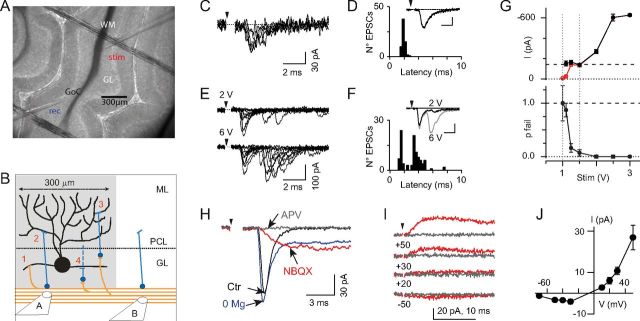
To record mf-EPSCs and GrC-EPSCs (from mf-GoC and GrC-GoC inputs, respectively), the mf bundle was stimulated using a large-bore (3–10 μm diameter) patch pipette, connected to a stimulus isolation unit and filled either with control saline, or with a HEPES-buffered extracellular saline (no appreciable differences between the two solutions). Individual stimuli were 200 μs monopolar square pulses. The stimulation pipette was positioned in the WM in an appropriate position to evoke monosynaptic mf inputs and disynaptic GrC inputs, as follows. A pipette positioned near to the recorded GoC (see Fig. 1B, pipette “A”) may evoke short-latency monosynaptic inputs from either mfs or GrCs, through pathways 1 and 2 in Figure 1B. Indeed, the stimulus may not only activate mfs, running in the WM to enter cerebellar folia, but also evoke spikes in GrC axons near the WM (as assessed in loose-cell attached recordings from GrC somata in the presence of NBQX; data not shown). However, in parasagittal slices, a GoC receives direct contacts only from GrCs in the longitudinal volume delimited by its dendritic arbor (see Fig. 1B, shaded area). Therefore, a stimulation pipette at sufficient distance from the recorded GoC (see Fig. 1B, pipette “B”) may activate monosynaptic inputs exclusively from mfs, which extend parasagittally in the WM and cross the border of the GoC dendritic territory, whereas GrC inputs are disynaptic (see Fig. 1B, pathways 3 and 4). In conclusion, the distance between stimulus and the soma of the recorded GoC was always set to exceed 300 μm, well above the extension of the apical GoC dendritic arbor (150 μm on each side of the cell body) (Dieudonné, 1998).
Figure 1.
Excitatory mossy fiber and granule cell synaptic inputs evoked in Golgi cells by WM stimulation. A, Low-magnification differential interference contrast image of a sagittal cerebellar slice. A patch pipette (rec) contacts a Golgi cell (GoC) in the granular layer (GL) while a stimulation pipette (stim) is immersed in the WM. B, Scheme of the afferent excitatory connections to a GoC activated by WM stimulation. Black cell, Golgi cell; orange fibers, mossy fibers; blue cells, granule cell; dotted line, Purkinje cell layer (PCL); ML, molecular layer. 1 and 2, mf-GoC and GrC-GoC monosynaptic pathways, respectively. 3 and 4, mf-GrC-GoC disynaptic pathways, where a GrC contacts either apical (3) or basolateral (4) GoC dendrites. Pipette “A” stimulates all pathways; pipette “B,” at ≥300 μm from the GoC soma, stimulates pathways 1, 3, and 4. C, EPSCs evoked by WM stimulation (with pipette configuration “B”) at threshold intensity (20 V) in a GoC (−70 mV), in the presence of blockers of inhibitory inputs (9 consecutive superimposed trials). Latency from stimulus is relatively invariant, suggesting a monosynaptic input from mfs. Here (and in all figures), arrowheads indicate stimulus onset; the stimulation artifact is blanked for clarity. D, EPSC latency histogram (same cell as in C). There is narrow unimodal distribution (mean latency, 1.9 ms; SD, 0.2 ms). Inset, Average of 66 EPSCs. Calibration in D, F insets, 2 ms, 25 pA. E, Multiple EPSCs in a different cell (same conditions) evoked by threshold (2 V; top) and suprathreshold (6 V; bottom) stimuli (10 consecutive trials each). The large jitter of late EPSCs suggests a disynaptic input from granule cells. Stronger stimuli preferentially enhance the frequency of long-latency EPSCs. F, Latency histogram for all EPSCs at 6 V (same cell as in E). Note the multimodal distribution, with an early narrow peak (modal latency, 1.4 ms) followed by a second broader peak (median latency, 3.5 ms). Inset, Average traces from 50 (black trace; 2 V) and 69 (gray trace; 6 V) EPSCs. Short- and long-latency EPSCs are present in both averages. G, Mean amplitude (top, red symbols), mean nonfailure amplitude (top, black symbols), and fiber failure probability (pfail, bottom) of mf-EPSCs, plotted versus stimulus intensity, for an exemplar GoC (−70 mV). Inhibitory blockers were omitted. The nonfailure mean is constant between threshold and 1.5 V (vertical dotted lines), suggesting that in this range a single mossy fiber input was activated. Error bars indicate SEM. H, Average mf-EPSC (−70 mV; threshold stimuli) in control saline (Ctr, black trace), after 15–20 min in Mg2+-free solution (0 Mg; blue trace), and after sequentially adding NBQX (2 μm; red trace) and d-APV (50 μm; gray trace). Mg2+ removal uncovers a large, NMDAR-mediated slow current component. Increase of this component in NBQX may result from ongoing Mg2+ removal. I, Mean mf-EPSCs evoked at threshold in control saline at various holding potentials (from top to bottom: +50 mV, +30 mV, +20 mV, −50 mV) in the presence of 2 μm NBQX, 10 μm gabazine, and 500 nm strychnine, before (red traces) and after (gray traces) adding 100 μm d-APV. J, I-V relationships for the NMDAR-mediated component of unitary mf-EPSCs, obtained in the presence of NBQX, gabazine, and strychnine. Each point is the average of 7–12 cells. Error bars indicate SEM.
Paired-pulse stimulation consisted of two stimuli, separated by 10 ms, and repeated every 10 s. The “minimal stimulation” protocol, designed to isolate the response to a single mossy fiber, consisted of runs of 10–50 stimuli, delivered at 0.1 Hz, for each one of a range of strengths, from just subthreshold (median, 7 V) to ∼10-fold larger values; the runs were given in random sequence.
Paired recordings
To study the GrC-GoC connection in paired recordings, GrCs located within a few tens of micrometers of the GoC body were specifically stimulated in the loose-cell attached configuration. This technique excluded activation of nearby mfs. The loose-cell attached configuration was obtained on GrC bodies using a 3–5 MΩ patch pipette. Large positive voltage jumps were applied to depolarize the GrC to threshold through its membrane capacitance while recording the GrC juxtacellular spike-induced current (Barbour and Isope, 2000). Simultaneous failure of the juxtacellular spike and of the evoked EPSC in the GoC at near-threshold stimulation was the criterion used to exclude a possible contamination by stimulation of nearby cells and fibers. GoCs were recorded with the Cs-BAPTA internal solution.
Drug application
Strychnine hydrochloride was obtained from Sigma. All other drugs were from Tocris Bioscience: QX-314, d-APV, NBQX, and 6-imino-3-(4-methoxyphenyl)-1(6H)-pyridazinebutanoic acid hydrobromide (SR 95531, gabazine). Stock solutions were prepared in water and stored at −20°C. During experiments, aliquots were diluted in control saline and bath-applied.
Analysis of synaptic currents
Latency distributions.
To study the latency distributions of evoked EPSCs (see Fig. 1), synaptic currents were detected in the 10 ms window after a stimulus using a threshold-above-baseline detector (Kudoh and Taguchi, 2002) implemented in the NeuroMatic package (J. Rothman, http://www.neuromatic.thinkrandom.com/), and their latency from stimulus onset was measured. To demonstrate the presence of an evoked disynaptic response, the number (Nobs) of EPSCs observed in the time window between 2.5 and 10 ms after the stimulus in N trials was compared with the number of spontaneous EPSCs expected to occur randomly in the same interval (μ = fs × (7.5 ms) × N), where fs is the observed sEPSC frequency. The probability that Nobs sEPSCs occurred by chance in the late time window (>2.5 ms) is given by the area under the tail of the Poisson distribution with parameter μ and n ≥ Nobs. Whenever this probability was <10−4, evoked disynaptic long-latency (LL) EPSCs were acknowledged. In latency histograms, multiple peaks (usually two) were always clearly separated by a short (∼0.5 ms) interval with no events. Events were visually assigned to the first or second peak, and the SD of the latencies in each group was used as a measure of peak dispersion.
Waveform analysis.
The waveform of evoked mf- and GrC-EPSCs was analyzed using Clampfit 10 (Molecular Devices) or a home-made detection and analysis routine running in IGOR (Wavemetrics). Current traces were baseline-subtracted. EPSC detection was performed in the 10 ms window after each stimulus. Events in the time window corresponding to the early narrow peak of the EPSC latency histogram (typically between 1.4 and 2.5 ms after stimulus; see Fig. 1C–F) were classified as short-latency (SL)-EPSCs (identified with mf-EPSCs); those detected in the rest of the interval as LL-EPSCs (identified with GrC-EPSCs). For detection, the peak of a candidate EPSC, defined as the mean current over 300 μs around a local minimum, was evaluated; an EPSC was acknowledged when this peak was >3 SD of the baseline current noise in a 2 ms window before stimulus.
For EPSC kinetics, the 20–80% rise time (t20–80) and the decay time constant(s) were studied. t20–80 was measured on each individual trace, whereas the decay phase was studied both in individual traces (Fig. 7D,E), and in averages. To average LL-EPSCs, events were aligned on the time of 20% rise; for SL-EPSCs, traces were aligned on the time of the stimulus. In the latter case, the latency jitter of individual responses is expected to significantly affect the rising, but not the decaying phase of the average. The decay was fitted, starting at 90% of peak amplitude, with 1–2 exponentials using nonlinear least-squares methods to yield one (τ) or two (τfast, τslow) decay time constants and their respective amplitudes (A; Aslow, Afast). The slow component was considered absent when τslow < 3 × τfast or when it had negative or irrelevant amplitude (≤2% of Afast). The fit was performed over 5–9 ms after the peak, with the exception of the fits in Fig. 7D, E. Two exponential components, when present, were only found in average traces (see Fig. 6A), whereas in the corresponding individual traces the second, slow component was not detected, most likely buried by noise.
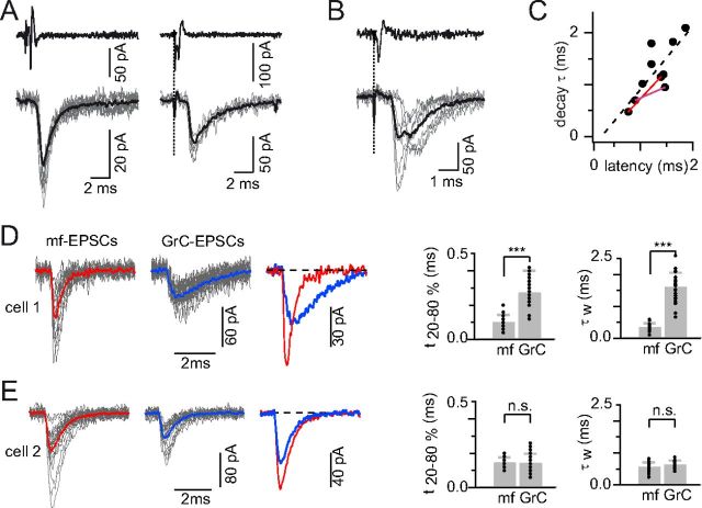
Figure 7.
Similar numbers of GrC-EPSCs with fast and slow kinetics revealed by granule-Golgi paired recordings and extracellular stimulation recordings. A, Exemplar recordings from two different GrC-GoC pairs, illustrating fast (left) and slow (right) EPSC kinetics. Top, Loose-cell attached recordings of juxtacellular spike-induced current in two GrCs. Bottom, Simultaneous EPSCs (gray traces) and their averages (black traces) recorded in postsynaptic GoCs clamped at −70 mV. Decay time constant: 0.7 ms (bottom left) and 1.8 ms (bottom right). B, In another pair, presynaptic GrC spikes (top) evoke two distinct EPSC responses with different latencies. Bottom, Superimposed individual responses (gray traces) and their average (black trace). C, Scatter plot of the mean EPSC decay time constant versus latency from presynaptic spike in 8 paired recordings. Red lines connect measurements for 2 distinct EPSCs obtained in single pairs. The dashed line indicates the linear regression through all points (see Results). D, E, Kinetic properties of mf- and GrC-EPSCs evoked by extracellular WM stimulation in two exemplar GoCs (D, cell 1; E, cell 2). Traces: individual EPSCs (in gray) and their averages (in color) aligned at the time of 20% of rise. Left column: mf-EPSCs. Middle column: GrC-EPSCs. Averages are superimposed in the right column. Right, Summary plots of the 20–80% rise time (t20–80) and weighted decay time (τw; see Materials and Methods) of all mf- and GrC-EPSCs in cell 1 (D) and cell 2 (E). Gray bars indicate mean ± SD; dots, individual values. In cell 1, kinetic parameters indicate much slower GrC- than mf-EPSCs (t20–80, 0.3 ± 0.1 vs 0.1 ± 0.04 ms; τw, 1.6 ± 0.4 vs 0.3 ± 0.1 ms), whereas in cell 2 the two inputs are similarly fast (t20–80, 0.1 ± 0.05 vs 0.1 ± 0.03 ms; τw, 0.6 ± 0.1 vs 0.6 ± 0.2 ms). ***, p <0.0005; n.s., not significant.
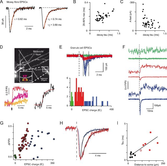
Figure 6.
The decay kinetics of mf-EPSCs and GrC-EPSCs, and the relationship between EPSC decay time and synapse distance from the GoC soma. A, Unitary mf-EPSCs evoked by minimal WM stimulation, from two different cells. Black traces represent individual trials. Red traces represent best fits with decaying exponentials. Left, Monoexponentially decaying EPSC (average of 50 trials). Right, Biexponentially decaying EPSC (Aslow /Afast = 0.19; average of 20 trials). Most (43 of 48) cells displayed biexponential mf-EPSC decay. B, 20–80% rise time of unitary mf-EPSCs versus the respective fast decay time constant, for 43 biexponentially decaying cells. Rise and decay are positively correlated (r = 0.55, p < 0.001, Spearman rank correlation). C, In the same cells, amplitude of the unitary mf-EPSC fast component (Afast) versus the respective time constant (τfast). Afast and τfast are negatively correlated (r = −0.41, p < 0.01). D, Projected morphology of a GoC obtained by multiphoton imaging of Alexa-594 introduced through the recording pipette in a parasagittal slice. Apical dendrites ascending to the molecular layer cross the Purkinje cell layer (dotted line indicates the apex of Purkinje cells). A stimulation electrode (white arrowhead) was placed at the ML surface above a deep-running dendrite (boxed region, inset). Ca2+ influx was imaged quasi-simultaneously at POIs indicated by colored arrows in the inset. Lower left panel, The corresponding fluorescence traces are displayed (color coded; average of 65 stimulations). Black arrow under the traces represent stimulation time. Lower right, Sum of the fluorescence signal over all responding points (black trace), representing the total Ca2+ influx. The charge integral of the average AMPA EPSC (red trace) is superimposed on the fluorescence Ca2+ transient. There is similarity in their rise kinetics. E, Top, Electrophysiological traces with evoked EPSCs. Bottom, Histogram of EPSC charges (measured between the two vertical lines in the top traces). Three types of responses can be discriminated based on the magnitude of the charge integral: failures (0–15 fC; green traces; n = 7), low amplitude slow events (15–120 fC; red traces; n = 42), and large amplitude fast events (>120 fC; blue traces; n = 16). F, Average Ca2+ transients (top traces; averaged for 5 POIs around the entry site) and corresponding EPSCs (bottom traces) for the three types of responses in E. G, Correlation between the EPSC charge (measured between the dotted lines of E) and the Ca2+ influx (ΔF/F0) recorded in the apical dendrite underlying the stimulation electrode. ΔF/F0 here is the average over 1.45 ms around the peak for the sum of all responding POIs. The charge of small EPSCs is significantly correlated to Ca2+ influx (red points and black line; Pearson's correlation coefficient 0.793; p < 0.001; n = 42), indicating a synaptic contact at the imaging site. Large EPSCs are not correlated with Ca2+ influx (blue points; Pearson's correlation coefficient 0.371; p > 0.1; n = 16), indicating that large EPSCs do not occur at the Ca2+ imaging site but may crown smaller EPSCs occurring at this site, which are responsible for the Ca2+ influx. H, Peak-scaled averaged EPSCs showing that the local pf events (red) decay with slower kinetics than the distal aa events (blue). Black lines indicate biexponential fits to the decay phase (blue EPSC, first component: 0.6 ms; red EPSC, first component: 1.3 ms). I, First decay time constant of pf-EPSCs (n = 6; red symbols) and mf-EPSCs (n = 6; black symbols) as a function of the distance of the synaptic site (identified by Ca2+ imaging) from the soma. The hollow red circle represents the experiment illustrated in this figure (D–H).
For the comparative analysis of amplitude and kinetics of mf-EPSCs and GrC-EPSCs (see Fig. 7D,E), we selected all the isolated events in the first 2.5 ms after each stimulus and in the subsequent 7.5 ms, respectively. To maximize the number of analyzable mf-EPSCs while avoiding cross-contamination, a weighted time constant (τw) was measured in 1.2–2.4 ms after the peak (starting at 90% peak amplitude). For mf-EPSCs, τw was equivalent to the fast decay time constant obtained when fitting over longer time windows (see Fig. 6A–C). The 8 minimal stimulation experiments used for this analysis were selected with the requirement of a sufficient number (12–84) of well-isolated GrC-EPSCs, to allow waveform analysis. Within this selection, the coefficient of variation of 20–80% rise times (CVrise) of GrC-EPSCs was not significantly different from CVrise of mf-EPSCs, whereas both were significantly smaller than CVrise of spontaneous EPSCs (sEPSCs) (mf-EPSCs, 0.36 ± 0.12; GrC-EPSCs, 0.42 ± 0.13; sEPSCs, 0.75 ± 0.27, mean ± SD, n = 8 cells; pGrC/sEPSC < 0.05; pmf/sEPSC<0.01; pGrC-/mf-EPSC > 0.05, nonparametric ANOVA), suggesting that mf- and GrC-EPSCs were generated in a few contacts at well-defined electrotonic distance, whereas sEPSCs reflected the activity of several synapses at variable distances from the GoC soma.
In whole-cell recordings aimed at identifying pharmacological components of the mf-EPSC (see Fig. 1H–J), all parameters were measured on averages from a few tens of individual responses, selected to exclude disynaptic GrC-EPSCs.
Two-photon Ca2+ imaging
All experiments were performed with a custom-built random-access two-photon laser-scanning microscope (Otsu et al., 2008). In this instrument, both X and Y scanning are operated by acousto-optic deflectors (AODs). These custom-built nonmechanical beam-steering devices (A-A Opto-Electronic) can redirect the laser beam in 6.5 μs. Two-photon excitation was produced by an infrared Ti-Sa pulsed Tsunami laser pumped by a Millennia VI (Spectra-Physics) and coupled to the transmitted light port of a BX51W1 microscope (Olympus). The microscope was equipped with a 40× LUMPlanFL/IR objective with 0.8 numerical aperture (Olympus). Fluorescence photons were detected by a cooled AsGaP H7421–40 photomultiplier (Hamamatsu). To operate the AODs and run the scanning procedures, a custom-made user interface was programmed in LabView (National Instruments). The AOD acoustic frequency drive was generated by a Direct Digital Synthesizer and a fast (10 ns) power amplifier (A-A Optoelectronics).
GoCs were filled through the whole-cell patch pipette with the morphological dye Alexa-594 (15 μm; Invitrogen) and the high-affinity Ca2+ dye Fluo-4 (200 μm; Invitrogen). Cells were clamped at −68, −73, or −78 mV. For GL imaging (see Fig. 4), after evoking stable unitary mf-EPSCs with minimal electrical stimulation in the WM (as above) at 0.2 Hz, Mg2+ was washed away from the control saline for at least 20 min to remove block of NMDA receptors (NMDARs). Upon appearance of the NMDA component of the mf-EPSC, the dendrites were systematically scanned in the GL by performing continuous Ca2+ imaging at ∼10 Hz, looking for site(s) of mf-evoked Ca2+ entry. For molecular layer (ML) imaging (see Fig. 6), slices were continuously bathed in control saline. The stimulation electrode, filled with a HEPES-buffered solution and Alexa-594, was placed at the surface of the slice, well above a deep-running GoC apical dendrite, to avoid direct dendrite stimulation. The distance from the stimulation electrode to the imaged dendrite in the z-plane varied from 18 to 41 μm. The distance from the dendrite to the lower Purkinje cell layer in the x-y plane varied between 37.5 and 117 μm. The presumed target apical dendrite was scanned, looking for local Ca2+ transients, whereas EPSCs were evoked by juxta-threshold stimulations at 0.5 Hz.
Figure 4.
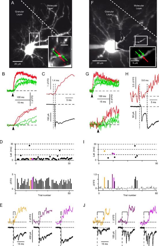
Ultrafast 2-photon imaging of postsynaptic Ca2+ influx at mf- and GrC-Golgi contacts in the GL. Analysis of Ca2+ fluorescence transients in the basolateral dendrites of two different GoCs (A–E and F–J) during WM stimulation in Mg2+-free saline. Fluorescence multiphoton images were obtained from the dendrites of cells filled with Alexa-594 and Fluo-4 through the recording pipette. A, F, Projected fluorescence images of two Golgi cells, extending apical dendrites toward the ML and basolateral dendrites in the GL. The proximal portion of an ascending GoC dendrite (white rectangle) is magnified (bottom right rectangle). Arrows indicate analyzed points of interest (POIs). B, G, Top graphs, Mean fluorescence transients, from corresponding color-coded POIs in A and F; averages of 50 (B) and 8 (G) stimuli. Black arrows indicate stimulation time. POIs marked in red indicate responses with shorter delay from stimulus and faster rise than flanking POIs marked in green, as better shown in the bottom graphs (expanded traces), indicating that these POIs are the sites of synaptic Ca2+ entry. Traces from sites marked 2 and 3 in A are nearly superimposed in B, implying the presence of an entry site between the two POIs. C, H, Average EPSC (bottom black trace) (n = 50 in C; n = 80 in H) and the corresponding fluorescence transient (top red trace; same as in B,G) from the Ca2+ entry sites marked by red arrows in A, F. Vertical dotted lines indicate stimulation time (left line) and time of the first detected point in the fluorescence response (right line; see Materials and Methods). Numbers indicate latency (in ms). The evoked EPSC has a fast component (mf-EPSC), which peaks and decays in 2–3 ms, followed by a second slower component (the compound disynaptic GrC EPSC). The fluorescence signal in C rises around the time of the mf-EPSC peak, whereas the signal in H rises during the slow rising phase of the GrC-EPSC. D, I, Top graphs, Latency of fluorescence responses versus trial number for the Ca2+ entry sites in A and F. Bottom graphs, Fluorescence change (ΔF/F0) relative to the prestimulus baseline, averaged over 150 ms after stimulation, versus trial number, for the same sites. Gray bars represent detection failures. Colored dots (top) and bars (bottom) indicate trials described in E and J. In D, latencies show very small jitter; a few values exceed 5 ms, possibly representing failures of the local mf-GoC input in the presence of late Ca2+ diffusion from a nearby activated site. In I, most trials are classified as failures (in gray), except for a few detected responses with long and variable latencies. E, J, Fluorescence transients (top traces) and the corresponding EPSCs (bottom traces) in individual trials from the sites in D and I, respectively. Brackets over the fluorescence traces indicate poststimulus delay before signal detection. E, The fluorescence response starts around the time of the mf-EPSC peak. J, The fluorescence response occurs in correspondence of disynaptic EPSCs, long after the mf-EPSC has decayed.
Whenever the presence of a responding site in the GL or the ML was found, continuous 10 Hz-imaging was stopped, and temporally resolved optical recordings of Ca2+ influx, at and near the presumed synaptic site, was performed by setting 15 to 25 points of interest (POIs) along the GoC dendrites with a spacing of ∼1 μm. The imaging dwell time per POI, during which fluorescence photons were collected, was set between 20 and 50 μs, yielding sampling rates comprised between 0.98 and 3.2 kHz. In this way, fast biological signals could be simultaneously resolved at multiple POIs. Episodic acquisition of Ca2+ dye fluorescence transients (excitation wavelength 820 nm, emission bandpass 480–580 nm) was synchronized with electrophysiological stimulation protocols to time the averaging procedure. Fluorescence acquisition was routinely interrupted every 3–5 min to correct for possible drift of the preparation. The laser power (typical averaged square power <10 mW2 per point) provided good signal-to-noise ratio while limiting photodamage (Otsu et al., 2008). Optical recordings were stable for up to several hours, only limited by the duration of the patch recordings. z-stack acquisition of the Alexa-594 fluorescence was performed at the end of the experiment to reconstruct cell morphology (excitation 820 nm, emission bandpass 605–675 nm). The effect of d-APV or NBQX on fluorescence responses was measured on the average of 20–30 trials, taken at least 3 min after the start of drug application.
All data were analyzed with routines developed in-house using IGOR. Fluorescence intensity transients were measured as ΔF/F0 = (F − F0)/F0, where F0 and F are the background-subtracted photon number at rest and during the test period, respectively. Background fluorescence was acquired at a POI positioned far from dye-loaded structures. A fluorescence signal was acknowledged when ΔF/F0 was larger than twice the baseline SD all along a 20 ms “detection” window (otherwise, a failure was declared); the initial point in this window was taken as the event start. Latency from stimulus was the interval from the mid-time of the stimulus to the event start. Averages were formed from a few tens (15–50) of trials. Ca2+ entry sites were defined as POIs where the latency and the time of 20–80% rise to peak fluorescence, evaluated from average traces, were shorter than in neighboring POIs (see Fig. 4B,G). The correct identification of the synaptic contact site was confirmed by the correlation of stimulation and imaging failures. For GL imaging, peak Ca2+ flux at a given entry site was quantified as the average ΔF/F0 over the first 7 ms after latency (ΔF7ms), which is a measure of the initial rate of rise of fluorescence. This was preferred to evaluating peak fluorescence change because the latter is potentially affected by Ca2+ diffusion from nearby synaptic sites and out of the recorded site or by Ca2+ release phenomena, as suggested by the large variability of time-to-peak among sites (from ∼15 to ∼80 ms; data not shown), whereas the rate of rise of fluorescence at entry sites should be linearly correlated to the intensity of local Ca2+ influx. For molecular layer imaging, Ca2+ entry sites had stable time-to-peak fluorescence change, and Ca2+ flux was quantified as the average ΔF/F0 over 20–40 ms after stimulation.
Pre-embedding immunoelectron microscopy
GlyT2-EGFP mice, a transgenic mouse model expressing GFP in glycinergic interneurons (P52-P65; kind gift from H.U. Zeilhofer) (Zeilhofer et al., 2005), or Wistar rats (P24), were anesthetized with pentobarbital (60 mg/kg) and intracardially perfused with 4% PFA and 0.1% glutaraldehyde in PBS. After dissection, the cerebellum was kept overnight at 4°C in 4% PFA. Vibratome sagittal sections (100 μm) were collected in ice-cold PBS, cryoprotected for 3 h in 20% sucrose/20% glycerol at room temperature, permeabilized by freezing and thawing, and then incubated overnight at 4°C in anti-GFP rabbit polyclonal antibody (1:50; ref 132002, Synaptic Systems) or in anti-neurogranin polyclonal antibody (1:50; ref AB5620, Millipore Bioscience Research Reagents) diluted in PBS containing 0.1% gelatin (PBSg). After extensive washes, GFP or neurogranin expression was detected by using the avidin-biotin complex method (Elite Vectastain, Vector Laboratories). For this purpose, the sections were incubated for 4 h at room temperature with biotinylated horse anti-rabbit antibody (1:200 in PBSg, Vector Laboratories). After a preincubation for 20 min in 0.6% DAB in Tris-buffered saline (0.06 m, pH 7.4), the sections were incubated with DAB and hydrogen peroxide (Sigma; Fast DAB) in the same buffer and revealed under visual control. They were then postfixed for 1 h at 4°C in 2% osmium tetroxide in PBS with 0.07% glucose, dehydrated in ethanol, and finally flat-embedded in Araldite resin (Polysciences). After blocks were trimmed, ultrathin sections (∼70 nm) were collected on copper grids and counterstained for 10 min with 2% uranyl acetate in H2O and then with 0.2% Reynolds lead citrate (Reynolds, 1963). Observations of ultrathin sections were performed with a Philips Tecnaï 12 electron microscope (FEI). Micrographs were processed and analyzed using ImageJ free software.
Immunohistochemistry
Tissue fixation.
For immunohistochemistry, we used heterozygote transgenic mice of the GlyT2-EGFP line, which were backcrossed for >10 generations to the C57BL/6J background. GFP is expressed in 85% of GoCs in this mouse line, the others being pure GABAergic cells (Simat et al., 2007). Two males, 2 months of age, were deeply anesthetized by intraperitoneal injection of sodium pentobarbital (60 mg/kg body weight) and perfused through the ascending aorta with PBS, followed by 75 ml of 4% freshly depolymerized PFA in 0.1 m PBS, pH 7.4, both at 4°C. Brains were then dissected and postfixed in 4% PFA overnight at 4°C. They were then dehydrated in a gradient of alcohol (ethanol 70%, 80%, 95%, 100%) followed by butanol and xylene and embedded in paraffin (Paraplast X-TRA Tissue Embedding Medium, Leica Microsystems).
Tissue preparation and labeling.
Sagittal sections (20 μm thickness) were cut on a microtome and mounted on positively charged glass slides (Superfrost Plus, Thermo Scientific). After removing paraffin with xylene and rehydration in a graded series of ethanol (100%, 95%, 85%, 75%, 50%), sections were processed in a decloaking chamber (Biocare Medical) using a citrate buffer-based antigen retrieval medium (Biocare Medical) for 20 min at 110–115°C. Aldehyde groups were removed by incubating the sections in sodium borohydride (1%) in PBS. They were then processed in PBS with 15% methanol and 0.3% H2O2 to block the endogenous eroxidase activity. After these treatments, the slices were incubated in a blocking PBS-based solution containing cold-water fish-skin gelatin (0.1%) and 0.1% Triton X-100. Tissue was then incubated overnight at 4°C with the following primary antibodies: chicken anti-GFP (1:1000; Aves Laboratories), guinea pig anti-vGluT1 (1:1500; Millipore), and mouse anti-vGluT2 (1:1500; Millipore). Primary antibodies were revealed by incubation for 2 h with secondary antibodies coupled to either Alexa-488 (Invitrogen) or DyLight 488, DyLight 549, and DyLight 649 (Jackson ImmunoResearch Laboratories). Sections were mounted using Prolong Gold Antifade Reagent (Invitrogen).
Image acquisition and analysis.
Confocal stacks were acquired on a Leica SP5 microscope using a 63× oil-immersion objective (NA 1.4) and a pinhole aperture of 1 Airy. To resolve individual synaptic boutons, data were oversampled using a voxel size of 0.06 μm in x-y and 0.17 μm in z. Image stacks were analyzed in 3D using the software Imaris (Bitplane AG). vGluT1-positive hot spots were detected in the GL using the spot function of Imaris, adjusting size and intensity thresholds to select all extraglomerular varicose-like puncta. This also led to a large number of spurious intraglomerular hits. To exclude these puncta, the fluorescence was measured on the vGluT2 channel, and regions in which all mfs expressed vGluT2 were chosen. vGluT2 immunoreactivity always displayed a bimodal distribution, with a subpopulation of spots displaying only background staining (see Fig. 5D). Thresholding at twice the mode of this first population selectively highlighted extraglomerular vGluT1-only varicose profiles, tentatively identified as aa varicosities in the GL. GoCs, identified from the stack based on GFP immunoreactivity, were segmented using the surface function of Imaris (adaptive local thresholding). Distances were calculated between the center of putative aa varicosities and the surface of GoCs, both in the original stack and in a stack for which the GFP channel had been rotated by 180° to randomize the GFP signal relative to the vGluT1 signal.
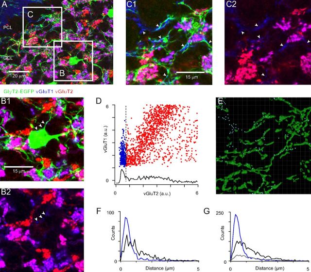
Figure 5.
Immunolocalization of granule cell ascending axon contacts with Golgi cells in the GL. Glutamatergic varicosities identified by immunostaining of the vesicular glutamate transporters vGluT1 (blue channel) and vGluT2 (red channel) in parasagittal sections obtained from GlyT2-EGFP mice (green channel: Golgi cell somata and processes). A, Large field of view (92 × 123 μm, projection depth 2.38 μm) showing the general organization of terminals in the cerebellar cortex. The GL is occupied by large mossy fiber rosettes displaying various ratios of vGluT1 and vGluT2 staining. There is absence of vGluT1-only rosettes in this region. vGluT1-only parallel fiber boutons are seen in the molecular layer (top left) together with rare vGluT2-only climbing fiber terminals. B1, B2, Enlargement of the region B, boxed in A, showing small vGluT1-only varicose profiles in the GL and their apposition to the cell body and dendrites of a GFP-positive GoC (projection depth 1.36 μm). vGluT1-only profiles most likely correspond to the boutons of GrC aa in the GL. In B2, the GFP channel was removed for better display of vGluT1-only profiles, which show weaker staining than the large mf rosettes. C1, C2, Same as B1, B2 for the region C (projection depth 1.36 μm), illustrating the dense innervation of the GFP-positive GoC neurites by vGluT1-only varicosities in the upper part of the GL. D, Relative intensity of vGluT1 and vGluT2 staining in punctuate profiles detected in the blue channel (vGluT1) in region C, both in mossy fiber rosettes and outside glomeruli. The vGluT2 intensity histogram (black curve) is bimodal, with a first population (nonglomerular puncta) showing background levels of immunoreactivity (vGluT1-only, blue dots). In contrast, glomerular puncta (red points) display fixed ratios of vGluT1 and vGluT2 staining, as evidenced by the linear correlation in their distribution. E, vGluT1-only puncta are displayed as blue dots on top of segmented GFP-positive GoC neurites, for the region displayed in C; distance of blue dots from the nearest GoC neurite is shown in F. By comparison with C2, all selected puncta in the GL are indeed extraglomerular. F, Blue curve represents the distribution of the distance of vGluT1-only puncta from the surface of GoC dendrites in the GL of the stack shown in A. Black curve represents randomized distance distribution obtained by rotating the GFP channel by 180°. G, Distance distribution (in blue) and randomized distance distribution (in black) for all the regions analyzed (6 confocal stacks, 2 animals). Gray curve represents the scaled randomized distance histogram accounting for the randomly located puncta in the blue distribution (see Results). The difference between the gray and blue curves is an estimate of the number of vGluT1-only puncta truly apposed to GFP-positive GoCs.
Statistical tests
Data are reported as mean ± SD unless otherwise stated. For statistical comparisons, nonparametric methods were used, and two-tailed p values were computed using GraphPad Instat software (GraphPad Software). The Mann–Whitney test and the Wilcoxon matched-pairs test were used to compare the means of two groups of unpaired and paired values, respectively; one-way ANOVA was used for >2 groups of paired values. Correlations were studied using Spearman's rank correlation test.
Results
mf and GrC excitatory synaptic inputs evoked in Golgi cells by WM stimulation
We set out to investigate the respective functional contributions of mf and GrC synaptic inputs to the excitation of GoCs and their contribution to feedforward and feedback inhibitory circuits in the GL. EPSCs were recorded from voltage-clamped GoCs at −70 mV in parasagittal slices, in the presence of gabazine (10 μm) and strychnine (500 nm), to exclude a possible contribution of inhibitory synapses (Dumoulin et al., 2001; Hull and Regehr, 2012). Excitatory inputs were activated by extracellular stimulation of the WM at least 300 μm from the soma of the recorded GoC (Fig. 1A), to avoid direct activation of GrC axons (Fig. 1B). This configuration recruited monosynaptic mf inputs, and GrC inputs through a disynaptic pathway (see Materials and Methods and Fig. 1B). It also minimized interference by direct antidromic stimulation of the GoC axon (Dieudonné, 1998), and by GoC-GoC electrical coupling, which extends for ∼100 μm (Dugué et al., 2009).
Threshold stimulation intensity (1.1–30 V) evoked in most GoCs (21 of 24) an isolated EPSC with short latency and little jitter (Fig. 1C). Latency histograms, obtained from a subset of cells in which a large dataset was available, showed a single narrow peak at 1.5 ± 0.3 ms, with a dispersion (SD of all latencies) of 0.2 ± 0.1 ms (n = 5 cells; Fig. 1D). In the remaining cells (3 of 24), stimulation evoked multiple EPSCs (Fig. 1E, top traces). The first EPSC occurred with short invariant latency (mean: 1.4–2.6 ms; SD: 0.1–0.2 ms, n = 3 cells), whereas subsequent EPSCs appeared only in a fraction (25.4 ± 9.8%) of the trials and displayed longer and more variable latencies (mean: 3.8–5.7 ms; SD: 0.6–1.7 ms). These late events were not spontaneous EPSCs (p < 10−4; see Materials and Methods) and gave rise to a second synaptic component in the average trace (Fig. 1F, inset). An increase in stimulation intensity (by up to 3 times the threshold; n = 20 cells) raised the frequency of late EPSCs in 65% of cells (13 of 20, Fig. 1E, bottom traces), to 45 ± 27% per trial. Latency histograms (from 7 cells) showed a multimodal distribution, with a first narrow peak at 1.6 ± 0.4 ms (dispersion: 0.2 ± 0.1 ms), followed by a late hump (median at 4.7 ± 2.4 ms). The two components were separated by 0.6 ± 0.3 ms without events (Fig. 1F, 0.5 ms in the example). The significantly larger jitter (dispersion: 1.3 ± 1.2 ms) observed in the late hump (vs first narrow peak, p = 0.015) is consistent with a disynaptic response (Fig. 1F). In summary, synaptic responses could be separated into monosynaptic short-latency EPSCs (SL-EPSCs) and disynaptic long-latency EPSCs (LL-EPSCs). Monosynaptic EPSCs most likely arose at the mf-GoC input (Fig. 1B, feedforward pathway 1), whereas LL-EPSCs, with their longer delay and larger jitter, are likely attributed to the disynaptic mf-GrC-GoC input (Fig. 1B, feedback pathways 3 and 4).
To investigate the properties of unitary mf-EPSCs (Fig. 1G), we examined the recruitment of mfs during WM stimulation. When gradually increasing stimulus strength above threshold, the failure probability of the SL-EPSC decreased steeply while the mean amplitude increased (Fig. 1G). The nonfailure mean amplitude remained approximately constant (p > 0.1, Spearman rank correlation test) for a range of strengths (up to ∼3 times the threshold; n = 12 cells; Fig. 1G). This range (“minimal stimulation”) therefore likely corresponds to the excitation of a single mf, producing a “unitary” EPSC. The unitary events had a fast rise (t20–80, 0.2 ± 0.1 ms), and their mean amplitude ranged from 28 to 456 pA (median, 62 pA). The percentage of failures, when using the largest intensity in the minimal stimulation range (Fig. 1G), was 26 ± 20% (range, 0–62%). When using pairs of stimuli at 100 Hz, the paired-pulse amplitude ratio (ratio of mean amplitude of second vs first response) was 1.03 ± 0.44 (range, 0.52–2.02). These properties are in agreement with a previous description of mf-EPSCs in GoCs (Kanichay and Silver, 2008). Application of the non-NMDA glutamate receptor blocker NBQX (2 μm) completely abolished mf-EPSCs recorded at −70 mV (3.2 ± 1.6% of pre-NBQX value, n = 15 cells), indicating that, at hyperpolarized potentials, mf-EPSCs are mediated by non-NMDA glutamate receptors. Removal of Mg2+ at −70 mV (in the presence of 10 μm d-serine) induced the appearance of a slow, NBQX-insensitive and d-APV-sensitive component (peak amplitude, measured in the presence of NBQX, −34 ± 41 pA; t20–80, 5.0 ± 3.1 ms; n = 9 cells; Fig. 1H). In control conditions (in the presence of Mg2+ and NBQX), the amplitude of the d-APV-sensitive component was 27 ± 6 pA at +50 mV (n = 12 cells) and −4 ± 1 pA at −30 mV (n = 7 cells; Fig. 1I). These NMDAR-mediated currents displayed a current-to-voltage relation typical of GluN2A/2B-containing receptors (Fig. 1J). Our results thus demonstrate the presence of active NMDA receptors at mf-GoC postsynaptic sites.
GrC- and mf-EPSCs contribute equivalent charge contents to GoC excitation during repetitive mf activity
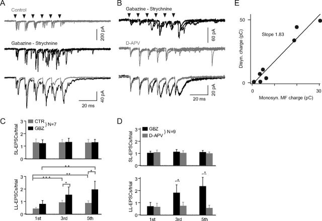
It is well known that the excitation of GrCs by mf inputs may require the temporal summation of multiple synaptic inputs during low-frequency asynchronous activity (Schwartz et al., 2012) or during high-frequency trains (D'Angelo et al., 1995). To evaluate the relative contribution of mf and GrC inputs to GoC excitation during physiological activity, we applied trains of WM stimulations at 100 Hz (5–20 stimuli; repeated at 0.1 Hz). In control conditions (without inhibition blockers), the number of LL-EPSCs increased significantly with stimulation number within the train (p = 0.0002; one-way ANOVA; Fig. 2A,C).
Figure 2.
GABAAR- and NMDAR-sensitive recruitment of disynaptic granule cell inputs during train stimulation. Whole-cell recording of EPSCs from GoCs at −70 mV during WM train stimulation (6 stimuli at 100 Hz, delivered every 10 s). Traces are aligned at time of the stimulus (arrowheads). A, Five superimposed consecutive traces showing EPSCs recorded in GoC in control saline (top) and after bath application of gabazine and strychnine (middle). Bottom, Averages (35 consecutive trials) from the same experiment (gray trace, control; black trace, inhibitory blockers present). The blockers produce a strong increase in the number of long-latency EPSCs. B, In another cell, EPSCs before (top) and after d-APV application (middle; 5 consecutive trials in each row), in the presence of gabazine and strychnine. Bottom, Average of 44 consecutive responses from the same cell before (black trace) and after (gray trace) d-APV. d-APV strongly and selectively reduces long-latency EPSCs. C, Summary plot of the effects of GABAA receptor block on the frequency of short-latency (SL; top) and long-latency (LL; bottom) EPSCs. Bars represent the averaged frequency (±SEM) over 7 cells, before (CTR; gray bars) and after (GBZ; black bars) perfusion of 10 μm gabazine and 500 nm strychnine. Frequency (events/trial) is measured over 10 ms after the first (1st), third (3rd), and fifth (5th) stimulus. The LL-EPSC frequency increases significantly with stimulus number (in CTR and GBZ), and with application of gabazine/strychnine (at the third and fifth stimulus). *, p < 0.05; **, p < 0.005; ***, p < 0.0005. SL-EPSCs are not affected in all cases. In a few cells, two distinct SL-EPSCs were present (as judged from their short, nonjittery latency), explaining the >1 value of mean SL-EPSC frequencies. D, Summary of the effects of NMDA receptor block on the frequency of SL- (top) and LL-EPSCs (bottom). Averaged frequency (±SEM) over 9 cells in gabazine/strychnine (GBZ; black bars) and after addition of 50 or 100 μm d-APV for at least 3 min (d-APV; gray bars). The increase of LL-EPSC frequency with stimulus number is abolished by d-APV. SL-EPSCs are not affected. *, p < 0.05. E, The relationship between the charge transfer resulting from disynaptic LL-EPSCs and monosynaptic SL-EPSCs, evoked by trains of 6–20 stimuli at 100 Hz in 8 cells, in the presence of inhibitory blockers. The monosynaptic charge is measured as the time integral of the average current along the train after bath perfusion of d-APV (artifacts blanked); the disynaptic charge is the difference between total and monosynaptic charge. Black line indicates linear regression; slope, 1.83 ± 0.17.
To confirm that disynaptic LL-EPSCs originated from GrC-GoC inputs, we monitored the frequency of LL-EPSCs during pharmacological manipulations of GrC synaptic drive. GrCs undergo inhibitory control by tonic GABAA conductances (Hamann et al., 2002) and by phasic inhibitory synaptic inputs resulting from the spontaneous and evoked discharge of GoCs (Forti et al., 2006; Dugué et al., 2009). Antagonism of inhibitory synaptic currents will thus augment GrC firing upon mf stimulation and preferentially increase the activation of the disynaptic mf-GrC-GoC pathway. Accordingly, the frequency of LL-EPSCs after the fifth stimulus in gabazine/strychnine was 256 ± 62% (p = 0.016) of its value before addition of the antagonists, whereas the frequency of SL-EPSCs was unchanged (105 ± 10%; p > 0.9; n = 7 cells; Fig. 2C). The jitter of LL-EPSCs increased during the train, as shown by the slow rise (latency to peak of the LL-EPSC, 3.9 ± 0.3 ms at second stimulus; 4.6 ± 0.3 ms at fifth stimulus; p < 0.05, n = 7 cells) and prolonged duration of the long-latency component in the averaged trace (Fig. 2A), consistent with synaptic summation in GrCs.
In GrCs, the temporal summation of mf inputs critically depends upon NMDAR activity (D'Angelo et al., 1995; Schwartz et al., 2012). Correspondingly, in GoCs, bath application of the NMDAR blocker d-APV (50 or 100 μm) reduced LL-EPSC frequency after the fifth stimulus by 80 ± 7% (p < 0.005, n = 9 cells; Fig. 2B,D). In contrast, the frequency of SL-EPSCs was nearly constant during the train (p = 0.46, one-way ANOVA) and was not significantly affected by d-APV (SL-EPSC frequency after the fifth stimulus in d-APV: 94 ± 9% of pre-d-APV value, p = 0.50, n = 9; Fig. 2B,D). The opposite effects of d-APV and gabazine/strychnine on the frequency of LL-EPSCs confirm that these events arise from activity at GrC-GoC synapses. Thus, in the rest of this study, LL-EPSCs will be identified with GrC-EPSCs and SL-EPSCs with mf-EPSCs.
The selective sensitivity of the disynaptic GrC relay to the NMDA antagonist d-APV was used to evaluate the relative contributions of mf and GrC inputs to the excitatory drive of GoCs during repetitive WM activation with variable stimulation strength. We first measured the total charge transfer to GoCs, provided jointly by mf- and GrC-EPSCs, during trains of stimulation in the WM (6, 10, or 20 stimuli at 100 Hz). For these experiments, gabazine and strychnine were added to avoid a potential contamination by evoked inhibitory conductances. We then measured the charge transfer, provided mostly by mf-EPSCs, after addition of d-APV (50–100 μm) (Fig. 2B). Calculating the difference in the synaptic charge before and after application of d-APV provided a lower estimate of the disynaptic charge transfer produced by GrCs in the absence of inhibition. The relation between disynaptic and monosynaptic charges (Fig. 2E; 8 cells) displays a significant linear correlation (R2 = 0.94, p < 10−4) with a slope of 1.83 ± 0.17. Because inhibition would approximately halve the number of disynaptic GrC inputs to GoCs (as assessed at the end of a train of 5 stimuli at 100 Hz; Fig. 2C) (Schwartz et al., 2012), we conclude that GoCs receive a similar amount of charge from mf inputs and disynaptic GrC inputs during repetitive activation in physiological conditions.
Ultrastructural identification of GrC ascending axon contacts onto GoCs in the GL
In the previous experiments (Figs. 1 and 2), the parasagittal orientation of the slice ensured that GrCs contributing disynaptic inputs are to be located in the same parasagittal volume as the recorded GoC. These local granule cells account for approximately half of the excitatory synaptic drive to GoCs during repetitive mf activation. The existence of en passant synapses formed by local GrC ascending axons onto basolateral and/or apical GoC dendrites was proposed in early morphological studies (Mugnaini et al., 1974; Palay and Chan-Palay, 1974) but remains to be confirmed. These proximal contacts could constitute a privileged synaptic connection between local GrCs and GoCs and therefore a potent inhibitory feedback circuit in the GL.
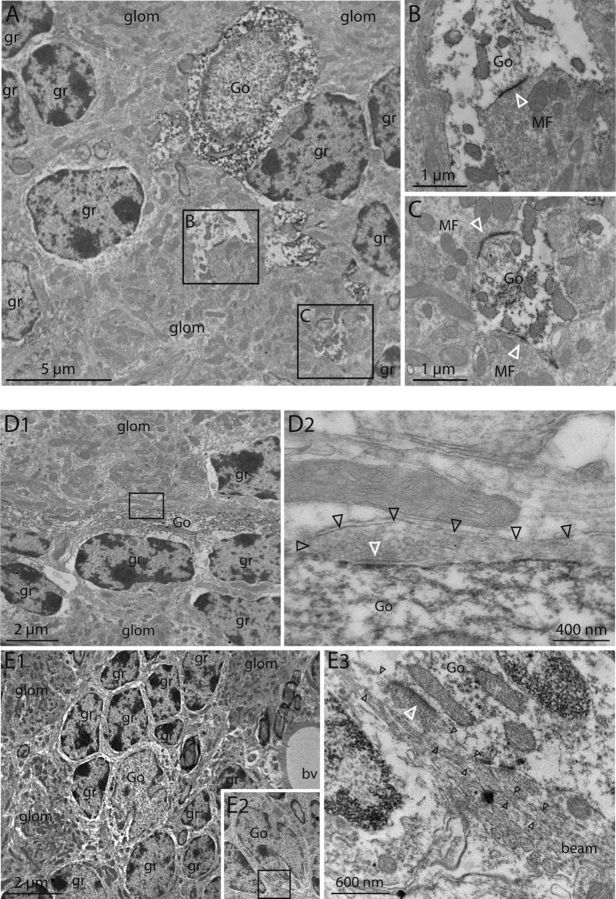
To selectively examine the morphology of contacts formed on GoCs in the GL, we unambiguously identified GoC dendrites in electron micrographs by performing pre-embedding immunostaining against neurogranin, a Ca2+ binding protein specific of GoCs in the cerebellar cortex, or against GFP in a transgenic mouse model expressing GFP in glycinergic interneurons (GlyT2-EGFP mice; see Materials and Methods). Immunopositive dendrites, generally of large diameter (Fig. 3A–D), were occasionally found inside glomeruli, where they were contacted by mf profiles (Fig. 3A–C). In several cases, excitatory synaptic contacts were found between those two elements, as judged from the presence of synaptic vesicles close to the presynaptic membrane and of a thick postsynaptic density. In some cases, more than one morphological contact site was found inside the same glomerulus, putatively on the same GoC dendrite (Fig. 3A,C). Overall, this EM analysis confirms the existence of glomerular mf contacts with GoC dendrites, including putative multiple contacts made by one rosette to a single dendrite.
Figure 3.
Excitatory contacts onto Golgi cells in the GL identified with electron microscopy. Analysis of ultrathin cuts of GlyT2-GFP mice cerebella stained with an anti-GFP antibody amplified with the peroxidasic system to identify Golgi cells. A, A peroxidase-stained GoC dendrite (boxed regions B and C) penetrates a glomerulus (glom), well differentiated from surrounding GrC somata (gr), in the proximity of a GoC soma (Go). B, C, Intraglomerular articulations formed by the dendrites boxed in A shown at higher magnification. GoC dendrites (Go) receive asymmetric synapses from a mossy fiber terminal (MF) at both locations. There are numerous and densely packed vesicles in mossy fiber terminals. Note the postsynaptic densities on Golgi cell dendrites (white arrowheads). D1, Low-magnification view of a stained GoC dendrite running in the GL in between GrC bodies and outside of a nearby glomerulus. D2, Enlargement of the region boxed in D1, showing that the GoC dendrite is separated from the glomerulus by two fibers, running parallel to each other. One of these fibers (black arrowheads), putatively a GrC ascending axon, forms an asymmetric synaptic contact with the GoC dendrite (white arrowhead). E1, Low-magnification view of a GoC body surrounded by granule cell bodies (gr) in the GL. E2, Serial sections were acquired, and a low resolution image of another section is shown. E3, Magnification of the boxed region in E2 showing an aa bundle (“beam”) running outside of glomeruli and forming asymmetric synapses with the GoC body. Black arrowheads indicate an individual fiber; white arrowhead indicates the corresponding postsynaptic density.
Another type of synaptic contact was also found on GoC in the GL (Fig. 3D,E). In several cases, bundles of thin axons running in the GL were found to contact thick postsynaptic densities of basolateral GoC dendrites (Fig. 3D) or GoC somata (Fig. 3E). Each synaptic articulation was composed of a simple en passant bouton occurring outside glomeruli and containing a small number of vesicles, a typical GrC axon feature. In contrast, climbing fiber (CF), unipolar brush cell, or mf terminals contain high numbers of densely packed vesicles (Palay and Chan-Palay, 1974). The presence of bundles of axons excludes CF thin collaterals and clearly identifies GrC ascending axons. Thus, in contradiction with the general belief that functional GrC-GoC connections are formed exclusively by pfs on apical GoC dendrites (Dieudonné, 1998; Bureau et al., 2000; Misra et al., 2000; Brickley et al., 2003; Watanabe and Nakanishi, 2003; Beierlein et al., 2007; Kanichay and Silver, 2008; Menuz et al., 2008; Robberechts et al., 2010), our results now establish the existence of synapses formed by GrC ascending axons on GoC dendrites in the GL.
Ca2+ imaging reveals functional GrC ascending axon synapses in the GL
To determine the functional contribution of GrCs ascending axon synapses in the GL to the disynaptic activity evoked by WM stimulations, we combined ultrafast 2-photon Ca2+ imaging and patch-clamp recording of GoCs. Juxta-threshold WM stimulations were applied in a Mg2+-free extracellular medium complemented with 10 μm d-serine, which potentiates NMDA currents. These conditions maximize the frequency of disynaptic GrC-EPSCs in low-intensity stimulation conditions (Fig. 1) (Schwartz et al., 2012) and increase postsynaptic Ca2+ influx. Individual GoCs were clamped at subthreshold potential and filled through the pipette with the high-affinity Ca2+-sensitive dye Fluo-4 (100 μm) and the morphological dye Alexa-594 (Fig. 4A,F). We then scanned systematically the basolateral dendrites in the GL in search of fluorescence transients correlated with the stimulation. In the majority of cells, we could identify at least one area of the dendrites with transients correlated in time with the stimulation. The fluorescence signal of this area was then acquired at submillisecond sampling intervals, quasi-simultaneously from a small number (15–25) of closely spaced (∼1 μm) POIs covering the nearby dendrites (Fig. 4A,F). To identify the precise location of the active synaptic contacts, the latency and rise time of the fluorescence transients averaged over all trials were compared between POIs (see Materials and Methods). A “Ca2+ entry site” was defined as a POI where the latency from stimulus was smaller, and the kinetics of rise was faster, than in flanking POIs (Fig. 4B,G).
We detected 16 entry sites on basolateral dendrites (20–80% rise time, 10.8 ± 5.3 ms; rise time in flanking POIs, 28 ± 12.3 ms, p < 0.002). In 10 of these sites, from 6 cells, the latency of the mean fluorescence change matched the latency of the electrophysiological mean mf-EPSC (Ca2+ transient latency, 2.3 ± 0.6 ms; mf-EPSC latency, 1.5 ± 0.6 ms; delay from the onset of the mf-EPSC, 0.9 ± 0.2 ms; Fig. 4C). Latencies, measured from the largest individual responses at each site, had a small variability (SD of the 10 largest responses, 1.8 ± 1.1 ms, n = 10 sites; Fig. 4D,E). Moreover, mf-EPSC failures were in all cases accompanied by a Ca2+ transient failure (data not shown, see Materials and Methods), confirming that fluorescence responses at short-latency entry sites corresponded to activation of mf-GoC synaptic contacts.
In the remaining 6 entry sites (from 3 cells), Ca2+ rise occurred after long, variable latencies from WM stimulation (“long-latency entry sites”), consistent with influx evoked by disynaptic GrC-EPSCs. Figure 4F–J illustrates the behavior of one of these sites, where the latency of the mean response was 5.6 ms, with a delay of 4.6 ms from the onset of the mean SL-EPSC (i.e., the mf-EPSC; Fig. 4H), and latencies measured in individual trials varied largely (between 3.9 ms and 26.8 ms; Fig. 4I). The electrophysiological responses showed both mf- and GrC-EPSCs, but no fluorescence signal was detected at short latency (Fig. 4J). In a total of 6 sites, the latency of the mean Ca2+ transient varied between 5.6 and 8 ms, with a delay from the mean mf-EPSC rise between 3.9 and 6.4 ms. The latencies measured in individual trials were highly variable (SD of the 10 largest responses at each site, 12.7 ± 10.1 ms, n = 5), in contrast with the small variability observed at short-latency entry sites (p < 0.01). Furthermore, the probability of observing a detectable response at long-latency entry sites was very low in 4 of 5 sites examined (between 10% and 37%; mean, 24 ± 13%; Fig. 4I), as expected from a disynaptic relay (mf-GrC-GoC). At the fifth site, the probability of response was 93%.
The amplitude of fluorescence transients at entry sites was quantified as the average ΔF/F0 over 7 ms after rise (ΔF7ms; see Materials and Methods). At short-latency entry sites (n = 10), ΔF7ms of the mean response was 0.55 ± 0.27 (range, 0.25–1.01), or 0.7 ± 0.29 (range, 0.34–1.16) when averaging nonfailure responses only. At long-latency entry sites, ΔF7ms of nonfailure responses was 0.36 ± 0.19 (range, 0.10–0.56; n = 6), significantly smaller (p = 0.04) than at short-latency sites. This may be explained by the larger jitter of long-latency responses and/or by genuinely smaller Ca2+ rise. To distinguish between these possibilities, ΔF7ms was measured individually in the 10 largest responses in short- and long-latency entry sites, yielding significantly larger mean values in the first group (1.46 ± 0.67, n = 10, vs 0.61 ± 0.22, n = 5; p = 0.027). These results suggest that postsynaptic Ca2+ influx in basolateral GoC dendrites is larger at contacts made by mfs than at en passant contacts made by GrCs, at least when NMDA currents are potentiated.
ΔF7ms at short-latency entry sites was sensitive to block of NMDARs. Application of 50 μm d-APV reduced ΔF7ms by 81.4 ± 11% (range, 72–100%; n = 7 sites in 4 cells, p < 0.01), suggesting that Ca2+ influx at mf-GoC contacts mainly occurs through NMDA receptors. In parallel, as expected (Fig. 2), d-APV practically eliminated disynaptic EPSCs (data not shown).
These data established the presence of functional en passant synapses formed by GrC ascending axons (“aa synapses”) in the GL. We have thus established that local excitation onto basolateral GoC dendrites arises from two types of contacts that may play functionally distinct roles in the modular regulation of GoC activity.
3D-immunohistochemical quantification of ascending axon contacts onto GoCs in the GL
The presence of aa synapses onto GoCs constitutes a substrate for a specific feedback circuit onto local GrCs. Assessing the number of aa contacts to GoCs is crucial to evaluate the functional impact of this circuit. To this end, we performed an immunohistochemical identification of aa varicosities followed by quantitative 3D confocal microscopy. Glutamatergic neurons express at least one of three vesicular transporters, vGluT1–3, which are accumulated presynaptically at synaptic contacts (Takamori, 2006). In the cerebellar cortex, each of the three glutamatergic elements (mfs, GrCs, and CFs) has a specific phenotype. GrCs express exclusively vGluT1, whereas CFs express vGluT2 only (Fremeau et al., 2001; Hioki et al., 2003). Mossy fibers, depending on their nuclei of origin, can express vGluT1 alone, vGluT2 alone, or both transporters (Fremeau et al., 2001; Hisano et al., 2002; Varoqui et al., 2002; Hioki et al., 2003; Gebre et al., 2012). However, because of the modular organization of the cerebellar cortex, some regions are completely devoid of mfs expressing only vGluT1 (Gebre et al., 2012). In these regions, GrC varicosities can be identified unambiguously as the only profile positive for vGluT1 but negative for vGluT2 (vGluT1-only phenotype).
We performed triple immunostaining against vGluT1, vGluT2, and GFP in GlyT2-EGFP animals, which express GFP in ∼85% of the GoCs. We found that the upper part of the GL was sprinkled with vGluT1-only puncta similar to pf varicosities in the ML and located outside of the glomeruli (Fig. 5B,C, arrows). In regions innervated by vGluT2-expressing mfs, these puncta were the sole vGluT1-only profiles in the GL (Fig. 5A–C). vGluT1-only puncta, which shall be called aa varicosities, were much less abundant in the lower half of the GL, as expected from the lower density of ascending axons (and of GoC dendrites which they may contact). A striking feature of aa varicosity localization seemed to be their close apposition to GoC neurites stained with GFP (Fig. 5B,C). To quantify this aspect, vGluT1-only puncta were automatically detected in high-resolution 3D confocal stacks (see Materials and Methods; Fig. 5D,E), and the surfaces of GFP-positive neurites were segmented (Fig. 5E). The ML was systematically excluded from analysis. In the GL, 85% of aa varicosities were located at <1 μm (center to surface) from a GFP-positive neurite (Fig. 5E,F), with a mode of the distribution of 0.4 μm (Fig. 5F; 1488 varicosities in 6 confocal stacks from 2 animals; total volume 188 × 103 μm3). Only 15% of the varicosities were located at >1 μm, in the tail of the distribution. However, this represents an underestimate of the varicosities that are not in synaptic contact with a GFP-positive neurite, as short distance measurements may result from fortuitous proximity of a varicosity to a dendrite. Therefore, we computed the distribution of a “random distance,” obtained by rotating the GFP channel relative to the vGluT1 channel by 180°. In this random histogram, 51% of the varicosities are located at >1 μm from GFP neurites. Hence, assuming that all distances >1 μm in the original data represent varicosities not in synaptic contact, a scaling factor of ∼0.3 (15% over 51%) is needed to fit the tail of the random distance distribution to the original distribution, indicating that a maximum of 30% of the varicosities in the original histogram may be located randomly relative to the GFP-positive dendrites (Fig. 5G, scaled gray histogram). This may correspond to contacts made with GFP-negative GoCs (15% of Golgi cells) (Simat et al., 2007), to intracellular vGluT1 staining outside of synaptic varicosities or to contacts with other cell types.
We have previously shown that ∼1000 GoCs are found in the GL volume underlying 1 mm2 of Purkinje cell layer (Dugué et al., 2009). Here, the projected area explored during immunohistochemical quantification was 2480 μm2, corresponding to ∼2.5 GoCs. A total of 1489 vGluT1-only puncta were counted, ∼1000 of which were nonrandomly apposed to GoC dendrites. Hence, the number of morphological aa contacts can be estimated at ∼400 per GoC. This number represents a lower estimate, as the contacts made by aa in the lower GL or in the ML were not considered.
Fast kinetics of basolateral mf synaptic inputs recorded at the soma
We sought a strategy to evaluate the relative contribution of aa contacts onto basolateral dendrites to the total excitatory inputs received by GoCs. The passive properties of the thin apical dendrites of GoCs have been shown to exert significant low-pass filtering on synaptic currents recorded at the soma (Kanichay and Silver, 2008; Vervaeke et al., 2012). We reasoned that the decay kinetics of GrC ascending axon EPSCs arising in basolateral GoC dendrites should be faster than that of pf-EPSCs, which arise in apical dendrites, and similar to that of mf-EPSCs.
We started with the examination of mf-EPSCs kinetics in a large experimental series using juxta-threshold WM stimulations (n = 55). The mean amplitude (72 pA; median value across cells), the percentage of failures (23 ± 19%), and the 20–80% rise time (0.2 ± 0.1 ms) of mf-EPSCs in this series were similar to the corresponding values in the minimal stimulation series (p = 0.87, p = 0.54, and p = 0.82, respectively; Fig. 1G), consistent with the stimulation of unitary mf-EPSCs. The decay of the average mf-EPSCs was in most cases (43 of 48 cells examined) well fitted by a double exponential (see Materials and Methods; Fig. 6A). The slower decay time constant, which accounted for 19 ± 14% of the total amplitude and for 54 ± 16% of the total charge (154 ± 148 fC; median 125 fC), was highly variable (τslow = 4.9 ± 3.0 ms, range, 1.6–13.2 ms). The slow EPSC component was generally not observed in isolation (absent in 32 of 48 cells; <5% of the failures in the remaining 16 cells), in accordance with results of Kanichay and Silver (2008). This contrasts with the case at mf-GrC synapses, where the frequently observed isolated slow component was attributed to glutamate spillover from nearby mf-GrC contacts (DiGregorio et al., 2002).
The fast decay time constant (τfast) of mf-EPSCs in connections displaying a biexponential decay was 0.6 ± 0.2 ms (range, 0.3–1.2 ms; n = 43), similar to the time constant in connections with a monoexponential decay (τfast = 0.6 ± 0.1, range, 0.5–0.8 ms; n = 5) (Fig. 6A). The 20–80% rise time (averaged from measurements in individual trials, to avoid the impact of release jitter) varied from 0.1 to 0.5 ms (0.2 ± 0.1 ms, n = 48 cells) and was positively correlated to the time constant of fast decay (p < 0.001; n = 43 biexponentially decaying EPSCs) (Fig. 6B). In addition, the amplitude of the fast decay component was inversely correlated to its time constant (p < 0.01; Fig. 6C). These data indicate that the kinetics of mf-EPSCs are minimally filtered by basolateral GoC dendrites (filtering τfast to an upper limit of 1.2 ms, 42 of 43 connections).
Slow kinetics of pf-EPSCs arising in apical dendrites: characterization through postsynaptic Ca2+ imaging in the molecular layer
Electrical activation of pf bundles in the ML often generates spillover transmission characterized by the slow time course of the postsynaptic current (Marcaggi et al., 2003). Therefore, to assess properly the distance-dependent filtering of EPSCs in apical dendrites, we analyzed the kinetics of pf-EPSCs evoked by juxta-threshold electrical stimulations in the ML. Because such stimulations can evoke back-propagating action potentials in GrC ascending axons, resulting in aa-EPSCs in the GL, we used Ca2+ imaging to identify true pf-EPSCs. GoCs were filled with morphological and Ca2+ dyes, and the stimulation pipette was placed at the surface of the parasagittal brain slice above a deep-running GoC apical dendrite, favoring pf inputs and preventing direct electrical stimulation of the dendrite (Fig. 6D; see Materials and Methods). Evoked Ca2+ transients were imaged simultaneously at POIs regularly spaced over a stretch of the apical dendrites, around the presumed pf input site (Fig. 6D). In many cases, threshold stimulations evoked fast-decaying EPSCs that were not associated with Ca2+ transients in the underlying dendrites, suggesting the involvement of backfired aa synapses. In 6 cases, which were analyzed in further detail, juxta-threshold stimulations evoked fast Ca2+ influx at a single dendritic entry site (Fig. 6D, entry sites detected as in Fig. 4B,G or inputs to GL). Postsynaptic pf-Ca2+ transients displayed a latency of 1.4 ± 0.4 ms from stimulation onset (n = 6), similar to the latency of AMPA EPSCs (1.0 ± 0.1 ms; n = 6). The total fluorescence signal, summed over all the responding points, rose with an exponential time constant of 3.5 ± 0.7 ms (n = 5) (Fig. 6D), similar to the time course of the integral of the AMPA EPSCs (2.5 ± 0.7 ms, n = 5). As Ca2+ transients were recorded at hyperpolarized potentials in the presence of 1 mm external Mg2+, conditions in which NMDA receptors and voltage-gated Ca2+ channels are blocked, our data suggest that Ca2+-permeable AMPA receptors (AMPARs) constitute a major source of Ca2+ influx at pf synapses onto GoCs.
The analysis of the pf-EPSCs kinetics was complicated by the finding that, in half of the cases (3 of 6), both small-amplitude slow EPSCs and large-amplitude fast EPSCs were evoked (Fig. 6E). In those cases, “fast” and “slow” EPSCs were separated according to their charge transfer during the first millisecond (Fig. 6E, legend). Ca2+ influx occurred in most trials, whether slow EPSCs or both types of EPSCs were evoked (Fig. 6F). However, the peak amplitude of the Ca2+ influx (see Materials and Methods) was only correlated to the charge of slow EPSCs but not to the charge of the fast EPSCs (Fig. 6G). We conclude that fast EPSCs do not arise at the site of Ca2+ imaging but most likely at a contact formed on more proximal dendrites by a back-fired GrC ascending axon. Indeed, the latency of the fast EPSCs, measured at 20% of the peak, was always longer (by 0.1 ± 0.01 ms; n = 3) than the latency of slow EPSCs. Moreover, the amplitude (178.7 ± 57 pA) and decay time constant (1.0 ± 0.25 ms; n = 3; Fig. 6H) of fast EPSCs were reminiscent of proximal inputs (Fig. 6A–C). Slow pf-EPSCs were averaged over the trials at which fast EPSCs did not occur and their decay was fitted by a double exponential function (Fig. 6H), yielding a first time constant of 2.7 ± 0.6 ms (range, 1.3–3.8 ms; n = 6).
To quantify the dependence of EPSC decay kinetics on dendritic filtering, the distance from the GoC soma to the imaged pf entry sites was measured on 3D morphological stacks (minimum 91 μm; maximum 210 μm). The relationship between EPSC decay time constant and distance to the soma is shown in Fig. 6I for the apical GrC-GoC entry sites (red symbols) and for the basolateral mf-GoC entry sites detected with GL imaging and WM stimulation (experiments of Fig. 4, black symbols). The decay time constant was always faster for sites on the basolateral dendrites (0.7 ± 0.2 ms, range, 0.3–1.0 ms; n = 6; p < 0.01), with a significant linear correlation with the somatic distance of slope 12.8 ± 2.7 ms/mm (p < 0.01). These data show that pf-EPSCs arising in apical dendrites can be separated from EPSCs arising in the basolateral dendrites, like mf-EPSCs, based on their decay kinetics. In the following section, we assume that EPSCs with a decay time constant faster than 1.2 ms arise from basolateral dendrites (because 98% of mf-EPSCs, but none of the pf-EPSCs, measured in the lower ML have a decay time constant <1.2 ms).
Ascending axon contacts contribute half of the excitatory synaptic drive provided to GoCs by local GrCs
Having set a kinetic criterion for EPSCs occurring in basolateral dendrites, we analyzed the contribution of aa synapses to the synaptic drive of GoCs by local GrCs. We first performed patch-clamp recordings from pairs of synaptically coupled GrCs and GoCs. GrCs located within a few tens of micrometers from the GoC body were stimulated to threshold in the loose-cell attached configuration, while recording the GrC juxtacellular spike-induced current (Barbour and Isope, 2000) (see Materials and Methods; Fig. 7A–C). In 8 pairs, GrC spikes evoked EPSCs in voltage-clamped GoCs, at −70 mV, with a mean amplitude of −45 ± 27 pA (range, − 10 to −77 pA) and mean charge transfer of 55 ± 38 fC (range, 5–121 fC). The paired-pulse amplitude ratio of 1.46 ± 0.82 (range, 0.26–2.96) was similar, in average and variability, to that of the mf inputs. The decay phase could be fitted by a single exponential, consistent with the nonglomerular nature of the GrC-GoC synaptic contacts. Both the latency of the evoked EPSCs, 1.3 ± 0.3 ms (range, 0.8–1.9 ms), their 20–80% rise time, 0.3 ± 0.1 ms (range, 0.2–0.6 ms), and their decay time constants, 1.3 ± 0.4 ms (range, 0.5–2.1 ms), were extremely variable. The decay time constant of the GrC-EPSCs was linearly correlated to the latency from the presynaptic GrC spike, with a slope of 1.22 ± 0.32 (R2 = 0.615, p = 0.023; Fig. 7C). In 2 cell pairs, the synaptic response consisted of doublets of EPSCs (Fig. 7B), both with mean latency differences of 0.6 ms. As expected, rise and decay times were longer for the longer-latency EPSC (Fig. 7C). This correlation is consistent with the fact that synapses formed further along the ascending axon, hence activated with larger delay, will impinge on more distal dendrites of the GoC, generating more filtered somatic EPSCs (Fig. 6). In contrast, propagation along the pf should increase the delay, but should only marginally increase filtering, because consecutive synapses are formed at approximately the same distance from the soma. Given the distance dependence of dendritic filtering (Fig. 6I), the latency-decay relation in paired GrC-GoC recordings (Fig. 7C) can be converted into a latency-distance relation, providing an estimate of propagation speed of ∼100 μm/ms, close to the value for GrC axon conduction speed (Eccles et al., 1967; Gardner-Medwin, 1972; Llinas, 1982). Overall, these data are consistent with the proposal that most contacts between GoC and local GrCs are made by aa. The fastest-decaying GrC-EPSCs were as fast as the fast component of proximal mf-EPSCs (Fig. 6A–C), compatible with an origin at juxta-somatic sites. Furthermore, the decay time constant of 60% of the EPSCs (6 of 10 connections) was faster than 1.2 ms, indicating that more than half of the local GrC synapses are made by the aa axon on basolateral GoC dendrites. The other synapses may be formed by distal aa contacts or by pf contacts on GoC apical dendrites in the ML.
To quantify the contribution of aa synapses in the GL to the excitation of GoCs during evoked synaptic activity, we also compared the rise and decay kinetics of disynaptic, long-latency GrC-EPSCs and short-latency mf-EPSCs in WM minimal stimulation experiments. Recordings were obtained in the absence of d-APV and in the presence of inhibitory blockers, to maximize the occurrence of GrC-EPSCs. The mean nonfailure amplitude of GrC-EPSCs was 47 pA (median across cells; range, 20–92 pA), corresponding to a charge transfer of 76 fC (median; range, 31–172 fC), similar to the value obtained with paired recordings. The kinetics were quantified in 8 cells, by measuring the 20–80% rise time and the time constant of fast decay (see Materials and Methods).
On average, rise and decay times were slower for GrC- than mf-EPSCs (t20–80, 0.3 ± 0.2 ms vs 0.1 ± 0.03 ms, p = 0.016; fast decay time constant: 1.0 ± 0.6 ms vs 0.4 ± 0.1 ms, n = 8 cells, p < 0.01). However, comparing rise time values within individual cells yielded contrasting results. In 3 of 8 cells, rise times were systematically longer in GrC-EPSCs than mf-EPSCs (p < 0.0001 in each cell; mean t20–80, 0.4 ± 0.3 ms vs 0.1 ± 0.03 ms, “slowly rising GrC-EPSCs”; Fig. 7D, cell 1). In the remaining 5 cells, rise times were equally fast for GrC-EPSCs and mf-EPSCs (p > 0.05 in each cell; mean t20–80, 0.1 ± 0.03 ms vs 0.1 ± 0.03 ms, “fast-rising GrC-EPSCs”; Fig. 7E, cell 2). Mean decay times were 1.5 ± 0.6 ms for slowly rising and 0.7 ± 0.3 ms for fast-rising GrC-EPSCs. In 2 of the 5 cells with fast-rising GrC-EPSCs, the decay phase of monosynaptic and disynaptic EPSCs were not significantly different (p > 0.2; Fig. 7E). Overall, the similarity of kinetic parameters of monosynaptic and disynaptic EPSCs in cells with fast-rising GrC-EPSCs suggests that, in those cells, both inputs were located at short electrotonic distance from the soma, and GrC-EPSCs arose from basolateral dendrites in the GL, at sites of contact with a GrC aa. In summary, we have shown that fast disynaptic EPSCs were present in the majority (5 of 8) of analyzed GoCs upon minimal stimulation of mf afferents in the WM. The slow-rising disynaptic EPSCs were apparently less frequent (3 of 8 cells), although we cannot rule out that small, slow EPSCs were undetected in the baseline noise in these recordings. Overall, the results indicate that approximately half of the contacts of local GrCs onto GoCs are made by their aa on the basolateral dendrites.
Discussion
In this study, we combined morphological, imaging, and electrophysiological approaches to dissect the contribution of mf and GrC excitatory inputs in controlling feedforward/feedback inhibitory circuits in the GL of the cerebellar cortex. We show, for the first time, that aa of GrCs form functional synapses on the basolateral dendrites of GoCs in the GL.
GoC dendrites are confined within a parasagittal cerebellar module (Sillitoe et al., 2008), where they receive both local inputs from neurons within the module and distal inputs from other modules via the parallel fiber pathway. We found that aa synapses in the GL represent approximately half of all the contacts made by local GrCs on GoCs, the other half being formed in the ML by aa and pf. The abundance of these local GrC connections constitutes the first evidence for a powerful, bona fide feedback inhibitory circuit in the GL.
We estimate that, during sustained mf activity, the feedback circuit, via local GrC synapses, contributes approximately half of the excitatory synaptic charge transferred to GoCs. A large fraction of the corresponding EPSCs are produced at aa synapses in the GL and are as fast as mf-EPSCs, arguing for a strong and temporally precise influence of this feedback inhibitory circuit over GrC population dynamics.
Quantitative evidence for a local feedback circuit in the GL
Previous studies of GrC inputs to GoCs mostly used extracellular stimulation in the ML (Bureau et al., 2000; Misra et al., 2000; Brickley et al., 2003; Watanabe and Nakanishi, 2003; Beierlein et al., 2007; Menuz et al., 2008; Robberechts et al., 2010), which does not allow separation of local from distal inputs. The ability to evoke EPSCs in GoCs through chemical GrC stimulation in parasagittal slices (Dieudonné, 1998) suggested the presence of local GrC-GoC contacts. We show here that mf stimulation within a parasagittal cerebellar module evokes disynaptic local GrC-EPSCs in GoCs. We establish the presence of this local feedback circuit in several steps. First, through EM analysis of stained GoC basolateral dendrites, we show that ascending GrC axons form synaptic GrC-GoC contacts in the GL, confirming early morphological studies (in the rat: Palay and Chan-Palay, 1974, their Figs. 68, 99, and 115; in the turtle: Mugnaini et al., 1974). Second, we demonstrate by Ca2+ imaging that local GrC contacts on GoC basolateral dendrites form functional synapses (Fig. 4). Third, quantitative immunohistochemical analysis yields a lower estimate of 400 aa contacts/GoC in the GL (Fig. 5). Fourth, we found that EPSCs originating in the GL and ML can be distinguished upon their kinetics (Fig. 6). Examining the kinetics of local GrC EPSCs evoked by mf stimulation or by loose-cell attached GrC stimulation, we conclude that aa contacts in the GL represent approximately half of all local GrC-GoC contacts (Fig. 7). Hence, a GoC might receive as much as 800 contacts from local GrCs.
An upper limit to the number of distal GrC contacts is given by the number of pf contacts to GoCs in the ML, derived from published data. The density of pf varicosities synapsing on ML interneurons (MLIs) or GoC dendrites in the ML of adult rats is estimated to 2.66 × 10⁷ boutons/mm2 of Purkinje cell layer (Napper and Harvey, 1988). There are ∼42 MLIs/GoC in the cerebellar cortex (in the cat: ∼22 MLIs/Purkinje cell, Palkovits et al., 1971; 1.9 Purkinje cells/GoC, Lange, 1974). Assuming the dendritic surface of GoCs is twice that of MLIs (MLI capacitance: ∼20 pF; GoCs capacitance: ∼55 pF; Dieudonné, 1998), ∼12 × 105 pf contacts/mm2 of Purkinje cell layer (2.66 × 107 × 2/43) would be formed on GoCs. With ∼1000 GoCs/mm2 of Purkinje cell layer (Dugué et al., 2009), each GoC may receive ∼1200 pf contacts. Assuming a 1 mm total length for GoC apical dendrites, the density of pf synapses would be similar to that found in MLIs (mean distance: 0.7 μm) (Abrahamsson et al., 2012). In conclusion, local GrCs within a 200 μm parasagittal module may provide as many synaptic inputs to a GoC as the distal GrCs distributed along a 2 mm pf beam, suggesting a 10 times lower pf connectivity.
Synaptic properties of GrC and mf afferences to GoCs
GrC-EPSCs (Dieudonné, 1998; Bureau et al., 2000; Misra et al., 2000; Kanichay and Silver, 2008) and mf-EPSCs (Kanichay and Silver, 2008) (Fig. 1) recorded from GoCs at hyperpolarized potentials are mediated by AMPARs. Our data show that unitary mf inputs transfer three times the charge of unitary GrC inputs (∼155 fC vs ∼55 fC), probably because of the presence of multiple sites of contact between mfs and GoCs within glomeruli (Fig. 3A,C). Both GrC-GoC and mf-GoC connections can display widely variable paired-pulse plasticity. The absence of monosynaptic EPSCs presenting the characteristic paired-pulse depression, observed at CF inputs to Purkinje cells (Konnerth et al., 1990) and to MLIs (Szapiro and Barbour, 2007), argues against the presence of functional CF-GoC inputs. Despite the morphological description of contacts made on GoC somata by thin CF collaterals (Hamori and Szentagothai, 1966; Eccles et al., 1967; Chan-Palay and Palay, 1971; Altman and Bayer, 1997; Sugihara et al., 1999; but see Galliano et al., 2013), the existence of functional CF-GoC inputs remains unlikely, as also suggested by the depression of GoC firing evoked by in vivo stimulations of olivary neurons or CFs (Schulman and Bloom, 1981; Xu and Edgley, 2008).
AMPAR-mediated mf-EPSCs commonly showed biphasic decay, the slow component transferring ∼55% of the EPSC charge. The role of glutamate spillover in generating this component remains unclear. At mf-GrC (DiGregorio et al., 2002) and GoC-GrC (Rossi and Hamann, 1998) contacts, isolated slow synaptic currents resulting from pure spillover were observed, whereas they were scarce at mf-GoC contacts; this may result from morpho-functional peculiarities of the mf-GoC synapse (Fig. 3A–C) (Kanichay and Silver, 2008, their Discussion). Alternatively, perisynaptic kainate receptors (Bureau et al., 2000), rather than spillover, may explain the slow mf-EPSC component in GoCs. A slow component was not detected in paired GrC-GoC recordings but was present in minimal pf stimulations.
We show here, for the first time, that AMPARs present at the pf synapse are strongly Ca2+ permeable (Fig. 6). A small and variable d-APV-resistant Ca2+ influx component was also recorded at some mf synapses. The degree of Ca2+ permeability of AMPARs at different synapses may be regulated by γ2–3 TARPs, which control the synaptic expression of AMPARs in GoCs (Menuz et al., 2008). An NMDAR-mediated component of GrC-EPSCs was described in immature GoCs (Misra et al., 2000; Brickley et al., 2003) but was highly variable in more mature GoCs, both in chemically evoked GrC-EPSCs (Dieudonné, 1998) and in GrC-GoC pairs (data not shown). In contrast, all mf contacts displayed a strongly Mg2+-sensitive NMDAR-mediated component (Fig. 1); its role in physiological conditions, however, remains to be determined. Differential mechanisms of Ca2+ signaling at GrC and mf synapses need to be further studied in relation with plasticity (Kelly et al., 2009; Robberechts et al., 2010).
Physiological implications of a feedback inhibitory circuit in the cerebellar GL
GoC apical dendrites exert low-pass filtering on pf-EPSCs (Fig. 6) (Vervaeke et al., 2012), whereas basolateral dendrites operate more modest filtering (Fig. 6) (Kanichay and Silver, 2008). Local GrC-EPSCs can display similar kinetics as mf-EPSCs (Figs. 6 and 7), as expected from aa synapse location on basolateral GoC dendrites. The fast kinetics of mf-EPSCs was proposed to enhance the temporal precision of GoC spiking upon synchronization of ∼4 inputs (Kanichay and Silver, 2008). However, submillisecond mf synchronization is unlikely in physiological conditions. We show here that monosynaptic mf-EPSCs are followed within a millisecond by disynaptic local GrC-EPSCs, transferring on average, during an input burst, a similar charge as mf-EPSCs (Fig. 2). Given the density of GrCs (2 × 10⁶ GrC/μl in the rat) (Harvey and Napper, 1988) and the GL volume occupied by a GoC dendritic arbor (∼200 × 100 × 180 μm ∼4 × 10⁻³ μl) (Dieudonné, 1998; Barmack and Yakhnitsa, 2008), approximately one-tenth of local GrCs (800 of 8000) may be connected to each GoC. A single mf contacts ∼53 GrCs/glomerulus (Jakab and Hamori, 1988) and may form ∼2 or 3 glomeruli inside the GL volume spanned by a GoC dendritic arbor (Palay and Chan-Palay, 1974; their Fig. 126), therefore contacting ∼100–150 GrCs and potentially synchronizing 10–15 disynaptic GrC inputs to the GoC. We propose that these synchronized disynaptic EPSCs are essential components of the computational unit driving GoC firing during a mf input burst. A similar clustered organization was recently shown to optimize passage-of-time encoding by GL dynamics (Yamazaki and Tanaka, 2007).
Computer models of the GL have often used unrealistically high (Maex and De Schutter, 1998) or low (Medina et al., 2000; Kalmbach et al., 2011) levels of pf-GoC connections. Interestingly, decreasing GrC-GoC connectivity to 10% (Simoes de Souza and De Schutter, 2011), the value estimated here for local GrC connectivity, generates gap junction-dependent weak synchronization as observed in situ (Dugué et al., 2009). In addition, weak common pf inputs (Maex et al., 2000) are thought to explain GoC synchrony in longitudinally aligned pairs (Vos et al., 1999b). More efforts toward realistic GL models were made recently (Gleeson et al., 2007; Solinas et al., 2010) and should generate further insights on GL dynamics within and across cerebellar modules.
Footnotes
This work was supported by the European Union Grants SENSOPAC, FP6-IST028056, CEREBNET FP7-ITN238686, and REALNET FP7-ICT270434 to E.D. and ANR Grants 09-MNPS-038 Cecomod and 11-BSV4-010 Innet to S.D.K.P was supported by CEREBNET FP7-ITN238686. E.C. was supported by the 20072CN2HP_002 of the Italian Ministero dell'Università e della Ricerca. Preliminary versions of this work were presented at the 2009 Society for Neuroscience Meeting (Chicago), at the 2010 Meeting of the Federation of European Neuroscience Societies (Amsterdam), and at the 2011 Meeting of the Italian Group for Cooperation in Molecular Neurosciences (Rome). We thank Eric Schwartz and Riccardo Fesce for helpful discussion and for critically reading the manuscript, as well as Maurizio Rossin and Gabriele Ferrari for technical assistance.
The authors declare no competing financial interests.
References
- Abrahamsson T, Cathala L, Matsui K, Shigemoto R, Digregorio DA. Thin dendrites of cerebellar interneurons confer sublinear synaptic integration and a gradient of short-term plasticity. Neuron. 2012;73:1159–1172. doi: 10.1016/j.neuron.2012.01.027. [DOI] [PubMed] [Google Scholar]
- Albus JS. A theory of cerebellar function. Math Biosci. 1971;10:26–61. [Google Scholar]
- Altman J, Bayer SA. Development of the cerebellar system in relation to its evolution, structure and functions. Boca Raton, FL: CRC; 1997. [Google Scholar]
- Apps R, Hawkes R. Cerebellar cortical organization: a one-map hypothesis. Nat Rev Neurosci. 2009;10:670–681. doi: 10.1038/nrn2698. [DOI] [PubMed] [Google Scholar]
- Barbour B, Isope P. Combining loose cell-attached stimulation and recording. J Neurosci Methods. 2000;103:199–208. doi: 10.1016/S0165-0270(00)00318-6. [DOI] [PubMed] [Google Scholar]
- Barmack NH, Yakhnitsa V. Functions of interneurons in mouse cerebellum. J Neurosci. 2008;28:1140–1152. doi: 10.1523/JNEUROSCI.3942-07.2008. [DOI] [PMC free article] [PubMed] [Google Scholar]
- Beierlein M, Fioravante D, Regehr WG. Differential expression of posttetanic potentiation and retrograde signaling mediate target-dependent short-term synaptic plasticity. Neuron. 2007;54:949–959. doi: 10.1016/j.neuron.2007.06.002. [DOI] [PMC free article] [PubMed] [Google Scholar]
- Brickley SG, Misra C, Mok MH, Mishina M, Cull-Candy SG. NR2B and NR2D subunits coassemble in cerebellar Golgi cells to form a distinct NMDA receptor subtype restricted to extrasynaptic sites. J Neurosci. 2003;23:4958–4966. doi: 10.1523/JNEUROSCI.23-12-04958.2003. [DOI] [PMC free article] [PubMed] [Google Scholar]
- Bureau I, Dieudonne S, Coussen F, Mulle C. Kainate receptor-mediated synaptic currents in cerebellar Golgi cells are not shaped by diffusion of glutamate. Proc Natl Acad Sci U S A. 2000;97:6838–6843. doi: 10.1073/pnas.97.12.6838. [DOI] [PMC free article] [PubMed] [Google Scholar]
- Chan-Palay V, Palay SL. Tendril and glomerular collaterals of climbing fibers in the granular layer of the rat's cerebellar cortex. Z Anat Entwicklungsgesch. 1971;133:247–273. doi: 10.1007/BF00519302. [DOI] [PubMed] [Google Scholar]
- Crowley JJ, Fioravante D, Regehr WG. Dynamics of fast and slow inhibition from cerebellar Golgi cells allow flexible control of synaptic integration. Neuron. 2009;63:843–853. doi: 10.1016/j.neuron.2009.09.004. [DOI] [PMC free article] [PubMed] [Google Scholar]
- D'Angelo E, De Zeeuw CI. Timing and plasticity in the cerebellum: focus on the granular layer. Trends Neurosci. 2009;32:30–40. doi: 10.1016/j.tins.2008.09.007. [DOI] [PubMed] [Google Scholar]
- D'Angelo E, De Filippi G, Rossi P, Taglietti V. Synaptic excitation of individual rat cerebellar granule cells in situ: evidence for the role of NMDA receptors. J Physiol. 1995;484:397–413. doi: 10.1113/jphysiol.1995.sp020673. [DOI] [PMC free article] [PubMed] [Google Scholar]
- Dieudonné S. Submillisecond kinetics and low efficacy of parallel fibre-Golgi cell synaptic currents in the rat cerebellum. J Physiol. 1998;510:845–866. doi: 10.1111/j.1469-7793.1998.845bj.x. [DOI] [PMC free article] [PubMed] [Google Scholar]
- DiGregorio DA, Nusser Z, Silver RA. Spillover of glutamate onto synaptic AMPA receptors enhances fast transmission at a cerebellar synapse. Neuron. 2002;35:521–533. doi: 10.1016/S0896-6273(02)00787-0. [DOI] [PubMed] [Google Scholar]
- Dugué GP, Dumoulin A, Triller A, Dieudonné S. Target-dependent use of co-released inhibitory transmitters at central synapses. J Neurosci. 2005;25:6490–6498. doi: 10.1523/JNEUROSCI.1500-05.2005. [DOI] [PMC free article] [PubMed] [Google Scholar]
- Dugué GP, Brunel N, Hakim V, Schwartz E, Chat M, Lévesque M, Courtemanche R, Léna C, Dieudonné S. Electrical coupling mediates tunable low-frequency oscillations and resonance in the cerebellar Golgi cell network. Neuron. 2009;61:126–139. doi: 10.1016/j.neuron.2008.11.028. [DOI] [PubMed] [Google Scholar]
- Dumoulin A, Triller A, Dieudonné S. IPSC kinetics at identified GABAergic and mixed GABAergic and glycinergic synapses onto cerebellar Golgi cells. J Neurosci. 2001;21:6045–6057. doi: 10.1523/JNEUROSCI.21-16-06045.2001. [DOI] [PMC free article] [PubMed] [Google Scholar]
- Eccles JC, Ito M, Szentagothai J. The cerebellum as a neuronal machine. New York: Springer Verlag; 1967. [Google Scholar]
- Edgley SA, Lidierth M. The discharges of cerebellar Golgi cells during locomotion in the cat. J Physiol. 1987;392:315–332. doi: 10.1113/jphysiol.1987.sp016782. [DOI] [PMC free article] [PubMed] [Google Scholar]
- Forti L, Cesana E, Mapelli J, D'Angelo E. Ionic mechanisms of autorhythmic firing in rat cerebellar Golgi cells. J Physiol. 2006;574:711–729. doi: 10.1113/jphysiol.2006.110858. [DOI] [PMC free article] [PubMed] [Google Scholar]
- Fremeau RT, Jr, Troyer MD, Pahner I, Nygaard GO, Tran CH, Reimer RJ, Bellocchio EE, Fortin D, Storm-Mathisen J, Edwards RH. The expression of vesicular glutamate transporters defines two classes of excitatory synapse. Neuron. 2001;31:247–260. doi: 10.1016/S0896-6273(01)00344-0. [DOI] [PubMed] [Google Scholar]
- Galliano E, Baratella M, Sgritta M, Ruigrok THJ, Haasdijk, Hoebeek FR, D'Angelo E, Jaarsma D, De Zeeuw CI. Anatomical investigation of potential contacts between climbing fibers and cerebellar Golgi cells in the mouse. Front Neural Circuits. 2013;7:59. doi: 10.3389/fncir.2013.00059. [DOI] [PMC free article] [PubMed] [Google Scholar]
- Gardner-Medwin AR. An extreme supernormal period in cerebellar parallel fibres. J Physiol. 1972;222:357–371. doi: 10.1113/jphysiol.1972.sp009802. [DOI] [PMC free article] [PubMed] [Google Scholar]
- Gebre SA, Reeber SL, Sillitoe RV. Parasagittal compartmentation of cerebellar mossy fibers as revealed by the patterned expression of vesicular glutamate transporters VGLUT1 and VGLUT2. Brain Struct Funct. 2012;217:165–180. doi: 10.1007/s00429-011-0339-4. [DOI] [PubMed] [Google Scholar]
- Gleeson P, Steuber V, Silver RA. neuroConstruct: a tool for modeling networks of neurons in 3D space. Neuron. 2007;54:219–235. doi: 10.1016/j.neuron.2007.03.025. [DOI] [PMC free article] [PubMed] [Google Scholar]
- Hamann M, Rossi DJ, Attwell D. Tonic and spillover inhibition of granule cells control information flow through cerebellar cortex. Neuron. 2002;33:625–633. doi: 10.1016/S0896-6273(02)00593-7. [DOI] [PubMed] [Google Scholar]
- Hámori J, Szentágothai J. Identification under the electron microscope of climbing fibers and their synaptic contacts. Exp Brain Res. 1966;1:65–81. doi: 10.1007/BF00235210. [DOI] [PubMed] [Google Scholar]
- Harvey RJ, Napper RM. Quantitative study of granule and Purkinje cells in the cerebellar cortex of the rat. J Comp Neurol. 1988;274:151–157. doi: 10.1002/cne.902740202. [DOI] [PubMed] [Google Scholar]
- Heine SA, Highstein SM, Blazquez PM. Golgi cells operate as state-specific temporal filters at the input stage of the cerebellar cortex. J Neurosci. 2010;30:17004–17014. doi: 10.1523/JNEUROSCI.3513-10.2010. [DOI] [PMC free article] [PubMed] [Google Scholar]
- Hioki H, Fujiyama F, Taki K, Tomioka R, Furuta T, Tamamaki N, Kaneko T. Differential distribution of vesicular glutamate transporters in the rat cerebellar cortex. Neuroscience. 2003;117:1–6. doi: 10.1016/S0306-4522(02)00943-0. [DOI] [PubMed] [Google Scholar]
- Hisano S, Sawada K, Kawano M, Kanemoto M, Xiong G, Mogi K, Sakata-Haga H, Takeda J, Fukui Y, Nogami H. Expression of inorganic phosphate/vesicular glutamate transporters (BNPI/VGLUT1 and DNPI/VGLUT2) in the cerebellum and precerebellar nuclei of the rat. Brain Res Mol Brain Res. 2002;107:23–31. doi: 10.1016/S0169-328X(02)00442-4. [DOI] [PubMed] [Google Scholar]
- Holtzman T, Rajapaksa T, Mostofi A, Edgley SA. Different responses of rat cerebellar Purkinje cells and Golgi cells evoked by widespread convergent sensory inputs. J Physiol. 2006;574:491–507. doi: 10.1113/jphysiol.2006.108282. [DOI] [PMC free article] [PubMed] [Google Scholar]
- Holtzman T, Sivam V, Zhao T, Frey O, van der Wal PD, de Rooij NF, Dalley JW, Edgley SA. Multiple extra-synaptic spillover mechanisms regulate prolonged activity in cerebellar Golgi cell-granule cell loops. J Physiol. 2011;589:3837–3854. doi: 10.1113/jphysiol.2011.207167. [DOI] [PMC free article] [PubMed] [Google Scholar]
- Hull C, Regehr WG. Identification of an inhibitory circuit that regulates cerebellar Golgi cell activity. Neuron. 2012;73:149–158. doi: 10.1016/j.neuron.2011.10.030. [DOI] [PMC free article] [PubMed] [Google Scholar]
- Jakab RL, Hámori J. Quantitative morphology and synaptology of cerebellar glomeruli in the rat. Anat Embryol (Berl) 1988;179:81–88. doi: 10.1007/BF00305102. [DOI] [PubMed] [Google Scholar]
- Kalmbach BE, Voicu H, Ohyama T, Mauk MD. A subtraction mechanism of temporal coding in cerebellar cortex. J Neurosci. 2011;31:2025–2034. doi: 10.1523/JNEUROSCI.4212-10.2011. [DOI] [PMC free article] [PubMed] [Google Scholar]
- Kanichay RT, Silver RA. Synaptic and cellular properties of the feedforward inhibitory circuit within the input layer of the cerebellar cortex. J Neurosci. 2008;28:8955–8967. doi: 10.1523/JNEUROSCI.5469-07.2008. [DOI] [PMC free article] [PubMed] [Google Scholar]
- Katz B. The release of neural transmitter substances. Liverpool: Liverpool UP; 1969. [Google Scholar]
- Kelly L, Farrant M, Cull-Candy SG. Synaptic mGluR activation drives plasticity of calcium-permeable AMPA receptors. Nat Neurosci. 2009;12:593–601. doi: 10.1038/nn.2309. [DOI] [PubMed] [Google Scholar]
- Konnerth A, Llano I, Armstrong CM. Synaptic currents in cerebellar Purkinje cells. Proc Natl Acad Sci U S A. 1990;87:2662–2665. doi: 10.1073/pnas.87.7.2662. [DOI] [PMC free article] [PubMed] [Google Scholar]
- Kudoh SN, Taguchi T. A simple exploratory algorithm for the accurate and fast detection of spontaneous synaptic events. Biosens Bioelectron. 2002;17:773–782. doi: 10.1016/S0956-5663(02)00053-2. [DOI] [PubMed] [Google Scholar]
- Lange W. Regional differences in the distribution of Golgi cells in the cerebellar cortex of man and some other mammals. Cell Tissue Res. 1974;153:219–226. doi: 10.1007/BF00226610. [DOI] [PubMed] [Google Scholar]
- Llinas R. General discussion: radial connectivity in the cerebellar cortex: a novel view regarding the functional organization of the molecular layer. In: Palay SL, Chan-Palay V, editors. The cerebellum: new vistas. Berlin: Springer; 1982. pp. 189–194. [Google Scholar]
- Maex R, De Schutter E. Synchronization of Golgi and granule cell firing in a detailed network model of the cerebellar granule cell layer. J Neurophysiol. 1998;80:2521–2537. doi: 10.1152/jn.1998.80.5.2521. [DOI] [PubMed] [Google Scholar]
- Maex R, Vos BP, De Schutter E. Weak common parallel fibre synapses explain the loose synchrony observed between rat cerebellar Golgi cells. J Physiol. 2000;523:175–192. doi: 10.1111/j.1469-7793.2000.t01-1-00175.x. [DOI] [PMC free article] [PubMed] [Google Scholar]
- Mapelli J, D'Angelo E. The spatial organization of long-term synaptic plasticity at the input stage of cerebellum. J Neurosci. 2007;27:1285–1296. doi: 10.1523/JNEUROSCI.4873-06.2007. [DOI] [PMC free article] [PubMed] [Google Scholar]
- Marcaggi P, Billups D, Attwell D. The role of glial glutamate transporters in maintaining the independent operation of juvenile mouse cerebellar parallel fibre synapses. J Physiol. 2003;552:89–107. doi: 10.1113/jphysiol.2003.044263. [DOI] [PMC free article] [PubMed] [Google Scholar]
- Marr D. A theory of cerebellar cortex. J Physiol. 1969;202:437–470. doi: 10.1113/jphysiol.1969.sp008820. [DOI] [PMC free article] [PubMed] [Google Scholar]
- Medina JF, Garcia KS, Nores WL, Taylor NM, Mauk MD. Timing mechanisms in the cerebellum: testing predictions of a large-scale computer simulation. J Neurosci. 2000;20:5516–5525. doi: 10.1523/JNEUROSCI.20-14-05516.2000. [DOI] [PMC free article] [PubMed] [Google Scholar]
- Menuz K, O'Brien JL, Karmizadegan S, Bredt DS, Nicoll RA. TARP redundancy is critical for maintaining AMPA receptor function. J Neurosci. 2008;28:8740–8746. doi: 10.1523/JNEUROSCI.1319-08.2008. [DOI] [PMC free article] [PubMed] [Google Scholar]
- Misra C, Brickley SG, Farrant M, Cull-Candy SG. Identification of subunits contributing to synaptic and extrasynaptic NMDA receptors in Golgi cells of the rat cerebellum. J Physiol. 2000;524:147–162. doi: 10.1111/j.1469-7793.2000.00147.x. [DOI] [PMC free article] [PubMed] [Google Scholar]
- Mugnaini E, Atluri RL, Houk JC. Fine structure of granular layer in turtle cerebellum with emphasis on large glomeruli. J Neurophysiol. 1974;37:1–29. doi: 10.1152/jn.1974.37.1.1. [DOI] [PubMed] [Google Scholar]
- Napper RM, Harvey RJ. Number of parallel fiber synapses on an individual Purkinje cell in the cerebellum of the rat. J Comp Neurol. 1988;274:168–177. doi: 10.1002/cne.902740204. [DOI] [PubMed] [Google Scholar]
- Otsu Y, Bormuth V, Wong J, Mathieu B, Dugué GP, Feltz A, Dieudonné S. Optical monitoring of neuronal activity at high frame rate with a digital random-access multiphoton (RAMP) microscope. J Neurosci Methods. 2008;173:259–270. doi: 10.1016/j.jneumeth.2008.06.015. [DOI] [PubMed] [Google Scholar]
- Palay SL, Chan-Palay V. Cerebellar cortex. New York: Springer; 1974. [Google Scholar]
- Palkovits M, Magyar P, Szentágothai J. Quantitative histological analysis of the cerebellar cortex in the cat: 3. Structural organization of the molecular layer. Brain Res. 1971;34:1–18. doi: 10.1016/0006-8993(71)90347-7. [DOI] [PubMed] [Google Scholar]
- Pichitpornchai C, Rawson JA, Rees S. Morphology of parallel fibres in the cerebellar cortex of the rat: an experimental light and electron microscopic study with biocytin. J Comp Neurol. 1994;342:206–220. doi: 10.1002/cne.903420205. [DOI] [PubMed] [Google Scholar]
- Prsa M, Dash S, Catz N, Dicke PW, Thier P. Characteristics of responses of Golgi cells and mossy fibers to eye saccades and saccadic adaptation recorded from the posterior vermis of the cerebellum. J Neurosci. 2009;29:250–262. doi: 10.1523/JNEUROSCI.4791-08.2009. [DOI] [PMC free article] [PubMed] [Google Scholar]
- Reynolds ES. The use of lead citrate at high pH as an electron-opaque stain in electron microscopy. J Cell Biol. 1963;17:208–212. doi: 10.1083/jcb.17.1.208. [DOI] [PMC free article] [PubMed] [Google Scholar]
- Robberechts Q, Wijnants M, Giugliano M, De Schutter E. Long-term depression at parallel fiber to Golgi cell synapses. J Neurophysiol. 2010;104:3413–3423. doi: 10.1152/jn.00030.2010. [DOI] [PMC free article] [PubMed] [Google Scholar]
- Ros H, Sachdev RN, Yu Y, Sestan N, McCormick DA. Neocortical networks entrain neuronal circuits in cerebellar cortex. J Neurosci. 2009;29:10309–10320. doi: 10.1523/JNEUROSCI.2327-09.2009. [DOI] [PMC free article] [PubMed] [Google Scholar]
- Rossi DJ, Hamann M. Spillover-mediated transmission at inhibitory synapses promoted by high affinity alpha6 subunit GABA(A) receptors and glomerular geometry. Neuron. 1998;20:783–795. doi: 10.1016/S0896-6273(00)81016-8. [DOI] [PubMed] [Google Scholar]
- Schulman JA, Bloom FE. Golgi cells of the cerebellum are inhibited by inferior olive activity. Brain Res. 1981;210:350–355. doi: 10.1016/0006-8993(81)90908-2. [DOI] [PubMed] [Google Scholar]
- Schwartz EJ, Rothman JS, Dugué GP, Diana M, Rousseau C, Silver RA, Dieudonné S. NMDA receptors with incomplete Mg(2) (+) block enable low-frequency transmission through the cerebellar cortex. J Neurosci. 2012;32:6878–6893. doi: 10.1523/JNEUROSCI.5736-11.2012. [DOI] [PMC free article] [PubMed] [Google Scholar]
- Sillitoe RV, Chung SH, Fritschy JM, Hoy M, Hawkes R. Golgi cell dendrites are restricted by Purkinje cell stripe boundaries in the adult mouse cerebellar cortex. J Neurosci. 2008;28:2820–2826. doi: 10.1523/JNEUROSCI.4145-07.2008. [DOI] [PMC free article] [PubMed] [Google Scholar]
- Simat M, Parpan F, Fritschy JM. Heterogeneity of glycinergic and GABAergic interneurons in the granule cell layer of mouse cerebellum. J Comp Neurol. 2007;500:71–83. doi: 10.1002/cne.21142. [DOI] [PubMed] [Google Scholar]
- Simões de Souza FM, De Schutter E. Robustness effect of gap junctions between Golgi cells on cerebellar cortex oscillations. Neural Syst Circuits. 2011;1:7. doi: 10.1186/2042-1001-1-7. [DOI] [PMC free article] [PubMed] [Google Scholar]
- Solinas S, Nieus T, D'Angelo E. A realistic large-scale model of the cerebellum granular layer predicts circuit spatio-temporal filtering properties. Front Cell Neurosci. 2010;4:12. doi: 10.3389/fncel.2010.00012. [DOI] [PMC free article] [PubMed] [Google Scholar]
- Sugihara I, Wu H, Shinoda Y. Morphology of single olivocerebellar axons labeled with biotinylated dextran amine in the rat. J Comp Neurol. 1999;414:131–148. doi: 10.1002/(SICI)1096-9861(19991115)414:2<131::AID-CNE1>3.0.CO;2-F. [DOI] [PubMed] [Google Scholar]
- Szapiro G, Barbour B. Multiple climbing fibers signal to molecular layer interneurons exclusively via glutamate spillover. Nat Neurosci. 2007;10:735–742. doi: 10.1038/nn1907. [DOI] [PubMed] [Google Scholar]
- Takamori S. VGLUTs: ‘exciting’ times for glutamatergic research? Neurosci Res. 2006;55:343–351. doi: 10.1016/j.neures.2006.04.016. [DOI] [PubMed] [Google Scholar]
- van Kan PL, Gibson AR, Houk JC. Movement-related inputs to intermediate cerebellum of the monkey. J Neurophysiol. 1993;69:74–94. doi: 10.1152/jn.1993.69.1.74. [DOI] [PubMed] [Google Scholar]
- Varoqui H, Schäfer MK, Zhu H, Weihe E, Erickson JD. Identification of the differentiation-associated Na+/PI transporter as a novel vesicular glutamate transporter expressed in a distinct set of glutamatergic synapses. J Neurosci. 2002;22:142–155. doi: 10.1523/JNEUROSCI.22-01-00142.2002. [DOI] [PMC free article] [PubMed] [Google Scholar]
- Vervaeke K, Lorincz A, Gleeson P, Farinella M, Nusser Z, Silver RA. Rapid desynchronization of an electrically coupled interneuron network with sparse excitatory synaptic input. Neuron. 2010;67:435–451. doi: 10.1016/j.neuron.2010.06.028. [DOI] [PMC free article] [PubMed] [Google Scholar]
- Vervaeke K, Lorincz A, Nusser Z, Silver RA. Gap junctions compensate for sublinear dendritic integration in an inhibitory network. Science. 2012;335:1624–1628. doi: 10.1126/science.1215101. [DOI] [PMC free article] [PubMed] [Google Scholar]
- Vos BP, Volny-Luraghi A, De Schutter E. Cerebellar Golgi cells in the rat: receptive fields and timing of responses to facial stimulation. Eur J Neurosci. 1999a;11:2621–2634. doi: 10.1046/j.1460-9568.1999.00678.x. [DOI] [PubMed] [Google Scholar]
- Vos BP, Maex R, Volny-Luraghi A, De Schutter E. Parallel fibers synchronize spontaneous activity in cerebellar Golgi cells. J Neurosci. 1999b;19:RC6. doi: 10.1523/JNEUROSCI.19-11-j0003.1999. [DOI] [PMC free article] [PubMed] [Google Scholar]
- Watanabe D, Nakanishi S. mGluR2 postsynaptically senses granule cell inputs at Golgi cell synapses. Neuron. 2003;39:821–829. doi: 10.1016/S0896-6273(03)00530-0. [DOI] [PubMed] [Google Scholar]
- Watanabe D, Inokawa H, Hashimoto K, Suzuki N, Kano M, Shigemoto R, Hirano T, Toyama K, Kaneko S, Yokoi M, Moriyoshi K, Suzuki M, Kobayashi K, Nagatsu T, Kreitman RJ, Pastan I, Nakanishi S. Ablation of cerebellar Golgi cells disrupts synaptic integration involving GABA inhibition and NMDA receptor activation in motor coordination. Cell. 1998;95:17–27. doi: 10.1016/S0092-8674(00)81779-1. [DOI] [PubMed] [Google Scholar]
- Xu W, Edgley SA. Climbing fibre-dependent changes in Golgi cell responses to peripheral stimulation. J Physiol. 2008;586:4951–4959. doi: 10.1113/jphysiol.2008.160879. [DOI] [PMC free article] [PubMed] [Google Scholar]
- Xu W, Edgley SA. Cerebellar Golgi cells in the rat receive convergent peripheral inputs via a lateral reticular nucleus relay. Eur J Neurosci. 2010;32:591–597. doi: 10.1111/j.1460-9568.2010.07307.x. [DOI] [PMC free article] [PubMed] [Google Scholar]
- Yamazaki T, Tanaka S. A spiking network model for passage-of-time representation in the cerebellum. Eur J Neurosci. 2007;26:2279–2292. doi: 10.1111/j.1460-9568.2007.05837.x. [DOI] [PMC free article] [PubMed] [Google Scholar]
- Zeilhofer HU, Studler B, Arabadzisz D, Schweizer C, Ahmadi S, Layh B, Bösl MR, Fritschy JM. Glycinergic neurons expressing enhanced green fluorescent protein in bacterial artificial chromosome transgenic mice. J Comp Neurol. 2005;482:123–141. doi: 10.1002/cne.20349. [DOI] [PubMed] [Google Scholar]


