Abstract
CYP2D6, a cytochrome P450 (CYP) enzyme, metabolizes codeine to morphine. Within the human body, 0–15% of codeine undergoes O-demethylation by CYP2D6 to form morphine, a far stronger analgesic than codeine. Genetic polymorphisms in wild-type CYP2D6 (CYP2D6-wt) are known to cause poor-to-extensive metabolism of codeine and other CYP2D6 substrates. We have established a platform technology that allows stable expression of human CYP genes from chromosomal loci of baker’s yeast cells. Four CYP2D6 alleles, (i) chemically synthesized CYP2D6.1, (ii) chemically synthesized CYP2D6-wt, (iii) chemically synthesized CYP2D6.10, and (iv) a novel CYP2D6.10 variant CYP2D6-C (i.e., CYP2D6.10A122V) isolated from a liver cDNA library, were cloned for chromosomal integration in yeast cells. When expressed in yeast, CYP2D6.10 enzyme shows weak activity compared with CYP2D6-wt and CYP2D6.1 which have moderate activity, as reported earlier. Surprisingly, however, the CYP2D6-C enzyme is far more active than CYP2D6.10. More surprisingly, although CYP2D6.10 is a known low metabolizer of codeine, yeast cells expressing CYP2D6-C transform >70% of codeine to morphine, which is more than twice that of cells expressing the extensive metabolizers, CYP2D6.1, and CYP2D6-wt. The latter two enzymes predominantly catalyze formation of codeine’s N-demethylation product, norcodeine, with >55% yield. Molecular modeling studies explain the specificity of CYP2D6-C for O-demethylation, validating observed experimental results. The yeast-based CYP2D6 expression systems, described here, could find generic use in CYP2D6-mediated drug metabolism and also in high-yield chemical reactions that allow the formation of regio-specific dealkylation products.
Introduction
Xenobiotics are chemicals that are foreign to the human body and are considered harmful and toxic if they were retained permanently within the body. Endobiotics, in contrast, are endogenous chemicals which have important functional roles in the body. The human cytochrome P450 (CYP) family of enzymes, belonging to a superfamily of heme-thiolate proteins that are present in all living genera of organisms, catalyze the biotransformation (i.e., metabolism) of a diverse range of xenobiotics and endobiotics in humans.1
There are 57 human CYP genes which are differentially expressed in diverse tissues. The 12–15 human CYP enzymes, which metabolize 75% of all approved drugs (i.e., pharmaceuticals), are naturally bound to the liver’s intracellular endoplasmic reticular (ER) membranes. Hence, the enzyme activities of these CYPs are expressed within liver cells. Among the hepatic CYPs that are involved in drug metabolism, the enzymes that play the most important roles are CYPs 1A2, 3A4, 2C9, 2C19, 2D6, and 2E1, metabolizing over 90% of all drugs in the urine that undergo CYP-mediated biotransformation. Although CYP2D6 is only 2–4% of the total CYP content in the liver, it is responsible for the metabolism of at least 25–30% of currently approved pharmaceuticals.2
Genetic polymorphisms in a CYP gene can significantly alter the corresponding CYP enzyme’s ability to metabolize drugs. It has widely been reported that the gene coding for the CYP2D6 protein is highly polymorphic and has more than 100 alleles (http://www.cypalleles.ki.se/cyp2d6.htm). Extensive studies on the effects of CYP2D6 genetic polymorphisms in the metabolism of drugs, that are substrates of the CYP2D6 enzyme, have led to the conclusion that polymorphisms in the CYP2D6 gene can lead to poor (i.e., null), low, and extensive (i.e., normal) metabolizer phenotypes.3 Individuals who are poor metabolizers carry a nonfunctional CYP2D6 gene, incapable of producing an active enzyme. This is either because there are deletions in the genetic sequence or there are nucleotide substitutions that translate to a nonfunctional truncated protein. Across dissimilar ethnic groups, the percentages of individuals carrying the CYP2D6 gene with poor, low, or extensive metabolizer phenotypes vary.4 For instance, as high as 7.7% of the Caucasian population has no CYP2D6 activity (i.e., are poor metabolizers), whereas less than 1% lack CYP2D6 activity amongst the Chinese, Japanese, and South Indian people.4,5 However, even in populations which are generally classified as CYP2D6 extensive metabolizers, there is a noticeable variation in metabolic capability. Although the percentage of individuals with poor CYP2D6 metabolizer phenotype is low in the Chinese, Japanese, and South Indian population,4,5 surprisingly, on average, they generally exhibit lower CYP2D6 activity than Caucasians who carry the extensive metabolizer allele, CYP2D6.1 (http://www.cypalleles.ki.se/cyp2d6.htm). The CYP2D6.1 protein has one change in amino acid, Met374Val, with respect to the wild-type allele which is referred here as CYP2D6-wt.6 The Met has been replaced by Val at position 374 in 2D6.1 (i.e., it has Met374Val change in its sequence with respect to the wt).7
The observed overall decrease in the CYP2D6 activity, in Asian populations, is manifested as an increase in the metabolic ratio (i.e., ratio of parent compound/metabolite) for several approved drugs that are known to be CYP2D6 substrates, such as debrisoquine, sparteine, metoprolol, and dextromethorphan.2a It has been suggested that the CYP2D6 activity diminishes in Asian populations primarily because of the prevalence of the low metabolizer allele, CYP2D6.10 (http://www.cypalleles.ki.se/cyp2d6.htm), which has appreciably less enzymatic activity than the extensive metabolizers CYP2D6-wt and CYP2D6.1. CYP2D6.10 is present in around 73% of Taiwanese Chinese,8 56% of mainland Chinese,9 45% of Koreans,10 39% of Iranians,11 38% of Japanese,12 and 10.2% of South Indians.5b The frequency of the CYP2D6.10 allele occurring in populations other than Asians is remarkably low from 1–5% in Caucasians,13 around 2.7% in African Americans14 and 2.8% in Mexican Americans.15 The low metabolizer CYP2D6.10 allele codes for a protein that has the amino acid substitutions Pro34Ser and Ser486Thr when aligned with the CYP2D6.1 protein sequence, and the amino acid substitutions Pro34Ser, Met374Val, and Ser486Thr when aligned with CYP2D6-wt (http://www.cypalleles.ki.se/cyp2d6.htm).
Codeine and morphine are analgesics of the opiate family. However, morphine has 200-fold greater affinity as an agonist for the μ-opioid receptors, the mediators of nullifying pain, compared with codeine.16 Both drugs are found naturally in the poppy plant, Papaver somniferum,17 with morphine being much more abundant than codeine. Because the CYP2D6 enzyme in the human liver can naturally transform codeine to morphine, codeine is considered a safer alternative, to the direct use of morphine, in an outpatient setting.1b For commercial use, codeine is, therefore, synthesized from morphine.18 It has been reported that, within the human body, depending on the CYP2D6 polymorphism, 0–15% of codeine is O-demethylated to morphine, codeine’s most active metabolite.1b,19 The biotransformation reaction of codeine to morphine has been extensively studied using CYP2D6 enzymes that are encoded by diverse CYP2D6 polymorphic alleles. Pain relief is inadequate in individuals who carry a poor metabolizer incapable of synthesizing morphine, whereas individuals with a low metabolizer, such as CYP2D6.10, are substantially less efficient at synthesizing endogenous morphine than the extensive metabolizers.20
Herein, we report the metabolic study of codeine using four CYP2D6 alleles expressed within recombinant baker’s yeast cells: (i) CYP2D6-wt which bears methionine (M) at position 374;6 (ii) CYP2D6.1 which bears valine (V) at position 374 instead of Met which is in CYP2D6-wt (http://www.cypalleles.ki.se/cyp2d6.htm); (iii) CYP2D6.10 which bears serine (S), V and threonine (T) at positions 34, 374, and 486 instead of Pro (P), Met (M), and S at the same positions of CYP2D6-wt (http://www.cypalleles.ki.se/cyp2d6.htm); and (iv) a novel CYP2D6.10 variant, CYP2D6-C, which bears V at position 122 instead of alanine (A) present in CYP2D6.10. Surprisingly, we find that CYP2D6-C is a far superior metabolizer of codeine to morphine than either CYP2D6-wt or CYP2D6.1, although CYP2D6.10 is a known low metabolizer of codeine.20 The cDNA coding for the variant was isolated from a human liver cDNA library (constructed by GATC Biotech, Germany). Fortuitously, it was found out later that the isolated cDNA was identical to the clone TC104446 distributed by OriGene (Rockville, Maryland, USA), purportedly as the wild-type CYP2D6 gene. The experimental results obtained have been validated via molecular modeling. The chemical structures of codeine and its metabolites, that have been identified in the human organism, are shown in Figure 1.
Figure 1.
Chemical structures of codeine and its metabolites.
Results and Discussion
CYP enzymes are known for their exceptional ability to carry out diverse sets of chemical reactions, for example, hydroxylation, epoxidation, or demethylation, in a stereo-selective and regio-selective manner, both in plants and humans. The human liver CYP enzymes, involved in drug metabolism, are naturally bound to the intracellular ER membranes. Loss of membrane integrity results in complete loss of CYP enzymatic activity. It is, therefore, essential that, for heterologous expression of human CYPs in recombinant organisms, they are integrated into the ER membranes so that their native activity is manifested. The intracellular architecture of eukaryotic baker’s yeast (Saccharomyces cerevisiae) cells and human cells is alike. Because yeast and human cells also possess structurally similar ER membranes, it would be expected that human CYPs expressed in yeast would closely resemble in function the CYP enzymes present in human cells. This would not be the case for human CYPs expressed in bacterial (Escherichia coli) cells because they are prokaryotic and, therefore, do not possess any ER membranes.
We have created a platform technology that allows stable expression, in baker’s yeast, of human CYP genes from yeast’s different chromosomal loci. The ability of four CYP2D6 variant enzymes to transform codeine to morphine was compared using this technology. The four alleles, which produced active enzymes in yeast, were CYP2D6.1, CYP2D6-wt, CYP2D6.10, and CYP2D6-C. The CYP2D6.1, CYP2D6-wt, and CYP2D6.10 genes were chemically synthesized (by GENEWIZ, USA), whereas CYP2D6-C was isolated from a human liver cDNA library (obtained from GATC Biotech, Germany). Comparisons of the amino acid sequences of the four variant proteins are shown in Figure S1A,B of the Supporting Information.
Cloning of CYP2D6 Allelic Genes, Transformation, and Growth of Yeast Cells, and Determination of Cellular CYP2D6 Enzyme Activities
Extrachromosomal 2-micron (2 μ) plasmids are normally used for expression of heterologous (i.e., human) proteins in yeast.21 Unfortunately, a 2 μ plasmid can be maintained within yeast cells only when grown in selective synthetic defined (SD) minimal medium.22 Because such a medium lacks essential nutrients, a CYP gene expression cassette borne on a 2 μ plasmid would produce very little CYP protein/enzyme simply because of poor yeast cell growth. As an alternative, complete full YPD medium (containing yeast extract, peptone, dextrose/glucose), which cannot select for the presence of a 2 μ plasmid, could be used for short-term growth of cells which bear a CYP gene expression cassette on a 2-micron plasmid. However, over a time period of 24–48 h, there would be a huge loss of plasmid from the cells, resulting in yeast cells continuing to grow in the absence of the 2 μ plasmid that bears the heterologous human CYP gene.
To maximize the production of human CYP proteins within yeast cells, we have established a platform technology that creates stable yeast strains which can be grown in complete nutritious, nonselective YPD medium, over many days even weeks, without any loss of genetic information pertaining to the heterologous human CYP genes. Stable yeast strains were created by integrating copies of CYP2D6 gene expression cassettes into yeast cells’ different chromosomal loci, namely, for this investigation, at the HIS3 (on chromosome XV) and URA3 (on chromosome V) loci, via homologous recombination.23 Chromosomal integration of CYP gene expression cassettes would allow stable propagation of the cells’ genetic information during cell division, in YPD medium. Thus, it would permit yeast cells containing integrated copies of human CYP gene expression cassettes to be used routinely for (i) diverse biotransformation reactions using live whole cells and (ii) production, on a large scale, of active human CYP enzymes which closely mimic the human enzymes in the liver.
For driving expression of human CYP genes in yeast, the ADH2 promoter (ADH2p; Saccharomyces Genome Database; YMR303C) was used. The ADH2p is repressed in glucose-containing medium and is induced by ethanol. The ADH2 promoter is gradually induced through steady conversion of glucose, present in the yeast cell culture medium, to ethanol. Thus, yeast cells slowly adapt to overexpression of the toxic, heterologous human CYP enzymes which have strong oxidoreductive properties.24 The transcription termination signal that was used for expression of human CYP genes in yeast was the SUC2 terminator (SUC2t; Saccharomyces Genome Database; S000001424). Both ADH2p and SUC2t were isolated by the polymerase chain reaction (PCR) using genomic DNA isolated from the wild-type yeast strain S288C (ATCC 204508) as the template and DNA-specific primers.
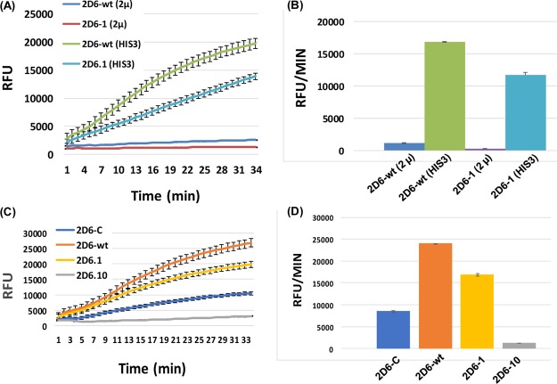
Four pairs of integrative plasmids, bearing the four CYP2D6 alleles on plasmids encoding the HIS3 or URA3 genes (Figure S2A,B), were created for use in integration into the yeast strain YY7, derived from the strain W303-1a (ATCC 208352) which already contained a modified human CYP450 reductase (CPR) gene.25 The plasmids were integrated at the HIS3 and the URA3 chromosomal loci of the yeast strain YY7. Episomal plasmids, bearing the URA3 gene as selection marker and encoding the four different CYP2D6 alleles, were also constructed (Figure S2C,D). Corresponding strains containing these plasmids were created for the sake of comparison with yeast strains that contain integrative copy/copies of CYP2D6 gene expression cassettes. Figure 2A,B shows a comparison of CYP2D6 activities obtained from yeast strains containing expression cassettes for the CYP2D6-wt and CYP2D6.1 alleles, integrated at the HIS3 chromosomal locus or borne on an episomal plasmid. The results in Figure 2A,B clearly indicate that over an identical period of time, a copy of either of the two CYP2D6 alleles, CYP2D6-wt and CYP2D6.1, integrated at the HIS3 chromosomal locus produces far more CYP2D6 enzyme activity than from an extrachromosomal episomal plasmid. It could be inferred that this was primarily because of the stability of the integrated copy of the CYP2D6 gene expression cassettes during expression of the protein. Similar results were obtained for the other two CYP2D6 alleles, CYP2D6.10 and CYP2D6-C. This is shown in Figures S3A,B and S4A,B, in the Supporting Information.
Figure 2.
Graph (A) compares the kinetics of enzyme activities of the two alleles CYP2D6-wt (2D6-wt) and CYP2D6.1 (2D6.1), produced in the strain YY7, from gene expression cassettes either (i) integrated at the HIS3 chromosomal locus or (ii) borne on an episomal plasmid. The kinetics of enzyme activity, present in ∼1 × 10⁶ cells, was followed over a time course of 34 min. The concentration of the fluorogenic substrate, EOMCC, used for each assay was 2 μM. The amount of fluorescent product, 7-HCC, formed was monitored at each time point using a fluorescent plate reader. The graphs represent the average of results obtained from three independent experiments. The bar plot (B) mirrors the fluorescence values of the graphs in (A), at time point 32 min. The graph (C) compares the kinetics of enzyme activities of the four alleles CYP2D6-C (2D6-C), CYP2D6-wt (2D6-wt), CYP2D6.1 (2D6.1), and CYP2D6.10 (2D6.10), expressed as two copies of each gene in the strain YY7, from the HIS3 and URA3 chromosomal loci. The kinetics of enzyme activity, present in ∼1 × 10⁶ cells, was followed over a time course of 33 min. The concentration of the fluorogenic substrate, EOMCC, used for each assay was 2 μM. The amount of the fluorescent product, 7-HCC, formed was monitored at each time point using a fluorescent plate reader. The graphs represent the average of results obtained from three independent experiments. Bar plot (D) mirrors the fluorescence values of the graphs in (C), at time point 32 min. The data represent mean ± SD of three independent experiments. “RFU” represents relative fluorescence units. Data were assembled from three independent experimental groups, and a between group ANOVA test identified significant differences (P < 0.05) between strains, in all comparative analyses performed.
Two copies of each of the four CYP2D6 alleles, CYP2D6-wt, CYP2D6.1, CYP2D6.10, and CYP2D6-C, were then integrated at the HIS3 and URA3 loci of the yeast strain YY7 that already contained a modified human CPR.25 The enzyme activities produced by these strains after 18 h of culture were determined, as described in the Experimental Section. Using EOMCC as a fluorogenic substrate, comparison of the enzymatic activities of the four CYP2D6 proteins, encoded by the four different alleles, are depicted in Figure 2C,D. The results show that CYP2D6.10 possesses the weakest enzymatic activity and that CYP2D6-C has ∼eightfold more activity than CYP2D6.10, a known CYP2D6 low metabolizer allele.1a,8−15
The western blot experiment confirmed that levels of expression in yeast of the four CYP2D6 proteins, from the different alleles, do not vary (Figure 3A). Total protein (10 μg), obtained from lysis of ∼1 × 10⁶ cells from each of the four strains expressing CYP2D6-wt, CYP2D6.1, CYP2D6.10 and CYP2D6-C alleles, was analyzed by Western blotting (Figure 3A). The results show that roughly equal amounts of CYP2D6 protein are produced by each yeast strain, from the same number of cells. This would indicate that differences in the levels of enzymatic activities between the different alleles, seen in yeast (Figure 2C,D), are a genuine reflection of the allelic phenotypes. Next, we estimated the amount of enzyme, produced within yeast cells, by genes coding for CYP2D6-wt, CYP2D6.1, and CYP2D6-C (CYP2D6.10A122V) alleles. Figure 3B,C attempts to quantify the amounts of CYP2D6 enzyme activities produced by a defined number of recombinant yeast cells, that is, 1 × 10⁷ cells (approximately 1 OD600 of cells), harboring the three different CYP2D6 alleles, CYP2D6-wt, CYP2D6.1, and CYP2D6-C (CYP2D6.10A122V). The cells were grown in YPD cultures over 54 h. The cells were provided with fresh YPD medium, every 18 h. It was observed that after, every 18 h of growth, enzymatic activity, per OD600 of cells, was gradually augmented as cell density dramatically increased with time (unpublished observations; I.S.W., PhD Thesis). From Figure 3B,C, it is observed that the amount of CYP2D6-wt enzyme present in 1 × 10⁷ cells, that are involved in the metabolism of the fluorogenic substrate EOMCC to form the product 7-HCC, is equivalent to around 3 pmol of CYP2D6 Supersomes (Corning). Similarly, 1 × 10⁷ cells expressing CYP2D6-C (2D6-C) equates to around 1.5 pmol of CYP2D6 Supersomes, whereas the same volume of cells expressing CYP2D6.1 (2D6.1) equates to approximately 2.5 pmol of CYP2D6 Supersomes. These results would indicate that the CYP2D6 variant enzymes, produced within yeast cells, may be remarkably proficient in performing biotransformation reactions on CYP2D6 substrates that would allow formation of products with high yields. The results would also indicate that with chromosomal integrations of a CYP gene, a large amount of microsomal enzyme, with high activity, can possibly be isolated from recombinant baker’s yeast. In fact, the CYP2D6-wt enzyme was obtained at ∼130 nmol/L of yeast cell culture with activities that were at least twofold better than the three commercially available enzymes, Supersomes (Corning), Baculosomes (Invitrogen), and Bactosomes (Cypex). Production of such large amounts of highly active human CYP enzymes in recombinant eukaryotic cells has never been reported before.
Figure 3.
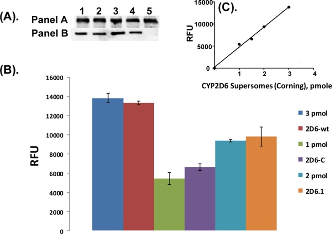
(A) Western blots of lysates of yeast cells expressing two copies of the CYP2D6-wt, CYP2D6.1, CYP2D6.10, and CYP2D6-C proteins, from the HIS3 and URA3 chromosomal loci of the strain YY7. Total protein (10 μg) from ∼1 × 10⁶ cells, obtained after lysis of the four strains (expressing 2 copies of each the four CYP2D6 alleles), was probed with a CYP2D6 antibody (panel A; Santa Cruz Biotechnology, sc-130366) and a β-actin antibody (panel B; Proteintech, 60008-1-Ig). Lane 1, CYP2D6-wt; lane 2, CYP2D6.1; lane 3, CYP2D6.10; lane 4, CY2D6-C; and lane 5, 0.3 pmol of CYP2D6-wt Sacchrosomes (i.e., microsomal CYP2D6-wt enzyme, isolated from baker’s yeast; CYP Design Ltd), as a positive control. (B,C) Calibration of CYP2D6 activities produced by 1/ × 107 cells that express CYP2D6-wt (2D6-wt), CYP2D6-C (2D6-C), and CYP2D6.10 (2D6.10) proteins (B), using a standard curve (C) drawn using specific amounts (1, 1.5, 2, and 3 pmol) of CYP2D6 Supersomes (Corning, #456217); CYP enzymes from Corning are widely considered to be the benchmark in the area of recombinant human CYP microsomal enzymes.
Biotransformation of Codeine to Morphine by Baker’s Yeast Cells Expressing the CYP2D6-wt, CYP2D6.1, CYP2D6.10, and CYP2D6-C Enzymes
Using the four variant CYP2D6 enzymes, expressed in yeast from the four different CYP2D6 alleles, the metabolism of codeine to morphine was investigated. For the biotransformation reaction, 10 μM of codeine (final concentration) was added to each yeast cell culture that expressed different CYP2D6 alleles. After completion of cell culture at defined time points (24, 48, and 72 h), the media were extracted with an organic solvent, ethyl acetate. The residues obtained after evaporation of extracts were redissolved in 1 mL of ethanol and then analyzed by thin-layer chromatography (TLC) and high-performance liquid chromatography (HPLC).
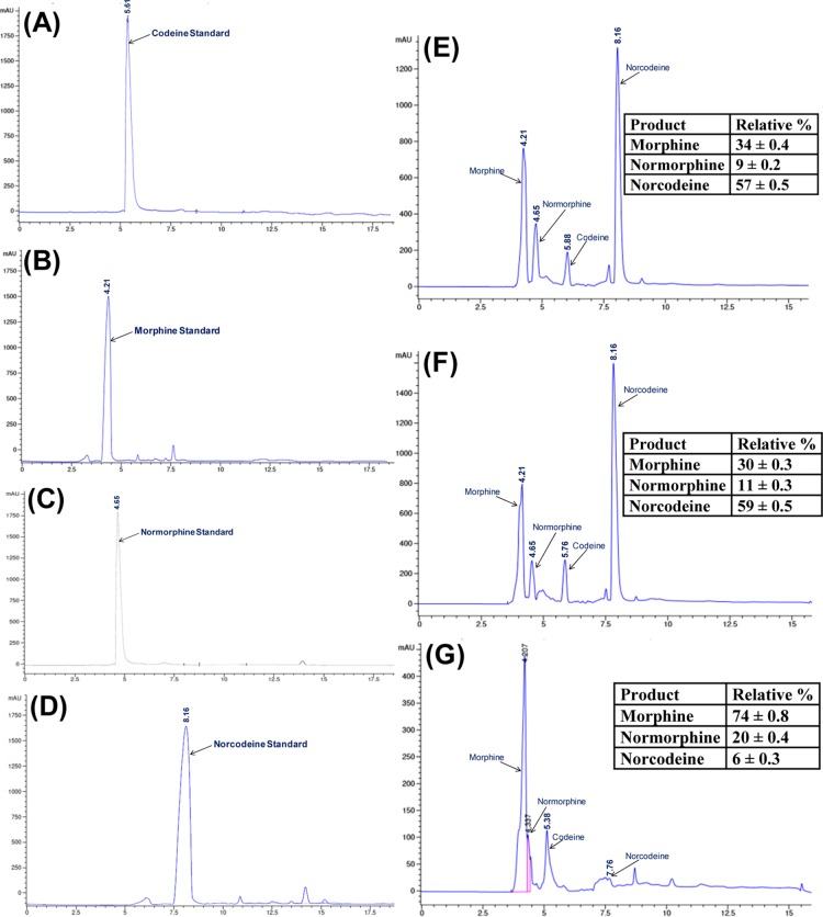
For TLC analyses, the equal volumes (5 μL) of the dissolved residues, obtained after extraction, were spotted on to TLC plates. The TLC images are shown in section S5 of the Supporting Information. Figure S5A shows that cells expressing CYP2D6.wild are hardly able to convert codeine to morphine. In contrast, CYP2D6-C, which is CYP2D6.10A112V produces more morphine than the other two CYP2D6 variant enzymes, CYP2D6-wt and CYP2D6.1. This is shown on the TLC plate in Figure S5B (lanes 5, 6, and 7) which also has as reference standards, codeine and its metabolites, that is, morphine, norcodeine, and normorphine (lanes 1–4). All cell culture extracts were then analyzed by HPLC, along with reference standards of codeine and its three metabolites, morphine, normorphine, and norcodeine.26 Equal volumes of media extracts (10 μL), from the 72 h cultures, were injected into the HPLC column. HPLC results are shown in Figure 4 and the percentage of the metabolites formed are shown on the chromatograms. Data obtained from HPLC quantified the observations made with TLC. It was confirmed that the CYP2D6-C enzyme, a variant of CYP2D6.10 (i.e., CYP2D6.10A112V), was superior in activity than the CYP2D6-wt and CYP2D6.1 enzymes, in the formation of morphine from codeine, although CYP2D6-C showed weaker activity compared with the other two variant enzymes when using EOMCC as a fluorogenic substrate (Figure 2C,D).
Figure 4.
HPLC analysis of the biotransformation of codeine using three different CYP2D6 variant enzymes, CYP2D6-wt, CYP2D6.1, and CYP2D6-C (i.e., CYP2D6.10A122V) expressed within yeast. Redissolved residues (10 μL), obtained after extracting the reaction medium from 72 h cell cultures, were injected into the column. (A–D) HPLC chromatograms of codeine, morphine, normorphine, and norcodeine which are reference standards. (E–G) HPLC chromatograms of biotransformation reactions mediated by the CYP2D6 variants, CYP2D6-wt (E) CYP2D6.1 (F), CYP2D6C (G), using codeine as a substrate. The relative percentages of morphine, normorphine, and norcodeine obtained from each CYP2D6-mediated reaction are shown on the chromatograms. The percentage values represent mean and standard deviations (±SD) from three independent experiments. In all experiments, 10 μM (final concentration) of codeine was added to each yeast cell culture, bearing different CYP2D6 alleles.
The results obtained from the biotransformation of codeine, using live yeast cells expressing different CYP2D6 alleles, clearly show that the CYP2D6-C (CYP2D6.10A112V) allele is more efficient in the conversion of codeine to morphine than the known extensive metabolizers CYP2D6.1 and CYP2D6-wt, although CYP2D6.10 is known to be a low metabolizer of codeine. This would suggest that the active site of the CYP2D6-C enzyme has a higher activity for the O-demethylation of codeine than the CYP2D6-wt and CYP2D6.1 enzymes.
Molecular Modeling Studies
A total of 10 crystal structures (1 without any ligand and 9 with a substrate or an inhibitor) have been reported.6,27 The key sites in the CYP2D6 enzyme that are utilized for demethylation of specific substrates have been established in the literature. Several site-directed mutagenesis and computational studies on the CYP2D6 cDNA and protein, respectively, have demonstrated that Glu 216, Asp 301, Phe 120, and Met 374 are key determinants of substrate specificity and product regioselectivity in CYP2D6.28 Because of the significant contribution of CYP2D6 in the metabolism of drugs, discovery of its inhibitors is a key research area in drug discovery programs.28d Furthermore, the CYP2D6 enzyme has extensively been studied for delineation of the metabolism (O-demethylation and N-demethylation) of approved drugs viz metoprolol,29 gefitinib,30 and natural products.31
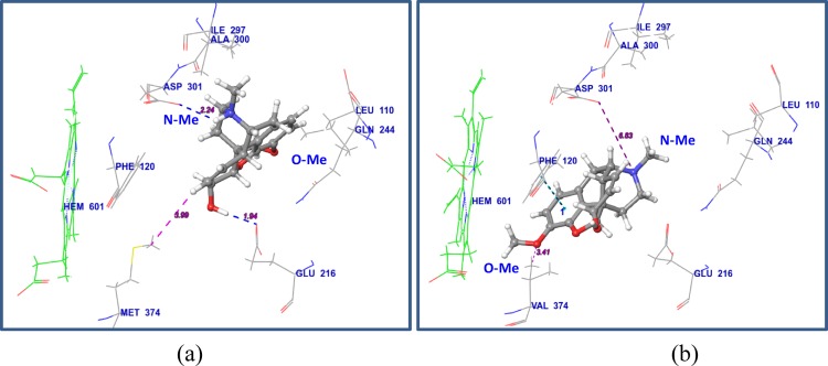
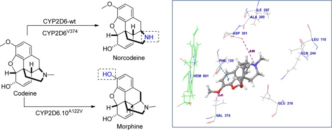
To understand the observed trend in differential demethylation reactions (O vs N-demethylation) using CYP2D6-wt and CYP2D6-C (CYP2D6.10A112V), molecular modeling studies were conducted. The crystal structure of CYP2D6.1 (PDB: 4WNW), comprising thioridazine (CYP2D6 substrate) as a cocrystallized ligand, was selected. The required mutations were performed in Schrodinger to generate CYP2D6-wt (Val374Met) and CYP2D6-C (Pro34Ser, Ser486Thr, and Ala122Val) variants. The CYP2D6 structure has a well-defined active site cavity which is around 6–8 Å away from the haem group and contains important residues that have been implicated in substrate recognition and binding. They include Asp-301, Glu-216, Phe-483, and Phe-120. The active site, where the substrate is accommodated, contains a hydrophobic binding pocket and comprises residues Phe-110, Gly-244, Leu-248, Ile-297, and Ala-300.6 The molecular docking of codeine with the active site of wild-type CYP2D6-wt (Figure 5a) shows that the hydrogen of the protonated nitrogen of codeine forms a H-bond with the carbonyl oxygen of Asp-301 (distance 2.24 Å). It has been reported in the literature that Asp-301 plays a key role in CYP2D6-mediated demethylation reactions.6 Thus, it is clear that the N-demethylation of codeine is mediated via its interaction with Asp-301. This observation supports the experimental results obtained with CYP2D6-wt, which favors N-demethylation to produce norcodeine as the major product. When Met (M) residue is substituted by Val (V) residue at position 374 in the CYP2D6.1 and CYP2D6.C amino acid sequence, the location of codeine is shifted toward haem along with a 90° flip, resulting in loss of a few crucial van der Waals interactions with the residues at the hydrophobic binding cavity. In 2D6-C, the OMe group of codeine is present in close proximity to Val-374 (3.41 Å) and the OMe group containing Ar ring displays π–π interaction with Phe-120 residue. It has been reported that the 374 residue has influence on cytochrome P450 expression, ligand binding, catalysis, and functional activity of the CYP2D6 enzyme.32 The interaction of OMe group with Val 374 could be accounting for enhanced O-demethylation in case of 2D6-C in comparison to 2D6-wild. The interaction pattern of codeine in CYP2D6-C (i.e., CYP2D6.10A122V) is shown in Figure 5b. The docked images of thioridazine (CYP2D6 substrate) are provided in Figure S6a–c.
Figure 5.
Interaction pattern of codeine with CYP2D6-wt (a), and CYP2D6-C (CYP2D6.10A122V) (b). The pink dotted lines indicate specific distances between ligand and residues.
The interaction of EOMCC (7-ethoxy-methyloxy-3-cyanocoumarin), a substrate which is used in the biochemical assay, was also studied (Figure S7a,b). The interaction of CH2 with Glu 216 is important for cleavage of ether bond (C–O–C bond) to form 7-hydroxy-3-cyanocoumarin metabolite, which is measured as an output of the assay, at excitation and emission wavelengths of 410 and 460 nM. In case of wild-type 2D6 (Met374), the CH2 group is closer to the Glu 216 (3.07 Å), in comparison to 2D-C (distance is 3.55 Å). This difference could be attributing to the lesser activity of EOMCC in case of 2D6-C variant in comparison to wild-type 2D6.
Conclusions
In summary, we have demonstrated that the CYP2D6.10 variant enzyme, CYP2D6.10A122V (CYP2D6-C), which has never been reported to exist in a human cDNA library, is a far superior metabolizer of codeine to morphine than the CYP2D6-wt (CYP2D6M374) or the CYP2D6.1 (CYP2D6V374) enzymes. This result is quite unusual because published observations categorically show CYP2D6.10 as a low (i.e., weak) metabolizer of codeine to morphine. The enzyme expressed from CYP2D6.10A122V yielded >70% of morphine from codeine in comparison to only ∼30–35% of morphine using the other two variant enzymes, CYP2D6-wt and CYP2D6.1, which have been reported to be extensive metabolizers of codeine.2,26
Yeast cell-based systems, described in this report, are amenable to scale-up in fed-batch fermentors. Cells which individually express the newly identified CYP2D6 allele, CYP2D6.10A122V, and the CYP2D6-wt and CYP2D6.1 alleles, could find wider applicability in the identification, characterization, and large-scale isolation of CYP2D6-mediated formation of drug metabolites that would facilitate studies in the preclinical/clinical interface and in the clinic. They could also be used for regio-selective bioorganic syntheses of high-value natural products from more abundantly available chemicals available in nature.33
Experimental Section
Chemicals
Codeine, morphine, norcodeine, and normorphine were bought from Sigma-Aldrich on a license received by the Leicester School of Pharmacy, UK. All other chemicals used in this study were also obtained from Sigma-Aldrich.
Gene Synthesis
The three CYP2D6 alleles, CYP2D6-wt (NCBI Accession No M20403) for DNA sequence; NCBI Accession No AAA52153 for debrisoquine 4-hydroxylase_Homo sapiens protein sequence; 6 CYP2D6.1 (NCBI Accession No NM_000106 for DNA sequence; NCBI Accession No NP_000097 for protein sequence;34 and CYP2D6.10 (NCBI Accession No ABB01372), were chemically synthesized by GENEWIZ, USA. Tailor-made chemically synthesized genes are routinely used in molecular biology laboratories around the world.35 The synthesized genes for this manuscript were sourced from GENEWIZ (details can be obtained at https://www.genewiz.com/Public/Services/Gene-Synthesis).
Isolation of CYP2D6-C (CYP2D6.10A122V) cDNA
The CYP2D6-C cDNA was isolated by PCR using a human liver cDNA library, obtained from GATC Biotech (Germany), as template and CYP2D6-specific 5′ and 3′-end primers. The DNA of the isolated clone was confirmed by sequencing. The sequence was identical to the sequence of the TC104446 clone distributed by OriGene (Rockville, Maryland, USA).
Cloning of CYP2D6 Allelic Genes
The basic vectors (integrative plasmids used for chromosomal integration at the HIS3 and URA3 loci of baker’s yeast, S. cerevisiae, and the episomal plasmid for extra-chromosomal replication), used for cloning of CYP2D6 gene expression cassettes, were obtained from CYP Design Ltd. The resultant plasmids, after cloning of CYP2D6 gene expression cassettes in respective vectors, were used for yeast transformation.
Transformation of Yeast Cells
The yeast strain YY7, carrying the nonfunctional alleles ade2, leu2, his3, trp1, and ura3 which act as auxotrophic markers, already carried a modified less toxic version of the human P450 reductase (CPR) gene.25 Yeast transformation was carried out by the lithium acetate protocol.36 For integration of plasmids, bearing CYP2D6 alleles, into chromosomal locations where the HIS3 and URA3 genes exist (i.e., on chromosomes XV and V, respectively), the integrative plasmids were linearized with an appropriate restriction enzyme within the nutritional selection marker (HIS3 or URA3), in a way that at least 200 bps are at both 5′ and 3′ segments of the fragmented gene, before the DNA was introduced into yeast cells. The linearized DNA underwent allelic homologous recombination23 with the genomic DNA at the chromosomal locations of the his3 or ura3 genes in the strain YY7.
Growth of Cells for Comparison of Cellular CYP2D6 Enzyme Activities
Yeast strains, containing the CYP2D6 alleles, were at first streaked out on SD minimal medium agar plates (6.7 g/L yeast nitrogen base without amino acids; 20 g/L glucose; 20 g/L agar) with appropriate nutrients depending on the auxotroph. The plates were incubated for 3 days at 30 °C. Three individual colonies were inoculated into in 5 mL of SD medium (6.7 g/L yeast nitrogen base without amino acids; 20 g/L glucose) containing appropriate nutrients. Cells were grown for 20 h at 30 °C in an orbital shaker at 220 rpm. Cells with 0.1 OD600 (optical density of cells measured at 600 nm) from the minimal medium SD pre-cultures were inoculated into 5 mL of full (nonselective) YPD medium (10 g/L yeast extract; 20 g/L peptone; 20 g/L glucose) for expression of the CYP2D6 alleles and were grown overnight for 18 h at 30 °C. At this time point, the cells were still in the late exponential growth phase (thus preventing the decay of CYP enzyme activity that occurs at the stationary phase), and glucose in the medium was fully exhausted. Cells were counted so that exactly the same number of cells were used for all fluorescence-based CYP2D6 enzyme activity assays.
Determination of Cellular CYP2D6 Enzyme Activities
A total of 1 × 10⁷ (∼1 OD600) cells was aliquoted into Eppendorf tubes and centrifuged for 1 min on a bench-top centrifuge. The pellets were resuspended in 500 μL 1× TE (50 mM Tris-HCl, 1 mM EDTA, pH 7.4) and centrifuged for 1 min. This was repeated twice. The supernatants were removed carefully, and the pellets were finally resuspended in 500 μL of 1× TE. Cell suspensions (50 μL) were transferred into a sterile 96-well microtiter plate to which 50 μL of a 4 μM EOMCC (7-ethoxymethoxy-3-cyanocoumarin) containing solution was added. Plates were incubated at 30 °C for 30 min before the fluorescence excitation/emissions of the CYP2D6-mediated fluorescent product, 7-HCC (7-hydroxy-3-cyanocoumarin), were monitored at 410/460 nm on a fluorescence plate reader (Synergy HT BioTek), over a period of time. The gain sensitivities of the plate reader were set in a way that allowed the best kinetic output to be obtained. The plates were incubated at 30 °C for 30 s before the fluorescence emissions were measured.
Western Blotting
A total of 1 × 10⁷ cells, bearing CYP2D6 alleles, after growth in YPD medium for 18 h at 30 °C, was harvested. TE-washed cells were suspended in 200 μL of SD (Sodium dodecyl sulfate)–TE buffer (5.7 μL 10× SDS, 1 mL 10× TE buffer, made up to 10 mL with deionized pure water). The cell suspension was sonicated (Grant Ultrasonic bath XUBA1), twice for 10 s, and then incubated at 95 °C for 10 min. Proteins in the supernatants were estimated via Lowry’s method (Pierce; Thermo Fisher Scientific) and were diluted 1:10 in SDS sample buffer before loading on a 7.5% SDS–polyacrylamide gel for electrophoresis.
Biotransformation Experiments
Strains, containing a chromosomally integrated CYP2D6 allele (one or two copies), or an episomal plasmid bearing a CYP2D6 gene expression cassette, were revived on YPD plates. Typically, a loopful of CYP2D6-containing freshly grown yeast cells was inoculated in a 500 mL Erlenmeyer baffled flask containing 100 mL YPD medium (pre-culture-1) at 28 °C for 24 h. Baffled flasks allow vigorous aeration. The cells were harvested after 24 h and inoculated into a new 500 mL baffled flask containing 100 mL YPD medium (pre-culture-2) at 30 °C for 18 h. The process was repeated a third time (pre-culture-3) for the cells to reach an OD600, of ∼90/mL. Cells (100 OD600) from pre-culture-3 were resuspended in 25 mL of SD medium contained in a 250 mL baffled flask. A final concentration of 10 μM codeine was added to the cultures. Flasks were shaken at 30 °C, 220 rpm, for 24, 48, and 72 h. Cells were harvested every 24 h for assessing biotransformation (i.e., for amounts of formed products). After every 24 h of growth, 1 mL of 50% glucose was added to the 25 mL cultures to replenish levels of glucose to 2%. The SD reaction medium was extracted with ethyl acetate (3 times). The combined ethyl acetate layer was concentrated on vacuo-rotavapor to obtain residues that contained the biotransformation products. The residues, redissolved in 1 mL of ethanol, were analyzed on TLC plates and with HPLC.
TLC Analysis
Equal volumes (5 μL) of the redissolved residue, obtained after extraction of the reaction media, were spotted on to lanes of TLC plates (Sigma Aldrich #Z193275-1). For TLC detection of codeine and its metabolites formed in the biotransformation experiments, the solvent system was CHCl3/MeOH/NH3 = 36:1:0.6.
HPLC Analysis
HPLC analysis was performed on a Shimadzu LC-6AD system connected with a C18 column (4.6 × 25 mm, 5 μ). Mobile phase consisted of A: 0.1% formic acid and B: methanol using isocratic elution (30:70—A/B). The flow rate was 1 mL/min. The detection wavelength was 270 nm. The crude residue, obtained after extraction of the reaction media, was loaded on a reverse phase (C18) silica gel column packed in water. The crude extract was loaded on the column by making slurry with C18 silica gel. The column was then eluted with increasing concentrations of methanol in water.
Molecular Modeling
The docking of codeine with CYP2D6 (PDB ID: 4WNT) was performed using GLIDE module of Schrodinger molecular modeling software, using the protocols as described in our earlier publications.37 The docking protocol was validated by redocking known ligand ajmalicine. The interaction pattern of docked ajmalicine and ligand from cocrystallized protein (4WNT) are shown in the Supporting Information.
Acknowledgments
I.S.W. would like to thank Dr. R. Arroo of the Leicester School of Pharmacy for allowing the use of facilities in his laboratory and providing codeine and morphine, bought on license by the Leicester School of Pharmacy. I.S.W. also acknowledges the technical assistance of Hajara Alfa in performing the HPLC experiments.
Supporting Information Available
The Supporting Information is available free of charge on the ACS Publications website at DOI: 10.1021/acsomega.8b00809.
Additional biology details; TLC images of biotransformation experiment; additional molecular modeling results; and comparisons of the amino acid sequences of the four CYP2D6 proteins (PDF)
Author Present Address
§ Institute of Bio-sensing Technology, University of the West of England, Frenchay Campus, Filton Road, Bristol, BS34 8QZ, UK.
The authors declare no competing financial interest.
Supplementary Material
References
- a Yu A.; Kneller B. M.; Rettie A. E.; Haining R. L. Expression, purification, biochemical characterization, and comparative function of human cytochrome P450 2D6.1, 2D6.2, 2D6.10, and 2D6.17 allelic isoforms. J. Pharmacol. Exp. Ther. 2002, 303, 1291–1300. 10.1124/jpet.102.039891. [DOI] [PubMed] [Google Scholar]; b Holmquist G. L. Opioid Metabolism and Effects of Cytochrome P450. Pain Med. 2009, 10, S20–S29. 10.1111/j.1526-4637.2009.00596.x. [DOI] [Google Scholar]
- a Shen H.; He M. M.; Liu H.; Wrighton S. A.; Wang L.; Guo B.; Li C. Comparative metabolic capabilities and inhibitory profiles of CYP2D6.1, CYP2D6.10, and CYP2D6.17. Drug Metab. Dispos. 2007, 35, 1292–1300. 10.1124/dmd.107.015354. [DOI] [PubMed] [Google Scholar]; b Zhuge J.; Yu Y.-N.; Wu X.-D. Stable expression of human cytochrome P450 2D6*10 in HepG2 cells. World J. Gastroenterol. 2004, 10, 234–237. 10.3748/wjg.v10.i2.234. [DOI] [PMC free article] [PubMed] [Google Scholar]
- a Ingelman-Sundberg M.; Daly A. K.; Oscarson M.; Nebert D. W. Human cytochrome P450 (CYP) genes: recommendations for the nomenclature of alleles. Pharmacogenetics 2000, 10, 91–93. 10.1097/00008571-200002000-00012. [DOI] [PubMed] [Google Scholar]; b Zanger U. M.; Raimundo S.; Eichelbaum M. Cytochrome P450 2D6: overview and update on pharmacology, genetics, biochemistry. Naunyn-Schmiedeberg’s Arch. Pharmacol. 2004, 369, 23–37. 10.1007/s00210-003-0832-2. [DOI] [PubMed] [Google Scholar]
- Neafsey P.; Ginsberg G.; Hattis D.; Sonawane B. Genetic polymorphism in cytochrome P450 2D6 (CYP2D6): Population distribution of CYP2D6 activity. J. Toxicol. Environ. Health, Part B 2009, 12, 334–361. 10.1080/10937400903158342. [DOI] [PubMed] [Google Scholar]
- a Shimizu T.; Ochiai H.; Åsell F.; Yokono Y.; Kikuchi Y.; Nitta M.; Hama Y.; Yamaguchi S.; Hashimoto M.; Taki K.; Nakata K.; Aida Y.; Ohashi A.; Ozawa N. Bioinformatics research on inter-racial difference in drug metabolism II. Analysis on relationship between enzyme activities of CYP2D6 and CYP2C19 and their relevant genotypes. Drug Metab. Pharmacokinet. 2003, 18, 71–78. 10.2133/dmpk.18.71. [DOI] [PubMed] [Google Scholar]; b Naveen A. T.; Adithan C.; Soya S. S.; Gerard N.; Krishnamoorthy R. CYP2D6 genetic polymorphism in South Indian populations. Biol. Pharm. Bull. 2006, 29, 1655–1658. 10.1248/bpb.29.1655. [DOI] [PubMed] [Google Scholar]
- Rowland P.; Blaney F. E.; Smyth M. G.; Jones J. J.; Leydon V. R.; Oxbrow A. K.; Lewis C. J.; Tennant M. G.; Modi S.; Eggleston D. S.; Chenery R. J.; Bridges A. M. Crystal structure of human cytochrome P450 2D6. J. Biol. Chem. 2006, 281, 7614–7622. 10.1074/jbc.m511232200. [DOI] [PubMed] [Google Scholar]
- Corning-Gentest’s Product List (https://www.corning.com/media/worldwide/cls/documents/CLS-DL-GT-038%20ROW%20REV1.pdf; Product # 456217) states clearly that the Val374 variant is CYP2D6*1 (i.e. CYP2D6–1). Corning sells this product as the only 2D6 enzyme.
- Chen C.-H.; Hung C.-C.; Wei F.-C.; Koong F.-J. Debrisoquine 4-hydroxylase (CYP2D6) genetic polymorphisms and susceptibility to schizophrenia in Chinese patients from Taiwan. Psychiatr. Genet. 2001, 11, 153–155. 10.1097/00041444-200109000-00007. [DOI] [PubMed] [Google Scholar]
- Cai W. M.; Chen B.; Zhang W. X. Frequency of CYP2D6*10 and *14 alleles and their influence on the metabolic activity of CYP2D6 in a healthy Chinese population. Clin. Pharmacol. Ther. 2007, 81, 95–98. 10.1038/sj.clpt.6100015. [DOI] [PubMed] [Google Scholar]
- Lee S.-Y.; Sohn K. M.; Ryu J. Y.; Yoon Y. R.; Shin J. G.; Kim J.-W. Sequence-based CYP2D6 genotyping in the Korean population. Ther. Drug Monit. 2006, 28, 382–387. 10.1097/01.ftd.0000211823.80854.db. [DOI] [PubMed] [Google Scholar]
- Bagheri A.; Kamalidehghan B.; Haghshenas M.; Azadfar P.; Akbari L.; Sangtarash M. H.; Vejdandoust F.; Ahmadipour F.; Meng G. Y.; Houshmand M. Prevalence of the CYP2D6*10 (C100T), *4 (G1846A), and *14 (G1758A) alleles among Iranians of different ethnicities. Drug Des., Dev. Ther. 2015, 9, 2627–2634. 10.2147/dddt.s79709. [DOI] [PMC free article] [PubMed] [Google Scholar]
- Nishida Y.; Fukuda T.; Yamamoto I.; Azuma J. CYP2D6 genotypes in a Japanese population: low frequencies of CYP2D6 gene duplication but high frequency of CYP2D6*10. Pharmacogenetics 2000, 10, 567–570. 10.1097/00008571-200008000-00010. [DOI] [PubMed] [Google Scholar]
- a Sachse C.; Brockmoller J.; Bauer S.; Roots I. Cytochrome P450 2D6 variants in a Caucasian population: allele frequencies and phenotypic consequences. Am. J. Hum. Genet. 1997, 60, 284–295. [PMC free article] [PubMed] [Google Scholar]; b Gaikovitch E. A.; Cascorbi I.; Mrozikiewicz P. M.; Brockmöller J.; Frötschl R.; Köpke K.; Gerloff T.; Chernov J. N.; Roots I. Polymorphisms of drug-metabolizing enzymes CYP2C9, CYP2C19, CYP2D6, CYP1A1, NAT2 and of P-glycoprotein in a Russian population. Eur. J. Clin. Pharmacol. 2003, 59, 303–312. 10.1007/s00228-003-0606-2. [DOI] [PubMed] [Google Scholar]
- Cai W.-M.; Nikoloff D. M.; Pan R.-M.; de Leon J.; Fanti P.; Fairchild M.; Koch W. H.; Wedlund P. J. CYP2D6 genetic variation in healthy adults and psychiatric African-American subjects: implications for clinical practice and genetic testing. Pharmacogenomics J. 2006, 6, 343–350. 10.1038/sj.tpj.6500378. [DOI] [PubMed] [Google Scholar]
- Luo H.-R.; Gaedigk A.; Aloumanis V.; Wan Y.-J. Y. Identification of CYP2D6 impaired functional alleles in Mexican Americans. Eur. J. Clin. Pharmacol. 2005, 61, 797–802. 10.1007/s00228-005-0044-4. [DOI] [PubMed] [Google Scholar]
- Pasternak G. W.; Pan Y.-X. Mu opioids and their receptors: evolution of a concept. Pharmacol. Rev. 2013, 65, 1257–1317. 10.1124/pr.112.007138. [DOI] [PMC free article] [PubMed] [Google Scholar]
- a elSohly H. N.; Stanford D. F.; Jones A. B.; elSohly M. A.; Snyder H.; Pedersen C. Gas chromatographic/mass spectrometric analysis of morphine and codeine in human urine of poppy seed eaters. J. Forensic Sci. 1988, 33, 347–356. 10.1520/jfs11948j. [DOI] [PubMed] [Google Scholar]; b Grove M. D.; Spencer G. F.; Wakeman M. V.; Tookey H. L. Morphine and codeine in poppy seed. J. Agric. Food Chem. 1976, 24, 896–897. 10.1021/jf60206a022. [DOI] [PubMed] [Google Scholar]
- Ayyangar N. R.; Choudhary A. R.; Kalkote U. R.; Sharma V. K.. Process for the preparation of codeine from morphine. EP 0268710 B1, Nov 25, 1986.
- a Yee D. A.; Atayee R. S.; Best B. M.; Ma J. D. Observations on the urine metabolic profile of codeine in pain patients. J. Anal. Toxicol. 2014, 38, 86–91. 10.1093/jat/bkt101. [DOI] [PubMed] [Google Scholar]; b Trescot A. M.; Datta S.; Lee M.; Hansen H. Opioid pharmacology. Pain Physic. 2008, 11, S133–S153. [PubMed] [Google Scholar]
- a Zahari Z.; Ismail R. Influence of Cytochrome P450, Family 2, Subfamily D, Polypeptide 6 (CYP2D6) polymorphisms on pain sensitivity and clinical response to weak opioid analgesics. Drug Metab. Pharmacokinet. 2014, 29, 29–43. 10.2133/dmpk.dmpk-13-rv-032. [DOI] [PubMed] [Google Scholar]; b Wu X.; Yuan L.; Zuo J.; Lv J.; Guo T. The impact of CYP2D6 polymorphisms on the pharmacokinetics of codeine and its metabolites in Mongolian Chinese subjects. Eur. J. Clin. Pharmacol. 2014, 70, 57–63. 10.1007/s00228-013-1573-x. [DOI] [PubMed] [Google Scholar]
- Karim A. S.; Curran K. A.; Alper H. S. Characterization of plasmid burden and copy number inSaccharomyces cerevisiaefor optimization of metabolic engineering applications. FEMS Yeast Res. 2013, 13, 107–116. 10.1111/1567-1364.12016. [DOI] [PMC free article] [PubMed] [Google Scholar]
- Van Der Sand S. T.; Greenhalf W.; Gardner D. C. J.; Oliver S. G. The maintenance of self-replicating plasmids inSaccharomyces cerevisiae: Mathematical modelling, computer simulations and experimental tests. Yeast 1995, 11, 641–658. 10.1002/yea.320110705. [DOI] [PubMed] [Google Scholar]
- Joska T. M.; Mashruwala A.; Boyd J. M.; Belden W. J. A universal cloning method based on yeast homologous recombination that is simple, efficient, and versatile. J. Microbiol. Methods 2014, 100, 46–51. 10.1016/j.mimet.2013.11.013. [DOI] [PMC free article] [PubMed] [Google Scholar]
- Guengerich F. P.; Munro A. W. Unusual cytochrome p450 enzymes and reactions. J. Biol. Chem. 2013, 288, 17065–17073. 10.1074/jbc.r113.462275. [DOI] [PMC free article] [PubMed] [Google Scholar]
- Chaudhuri B.Method for production of cytochrome p450 with n-term. truncated p450 reductase. WO 2007129050 A3, May 5, 2006.
- Verwey-Van Wissen C. P. W. G. M.; Koopman-Kimenai P. M.; Vree T. B. Direct determination of codeine, norcodeine, morphine and normorphine with their corresponding O-glucuronide conjugates by high-performance liquid chromatography with electrochemical detection. J. Chromatogr. 1991, 570, 309–320. 10.1016/0378-4347(91)80534-j. [DOI] [PubMed] [Google Scholar]
- a Wang A.; Savas U.; Hsu M.-H.; Stout C. D.; Johnson E. F. Crystal structure of human cytochrome P450 2D6 with prinomastat bound. J. Biol. Chem. 2012, 287, 10834–10843. 10.1074/jbc.m111.307918. [DOI] [PMC free article] [PubMed] [Google Scholar]; b Wang A.; Stout C. D.; Zhang Q.; Johnson E. F. Contributions of ionic interactions and protein dynamics to cytochrome P450 2D6 (CYP2D6) substrate and inhibitor binding. J. Biol. Chem. 2015, 290, 5092–5104. 10.1074/jbc.m114.627661. [DOI] [PMC free article] [PubMed] [Google Scholar]; c Brodney M. A.; Beck E. M.; Butler C. R.; Barreiro G.; Johnson E. F.; Riddell D.; Parris K.; Nolan C. E.; Fan Y.; Atchison K.; Gonzales C.; Robshaw A. E.; Doran S. D.; Bundesmann M. W.; Buzon L.; Dutra J.; Henegar K.; LaChapelle E.; Hou X.; Rogers B. N.; Pandit J.; Lira R.; Martinez-Alsina L.; Mikochik P.; Murray J. C.; Ogilvie K.; Price L.; Sakya S. M.; Yu A.; Zhang Y.; O’Neill B. T. Utilizing structures of CYP2D6 and BACE1 complexes to reduce risk of drug-drug interactions with a novel series of centrally efficacious BACE1 inhibitors. J. Med. Chem. 2015, 58, 3223–3252. 10.1021/acs.jmedchem.5b00191. [DOI] [PMC free article] [PubMed] [Google Scholar]; d Butler C. R.; Ogilvie K.; Martinez-Alsina L.; Barreiro G.; Beck E. M.; Nolan C. E.; Atchison K.; Benvenuti E.; Buzon L.; Doran S.; Gonzales C.; Helal C. J.; Hou X.; Hsu M.-H.; Johnson E. F.; Lapham K.; Lanyon L.; Parris K.; O’Neill B. T.; Riddell D.; Robshaw A.; Vajdos F.; Brodney M. A. Aminomethyl-Derived Beta Secretase (BACE1) Inhibitors: Engaging Gly230 without an Anilide Functionality. J. Med. Chem. 2017, 60, 386–402. 10.1021/acs.jmedchem.6b01451. [DOI] [PMC free article] [PubMed] [Google Scholar]
- a Paine M. J. I.; McLaughlin L. A.; Flanagan J. U.; Kemp C. A.; Sutcliffe M. J.; Roberts G. C. K.; Wolf C. R. Residues glutamate 216 and aspartate 301 are key determinants of substrate specificity and product regioselectivity in cytochrome P450 2D6. J. Biol. Chem. 2003, 278, 4021–4027. 10.1074/jbc.m209519200. [DOI] [PubMed] [Google Scholar]; b Wang B.; Yang L.-P.; Zhang X.-Z.; Huang S.-Q.; Bartlam M.; Zhou S.-F. New insights into the structural characteristics and functional relevance of the human cytochrome P450 2D6 enzyme. Drug Metab. Rev. 2009, 41, 573–643. 10.1080/03602530903118729. [DOI] [PubMed] [Google Scholar]; c de Graaf C.; Oostenbrink C.; Keizers P. J.; van Vugt-Lussenburg B. A.; van Waterschoot R. B.; Tschirret-Guth R.; Commandeur J. M.; Vermeulen N. E. Molecular modeling-guided site-directed mutagenesis of cytochrome P450 2D6. Curr. Drug Metab. 2007, 8, 59–77. 10.2174/138920007779315062. [DOI] [PubMed] [Google Scholar]; d Hochleitner J.; Akram M.; Ueberall M.; Davis R. A.; Waltenberger B.; Stuppner H.; Sturm S.; Ueberall F.; Gostner J. M.; Schuster D. A combinatorial approach for the discovery of cytochrome P450 2D6 inhibitors from nature. Sci. Rep. 2017, 7, 8071. 10.1038/s41598-017-08404-0. [DOI] [PMC free article] [PubMed] [Google Scholar]
- Uehara S.; Ishii S.; Uno Y.; Inoue T.; Sasaki E.; Yamazaki H. Regio- and Stereo-Selective Oxidation of a Cardiovascular Drug, Metoprolol, Mediated by Cytochrome P450 2D and 3A Enzymes in Marmoset Livers. Drug Metab. Dispos. 2017, 45, 896–899. 10.1124/dmd.117.075630. [DOI] [PubMed] [Google Scholar]
- Fang P.; Zheng X.; He J.; Ge H.; Tang P.; Cai J.; Hu G. Functional characterization of wild-type and 24 CYP2D6 allelic variants on gefitinib metabolism in vitro. Drug Des., Dev. Ther. 2017, 11, 1283–1290. 10.2147/dddt.s133814. [DOI] [PMC free article] [PubMed] [Google Scholar]
- a Li Y.; Zhang Y.; Wang R.; Wei L.; Deng Y.; Ren W. Metabolic profiling of five flavonoids from Dragon’s Blood in human liver microsomes using high-performance liquid chromatography coupled with high resolution mass spectrometry. J. Chromatogr. B: Anal. Technol. Biomed. Life Sci. 2017, 1052, 91–102. 10.1016/j.jchromb.2017.03.022. [DOI] [PubMed] [Google Scholar]; b Jiang R.; Yamaori S.; Takeda S.; Yamamoto I.; Watanabe K. Identification of cytochrome P450 enzymes responsible for metabolism of cannabidiol by human liver microsomes. Life Sci. 2011, 89, 165–170. 10.1016/j.lfs.2011.05.018. [DOI] [PubMed] [Google Scholar]; c Renwick A. B.; Surry D.; Price R. J.; Lake B. G.; Evans D. C. Metabolism of 7-benzyloxy-4-trifluoromethylcoumarin by human hepatic cytochrome P450 isoforms. Xenobiotica 2000, 30, 955–969. 10.1080/00498250050200113. [DOI] [PubMed] [Google Scholar]; d Tang L.; Ye L.; Lv C.; Zheng Z.; Gong Y.; Liu Z. Involvement of CYP3A4/5 and CYP2D6 in the metabolism of aconitine using human liver microsomes and recombinant CYP450 enzymes. Toxicol. Lett. 2011, 202, 47–54. 10.1016/j.toxlet.2011.01.019. [DOI] [PubMed] [Google Scholar]
- Ellis S. W.; Rowland K.; Ackland M. J.; Rekka E.; Simula A. P.; Lennard M. S.; Wolf C. R.; Tucker G. T. Influence of amino acid residue 374 of cytochromeP-450 2D6 (CYP2D6) on the regio- and enantio-selective metabolism of metoprolol. Biochem. J. 1996, 316, 647–654. 10.1042/bj3160647. [DOI] [PMC free article] [PubMed] [Google Scholar]
- Williams I. S.; Chib S.; Nuthakki V. K.; Gatchie L.; Joshi P.; Narkhede N. A.; Vishwakarma R. A.; Bharate S. B.; Saran S.; Chaudhuri B. Biotransformation of Chrysin to Baicalein: Selective C6-Hydroxylation of 5,7-Dihydroxyflavone Using Whole Yeast Cells Stably Expressing Human CYP1A1 Enzyme. J. Agric. Food Chem. 2017, 65, 7440–7446. 10.1021/acs.jafc.7b02690. [DOI] [PubMed] [Google Scholar]
- McLaughlin P.; Mactier H.; Gillis C.; Hickish T.; Parker A.; Liang W.-J.; Osselton M. D. Increased DNA Methylation of ABCB1 , CYP2D6 , and OPRM1 Genes in Newborn Infants of Methadone-Maintained Opioid-Dependent Mothers. J. Pediatr. 2017, 190, 180–184.e1. 10.1016/j.jpeds.2017.07.026. [DOI] [PubMed] [Google Scholar]
- a Czar M. J.; Anderson J. C.; Bader J. S.; Peccoud J. Gene synthesis demystified. Trends Biotechnol. 2009, 27, 63–72. 10.1016/j.tibtech.2008.10.007. [DOI] [PubMed] [Google Scholar]; b Xiong A.-S.; Peng R.-H.; Zhuang J.; Gao F.; Li Y.; Cheng Z.-M.; Yao Q.-H. Chemical gene synthesis: strategies, softwares, error corrections, and applications. FEMS Microbiol. Rev. 2008, 32, 522–540. 10.1111/j.1574-6976.2008.00109.x. [DOI] [PubMed] [Google Scholar]; c Greenhalf W.; Stephan C.; Chaudhuri B. Role of mitochondria and C-terminal membrane anchor of Bcl-2 in Bax induced growth arrest and mortality inSaccharomyces cerevisiae. FEBS Lett. 1996, 380, 169–175. 10.1016/0014-5793(96)00044-0. [DOI] [PubMed] [Google Scholar]
- Gietz R. D.; Woods R. A. Transformation of yeast by lithium acetate/single-stranded carrier DNA/polyethylene glycol method. Methods Enzymol. 2002, 350, 87–96. 10.1016/s0076-6879(02)50957-5. [DOI] [PubMed] [Google Scholar]
- a Joshi P.; McCann G. J. P.; Sonawane V. R.; Vishwakarma R. A.; Chaudhuri B.; Bharate S. B. Identification of Potent and Selective CYP1A1 Inhibitors via Combined Ligand and Structure-Based Virtual Screening and Their in Vitro Validation in Sacchrosomes and Live Human Cells. J. Chem. Inf. Model. 2017, 57, 1309–1320. 10.1021/acs.jcim.7b00095. [DOI] [PubMed] [Google Scholar]; b Horley N. J.; Beresford K. J. M.; Chawla T.; McCann G. J. P.; Ruparelia K. C.; Gatchie L.; Sonawane V. R.; Williams I. S.; Tan H. L.; Joshi P.; Bharate S. S.; Kumar V.; Bharate S. B.; Chaudhuri B. Discovery and characterization of novel CYP1B1 inhibitors based on heterocyclic chalcones: Overcoming cisplatin resistance in CYP1B1-overexpressing lines. Eur. J. Med. Chem. 2017, 129, 159–174. 10.1016/j.ejmech.2017.02.016. [DOI] [PubMed] [Google Scholar]
Associated Data
This section collects any data citations, data availability statements, or supplementary materials included in this article.


