Congenital dyserythropoietic anemias (CDA) are a group of heterogeneous disorders characterized by hyporegenerative anemia and ineffective erythropoiesis, with related reticulocytopenia, and iron overload.1 Specific morphological aspects of late erythroblasts in the bone marrow form the most important, albeit non-specific, feature of the disease. Morphology has always been the most important tool for diagnosis and is still used to classify the disease into three classical forms and variants.2 Nonetheless, in the last few years, mutations of six specific genes related to the regulation of DNA and cell division have been identified as being causative.3–5
Patients with CDA usually show anemia, jaundice, splenomegaly, ineffective erythropoiesis and the typical marrow features. Somatic abnormalities involving fingers and nails can be part of the clinical features.
The management of the disease is generally limited to blood transfusion and iron chelation. Interferon-α treatment has also been successfully used in patients with CDA type I,6,7 whereas splenectomy has been proved to reduce the number of transfusions in CDA II.8
Overall, the prognosis of CDA patients is good;2 however, stem cell transplantation (SCT) represents the only curative option for this disease. Some reports have shown its efficacy, but data from the literature are scarce and limited to a very small number of patients, mostly transplanted from sibling donors.9–16
In this retrospective study, we describe the outcome of SCT in a large cohort of patients with CDA. The study was conducted on behalf of the Severe Aplastic Anemia Working Party (SAAWP) of the European Society for Bone and Marrow Transplantation (EBMT) and relied on data from patients affected with CDA who underwent SCT and were registered in the EBMT Database. Clinical information on the disease was collected by a questionnaire distributed to participating centers and the details on transplant procedures were obtained by analyzing the database.
Engraftment was defined as neutrophil count >0.5 × 10⁹/L for at least three consecutive days. Primary graft failure was defined as neutrophil count never reaching >0.5×10⁹/L and secondary graft failure was defined as a decrease in neutrophil count to a lower level after initial engraftment. Overall survival (OS) and event-free survival (EFS), defined as survival without rejection, graft loss or a second transplant, were calculated using the Kaplan-Meier product limit estimation method; differences in subgroups were assessed by the Log rank test. Median follow up was estimated using the reverse Kaplan-Meier method. Cumulative incidences of grade II-IV acute graft-versus-host disease (aGvHD), of limited/extensive chronic GvHD (cGvHD), and of graft failure were analyzed separately in a competing risks framework, and subgroup differences were assessed by Gray test. Competing events for graft failure, acute/chronic GvHD, include second transplant, relapse and death. All estimates were reported with a corresponding 95% confidence interval; P<0.05 was considered significant. Iron overload was defined as ferritin serum level >1000 mg/dL or as the presence of pathological liver iron concentration by magnetic resonance imaging (MRI).
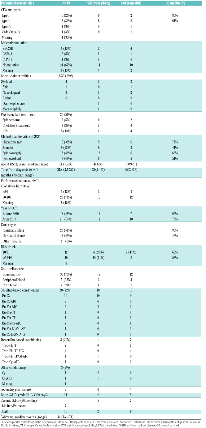
Between 1996 and 2016, 39 patients (22 males), with a median age of 5.1 years (range, 0.9-38.2) underwent SCT following a conditioning regimen that was myeloablative in all but one case. SCT was performed because of transfusion dependency alone (n=34) or associated with iron overload (n=5). Patients’ clinical characteristics are shown in Table 1. Only one patient in this cohort has been previously reported.10
Table 1.
Patients’ characteristics and transplant features.
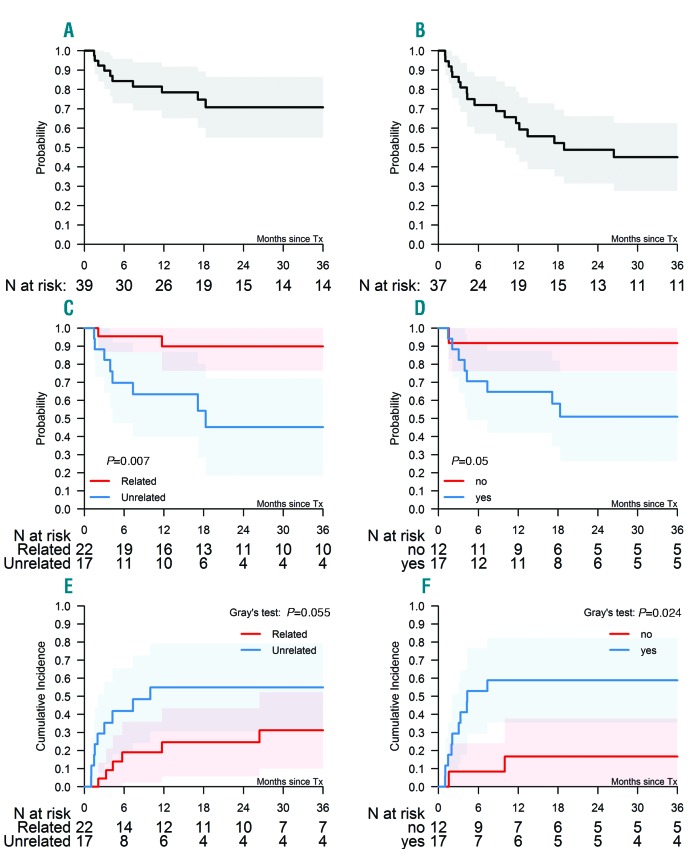
All patients were engrafted. The 3-year incidence of secondary graft failure was 12% (1-23%). Median days to neutrophil and platelet recovery were 21 (range, 10-75) and 34 (range, 16-399), respectively. Conditioning regimens and transplant features are shown in Table 1. Median follow up was 44 months (range, 13-71, with 13 transplants performed in the last two years of the study period). OS and EFS at 36 months were 71% (55-87%) and 45% (45-63%), respectively (Figure 1A and B). Ten patients died; death was due to GvHD (n=6), infection (n=3), or multi-organ failure (n=1). Patients who were transplanted from unrelated donor (UD) had an inferior outcome compared with those engrafted from matched sibling donors (MSD) [OS 51% (range, 26-76%) vs. 92% (range, 76-100%); P=0.05]. OS of patients with iron overload was significantly worse [45% (range, 18-2%) vs. 90% (range, 76-100%) P=0.007] compared with those transplanted without (Figure 1C and D). There was no difference in survival between patients transplanted before and after 2010, or between patients conditioned with or without fludarabine. The incidence of graft failure in patients transplanted with and without iron overload was 17% (range, 0-38%) and 6% (range, 0-17%), respectively (P=0.382). Graft failure was 6% (range, 0-17%) and 16% (range, 0-32%) in patients transplanted from MSD and UD, respectively (P=0.497). Of note, this probability, due to competitive causes, was significantly higher in patients transplanted with versus without iron overload (59% vs.17%; P=0.024) and from a UD versus a MSD, respectively (55% vs. 31%; P=0.05) (Figure 1E and F). A trend towards a higher incidence of grade II-IV aGvHD was also seen in patients transplanted from a UD (59% vs. 25%; P=0.06) and with iron overload (53% vs. 25%; P=0.1), as compared to those engrafted from MSD and with no iron overload, respectively. Severe (Grade III-IV) aGvHD incidence was 29%, and was significantly higher (P=0.005) in UD (53%) than in MSD transplants (10%).
Figure 1.
Outcome of patients with congenital dyserythropoietic anemias (CDA) undergoing stem cell transplantation (SCT). (A) Overall survival (OS). (B) Event-free survival. (C) OS according to donor type. (D) OS according to iron overload. (E) Graft failure incidence according to donor type. (F) Graft failure incidence according to iron overload.
To the best of our knowledge, this is the largest reported cohort of patients transplanted for CDA. Previous publications have been anecdotal experiences including a total of thirteen children, composed of CDA type I,9 type II,10–13 and type III,16 transplanted from sibling donors in all cases except for three patients who were transplanted from a UD.11,12,16 Of particular note, our study, which included 44% of patients transplanted from a UD, showed that donor type still affects the outcome since MSD SCT patients have superior OS and engraftment.
Another factor affecting the outcome of our cohort was iron overload, which is an intrinsic feature of patients with CDA due to hepcidin downregulation and ineffective erythropoiesis. We also found an association between iron overload and GvHD that might become fully significant if a larger number of patients was available. Previous reports on thalassemia17,18 showed that tissue damage secondary to iron accumulation may enhance the risk of transplant-related toxicity and predispose patients to a higher incidence of aGvHD. This outlines the importance of a pre-transplant definition of the risk the patients may incur, which generated a dramatic improvement in the outcome in the case of thalassemia patients (Pesaro risk classes).17 The lack of such a pre-SCT selection in CDA patients might explain their worse outcome compared with other transfusion-dependent, hemolytic anemias like the thalassemias; indeed, the better outcome of CDA patients transplanted without iron overload seems to support this hypothesis. Amongst the risk factors, iron overload in CDA candidates for SCT needs to be carefully evaluated and reduced with effective chelation.
Graft rejection of this CDA cohort was comparable to that seen in thalassemia patients19 and was negatively affected by donor type (worse with UD) and iron overload. This points to the need for careful evaluation of the choice of donor type in the decision-making process.
Most patients were conditioned with myeloablative regimens including busulfan/treosulfan. In the limited number of cases included in this study, no difference was noted compared to patients who, in addition, received fludarabine or cyclophosphamide. Of note, the only patients of our cohort who received a non-myeloablative conditioning regimen had a successful outcome, as did the only previously reported CDA subject,16 who was conditioned with fludarabine-cyclophosphamide and a-day course of total body irradiation. This raises the issue of the appropriateness of reduced-intensity conditioning in CDA. Unfortunately, we could not address this aspect in this study due to the lack of available data, but it might be worth investigating in future analyses.
In conclusion, despite the lower overall outcome compared to previous reports on thalassemia patients, SCT may represent an alternative therapeutic option in CDA, with outcomes appearing to be superior in patients transplanted from MSD and without iron overload. Indeed, iron overload is an important factor that needs to be controlled and eventually treated before transplant. In this respect, CDA patients might benefit from pre-SCT risk assessment, e.g. like the Pesaro criteria, which may have a favorable impact on the overall outcome. The high incidence of graft failure and aGvHD, and the lower overall survival observed in patients transplanted from UD, should be taken into consideration and discussed with parents if a familial donor is not available.
Footnotes
Information on authorship, contributions, and financial & other disclosures was provided by the authors and is available with the online version of this article at www.haematologica.org.
References
- 1.Wickramasinghe SN, Wood WG. Advances in the understanding of the congenital dyserythropoietic anemias. Br J Haematol. 2005;131(4):431–446. [DOI] [PubMed] [Google Scholar]
- 2.Gambale A, Iolascon A, Andolfo I, Russo R. Diagnosis and management of congenital dyserythropoietic anemias. Exp Rev Hematol. 2016;9(3):283–296. [DOI] [PubMed] [Google Scholar]
- 3.Iolascon A, Delaunay J, Wickramasinghe SN, et al. Natural history of congenital dyserythropoietic anemia type II. Blood. 2001:98(4):1258–1260. [DOI] [PubMed] [Google Scholar]
- 4.Tamary H, Offret H, Dgany O, et al. Congenital dyserythropoietic anemia, type I, in a Caucasian patient with retinal angioid streaks (homozygous Arg1042Trp mutation in codanin-1). Eur J Haematol. 2008;80(3):271–274. [DOI] [PubMed] [Google Scholar]
- 5.Russo R, Gambale A, Esposito MR, et al. Two founder mutations in the SEC23B gene account for the relatively high frequency of CDA II in the Italian population. Am J Hematol. 2011;86(9):727–732. [DOI] [PMC free article] [PubMed] [Google Scholar]
- 6.Lavabre-Bertrand T, Ramos J, Delfour C, et al. Long-term alpha interferon treatment is effective on anaemia and significantly reduces iron overload in congenital dyserythropoiesis type I. Eur J Haematol. 2004;73(5):380–383. [DOI] [PubMed] [Google Scholar]
- 7.Goede JS, Benz R, Fehr J, Schwarz K, Heimpel H. Congenital dyserythropoietic anemia type I with bone abnormalities, mutations of the CDAN I gene, and significant responsiveness to alpha-interferon therapy. Ann Hematol. 2006:85(9):591–595. [DOI] [PubMed] [Google Scholar]
- 8.Choudhry VP, Saraya AK, Kasturi J, Rath PK. Congenital dyserythropoietic anemias: splenectomy as a mode of therapy. Acta Haematol. 1981;66(3):195–201. [DOI] [PubMed] [Google Scholar]
- 9.Ayas M, Al-Jefri A, Baothman A, et al. Transfusion-dependent congenital dyserythropoietic anemia type I successfully treated with allogeneic stem cell transplantation. Bone Marrow Transpl. 2002:29(8):681–682. [DOI] [PubMed] [Google Scholar]
- 10.Unal S, Russo R, Gumruk F, et al. Successful hematopoietic stem cell transplantation in a patient with congenital dyserythropoietic anemia type II. Pediatr Transplant. 2014;18(4):E130–133. [DOI] [PubMed] [Google Scholar]
- 11.Braun M, Wölfl M, Wiegering V, et al. Successful treatment of an infant with CDA type II by intrauterine transfusions and postnatal stem cell transplantation. Pediatr Blood Cancer. 2014;61(4):743–745. [DOI] [PubMed] [Google Scholar]
- 12.Buchbinder D, Nugent D, Vu D, et al. Unrelated hematopoietic stem cell transplantation in a patient with congenital dyserythropoietic anemia and iron overload. Pediatr Transplant. 2012;16(3):E69–73. [DOI] [PubMed] [Google Scholar]
- 13.Modi G, Shah S, Madabhavi I, et al. Succesful allogeneic hematopoietic stem cell transplantation of a patient suffering from type II congenital dyserythropoietic anemia a rare case report from Western India. Case Rep Hematol. 2015;2015:792485. [DOI] [PMC free article] [PubMed] [Google Scholar]
- 14.Remacha AF, Badell I, Pujol-Moix N, et al. Hydrops fetalis-associated congenital dyserythropoietic anemia treated with intrauterine transfusions and bone marrow transplantation. Blood. 2002;100(1):356–358. [DOI] [PubMed] [Google Scholar]
- 15.Iolascon A, Sabato V, de Mattia D, Locatelli F. Bone marrow transplantation in a case of severe, Type II congenital dyserythropoietic anemia (CDA II). Bone Marrow Transpl. 2001;27(2):213–215. [DOI] [PubMed] [Google Scholar]
- 16.Oh A, Patel PR, Aardsma N, et al. Non-myeloablative allogeneic stem cell transplant with post-transplant cyclophosphamide cures the first adult patient with congenital dyserythropoietic anemia. Bone Marrow Transpl. 2017;52(6):905–906. [DOI] [PubMed] [Google Scholar]
- 17.Luccarelli G, Galimberti M, Polchi P, et al. Bone Marrow Transplantation in patients with thalassemia. N Eng J Med. 1990; 322(7):417–421. [DOI] [PubMed] [Google Scholar]
- 18.Luccarelli G, Clift RA, Galimberti M, et al. Bone Marrow Transplantation in adult thalassemic patients. Blood. 1999; 93(4);1164–1167. [PubMed] [Google Scholar]
- 19.Angelucci E, Matthes-Martin S, Baronciani D, et al. EBMT Inborn Error and EBMT Pediatric Working Parties Hematopoietic stem cell transplantation in thalassemia major and sickle cell disease: indications and management recommendations from an international expert panel. Haematologica. 2014;99(5):811–820. [DOI] [PMC free article] [PubMed] [Google Scholar]


