Abstract
Purpose
Bacillus causes a sight-threating infection of the posterior segment of the eye. The robust intraocular inflammatory response in this disease is likely activated via host innate receptor interactions with components of the Bacillus cell envelope. S-layer proteins (SLPs) of some Gram-positive pathogens contribute to the pathogenesis of certain infections. The potential contributions of SLPs in eye infection pathogenesis have not been considered. Here, we explored the role of a Bacillus SLP (SlpA) in endophthalmitis pathogenesis.
Methods
The phenotypes and infectivity of wild-type (WT) and S-layer deficient (ΔslpA) Bacillus thuringiensis were compared. Experimental endophthalmitis was induced in C57BL/6J mice by intravitreally injecting 100-CFU WT or ΔslpA B. thuringiensis. Infected eyes were analyzed by bacterial counts, retinal function analysis, histology, and inflammatory cell influx. SLP-induced inflammation was also analyzed in vitro. Muller cells (MIO-M1) were treated with purified SLP. Nuclear factor-κB (NF-κB) DNA binding was measured by ELISA and expression of proinflammatory mediators from Muller cells was measured by RT-qPCR.
Results
Tested phenotypes of WT and ΔslpA B. thuringiensis were similar, with the exception of absence of the S-layer in the ΔslpA mutant. Intraocular growth of WT and ΔslpA B. thuringiensis was also similar. However, eyes infected with the ΔslpA mutant had significantly reduced inflammatory cell influx, less inflammatory damage to the eyes, and significant retention of retinal function compared with WT-infected eyes. SLP was also a potent stimulator of the NF-κB pathway and induced the expression of proinflammatory mediators (IL6, TNFα, CCL2, and CXCL-1) in human retinal Muller cells.
Conclusions
Taken together, our results suggest that SlpA contributes to the pathogenesis of Bacillus endophthalmitis, potentially by triggering innate inflammatory pathways in the retina.
Keywords: endophthalmitis, bacteria, Bacillus, inflammation, retina, blindness
Severe inflammation and rapid vision loss are destructive consequences of Bacillus endophthalmitis.1–3 This disease typically results from a traumatic ocular injury with a foreign body contaminated with this organism. Bacillus endophthalmitis is particularly devastating, as greater than 70% of patients were reported to have lost significant vision, and 50% of those cases resulted in enucleation of the infected eye.4–10 Treatment strategies for traumatic ocular injuries include the use of antibiotics, anti-inflammatory drugs, and in severe cases, vitrectomy surgery.11–17 However, the potentially blinding outcome for Bacillus endophthalmitis has been difficult to prevent, emphasizing the importance of identifying unique virulence factors of Bacillus that might be targeted in developing better treatment strategies for this disease.
B. cereus and B. thuringiensis are two of the most virulent organisms reported to cause bacterial endophthalmitis. These members of the Bacillus cereus sensu lato group are Gram-positive, facultative aerobic, spore-forming rods, and are widely distributed in the environment.18,19 Other than the presence of crystal toxins in B. thuringiensis, the genomes and phenotypes of B. cereus and B. thuringiensis are highly similar and, on a genetic basis together with B. anthracis, are considered a single species.20–22 These organisms express a comparable cohort of virulence factors that have the potential to contribute to disease. During infection, Bacillus spp. replicates and expresses harmful toxins and enzymes in the vitreous.23–27 Bacillus spp. also possesses a quorum sensing system (PlcR) that regulates the coordinated synthesis of almost all extracellular virulence factors and is important for intraocular virulence.27,28
We reported that an absence of individual Bacillus toxins, such as hemolysin BL, phosphatidylinositol-specific phospholipase C (PI-PLC), or phosphatidylcholine-specific phospholipase C (PC-PLC), did not completely eliminate endophthalmitis pathology.24,25 We also reported delayed evolution of Bacillus endophthalmitis in the absence of the PlcR quorum sensing system.23,27,29,30 In these cases, complete elimination of disease pathology did not occur, suggesting the contribution of other nontoxin bacterial products or perhaps Bacillus cell wall components in this disease.
During experimental endophthalmitis, Bacillus induces a rapid inflammatory response, which is more aggressive than that of other common pathogens associated with this disease.2,3,31,32 We reported that these inflammatory responses were mediated, in part, through innate receptors, such as Toll-like receptor 2 (TLR2), TLR4, and their adaptors, myeloid differentiation primary response gene-88 (MyD88), and Toll/interleukin-1 receptor (TIR) domain containing adaptor-inducing interferon-β (TRIF).33,34 Bacillus endophthalmitis in mice deficient in TLR2, TLR4, MyD88, or TRIF was significantly less severe than infections in the eyes of WT mice. We also reported that nonviable B. cereus cell walls induced a greater degree of intraocular inflammation than cell walls of other Gram-positive pathogens associated with endophthalmitis,2 suggesting that this difference in inflammation potential may be attributed to variations in cell envelope constituents.
The Bacillus cell envelope varies structurally from other Gram-positive ocular pathogens, such as staphylococci or streptococci.35–38 The envelopes of Bacillus and other Gram-positive organisms have an inner membrane, a thick layer of peptidoglycan (PGN), teichoic acids (TA), and lipoproteins (Lpp), and proteinaceous adhesive appendages called pili.38–42 Unlike other Gram-positive ocular pathogens, Bacillus has peritrichous flagella. Bacillus species, including some strains of the Bacillus cereus sensu lato group, have a paracrystalline surface layer composed of S-layer proteins (SLPs).43–45 During infection, this pathogen migrates from the posterior to the anterior segment.2,23 Nonmotile Bacillus were less virulent and a deficiency in swarming movement prevented the pathogen from migrating to the anterior segment, resulting in less severe disease.23,46,47 Flagella aid this migration throughout the eye, but are weak activators of TLR5.23,47 Recently, we reported a potential protective role for Bacillus pili in the clearance of the pathogen during the early stages of endophthalmitis.48 The inflammatory capacities of common Gram-positive envelope components (Lpp, PGN, and TA) are well documented,49–52 but the role of the SLPs in the context of Bacillus endophthalmitis has not been addressed.
SLPs are cell surface proteins present in Gram-positive and -negative bacteria, as well as in Archaea.53 A spontaneous self-assembly of one or more SLPs forms a regularly spaced array on the surface of the bacterial cell. This array links with the underlying cell surface through noncovalent forces.54,55 Firmicutes SLPs possess two domains, a conserved anchoring domain composed of three repetitions of approximately 50 residues followed by the crystallization domain. Sequence similarities of crystallization domains from different species are low because there are no universal signature sequences.56 Archaea, such as Thermoproteus, Methanocorpusculum, and Sulfolobus, possess SLPs as a sole constituent of their cell wall, suggesting its role in determination and maintenance of cell shape and function as a molecular sieve.57–59 SLPs also contribute to microbial pathogenesis through several mechanisms, such as promoting bacterial adherence to host cells and extracellular matrix components, and biofilm formation.60–64 Bacterial SLPs also provide protection from complement-mediated killing and phagocytosis.63,65 SLPs protect micro-organisms from harmful environmental changes, such as abrupt changes in pH, radiation, and mechanical and osmotic stresses,66–70 and safeguard the pathogen from bacteriolytic enzymes or antimicrobial peptides, bacteriophages, and other bacterial predators.71,72
Because bacterial SLPs are major cell wall proteins of certain microorganisms and contribute to their pathogenesis, and because the Bacillus cell wall is highly inflammogenic, we hypothesized that an SLP of B. thuringiensis (SlpA) contributes to the pathogenesis of endophthalmitis. Using a well-characterized experimental mouse model of endophthalmitis, we demonstrated that the absence of SlpA impacted B. thuringiensis virulence, significantly blunting the severity of experimental endophthalmitis caused by this pathogen. Further exploration of a role for SlpA in B. thuringiensis endophthalmitis may identify a new virulence determinant for this pathogen, potentially paving the way for SLPs as novel therapeutic targets for this blinding disease.
Materials and Methods
Ethics Statement
The in vivo experiments described in these studies involved the use of mice. All animal experiments were performed following the recommendations of the Guide for the Care and Use of Laboratory Animals, the ARVO Statement for the Use of Animals in Ophthalmic and Vision Research, and the University of Oklahoma Health Sciences Center Institutional Animal Care and Use Committee (approved protocols 16-086, 18-079).
Bacterial Strains
B. thuringiensis subsp. galleriae NRRL 4045 (WT) or its isogenic S-layer (SL)-deficient mutant (ΔslpA)73 were used to initiate experimental endophthalmitis in mice, as previously described.1,30,31,34,47,48,74,75 Phenotypes of WT and ΔslpA B. thuringiensis were compared by transmission electron microscopy (TEM), growth curves, hemolytic analysis, gel electrophoresis, cytotoxicity, and motility analysis, as described below.
Transmission Electron Microscopy
WT and ΔslpA B. thuringiensis were compared by thin-section TEM. Overnight (18 hour) cultures of WT and ΔslpA B. thuringiensis were centrifuged and resuspended in PBS, fixed with 2% paraformaldehyde (EM grade), 2.5% glutaraldehyde (EM grade), in 0.1 M sodium cacodylate buffer (pH 7.2) overnight at 4°C. Samples were then post-fixed for 90 minutes in 1% osmium tetroxide (OsO4) in 0.1 M sodium cacodylate, and rinsed three times for 5 minutes each in the same buffer. The samples were then dehydrated in a graded ethanol series (50%–100%) for 15 minutes each. Bacteria were then dehydrated twice for 15 minutes each in 100% propylene oxide. Following dehydration, the samples were infiltrated in a graded epon/araldite (EMS) resin/propylene oxide series (1:3, 1:1, 3:1) for 60 and 120 minutes, and overnight, respectively. The following day, samples were further infiltrated with pure resin for 45 and 90 minutes, and overnight. The samples were then infiltrated with resin plus benzyl dimethylamine (BDMA, accelerator) for 4 hours and then embedded in resin plus BDMA and polymerized at 60°C for 48 hours. Ultrathin sections were stained with Sato's lead and saturated uranyl acetate in 50% methanol before viewing on a Hitachi H7600 TEM (Hitachi, Tokyo, Japan). All procedures were performed at the Oklahoma Medical Research Foundation Imaging Core, Oklahoma City, Oklahoma, United States.
Bacterial Growth Curves
WT and ΔslpA B. thuringiensis strains were cultured for 18 hours at 37°C with aeration in brain heart infusion medium (BHI; VWR, Radnor, PA, USA). Strains were then subcultured in fresh BHI to an approximate concentration of 103 CFU/mL and incubated for 18 hours as mentioned above. Every 2 hours, 20-μL aliquots were diluted 10-fold in PBS, plated onto BHI agar plates, and quantified.25,27,48
Hemolytic Analysis
WT and ΔslpA B. thuringiensis were cultured for 18 hours as mentioned above, then centrifuged at 3000g for 15 minutes. Supernatants were then filter sterilized (0.22 μm; Millex-GP, Merck Millipore Ltd., Cork, Ireland) and serially diluted 1:2 in PBS (pH 7.4). Diluted supernatants were incubated 1:1 with 4% (vol/vol) sheep erythrocytes (Rockland Immunochemicals, Pottstown, PA, USA) in a 96-well round bottom plate for 30 minutes at 37°C. The plate was centrifuged at 1892g for 10 minutes to remove the unlysed erythrocytes.
The supernatants were carefully transferred into a 96-well, flat-bottom plate and hemoglobin release was measured spectrophotometrically at 490 nm by using a FLUOstar Omega microplate spectrophotometer (BMG Labtech, Cary, NC, USA). Values are expressed as the percent hemolysis relative to a 100% lysis control in which 5% rabbit erythrocytes were lysed in double-distilled H2O.23,27,48,76,77 Values represent the mean results ± SEM of two independent experiments. These strains were also incubated on tryptic soy agar (TSA) supplemented with 5% sheep blood (Hardy Diagnostics, Santa Maria, CA, USA) for 18 hours at 37°C, and colony morphology and hemolytic phenotypes were compared.
Motility Analysis
WT and ΔslpA B. thuringiensis were inoculated at the center of motility agar (0.75% agar in BHI) plates using a sterile toothpick and incubated for 24 hours at 37°C. Distances of each colony from the center were measured in millimeters.23,27,46
Muller Cell Cytotoxicity
Immortalized human Muller cells (MIO-M1; a kind gift from Dr. Astrid Limb, UCL Institute of Ophthalmology, London) were maintained in DMEM/F-12 (GIBCO, Grand Island, NY, USA) supplemented with 10% fetal bovine serum (FBS; Sigma-Aldrich Corp., St. Louis, MO, USA) and 1% Pen Strep (GIBCO) in a humidified 5% CO2 incubator at 37°C.78,79
The cytotoxicity of WT and ΔslpA B. thuringiensis on human retinal Muller cells was measured using a Pierce Lactate Dehydrogenase (LDH) Cytotoxicity Assay Kit (ThermoFisher Scientific, Waltham, MA, USA) according to the manufacturer's instructions. Briefly, 20,000 cells/100 μL were seeded in triplicate wells for overnight incubation. WT and ΔslpA B. thuringiensis were cultured for 18 hours and centrifuged at 3000g for 15 minutes. Supernatants were then filter sterilized (0.22 μm; Millex-GP, Merck Millipore Ltd.) and added into respective wells. Positive and negative controls were 10 μL lysis buffer (10×) and 10 μL of sterile ultrapure water, respectively. Cytotoxicity (%) was calculated by subtracting the LDH activity of negative control from sample LDH activity, divided by the total LDH activity (positive control − negative control), and multiplied by 100.29,79
Purification of Bacillus S-layer Protein
WT and ΔslpA B. thuringiensis were grown for 18 hours at 37°C in BHI, harvested by centrifugation (at 3000g for 15 minutes at 4°C), and washed twice with chilled HyPure cell culture grade water (GE Healthcare Life Science, Logan, UT, USA). Pellets were then resuspended in one-tenth of the initial volume of 3 M guanidine hydrochloride (GHCL; pH 2.5; Sigma-Aldrich Corp.) and incubated for 1 hour at 37°C. The extracted SLP was separated from the cell pellets by centrifugation (18,000g for 15 minutes, 4°C). The supernatants were carefully transferred and dialyzed (Pur-A-Lyzer 50-kDa dialysis kit; Sigma-Aldrich Corp.) for 24 hours at 4°C against 2 L of tris/HCL (pH 8.0; Research Products International Corporation, Mt. Prospect, IL, USA) with four exchanges of dialysis buffer to remove the residual GHCL. Protein concentrations were measured with a bicinchoninic acid kit (Sigma-Aldrich Corp.) according to the manufacturer's instructions. Endotoxin level was measured using Pierce LAL chromogenic endotoxin quantitation kit (ThermoFisher Scientific) according to the manufacturer's instructions. Samples were then resuspended in Laemmli buffer, boiled for 10 minutes, loaded onto a 12% PAGE. The gels were fixed and stained with Coomassie brilliant blue.44,73,80
Isolation of Bacillus Peptidoglycan (PGN)
PGN was prepared by the method described in Langer et al.81 BHI agar plates were inoculated with 0.1 mL of an overnight culture of WT or ΔslpA B. thuringiensis and incubated at 37°C overnight. Lawns were scraped from the plates into 50-mL cold BHI and harvested by centrifugation (5000g, 37°C, 10 minutes). Pellets were washed in endotoxin free water (GE Healthcare Life Science), resuspended in 5 mL 8% SDS (Sigma-Aldrich Corp.), and boiled for 30 minutes. Lysed cells were then centrifuged at 25,000g for 20 minutes and washed 3× with endotoxin-free water. Washed pellets were resuspended with 40 U of DNase I and 7 U of RNase A (ThermoFisher Scientific) and incubated for 15 minutes at room temperature. To remove the nucleases, samples were resuspended in 4% SDS (Sigma-Aldrich Corp.), boiled for 15 minutes, and washed 3× with endotoxin free water, 1× with 2M NaCl, and 6× with endotoxin-free water. Pellets were then resuspended in endotoxin-free water, boiled for 5 minutes, and centrifuged as above. PGN was then dried, weighed, and resuspended in endotoxin-free water to a concentration of 40 mg/mL. Sterility was tested by spread plating on BHI agar. The level of endotoxin was measured by Pierce LAL chromogenic endotoxin quantitation kit (ThermoFisher Scientific) according to the manufacturer's instructions.
TLR2 Reporter Assay for PGN
HEK-Blue reporter cell lines are designed to measure the stimulation of human TLRs by monitoring the activation of Nuclear factor-κB (NF-κB; Invivogen, San Diego, CA, USA). These cells are transfected with a reporter gene encoding secreted embryonic alkaline phosphatase (SEAP), whose transcriptional activation is under the control of a TLR-inducible gene promoter. HEK-293 cells, which do not normally express TLRs, were co-transfected with an expression plasmid encoding specific TLRs. In this study, we used HEK-Blue hTLR2 (named here hTLR2) for the recognition of TLR2 agonists. hTLR2 cells were routinely cultured (up to 20 passages) in Dulbecco's modified Eagle's medium (DMEM) containing GlutaMAX-l (GIBCO), supplemented with 10% (vol/vol) FBS and HEK-Blue Selection antibiotics (Invivogen) in a humidified 5% CO2 incubator at 37°C.
PGN (0.1, 1, 10, and 100 μg/mL) from WT and ΔslpA B. thuringiensis, 0.25 ng/mL Pam3Csk4 (TLR2 agonist; positive control for hTLR2), and endotoxin-free water (negative control for hTLR2; GE Healthcare Life Science) were added to wells of a 96-well tissue culture plate. Growth medium from 50% to 80% confluent TLR2 reporter cells was discarded and gently washed with warm PBS (pH 7.4; GIBCO). After detaching the cells with PBS (pH 7.4) a new cell suspension of each cell type was prepared (∼50,000/180 μL TLR2) in HEK-Blue detection medium (Invivogen). Each cell (180 μL/well) suspension was immediately added into each well and the plates were incubated for 14 to 16 hours at 37°C in 5% CO2. Production of SEAP from hTLTR2 cells following PGN exposure was measured using a spectrophotometer at 620 to 655 nm.82 TLR2 activation was presented as percent of TLR2 activation relative to the positive-control Pam3Csk4.
Mice and Intraocular Infection
All in vivo experiments were performed with C57BL/6J mice purchased from commercially available colonies (Stock No. 000664; Jackson Labs, Bar Harbor, ME, USA). Mice were kept on a 12 hour on/12 hour off light cycle, acclimated to housing conditions for at least 2 weeks to equilibrate their microbiota, and maintained under biosafety level 2 conditions during the experiments. All animals were 8- to 10-weeks old at the time of the experiments. Mice were sedated using a combination of ketamine (85 mg/kg body weight; Ketathesia, Henry Schein Animal Health, Dublin, OH, USA) and xylazine (14 mg/kg body weight; AnaSed; Akorn Inc., Decatur, IL, USA). Experimental endophthalmitis was induced by intravitreally injecting approximately 100 CFU WT or ΔslpA B. thuringiensis/0.5 μL BHI into one eye using a sterile glass capillary needle, as previously described.1,33,34,47,48,74–77,83 The uninjected left eye served as the uninfected control. Electroretinography (ERG) was performed prior to euthanasia by CO2 inhalation and harvesting for subsequent analysis. As described below, quantitation of intraocular bacterial growth, retinal function, polymorphonuclear leukocyte (PMN) infiltration (myeloperoxidase [MPO] activity), and analysis of ocular architecture (histology) were conducted at different time points postinfection.
Intraocular Bacterial Growth
Intraocular bacteria were quantified as previously described.1,33,34,47,48,74–77,83 Briefly, infected eyes were harvested from euthanized mice at 0, 2, 6, 8, 10, and 12 hours postinfection. Infected eyes were then homogenized in 400 μL PBS with sterile 1-mm glass beads (BioSpec Products, Inc., Bartlesville, OK, USA). Eye homogenates were then diluted 10-fold in PBS, plated onto BHI agar plates and quantified by track dilution.
Retinal Function Analysis by Electroretinography
ERG was used to quantify retinal function in eyes infected with WT or ΔslpA B. thuringiensis, as previously described.1,33,34,47,48,74–77,83 Scotopic ERGs were performed at 6, 8, 10, and 12 hours postinfection using Espion E2 software (Diagnosys LLC, Lowell, MA, USA). After infection, mice were dark adapted for at least 6 hours. Infected mice were anesthetized as previously described and pupils were dilated with topical phenylephrine (Akorn, Inc.). Two gold wire electrodes were placed on each cornea and reference electrodes were place on the forehead and on the tail. Eyes were then stimulated by five flashes of white light (1200 cd s/m2) and retinal responses were recorded as A-wave and B-wave amplitudes for infected eyes and compared with the uninfected eyes of the same animal.
Histology
Infected eyes were harvested from euthanized mice at 6, 8, 10, and 12 hours postinfection. Harvested eyes were then incubated in low-alcoholic fixative for 30 minutes and then transferred to 70% ethanol. Eyes were embedded in paraffin, sectioned, and stained with hematoxylin and eosin (H&E) or tissue Gram stain.1,33,34,47,48,74–77,83
Inflammatory Cell Influx
Ocular PMN infiltration was estimated by quantifying MPO using a sandwich ELISA (Hycult Biotech, Plymouth Meeting, PA, USA), as previously described.1,33,34,47,74 At 6, 8, 10, and 12 hours postinfection, eyes were harvested, placed into PBS-containing proteinase inhibitor cocktail (Roche Diagnostics, Indianapolis, IN, USA) and homogenized using sterile glass beads (BioSpec Products, Inc.). Eye homogenates were tested with the MPO ELISA. Uninfected eye homogenates served as negative controls. The lower limit of detection for this assay was 2 ng/mL.
NF-κB-DNA Binding Assay
NF-κB-DNA binding following SlpA stimulation of MIO-M1 cells was measured by an NF-κB transcription factor assay (Cayman Chemical Co., Ann Arbor, MI, USA), a nonradioactive, sensitive method for detecting specific NF-κB-DNA binding in nuclear extracts.84 Briefly, 3 × 10⁶ human Muller cells/well were seeded in a 6-well plate and treated with 10 μg/mL SLP for 15 minutes to 2 hours. Untreated wells were negative controls. Nuclear extracts were prepared at the indicated time points using a chemical nuclear extraction kit (Cayman Chemical Co.) according to the manufacturer's instructions. Briefly, cell pellets were washed with PBS/phosphatase inhibitor solution, swelled with hypotonic buffer, lysed with NP-40, centrifuged at 14,000g, and resuspended in complete nuclear extraction buffer. After 30 minutes of incubation, suspensions were centrifuged at 14,000g and supernatants containing nuclear proteins were collected. To measure NF-κB-DNA binding, 50 μg of nuclear proteins was added to the wells containing transcription factor buffer and incubated overnight at 4°C. Controls included blank wells (negative) and TNFα-stimulated HeLa cell nuclear extract (positive). NF-κB binding was detected with NF-κB primary antibody and goat anti-rabbit horseradish peroxidase conjugate secondary antibody. Absorbance was measured at 450 nm. Changes in NF-κB-DNA binding for each time point were quantified by calculating the percentage of binding relative to the untreated group.
RNA Isolation and Quantitative PCR
Expression of proinflammatory cytokines (IL-6, TNF-α) and chemokines (CCL2, CXCL-1) from MIO-M1 cells treated with SLP were measured by real-time quantitative PCR. MIO-M1 cells were seeded (2 × 10⁶ cells/well) into a 6-well plate and treated with 10 μg/mL SLP from WT B. thuringiensis, 10 μg/mL SLP extract control (SLP fraction from ΔslpA B. thuringiensis), or PBS for 10 hours. Untreated cells were the negative control. Total RNA was isolated using a QIAGEN RNeasy Mini Kit (QIAGEN, Hilden, Germany). DNA was removed by using a TURBO DNA-free Kit (Invitrogen, Carlsbad, CA, USA). Using an RNA Clean & Concentrator-5 Kit (Zymo Research, Irvine, CA, USA), 50 μL RNA was purified. The concentration and purity were confirmed on a Nanodrop. All procedures were performed following the manufacturer's instructions. Real-time quantitative PCR was performed with the Applied Biosystems 7500, using an iTaq Universal SYBR Green One-Step Kit (Bio-Rad, Hercules, CA, USA) and specific primers (Table) following the manufacturer's instructions.75 PCR amplification was performed in triplicate and water was used to replace RNA in each run as negative control. Relative gene expression was determined using ΔCT method using GAPDH as a reference housekeeping gene.75
Table.
Primer Sequence Used in Quantitative PCR
|
Gene |
Species |
Sequence (5′–3′) |
Temperature (°C) |
| GAPDH | Human | ACA TCG CTC AGA CAC CAT G | 55 |
| TGT AGT TGA GGT CAA TGA AGG G | |||
| TNFα | Human | TGC ACT TTG GAG TGA TCG G | 55 |
| TCA GCT TGA GGG TTT GCT AC | |||
| IL-6 | Human | CCT TCC CTG CCC CAG TA | 55 |
| ATT CGT TCT GAA GAG GTG AGT G | |||
| CXCL-1 | Human | CTC TTC TTC CCT AGG AGC GT | 55 |
| TGT TCT TGG GGT GAA TTC CC | |||
| CCL2 (MCP-1) | Human | AGT GTC CCA AAG AAG CTG TG | 55 |
| AAT CCT GAA CCC ACT TCT GC |
Statistics
GraphPad Prism 7 was used for the statistical analysis (GraphPad Software, Inc., La Jolla, CA, USA). Mann-Whitney U test was used for statistical comparisons48,74,77 unless otherwise specified. P values of < 0.05 were considered significant.
Results
Absence of S-layer Does Not Alter Bacterial Growth or Phenotypes
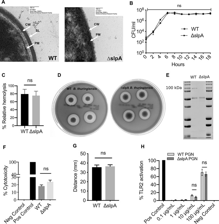
The phenotypes of B. thuringiensis subsp. galleriae NRRL 4045 (WT) and its isogenic SlpA-deficient mutant (ΔslpA) were compared. Thin-section TEM showed the absence of SL in ΔslpA B. thuringiensis and its presence in the WT strain (Fig. 1A). In vitro growth and other virulence phenotypes were also compared. Overnight cultures of each strain were subcultured into fresh BHI and bacteria were quantified every 2 hours for 18 hours. Figure 1B demonstrates that WT and ΔslpA B. thuringiensis grew to similar concentrations in BHI at all time points, and both strains reached stationary phase growth at 6 hours. In Figure 1C, hemolytic titers of 18-hour supernatants of WT and ΔslpA B. thuringiensis were compared by hemolytic assay. Supernatants of these strains were similarly hemolytic. This finding was confirmed by the presence of similarly sized hemolytic zones on TSA supplemented with 5% sheep blood. Figure 1D depicts the characteristic double zones of hemolysis surrounding each colony of WT and ΔslpA B. thuringiensis after 18-hour incubation at 37°C. SLPs from both strains were purified, loaded onto a 12% PAGE. The Coomassie stained gel in Figure 1E depicts the presence of a 91.4-kDa band indicating the existence of SlpA in the WT strain. As expected, this band was absent in the ΔslpA strain, confirming our TEM data. We also noted the presence of a 49-kDa flagellar band in both WT and ΔslpA strains, similar to what has been previously described.44,80 We measured the cytotoxicity of WT and ΔslpA B. thuringiensis supernatants on human retinal Muller cells, and demonstrated that these strains were similarly cytotoxic (Fig. 1F). We also compared the motility of WT and ΔslpA B. thuringiensis on motility agar (0.75% agar in BHI). Figure 1G demonstrates that the distances of the colonies from the center to the edge were similar. To determine whether the absence of SlpA altered the Bacillus cell wall composition, we purified PGN from WT and ΔslpA B. thuringiensis. PGN yields from WT and ΔslpA B. thuringiensis were approximately 7.4% and 8.5%, respectively. Because PGN is a universal TLR2 agonist, we measured TLR2 activation by equal concentrations WT and ΔslpA PGN in the hTLR2 reporter assay. Figure 1H demonstrates that PGN from both WT and ΔslpA activated TLR2 to a similar degree. Together, these results suggested that the absence of SlpA did not affect the in vitro bacterial growth, hemolytic phenotypes, cytotoxic potential of secreted bacterial products, motility, or cell wall composition. These similarities suggested that in vivo infections with WT and ΔslpA B. thuringiensis might be similar.
Figure 1.
Absence of SLP does not alter bacterial growth and phenotypes. (A) Electron micrograph of thin-sections of WT and ΔslpA B. thuringiensis. CW, Cell wall; PM, plasma membrane. Magnification, ×25,000 (WT) and ×20,000 (ΔslpA). (B) In vitro growth curve of WT B. thuringiensis and its isogenic SlpA-deficient mutant (ΔslpA) in BHI broth. CFU of both WT and ΔslpA B. thuringiensis were similar at each time point (P > 0.05). Values represent the mean ± SEM for N = 3 samples per time point. (C) WT and ΔslpA B. thuringiensis were compared for their hemolytic activities (2-way ANOVA, P > 0.05). (D) WT and ΔslpA B. thuringiensis exhibited colonies with characteristic double zones of hemolysis on TSA containing 5% sheep blood. (E) Samples of the final purification step from WT and ΔslpA B. thuringiensis were loaded onto 12% polyacrylamide gel and subjected to PAGE. The 91.4-kDa band indicates the presence of SlpA in the WT B. thuringiensis, which is absent in the isogenic mutant. A 49.1-kDa flagellar protein band in both WT and ΔslpA B. thuringiensis serves as a standard. (F) Cytotoxicity of filter sterilized overnight supernatants from WT and ΔslpA B. thuringiensis in human retinal Muller cells. No significant difference was observed in the cytotoxicity of these strains (P = 0.1297). Data represents the mean ± SEM of percent of cytotoxicity for N > 5 samples. (G) The motility of WT and ΔslpA B. thuringiensis was compared. Both strains exhibited similar levels of motility (P = 0.9429). (H) Cell-wall PGN from WT and ΔslpA B. thuringiensis activated TLR2 (P = 0.8857). Data represents the mean ± SEM for N ≥ 4 samples).
Absence of S-layer Does Not Significantly Alter Intraocular Bacterial Growth
The intraocular growth of WT and ΔslpA B. thuringiensis was analyzed in a well-established model of experimental endophthalmitis in C57BL/6J mice (Fig. 2). Eyes were infected with 108 ± 4 CFU/eye of WT or 96 ± 4 CFU/eye of ΔslpA B. thuringiensis (P = 0.0952). At 2, 6, 8, 10, and 12 hours, infected eyes were harvested, homogenized, and plated onto BHI agar. Intraocular bacterial loads in eyes infected with WT or ΔslpA B. thuringiensis were similar, except at 8 hours postinfection. These results also suggested that intraocular infections with WT and ΔslpA B. thuringiensis may be similar.
Figure 2.
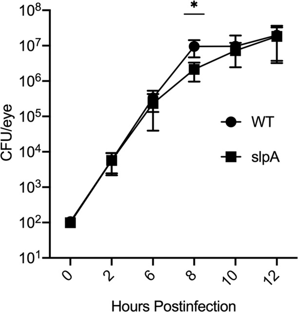
Bacillus SLP does not influence intraocular growth in endophthalmitis. C57BL/6J mouse eyes were injected with 100 CFU WT B. thuringiensis or its isogenic SLP-deficient mutant (ΔslpA). At the indicated times postinfection, eyes were harvested and CFU quantified for bacterial intraocular growth. Data represents the mean ± SEM of log10 CFU/eye of N ≥ 4 eyes per time point for at least two separate experiments. ns: P > 0.05 at 0, 2, 6, 10, and 12 hours postinfection. *P = 0.0087 at 8 hours postinfection.
Retinal Function is Retained in the Absence of S-layer Protein
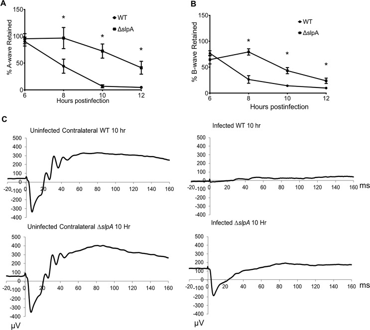
Analysis of retinal function and the representative waveforms of eyes infected with WT or ΔslpA B. thuringiensis is depicted in Figure 3. Infected mice were dark-adapted for at least 6 hours and ERG was performed at 6, 8, 10, and 12 hours postinfection. Retention of retinal function, as demonstrated by A- and B-wave amplitudes in WT and ΔslpA-infected eyes, was similar at 6 hours postinfection (Fig. 3A). A-wave function, which represents the function of retinal photoreceptor cells, rapidly decreased in eyes infected with WT B. thuringiensis from 6 to 12 hours postinfection, to a retained response of approximately 5% (Fig. 3A). This function in ΔslpA-infected eyes was also decreased over time, but was retained to a significantly greater degree compared with that of WT B. thuringiensis–infected eyes. B-wave function, which represents the function of bipolar cells, Muller cells, and second order neurons, also rapidly decreased in the WT B. thuringiensis–infected eyes from 6 to 12 hours postinfection, to a retained response of approximately 10% (Fig. 3B). This response over the same period of time was also decreased in eyes infected with ΔslpA B. thuringiensis, but function was retained to a significantly greater degree compared with that of WT-infected eyes. The retained responses of A- and B-waves in ΔslpA-infected eyes at 12 hours postinfection were approximately 41% and 24%, respectively. Representative waveforms demonstrating the stark differences in A- and B-wave amplitudes of eyes infected with WT or ΔslpA B. thuringiensis and uninfected controls at 10 hours postinfection are shown in Figure 3C. Together, these results demonstrated that eyes infected with ΔslpA B. thuringiensis retained greater retinal function compared with eyes infected with WT B. thuringiensis, suggesting that the presence of SlpA in B. thuringiensis influenced the loss of retinal function during experimental endophthalmitis.
Figure 3.
Absence of SLP impacts retinal function loss in Bacillus endophthalmitis. C57BL/6J mouse eyes were injected with 100 CFU WT or ΔslpA B. thuringiensis and retinal function was assessed by ERG. (A) Retained A-wave function was significantly greater in eyes infected with ΔslpA B. thuringiensis at 8, 10, and 12 hours postinfection (*P < 0.04). (B) B-wave function was also significantly greater in eyes infected with ΔslpA B. thuringiensis at 8, 10, and 12 hours postinfection (*P < 0.04). (C) Representative waveforms from eyes infected with WT or ΔslpA B. thuringiensis at 10 hours postinfection. In these mice, one eye was infected and the contralateral eye served as the uninfected control. Values represent the mean ± SEM of percentage amplitude retained per time point for at least two separate experiments. Data are representative of N ≥ 8 eyes per time point.
Ocular Architecture is Preserved in the Absence of S-layer Protein
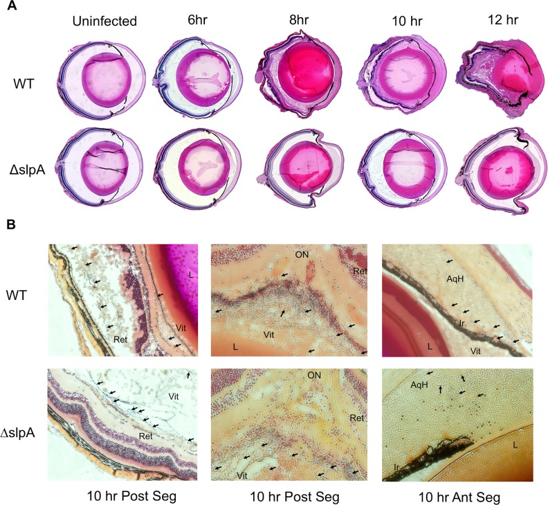
A histologic comparison of WT or ΔslpA B. thuringiensis-infected C57BL/6J mouse eyes is depicted in Figure 4. At 6, 8, 10, and 12 hours postinfection, eyes were harvested, fixed, sectioned, and stained with H&E. The ocular architecture of uninfected control eyes in both groups was identical, with no signs of inflammation (Fig. 4A). At 6 hours postinfection, the anterior and posterior segments of both WT and ΔslpA-infected eyes were similar. At this time point, corneas and posterior segments of infected eyes were not significantly inflamed, retinas were intact, and retinal layers were distinguishable in both groups. At 8 hours postinfection in eyes infected with WT B. thuringiensis, significant accumulation of fibrin and infiltrating cells in the posterior segment was observed. Retinas were also partially detached and the anterior chambers were occluded with fibrin. In contrast, ocular structures were well-preserved in ΔslpA-infected eyes. These eyes exhibited minimal fibrin deposition and inflammation, intact retinas, and distinguishable retinal layers (Fig. 4A). At 10 hours postinfection, significant fibrin deposition throughout the vitreous and inflammatory cell infiltration were observed in eyes infected with WT B. thuringiensis. Corneas in these eyes were highly edematous, and complete retinal detachments and indistinguishable retinal architecture were observed. In stark contrast, retinas were well-preserved and retinal layers were distinguishable in ΔslpA-infected eyes at 10 hours postinfection (Fig. 4B). In both infection groups at 10 hours postinfection, bacteria were localized (black arrow) along the posterior capsule, in the midvitreous, and near the inner limiting membrane (ILM) (Fig. 4B). Bacteria were located inside the retinal layers in WT-infected eyes, whereas in ΔslpA-infected eyes, no bacteria were found inside the retinal layers and the retinas were intact (Fig. 4B, first column). Bacteria were also located in the anterior chamber and near the iris and ciliary body in eyes infected with either strain (Fig. 4B, third column). At 12 hours postinfection, inflammation pervaded all ocular structures and retinal architecture was completely lost in eyes infected with WT B. thuringiensis (Fig. 4A). In eyes infected with ΔslpA, retinal layers and architecture remained well-preserved, and these eyes looked similar to that of 6-hour WT-infected eyes (Fig. 4A). These results demonstrated that, in contrast to what was observed in the presence of SlpA, ocular architecture was preserved during Bacillus infection, further supporting a contribution of SlpA to the pathogenesis of this disease.
Figure 4.
Absence of SLP preserves ocular architecture in Bacillus endophthalmitis. (A) C57BL/6J mouse eyes were infected with 100 CFU of WT or ΔslpA B. thuringiensis. Infected eyes were harvested at 6, 8, 10, and 12 hours postinfection and processed for H&E and Gram staining. Magnification, ×10. (B) Bacteria were observed in the midvitreous of eyes infected with each strain at 10 hours postinfection. At this time point, WT and ΔslpA mutant were each observed in the aqueous humor of the anterior segment (Ant Seg), as well as in the midvitreous and near the retina of the posterior segment (Post Seg). Sections are representative of three eyes per time point. WT and ΔslpA B. thuringiensis are denoted by black arrows. Magnification, ×200. Ret, retina; Vit, vitreous; AqH, aqueous humor; Ir, iris; L, lens.
Absence of S-layer Protein Reduces Intraocular Inflammation
We examined the levels of inflammatory cell influx (Fig. 5) in eyes infected with WT or ΔslpA B. thuringiensis. The primary infiltrating inflammatory cells in Bacillus endophthalmitis are PMN.1 Infected eyes were harvested at 0, 4, 8, and 12 hours postinfection and PMN influx was estimated by quantifying MPO85 in eye homogenates by ELISA. MPO concentrations were significantly greater at 4, 8, and 12 hours postinfection in WT-infected eyes compared with that of ΔslpA-infected eyes. These results demonstrated that an absence of SlpA in these infections resulted in reduced MPO enzyme levels, indicating a subdued PMN response in these eyes. These results suggest a role for SlpA in the recruitment of PMN into the posterior segment of the eye during infection. Overall, this result corroborates our findings of less retinal function loss and less ocular pathology in eyes infected with SlpA-deficient B. thuringiensis, strongly suggesting that SlpA is important to the virulence of this organism during endophthalmitis.
Figure 5.
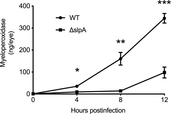
Absence of SLP blunts the intraocular infiltration of PMN during Bacillus endophthalmitis. C57BL/6J mouse eyes were injected with 100 CFU WT or ΔslpA B. thuringiensis. At 4, 8, and 12 hours postinfection, infected eyes were harvested and infiltration of PMN was assessed by quantifying MPO in whole eyes by sandwich ELISA. MPO levels were significantly greater in eyes infected with WT strains at 4, 8, and 10 hours postinfection compared with the eyes infected with ΔslpA B. thuringiensis. Values represent the mean ± SEM of MPO (ng/eye) of N ≥ 4 per time point for at least two separate experiments. *P = 0.0095; **P = 0.0022; ***P = 0.0043.
S-layer Protein Activates the NF-κB Pathway in Retinal Cells
Transcription factor NF-κB is a crucial component in human inflammation and disease, and is often considered a target for potential therapeutics.86 Under normal physiological conditions, inactive NF-κB is present in the cytoplasm as a complex with members of IκB inhibitor family of proteins.87 When cells receive any multitude of extracellular signals, NF-κB becomes active, enters into the nucleus, and binds to DNA, activating inflammatory gene expression.86,88 During Bacillus endophthalmitis, bacteria have been observed in close proximity to the ILM, as well as within retinal layers when retinas are detached.2 Because the end feet of retinal Muller cells lie at the ILM/vitreous interface, bacteria in their proximity can adhere to or interact with these surfaces, increasing the possibility of receptor-agonist interactions. Activation of the NF-κB signaling pathway is a sign of receptor-agonist interactions. To determine whether SlpA could activate canonic NF-κB inflammatory pathways, we measured NF-κB DNA binding activity in human retinal Muller cells treated with SlpA from WT B. thuringiensis. Figure 6 depicts the percent of NF-κB-DNA binding relative to the untreated control. The activation of NF-κB upon an environmental stimulus is a very rapid process, and we observed a 46% increase in NF-κB-DNA binding within 15 minutes. Maximum DNA binding was achieved at 45 minutes. At 30 and 45 minutes, 84.7% and 96.35% DNA binding were observed, respectively. At 1 and 2 hours in the SLP-treated group, DNA binding was approximately 85%. At all time points, NF-κB-DNA binding was significantly different compared with the untreated control. This result suggests that SLP of B. thuringiensis is a potent stimulator of the NF-κB signaling pathway in human Muller cells and may contribute to the regulation of inflammatory modulator release and inflammation.
Figure 6.
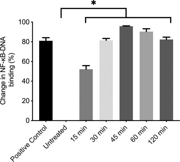
Bacillus SLP induces NF-κB-DNA binding in human retinal Muller cells. NF-κB-DNA binding in human retinal Muller cells (MIO-M1) treated with 10 μg/mL SlpA for 15, 30, 45, 60, and 120 minutes was quantified by ELISA. The percent of change in NF-κB-DNA binding for each time point relative to untreated control is presented. Values represent the mean ± SEM of change in NF-κB-DNA binding for at least three separate experiments. *P = 0.0286.
S-layer Protein Induces the Expression of Inflammatory Modulators From Retinal Cells
Host/pathogen interactions in infections typically involve the activation of the NF-κB pathway, leading to the production of inflammatory mediators to regulate inflammation.89,90 This is a downstream effect of a receptor/agonist interaction. It has been reported that SLPs of Lactobacillus helveticus induce inflammatory mediator production in human and mouse macrophages.91 Cytokine expression was also observed when mouse bone marrow–derived immature DCs were exposed to SLP from C. difficile.92,93 We therefore analyzed whether SlpA from B. thuringiensis induced the expression of inflammatory mediators from human retinal Muller cells. The expression of proinflammatory cytokines IL-6 and TNFα in SlpA-treated Muller cells was significantly greater than that of the extract control (Fig. 7). The expression of proinflammatory chemokines (CCL2 and CXCL-1) was also significantly greater compared with that of the extract control (Fig. 7). This finding demonstrates that B. thuringiensis SlpA induces the expression of inflammatory mediators from human retinal Muller cells, which is an outcome of NF-κB signaling pathway activation.
Figure 7.
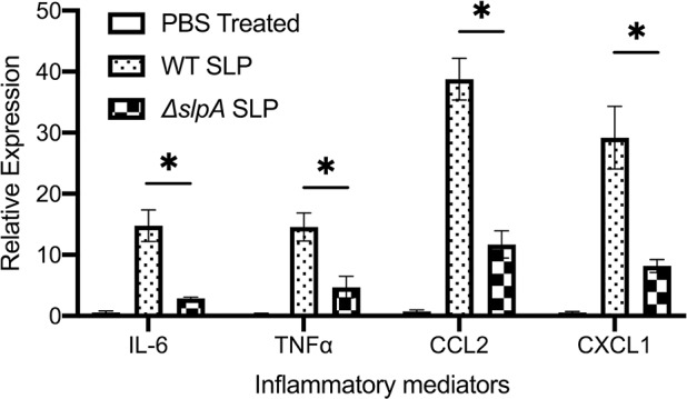
Bacillus SLP induces the expression of inflammatory mediators in human retinal Muller cells. Expression of proinflammatory cytokines (IL-6, TNFα) and chemokines (CCL2, CXCL-1) from human retinal Muller cells (MIO-M1) was quantified following treatment with 10 μg/mL SlpA or PBS for 10 hours. The “SlpA” fraction from ΔslpA B. thuringiensis was used as an extract control. Figures are representative of at least four independent experiments. Relative expression of these genes was determined by RT-PCR using ΔCT method using GAPDH as a housekeeping gene. Data are represented as the mean ± SEM of gene expression relative to GAPDH expression. *P = 0.0286.
Discussion
As one of the most immune privileged sites of the body, the eye contains innate defense systems designed to confront and eliminate invading microorganisms without the development of an overwhelming inflammatory response that might disrupt the clarity of visual axis.94,95 When introduced into the eye, Bacillus compromises these defenses, resulting in an explosive inflammatory response. Intraocular inflammation in endophthalmitis caused by members of the Bacillus cereus sensu lato group goes hand in hand with severe and irreversible retinal damage. The evolution of endophthalmitis typically depends on the species of the infecting pathogen.2,31,96 Compared with other pathogens associated with endophthalmitis, intraocular infection with Bacillus can reach severe levels quite rapidly. Within 12 to 48 hours, up to two-thirds of Bacillus-infected eyes have been reported to have lost useful vision, often leading to enucleation of the eye.4–10,97,98 The B. cereus envelope induced robust intraocular inflammation in a rabbit model of experimental endophthalmitis,2 presumably due to retinal innate immune recognition of pathogen associated molecular patterns on the organism's surface.33,34,99,100 Effective therapeutic options for endophthalmitis depend on identifying bacterial and host targets that can be used to modify disease outcomes and prevent vision loss. Therefore, identifying unique bacterial components that trigger the innate immune response in Bacillus endophthalmitis is crucial for understanding the interplay of the host and microbes in this disease.
The outer surfaces of some Gram-positive and -negative bacteria and Archaea have a proteinaceous coat known as an SL, formed by the self-assembly of monomeric proteins into a repeatedly spaced, two-dimensional array. As a protective coat, SL contributes not only to bacterial shape, cell wall integrity, and survival, but also to pathogenicity through several mechanisms.53,56 The function of SL in Bacillus endophthalmitis has never been studied. We previously used B. thuringiensis as a surrogate pathogen for B. cereus endophthalmitis and reported that both pathogens are capable of infecting human, rabbit, and mouse eyes.23,24,27,79 Here, studying SLP of B. thuringiensis will shed light on the role of this protein in Bacillus endophthalmitis.
If present, SLPs can constitute up to 15% of the total protein of a bacterial cell during exponential growth.53,55,56 Using TEM, we confirmed the presence or absence of SL in WT or ΔslpA B. thuringiensis, respectively. It has been reported that in artificial growth media, SLP-deficient bacteria grew faster than the WT strain.43,101 Here, the absence of SlpA did not result in changes in in vitro growth, as WT and ΔslpA B. thuringiensis grew at similar concentrations in BHI media. Both strains had similar hemolytic activities on red blood cells and cytotoxic effects on human retinal Muller cells. The SLP of Synechococcus, a marine cyanobacteria, was reported to contribute to the motility of this pathogen.102 However, we found that WT and ΔslpA B. thuringiensis were similarly motile. Moreover, we observed that during infection, both WT and ΔslpA penetrated the posterior capsule and entered the anterior segment, further supporting the similar motility of these strains. Overall, the absence of SlpA in B. thuringiensis resulted in no obvious phenotypic or biochemical differences.
Pathogenic Bacillus species and many other pathogenic bacteria have SLPs. SLPs are important virulence factors and may be required for an infection to occur,43 but the contributions of SLP to bacterial pathogenesis are poorly understood. To study this in the context of experimental endophthalmitis, we compared infections with WT and ΔslpA B. thuringiensis in mouse eyes. Intraocular growth of WT and ΔslpA B. thuringiensis was not statistically different at most time points, but at 8 hours, less bacilli in ΔslpA-infected eyes was observed. We reported a similar observation in experimental endophthalmitis initiated by pili-deficient B. cereus.48 One of the major functions of SLP is to protect the bacteria from phagocytosis and serum killing by binding with complement factor C3b.56,103 PMN are the primary infiltrating cells during B. cereus endophthalmitis1 and are phagocytic in nature. We reported that B. cereus are readily ingested by neutrophils from C57BL/6J mice in vitro.34 Whether B. cereus SLP has any role in protecting Bacillus from complement and neutrophil-mediated killing during experimental endophthalmitis is an open question.
The key event in the visual cycle is the phototransduction cascade that occurs within the retina. Therefore, any damage to the retina may cause significant and permanent vision loss or blindness. This is unfortunately a common occurrence in Bacillus endophthalmitis. Because the production of bacterial toxins, enzymes, and the inflammatory response in the eye during Bacillus endophthalmitis collectively contributes to retinal damage,23,27,104 we compared the retinal function in the eyes infected with WT or ΔslpA B. thuringiensis. Our results demonstrated that an absence of SlpA resulted in significant retention of retinal function, likely due to the preservation of retinal structure in these eyes (Figs. 3, 4). Eyes infected with the WT strain had significant retinal damage and detachments, likely contributing to significant retinal function loss in these eyes. Because WT and SlpA-deficient B. thuringiensis had similar cytotoxicity phenotypes, it is unlikely that the stark contrast in retinal damage and function loss were due to differences in toxin production by these strains.
We reported the importance of envelope components of Bacillus to intraocular inflammation.2 Flagella decorate B. cereus and B. thuringiensis and are essential for motility and migration throughout the eye during endophthalmitis,23 but are not involved in TLR5-mediated inflammation.47 The 49-kDa band observed in WT and ΔslpA (Fig. 1E) suggests that flagellin may have been present in the ΔslpA fractions. However, the presence of flagellin in these preparations was likely negligible based on less severe infections caused by ΔslpA (Figs. 3–5), the muted cytokine/chemokine response after exposure to the control ΔslpA fraction (Fig. 7), and our report that high, nonphysiological concentrations of flagellin are needed to induce intraocular inflammation.47
To date, studies on SLP have not addressed the recruitment of inflammatory cells in any infection model. Here, we demonstrated that a SlpA deficiency blunted the inflammatory response to Bacillus infection. ΔslpA-infected eyes had significantly less inflammatory influx compared with WT-infected eyes (Figs. 4, 5). The greater retained retinal function and reduced inflammatory influx in ΔslpA-infected groups were similar to what we previously observed in immune-deficient (TLR2−/−,33 TLR4−/−,34 MyD88−/−,34 TRIF−/−,34 and CXCL-1−/−74) mice, suggesting a potential link between SLPs and these innate immune systems. Together, these results suggest that in the absence of B. thuringiensis SlpA, a muted inflammatory response led to preserved retinal function.
During infection, B. cereus and B. thuringiensis migrate from the initial infection site to the midvitreous and localizes in close proximity near the retinal ILM.2 Here, both WT and ΔslpA were present adjacent to the ILM, suggesting a potential interaction between migrating organisms and the retinal cells constituting the ILM. We also observed that SLPs of B. thuringiensis induced NF-κB-DNA binding in human retinal Muller cells and stimulated the production of proinflammatory mediators (IL-6, TNFα, CCL2, and CXCL-1). All of these mediators have been reported to be present in mouse1,33,34,74,75,83 and human eyes105 with endophthalmitis. These findings imply that SLPs are immune modulators that may interact with innate immune pathways, and may do so in the eye via Muller cells during endophthalmitis.
Our present findings have demonstrated, for the first time, that the absence of a single virulence factor, the SLP, resulted in a significantly better clinical outcome for experimental Bacillus endophthalmitis. Because current anti-inflammatory therapeutics used this disease (i.e., corticosteroids) are relatively ineffective in altering rapidly evolving inflammation and subsequent vision loss,15,16,31,32 it is important to identify the bacterial components that interact with innate immune pathways to trigger this robust inflammation. Due to its rapidly blinding course, Bacillus endophthalmitis requires prompt and accurate treatment, which should be based largely on an understanding of the bacterial and host factors, which contribute to disease pathogenesis. Further understanding of the mechanisms by which SLPs contributes to the pathogenesis of Bacillus endophthalmitis could be instrumental in the development of new therapeutic regimens to block inflammation and prevent vision loss during this blinding infection.
Acknowledgments
The authors thank Mark K. Coggeshall, Tarea Burgett, and Alanson W. Girton (Oklahoma Medical Research Foundation) for stimulating discussions on peptidoglycan purification, Feng Li and Mark Dittmar (OUHSC Live Animal Imaging Core, Dean A. McGee Eye Institute, Oklahoma City, OK, USA) for invaluable technical assistance, and Excalibur Pathology (Moore, OK, USA) and the OUHSC Cellular Imaging Core (Dean A. McGee Eye Institute, Oklahoma City, OK, USA) for histology expertise. The human Muller cell line (MIO-M1) was a kind gift from Astrid Limb (Institute of Ophthalmology, Moorfields Eye Hospital, London).
Supported by National Institutes of Health grants R01EY028810 and R01EY024140 (to MCC). Our research is also supported in part by National Institutes of Health grants R01EY025947 and R21EY028066 (to MCC), National Eye Institutes Vision Core Grant P30EY027125 (to MCC), a Presbyterian Health Foundation Research Support Grant Award (to MCC), a Presbyterian Health Foundation Equipment Grant (to Robert E. Anderson, OUHSC), and an unrestricted grant to the Dean A. McGee Eye Institute from Research to Prevent Blindness.
Disclosure: M.H. Mursalin, None; P.S. Coburn, None; E. Livingston, None; F.C. Miller, None; R. Astley, None; A. Fouet, None; M.C. Callegan, None
References
- 1.Ramadan RT, Ramirez R, Novosad BD, Callegan MC. Acute inflammation and loss of retinal architecture and function during experimental Bacillus endophthalmitis. Curr Eye Res. 2006;31:955–965. doi: 10.1080/02713680600976925. [DOI] [PubMed] [Google Scholar]
- 2.Callegan MC, Booth MC, Jett BD, Gilmore MS. Pathogenesis of gram-positive bacterial endophthalmitis. Infect Immun. 1999;67:3348–3356. doi: 10.1128/iai.67.7.3348-3356.1999. [DOI] [PMC free article] [PubMed] [Google Scholar]
- 3.Parkunan SM, Callegan MC. The pathogenesis of bacterial endophthalmitis. In: Durand ML, Miller JW, Young LH, editors. Endophthalmitis. Switzerland: Springer International Publishing; 2016. pp. 17–47. [Google Scholar]
- 4.Boldt HC, Pulido JS, Blodi CF, Folk JC, Weingeist TA. Rural endophthalmitis. Ophthalmology. 1989;96:1722–1726. doi: 10.1016/s0161-6420(89)32658-3. [DOI] [PubMed] [Google Scholar]
- 5.David DB, Kirkby GR, Noble BA. Bacillus cereus endophthalmitis. Br J Ophthalmol. 1994;78:577–580. doi: 10.1136/bjo.78.7.577. [DOI] [PMC free article] [PubMed] [Google Scholar]
- 6.Shamsuddin D, Tuazon CU, Levy C, Curtin J. Bacillus cereus panophthalmitis: source of the organism. Rev Infect Dis. 1982;4:97–103. doi: 10.1093/clinids/4.1.97. [DOI] [PubMed] [Google Scholar]
- 7.Cowan CL, Jr, Madden WM, Hatem GF, Merritt JC. Endogenous Bacillus cereus panophthalmitis. Ann Ophthalmol. 1987;19:65–68. [PubMed] [Google Scholar]
- 8.Ho PC, O'Day DM, Head WS. Fulminating panophthalmitis due to exogenous infection with Bacillus cereus: report of 4 cases. Br J Ophthalmol. 1982;66:205–208. doi: 10.1136/bjo.66.3.205. [DOI] [PMC free article] [PubMed] [Google Scholar]
- 9.Davey RT, Jr, Tauber WB. Posttraumatic endophthalmitis: the emerging role of Bacillus cereus infection. Rev Infect Dis. 1987;9:110–123. doi: 10.1093/clinids/9.1.110. [DOI] [PubMed] [Google Scholar]
- 10.Vahey JB, Flynn HW., Jr Results in the management of Bacillus endophthalmitis. Ophthalmic Surg. 1991;22:681–686. [PubMed] [Google Scholar]
- 11.Bhagat N, Li X, Zarbin MA. Post-traumatic endophthalmitis. In: Durand ML, Miller JW, Young LH, editors. Endophthalmitis. Switzerland: Springer; 2016. pp. 151–170. [Google Scholar]
- 12.Carifi G. Bacterial post-traumatic endophthalmitis. Surv Ophthalmol. 2012;57:85–86. doi: 10.1016/j.survophthal.2011.09.005. author reply 86–88. [DOI] [PubMed] [Google Scholar]
- 13.Callegan MC, Guess S, Wheatley NR, et al. Efficacy of vitrectomy in improving the outcome of Bacillus cereus endophthalmitis. Retina. 2011;31:1518–1524. doi: 10.1097/IAE.0b013e318206d176. [DOI] [PMC free article] [PubMed] [Google Scholar]
- 14.Bialasiewicz AA, Al-Zuhaibi SM, Ganesh A. Post-traumatic inflammation with an intraocular foreign body. Ophthalmologe. 2008;105:669–673. doi: 10.1007/s00347-007-1621-y. [DOI] [PubMed] [Google Scholar]
- 15.Callegan MC, Gilmore MS, Gregory M, et al. Bacterial endophthalmitis: therapeutic challenges and host-pathogen interactions. Prog Retinal Eye Res. 2007;26:189–203. doi: 10.1016/j.preteyeres.2006.12.001. [DOI] [PMC free article] [PubMed] [Google Scholar]
- 16.Callegan MC, Engelbert M, Parke DW, II, Jett BD, Gilmore MS. Bacterial endophthalmitis: epidemiology, therapeutics, and bacterium-host interactions. Clin Microbiol Rev. 2002;15:111–124. doi: 10.1128/CMR.15.1.111-124.2002. [DOI] [PMC free article] [PubMed] [Google Scholar]
- 17.Huber-Spitzy V, Grabner G, Haddad R, Haselberger C. Post-traumatic endophthalmitis caused by Bacillus cereus. Klin Monbl Augenheilkd. 1986;188:52–54. doi: 10.1055/s-2008-1050574. [DOI] [PubMed] [Google Scholar]
- 18.McDowell RH, Friedman H. Bacillus cereus. Treasure Island, FL: StatPearls; 2018. [PubMed] [Google Scholar]
- 19.Granum PE. Bacillus cereus and its toxins. Soc Appl Bacteriol Symp Ser. 1994;23:61S–66S. [PubMed] [Google Scholar]
- 20.Carlson CR, Caugant DA, Kolstø AB. Genotypic diversity among Bacillus cereus and Bacillus thuringiensis strains. Appl Env Microbiol. 1994;60:1719–1725. doi: 10.1128/aem.60.6.1719-1725.1994. [DOI] [PMC free article] [PubMed] [Google Scholar]
- 21.Baumann L, Okamoto K, Unterman BM, Lynch MJ, Baumann P. Phenotypic characterization of Bacillus thuringiensis and Bacillus cereus. J Invertebrate Pathol. 1984;44:329–341. [Google Scholar]
- 22.Helgason E, Okstad OA, Caugant DA, et al. Bacillus anthracis, Bacillus cereus, and Bacillus thuringiensis–one species on the basis of genetic evidence. Appl Environ Microbiol. 2000;66:2627–2630. doi: 10.1128/aem.66.6.2627-2630.2000. [DOI] [PMC free article] [PubMed] [Google Scholar]
- 23.Callegan MC, Kane ST, Cochran DC, et al. Bacillus endophthalmitis: roles of bacterial toxins and motility during infection. Invest Ophthalmol Vis Sci. 2005;46:3233–3238. doi: 10.1167/iovs.05-0410. [DOI] [PubMed] [Google Scholar]
- 24.Callegan MC, Cochran DC, Kane ST, Gilmore MS, Gominet M, Lereclus D. Contribution of membrane-damaging toxins to Bacillus endophthalmitis pathogenesis. Infect Immun. 2002;70:5381–5389. doi: 10.1128/IAI.70.10.5381-5389.2002. [DOI] [PMC free article] [PubMed] [Google Scholar]
- 25.Callegan MC, Jett BD, Hancock LE, Gilmore MS. Role of hemolysin BL in the pathogenesis of extraintestinal Bacillus cereus infection assessed in an endophthalmitis model. Infect Immun. 1999;67:3357–3366. doi: 10.1128/iai.67.7.3357-3366.1999. [DOI] [PMC free article] [PubMed] [Google Scholar]
- 26.Beecher DJ, Pulido JS, Barney NP, Wong AC. Extracellular virulence factors in Bacillus cereus endophthalmitis: methods and implication of involvement of hemolysin BL. Infect Immun. 1995;63:632. doi: 10.1128/iai.63.2.632-639.1995. [DOI] [PMC free article] [PubMed] [Google Scholar]
- 27.Callegan MC, Kane ST, Cochran DC, Gilmore MS, Gominet M, Lereclus D. Relationship of plcR-regulated factors to Bacillus endophthalmitis virulence. Infect Immun. 2003;71:3116–3124. doi: 10.1128/IAI.71.6.3116-3124.2003. [DOI] [PMC free article] [PubMed] [Google Scholar]
- 28.Slamti L, Lereclus D. A cell-cell signaling peptide activates the PlcR virulence regulon in bacteria of the Bacillus cereus group. EMBO J. 2002;21:4550–4559. doi: 10.1093/emboj/cdf450. [DOI] [PMC free article] [PubMed] [Google Scholar]
- 29.Moyer AL, Ramadan RT, Thurman J, Burroughs A, Callegan MC. Bacillus cereus induces permeability of an in vitro blood-retina barrier. Infect Immun. 2008;76:1358–1367. doi: 10.1128/IAI.01330-07. [DOI] [PMC free article] [PubMed] [Google Scholar]
- 30.Moyer AL, Ramadan RT, Novosad BD, Astley R, Callegan MC. Bacillus cereus-induced permeability of the blood-ocular barrier during experimental endophthalmitis. Invest Ophthalmol Vis Sci. 2009;50:3783–3793. doi: 10.1167/iovs.08-3051. [DOI] [PMC free article] [PubMed] [Google Scholar]
- 31.Astley RA, Coburn PS, Parkunan SM, Callegan MC. Modeling intraocular bacterial infections. Prog Retinal Eye Res. 2016;54:30–48. doi: 10.1016/j.preteyeres.2016.04.007. [DOI] [PMC free article] [PubMed] [Google Scholar]
- 32.Miller FC, Coburn PS, Huzzatul MM, LaGrow AL, Livingston E, Callegan MC. Targets of immunomodulation in bacterial endophthalmitis. Prog Retinal Eye Res. 2019 doi: 10.1016/j.preteyeres.2019.05.004. published online ahead of print May 29. [DOI] [PMC free article] [PubMed] [Google Scholar]
- 33.Novosad BD, Astley RA, Callegan MC. Role of Toll-like receptor (TLR) 2 in experimental Bacillus cereus endophthalmitis. PLoS One. 2011;6:e28619. doi: 10.1371/journal.pone.0028619. [DOI] [PMC free article] [PubMed] [Google Scholar]
- 34.Parkunan SM, Randall CB, Coburn PS, Astley RA, Staats RL, Callegan MC. Unexpected roles for Toll-like receptor 4 and TRIF in intraocular infection with Gram-positive bacteria. Infect Immun. 2015;83:3926–3936. doi: 10.1128/IAI.00502-15. [DOI] [PMC free article] [PubMed] [Google Scholar]
- 35.Sankararaman S, Velayuthan S. Bacillus cereus. Pediatr Rev. 2013;34:196–197. doi: 10.1542/pir.34-4-196. [DOI] [PubMed] [Google Scholar]
- 36.Dufresne K, Paradis-Bleau C. Biology and assembly of the bacterial envelope. Adv Exp Med Biol. 2015;883:41–76. doi: 10.1007/978-3-319-23603-2_3. [DOI] [PubMed] [Google Scholar]
- 37.Fouet A, Mesnage S. Bacillus anthracis cell envelope components. Curr Top Microbiol Immunol. 2002;271:87–113. doi: 10.1007/978-3-662-05767-4_5. [DOI] [PubMed] [Google Scholar]
- 38.Beeby M, Gumbart JC, Roux B, Jensen GJ. Architecture and assembly of the Gram-positive cell wall. Mol Microbiol. 2013;88:664–672. doi: 10.1111/mmi.12203. [DOI] [PMC free article] [PubMed] [Google Scholar]
- 39.Budzik JM, Marraffini LA, Schneewind O. Assembly of pili on the surface of Bacillus cereus vegetative cells. Mol Microbiol. 2007;66:495–510. doi: 10.1111/j.1365-2958.2007.05939.x. [DOI] [PubMed] [Google Scholar]
- 40.Malanovic N, Lohner K. Gram-positive bacterial cell envelopes: the impact on the activity of antimicrobial peptides. Biochimica et Biophysica Acta Biomembranes. 2016;1858:936–946. doi: 10.1016/j.bbamem.2015.11.004. [DOI] [PubMed] [Google Scholar]
- 41.Shockman GD, Barrett JF. Structure, function, and assembly of cell walls of Gram-positive bacteria. Annu Rev Microbiol. 1983;37:501–527. doi: 10.1146/annurev.mi.37.100183.002441. [DOI] [PubMed] [Google Scholar]
- 42.Desvaux M, Dumas E, Chafsey I, Hébraud M. Protein cell surface display in Gram-positive bacteria: from single protein to macromolecular protein structure. FEMS Microbiol Lett. 2006;256:1–15. doi: 10.1111/j.1574-6968.2006.00122.x. [DOI] [PubMed] [Google Scholar]
- 43.Sidhu MS, Olsen I. S-layers of Bacillus species. Microbiology. 1997;143(Pt 4):1039–1052. doi: 10.1099/00221287-143-4-1039. [DOI] [PubMed] [Google Scholar]
- 44.Mignot T, Denis B, Couture-Tosi E, Kolsto AB, Mock M, Fouet A. Distribution of S-layers on the surface of Bacillus cereus strains: phylogenetic origin and ecological pressure. Environ Microbiol. 2001;3:493–501. doi: 10.1046/j.1462-2920.2001.00220.x. [DOI] [PubMed] [Google Scholar]
- 45.Rasko DA, Altherr MR, Han CS, Ravel J. Genomics of the Bacillus cereus group of organisms. FEMS Microbiol Rev. 2005;29:303–329. doi: 10.1016/j.femsre.2004.12.005. [DOI] [PubMed] [Google Scholar]
- 46.Callegan MC, Novosad BD, Ramirez R, Ghelardi E, Senesi S. Role of swarming migration in the pathogenesis of Bacillus endophthalmitis. Invest Ophthalmol Vis Sci. 2006;47:4461–4467. doi: 10.1167/iovs.06-0301. [DOI] [PubMed] [Google Scholar]
- 47.Parkunan SM, Astley R, Callegan MC. Role of TLR5 and flagella in Bacillus intraocular infection. PLoS One. 2014;9:e100543. doi: 10.1371/journal.pone.0100543. [DOI] [PMC free article] [PubMed] [Google Scholar]
- 48.Callegan MC, Parkunan SM, Randall CB, et al. The role of pili in Bacillus cereus intraocular infection. Exp Eye Res. 2017;159:69–76. doi: 10.1016/j.exer.2017.03.007. [DOI] [PMC free article] [PubMed] [Google Scholar]
- 49.Nguyen MT, Götz F. Lipoproteins of Gram-positive bacteria: key players in the immune response and virulence. Microbiol Mol Biol Rev. 2016;80:891. doi: 10.1128/MMBR.00028-16. [DOI] [PMC free article] [PubMed] [Google Scholar]
- 50.Boneca IG. The role of peptidoglycan in pathogenesis. Curr Opin Microbiol. 2005;8:46–53. doi: 10.1016/j.mib.2004.12.008. [DOI] [PubMed] [Google Scholar]
- 51.Kumar A, Kumar A. Role of Staphylococcus aureus virulence factors in inducing inflammation and vascular permeability in a mouse model of bacterial endophthalmitis. PLoS One. 2015;10:e0128423. doi: 10.1371/journal.pone.0128423. [DOI] [PMC free article] [PubMed] [Google Scholar]
- 52.Suzuki T, Campbell J, Swoboda JG, Walker S, Gilmore MS. Role of wall teichoic acids in Staphylococcus aureus endophthalmitis. Invest Ophthalmol Vis Sci. 2011;52:3187–3192. doi: 10.1167/iovs.10-6558. [DOI] [PMC free article] [PubMed] [Google Scholar]
- 53.Sleytr UB, Schuster B, Egelseer E-M, Pum D. S-layers: principles and applications. FEMS Microbiol Rev. 2014;38:823–864. doi: 10.1111/1574-6976.12063. [DOI] [PMC free article] [PubMed] [Google Scholar]
- 54.Mesnage S, Fontaine T, Mignot T, Delepierre M, Mock M, Fouet A. Bacterial SLH domain proteins are non-covalently anchored to the cell surface via a conserved mechanism involving wall polysaccharide pyruvylation. EMBO J. 2000;19:4473–4484. doi: 10.1093/emboj/19.17.4473. [DOI] [PMC free article] [PubMed] [Google Scholar]
- 55.Sara M, Sleytr UB. S-Layer proteins. J Bacteriol. 2000;182:859–868. doi: 10.1128/jb.182.4.859-868.2000. [DOI] [PMC free article] [PubMed] [Google Scholar]
- 56.Gerbino E, Carasi P, Mobili P, Serradell MA, Gómez-Zavaglia A. Role of S-layer proteins in bacteria. World J Microbiol Biotechnol. 2015;31:1877–1887. doi: 10.1007/s11274-015-1952-9. [DOI] [PubMed] [Google Scholar]
- 57.Pum D, Messner P, Sleytr UB. Role of the S layer in morphogenesis and cell division of the archaebacterium Methanocorpusculum sinense. J Bacteriol. 1991;173:6865–6873. doi: 10.1128/jb.173.21.6865-6873.1991. [DOI] [PMC free article] [PubMed] [Google Scholar]
- 58.Messner P, Pum D, Sara M, Stetter KO, Sleytr UB. Ultrastructure of the cell envelope of the archaebacteria Thermoproteus tenax and Thermoproteus neutrophilus. J Bacteriol. 1986;166:1046–1054. doi: 10.1128/jb.166.3.1046-1054.1986. [DOI] [PMC free article] [PubMed] [Google Scholar]
- 59.Sleytr UB, Beveridge TJ. Bacterial S-layers. Trends Microbiol. 1999;7:253–260. doi: 10.1016/s0966-842x(99)01513-9. [DOI] [PubMed] [Google Scholar]
- 60.Ethapa T, Leuzzi R, Ng YK, et al. Multiple factors modulate biofilm formation by the anaerobic pathogen Clostridium difficile. J Bacteriol. 2013;195:545–555. doi: 10.1128/JB.01980-12. [DOI] [PMC free article] [PubMed] [Google Scholar]
- 61.Hynonen U, Palva A. Lactobacillus surface layer proteins: structure, function and applications. Appl Microbiol Biotechnol. 2013;97:5225–5243. doi: 10.1007/s00253-013-4962-2. [DOI] [PMC free article] [PubMed] [Google Scholar]
- 62.Sakakibara J, Nagano K, Murakami Y, et al. Loss of adherence ability to human gingival epithelial cells in S-layer protein-deficient mutants of Tannerella forsythensis. Microbiology. 2007;153:866–876. doi: 10.1099/mic.0.29275-0. [DOI] [PubMed] [Google Scholar]
- 63.Shimotahira N, Oogai Y, Kawada-Matsuo M, et al. The surface layer of Tannerella forsythia contributes to serum resistance and oral bacterial coaggregation. Infect Immun. 2013;81:1198–1206. doi: 10.1128/IAI.00983-12. [DOI] [PMC free article] [PubMed] [Google Scholar]
- 64.Zhang W, Wang H, Liu J, Zhao Y, Gao K, Zhang J. Adhesive ability means inhibition activities for Lactobacillus against pathogens and S-layer protein plays an important role in adhesion. Anaerobe. 2013;22:97–103. doi: 10.1016/j.anaerobe.2013.06.005. [DOI] [PubMed] [Google Scholar]
- 65.Thompson SA. Campylobacter Surface-layers (S-layers) and immune evasion. Ann Periodontol. 2002;7:43–53. doi: 10.1902/annals.2002.7.1.43. [DOI] [PMC free article] [PubMed] [Google Scholar]
- 66.Claus H, Akca E, Debaerdemaeker T, Evrard C, Declercq JP, Konig H. Primary structure of selected archaeal mesophilic and extremely thermophilic outer surface layer proteins. Syst Appl Microbiol. 2002;25:3–12. doi: 10.1078/0723-2020-00100. [DOI] [PubMed] [Google Scholar]
- 67.Engelhardt H. Mechanism of osmoprotection by archaeal S-layers: a theoretical study. J Struct Biol. 2007;160:190–199. doi: 10.1016/j.jsb.2007.08.004. [DOI] [PubMed] [Google Scholar]
- 68.Kotiranta A, Haapasalo M, Kari K, et al. Surface structure, hydrophobicity, phagocytosis, and adherence to matrix proteins of Bacillus cereus cells with and without the crystalline surface protein layer. Infect Immun. 1998;66:4895–4902. doi: 10.1128/iai.66.10.4895-4902.1998. [DOI] [PMC free article] [PubMed] [Google Scholar]
- 69.Engelhardt H, Peters J. Structural research on surface layers: a focus on stability, surface layer homology domains, and surface layer-cell wall interactions. J Struct Biol. 1998;124:276–302. doi: 10.1006/jsbi.1998.4070. [DOI] [PubMed] [Google Scholar]
- 70.Gilmour R, Messner P, Guffanti AA, et al. Two-dimensional gel electrophoresis analyses of pH-dependent protein expression in facultatively alkaliphilic Bacillus pseudofirmus OF4 lead to characterization of an S-layer protein with a role in alkaliphily. J Bacteriol. 2000;182:5969–5981. doi: 10.1128/jb.182.21.5969-5981.2000. [DOI] [PMC free article] [PubMed] [Google Scholar]
- 71.Lortal S, Van Heijenoort J, Gruber K, Sleytr UB. S-layer of Lactobacillus helveticus ATCC 12046: Isolation, chemical characterization and re-formation after extraction with lithium chloride. Microbiology. 1992;138:611–618. [Google Scholar]
- 72.Koval SF, Hynes SH. Effect of paracrystalline protein surface layers on predation by Bdellovibrio bacteriovorus. J Bacteriol. 1991;173:2244–2249. doi: 10.1128/jb.173.7.2244-2249.1991. [DOI] [PMC free article] [PubMed] [Google Scholar]
- 73.Mesnage S, Haustant M, Fouet A. A general strategy for identification of S-layer genes in the Bacillus cereus group: molecular characterization of such a gene in Bacillus thuringiensis subsp. galleriae NRRL 4045. Microbiology. 2001;147:1343–1351. doi: 10.1099/00221287-147-5-1343. [DOI] [PubMed] [Google Scholar]
- 74.Parkunan SM, Randall CB, Astley RA, Furtado GC, Lira SA, Callegan MC. CXCL1, but not IL-6, significantly impacts intraocular inflammation during infection. J Leukoc Biol. 2016;100:1125–1134. doi: 10.1189/jlb.3A0416-173R. [DOI] [PMC free article] [PubMed] [Google Scholar]
- 75.Coburn PS, Miller FC, LaGrow AL, et al. TLR4 modulates inflammatory gene targets in the retina during Bacillus cereus endophthalmitis. BMC Ophthalmol. 2018;18:96. doi: 10.1186/s12886-018-0764-8. [DOI] [PMC free article] [PubMed] [Google Scholar]
- 76.Coburn PS, Miller FC, LaGrow AL, et al. Disarming pore-forming toxins with biomimetic nanosponges in intraocular infections. mSphere. 2019;2:e00262–19. doi: 10.1128/mSphere.00262-19. [DOI] [PMC free article] [PubMed] [Google Scholar]
- 77.LaGrow AL, Coburn PS, Miller FC, et al. A novel biomimetic nanosponge protects the retina from the Enterococcus faecalis cytolysin. mSphere. 2017;2:e00335–17. doi: 10.1128/mSphere.00335-17. [DOI] [PMC free article] [PubMed] [Google Scholar]
- 78.Limb GA, Salt TE, Munro PM, Moss SE, Khaw PT. In vitro characterization of a spontaneously immortalized human Muller cell line (MIO-M1) Invest Ophthalmol Vis Sci. 2002;43:864–869. [PubMed] [Google Scholar]
- 79.Callegan MC, Cochran DC, Kane ST, et al. Virulence factor profiles and antimicrobial susceptibilities of ocular Bacillus isolates. Curr Eye Res. 2006;31:693–702. doi: 10.1080/02713680600850963. [DOI] [PubMed] [Google Scholar]
- 80.Luckevich MD, Beveridge TJ. Characterization of a dynamic S layer on Bacillus thuringiensis. J Bacteriol. 1989;171:6656–6667. doi: 10.1128/jb.171.12.6656-6667.1989. [DOI] [PMC free article] [PubMed] [Google Scholar]
- 81.Langer M, Malykhin A, Maeda K, et al. Bacillus anthracis peptidoglycan stimulates an inflammatory response in monocytes through the p38 mitogen-activated protein kinase pathway. PLoS One. 2008;3:e3706. doi: 10.1371/journal.pone.0003706. [DOI] [PMC free article] [PubMed] [Google Scholar]
- 82.Wampach L, Heintz-Buschart A, Fritz JV, et al. Birth mode is associated with earliest strain-conferred gut microbiome functions and immunostimulatory potential. Nat Comm. 2018;9:5091. doi: 10.1038/s41467-018-07631-x. [DOI] [PMC free article] [PubMed] [Google Scholar]
- 83.Ramadan RT, Moyer AL, Callegan MC. A role for tumor necrosis factor-alpha in experimental Bacillus cereus endophthalmitis pathogenesis. Invest Ophthalmol Vis Sci. 2008;49:4482–4489. doi: 10.1167/iovs.08-2085. [DOI] [PMC free article] [PubMed] [Google Scholar]
- 84.Kodela R, Nath N, Chattopadhyay M, Nesbitt DE, Velazquez-Martinez CA, Kashfi K. Hydrogen sulfide-releasing naproxen suppresses colon cancer cell growth and inhibits NF-kappaB signaling. Drug Design Dev Ther. 2015;9:4873–4882. doi: 10.2147/DDDT.S91116. [DOI] [PMC free article] [PubMed] [Google Scholar]
- 85.Aratani Y. Myeloperoxidase: its role for host defense, inflammation, and neutrophil function. Arch Biochem Biophys. 2018;640:47–52. doi: 10.1016/j.abb.2018.01.004. [DOI] [PubMed] [Google Scholar]
- 86.Bharti AC, Aggarwal BB. Nuclear factor-kappa B and cancer: Its role in prevention and therapy. Biochem Pharmacol. 2002;64:883–888. doi: 10.1016/s0006-2952(02)01154-1. [DOI] [PubMed] [Google Scholar]
- 87.Gilmore TD. Introduction: the Rel/NF-kappaB signal transduction pathway. Semin Cancer Biol. 1997;8:61–62. doi: 10.1006/scbi.1997.0056. [DOI] [PubMed] [Google Scholar]
- 88.Pahl HL. Activators and target genes of Rel/NF-kappaB transcription factors. Oncogene. 1999;18:6853–6866. doi: 10.1038/sj.onc.1203239. [DOI] [PubMed] [Google Scholar]
- 89.Liu T, Zhang L, Joo D, Sun S-C. NF-κB signaling in inflammation. Signal Transduction Targeted Ther. 2017;2:17023. doi: 10.1038/sigtrans.2017.23. [DOI] [PMC free article] [PubMed] [Google Scholar]
- 90.Tak PP, Firestein GS. NF-κB: a key role in inflammatory diseases. J Clin Invest. 2001;107:7–11. doi: 10.1172/JCI11830. [DOI] [PMC free article] [PubMed] [Google Scholar]
- 91.Taverniti V, Stuknyte M, Minuzzo M, et al. S-layer protein mediates the stimulatory effect of Lactobacillus helveticus MIMLh5 on innate immunity. Appl Environ Microbiol. 2013;79:1221–1231. doi: 10.1128/AEM.03056-12. [DOI] [PMC free article] [PubMed] [Google Scholar]
- 92.Lynch M, Walsh TA, Marszalowska I, et al. Surface layer proteins from virulent Clostridium difficile ribotypes exhibit signatures of positive selection with consequences for innate immune response. BMC Evol Biol. 2017;17:90. doi: 10.1186/s12862-017-0937-8. [DOI] [PMC free article] [PubMed] [Google Scholar]
- 93.Collins LE, Lynch M, Marszalowska I, et al. Surface layer proteins isolated from Clostridium difficile induce clearance responses in macrophages. Microbes Infect. 2014;16:391–400. doi: 10.1016/j.micinf.2014.02.001. [DOI] [PubMed] [Google Scholar]
- 94.Zhou R, Caspi RR. Ocular immune privilege. F1000 Biol Rep. 2010;2:3. doi: 10.3410/B2-3. [DOI] [PMC free article] [PubMed] [Google Scholar]
- 95.Taylor AW, Ng TF. Negative regulators that mediate ocular immune privilege. J Leukoc Biol. 2019 doi: 10.1002/JLB.3MIR0817-337R. published online ahead of print February 12. [DOI] [PMC free article] [PubMed] [Google Scholar]
- 96.Kernt M, Kampik A. Endophthalmitis: pathogenesis, clinical presentation, management, and perspectives. Clin Ophthalmol. 2010;4:121–135. doi: 10.2147/opth.s6461. [DOI] [PMC free article] [PubMed] [Google Scholar]
- 97.Duch-Samper AM, Chaques-Alepuz V, Menezo JL, Hurtado-Sarrio M. Endophthalmitis following open-globe injuries. Curr Opin Ophthalmol. 1998;9:59–65. doi: 10.1097/00055735-199806000-00011. [DOI] [PubMed] [Google Scholar]
- 98.Reynolds DS, Flynn HW., Jr Endophthalmitis after penetrating ocular trauma. Curr Opin Ophthalmol. 1997;8:32–38. doi: 10.1097/00055735-199706000-00006. [DOI] [PubMed] [Google Scholar]
- 99.Benhar I, London A, Schwartz M. The privileged immunity of immune privileged organs: the case of the eye. Front Immunol. 2012;3:296–296. doi: 10.3389/fimmu.2012.00296. [DOI] [PMC free article] [PubMed] [Google Scholar]
- 100.Gilger BC. Immunology of the ocular surface. Vet Clin North Am Small Anim Pract. 2008;38:223–231. doi: 10.1016/j.cvsm.2007.11.004. [DOI] [PubMed] [Google Scholar]
- 101.Koval SF, Murray R. The superficial protein arrays on bacteria. Microbiol Sci. 1986;3:357–361. [PubMed] [Google Scholar]
- 102.McCarren J, Brahamsha B. Swimming motility mutants of marine Synechococcus affected in production and localization of the S-layer protein SwmA. J Bacteriol. 2009;191:1111–1114. doi: 10.1128/JB.01401-08. [DOI] [PMC free article] [PubMed] [Google Scholar]
- 103.Thompson SA. Campylobacter surface-layers (S-layers) and immune evasion. Ann Periodontal. 2002;7:43–53. doi: 10.1902/annals.2002.7.1.43. [DOI] [PMC free article] [PubMed] [Google Scholar]
- 104.Gregory M, Callegan MC, Gilmore MS. Role of bacterial and host factors in infectious endophthalmitis. Chem Immunol Allergy. 2007;92:266–275. doi: 10.1159/000099277. [DOI] [PubMed] [Google Scholar]
- 105.Escarião P, Commodaro AG, Arantes T, de Castro CMMB. Diniz MdeFA, Brandt CT. Analysis of cytokines in presumed acute infectious endophthalmitis following cataract extraction. J Clin Exp Ophthalmol. 2014;5:335. [Google Scholar]


