Abstract
Whole mitogenome sequences (mtDNA) have been exploited for insect ecology studies, using them as molecular markers to reconstruct phylogenies, or to infer phylogeographic relationships and gene flow. Recent Anopheles phylogenomic studies have provided information regarding the time of deep lineage divergences within the genus. Here we report the complete 15,393 bp mtDNA sequences of Anopheles aquasalis, a Neotropical human malaria vector. When comparing its structure and base composition with other relevant and available anopheline mitogenomes, high similarity and conserved genomic features were observed. Furthermore, 22 mtDNA sequences comprising anopheline and Dipteran sibling species were analyzed to reconstruct phylogenies and estimate dates of divergence between taxa. Phylogenetic analysis using complete mtDNA sequences suggests that A. aquasalis diverged from the Anopheles albitarsis complex ~28 million years ago (MYA), and ~38 MYA from Anopheles darlingi. Bayesian analysis suggests that the most recent ancestor of Nyssorhynchus and Anopheles + Cellia was extant ~83 MYA, corroborating current estimates of ~79–100 MYA. Additional sampling and publication of African, Asian, and North American anopheline mitogenomes would improve the resolution of the Anopheles phylogeny and clarify early continental dispersal routes.
Introduction
The mitogenome of most insects is composed of a small double-stranded circular molecule of 14–20 kb in length. It contains 37 genes including 13 protein-coding genes (PCG), 22 transfer RNA genes (tRNA) and two ribosomal RNA genes (small (srRNA) and large (lr-RNA) ribosomal subunits). Additionally, it contains an A+T rich control region that is involved in the initiation of transcription and replication [1, 2]. The order of the genes within the mitogenome is highly conserved and can be traced to the ancestral gene arrangement from the Bilateria [3], which differs slightly from ancestral ecdysozoan and arthropod mitogenomes [4].
A total of 475 formally recognized species of Anopheles are currently known [5]. Until recently, knowledge regarding the evolution, divergence time, and phylogenetic relationships among representative species within this genus were scarce. Despite its medical importance worldwide, this gap has been due to the relevance and focus on African anophelines [6–9]. The widespread existence of cryptic species complicates taxonomic and phylogenetic analyses in the genus Anopheles [6, 7, 10, 11]. Nevertheless, the recent publication of 16 anopheline genomes [8] may provide, in the near future, new full mitogenomes as parallel assemblies of the genomic data produced. This would enable more accurate phylogenomic reconstructions and enhance estimates of the divergence times among members of this genus.
The current hypothesis regarding Anopheles evolution is mostly based on the geographic distribution of extant species [12]. It proposes that major mosquito lineages, including Anopheles, originated in Western Gondwana approximately 145 to 100 million years ago (MYA) during the periods recognized as late Jurassic or early Cretaceous [12, 13]. The genus Anopheles would have emerged in what now is South America and, following rapid diversification and migration across land bridges, colonized most of the Earth’s favorable habitats [12, 14]. Recent publications hypothesize that the Anopheles subgenera Nyssorhynchus and Anopheles + Cellia diverged between 79–100 MYA [6, 15, 16], suggesting that their most recent common ancestor might have lived before the geological split of Western Gondwana ~95 MYA [16].
The human malaria cycle probably evolved in Africa, where first interactions between parasites, their anopheline hosts, and hominids occurred [7]. As malaria-infected humans migrated out of Africa, they carried Plasmodium parasites with them, leaving their African mosquito vectors behind. This migration gave rise to a journey of adaptation between the Plasmodium parasite and no less than 34 different anopheline mosquito species worldwide [17]. Based on available evidence of historical, archaeological and genetic type, it is believed that human malaria was introduced into the Americas by Europeans who transferred both, Plasmodium falciparum and Plasmodium vivax (the most prevalent malaria parasite species) to the indigenous population [18–20]. Although Neotropical anophelines were already adapted to feed upon primates, including humans, interactions between Neotropical malaria vectors, humans, and malaria parasites, can be considered geologically and evolutionarily recent [7, 16].
Anopheles aquasalis is a relevant Neotropical malaria vector of P. vivax on the Atlantic and Pacific coasts from Central America to Southern Brazil. An. aquasalis has been reared under laboratory conditions since 1995, being a well-established model for experimental studies involving the interaction of malaria vectors with several Plasmodium species [21]. Nonetheless, little is known about its evolutionary relationship with other Anopheles sibling species.
Mitochondrial genome sequence data are useful to infer phylogenetic and phylogeographic relationships [22–26]. Here we present a preliminary characterization of the mitogenome of A. aquasalis assembled by Next Generation shotgun sequencing (NGS). We compared the mtDNA genome sequence, and some of its features, with other selected anopheline mitogenomes. We applied Bayesian analysis to reconstruct phylogenetic relationships and estimate divergence times amongst other human malaria vectors. The implications of these findings were briefly discussed regarding the evolutionary history of anophelines in general. A more thorough and balanced analysis of anopheline mitogenomes, including representatives from North America, Asia and Africa would provide an in-depth description of dispersal routes throughout evolutionary and geological times.
Methods
Anopheles aquasalis colony
A. aquasalis were obtained from a colony established at the Medical Entomology Laboratory at Instituto de Pesquisas René Rachou-FIOCRUZ (Fiocruz, Minas Gerais). The mosquitoes originally came from a colony established in 1995 in Rio de Janeiro [27, 28], and are currently kept under laboratory conditions as previously described [29].
Single mosquito DNA extraction
Genomic DNA from a single adult female A. aquasalis was extracted using the QiagenDNeasy blood and tissue kit (Qiagen, Hilden, Germany) according to the protocol for purification of insect DNA with a minor modification: Qiagen EB buffer rather than AE buffer was used to avoid possible interference of EDTA with the Nextera kit enzymes. The purified Anopheles genomic DNA was then quantified with the Qubit HS (Life Technologies, USA) system and used to construct the genomic library.
Whole genome shotgun sequencing
The genomic DNA was processed using the Nextera DNA sample preparation kit (Epicentre Biotechnologies, Madison, WI). Thirty nanograms of sample DNA were fragmented utilizing 5 μl of Tagment DNA Enzyme with 25 μl of Tagment Buffer. Tagmentation reactions included in the Nextera kit were performed by incubating the sample for 5 min at 55°C. The tagmented DNA was purified using the QiagenMinElute protocol (QIAGEN, Germany). Purified DNA was eluted from the column with 11 μl of nuclease-free water. Purified DNA (5 μl) was used as the template in a 20-μl volume for limited-cycle PCR (5 cycles) and processed as outlined in the Nextera protocol (Illumina). Amplified DNA was purified using the AMPure Bead cleanup (Beckman Coulter, USA) according to the manufacturer's protocol. The fragment size distribution of the tagmented DNA was analyzed utilizing a 2100 Bioanalyzer with a 7500 DNA assay kit (Agilent Technologies, Santa Clara, CA). Fragments of ~600 bp long were carried out for sequencing. The library was sequenced on one lane of an Illumina HiSeq2000 instrument to generate paired-end reads. Sequencing was performed by The Vincent J. Coates Genomics Sequencing Laboratory (GSL) at the University of California, Berkeley.
Mitochondrial genome assembly
Sequences were assembled de novo using Velvet v1.2.10 [30] with a k-mer size of 41, according to the scripts and parameters suggested by the Velvet Manual (https://www.ebi.ac.uk/~zerbino/velvet/Manual.pdf) and in-house protocols from the UC Davis Vector Genetics Laboratory. The assembled contigs were aligned to the mtDNA sequence of An. gambiae (GenBank No. L20934.1) and A. darlingi (GenBank No.GQ918273.1) using the MUMmerv3.0 software [31] to identify and confirm the de novo assembly of the A. aquasalis mitogenome.
Sequence analysis: Composition and genomic features of the A. aquasalis mitogenome
The assembled mitogenome was manually inspected for repeats at the beginning and end of the assembly to infer circularity. Automatic annotation of the mitogenome fasta file was performed with MITOS [32], followed by a manual curation based on the GenBank file format. Manual inspection of the predicted Protein-Coding Genes (PCG's), ribosomal RNA (rRNA) genes, transfer RNA (tRNA) genes, and the AT rich region was performed with Artemis -release 16- [33]. The nucleotide sequences of PCGs were translated based on the invertebrate mtDNA genetic code.
The manual curation of coding regions and rRNA genes was mainly carried by sequence comparison with published insect mitogenome sequences such as A. gambiae (GenBank No. L20934.1) and A. darlingi (GenBank No.GQ918273.1) amongst others. Careful attention was given to PCGs comparing the predicted Open reading frames (ORFs) to the UniProt database (http://www.uniprot.org/) giving more weight to similarity hits that had experimental validation.
The MITOS annotated tRNAs were verified and their secondary structures predicted with the tRNAscan-SE search server v1.21 [34] with default settings: the invertebrate mitochondrial codon predictors, and a cove score cut off of 5 (software tool available at: http://lowelab.ucsc.edu/tRNAscan-SE/). Some tRNA genes could not be detected by tRNAscan-SE. They were identified by direct comparison and sequence similarity to tRNAs of other dipterans or anophelines. These tRNA genes were modeled with RNAStructure [35]. To maintain a uniform format, all of the 22 figures were generated with RNAStructure.
To visualize the annotated mitogenome, circular representation of it was generated with Blast Ring Image Generator (BRIG) [36]. For such purpose, a GenBank formatted file and the fasta file were employed according to the Brig 0.95 Manual. Available at: http://ufpr.dl.sourceforge.net/project/brig/BRIGMANUAL.pdf.
Nucleotide composition analyses, expressed as AT%, were performed for individual PCGs, full mtDNA, concatenated PCGs, concatenated tRNAs, lrRNA (16S), srRNA (12S) and concatenated rRNAs. For the aforementioned targets, composition bias based on strand asymmetry values were estimated using the following formulae for skews: AT skew = [A-T]/[A+T] and GC skew = [G-C]/[G+C] as proposed by Perna & Kocher (1995) [37] on an Excel spreadsheet. Codon bias was assessed estimating the relative synonymous codon usage (RSCU). All the above compositional analyses (except bias estimation) were performed using MEGA v6.0 [38]. The phylogenetic and comparative analyses performed henceforth relied upon available sequences and published literature regarding anopheline and culicine species detailed in Table 1.
Table 1. List of the insect species used in this study with their corresponding GenBank number.
| Species | Family | Length (bp) | GenBank No. | Vector of malaria in (continent) | Reference |
|---|---|---|---|---|---|
| Anopheles aquasalis | Culicidae | 15393 | NJHH00000000 | South America | This study |
| Anopheles punctulatus (isolate ITN_PNG-18) | Culicidae | 15412 | JX219738.1 | Oceania | [6] |
| Anopheles farauti 4 (isolate 7_10–11) | Culicidae | 15412 | JX219735.1 | Oceania | [6] |
| Anopheles farauti 4 (isolate 8_11–12) | Culicidae | 15412 | JX219736.1 | Oceania | [6] |
| Anopheles hinesorum | Culicidae | 15336 | JX219734.1 | Oceania | [6] |
| Anopheles koliensis (isolate ESP001B) | Culicidae | 15412 | JX219743.1 | Oceania | [6] |
| Anopheles dirus A (isolate A1) | Culicidae | 15404 | JX219731.1 | Southeast Asia | [6] |
| Anopheles dirus A (isolate A2) | Culicidae | 15404 | JX219732.1 | Southeast Asia | [6] |
| Anopheles cracens (isolate B1) | Culicidae | 15412 | JX219733.1 | Southeast Asia | [6] |
| Anopheles albitarsis F | Culicidae | 15418 | HQ335349.1 | South America | [39] |
| Anopheles albitarsis G | Culicidae | 15474 | HQ335346.1 | South America | [39] |
| Anopheles deaneorum | Culicidae | 15424 | HQ335347.1 | South America | [39] |
| Anopheles janconnae | Culicidae | 15425 | HQ335348.1 | South America | [39] |
| Anopheles oryzalimnetes | Culicidae | 15422 | HQ335345.1 | South America | [39] |
| Anopheles darlingi North | Culicidae | 15386 | GQ918272.1 | Central America | [40] |
| Anopheles darlingi South | Culicidae | 15385 | GQ918273.1 | South America | [40] |
| Anopheles quadrimaculatus (A strain Orlando) | Culicidae | 15455 | L04272.1 | North America | [41] |
| Anopheles gambiae | Culicidae | 15363 | L20934.1 | Africa | [42] |
| Culex pipiens | Culicidae | 14856 | HQ724614.1 | NA | [43] |
| Aedes aegypti | Culicidae | 16655 | EU352212.1 | NA | Unpublished |
| Aedes albopictus | Culicidae | 16665 | NC_006817.1 | NA | Unpublished |
| Drosophila melanogaster | Drosophillidae | 19517 | U37541.1 | NA | [44] |
For anopheline species, the continent in which they exert their malaria vectorial activity is shown. The sequence length reflects the number of base pairs assembled not considering Ns.
Comparative analyses between anophelines from different geographic regions
Comparative analyses regarding nucleotide composition and strand asymmetry were performed between A. aquasalis and four other anophelines representative of different geographic regions: Anopheles punctulatus (GenBank No. JX219738.1) from South East Asia, A. gambiae (GenBank No. L20934.1) from Africa, A. darlingi North (GenBank No. GQ918272.1) from Central America, and A. darlingi South (GenBank No. GQ918273.1) from South America. For each mtDNA genome, base composition (expressed as AT%) and strand asymmetry (AT and GC skew) were calculated as explained above.
Additionally, a CDS nucleotide similarity comparison between A. aquasalis, the above-cited anophelines, plus Anopheles albitarsis (GenBank No. HQ335349.1)—another South American brackish-water tolerant species—was performed using the Blast2sequence online tool [45] available at http://blast.ncbi.nlm.nih.gov/Blast.cgi#.
Phylogenetic analysis and molecular dating
To further our knowledge on the Anopheles genus phylogeny, as well as to estimate the divergence time or split between A. aquasalis and other Neotropical anophelines, we reproduced the phylogeny and molecular dating analyses performed by Logue et al., (2013) [6] using the tools and parameters suggested by the authors adding the assembled A. aquasalis mitogenome.
Briefly, 21 insect mitogenomes were selected from the ones used by Logue et al., (2013) [6] and their sequences retrieved from the NCBI databank (Table 1). Then, for each of the 13 PCGs, the following actions were performed: the DNA sequences were translated into amino acid sequences using the invertebrate mt genetic code, then they were aligned to each other with the MAFFT alignment engine, and the aligned amino acid sequences were reverse-translated back into nucleotide sequences. All the above steps were performed with the online tool Translator X [46] using default parameters (available at: http://translatorx.co.uk/). Afterward, the aligned sequences from all 13 mt genes were concatenated using FASconCAT [47]. The concatenated PCG sequences from the 22 mitogenomes were analyzed with jModeltest v0.1.1 [48] to determine the best nucleotide substitution model according to the Akaike Information Criterion.
Bayesian phylogenies were reconstructed, and node ages inferred, using BEAST v1.7.5 [49]. The following program parameters were used: an uncorrelated lognormal relaxed clock model allowing for rate heterogeneity among species; the GTR + G substitution model; the SRD06 model of partitioning, which enables estimation of nucleotide substitution parameters separately for the 1st + 2nd and 3rd codon positions (this apparently provides a better fit for protein-coding nucleotide data), and a Yule model for tree reconstruction. With the above parameters, three independent runs of 20 million generations were performed, saving the generated trees every 1,000 generations. All runs were then combined after a burn-in of 10% using LogCombiner v1.7.2 and afterwards, Tracer v1.5 was used to verify the mixing of the Markov chains (both tools are part of the BEAST pipeline). The maximum credibility tree was determined using TreeAnnotator v1.7.2 and visualized with FigTree v1.4.3 available at http://tree.bio.ed.ac.uk/software/figtree/. Divergence times were estimated using BEAST v1.7.5 following the instructions provided by the developers at http://beast.bio.ed.ac.uk/. In addition to the aforementioned parameters, the Drosophila-Anopheles divergence time was set as the calibration point using a prior distribution normally distributed around a mean of 260 million years ago (MYA) ranging from 243 to 276 MYA as suggested by Gaunt & Miles (2002) [50].
Results
Composition and genomic features
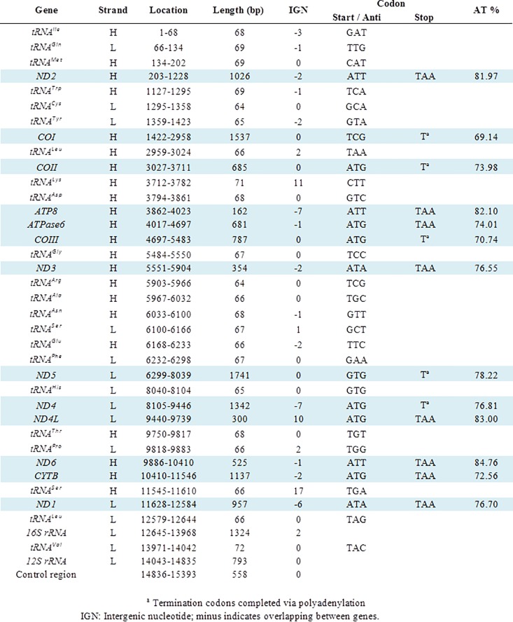
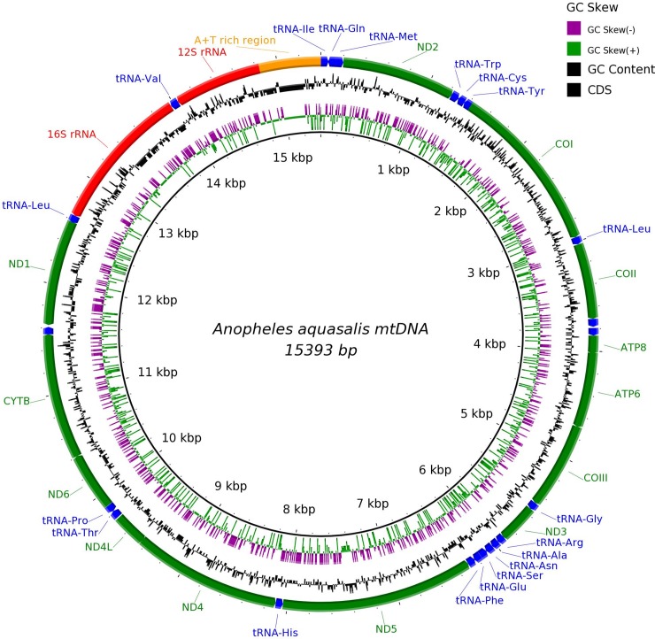
The complete mitogenome of A. aquasalis was assembled into a single contig of 15,393bp. The expected 37 genes in animal mtDNA, comprising 13 protein-coding genes, two rRNA genes (12S and 16S), 22 tRNA genes and a control region were identified (shown in Figs 1 and 2). Aaquasalis_mt.gb, shows the annotated features in GenBank format. [NCBI as part of the Anopheles aquasalis whole genome shotgun sequencing project; accession number NJHH00000000]. A short region of 229 bp, located within the A-T rich region, failed to be assembled due to low coverage. That is consistent with the report showing lower coverage of the AT-rich region using Illumina Nextera kit than some other enzymatic sheering protocols [51].
Fig 1. Organization and gene features of the A. aquasalis mitochondrial genome.
Fig 2. The complete mitogenome of A. aquasalis.
BRIG visualization showing the protein coding genes, rRNAs and tRNAs in the mtDNA genome of A. aquasalis. The black inner ring shows the GC content on the outer surface, whereas AT content is shown on the inner surface. Strand asymmetry of the mitogenome is shown by the GC (+) and (-) skews according to the color key shown in the legend.
The annotated genes are encoded on both the heavy (22 genes) and light (15 genes) strands with some ORFs overlapping adjacent genes. In total, there are 38 overlapping nucleotides between 14 neighboring genes with the junctions spanning from 1 to 7 bp. Excluding the control region, we found 44 intergenic nucleotides (IGNs) at 7 locations with their lengths ranging from 1 to 17 bp. As in other dipterans and metazoans [2, 25, 52], the most common start codon was ATG (6 PCGs). Incomplete or truncated termination codons were annotated in the following PCGs: COI, COII, COIII, ND5, and ND4 (Fig 1). Other Anopheline mosquitoes also shared the same incomplete termination codons.
The mtDNA of A. aquasalis, as in other anophelines or insects [15, 53] includes 22 tRNA genes with anticodons representing 20 different amino acids, with a length ranging from 64–72 bp, and a total length of 1477 bp when concatenated. The lengths of the 12S and 16SrRNA genes are 793 and 1324 bp respectively, both being encoded on the light strand (L). As suggested for metazoans, the ends of both rRNA genes were assumed to stretch up to the boundaries of flanking genes [54]. Like reported for other dipterans species, the 16SrRNA gene is flanked by the tRNALeu and tRNAVal genes while the 12SrRNA gene is placed between tRNAVal and the control region. The A-T content for both was 82.5% and 79.9% respectively, resembling the composition of other dipterans as compared by Zhao et al., (2013). [25]. The 22 predicted secondary structures of individual tRNAs are shown in S1 Fig. All folded into the classic cloverleaf secondary structure except tRNA-Ser (GCT) in which the dihydrouridine stem “simply” formed a loop.
The nucleotide composition of the A. aquasalis mtDNA is biased towards a high A+T content. The overall AT% content in the whole mitochondrion sequence was 77.16%, and concordantly it had a 22.84% GC. The AT% for individual PCG's, long and short rRNA genes, and concatenated rRNAs and tRNAs are summarized in S1 Table.
Base composition, measured by strand asymmetry analyses (AT and GC skews), were also computed and are shown in S1 Table. Regarding strand compositional biases, whole anopheline mitogenomes have AT- and GC-skews that are similar to the reported for other metazoans [16, 55]. The complete A. aquasalis mitogenome has a positive AT skew and a negative GC skew for the majority strand (also known as light -L-), which means that this strand is richer in A and C nucleotides.
The A. aquasalis mitogenome contains 3743 codons whose usage is shown in S2 Table. Almost all the codons were present except AGG (S). There are 29 codons (out of 32) of the NNU and NNA types with a relative codon usage bias (RSCU) greater than 1. A strong bias towards A+T rich codons was observed, being the five most prevalent codons (in descending order): UUA (L), UUU (F), AUU (I), AUA (M), and AAU (N) as shown in S3 Table. The most used codon is UUA (L) and the less used codon in the genome is CGC (R). The mitogenome is rich in leucine while cysteine is the rarest amino acid.
Comparative composition and identity analyses between selected anophelines
Compositional patterns based upon %AT and strand asymmetries were sought by comparing the mtDNA sequences from A. aquasalis and other human malaria vectors selected for their representative geographic distribution. The computed data and comparative approach rendered the plots shown in S2 Fig. Overall, the plots represent the similar trends of AT% and skew patterns between the compared features, amongst the evaluated anophelines. Matching profiles between invertebrates (mollusks) had been reported before by Plazzi et al., (2013) [52] via these types of plots. The observed trend had a few exceptions like the estimated GC skews from COI (from A. darlingi North and A. darlingi South) and COII (from A. darlingi South).
When comparing A. aquasalis coding nucleotide sequences and rRNA genes with their corresponding counterparts from the selected anopheline species, higher identity values were obtained with the American anophelines than with those from Africa and Asia. This was the general trend across all the compared PCGs and rRNA genes (S4 Table and S3 Fig).
Phylogeny and molecular dating analyses
The PCG sequences of 18 Anopheles mtDNA genomes were analyzed including the following outgroups: D. melanogaster, C. pipiens, Ae. albopictus, and Ae. aegypti (GenBank numbers are shown in Table 1). The aligned and concatenated sequences from the 22 mitogenomes resulted in a block of 11,514 nucleotides. According to the Akaike Information Criterion, the best nucleotide substitution model for this data set was the General Time Reversible with gamma distribution (GTR + G) model.
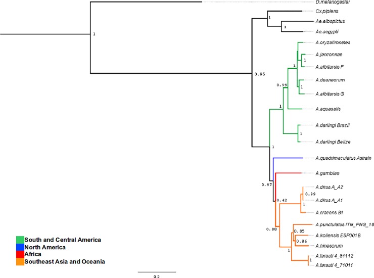
A phylogenetic tree was reconstructed using Bayesian analysis with BEAST v1.7.5 [49]. All the phylogenetic relationships were supported with robust posterior probabilities greater than 90%, with the exception of the position of A. gambiae (42%) and the internal nodes amongst the South East Asia and Oceania anophelines included (ranging from 85 to 88%). The reconstructed Anopheles phylogenetic tree is shown in Fig 3. From examining the tree topology, and considering the current continental distribution, a deep divergence between two Anopheles geographical lineages was observed. One clear monophyletic branch grouping Central and South American anophelines, and a second lineage grouping North American, Asian/Oceania, and African anophelines. As expected, this radiation pattern resembles the one published by Logue et al., (2013) [6]. The monophyletic clades corresponding to the A. punctulatus, A. dirus and A. albitarsis groups are also evident.
Fig 3. By-geographic region phylogeny of representative Anopheles, using the concatenated DNA sequences of all the mitochondrial protein coding genes.
The values on the nodes correspond to the posterior probabilities supporting the tree topology. The phylogenetic tree was reconstructed using the concatenated PCGs and the Bayesian Markov Chain Monte Carlo approach (MCMC) analysis implemented in BEAST v1.7.5.
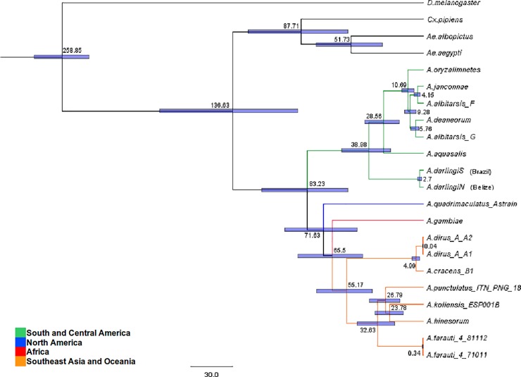
The currently available fossil record for mosquitoes is poor [7, 56]. Thus, we estimated the divergence times amongst anopheline species using the Drosophila-Anopheles divergence time (set at 260 MYA) as the only calibration point time as previously suggested [6, 50]. We dated the most recent common ancestor (MRCA) of all Anopheles to 83.23 MYA with a 95% credibility interval ranging from 54.33 to 115.88 MYA as shown in Table 2. Amongst Neotropical anophelines, the MRCA within the A. albitarsis complex and A. aquasalis dates to 28.56 MYA with a 95% credibility interval ranging from 17.10 to 42.12 MYA. This MRCA is younger than the one shared between A. darlingi and the A. albitarsis complex, which dates back to 38.98 MYA as it can be observed in Table 2 and Fig 4.
Table 2. Mean divergence times in MYA (million of years ago), and 95% credibility intervals for selected nodes.
| Drosophila / Anopheles (Calibration point ~260mya) | 258.85 | [239.70–278.86] |
| Anophelinae / Culicinae | 136.63 | [90.13–189.15] |
| Anopheles genus | 83.23 | [54.33–115.88] |
| Anopheles gambiae / SE Asia—Oceania anophelines | 65.50 | [43.54–89.80] |
| Anopheles darlingi / Anopheles albitarsis complex | 38.98 | [23.54–58.32] |
| Anopheles aquasalis / Anopheles albitarsis complex | 28.56 | [17.10–42.12] |
Output of the analysis performed with BEAST v1.7.5 as explained in the Methods section. The measures in the second and third column correspond to the mean ages, and 95% credibility intervals determined for each of the selected nodes respectively.
Fig 4. Phylogenetic tree of selected Anopheles using the concatenated DNA sequences of all the mitochondrial protein coding genes.
The values on the tree nodes correspond to the mean divergence time (MYA) estimated for each event. The bars illustrate the 95% credibility intervals of the divergence times. (Table 2 presents the selected key divergence events).
Discussion
Greater integration of nuclear and mitochondrial genomic studies is necessary to further our understanding of the Anopheles genomic evolution, the phylogenetic relationships between vectorial competence traits, and the co-evolutionary history of this genus with the human malaria parasites they may harbor. Thus, in the current “omics” era, extending the wealth of representative anopheline mitogenomes available is a necessary and feasible task.
Like in most metazoans, the assembled mitochondrial genome of A. aquasalis is a small, double-stranded, circular DNA molecule. Encompassed in 15,393 base pairs, we annotated a densely packaged set of 13 protein-coding genes, 22 tRNA genes, and two rRNA genes as shown in Fig 1. Genome structures with highly conserved features, composition, and organization have been reported among insects [51] such as anophelines [15].
When regarding genome architecture, the occurrence of overlapping open reading frames implies a genome with a compact structure. This feature can be observed by mtDNA sequences of culicines, anophelines, and other insect mitogenomes [47, 52, 53]. The reason for such characteristic can be (structurally) attributed to the small intergenic distances between consecutive genes (intergenic distance < x bp) as the following show: < 23 bp in Ae. aegypti; < 24 bp in C. quinquefasciatus; < 21 bp in Ae. albopictus; < 16 bp in A. gambiae; < 18 bp in Anopheles quadrimaculatus; < 30 bp in Drosophila yakuba, and < 30 bp in D. melanogaster (as recounted by [53]), and in the case of A. aquasalis< 17 bp. Once a free-living prokaryote, mitochondria underwent genome reduction as it transitioned into an obligate endosymbiont harboring tightly packed remnants from the eubacterial genome of its ancestor [54].
The intergenic distances do not account for the control region, which in the A. aquasalis mitogenome spans 558bp. This major non-coding region is also known as the A+T-rich region, and it plays a role in the initiation of transcription and replication [55]. The length of this region is highly variable among different insects due to the high rate of nucleotide substitutions, insertions/deletions, and a variable number of tandem repeats it can have [56, 57].
Almost all the tRNA sequences annotated could be folded into typical cloverleaf secondary structures (S1 Fig) exposing the adequate anticodon triplet, except for the DHU-arm of tRNASer, which is absent as it has been observed in other insects Li et al., 2013; Zhao et al., 2013 [25, 47] and references therein. Deficiencies regarding secondary structures of tRNA are often observed in protozoans, fungi, algae, plants and low metazoans [58]. The causes of such deficiencies range from aberrant loops and non-Watson-Crick matches to short arms, and all may induce aberrant tRNAs to lose their function, but a “corrective” post-transcriptional RNA editing mechanism has been proposed [59, 60]. The tRNA genes are embedded in variable regions within the mitogenome [51]. Throughout evolution, these regions underwent rearrangements more often than protein-coding regions. Consequently, tRNA order is nowadays explored as an additional tool for enhanced comparative phylogenetic analysis as between species [61].
The nucleotide composition of the complete A. aquasalis mtDNA sequence is clearly biased towards a high A+T content (77.16%) as it can be seen in S1 Table. This result was similar to the base composition described for other anophelines. The majority strand (L) of the A. aquasalis mtDNA has a compositional bias (positive AT skew and a negative GC skew). This means that this strand is richer in A and C. Though this is the tendency in insects, exceptions exist in arthropod mitogenomes in which the trend is reversed towards a composition with less A & C than T & G on the majority strand [62]. The underlying mechanism that leads to the strand bias, though unknown, has been linked to asymmetric replication and transcription processes. During both, one strand is transiently in a single-stranded state, and thus, it is left more prone to DNA damage. This phenomenon has been widely considered to account for the biased occurrence of mutations between the mtDNA strands [63].
Most of the start codons of the annotated PCGs followed the ATN rule, as described previously for other anophelines [15, 64]. Nonetheless, this was not the case for COI and ND5; genes that have TCG and GTG start respectively (Fig 1). The latter start codon characteristic has also been reported in other anopheline mitogenomes [15, 64, 65]. As observed in A. darling [15], eight genes use the complete stop codon TAA except for COI, COII, COIII, ND5, and ND4, which terminate with a single T (Fig 1). The incomplete stop codon is a reported phenomenon for insect mitogenomes [15, 25, 49, 66]. It has been proposed that the completion of the TAA termination codon is due to a post-transcriptional polyadenylation synthesis [67].
The total number of non-stop codons within the predicted PCGs (3743) was similar to that reported for A. darlingi and A. gambiae (3733), differing slightly to that of A. quadrimaculatus (3715). The nucleotide bias was also reflected in the codon usage within the annotated PCGs. A strong bias towards A+T rich codons like TTA (L), TTT (F), ATT (I), ATA (M), and AAT (N) is a compositional tendency that follows what has been reported as the canonical pattern for Culicidae species [15, 66]. When compared to other insects, such as aphids, the same five codons were the most prevalent, differing only in the exchanged F and I positions [49].
Among the 32 most frequently used codons (RSCU > 1) 29 were of the NNU and NNA type. This indicates that the third position of the codons mostly are either U or A. This differs from the expected trend for dipterans in which, frequently used codons present G or C in the third position [53].
The mitochondrial genome of insects displays unique characteristics such as high codon bias, low GC3content, and a highly conserved gene arrangement [51]. These characteristics are associated with the conserved patterns of Shine-Dalgarno sequences found in the transcripts of functional proteins. In addition, the aforementioned characteristics promote the high expression level of the mitochondrial genes [68, 69]. As a consequence of the above, mitochondrial activity of insects and nematodes would render them more tolerant to increased concentrations of reactive oxygen species (ROS) [70]. In the case of anophelines, it is interesting to consider linking this enhanced cellular resistance hypothesis with the detoxification mechanisms [71] towards the oxidative stress triggered as a response to the immune challenge posed by the Plasmodium infection [72, 73].
The sequence identity comparison and compositional patterns analyses we performed showed how the nucleotide sequence of protein-coding genes are a conserved feature of anopheline mitogenomes, in which most differences have been reported allocated within the control region [15, 74]. In terms of sequence identity (all values > 86%), a pattern was observed (S3 Fig). In general, the A. aquasalis PCGs are more similar to those of the other Neotropical anophelines tested, when compared to those from A. gambiae and A. punctulatus (African and Asian malaria vectors). Higher sequence similarity could be obeying the phylogeographic and divergence history of anophelines. Considering that, on average, the highest similarity values were obtained with A. albitarsis, another brackish-water Neotropical anopheline, from which A. aquasalis diverged ~28 MYA (Fig 4). The identity between rRNA sequences ranged from 93% to 98% (S4 Table). Similar identity values described in aphids prompted Wang et al., (2013) [49] to suggest that the predicted and potentially conserved rRNA secondary structures could improve phylogenetic analyses between insect species.
Human malaria vectors from different species complexes are not closely related. This implies that some of the competence traits potentially arose (or were lost) independently, multiple times within the different extant anopheline lineages. Therefore, the evolutionary history of genes associated with vector competence might have been driven by rapid processes. This would imply that such genes are not highly conserved sequences with a single ancestor as origin [75]. The publication of 16 anopheles genomes [8] might have generated, concomitantly, mitogenomes that will broaden our perspective on the phylogenetic and divergence relationships between the members of the Anopheles genus. This would be particularly important since both our study, and Logue et al., (2013) [6], encountered the paradoxical situation regarding the scarcity of mitogenomes from dominant malaria vectors pertaining to Africa (until recently) [76, 77] and the East Mediterranean regions, areas still burdened by human malaria [78]. For instance, at the time of our study, we opted to leave out the mitogenome of Anopheles funestus due the amount of gaps in the mtDNA PCGs sequences (GenBank accession number NC008070). It was not until recently that mitochondrial lineages of this important vector were thoroughly studied [77].
The reconstructed Anopheles phylogeny showed a deep divergence between two main Anopheles lineages (Fig 3) seemingly driven by their phylogeographic relations and the earth's geologic eras. The tree topology obtained is consistent with the current hypothesis regarding the origin of Anopheles mosquitoes in the Gondwana supercontinent during the Cretaceous period [12, 14]. Anopheles mosquitoes would have radiated from what is recognized as South America, then arrived in Africa and from there then colonized Europe and North America (with the aid of land bridges), also migrating through Asia, and into the Pacific [6]
However, as previously mentioned, paucity regarding mitogenomes from African, Asian and European anophelines, hindered the chance of determining accurately if African anophelines are the ancestors of those in Europe and North America, or if North American anophelines radiated from South America. For instance, both our results and those of Logue et al., (2013) [6], represent a phylogeny in which the position of Anopheles quadrimaculatus seems unresolved.
Nonetheless, Freitas et al., (2015) [7] provides a thorough revision of phylogenetic relations based on COI, COII and 5.8S rRNA genes which allowed for more species to be tested. When comparing with it, we observed how the mitogenome based relations here reported are the same at the subgenus level as Neafsey et al., (2015) [8] and Krzywinski et al., (2001) [10], being (Nyssorhynchus, (Anopheles, Cellia)). Whereas their study was not able to significantly resolve the evolutionary relationship within this taxonomic level, other studies have reported different associations too, namely, Cellia, (Nyssorhynchus, Anopheles) [79]. A recent comparative evolutionary mitochondriomic study strongly supported the sister relationship of Nyssorhynchus + Kerteszia and Cellia + Anopheles based on 50 complete mitogenomes [80]. Thus, this evolutionary relationship seems to be conclusively elucidated at this taxonomic level.
It has been stated that interpreting the current distributions of anophelines from an evolutionary context, may be quite problematic [7]. This author reviewed existing alternative hypotheses, some suggesting a different scenario for the evolution of the extant groups of the Anophelinae subfamily. The scenario would reflect the ideas presented by Christophers (1933) [81], in which the ancestral lineage of Anopheles existed prior to the split of Pangaea, diversifying into the modern species after the breakup of the continents. Nonetheless, it is not the scope of this study to dig deeper into this matter.
Regarding the estimated divergence times, we dated the Anopheles MRCA to ~ 83 MYA. The node age (Fig 4) differs from the ~ 79 MYA reported by Moreno et al., (2010) [16], the ~ 93 MYA estimated by Logue et al., (2013) [6] and 110 MYA by Freitas et al., (2015) [78]. These molecular dating estimates, though different, are in a degree of agreement with the breakup of Western Gondwana and the loss of land connections between South America and Africa (90–95 MYA). This geologic event might have prompted the divergence between both Anopheles + Cellia from Nyssorhynchus, and further on, the divergence between Cellia and Anopheles ~ 71 MYA or ~ 81 MYA [6]. The geographic sorting of lineages thus would coincide with the loss of land bridges connecting Africa and Europe, as well as between Europe and North America. This, in turn, could account for the absence of Cellia in the New World and Nyssorhynchus in the Afro-Eurasian continents [10, 15]. In agreement with the above Freitas et al., (2015) [78] determined, by ancestral area reconstruction, that a monophyletic clade composed of the Neotropical subgenera Stethomyia, Kerteszia and Nyssorhynchus had a common ancestor distributed along the Americas, and whose early radiation in the continent began around the Late Cretaceous (~ 90 MYA).
The data suggest that A. aquasalis diverged from the A. albitarsis complex ~ 28 MYA. The node age implies that their MRCA would be younger than the one shared between A. darlingi and the A. albitarsis complex which dates back to ~ 38 MYA (Fig 4). If Neotropical anophelines bionomics are considered, then A. aquasalis would have diverged from the A. albitarsis complex, adapting to its narrow coastal ecological niche, outcompeted by inland (A. darlingi) and brackish-water adapted (A. albitarsis) sibling species [82].
As brought forward by Freitas et al., (2015) [78], when comparing their results to Fontaine et al., (2015) [9], phylogenetic reconstructions for Anopheles require robust data from multiple molecular markers, and individuals per species. It is highlighted by the authors that unclear phylogenies regarding African anophelines, are due to complex speciation processes that have been permeated by recurring events of introgressive hybridization. Nonetheless, it drew our attention that, based on X chromosome phylogenies (which reflect species branching order), Fontaine et al., (2015) [9], determined that brackish water adapted Anopheles merus and Anopheles melas [83], diverged from bionomical different inland sibling species ~1.85–1.47 MYA, whereas A. aquasalis might have diverged from inland sibling A. darlingi ~38.98 MYA. As evidence for phylosymbiosis emerges, including examples of non-brackish water adapted or Neotropical anophelines [84], it becomes relevant to assess if co-evolution of A. aquasalis and its aquatic niche acquired microbiota, follow this trend. As a growing field of interest [85–87], exploring the ecological adaptations of anophelines to particular breeding sites, and its connection to their microbiota signature profiles, should generate interesting insights when comparing similar bionomical species for example.
The observed differences regarding radiation and divergence node ages have been reported by other authors [7, 12, 15]. Notably, the choices regarding data inclusion have a direct effect on phylogenomic studies. Therefore, the use of full mitogenomes, individual or concatenated genes (PCGs, rRNAs, tRNAs) or nuclear genes, can be accounted as the reason for the differences between molecular evolutionary histories reported to date [16, 51]. Additionally, observed discrepancies may also be due to: depth of species sampling while seeking for representatives of the geographical regions and /or the subgenera studied; the choice (and use) of mutation rates; and the amount of calibration points employed [6, 12, 15]. For example, Moreno et al., (2010) [15] used as an additional calibration point, the Anophelinae and Culicinae divergence age estimated to be 120 MYA [88]. Nevertheless, the most rigorously calculated date available involving the split between a mosquito lineage and a sister taxon is the divergence time of 259.9 MYA between Drosophila and Anopheles estimated by Gaunt & Miles, (2002), as highlighted by Krzywinski et al., (2006) [12, 44]. As there are many pitfalls inherent to the use of molecular clocks to date divergences, the outcome of age estimation should be interpreted with caution [12].
Molecular dating of the divergence events reported in this study occurred long before humans arrived in America 15–20 millennia ago [89]. Evidence suggests that Plasmodium parasites were introduced into the Americas via African slave trade routes during the European invasion [20]. In order to the transmission cycle to establish, the parasite had to adapt to the populations of local mosquitoes. Then, in accordance with the present geographic distribution of anophelines [17], and the known slave trade disembarking ports, some of the first indigenous vectors that P. falciparum-infected humans encountered would have been: A. albimanus, A. aquasalis, A. darlingi, A. albitarsis and A. quadrimaculatus [90].
Conclusion
Considering the evolutionary distance between Old and New World anophelines, it is not surprising that there are marked differences at genetic, ecological and behavioral levels between them [17]. This probably resulted in a more stringent selection upon Plasmodium as it adapted to these new vectors [90]. Multiple Neotropical anophelines became human malaria vectors independently from each other, including the anophelines from South East Asian and Oceania [6]. This suggests that co-occurrences of anopheline traits related to malaria transmission would be the outcome of convergent evolution [91].
Readily available anopheline phylogenies are key to perform co-evolutionary studies between parasites and vectors to gain insights into their interspecific relationships [7]. Recently, Molina-Cruz & Barillas-Mury, (2014) [90], proposed a hypothesis linking the Plasmodium protein PFS47, and a still unknown interacting protein from Anopheles, acting as a critical determinant of mosquito-parasite compatibility and thus the “emergence” of vector competence in this genus [90, 92, 93]. Considering that the Pfs47 gene is polymorphic, and presents a robust clonal distribution, we support the need of a better and more accurate Anopheles phylogeny. This, in turn, could enable the reconstruction of the history of malaria transmission in the New World.
Supporting information
22 tRNAs were identified in the mitogenome of A. aquasalis and their cloverleaf secondary structures predicted with RNAStructure. Using Dayhoff’s single letter amino acid code.
(TIF)
A-T content expressed as AT%, AT skew, and GC skew were estimated and plotted for each single PCG and for other genomic regions according to the legend below the chart. Refer to the S5 and S6 Tables to access the tabular data regarding each parameter.
(TIF)
Nucleotide identity (%) between A. aquasalis and selected Anopheles species ranging from 80 to 100%. The scale is shown along the main vertical axis of the radar plot.
(TIF)
(XLSX)
Codon frequency and Relative synonymous Codon Usage (RSCU) are shown for each codon.
(XLSX)
(XLSX)
(XLSX)
(XLSX)
(XLSX)
Acknowledgments
The mitogenome characterization was performed at the Computational Biology facility at FIOCRUZ-MG. Authors express their gratitude to Dr. Guilherme Oliveira, who lead this group at the time.
Data Availability
The data underlying the results of this study are available in the NCBI Database as part of the Anopheles aquasalis whole genome shotgun sequencing project; accession number: NJHH00000000.
Funding Statement
This work used the Vincent J. Coates Genomics Sequencing Laboratory at UC Berkeley - USA supported by NIH S10 OD018174 Instrumentation Grant. The following Brazilian agencies partially funded this study: Fundação do Instituto Oswaldo Cruz (FIOCRUZ – PAPES VI); Conselho Nacional de Desenvolvimento Científico e Tecnológico (CNPq); Coordenaçao de Aperfeicoamento de Pessoal de Nivel Superior (CAPES); Institutos Nacionais de Ciência e Tecnologia (INCTs Entomologia Molecular e Elimina Malária); Fundações de Amparo à Pesquisa dos Estados de Minas Gerais e Amazonas (FAPEMIG and FAPEAM). LMV was a Ph.D. student supported by the Programa de Estudantes - Convênio de Pos-Graduação (PEC-PG - CAPES). GCL was a fellow sponsored by Programme Science without Borders (CAPES); PFPP, NFCS, and WMM are research fellows supported by CNPq.
References
- 1.Wolstenholme DR. Genetic novelties in mitochondrial genomes of multicellular animals. Curr Opin Genet Dev.1992; 2(6): 918–925. [DOI] [PubMed] [Google Scholar]
- 2.Boore JL. Animal mitochondrial genomes. Nucleic Acids Res.1999; 27(8): 1767–1780. 10.1093/nar/27.8.1767 [DOI] [PMC free article] [PubMed] [Google Scholar]
- 3.Mwinyi A, Meyer A, Bleidorn C, Lieb B, Bartolomaeus T, and Podsiadlowski L. Mitochondrial genome sequence and gene order of Sipunculus nudus give additional support for an inclusion of Sipuncula into Annelida. BMC Genomics.2009; 10: 27 10.1186/1471-2164-10-27 [DOI] [PMC free article] [PubMed] [Google Scholar]
- 4.Braband A, Cameron SL, Podsiadlowski L, Daniels SR, and Mayer G. The mitochondrial genome of the onychophoran Opisthopatus cinctipes (Peripatopsidae) reflects the ancestral mitochondrial gene arrangement of Panarthropoda and Ecdysozoa. Mol Phylogenet Evol.2010; 57(1): 285–292. 10.1016/j.ympev.2010.05.011 [DOI] [PubMed] [Google Scholar]
- 5.Harbach RE. Genus Anopheles Meigen, 1818. Mosquito Taxonomic Inventory Book Genus Anopheles Meigen, 1818 Mosquito Taxonomic Inventory City.2011. [Google Scholar]
- 6.Logue K, Chan ER, Phipps T, Small ST, Reimer L, Henry-Halldin C et al. Mitochondrial genome sequences reveal deep divergences among Anopheles punctulatus sibling species in Papua New Guinea. Malar J.2013; 12: 64 10.1186/1475-2875-12-64 [DOI] [PMC free article] [PubMed] [Google Scholar]
- 7.Harbach RE. The phylogeny and classification of Anopheles. InTech;2013. [Google Scholar]
- 8.Neafsey DE, Waterhouse RM, Abai MR, Aganezov SS, Alekseyev MA, Allen JE et al. Mosquito genomics. Highly evolvable malaria vectors: the genomes of 16 Anopheles mosquitoes. Science.2015; 347(6217): 1258522 10.1126/science.1258522 [DOI] [PMC free article] [PubMed] [Google Scholar]
- 9.Fontaine MC, Pease JB, Steele A, Waterhouse RM, Neafsey DE, Sharakhov IV et al. Mosquito genomics. Extensive introgression in a malaria vector species complex revealed by phylogenomics. Science.2015; 347(6217): 1258524 10.1126/science.1258524 [DOI] [PMC free article] [PubMed] [Google Scholar]
- 10.Krzywinski J, Wilkerson RC, and Besansky NJ. Evolution of mitochondrial and ribosomal gene sequences in anophelinae (Diptera: Culicidae): implications for phylogeny reconstruction. Mol Phylogenet Evol.2001; 18(3): 479–487. 10.1006/mpev.2000.0894 [DOI] [PubMed] [Google Scholar]
- 11.Ad Torre, Fanello C, Akogbeto M, Dossou-Yovo J, Favia G, Petrarca V et al. Molecular evidence of incipient speciation within Anopheles gambiae ss in West Africa. Insect molecular biology. Wiley Online Library;2001; 10(1): 9–18. [DOI] [PubMed] [Google Scholar]
- 12.Krzywinski J, Grushko OG, and Besansky NJ. Analysis of the complete mitochondrial DNA from Anopheles funestus: an improved dipteran mitochondrial genome annotation and a temporal dimension of mosquito evolution. Mol Phylogenet Evol.2006; 39(2): 417–423. 10.1016/j.ympev.2006.01.006 [DOI] [PubMed] [Google Scholar]
- 13.Reidenbach KR, Cook S, Bertone MA, Harbach RE, Wiegmann BM, and Besansky NJ. Phylogenetic analysis and temporal diversification of mosquitoes (Diptera: Culicidae) based on nuclear genes and morphology. BMC Evol Biol.2009; 9: 298 10.1186/1471-2148-9-298 [DOI] [PMC free article] [PubMed] [Google Scholar]
- 14.Krzywinski J and Besansky NJ. Molecular systematics of Anopheles: from subgenera to subpopulations. Annu Rev Entomol.2003; 48: 111–139. 10.1146/annurev.ento.48.091801.112647 [DOI] [PubMed] [Google Scholar]
- 15.Moreno M, Marinotti O, Krzywinski J, Tadei WP, James AA, Achee NL et al. Complete mtDNA genomes of Anopheles darlingi and an approach to anopheline divergence time. Malar J.2010; 9: 127 10.1186/1475-2875-9-127 [DOI] [PMC free article] [PubMed] [Google Scholar]
- 16.Marinotti O, Cerqueira GC, de Almeida LG, Ferro MI, Loreto EL, Zaha A et al. The genome of Anopheles darlingi, the main neotropical malaria vector. Nucleic Acids Res.2013; 41(15): 7387–7400. 10.1093/nar/gkt484 [DOI] [PMC free article] [PubMed] [Google Scholar]
- 17.Sinka ME, Bangs MJ, Manguin S, Rubio-Palis Y, Chareonviriyaphap T, Coetzee M et al. A global map of dominant malaria vectors. Parasit Vectors.2012; 5: 69 10.1186/1756-3305-5-69 [DOI] [PMC free article] [PubMed] [Google Scholar]
- 18.De Castro MC and Singer BH. Was malaria present in the Amazon before the European conquest? Available evidence and future research agenda. Journal of Archaeological Science. Elsevier;2005; 32(3): 337–340. [Google Scholar]
- 19.Culleton R, Coban C, Zeyrek FY, Cravo P, Kaneko A, Randrianarivelojosia M et al. The origins of African Plasmodium vivax; insights from mitochondrial genome sequencing. PLoS One.2011; 6(12): e29137 10.1371/journal.pone.0029137 [DOI] [PMC free article] [PubMed] [Google Scholar]
- 20.Yalcindag E, Elguero E, Arnathau C, Durand P, Akiana J, Anderson TJ et al. Multiple independent introductions of Plasmodium falciparum in South America. Proc Natl Acad Sci USA.2012; 109(2): 511–516. 10.1073/pnas.1119058109 [DOI] [PMC free article] [PubMed] [Google Scholar]
- 21.Pimenta PF, Orfano AS, Bahia AC, Duarte AP, Rios-Velasquez CM, Melo FF et al. An overview of malaria transmission from the perspective of Amazon Anopheles vectors. Mem Inst Oswaldo Cruz.2015; 110(1): 23–47. 10.1590/0074-02760140266 [DOI] [PMC free article] [PubMed] [Google Scholar]
- 22.Masta SE, Longhorn SJ, and Boore JL. Arachnid relationships based on mitochondrial genomes: asymmetric nucleotide and amino acid bias affects phylogenetic analyses. Mol Phylogenet Evol.2009; 50(1): 117–128. 10.1016/j.ympev.2008.10.010 [DOI] [PubMed] [Google Scholar]
- 23.Hickerson MJ, Carstens BC, Cavender-Bares J, Crandall KA, Graham CH, Johnson JB et al. Phylogeography's past, present, and future: 10 years after Avise, 2000. Mol Phylogenet Evol.2010; 54(1): 291–301. 10.1016/j.ympev.2009.09.016 [DOI] [PubMed] [Google Scholar]
- 24.Santini F, Kong X, Sorenson L, Carnevale G, Mehta RS, and Alfaro ME. A multi-locus molecular timescale for the origin and diversification of eels (Order: Anguilliformes). Mol Phylogenet Evol.2013; 69(3): 884–894. 10.1016/j.ympev.2013.06.016 [DOI] [PubMed] [Google Scholar]
- 25.Zhao Z, Su TJ, Chesters D, Wang SD, Ho SY, Zhu CD et al. The mitochondrial genome of Elodia flavipalpis Aldrich (Diptera: Tachinidae) and the evolutionary timescale of Tachinid flies. PLoS One.2013; 8(4): e61814 10.1371/journal.pone.0061814 [DOI] [PMC free article] [PubMed] [Google Scholar]
- 26.Perseke M, Golombek A, Schlegel M, and Struck TH. The impact of mitochondrial genome analyses on the understanding of deuterostome phylogeny. Mol Phylogenet Evol.2013; 66(3): 898–905. 10.1016/j.ympev.2012.11.019 [DOI] [PubMed] [Google Scholar]
- 27.de Carvalho SC, Martins Junior AJ, Lima JB, and Valle D. Temperature influence on embryonic development of Anopheles albitarsis and Anopheles aquasalis. Mem Inst Oswaldo Cruz.2002; 97(8): 1117–1120. 10.1590/s0074-02762002000800009 [DOI] [PubMed] [Google Scholar]
- 28.da Silva AN, Dos Santos CC, Lacerda RN, Santa Rosa EP, De Souza RT, Galiza D et al. Laboratory colonization of Anopheles aquasalis (Diptera: Culicidae) in Belem, Para, Brazil. J Med Entomol.2006; 43(1): 107–109. [PubMed] [Google Scholar]
- 29.Rios-Velasquez CM, Martins-Campos KM, Simoes RC, Izzo T, dos Santos EV, Pessoa FA et al. Experimental Plasmodium vivax infection of key Anopheles species from the Brazilian Amazon. Malar J.2013; 12: 460 10.1186/1475-2875-12-460 [DOI] [PMC free article] [PubMed] [Google Scholar]
- 30.Zerbino DR and Birney E. Velvet: algorithms for de novo short read assembly using de Bruijn graphs. Genome Res.2008; 18(5): 821–829. 10.1101/gr.074492.107 [DOI] [PMC free article] [PubMed] [Google Scholar]
- 31.Kurtz S, Phillippy A, Delcher AL, Smoot M, Shumway M, Antonescu C et al. Versatile and open software for comparing large genomes. Genome Biol.2004; 5(2): R12 10.1186/gb-2004-5-2-r12 [DOI] [PMC free article] [PubMed] [Google Scholar]
- 32.Bernt M, Donath A, Juhling F, Externbrink F, Florentz C, Fritzsch G et al. MITOS: improved de novo metazoan mitochondrial genome annotation. Mol Phylogenet Evol.2013; 69(2): 313–319. 10.1016/j.ympev.2012.08.023 [DOI] [PubMed] [Google Scholar]
- 33.Rutherford K, Parkhill J, Crook J, Horsnell T, Rice P, Rajandream MA et al. Artemis: sequence visualization and annotation. Bioinformatics.2000; 16(10): 944–945. 10.1093/bioinformatics/16.10.944 [DOI] [PubMed] [Google Scholar]
- 34.Lowe TM and Eddy SR. tRNAscan-SE: a program for improved detection of transfer RNA genes in genomic sequence. Nucleic Acids Res.1997; 25(5): 955–964. 10.1093/nar/25.5.955 [DOI] [PMC free article] [PubMed] [Google Scholar]
- 35.Bellaousov S, Reuter JS, Seetin MG, and Mathews DH. RNAstructure: Web servers for RNA secondary structure prediction and analysis. Nucleic Acids Res.2013; 41(Web Server issue): W471–W474. 10.1093/nar/gkt290 [DOI] [PMC free article] [PubMed] [Google Scholar]
- 36.Alikhan NF, Petty NK, Ben Zakour NL, and Beatson SA. BLAST Ring Image Generator (BRIG): simple prokaryote genome comparisons. BMC Genomics.2011; 12: 402 10.1186/1471-2164-12-402 [DOI] [PMC free article] [PubMed] [Google Scholar]
- 37.Perna NT and Kocher TD. Patterns of nucleotide composition at fourfold degenerate sites of animal mitochondrial genomes. J Mol Evol.1995; 41(3): 353–358. [DOI] [PubMed] [Google Scholar]
- 38.Tamura K, Stecher G, Peterson D, Filipski A, and Kumar S. MEGA6: Molecular Evolutionary Genetics Analysis version 6.0. Mol Biol Evol.2013; 30(12): 2725–2729. 10.1093/molbev/mst197 [DOI] [PMC free article] [PubMed] [Google Scholar]
- 39.Zhang Z, Schwartz S, Wagner L, and Miller W. A greedy algorithm for aligning DNA sequences. Journal of Computational biology. Mary Ann Liebert, Inc.;2000; 7(1–2): 203–214. 10.1089/10665270050081478 [DOI] [PubMed] [Google Scholar]
- 40.Abascal F, Zardoya R, and Telford MJ. TranslatorX: multiple alignment of nucleotide sequences guided by amino acid translations. Nucleic Acids Res.2010; 38(Web Server issue): W7–13. 10.1093/nar/gkq291 [DOI] [PMC free article] [PubMed] [Google Scholar]
- 41.Kuck P and Meusemann K. FASconCAT: Convenient handling of data matrices. Mol Phylogenet Evol.2010; 56(3): 1115–1118. 10.1016/j.ympev.2010.04.024 [DOI] [PubMed] [Google Scholar]
- 42.Posada D. jModelTest: phylogenetic model averaging. Mol Biol Evol.2008; 25(7): 1253–1256. 10.1093/molbev/msn083 [DOI] [PubMed] [Google Scholar]
- 43.Drummond AJ and Rambaut A. BEAST: Bayesian evolutionary analysis by sampling trees. BMC evolutionary biology. BioMed Central;2007; 7(1): 214. [DOI] [PMC free article] [PubMed] [Google Scholar]
- 44.Gaunt MW and Miles MA. An insect molecular clock dates the origin of the insects and accords with palaeontological and biogeographic landmarks. Molecular Biology and Evolution.2002; 19(5): 748–761. 10.1093/oxfordjournals.molbev.a004133 [DOI] [PubMed] [Google Scholar]
- 45.Yamasaki YK, Nieman CC, Chang A, Collier TC, Main BJ, and Lee Y. Improved tools for genomic DNA library construction of small insects [version 1; not peer reviewed]. F1000Res.2016; (5): 211. [Google Scholar]
- 46.Plazzi F, Ribani A, and Passamonti M. The complete mitochondrial genome of Solemya velum (Mollusca: Bivalvia) and its relationships with conchifera. BMC Genomics.2013; 14: 409 10.1186/1471-2164-14-409 [DOI] [PMC free article] [PubMed] [Google Scholar]
- 47.Li T, Gao C, Cui Y, Xie Q, and Bu W. The complete mitochondrial genome of the stalk-eyed bug Chauliops fallax Scott, and the monophyly of Malcidae (Hemiptera: Heteroptera). PLoS One.2013; 8(2): e55381 10.1371/journal.pone.0055381 [DOI] [PMC free article] [PubMed] [Google Scholar]
- 48.Boore JL, Macey JR, and Medina M. Sequencing and comparing whole mitochondrial genomes of animals. Methods Enzymol.2005; 395: 311–348. 10.1016/S0076-6879(05)95019-2 [DOI] [PubMed] [Google Scholar]
- 49.Wang Y, Huang XL, and Qiao GX. Comparative analysis of mitochondrial genomes of five aphid species (Hemiptera: Aphididae) and phylogenetic implications. PLoS One.2013; 8(10): e77511 10.1371/journal.pone.0077511 [DOI] [PMC free article] [PubMed] [Google Scholar]
- 50.Poinar GO, Zavortinik TJ, Pike T, and Johnston PA. Paleoculicis minututs (Diptera: Culicidae) n. Gen., n. Sp., from Cretaceous Canadian amber, with a summary of described fossil mosquitoes. Acta Geologica Hispanica.2000; 35(1): 119–130. [Google Scholar]
- 51.Cameron SL. Insect mitochondrial genomics: implications for evolution and phylogeny. Annu Rev Entomol.2014; 59: 95–117. 10.1146/annurev-ento-011613-162007 [DOI] [PubMed] [Google Scholar]
- 52.Stewart JB and Beckenbach AT. Insect mitochondrial genomics: the complete mitochondrial genome sequence of the meadow spittlebug Philaenus spumarius (Hemiptera: Auchenorrhyncha: Cercopoidae). Genome. NRC Research Press;2005; 48(1): 46–54. 10.1139/g04-090 [DOI] [PubMed] [Google Scholar]
- 53.Behura SK and Severson DW. Comparative analysis of codon usage bias and codon context patterns between dipteran and hymenopteran sequenced genomes. PLoS One.2012; 7(8): e43111 10.1371/journal.pone.0043111 [DOI] [PMC free article] [PubMed] [Google Scholar]
- 54.Timmis JN, Ayliffe MA, Huang CY, and Martin W. Endosymbiotic gene transfer: organelle genomes forge eukaryotic chromosomes. Nature Reviews Genetics. Nature Publishing Group;2004; 5(2): 123 10.1038/nrg1271 [DOI] [PubMed] [Google Scholar]
- 55.Wolstenholme DR. Animal mitochondrial DNA: structure and evolution. Int Rev Cytol.1992; 141: 173–216. [DOI] [PubMed] [Google Scholar]
- 56.Fauron CM and Wolstenholme DR. Extensive diversity among Drosophila species with respect to nucleotide sequences within the adenine + thymine-rich region of mitochondrial DNA molecules. Nucleic Acids Res.1980; 8(11): 2439–2452. 10.1093/nar/8.11.2439 [DOI] [PMC free article] [PubMed] [Google Scholar]
- 57.Inohira K, Hara T, and Matsuura ET. Nucleotide sequence divergence in the A+T-rich region of mitochondrial DNA in Drosophila simulans and Drosophila mauritiana. Mol Biol Evol.1997; 14(8): 814–822. 10.1093/oxfordjournals.molbev.a025822 [DOI] [PubMed] [Google Scholar]
- 58.Schneider A and Marechal-Drouard L. Mitochondrial tRNA import: are there distinct mechanisms? Trends in cell biology. Elsevier;2000; 10(12): 509–513. [DOI] [PubMed] [Google Scholar]
- 59.Tomita K and Weiner AM. Collaboration between CC-and A-adding enzymes to build and repair the 3'-terminal CCA of tRNA in Aquifex aeolicus. Science.2001; 294(5545): 1334–1336. 10.1126/science.1063816 [DOI] [PubMed] [Google Scholar]
- 60.Li R and Guan MX. Human mitochondrial leucyl-tRNA synthetase corrects mitochondrial dysfunctions due to the tRNALeu(UUR) A3243G mutation, associated with mitochondrial encephalomyopathy, lactic acidosis, and stroke-like symptoms and diabetes. Mol Cell Biol.2010; 30(9): 2147–2154. 10.1128/MCB.01614-09 [DOI] [PMC free article] [PubMed] [Google Scholar]
- 61.Silverio MS, Rodovalho VdR, Bonetti AM, de Oliveira GC, Cuadros-Orellana S, Ueira-Vieira C et al. Preliminary characterization of mitochondrial genome of Melipona scutellaris, a Brazilian stingless bee. BioMed research international. Hindawi;2014; 2014: 1–6. [DOI] [PMC free article] [PubMed] [Google Scholar]
- 62.Wei SJ, Shi M, Chen XX, Sharkey MJ, van AC, Ye GY et al. New views on strand asymmetry in insect mitochondrial genomes. PLoS One.2010; 5(9): e12708 10.1371/journal.pone.0012708 [DOI] [PMC free article] [PubMed] [Google Scholar]
- 63.Hassanin A, Leger N, and Deutsch J. Evidence for multiple reversals of asymmetric mutational constraints during the evolution of the mitochondrial genome of metazoa, and consequences for phylogenetic inferences. Syst Biol.2005; 54(2): 277–298. 10.1080/10635150590947843 [DOI] [PubMed] [Google Scholar]
- 64.Beard CB, Hamm DM, and Collins FH. The mitochondrial genome of the mosquito Anopheles gambiae: DNA sequence, genome organization, and comparisons with mitochondrial sequences of other insects. Insect Mol Biol.1993; 2(2): 103–124. [DOI] [PubMed] [Google Scholar]
- 65.Mitchell SE, Cockburn AF, and Seawright JA. The mitochondrial genome of Anopheles quadrimaculatus species A: complete nucleotide sequence and gene organization. Genome.1993; 36(6): 1058–1073. [DOI] [PubMed] [Google Scholar]
- 66.Behura SK, Lobo NF, Haas B, deBruyn B, Lovin DD, Shumway MF et al. Complete sequences of mitochondria genomes of Aedes aegypti and Culex quinquefasciatus and comparative analysis of mitochondrial DNA fragments inserted in the nuclear genomes. Insect Biochem Mol Biol.2011; 41(10): 770–777. 10.1016/j.ibmb.2011.05.006 [DOI] [PMC free article] [PubMed] [Google Scholar]
- 67.Wei Sj, Shi M, He Jh, Sharkey M, and Chen Xx. The complete mitochondrial genome of Diadegma semiclausum (Hymenoptera: Ichneumonidae) indicates extensive independent evolutionary events. Genome. NRC Research Press;2009; 52(4): 308–319. 10.1139/g09-008 [DOI] [PubMed] [Google Scholar]
- 68.Duret L and Mouchiroud D. Expression pattern and, surprisingly, gene length shape codon usage in Caenorhabditis, Drosophila and Arabidopsis. Proc Natl Acad Sci USA.1999; 96(8): 4482 10.1073/pnas.96.8.4482 [DOI] [PMC free article] [PubMed] [Google Scholar]
- 69.Hiraoka Y, Kawamata K, Haraguchi T, and Chikashige Y. Codon usage bias is correlated with gene expression levels in the fission yeast Schizosaccharomyces pombe. Genes Cells.2009; 14(4): 499–509. 10.1111/j.1365-2443.2009.01284.x [DOI] [PubMed] [Google Scholar]
- 70.Pandey A, Suman S, and Chandna S. Predictive role of mitochondrial genome in the stress resistance of insects and nematodes. Bioinformation.2010; 5(1): 21–27. 10.6026/97320630005021 [DOI] [PMC free article] [PubMed] [Google Scholar]
- 71.Jaramillo-Gutierrez G, Molina-Cruz A, Kumar S, and Barillas-Mury C. The Anopheles gambiae oxidation resistance 1 (OXR1) gene regulates expression of enzymes that detoxify reactive oxygen species. PLoS One.2010; 5(6): e11168 10.1371/journal.pone.0011168 [DOI] [PMC free article] [PubMed] [Google Scholar]
- 72.Bahia AC, Oliveira JH, Kubota MS, Araujo HR, Lima JB, Rios-Velasquez CM et al. The role of reactive oxygen species in Anopheles aquasalis response to Plasmodium vivax infection. PLoS One.2013; 8(2): e57014 10.1371/journal.pone.0057014 [DOI] [PMC free article] [PubMed] [Google Scholar]
- 73.Shrinet J, Nandal UK, Adak T, Bhatnagar RK, and Sunil S. Inference of the oxidative stress network in Anopheles stephensi upon plasmodium infection. PLoS One. Public Library of Science;2014; 9(12): e114461 10.1371/journal.pone.0114461 [DOI] [PMC free article] [PubMed] [Google Scholar]
- 74.Oliveira MT, Azeredo-Espin AM, and Lessinger AC. The mitochondrial DNA control region of Muscidae flies: evolution and structural conservation in a dipteran context. J Mol Evol.2007; 64(5): 519–527. 10.1007/s00239-006-0099-6 [DOI] [PubMed] [Google Scholar]
- 75.Besansky NJ. Genome analysis of vectorial capacity in major Anopheles vectors of malaria parasites. Document hosted by VectorBase.2014. [Google Scholar]
- 76.Hanemaaijer MJ, Houston PD, Collier TC, Norris LC, Fofana A, Lanzaro GC et al. Mitochondrial genomes of Anopheles arabiensis, An. gambiae and An. coluzzii show no clear species division. F1000Res.2018; 7: 347 10.12688/f1000research.13807.2 [DOI] [PMC free article] [PubMed] [Google Scholar]
- 77.Jones CM, Lee Y, Kitchen A, Collier T, Pringle JC, Muleba M et al. Complete Anopheles funestus mitogenomes reveal an ancient history of mitochondrial lineages and their distribution in southern and central Africa. Scientific Reports.2018; 8(1): 9054 10.1038/s41598-018-27092-y [DOI] [PMC free article] [PubMed] [Google Scholar]
- 78.Freitas LA, Russo CA, Voloch CM, Mutaquiha OC, Marques LP, and Schrago CG. Diversification of the Genus Anopheles and a Neotropical Clade from the Late Cretaceous. PLoS One.2015; 10(8): e0134462 10.1371/journal.pone.0134462 [DOI] [PMC free article] [PubMed] [Google Scholar]
- 79.Sallum MAM, Schultz TR, Foster PG, Aronstein K, Wirtz RA, and Wilkerson RC. Phylogeny of Anophelinae (Diptera: Culicidae) based on nuclear ribosomal and mitochondrial DNA sequences. Systematic Entomology. Wiley Online Library;2002; 27(3): 361–382. [Google Scholar]
- 80.Hao YJ, Zou YL, Ding YR, Xu WY, Yan ZT, Li XD et al. Complete mitochondrial genomes of Anopheles stephensi and An. dirus and comparative evolutionary mitochondriomics of 50 mosquitoes. Sci Rep.2017; 7(1): 7666 10.1038/s41598-017-07977-0 [DOI] [PMC free article] [PubMed] [Google Scholar]
- 81.CHRISTOPHERS SR. The Fauna of British India, including Ceylon and Burma. Diptera Vol. IV Family Culicidae. Tribe Anophelini. London: Taylor and Francis;1933; 1–371. [Google Scholar]
- 82.Sinka ME, Rubio-Palis Y, Manguin S, Patil AP, Temperley WH, Gething PW et al. The dominant Anopheles vectors of human malaria in the Americas: occurrence data, distribution maps and bionomic precis. Parasit Vectors.2010; 3: 72 10.1186/1756-3305-3-72 [DOI] [PMC free article] [PubMed] [Google Scholar]
- 83.Sinka ME, Bangs MJ, Manguin S, Coetzee M, Mbogo CM, Hemingway J et al. The dominant Anopheles vectors of human malaria in Africa, Europe and the Middle East: occurrence data, distribution maps and bionomic precis. Parasit Vectors.2010; 3: 117 10.1186/1756-3305-3-117 [DOI] [PMC free article] [PubMed] [Google Scholar]
- 84.Brooks AW, Kohl KD, Brucker RM, van Opstal EJ, and Bordenstein SR. Correction: Phylosymbiosis: Relationships and Functional Effects of Microbial Communities across Host Evolutionary History. PLoS Biol.2017; 15(1): e1002587 10.1371/journal.pbio.1002587 [DOI] [PMC free article] [PubMed] [Google Scholar]
- 85.Ngo CT, Romano-Bertrand S, Manguin S, and Jumas-Bilak E. Diversity of the Bacterial Microbiota of Anopheles Mosquitoes from Binh Phuoc Province, Vietnam. Front Microbiol.2016; 7: 2095 10.3389/fmicb.2016.02095 [DOI] [PMC free article] [PubMed] [Google Scholar]
- 86.Buck M, Nilsson LK, Brunius C, Dabire RK, Hopkins R, and Terenius O. Bacterial associations reveal spatial population dynamics in Anopheles gambiae mosquitoes. Sci Rep.2016; 6: 22806 10.1038/srep22806 [DOI] [PMC free article] [PubMed] [Google Scholar]
- 87.Akorli J, Gendrin M, Pels NA, Yeboah-Manu D, Christophides GK, and Wilson MD. Seasonality and Locality Affect the Diversity of Anopheles gambiae and Anopheles coluzzii Midgut Microbiota from Ghana. PLoS One.2016; 11(6): e0157529 10.1371/journal.pone.0157529 [DOI] [PMC free article] [PubMed] [Google Scholar]
- 88.Rai KS and Black WC. Mosquito genomes: structure, organization, and evolution. Adv Genet.1999; 41: 1–33. [DOI] [PubMed] [Google Scholar]
- 89.Stringer C. The origin of our species. Penguin UK;2011; 1. [Google Scholar]
- 90.Molina-Cruz A and Barillas-Mury C. The remarkable journey of adaptation of the Plasmodium falciparum malaria parasite to New World anopheline mosquitoes. Mem Inst Oswaldo Cruz.2014; 109(5): 662–667. 10.1590/0074-0276130553 [DOI] [PMC free article] [PubMed] [Google Scholar]
- 91.White BJ, Collins FH, and Besansky NJ. Evolution of Anopheles gambiae in relation to humans and malaria. Annual review of ecology, evolution, and systematics. Annual Reviews.2011; 42: 111–132. [Google Scholar]
- 92.Molina-Cruz A, Garver LS, Alabaster A, Bangiolo L, Haile A, Winikor J et al. The human malaria parasite Pfs47 gene mediates evasion of the mosquito immune system. Science.2013; 340(6135): 984–987. 10.1126/science.1235264 [DOI] [PMC free article] [PubMed] [Google Scholar]
- 93.Canepa GE, Molina-Cruz A, and Barillas-Mury C. Molecular Analysis of Pfs47-Mediated Plasmodium Evasion of Mosquito Immunity. PLoS One.2016; 11(12): e0168279 10.1371/journal.pone.0168279 [DOI] [PMC free article] [PubMed] [Google Scholar]
Associated Data
This section collects any data citations, data availability statements, or supplementary materials included in this article.
Supplementary Materials
22 tRNAs were identified in the mitogenome of A. aquasalis and their cloverleaf secondary structures predicted with RNAStructure. Using Dayhoff’s single letter amino acid code.
(TIF)
A-T content expressed as AT%, AT skew, and GC skew were estimated and plotted for each single PCG and for other genomic regions according to the legend below the chart. Refer to the S5 and S6 Tables to access the tabular data regarding each parameter.
(TIF)
Nucleotide identity (%) between A. aquasalis and selected Anopheles species ranging from 80 to 100%. The scale is shown along the main vertical axis of the radar plot.
(TIF)
(XLSX)
Codon frequency and Relative synonymous Codon Usage (RSCU) are shown for each codon.
(XLSX)
(XLSX)
(XLSX)
(XLSX)
(XLSX)
Data Availability Statement
The data underlying the results of this study are available in the NCBI Database as part of the Anopheles aquasalis whole genome shotgun sequencing project; accession number: NJHH00000000.


