Abstract
Properties of atomic van der Waals heterostructures are profoundly influenced by interlayer coupling, which critically depends on stacking of the proximal layers. Rotational misalignment or lattice mismatch of the layers gives rise to a periodic modulation of the stacking, the moiré superlattice. Provided the superlattice period extends over many unit cells, the coupled layers undergo lattice relaxation, leading to the concentration of strain at line defects – solitons - separating large area commensurate domains. We visualize such long-range periodic superstructures in thin crystals of hexagonal boron nitride using atomic-force microscopy and nano-infrared spectroscopy. The solitons form sub-surface hexagonal networks with periods of a few hundred nanometers. We analyze the topography and infrared contrast of these networks to obtain spatial distribution of local strain and its effect on the infrared-active phonons of hBN.
Subject terms: Two-dimensional materials, Polaritons
Solitons may develop when strain forms at line defects separating commensurate domains in misaligned or lattice-mismatched van der Waals heterostructures. Here, the authors use atomic-force microscopy and nano-infrared spectroscopy to image solitons in thin hBN crystals in the form of long-range periodic superstructures, creating sub-surface hexagonal networks with periods of a few hundred nanometers.
Introduction
Periodic modulation of interlayer stacking in two-dimensional van der Waals (vdWs)-based systems provide a unique control of their physical properties1–18 not available in commonplace epitaxial heterostructures. Prominent examples include twisted bilayer graphene (TBG), graphene on lattice-mismatched hBN substrate1,3–8,10–18, and hetero-bilayers of transition metal dichalcogenides9,19. In particular, the moiré superlattice in TBG at the “magic” twist angle θ ≈ 1° gives rise to strong electron correlations and superconductivity2,20. On the other hand, a network of solitons forming in TBG at smaller θ2,11,13,14,21,22, radically alters its electronic13,21–24 and plasmonic14,23,25 properties. Here we report on nano-imaging of soliton superlattices in hexagonal boron nitride (hBN). The solitons appear to originate at a misfit atomic plane located ~15 nm beneath the surface of hBN crystals. Utilizing scattering-type scanning near-field optical microscope (s-SNOM), we uncovered that networks of these buried soliton superlattices are registered in infrared (IR) spectral features associated with dipole-active phonons of hBN. We modeled the near-field IR contrast of the solitons in terms of local hardening and broadening of the phonon modes, which we related to the distribution of the elastic strain in the system.
Results
Topography of hBN domain patterns
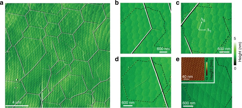
We begin with the topographic images of the hBN domain patterns (Fig. 1a–e) obtained with the atomic force microscope (AFM). The patterns extend over macroscopic areas (>104 μm2) and have periods Λ1, Λ2 ~ 300–800 nm. The domains vary from nearly perfectly hexagonal (Fig. 1b) to highly distorted, diamond-like motifs (Fig. 1e). The domain boundaries are demarcated by the dips in the topography, which are about w ~ 90 nm wide (Fig. S2 of Supplementary Information). Macroscopic domain patterns are not impacted by defects on the sample surface such as wrinkles26: the white lines of elevated topography in Fig. 1a–d. Similarly, the domain boundaries do not disturb the 14-nm-periodic moiré pattern formed by monolayer graphene deposited on top of hBN (Fig. 1e, inset). Both observations suggest that the long-range superlattices form at some depth d below the surface of the hBN crystal. Based on a theoretical model for the strain distribution around a soliton, we estimate d ~ w/7 ~ 15 nm (Supplementary Information Section 2).
Fig. 1.
AFM topography images. a Large-scale image of hBN crystals showing the periodic domain pattern. b–e Zoom-in view of different smaller regions revealing varied domain shapes. The dashed black lines are the contours of graphene epitaxial grown on hBN. Inset of e displays a high-resolution topography (friction-AFM) image demonstrating the small-period Moiré pattern at the graphene/hBN interface. The large-period domain patterns are influenced neither by the presence of graphene nor by hBN wrinkles (the white lines in a–d) on the top surface of the crystal
Origin of hBN soliton superlattices
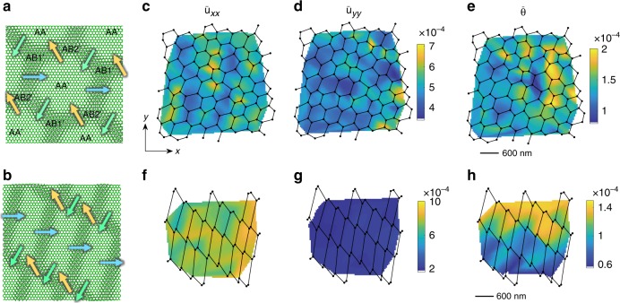
For an in-depth look at the solitons, we focus on two representative cases (Fig. 1b, e). The locations of the domain-wall junctions in these images, connected by straight lines, are plotted in Fig. 2c–h. These latter plots reveal somewhat deformed lattices whose primitive periods Λ1, Λ2 (Fig. 1c) vary in space. We can relate such variations to the interlayer strain and rotation in the system, or more precisely, to the averaged (coarse-grained) strain tensor and twist angle . Figure 2 illustrates the calculated color maps of and superimposed on the soliton meshes. Here α, β ∈ {x, y} and r = (x, y) is the in-plane position. Note that the angles at all the soliton junctions are close to 120°, which is analogous to the Plateau law of foam films27 (see Supplementary Information Section 3 for details). For domains shaped as unilateral hexagons, we find an isotropic and predominantly tensile average strain with the average twist angle . For diamond-like domains, the strain is anisotropic, almost uniaxial. A couple of remarks on these results are in order. First, the coarse-grained strain should not be confused with the local strain uαβ, which is to be discussed below. Second, the calculation of and requires as an input the Burgers vector bj of the solitons. These vectors connect the nearest identical atoms (e.g., borons) in the same atomic plane. They have magnitude b = 0.25 nm each and angular directions separated by 120°. However, since our AFM does not resolve the hBN crystal orientation, we do not know these directions. To generate Fig. 2 we chose one of these directions along the x-axis of the plot (Fig. 2a, b). If the Burgers vectors are rotated through some common angle, qualitatively similar and maps are obtained.
Fig. 2.
a, b Schematic diagram of the soliton superlattice in hBN. The shift of the lattice sites in the two adjacent misfit atomic layers (green and gray) is concentrated at the solitons (misfit dislocations). Each soliton is characterized by one of the three possible Burgers vectors displayed with arrows. The regions of AA’, AB1’, and AB2’ stacking are labeled. c–h Solitons (lines), their junctions (dots), and the corresponding maps of coarse-grained quantities: c, f x-axis average tensile strain , d, g y-axis average tensile strain , e, h average rotation angle (rad). The average shear strain is small everywhere and not shown. The top row panels c–e are deduced from Fig. 1b; the bottom row panels f–h are obtained from Fig. 1e
Nano-IR imaging of hBN superlattices
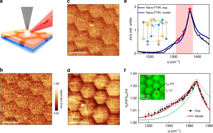
We proceed to the survey of the results obtained by the IR nano-imaging. We have carried out these scanning nano-spectroscopy experiments in the frequency region of hBN phonon polaritons28–32. In our s-SNOM apparatus the metalized AFM tip was illuminated by IR light thus generating a strong enhancement of the electric field underneath the tip (Fig. 3a)4,33. Such an antenna-based nano-IR setup solves the problem of the photon-polariton momentum mismatch33–35 and enables local spectroscopy of polariton modes in hBN with ~25 nm spatial resolution (see the “Methods” section). Employing tunable quantum cascade lasers, we acquired nano-IR images at more than 50 different discrete frequencies. We found that the domain patterns revealed by the AFM topography are also prominent in the nano-IR images (Fig. 3b–d). We obtained nano-IR data with both monochromatic tunable quantum cascade lasers (Fig. 3b–d, f) and also broadband difference frequency generation sources (Fig. 3e) using Fourier transform spectroscopy. The strongest contrast is observed at frequency ω = 1368 cm−1 (Fig. 3d), which corresponds to the phonon polariton resonance of hBN (Fig. 3e). The contrast systematically weakens as ω is shifted away from ω = 1368 cm−1. In all the images the back-scattering amplitude s(ω, r) is enhanced at the centers of the domains (s = sd(ω)) and reduced at the solitons (s = ssol(ω)). We therefore conclude that the lattice dynamics is modified near the solitons where the strain is concentrated. The frequency dependence of the ratio sd(ω)/ssol(ω) highlights subtle yet systematic variations of this modified lattice response. This ratio exhibits an asymmetric maximum, with a broader low-frequency side (Fig. 3f).
Fig. 3.
Solitons and lattice dynamics of hBN by nano-IR imaging and spectroscopy. a Schematic of the nano-IR imaging showing an AFM tip illuminated by a focused IR beam. The solitons reside at the atomic interface between two parts of the hBN slab (yellow and blue regions). The orange and blue cylinders delineating one hexagonal domain inside the slab represent the strain distribution around the solitons. b–d Nano-IR images of hBN domain patterns at frequencies 1320, 1344, and 1368 cm−1, respectively. These frequencies are marked with red stars and dots in panels e and f. e Typical nano-FTIR spectrum of an hBN crystal. Black line: experimental data, blue spectrum: theoretical model described in the text. The inset illustrates the AA′ stacking of hBN and the three possible Burgers vectors bj. The shaded area highlights the spectral range analyzed in panel f. f The sd(ω)/ssol(ω) spectra across the hBN phonon polariton band. The error bars represent the 90% confidence intervals. The red solid line is the best fit to the data (black squares) using the frequency and the damping rate of the hBN optical phonon as adjustable parameters. The green line is the best fit obtained by varying the damping rate only. The inset shows the AFM topography corresponding to the IR images in panels b–d
Elucidating nano-IR response
Quantitative modeling of the nano-IR response is a challenging task that goes beyond the scope of the present work. To analyze the sd(ω)/ssol(ω) spectra, we restricted ourselves to the simplified approach, similar those in recent nano-IR study of wrinkled and strained hBN crystals26 and an earlier work on nano-indented SiC36. We assumed that the hBN crystal can be described by a permittivity tensor εα(ω, r) varying only as a function of in-plane coordinates. Here α = ⊥(||) is the in-plane (out-of-the-plane) component. We further assumed that the near-field amplitude s(ω, r) depends only on the local value of εα(ω, r). We adopted the standard Lorentzian model for the hBN permittivity,
| 1 |
where ωTO,α (ωLO,α) is the transverse (longitudinal) optical phonon frequency and Γα is the broadening35, which we treated as adjustable parameters. We numerically simulated the near-field scattering amplitude s(ω) modeling the tip as an elongated conducting spheroid and taking into account the presence of the quartz substrate underneath hBN10. Through these simulations we found that the nano-IR contrast can be attributed to the broadening of the phonon resonance, from Γ⊥ = 6.5 cm−1 at the domain centers to 7.35 cm−1 at the walls. The dashed green trace in Fig. 3f illustrates the effect of this extra broadening alone. In addition, a minute blue shift of ωTO,⊥ from 1365.5 to 1365.8 cm−1 helps to better account for the spectral form of sd(ω)/ssol(ω), as shown by the red line in Fig. 3f (for more details of these fits, see Supplementary Information Section 4).
Our analysis of the nano-IR line-form in Fig. 3 is phenomenological. The microscopic approach can be developed by relating the phonon frequency shift ΔωTO,⊥ to the local strain. Notably, the frequency shift caused by the strain is not a single number. As shown by recent first-principles calculations26, a uniaxial strain splits the degenerate TO mode of hBN into two separate modes of orthogonal in-plane polarization. Moreover, from symmetry consideration we can predict that for an arbitrary strain, the fractional frequency shifts of these two modes should be:
| 2 |
where A, B, C ~ 1are constants (Supplementary Information Section 5). Effectively, the strain turns hBN from the uniaxial hyperbolic material into a bi-axial one, analogous to molybdenum trioxide37,38. Because of different polarization, the two modes have different coupling to the in-plane field, which complicates the modeling. Even more arduous task is to take into account the realistic three-dimensional strain distribution around the solitons. The dependence of the strain on the in-plane coordinates is relatively weak because the characteristic width w ~ 90 nm of the strained region near the surface is large (Figs. 1 and S2). Nevertheless, the dependence of the strain on the depth z is strong because of the anisotropic character of the strain (Fig. 3a and Supplementary Information Section 2). Leaving a quantitative study for future work, we limit ourselves to the following estimate. Qualitatively, the nano-IR amplitude measured above the soliton can be viewed as the sum of signals from all the underlying hBN layers, each with a shifted (and split) ωTO,⊥. Assuming the characteristic strain variation of δu ~ b/w ~ 0.3%, the nano-IR line-shape is expected to acquire an additional inhomogeneous broadening ΔΓ⊥ ~ ωTO,⊥δu ~ 4cm−1. This estimate is of the same order of magnitude as the fit parameter quoted above.
Discussion
The confinement of both the topographic and nano-IR contrast to narrow regions along the domain walls is consistent with the notion that the individual layers in hBN are not rigid but instead behave as deformable atomic membranes prone to incommensurate–commensurate transitions. The key signature of these transitions is the formation of structural solitons where mechanical strain is accumulated11–15. Previously, the transmission electron microscopy and second harmonic generation experiments have identified isolated solitons39 in few-layer-thin hBN specimens. Our work demonstrates that the solitons can form in the interior of a bulk vdW crystal, not just at the surface or in few-layer systems, and that they can organize themselves in large regular superlattices. We have also demonstrated that nano-IR imaging can be applied to map the local strain field in a polar crystal26,36.
The observed residual strain concentrated in the solitons, could originate from multiple sources, one of which is the difference in the thermal expansion coefficients of hBN and graphene deposited on top of it (−8 × 10⁻⁶/K for graphene40 and −3 × 10⁻⁶/K for hBN41). A rough estimate of the lattice mismatch that could develop during the cooling of the sample is [(−8)–(−3)] × 10−6/K×(1150–20) K = 0.6%, which is close to the critical mismatch for the commensurate–incommensurate transition in an hBN bilayer42.
Finally, we mention several predictions testable by atomic-resolution or stacking-sensitive probes. Unlike TBG, where the lowest-energy stacking is AB stacking and the domains are triangular1,6,14,18, the hexagonal domains in hBN are of AA′ type (Figs. 2a and 3e), the lowest-energy stacking in hBN43. For the domain-wall junctions in hBN, we expect two distinct stacking types, AB1′ and AB2′, that alternate in space (Fig. 2a). Future studies may also seek to observe changes in the electronic structure in hBN moiré superlattices44. More broadly, it will be fruitful to explore in operando tuning of the twist angle45 and attendant lattice, electronic2,46, plasmonic15 and possibly excitonic8,9 responses of various vdW materials.
Methods
Sample synthesis
The hBN flakes were prepared on quartz substrates using mechanical exfoliation. hBN/Quartz samples were then transferred into chemical vapor deposition chamber and annealed at 1150 °C at low pressure with continuous argon flow of 50 standard cubic centimeters per minute (s.c.c.m.) for 30 min. After that, CH4:H2 at 5:5 s.c.c.m. were injected into the chamber for 300 min at pressures below 12 mbar—a process used for graphene growth39. Finally, samples are cooled to room temperature in argon flow.
Nano-IR imaging
The IR nano-imaging experiments were performed using s-SNOM (neaspec.com) equipped with continuous wave mid-IR quantum cascade lasers (daylightsolutions.com). The s-SNOM is based on AFM with curvature radius ~ 25 nm operating in the tapping mode with a tapping frequency around 270 kHz. A pseudo-heterodyne interferometric detection module was implemented to extract both the scattering amplitude s and the phase of the near-field signal. In the current work, we discuss the amplitude of the signal. In order to subtract the background signal, we demodulated the near-field signal at the third harmonics of the tapping frequency. All the IR nano-imaging experiments were performed in ambient conditions. We used quantum cascade lasers with tunable frequency and a broad-band difference frequency generation laser systems.
Supplementary information
Acknowledgements
Authors acknowledge discussions with J.S. Wu, A.S. McLeod, S. Sunku, and Y.Q. Cai. Work at Columbia is supported as part of Programmable Quantum Materials, an Energy Frontier Research Center funded by the U.S. Department of Energy (DOE), Office of Science, Basic Energy Sciences (BES), under award DE-SC0019443. Work at UCSD is supported by ONR-N000014-18-1-2722. The development of mid-infrared polaritonic structures is supported by AFOSR: FA9550-15-1-0478. D.N.B. is supported by the Gordon and Betty Moore Foundation’s EPiQS Initiative through Grant GBMF4533.
Author contributions
G.X.N. performed the nanoscale infrared measurements and characterizations. B.-Y.J., Y.D., Z.Y.S. and M.M.F. provided theoretical calculations. H.W., L.X.C., and X.M.X. created the device structures. M.D.G. and A.J.F. helped with measurements. D.N.B. supervised the project. G.X.N., M.M.F., and D.N.B. co-wrote the manuscript with input from all co-authors.
Data availability
The data supporting the findings of this work are available from the corresponding author upon reasonable request.
Competing interests
The authors declare no competing interests.
Footnotes
Peer review information Nature Communications thanks the anonymous reviewers for their contribution to the peer review of this work
Publisher’s note Springer Nature remains neutral with regard to jurisdictional claims in published maps and institutional affiliations.
Supplementary information
Supplementary Information accompanies this paper at 10.1038/s41467-019-12327-x.
References
- 1.Bistritzer R, MacDonald AH. Moiré bands in twisted double-layer graphene. Proc. Natl Acad. Sci. USA. 2011;108:12233–12237. doi: 10.1073/pnas.1108174108. [DOI] [PMC free article] [PubMed] [Google Scholar]
- 2.Cao Y, et al. Unconventional superconductivity in magic-angle graphene superlattices. Nature. 2018;556:43–50. doi: 10.1038/nature26160. [DOI] [PubMed] [Google Scholar]
- 3.Geim AK, Grigorieva IV. Van der Waals heterostructures. Nature. 2013;499:419–425. doi: 10.1038/nature12385. [DOI] [PubMed] [Google Scholar]
- 4.Basov DN, Fogler MM, Garcia de Abajo FJ. Polaritons in van der Waals materials. Science. 2016;354:aag1992. doi: 10.1126/science.aag1992. [DOI] [PubMed] [Google Scholar]
- 5.Low T, et al. Polaritons in layered two-dimensional materials. Nat. Matters. 2017;16:182–194. doi: 10.1038/nmat4792. [DOI] [PubMed] [Google Scholar]
- 6.Yoo, H. et al. Atomic reconstruction at van der Waals interface in twisted bilayer graphene. arXiv:1804.03806 (2018). [DOI] [PubMed]
- 7.Fang H, et al. Strong interlayer coupling in van der Waals heterostructures built from single-layer chalcogenides. Proc. Natl Acad. Sci. USA. 2014;111:6198–6202. doi: 10.1073/pnas.1405435111. [DOI] [PMC free article] [PubMed] [Google Scholar]
- 8.Yu H, Liu GB, Tang J, Xu X, Yao W. Moiré excitons: from programmable quantum emitter arrays to spin–orbit-coupled artificial lattices. Sci. Adv. 2017;3:e1701696. doi: 10.1126/sciadv.1701696. [DOI] [PMC free article] [PubMed] [Google Scholar]
- 9.Wu F, Lovorn T, MacDonald AH. Topological exciton bands in moiré heterojunctions. Phys. Rev. Lett. 2017;118:147401. doi: 10.1103/PhysRevLett.118.147401. [DOI] [PubMed] [Google Scholar]
- 10.Song JCW, Shytov AV, Levitov LS. Electron interactions and gap opening in graphene superlattices. Phys. Rev. Lett. 2013;111:266801. doi: 10.1103/PhysRevLett.111.266801. [DOI] [PubMed] [Google Scholar]
- 11.Alden SJ, et al. Strain solitons and topological defects in bilayer graphene. Proc. Natl Acad. Sci. USA. 2013;110:11256–11260. doi: 10.1073/pnas.1309394110. [DOI] [PMC free article] [PubMed] [Google Scholar]
- 12.Woods CR, et al. Commensurate–incommensurate transition for graphene on hexagonal boron nitride. Nat. Phys. 2013;9:329–340. doi: 10.1038/nphys2615. [DOI] [Google Scholar]
- 13.San-Jose P, Prada E. Helical networks in twisted bilayer graphene under interlayer bias. Phys. Rev. B. 2013;88:121408(R). doi: 10.1103/PhysRevB.88.121408. [DOI] [Google Scholar]
- 14.Sunku SS, et al. Photonic crystals for nano-light in moiré graphene superlattices. Science. 2018;362:1153–1156. doi: 10.1126/science.aau5144. [DOI] [PubMed] [Google Scholar]
- 15.Ni GX, et al. Plasmons in graphene moiré superlattices. Nat. Matters. 2015;14:1217–1222. doi: 10.1038/nmat4425. [DOI] [PubMed] [Google Scholar]
- 16.Shi Z, et al. Gate-dependent pseudospin mixing in graphene/Boron nitride moiré superlattices. Nat. Phys. 2014;10:743–747. doi: 10.1038/nphys3075. [DOI] [Google Scholar]
- 17.Yankowitz M, Ma Q, Jarillo-Herrero P, LeRoy BJ. van der Waals heterostructures combining graphene and hexagonal boron nitride. Nat. Rev. Phys. 2019;1:112–125. doi: 10.1038/s42254-018-0016-0. [DOI] [Google Scholar]
- 18.Tomadin A, Polini M, Jung J. Plasmons in realistic graphene/hexagonal boron nitride moiré patterns. Phys. Rev. B. 2019;99:035432. doi: 10.1103/PhysRevB.99.035432. [DOI] [Google Scholar]
- 19.Schaibley JR, et al. Valleytronics in 2D materials. Nat. Rev. Mater. 2016;1:16055. doi: 10.1038/natrevmats.2016.55. [DOI] [Google Scholar]
- 20.Yankowitz Matthew, Chen Shaowen, Polshyn Hryhoriy, Zhang Yuxuan, Watanabe K., Taniguchi T., Graf David, Young Andrea F., Dean Cory R. Tuning superconductivity in twisted bilayer graphene. Science. 2019;363(6431):1059–1064. doi: 10.1126/science.aav1910. [DOI] [PubMed] [Google Scholar]
- 21.Huang S, et al. Topologically protected helical states in minimally twisted bilayer graphene. Phys. Rev. Lett. 2018;121:037702. doi: 10.1103/PhysRevLett.121.037702. [DOI] [PubMed] [Google Scholar]
- 22.Kim K, et al. Tunable moiré bands and strong correlations in small-twist-angle bilayer graphene. Proc. Natl Acad. Sci. USA. 2017;114:3364–3369. doi: 10.1073/pnas.1620140114. [DOI] [PMC free article] [PubMed] [Google Scholar]
- 23.Jiang B-Y, et al. Plasmon reflections by topological electronic boundaries in bilayer graphene. Nano. Lett. 2017;17:7080. doi: 10.1021/acs.nanolett.7b03816. [DOI] [PubMed] [Google Scholar]
- 24.Ju L, et al. Topological valley transport at bilayer graphene domain walls. Nature. 2015;520:650. doi: 10.1038/nature14364. [DOI] [PubMed] [Google Scholar]
- 25.Jiang L, et al. Soliton-dependent plasmon reflection at bilayer graphene domain walls. Nat. Mater. 2016;15:840–844. doi: 10.1038/nmat4653. [DOI] [PubMed] [Google Scholar]
- 26.Lyu B, et al. Phonon polariton-assisted infrared nanoimaging of local strain in hexagonal boron nitride. Nano Lett. 2019;19:1982–1989. doi: 10.1021/acs.nanolett.8b05166. [DOI] [PubMed] [Google Scholar]
- 27.Atkin JM, Berweger S, Jones AC, Raschke MB. Nano-optical imaging and spectroscopy of order, phases, and domains in complex solids. Adv. Phys. 2012;61:745–842. doi: 10.1080/00018732.2012.737982. [DOI] [Google Scholar]
- 28.Caldwell JD, et al. Atomic-scale photonic hybrids for mid-infrared and terahertz nanophotonics. Nat. Nano. 2016;11:9–15. doi: 10.1038/nnano.2015.305. [DOI] [PubMed] [Google Scholar]
- 29.Dai S, et al. Tunable phonon polaritons in atomically thin van der Waals crystals of boron nitride. Science. 2014;343:1125. doi: 10.1126/science.1246833. [DOI] [PubMed] [Google Scholar]
- 30.Caldwell JD, et al. Photonics with hexagonal boron nitride. Nat. Rev. Mater. 2019;4:552–567. doi: 10.1038/s41578-019-0124-1. [DOI] [Google Scholar]
- 31.Ambrosio A, et al. Selective excitation and imaging of ultraslow phonon polaritons in thin hexagonal boron nitride crystals. Light.: Sci. Appl. 2018;7:27. doi: 10.1038/s41377-018-0039-4. [DOI] [PMC free article] [PubMed] [Google Scholar]
- 32.Li, P. et al. Hyperbolic phonon-polaritons in boron nitride for near-field optical imaging and focusing. Nature Comm.6, 7507 (2015). [DOI] [PMC free article] [PubMed]
- 33.Fei Z, et al. Gate-tuning of graphene plasmons revealed by infrared nano-imaging. Nano Lett. 2011;11:4701–4705. doi: 10.1021/nl202362d. [DOI] [PubMed] [Google Scholar]
- 34.Ni GX, et al. Ultrafast optical switching of infrared plasmon polaritons in high-mobility graphene. Nat. Photon. 2016;10:244–247. doi: 10.1038/nphoton.2016.45. [DOI] [Google Scholar]
- 35.Ni GX, et al. Fundamental limits to graphene plasmonics. Nature. 2018;557:530–533. doi: 10.1038/s41586-018-0136-9. [DOI] [PubMed] [Google Scholar]
- 36.Huber AJ, Ziegler A, Köck T, Hillenbrand R. Infrared nanoscopy of strained semiconductors. Nat. Nano. 2009;4:153–157. doi: 10.1038/nnano.2008.399. [DOI] [PubMed] [Google Scholar]
- 37.Ma W, et al. In-plane anisotropic and ultra-low-loss polaritons in a natural van der Waals crystal. Nature. 2018;562:557–562. doi: 10.1038/s41586-018-0618-9. [DOI] [PubMed] [Google Scholar]
- 38.Zheng Z, et al. A mid-infrared biaxial hyperbolic van der Waals crystal. Sci. Adv. 2019;5:5. doi: 10.1126/sciadv.aav8690. [DOI] [PMC free article] [PubMed] [Google Scholar]
- 39.Kim C-J, et al. Stacking order dependent second harmonic generation and topological defects in h-BN bilayers. Nano Lett. 2013;13:5660–5665. doi: 10.1021/nl403328s. [DOI] [PubMed] [Google Scholar]
- 40.Yoon D, Son YW, Cheong H. Negative thermal expansion coefficient of graphene measured by Raman spectroscopy. Nano Lett. 2011;11:3227. doi: 10.1021/nl201488g. [DOI] [PubMed] [Google Scholar]
- 41.Paszkowicz W, Pelka JB, Knapp M, Szyszko T, Podsiadlo S. Lattice parameters and anistropic thermal expansion of the hexagonal boron nitride in the 10–297.5 K temperature range. Appl. Phys. A. 2002;75:431. doi: 10.1007/s003390100999. [DOI] [Google Scholar]
- 42.Lebedeva IV, Lebedev AV, Popov AM, Knizhnik AA. Dislocations in stacking and commensurate-incommensurate phase transition in bilayer graphene and hexagonal boron nitride. Phys. Rev. B. 2016;93:235414. doi: 10.1103/PhysRevB.93.235414. [DOI] [Google Scholar]
- 43.Xian, L., Kennes, D. M., Tancogne-Dejean, N., Altarelli, M. & Rubio, A. Multi-flat bands and strong correlations in twisted bilayer boron nitride. arXiv: 1812.08097 (2018). [DOI] [PMC free article] [PubMed]
- 44.Ribeiro-Palau R, et al. Twistable electronics with dynamically rotable heterostructures. Science. 2018;361:690–693. doi: 10.1126/science.aat6981. [DOI] [Google Scholar]
- 45.Yankowitz M, Xue J, LeRoy BJ. Graphene on hexagonal boron nitride. J. Phys. Condens. Matter. 2014;26:303201. doi: 10.1088/0953-8984/26/30/303201. [DOI] [PubMed] [Google Scholar]
- 46.Weaire D, Hutzler S. The Physics of Foams. Oxford: Oxford University Press; 1999. [Google Scholar]
Associated Data
This section collects any data citations, data availability statements, or supplementary materials included in this article.
Supplementary Materials
Data Availability Statement
The data supporting the findings of this work are available from the corresponding author upon reasonable request.


