ABSTRACT
The expression of env proteins that bind to viral cell receptors on avian leukosis virus (ALV)-susceptible cells can block ALV infection. In this study, we constructed a cell line (DF-1/B) by expressing the ALV-B env protein in DF-1 cells. PCR, immune fluorescence assay, Western blot, and immune electron microscopy results showed that the env gene can be stably expressed in DF-1cells and the env protein could be detected on the DF-1 cell membrane. An antiviral experiment concluded that the DF-1/B cell line could be resistant to 1 × 10⁴ TCID50 ALV-B virus infection, but had no inhibitory effect on other subgroup ALV. This means that the DF-1/B cell line is specifically resistant to ALV-B and can be used as a tool for ALV-B diagnosis.
Keywords: ALV-B, env protein, cell line
INTRODUCTION
Avian leukosis virus (ALV) belongs to the α retrovirus genus and can cause a variety of poultry diseases. Clinically, the main manifestations of ALV infection are immunosuppression, growth retardation, tumors in multiple organ tissues, and other characteristics (Hang et al., 2013). Since the discovery of the virus, it has caused enormous economic losses to the poultry industry. ALV are divided into 11 subgroups (A to K) (Payne et al., 1992; Wang et al., 2012) based on their host range, viral envelope glycoprotein antigenic structure, and receptor interference, of which subgroups A–E, J, and K infect chickens. Subgroups A, B, and J are the main pathogenic exogenous viruses and are the 3 most prevalent exogenous virus subgroups that can trigger tumors (Zhao et al., 2010; Wang et al., 2011), posing a significant threat to the poultry industry. Subgroups C and D also have pathogenic viruses, but rare in the flock. ALV-K is a novel ALV, and more and more ALV-K strains have been isolated in recent years (Wang et al., 2012; Cui et al., 2014; Li et al., 2016; Shao et al., 2017); subgroup E is an endogenous virus. In the mid-1980s, an international breeder company was successful in eradicating subgroup A and B ALV infection in chickens, but because of ALV purification, it could not be fully launched in China at that time, so the Chinese flocks, especially Chinese native chickens, still carry ALV-A, ALV-B, and ALV-J infection (Zhao et al., 2010). Over the past decade, ALV-B co-infection with ALV-A, a recombinant virus of ALV-B and ALV-A, and a recombinant virus of ALV-B and ALV-J have been isolated (Lupiani et al., 2006; Li et al., 2013; Wang et al., 2017). ALV-B is still a threat to the Chinese poultry industry and is part of a list of diseases necessary to be prevented and eradicated.
So far, there have been no effective vaccines and drugs specifically available for the prevention of ALV. The only option left for the prevention of ALV infection and, thus, evolution of flocks is the elimination of infected chickens. At present, the methods employed for the detection of exogenous ALV mainly include virus isolation, ELISA, PCR, and immune fluorescence assay (IFA) (Spencer and Gilka, 1982; Payne et al., 1993), and also some new detection methods have been recently established, such as RT-PCR, QC-RT-PCR, LAMP, and colloidal gold test strips (Kim and Brown, 2004; Zhang et al., 2010; He et al, 2013; Dai et al., 2015).
According to the ALV env protein, a glycoprotein encoded by the virus envelope (env) gene can be recognized by the specific viral receptors on the host cell membrane to block the same subgroup ALV infection of susceptible cells (Hunt et al., 1999; Holmen and Federspiel, 2000). A genetically engineered cell line (DF-1/J) resistant to ALV-J infection was developed and applied to screen large numbers of ALV-J field samples (Hunt et al., 1999). A cell line DF-1/A and a cell line DF-1/K resistant to ALV-A and novel subgroup ALV-K infection, respectively, have also been constructed and applied (Mingzhang et al., 2017). In order to establish a specific method for the detection of ALV-B, the env protein of ALV-B was required to be stably expressed on DF-1 cells to obtain an immortal cell line that can be exclusively used to resist ALV-B infection. This cell line would be further applied to evaluate clinical plasma samples and investigate the prevalence of ALV-B infection in Chinese native chickens. This is the first report of a cell line specifically resistant to infection by ALV-B.
MATERIALS AND METHODS
Viruses and Cell Lines
ALV-B (CD08) and ALV-J (CHN06) (GenBank accession number HQ900844) were isolated and maintained in our laboratory (Dai et al., 2015). The titers of CD08 and CHN06 strains were determined by ELISA (IDEXX, Inc., Westbrook, MA) and were presented as TCID50 mL−1 calculated using the Reed–Muench method. A DF-1 cell was grown in Dulbecco's modified Eagle medium (DMEM; GIBCO, NY, USA) supplemented with 10% fetal bovine serum (FBS, GIBCO, New Zealand) and maintained in DMEM supplemented with 1% FBS at 37°C with 5% CO2.
Plasmid Construction
The ALV-B env gene was amplified by PCR using the gene-specific primers (Table 1) and subcloned into the pMD-18T vector (TaKaRa, Dalian, China). It was then cloned into the eukaryotic expression vector pcDNA3.1 using the BamH I and Not I sites. The pcDNA-env-B vector was sequenced and found to have the predicted nucleotide sequence. The pcDNA-env-B-EGFP vector, which contains an EGFP tag, was constructed by the PCR amplification of EGFP fragment and ligated with the env gene, and the fusion fragment was cloned into the pcDNA3.1 vector.
Table 1.
Primers used for amplifying the ALV-B env gene and EGFP fragment in this study.
| Primer1 | Sequence (5′–3′) | Product size (bp) |
|---|---|---|
| B-env-F | CGGGGTACCGCCACCATGGAAGCCGTCATAAAGATGAGGCGA | 1845 |
| B-env-R | AAGGAAAAAAGCGGCCGCCTATACTGCTCTTTCGGGCTGCTTA | |
| EGFP-F | TTGCGGCCGCATGGTGAGCCAAGGGCGAGGA | 720 |
| EGFP-R | GCTCTAGATTACTTGTACAGCTCGTCCA |
F and R represent upstream primer and downstream primer, respectively. Italic for restriction sites, the underlined represents the Kozak sequence.
Screening Cells With Zeocin
The DF-1 cells were transfected with the pcDNA-env-B plasmid, pcDNA-env-B-EGFP plasmid, and pcDNA3.1/Zeo(+) plasmid. After 48 h, the transfected DF-1 cells grew to monolayer, and cells in one of the 6-well cell culture plates were digested with 0.25% trypsin (GIBCO). Then, the cells with a medium containing DMEM, 15% FBS, and 200 µg/mL zeocin were seeded into 500 µL/well 24-well tissue culture plates. The transfected DF-1 cells were selected for resistance to zeocin. Thereafter, cells were treated with a 500 µL/well medium containingDMEM + 15% FBS + 200 µg/mL zeocin, and then this medium was replaced every 3 D. The zeocin-resistant cells were passaged through 60 generations and then frozen. After 3 mo, these cells were refreshed and cultured in a medium free of zeocin.
PCR Assay
To demonstrate whether the DF-1/B cell line was constructed successfully, routine PCR tests were carried out with genomic DNA extracted from the ALV-B-resistant cell line, designated as DF-1/B cells. The DF-1 cells served as a negative control. The specific primers were as described in Table 1. Total cellular RNA was extracted from DF-1/B cells and DF-1 cells using the RNAfast200 kit (Fastagen, Shanghai, China), followed by cDNA synthesis using the RevertAid First strand cDNA synthesis kit (Fermentas, Burlington, Canada) according to the manufacturer's instructions. The cDNA was then used for routine PCR and real-time PCR amplification. Real-time reverse transcription (RT)-PCR was done with primers designed for the envelope gene and gene-specific primers (Table 2) synthesized by TaKaRa Company (Dalian, China). DNA sequences were determined by Invitrogen (Shanghai, China). For all reactions, PCR amplification and DNA sequencing were carried out at least twice independently to avoid PCR errors. Real-time PCR was performed on an ABI 7500 Real-time PCR System (Applied Bio systems) using Premix ExTaq (Probe qPCR) reagents (TaKaRa, Dalian, China) according to the manufacturer's specifications. Fluorescent signals were recorded during the elongation step. The β-actin gene served as a reference gene. The relative expression level of the env gene was normalized by β-actin. Finally, real-time quantitative PCR analysis was carried out using the 2−ΔΔCT method (Livak and Schmittgen, 2001).
Table 2.
Primers for real-time reverse transcription for amplifying the ALV-B env gene.
| Primer1 | Sequence (5′–3′) | Product size (bp) |
|---|---|---|
| q-B-env-F | ATAAGATCGGCGTGGACAAC | 1845 |
| q-B-env-R | TGGAATTTCCTGCATTCCTC |
F and R represent upstream primer and downstream primer, respectively.
Indirect IFA
DF-1/B and DF-1 cells were washed with pre-cold PBS once, fixed with cold paraformaldehyde for 20 min at –20°C, then washed with PBS 3 times and allowed to air-dry. The cells were then incubated with monoclonal antibodies for ALV-B (provided by Dr Jiaqian Cai, Shangdong Agricultural University) at 37°C for 1 h, washed 3 times with PBS, and further incubated with goat antimouse IgG conjugated with FITC (Sigma, Mannheim, Germany) at 37°C for 1 h. After 3 washes with PBS, the cells were observed using fluorescence microscopy (LEICA, Germany).
Western Blot Analysis
DF-1/B cells and DF-1 cells were once washed in 100-mm dishes with cold PBS and harvested by a cell-scraping device, then homogenized with NP-40 lysis buffer containing 1 × protease inhibitor cocktail (Roche) and incubated on ice for 30 min. Lysates were centrifuged at 12,000 × g for 5 min at 4°C. The supernatants were analyzed for total protein content using the BCA protein assay kit (Fermentas, Life Technologies). Total protein (20 μg) was resolved by 12% SDS-PAGE and transferred onto nitrocellulose membranes (Whatman, Maidstone, UK). The membranes were blocked with 5% (w/v) skimmed milk for 1 h at 37°C, and then incubated overnight at 4°C with monoclonal antibodies for ALV-B and β-actin (Santa Cruz, sc-1616-R). β-Actin served as a reference. After 3 washes with PBS Tween20 (PBST) buffer, the membranes were incubated at 37°C for 1 h with IRDye 800-conjugated anti-mouse IgG secondary antibody (1:10,000; Rockland Immunochemicals, Limerick, PA) diluted in PBS. Membranes were washed 3 times with PBST, then visualized and analyzed with an Odyssey infrared imaging system (LI-COR Biosciences, Lincoln, NE).
Immune Electron Microscopy
The DF-1 and DF-1/B cells were repaired and sliced with a slice thickness of 75 nm according to the preparation method of ordinary electron microscopy samples. The ultrathin slices were then transferred to a nickel mesh using a Fang Hua film (to ensure slice continuity) and then incubated with goat anti-mouse EGFP antibodies (Sigma) and monoclonal antibodies for ALV-B for 1 h at 37°C. After washing the sections with PBSA (PBS with 5% BSA) solution 6 times, they were incubated with 10 nm colloidal gold-labeled goat anti-mouse IgG (Sigma) for 1 h at 37°C. The sections were washed with ultrapure water and dried at room temperature. Finally, the samples were examined under a JEM-2010HR transmission electron microscope (JEPL, Japan).
Antiviral Experiment
ALV-B (CD08) and ALV-J (CHN06) were diluted from 105 TCID50 to 101 TCID50 for 5 gradients and then 100 µL per well of those cells were seeded in a 24-well plate containing 1 mL DF-1/B cells at 1.7 × 10⁵ cells/well. Three wells on each plate served as negative controls. Each dilution of virus was performed in triplicate. After 2 h of incubation, supernatants were removed and a maintenance medium containing DMEM with 1% FBS was added, and the plates were incubated at 37°C and 5% CO2 for another 6 D. The supernatant fluid was then harvested for ALV p27 antigen using the ELISA detection ALV-p27 Ag Test kit (IDEXX, Inc., Westbrook, MA). A mock-infected DF-1 cell group was established in parallel as a control.
RESULTS
Cell Line Screening
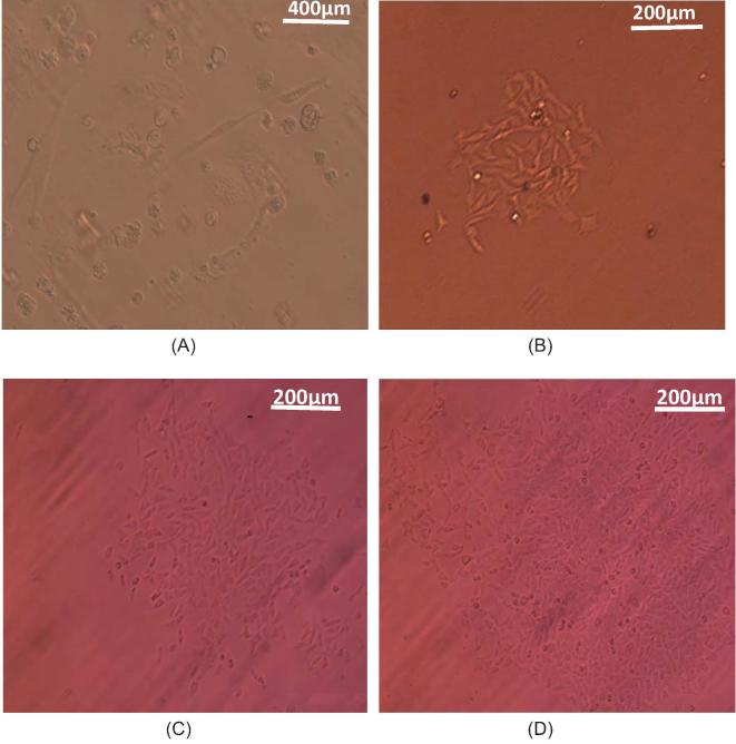
To establish single zeocin-resistant DF-1 clones, single cells from DF-1 cells transfected with the pcDNA-env-B plasmid and pcDNA-env-B-EGFP plasmid were picked, respectively. The picked single cells became actively proliferated (Figures 1A and 1B), and this single-cell colony appeared to increase in size over the next 6 to 10 D (Figures 1B and 1C). The cells grew to near completion by approximately 21 D in culture (Figure 1D). For 3 to 4 wk, the cells were digested with 0.25% trypsin (GIBCO), and then plated in 6-well cell culture plates at 37°C and 5% CO2 until they grew to monolayer. The zeocin-resistant cells were cultured in the medium containing zeocin, passaged continuously for 60 generations, and then frozen. After 3 mo, these cells were refreshed and cultured in a medium free of zeocin, and the env gene or env protein in the transfected DF-1 cell was detected.
Figure 1.
Zeocin selection of cell lines. (A) After zeocin selection, the transfected DF-1 cells formed a single cell colony within 10 to 15 D. The single cell colony appeared to increase in size over the following 6 to 10 D, and the differences in the size of the cell clones are shown on days 16 (B) and 20 (C). (D) The cells had nearly grown to a monolayer by approximately 21 D in culture. Magnification is ×400 for (A) and 200 × for (B–D).
PCR Detection of the env Gene in DF-1/B Cells
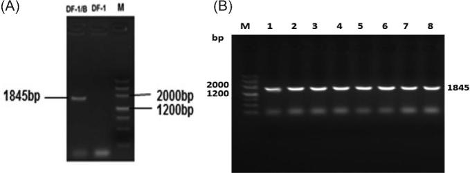
The ALV-B env gene in the first generation of DF-1/B cells was detected by the PCR method (Figure 2A) and the ALV-B env gene was still stable in the genome after 20 to 60 passages (Figure 2B). The ALV-B env fragment of 1845 bp in length was amplified from all DF-1/B DNA samples. This PCR product was purified, and sequencing analysis verified that the fragment corresponds to ALV-B env (Figure 2B). The results showed that the ALV-B env fragment was still able to inherit stably in DF-1/B cells during passages.
Figure 2.
(A) PCR amplification of the env gene from DF-1/B cells. (M) DNA marker 3; (DF-1/B): Genomic DNA extracted from DF-1/B cells; (DF-1): Genomic DNA extracted from DF-1 cells. (B) Verification of the stability of the ALV-B env gene in DF-1/B cells during passages. (M) DNA marker3; Lanes 1–8: ALV-B env gene cell-culture passage levels 15, 25, 30, 35, 40, 45, 50, 60, respectively.
Analysis of env Gene Transcription
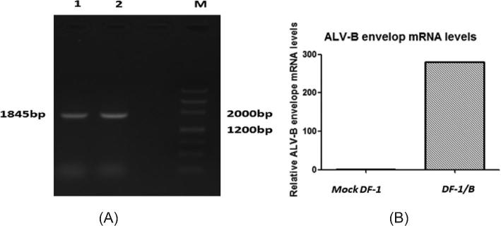
The viral envelope gene transcription level in DF-1/B cells was tested by conventional PCR and real-time RT-PCR, and the results are shown in Figure 3A. The ALV-B env gene fragment of 1,845 bp in length was amplified from RNA extracted from DF-1/B cells. Compared to the DF-1 cell negative control, the ALV-B env gene mRNA was highly expressed in DF-1/B cells (Figure 3B).
Figure 3.
(A) PCR amplification of the ALV-B env gene in DF-1/B cells. (M) DNA marker3; (1): RNA extracted from DF-1/B cells; (2): Genomic DNA extracted from DF-1/B cells; (3) RNA extracted from DF-1 cells. (B) Levels of ALV-B env gene transcription in DF-1/B cells were determined by real-time RT-PCR with gene specific primers. DF-1 cells served as a negative control. Data are representative of 2 independent experiments, both performed in triplicate.
Indirect IFA Testing of ALV-B env Gene Expression
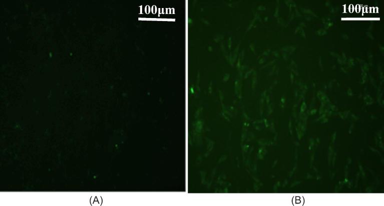
In the presence of env protein expression in the DF-1/B cells, the expression of env genes can be detected by specific monoclonal antibodies for ALV-B via IFA (Figure 4). The green fluorescence signal was bright in DF-1/B cells, but no green fluorescence in the cytoplasm could be observed in DF-1 cells. This indicates that the exogenous ALV-B env gene is successfully expressed in DF-1/B cells.
Figure 4.
Detection of ALV-B env protein in DF-1/B cells by IFA. (A) No green fluorescence was observed in DF-1 cells, which served as negative controls. (B) Green fluorescence was observed in DF-1/B cells. Magnification is × 100 for (A) and (B).
Western Blot Analysis of env Protein Expression in DF-1/B Cells
A Western blot experiment was performed to confirm the envelope protein expression in DF-1/B cells. The results of Western blot along with the lysate obtained from the DF-1/B cells transfected with the pcDNA-env-B plasmid showed the formation of a protein of 90 kDa. As expected, there was no protein band detected in DF-1 cells. This result demonstrated that the env protein had expressed in DF-1/B cells. β-Actin was used as a control for equal loading (Figure 5).
Figure 5.

Western blot analysis of env protein expression in DF-1/B cells. (DF-1/B): DF-1/B cell lysate; (DF-1): DF-1 cell lysate was used as a negative control. ALV-B env protein in the cell lysates were detected with monoclonal antibodies for ALV-B at a dilution of 1:200. β-actin cell lysates was also detected using actin antibodies for control for equal loading. A IRDye 800-conjugated anti-mouse IgG (1:10,000; Rockland Immunochemicals, Limerick, PA) diluted in PBS was used as the secondary antibody.
Antiviral Experiment
The antiviral effect of the DF-1 and DF-1/B cells was determined using representative strains of ALV-B (CD08) and ALV-J (CHN06). Both ALV-B and ALV-J are capable of infecting and replicating in DF-1 cells. In contrast, only ALV-J was able to infect DF-1/B cells, whereas ALV-B was effectively blocked from infecting DF-1/B cells. As shown in Figures 6A and 6B, DF-1/B cells inhibited the replication of ALV-B but not of ALV-J based on ELISA measurements of the viral p27 protein. To further validate the clinical utility of DF-1/B, ALV of clinically isolated unidentified subgroups were inoculated in DF-1 and DF-1/B cells. One of them was significantly blocked from infecting DF-1/B cells, but can infect DF-1cells (Figure 6C); the virus was identified as ALV-B by PCR. The antivirus assay showed that DF-1/B cells can have resistance to infection at a viral dose of 1 × 10⁴ TCID50 ALV-B, and at lower doses, infection was completely blocked. When the ALV-B infection dose reached 1 × 10⁵ TCID50, the ability of ALV-B to infect DF-1/B cells was still strongly inhibited (Figure 6A).
Figure 6.
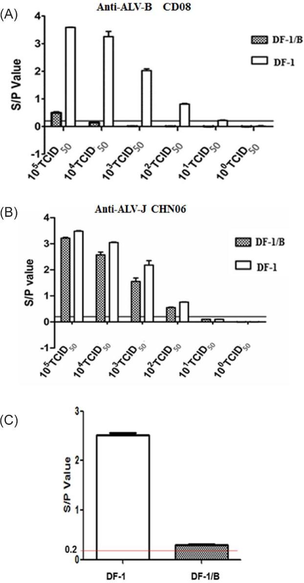
The result of antiviral experiment. Five different virus titers from 101 TCID50 to 105 TCID50 per 0.1 ml of ALV-B were inoculated per well in 24-well cell culture plates containing 1 mL 1.7 × 10⁵cells/well DF-1/B cells in triplicate. (A) ALV-B (CD08) replication was inhibited in DF-1/B, but not DF-1 cells. (B) Replication of ALV-J (CHN06) was not inhibited in either DF-1/B or DF-1 cells. (C) Infection by clinically isolated unidentified subgroups ALV, one of them was blocked in DF-1/B, but not in DF-1cells. In A–C, viral p27 protein levels determined by ELISA were reported. Black lines mean S/P = 0.2.
Immune Electron Microscopy
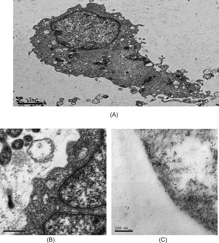
A colloidal gold immune electron microscopy examination revealed that the ALV-B env fusion protein is found in the DF-1/B cell membrane (Figure 7). The immune electron microscopy ALV-B fusion protein was detected with goat antimouse EGFP antibodies and monoclonal antibodies for ALV-B individually. As the secondary antibody, 10 nm of colloidal gold-labeled goat antimouse IgG was used. As a negative control, TBS (pH 7.4) was used in place of the primary antibody of control group. After uranyl acetate and lead citrate staining, images of the samples were obtained with a JEM-2000EX transmission electron microscope. Immunogold particles were observed in the cell membrane. In the controls, there were no immunogold particles in the cell membrane area.
Figure 7.
Electron microscopy image of DF-1/B cells that were pre-incubated with TBS (pH7.4) and subsequently incubated with 10 nm colloidal gold-labeled goat anti-mouse IgG. These samples served as negative controls. (A) TEM × 25,000, (B) TEM × 100,000. (C) Electron microscopy image of DF-1/B cells that were pre-incubated with goat anti-mouse EGFP antibodies and monoclonal antibodies for ALV-B individually and subsequently incubated with 10 nm colloidal gold-labeled goat anti-mouse IgG. TEM × 250,000. The arrows point to gold particles.
DISCUSSION
ALV is a retrovirus, which can be divided into 11 subgroups (subgroups A to K) according to their host range, patterns of receptor interference, and neutralization reaction (Payne et al., 1992; Wang et al., 2012). They have spread all over the world and caused enormous economic losses in the international poultry industry. In the mid-1980s, an international large-scale breeder company was successful in eradicating subgroup A and B ALV infection in chickens, but because of ALV purification, their drug could not be fully launched in China, so the Chinese chicken flocks, especially Chinese native chickens, still carry ALV-A (Zhang et al., 2010) and ALV-B (Zhao et al., 2010) infection. In recent years, ALV-B has been detected not only in chickens, but also in wild birds (Li et al., 2013) and has demonstrated the tendency of a recombinant virus (Lupiani et al., 2006; Wang et al., 2017). This indicates that the host range of ALV-B is changing. Traditional detection methods such as ELISA, PCR, real-time PCR, QC-RT-PCR, IFA, LAMP (Spencer and Gilka, 1982; Payne et al., 1993; Kim and Brown, 2004; Dai et al., 2015), and other methods have some limitations. The DF-1/B cell line described in this report has been genetically engineered to be selectively resistant to ALV-B through receptor interference. Whether it is the co-infection or single infection of the clinical sample, ALV-B can be definitively identified by inoculating the DF-1/B cell line and inoculating DF-1 as a control. The established immortal DF-1/B cell line, specifically resistant to ALV-B infection, not only enriches the diagnostic toolbox that distinguishes ALV subgroups, but also provides a rapid and reliable method to screen large numbers of samples.
In order to generate this cell line, which is genetically resistant to ALV-B, the env gene of ALV-B isolates, CD08, was cloned and expressed in the DF-1 cell line. The expressed env protein occupies the viral cell receptor binding sites and selectively interferes with the ALV-B infection cell line. To demonstrate whether the DF-1/B cell line was constructed successfully, PCR, IFA, Western blot, immune electron microscopy, and antivirus assay were used to detect the expression of env and the resistance to ALV-B infection. The results showed that DF-1/B cells could stably express the ALV-B env gene, and the env fusion protein is localized in the cell membrane area of DF-1/B cells. The antivirus assay showed that DF-1/B cells are resistant to 1 × 10⁴ TCID50 ALV-B infection, but not to ALV-J. To further verify this, we also tested with clinical samples. The result showed that ALV-B in field samples can be definitively identified. This means that the cell line is specifically resistant to ALV-B and can be used as a tool for ALV-B diagnosis.
Thus, the DF-1/B cell line has enriched the identification toolbox for identifying different subgroup ALV, provided a tool for identifying the molecular mechanism of ALV-B env protein interaction with host cells and the isolation and identification of viral cell surface receptors, provided a theoretical basis for the cultivation of disease-resistant chickens, and has also overcome the deficiencies of the current method of isolating and purifying ALV subgroups of mixed infection.
Supplementary Material
ACKNOWLEDGMENTS
This work was supported by the National Key R&D Project (2016YFD0501606); Poultry Industry Technology System of Guangdong (2018LM1114); the earmarked fund for China Agriculture Research System (CARS-41-G16); the Key Program of Science and Technology Development of Guangdong Province, China (2015A020209145).
REFERENCES
- Cui N., Su S., Chen Z., Zhao X., Cui Z.. 2014. Genomic sequence analysis and biological characteristics of a rescued clone of avian leukosis virus strain JS11C1, isolated from indigenous chickens. J. Gen. Virol. 95:2512. [DOI] [PubMed] [Google Scholar]
- Dai M., Min F., Liu D., Cao W. S., Liao M.. 2015. Development and application of SYBR Green I real-time PCR assay for the separate detection of subgroup J Avian leukosis virus and multiplex detection of avian leukosis virus subgroups A and B. Virol. J. 12:1–10. [DOI] [PMC free article] [PubMed] [Google Scholar]
- Hang B. L., Mei M., Fan Z. J.. 2013. Poultry Leukosis [M]. China Agricultural Science and Technology Press, Beijing. [Google Scholar]
- He C., Meng F. F., Chang S., Zhao P., Cui Z. Z.. 2013. Antigen ELISA Kit and Colloidal Gold Test Strips for the Detection of Avian Leukosis Virus Comparative Study [D]. Shandong Agricultural University; (in Chinese). [Google Scholar]
- Holmen S. L., Federspiel M. J.. 2000. Selection of a subgroup A avian leukosis virus [ALV(A)] envelope resistant to soluble ALV(A) surface glycoprotein. Virology 273:364–373. [DOI] [PubMed] [Google Scholar]
- Hunt H. D., Lee L. F., Foster D., Silva R. F., Fadly A. M.. 1999. A genetically engineered cell line resistant to subgroup j avian leukosis virus infection (C/J). Virology 264:205. [DOI] [PubMed] [Google Scholar]
- Kim Y., Brown T. P.. 2004. Development of quantitative competitive-reverse transcriptase-polymerase chain reaction for detection and quantitation of avian leukosis virus subgroup J. J. Vet. Diagn. Invest. 16:191. [DOI] [PubMed] [Google Scholar]
- Li D., Qin L., Gao H., Yang B., Liu W., Qi X., Wang Y., Zeng X., Liu S., Wang X., Gao Y. L.. 2013. Avian leukosis virus subgroup A and B infection in wild birds of Northeast China. Vet. Microbiol. 163:257–263. [DOI] [PubMed] [Google Scholar]
- Li X., Lin W., Chang S., Zhao P., Zhang X., Liu Y., Chen W., Li B., Shu D., Zhang H., Chen F., Xie Q. M.. 2016. Isolation, identification and evolution analysis of a novel subgroup of avian leukosis virus isolated from a local Chinese yellow broiler in South China. Arch. Virol 161:2717. [DOI] [PubMed] [Google Scholar]
- Livak K. J., Schmittgen T. D.. 2001. Analysis of relative gene expression data using real-time quantitative PCR and the 2(-Delta Delta C(T)) Method. Methods 25:402–408. [DOI] [PubMed] [Google Scholar]
- Lupiani B., Pandiri A. R., Mays J., Hunt H. D., Fadly A. M.. 2006. Molecular and biological characterization of a naturally occurring recombinant subgroup B avian leukosis virus with a subgroup J-like long terminal repeat. Avian Dis. 50:572–578. [DOI] [PubMed] [Google Scholar]
- Mingzhang R., Zijun Z., Lixia Y., Jian C., Min F., Jie Z., Ming L., Weisheng C.. 2018. The construction and application of a cell line resistant to novel subgroup avian leukosis virus (ALV-K) infection. Arch. Virol. 163:89–98. [DOI] [PMC free article] [PubMed] [Google Scholar]
- Payne L. N., Gillespie A. M., Howes K.. 1993. Unsuitability of chicken sera for detection of exogenous ALV by the group-specific antigen ELISA. Vet. Rec. 132:555–557. [DOI] [PubMed] [Google Scholar]
- Payne L. N., Howes K., Gillespie A. M., Smith L. M.. 1992. Host range of Rous sarcoma virus pseudotype RSV(HPRS-103) in 12 avian species: support for a new avian retrovirus envelope subgroup, designated J. J. Gen. Virol. 73:2995–2997. [DOI] [PubMed] [Google Scholar]
- Shao H., Wang L., Sang J., Li T., Liu Y., Wan Z., Qian K., Qin A., Ye J. Q.. 2017. Novel avian leukosis viruses from domestic chicken breeds in mainland China. Arch. Virol. 162:2073. [DOI] [PubMed] [Google Scholar]
- Spencer J. L., Gilka F.. 1982. Lymphoid leukosis: detection of group specific viral antigen in chicken spleens by immunofluorescence and complement fixation. Can. J. Comp. Med. 46:370–375. [PMC free article] [PubMed] [Google Scholar]
- Wang X., Peng-Fei Q. I., Yan D. U., Li W., Chuan L., De Q.. 2011. Pathological and viralogical analysis of a fibrosarcoma case induced by avian leucosis virus subgroup A. Chinese J. Anim. Infect. Dis. 19:11–16. [Google Scholar]
- Wang P., Yang Y., Lin L., Li H., Wei P.. 2017. Complete genome sequencing and characterization revealed a recombinant subgroup B isolate of avian leukosis virus with a subgroup J-like U3 region. Virus Genes 53:1–4. [DOI] [PubMed] [Google Scholar]
- Wang X., Zhao P., Cui Z. Z.. 2012. Identification of a new subgroup of avian leukosis virus isolated from Chinese indigenous chicken breeds. Bing Du Xue Bao = Chinese J. Virol. 28:609. [PubMed] [Google Scholar]
- Zhang X., Liao M., Jiao P., Luo K., Zhang H., Ren T., Zhang G., Xu C., Xin C., Cao W. S.. 2010. Development of a loop-mediated isothermal amplification assay for rapid detection of subgroup J avian leukosis virus. J. Clin. Microbiol. 48:2116–2121. [DOI] [PMC free article] [PubMed] [Google Scholar]
- Zhao D. M., Zhang Q. C., Cui Z. Z.. 2010. Isolation and identification of a subgroup B avian leukosis virus from chickens of Chinese native breed Luhua. Chinese J. Virol. 26:53. [PubMed] [Google Scholar]
Associated Data
This section collects any data citations, data availability statements, or supplementary materials included in this article.


