Significance
Recent live cell imaging has revealed that chromatin is not a static, rigid structure but is dynamically fluctuating in cells. We study chromatin motion by using single-nucleosome tracking data in living human cells. The distribution of single-nucleosome movement shows a distinct 2-peak feature: fast and slow fractions. Nucleosome movement is investigated using statistical analyses and a polymer model to elucidate the mechanism of dynamic organization of fast and slow chromatin.
Keywords: chromatin domains, nucleosomes, histone, cohesin, live cell imaging
Abstract
Understanding chromatin organization and dynamics is important, since they crucially affect DNA functions. In this study, we investigate chromatin dynamics by statistically analyzing single-nucleosome movement in living human cells. Bimodal nature of the mean square displacement distribution of nucleosomes allows for a natural categorization of the nucleosomes as fast and slow. Analyses of the nucleosome–nucleosome correlation functions within these categories along with the density of vibrational modes show that the nucleosomes form dynamically correlated fluid regions (i.e., dynamic domains of fast and slow nucleosomes). Perturbed nucleosome dynamics by global histone acetylation or cohesin inactivation indicate that nucleosome–nucleosome interactions along with tethering of chromatin chains organize nucleosomes into fast and slow dynamic domains. A simple polymer model is introduced, which shows the consistency of this dynamic domain picture. Statistical analyses of single-nucleosome movement provide rich information on how chromatin is dynamically organized in a fluid manner in living cells.
Three-dimensional organization of chromatin in nuclei influences DNA functions, such as transcription and replication (1–4), and hence, has been a focus of intensive investigation. In particular, the high-throughput chromosome conformation capture (Hi-C) approaches have revealed frequent chromatin–chromatin interactions within topologically associating domains (TADs) of several kb in size (5) or loop domains formed by looped chromatin chains of kb (6); these domains have come to be regarded as basic structural units of the genome chromatin. However, the single-cell Hi-C measurements (7, 8) and the microscopic observations with fluorescence in situ hybridization (FISH) (9, 10) have shown that positions of domain boundaries along the sequence are primarily different from cell to cell, showing large structural fluctuation in domain formation. Therefore, a fundamental question arises on how chromatin domains are formed with intense fluctuation and how such fluctuating domain formation affects gene regulation.
In order to answer this question on fluctuating chromatin organization, it is important to analyze chromatin movement in living cells. Highly dynamic chromatin movement has been so far observed with live cell imaging studies by using the LacO/LacI-GFP and related methods (11–16) and by more recent CRISPR-dCas9–based methods (17–19) and the single-nucleosome tracking techniques (20–23). In particular, Nozaki et al. (22) observed single-nucleosome movement in living human cells in a genome-wide manner and showed that nucleosomes are clustered in the nuclei to form chromatin domains. Here, we write displacement of the th nucleosome during time period as . When the nucleosome belongs to the domain , we can write for shorter than the timescale of domain formation/dissolution, where represents the intradomain displacement of the nucleosome and is the center of mass movement of the domain. Nozaki et al. (22) exemplified cases in which single-nucleosome movement is correlated with the domain movement for to 1 s, suggesting that domain movement is dominant with being sufficiently large. Theoretical polymer models also highlighted the effects of domain movement on chromatin dynamics (24–26); in the model of Di Pierro et al. (24), motions of different chromatin loci within the same domain are correlated with each other, showing significance of . Thus, quantitative analysis of chromatin dynamics is a key to understanding how chromatin domains are dynamically organized.
An important aspect of chromatin dynamics is their heterogeneity. Here, is mean square displacement (MSD) of the chromatin locus , and represents average over . In the previous study, at particular loci of chromosomes (15, 18, 27) and averaged over a wide region of the nucleus (28) or over the genome-wide single-nucleosome ensemble (20–23) were examined, showing that chromatin movement is subdiffusive as with ; various different values of were reported, suggesting diversity of chromatin movement. In the polymer model of Shi et al. (25), the value depends on whether the domain of the calculated locus is near the surface or in the interior of a chromosome structure. Shinkai et al. (26) argued that is smaller in compact heterochromatin-like domains, resulting in the smaller . Thus, the observed and calculated suggested that chromatin motion depends on interactions and environments of individual domains; therefore, chromatin movement is heterogeneous in a complex genomic structure. Indeed, heterogeneous distribution of movement was observed in fluorescent images of living mammalian cells using single-nucleosome tracking as “chromatin heat map” (22) and by flow-field monitoring (29). Thus, it is important to analyze heterogeneity in chromatin dynamics, which gives a clue to elucidate interactions and varied local environments of the chromatin domains.
In this study, we statistically analyze heterogeneity in chromatin dynamics by using the live cell imaging data of Nozaki et al. (22). These data are single-nucleosome trajectories obtained by tracking fluorescent images of nucleosomes in a thin layer (- to 250-nm thickness) of the HeLa cell nucleus. An example image is shown in SI Appendix, Fig. S1 and Movie S1. Using single-nucleosome trajectories, we extract distributions of MSD of individual nucleosomes, which allows for the nucleosome characterization as fast and slow. Based on this classification, features of nucleosome packing in chromatin domains are inferred by analyzing autocorrelations and pair correlations of nucleosome movement and by comparing cells in different conditions. Chromatin regions in which single nucleosomes show correlated movement are referred to as fast dynamic domains (f domains) and slow dynamic domains (s domains). A minimal polymer model is introduced to elucidate mechanics governing these domain organization.
Results
Fast and Slow Fractions of Nucleosomes.
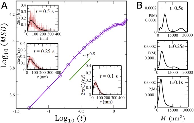
Shown in Fig. 1A is the average MSD of nucleosomes, , where is the average taken over and along the observed trajectories (22). From Fig. 1A, we find that the average movement of nucleosomes is subdiffusive for s with , although individual nucleosomes move with different exponents from 0.5 as shown below. For s, tends to saturate, suggesting that nucleosomes are caged in finite regions, and for s, nucleosomes begin to diffuse with . Here, we focus on the timescale s, where the data are experimentally well sampled by Nozaki et al. (22). SI Appendix, SI Text and Fig. S2 has the details of sampling the data.
Fig. 1.
MSD of nucleosome movement observed in live cell imaging of an example cell. (A) The MSD averaged over nucleosomes is plotted as a function of time. In Insets, the self-part of the vHC reproduced from using Eq. 1 (black) is superposed on the one obtained from the observed trajectories of single nucleosomes (red) at , , and s. (B) The distribution of the MSD of single nucleosomes at each corresponding time.
A remarkable feature is the diverse distribution of MSD of individual nucleosomes, which is captured by the distribution function, . Due to the short lifetime of observed fluorescence of single nucleosomes, the individual nucleosome MSD data are insufficient to provide for a clear . However, this problem is overcome by using the iterative algorithm of Richardson (30) and Lucy (31) (RL) to derive the smooth distribution from the noisy data. From the observed data, we first calculate the self-part of the van Hove correlation function (vHC), , where is the projected coordinate of the th nucleosome on the 2-dimensional imaging plane and is a constant to normalize as . The calculated vHC is shown at , 0.25, and 0.5 s in Fig. 1A, Insets. is expanded in Gaussian bases, , as
| [1] |
Given a noisy estimate of , is extracted as coefficients of expansion using the RL iterative scheme (Methods). The RL algorithm has been extensively used in image processing (32, 33) and also, in monitoring diffusion of liposomes in a nematic solution (34) and particles in simulated supercooled liquids (35–37). In SI Appendix, Figs. S3–S5, we show that this iterative method works well for evaluating the MSD distribution in example polymer systems.
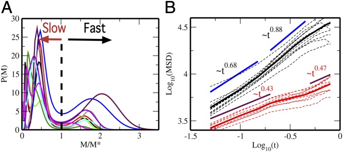
The MSD distribution, , obtained from the RL scheme is shown in Fig. 1B. At s, the distribution shows a single peak, but it splits into 2 peaks with increasing time. This bimodal feature is distinct at s, which allows for a characterization of nucleosomes as fast and slow. Mobility of nucleosomes varies from cell to cell, but the functional forms of become similar to each other when is scaled in a suitable way. In Fig. 2A, we plot for the 10 cells that we examined as functions of the scaled MSD, , where is defined as at the minimum of . Thus, we define fast (slow) nucleosomes as ones showing (). Then, the full vHC can be written in terms of sum of the vHC of the fast and slow nucleosomes as with and . SI Appendix, Fig. S4 shows the validity of this decomposition. With this characterization, we separately calculate the average MSD by for the fast () and slow () nucleosomes as shown in Fig. 2B. When we fit the MSD as , the exponent is to 0.88 for the fast nucleosomes and to 0.47 for the slow nucleosomes. This suggests that fast and slow nucleosomes move in different physical mechanisms.
Fig. 2.
Fast and slow nucleosomes. (A) The distribution of MSD of single nucleosomes, , is plotted for 10-cell samples as functions of , where is at the minimum between 2 peaks of . (B) The MSD averaged over fast nucleosomes, (black), and the MSD averaged over slow nucleosomes, (red), are shown for 10 individual cells (dashed lines) and the average over 10 cells (solid lines).
f and s Domains.
In order to understand the organization and underlying mechanisms that govern the dynamics of fast and slow movements, we analyze their temporal and spatial correlations. Shown in Fig. 3A are autocorrelations of displacement of nucleosomes:
| [2] |
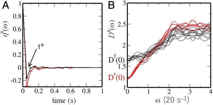
where with () is the average over and over the fast (slow) nucleosomes and is the average over 10 cells. is the displacement vector of the nucleosome , , with s. Fig. 3A shows that changes its sign for the first time at for both and , which indicates back scattering from the neighboring nucleosomes. Similar viscoelastic behavior has been seen in a polymer model of chromosomes (24). For , shows a damped oscillation with a period . From , a typical distance, , for nucleosomes to traverse between successive collisions is estimated as , showing to 88 nm for the fast nucleosomes and to 43 nm for the slow nucleosomes.
Fig. 3.
Autocorrelation functions of displacement of single nucleosomes and the density of vibrational modes. (A) The autocorrelation function of single-nucleosome displacement, , is plotted as a function of . Bars show the standard errors among 10 cells. (B) The density of vibrational modes, , is plotted as a function of frequency for 10 cells. In A and B, curves are plotted for fast (; black) and slow (; red) nucleosomes.
We can approximately regard as a velocity vector; then, the Fourier transform of , denoted here by , is the approximate density of vibrational modes (Fig. 3B). It is interesting to note that is finite, which is a signature of liquid-like behavior: represents self-diffusion in a fluid state, whereas in an amorphous solid state (38). shows that the fast nucleosomes are more fluid, and for the large shows that movement of the slow nucleosomes is more constrained. Nature of chromatin packing at the 30-nm scale has been under debate, with emerging evidence for the absence of the regular 30-nm fibers (39) in vivo and the fluid movement of chromatin in this length scale (4, 40). Here, the autocorrelations of displacement showed that the nucleosomes are back scattered at the 30-nm length scale along with the finite vibrational state density of , further providing evidence for the fluid nature of chromatin at this length scale.
Additional analyses with pair correlation functions are informative. The pair correlation functions of position (i.e., the radial distribution functions of fast and slow nucleosomes) are
| [3] |
with
| [4] |
where and are labels for either fast or slow nucleosomes and is a constant to normalize the function as for large-enough . In Eq. 4, is the average over and nucleosomes of type and . The nucleosome pair and was chosen to be averaged over in Eq. 4 only when both of them were observed in the same imaging frame.
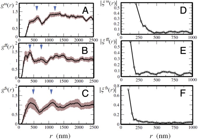
Due to the small number of sampled nucleosome pairs, a pair of nucleosomes was only infrequently observed in their mutual vicinity; therefore, as shown in Fig. 4 A–C, is small for nm. However, has peaks at and with nm (Fig. 4A), and with nm (Fig. 4B), and and (Fig. 4C); a straightforward interpretation is that fast and slow nucleosomes constitute domains with diameter and , respectively, and the oscillatory pattern of reflects liquid-like spatial arrangements of these domains. This domain picture is consistent with the pair correlations of displacement direction, , calculated as
| [5] |
with
| [6] |
Although is an oscillating function of , its oscillation profile is represented by as shown in Fig. 4 D–F. Correlation shown in is large only within a certain range . When we define as , we find that nm (Fig. 4D) and nm (Fig. 4 E and F), showing that and ; they imply that domains with radii and are domains of fast and slow nucleosomes, respectively, within which nucleosome dynamics are correlated with each other. We refer to these domains as f domains and s domains. Comparing and with the radius distribution observed in the FISH measurements (41), the size of f domains is estimated as to 300 kb, and that of s domains is to 500 kb; these findings suggest that the size of f domains is around the median size 185 kb of loop domains (6) and that the size of s domains is near to that of clusters of loop domains or TADs (5). A clear oscillatory behavior of in Fig. 4C shows that f domains and s domains form a mosaic arrangement. The peak of at shows density correlation between the adjacent f and s domains, and the peak at implies correlation between f domains and the next nearest s domains. Dominance of at over suggests that f domains are minor components in the mosaic arrangement.
Fig. 4.
Pair correlation functions of position and displacement of single nucleosomes. (A–C) Pair correlations of position: that is, the radial distribution functions, , of single nucleosomes. Triangles show the distances and in A, and in B, and and in C. The widths of brown shaded area show the standard errors among 10 cells. (D–F) Profile functions, , of pair correlation of displacement of single nucleosomes. Curves are shown with for the slow–slow correlation (A and D), for the fast–fast correlation (B and E), and for the fast–slow correlation (C and F).
We note that the displacement correlation function shows micrometer-scale correlations (SI Appendix, Fig. S6) consistent with the microscopically observed long length-scale correlations (28, 29, 42). However, such long-range correlations disappear when the numerator is averaged over multiple cells as in Eq. 5. The correlation remaining after this averaging over different cells is the core correlation common to those cells; as shown in Fig. 4 D–F, this correlation is large only within the range . Thus, these analyses of correlation functions provide a consistent picture that fast and slow nucleosomes show fluid movement to form dynamically correlated regions (i.e., f and s domains).
Perturbations on Chromatin Movement.
The relationship between chromatin dynamics and the physical features of domains is further examined by comparing the movement of nucleosomes under different cell conditions. Nozaki et al. (22) observed single-nucleosome movement in the following cases. (i) Cohesin knockdown (KD): cohesin action was suppressed by small interfering RNA (siRNA) KD of a cohesin subunit RAD21 (43, 44), which diminished the frequency of chromatin chain to be bundled by cohesin. (ii) Histone tail hyperacetylation: the frequency of histone tail acetylation was globally increased by adding a histone deacetylase inhibitor, Trichostatin A (TSA). Histone tail acetylation leads to weakening the histone H3 and H4 tail binding to the neighboring nucleosome and subsequent decondensation of chromatin (45). (iii) Cross-linking of chromatin: chromatin chains were cross-linked by treating the cells with formaldehyde (FA). Also compared was the case of (iv) focusing on heterochromatin: changing the height of the microscopic focal layer from the center to the periphery (PERI) of the nucleus, where these regions are enriched with heterochromatin regions or lamina-associated domains (46) tethered to inner nuclear membrane proteins (47).
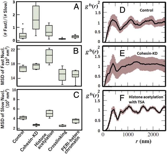
The distribution in these cases is bimodal or multimodal (SI Appendix, Fig. S7) so that the fast and slow nucleosomes are defined in the same way as in control described in the previous subsections. Features of are summarized in Fig. 5 A–C. The ratio of the number of fast nucleosomes to the number of slow nucleosomes is markedly large in cohesin KD and small in FA cross-linking cases. The average MSD for the fast nucleosomes is large in histone tail acetylation with TSA, and the average MSD for the slow nucleosomes is small in cohesin KD and FA.
Fig. 5.
Effects of perturbations on cells and effects of focusing on heterochromatin. (A–C) Features of the effects on the distribution of MSD, at s, of single nucleosomes: (A) the ratio of the number of fast nucleosomes to the number of slow nucleosomes, (B) the mean MSD of fast nucleosomes, and (C) the mean MSD of slow nucleosomes. Box plots of the data from 10 cells. (D–F) The radial distribution function in cases of (D) control, (E) cohesin KD, and (F) histone hyperacetylation with TSA. In D–F, widths of brown shaded area show the standard errors among 10 cells.
These features can be understood when chromatin domains are modified in a particular way by each perturbation. Cohesin KD diminishes the cohesin action to bundle the chromatin chains (43, 44), decreasing the constraint on the chain movement. Loosening the motional constraint increases the population of fast nucleosomes. It is intriguing to see that, with cohesin KD, the slow nucleosomes become slower and fast nucleosomes become faster, which may be due to the enhancement of A/B compartmentalization as found in the enhanced contrast of the Hi-C contact pattern on cohesin depletion (48, 49). Adding TSA, however, causes global decondensation of the compact domains; this structural loosening makes fast and slow nucleosomes faster. By contrast, FA cross-linking induces constraints on the movement, which severely decreases the population of fast nucleosomes and also, decreases the average MSD of fast and slow nucleosomes. The milder but similar effect is found in PERI heterochromatin, which should reflect the nucleosome tethering to the nuclear lamina and other heterochromatin proteins (46, 47).
With TSA, the first peak of the radial distribution function increases (SI Appendix, Fig. S8), which is consistent with the enhancement of fast nucleosomes with TSA. Shown in Fig. 5 D–F is the function . With TSA, has a shorter length scale of oscillation than in control. By contrast, the oscillation in diminishes with cohesin KD. Thus, different dynamic constraints give different effects on the f- and s-domain arrangements. A possible mechanism is that, with TSA, s domains are dissolved and that f domains and s domains are mixed, shortening the length scale of the oscillation, while with cohesin KD, the A/B compartmentalization is enhanced, which separates f domains and s domains, diminishing the mosaic-like arrangement and suppressing the oscillation. To examine this possibility, further comparison between changes in dynamic features and Hi-C contact maps on cell perturbations is desired.
The above analyses showed that the constraints on the motion of domains slow down the movement; the cohesin bundling of chromatin chains, which is diminished by cohesin KD, and the intradomain nucleosome–nucleosome interactions that decrease on the addition of TSA are the effective constraints on the movement. This supports a view that nucleosomes are driven primarily by thermal fluctuating motion and that physical or geometrical constraints on the motion are responsible for separating slow nucleosomes from the fast ones.
Discussion
These statistical analyses showed that physical or geometrical constraints on the motion are responsible for separating slow nucleosomes from the fast ones. Indeed, as shown in the TSA-treated cells, decondensing chromatin increased the mobility of nucleosomes, and as shown in PERI heterochromatin, tethering of chromatin to lamina and other proteins slows the domain movement. In addition to these factors described above, transcription machinery is likely to be another important factor. As well as the classical transcription factory model (50), the recent observations of the droplet-like assembly of RNA polymerase II and transcription coactivators/factors (51–54) suggested that enhancers/promoters can bind to such clusters/droplets to form a loosely connected network of chromatin chains, which slows the movement of transcriptionally active chromatin regions (23). Importantly, slowing down of the nucleosome movement is not induced by a sole mechanism, but there are multiple coexisting mechanisms; therefore, the interplay between these mechanisms is essential to understand the origin of the slow movement.
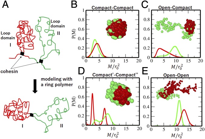
Here, we introduce a minimal model to discuss the interplay of multiple dynamical constraints. As illustrated in Fig. 6A, 2 consecutive loop domains with cohesin bound at their boundaries are represented by a ring having 2 regions, Region I and Region II. This ring is a bead-and-spring chain consisting of 300 beads, with each bead representing a -kb segment. Region I consists of 100 beads, and Region II consists of the rest (200 beads); 2 regions are separated by a contact between the 1st and 101st beads, which mimics the cohesin binding. We simulate movement of this chain by numerically integrating the Langevin dynamics by assuming the interaction potential, , where is the distance between the th and th beads and is a unit length, and we set . SI Appendix has more details. The coefficient for the attractive part of the potential is when , when , and when and belong to different regions. In SI Appendix, Fig. S9, we show the dependence of the simulated radius of gyration of the ring on the interaction strength of . This ring is open extended when and compact condensed when , showing a continuous coil–globule transition as in the microscopic observation of chromatin (55). We examine and around this transition regime. We represent the effects of tethering or interactions with the other nuclear structures by defining a reference point on the chain as the locus to be tethered. Although tethering/untethering can dynamically switch in cells, we here use a simple assumption that the coordinate system is fixed with its origin on a specific bead (reference point), and we monitor the MSD of the simulated polymer by using this coordinate system.
Fig. 6.
A polymer model of looped domains. (A) Two consecutive looped domains are represented by Region I and Region II in a model ring. The cohesin binding is represented by thick bars. (B–E) Distribution of MSD, , of beads in a polymer model. Connected 2-looped domains of B compact ()–compact (), (C) compact ()–open (), (D) compact′ ()–compact″ (), and (E) open ()–open () regions. In B–E, calculated from the reference point in Region I (red) and calculated from the reference point in Region II (green) are plotted. Insets are snapshots of the polymer ring; beads in Region I (red) and those in Region II (green) are shown with spheres.
Fig. 6 B–E shows obtained at a given time (SI Appendix) from the simulated movement of the chain. Two regions having interactions with represent 2 compact loop domains (Fig. 6B). These 2 regions tend to have a merged condensed configuration, and has a peak at a small . Two regions with and represent the connected compact and open loop domains (Fig. 6C). When the reference point is in the compact domain of Region I, both regions show the slow movement, while when the reference point is in the open domain of Region II, both regions show the fast movement. Therefore, the movement depends not only on whether the loop domain is open or compact but also, on the nature of the domain tethering. In a ring with and , both loop domains take compact configurations (Fig. 6D). However, Region I is a core globule, and Region II wraps the surface of Region I. In this case, shows a bimodal peak with the slow movement of Region I and the fast movement of Region II. The sensitivity of to tethering indicates that nucleosomes belonging to compact regions can be inferred as fast. When 2 loop domains are open with , both of them show the fast movement regardless of the position of the reference point. When 2 compact loop domains merge as shown in Fig 6B, their motions are correlated to form a single s domain having an effectively larger size than independent loop domains, making large. It is intriguing to examine with this polymer model whether the transitions between f and s domains take place as open–closed structural transitions suggested by a chromosome model (56).
Thus, the minimal polymer model elucidates the interplay of key mechanisms, such as geometry (compact or open and core or surface) of the chromatin chain and tethering in their role in the f- and s-domain organization. Further quantitative analyses are necessary to identify the precise molecular interactions that define chromosome organization geometry and tethering in living cells. The statistical analyses of single-nucleosome trajectories with the enhanced sampling (23) will allow for such quantitative analyses. As noted by Dubochet and Sartori Blanc (57), chemical fixation, such as with FA or glutaraldehyde, which is a standardized methodology in cell biology analyses, can have artifactual effects on chromatin interactions. To explore the “in vivo” organization, investigating chromatin in living cells is crucial. As demonstrated in this study, the statistical analyses of single-nucleosome trajectories provide a means for such exploration.
Conclusions
We investigated heterogeneity of chromatin dynamics in living human cells by analyzing single-nucleosome movement. The obtained nucleosome MSD distribution revealed that the nucleosomes are categorized into 2 types: fast and slow. This categorization of nucleosomes revealed aspects of organization of heterogeneous chromatin domains; nucleosome movements are correlated with each other within f and s domains. This analysis was applied to cells under various perturbations, and together with a simple polymer model, the method gave a consistent picture of organization of dynamic chromatin domains. Thus, the categorization of fast and slow nucleosome movement introduced in this analyses provides a basis for understanding chromatin organization.
Methods
of Eq. 1 was calculated in an iterative way with the RL algorithm: starting from the initial distribution, , at the th iteration was obtained by with . This equation was iterated under the constraints and .
Supplementary Material
Acknowledgments
This work was supported by Japan Science and Technology Agency (JST) Core Research for Evolutional Science and Technology Grant JPMJCR15G2; the Riken Pioneering Project; Japan Society for the Promotion of Science KAKENHI Grants JP19H01860, 19H05258, JP16H04746, 16H06279 (Platform for Advanced Genome Science), and 19H05273; the Takeda Science Foundation; and National Institute of Genetics Collaboration Grant (NIG-JOINT) 2016-A2 (6).
Footnotes
The authors declare no conflict of interest.
This article is a PNAS Direct Submission.
This article contains supporting information online at www.pnas.org/lookup/suppl/doi:10.1073/pnas.1907342116/-/DCSupplemental.
References
- 1.Bickmore W. A., The spatial organization of the human genome. Annu. Rev. Genom. Hum. Genet. 14, 67–84 (2013). [DOI] [PubMed] [Google Scholar]
- 2.Cardoso M. C., Schneider K., Martin R. M., Leonhardt H., Structure, function and dynamics of nuclear subcompartments. Curr. Opin. Cell Biol. 24, 79–85 (2012). [DOI] [PubMed] [Google Scholar]
- 3.Hübner M., Eckersley-Maslin M., Spector D., Chromatin organization and transcriptional regulation. Curr. Opin. Genet. Dev. 23, 89–95 (2013). [DOI] [PMC free article] [PubMed] [Google Scholar]
- 4.Maeshima K., Ide S., Hibino K., Sasai M., Liquid-like behavior of chromatin. Curr. Opin. Genet. Dev. 37, 36–45 (2016). [DOI] [PubMed] [Google Scholar]
- 5.Dekker J., Heard E., Structural and functional diversity of topologically associating domains. FEBS Lett. 589, 2877–2884 (2015). [DOI] [PMC free article] [PubMed] [Google Scholar]
- 6.Rao S. S., et al. , A 3D map of the human genome at kilobase resolution reveals principles of chromatin looping. Cell 159, 1665–1680 (2014). [DOI] [PMC free article] [PubMed] [Google Scholar]
- 7.Nagano T., et al. , Cell-cycle dynamics of chromosomal organization at single-cell resolution. Nature 547, 61–67 (2017). [DOI] [PMC free article] [PubMed] [Google Scholar]
- 8.Tan L., Xing D., Chang C. H., Li H., Xie X. S., Three-dimensional genome structures of single diploid human cells. Science 361, 924–928 (2018). [DOI] [PMC free article] [PubMed] [Google Scholar]
- 9.Bintu B., et al. , Super-resolution chromatin tracing reveals domains and cooperative interactions in single cells. Science 362, eaau1783 (2018). [DOI] [PMC free article] [PubMed] [Google Scholar]
- 10.Finn E. H., et al. , Extensive heterogeneity and intrinsic variation in spatial genome organization. Cell 176, 1502–1515 (2019). [DOI] [PMC free article] [PubMed] [Google Scholar]
- 11.Marshall W. F., et al. , Interphase chromosomes undergo constrained diffusional motion in living cells. Curr. Biol. 7, 930–939 (1997). [DOI] [PubMed] [Google Scholar]
- 12.Heun P., Laroche T., Shimada K., Furrer P., Gasser S. M., Chromosome dynamics in the yeast interphase nucleus. Science 294, 2181–2186 (2001). [DOI] [PubMed] [Google Scholar]
- 13.Chubb J.R., Boyle S., Perry P., Bickmore W. A., Ch6romatin motion is constrained by association with nuclear compartments in human cells. Curr. Biol. 12, 439–445 (2002). [DOI] [PubMed] [Google Scholar]
- 14.Levi V., Ruan Q., Plutz M., Belmont A. S., Gratton E., Chromatin dynamics in interphase cells revealed by tracking in a two-photon excitation microscope. Biophys. J. 89, 4275–4285 (2005). [DOI] [PMC free article] [PubMed] [Google Scholar]
- 15.Hajjoul H., et al. , High-throughput chromatin motion tracking in living yeast reveals the flexibility of the fiber throughout the genome. Genome Res. 23, 1829–1838 (2013). [DOI] [PMC free article] [PubMed] [Google Scholar]
- 16.Germier T., et al. , Real-time imaging of a single gene reveals transcription-initiated local confinement. Biophys. J. 113, 1383–1394 (2017). [DOI] [PMC free article] [PubMed] [Google Scholar]
- 17.Chen B., et al. , Dynamic imaging of genomic loci in living human cells by an optimized CRISPR/Cas system. Cell 155, 1479–1491 (2013). [DOI] [PMC free article] [PubMed] [Google Scholar]
- 18.Gu B., et al. , Transcription-coupled changes in nuclear mobility of mammalian cis-regulatory elements. Science 359, 1050–1055 (2018). [DOI] [PMC free article] [PubMed] [Google Scholar]
- 19.Ma H., et al. , Cell cycle- and genomic distance-dependent dynamics of a discrete chromosomal region. J. Cell Biol. 218, 1467–1477 (2019). [DOI] [PMC free article] [PubMed] [Google Scholar]
- 20.Hihara S., et al. , Local nucleosome dynamics facilitate chromatin accessibility in living mammalian cells. Cell Rep. 2, 1645–1656 (2012). [DOI] [PubMed] [Google Scholar]
- 21.Nozaki T., et al. , Flexible and dynamic nucleosome fiber in living mammalian cells. Nucleus 4, 349–356 (2013). [DOI] [PMC free article] [PubMed] [Google Scholar]
- 22.Nozaki T., et al. , Dynamic organization of chromatin domains revealed by super-resolution live-cell imaging. Mol. Cell 67, 282–293 (2017). [DOI] [PubMed] [Google Scholar]
- 23.Nagashima R., et al. , Single nucleosome imaging reveals loose genome chromatin networks via active RNA polymerase II. J. Cell Biol. 218, 1511–1530 (2019). [DOI] [PMC free article] [PubMed] [Google Scholar]
- 24.Di Pierro M., Potoyan D. A., Wolynes P. G., Onuchic J. N., Anomalous diffusion, spatial coherence, and viscoelasticity from the energy landscape of human chromosomes. Proc. Natl. Acad. Sci. U.S.A. 115, 7753–7758 (2018). [DOI] [PMC free article] [PubMed] [Google Scholar]
- 25.Shi G., Liu L., Hyeon C., Thirumalai D., Interphase human chromosome exhibits out of equilibrium glassy dynamics. Nat. Commun. 9, 3161 (2018). [DOI] [PMC free article] [PubMed] [Google Scholar]
- 26.Shinkai S., Nozaki T., Maeshima K., Togashi Y., Dynamic nucleosome movement provides structural information of topological chromatin domains in living human cells. PLoS Comput. Biol. 12, e1005136 (2016). [DOI] [PMC free article] [PubMed] [Google Scholar]
- 27.Bronstein I., Transient anomalous diffusion of telomeres in the nucleus of mammalian cells. Phys. Rev. Lett. 103, 018102 (2009). [DOI] [PubMed] [Google Scholar]
- 28.Zidovska A., Weitz D. A., Mitchison T. J., Micron-scale coherence in interphase chromatin dynamics. Proc. Natl. Acad. Sci. U.S.A. 110, 15555–15560 (2013). [DOI] [PMC free article] [PubMed] [Google Scholar]
- 29.Shaban H. A., Barth R., Bystricky K., Nanoscale mapping of DNA dynamics in live human cells. bioRxiv:10.1101/405969 (5 January 2019). [Google Scholar]
- 30.Richardson W. H., Bayesian-based iterative method of image restoration. J. Opt. Soc. Am. 62, 55–59 (1972). [Google Scholar]
- 31.Lucy L. B., An iterative technique for the rectification of observed distributions. Astron. J. 79, 745–754 (1974). [Google Scholar]
- 32.Dey N., et al. , Richardson-Lucy algorithm with total variation regularization for 3D confocal microscope deconvolution. Microsc. Res. Tech. 69, 260–266 (2006). [DOI] [PubMed] [Google Scholar]
- 33.Laasmaa M., Vendelin M., Peterson P., Application of regularized Richardson-Lucy algorithm for deconvolution of confocal microscopy images. J. Microsc. 243, 124–140 (2011). [DOI] [PMC free article] [PubMed] [Google Scholar]
- 34.Wang B., Kuo J., Bae S. C., Granick S., When Brownian diffusion is not Gaussian. Nat. Mater. 11, 481–485 (2012). [DOI] [PubMed] [Google Scholar]
- 35.Sengupta S., Karmakar S., Distribution of diffusion constants and Stokes-Einstein violation in supercooled liquids. J. Chem. Phys. 140, 224505 (2014). [DOI] [PubMed] [Google Scholar]
- 36.Bhowmik B. P., Das R., Karmakar S., Understanding Stokes-Einstein relation in supercooled liquids using random pinning. J. Stat. Mech. 2016, 074003 (2016). [Google Scholar]
- 37.Bhowmik B. P., Tah I., Karmakar S., Non-Gaussianity of van Hove function and dynamic heterogeneity length scale. Phys Rev E 98, 022122 (2018). [DOI] [PubMed] [Google Scholar]
- 38.Lin S. T., Blanco M., Goddard W. A., The two-phase model for calculating thermodynamic properties of liquids from molecular dynamics: Validation for the phase diagram of Lennard-Jones fluids. J. Chem. Phys. 119, 11792 (2003). [Google Scholar]
- 39.Finch J. T., Klug A., Solenoidal model for superstructure in chromatin. Proc. Natl. Acad. Sci. U.S.A. 73, 1897–1901 (1976). [DOI] [PMC free article] [PubMed] [Google Scholar]
- 40.Maeshima K., Ide S., Babokhov M., Dynamic chromatin organization without the 30-nm fiber. Curr. Opin. Cell Biol. 58, 95–104 (2019). [DOI] [PubMed] [Google Scholar]
- 41.Boettiger A. N., et al. , Super-resolution imaging reveals distinct chromatin folding for different epigenetic states. Nature 529, 418–422 (2016). [DOI] [PMC free article] [PubMed] [Google Scholar]
- 42.Shaban H. A., Barth R., Bystricky K., Formation of correlated chromatin domains at nanoscale dynamic resolution during transcription. Nucleic Acids Res. 46, e77 (2018). [DOI] [PMC free article] [PubMed] [Google Scholar]
- 43.Nasmyth K., Haering C. H., The structure and function of SMC and kleisin complexes. Annu. Rev. Biochem. 74, 595–648 (2005). [DOI] [PubMed] [Google Scholar]
- 44.Shintomi K., Hirano T., Sister chromatid resolution: A cohesion releasing network and beyond. Chromosoma 119, 459–467 (2010). [DOI] [PubMed] [Google Scholar]
- 45.Görisch S. M., Wachsmuth M., Tóth K. F., Lichter P., Rippe K., Histone acetylation increases chromatin accessibility. J. Cell Sci. 118, 5825–5834 (2005). [DOI] [PubMed] [Google Scholar]
- 46.van Steensel B., Belmont A. S., Lamina-associated domains: Links with chromosome architecture, heterochromatin, and gene repression. Cell 169, 780–791 (2017). [DOI] [PMC free article] [PubMed] [Google Scholar]
- 47.Lemaître C., Bickmore W. A., Chromatin at the nuclear periphery and the regulation of genome functions. Histochem. Cell Biol. 144, 111–122 (2015). [DOI] [PubMed] [Google Scholar]
- 48.Schwarzer W., et al. , Two independent modes of chromatin organization revealed by cohesin removal. Nature 551, 51–56 (2017). [DOI] [PMC free article] [PubMed] [Google Scholar]
- 49.Rao S. S. P., et al. , Cohesin loss eliminates all loop domains. Cell 171, 305–320 (2017). [DOI] [PMC free article] [PubMed] [Google Scholar]
- 50.Feuerborn A., Cook P. R., Why the activity of a gene depends on its neighbors. Trends Genet. 31, 483–490 (2015). [DOI] [PubMed] [Google Scholar]
- 51.Lu H., et al. , Phase-separation mechanism for C-terminal hyperphosphorylation of RNA polymerase II. Nature 558, 318–323 (2018). [DOI] [PMC free article] [PubMed] [Google Scholar]
- 52.Cho W. K., et al. , Mediator and RNA polymerase II clusters associate in transcription-dependent condensates. Science 361, 412–415 (2018). [DOI] [PMC free article] [PubMed] [Google Scholar]
- 53.Boija A., et al. , Transcription factors activate genes through the phase-separation capacity of their activation domains. Cell 175, 1842–1855 (2018). [DOI] [PMC free article] [PubMed] [Google Scholar]
- 54.Sabari B. R., et al. , Coactivator condensation at super-enhancers links phase separation and gene control. Science 361, eaar3958 (2018). [DOI] [PMC free article] [PubMed] [Google Scholar]
- 55.Zinchenko A., Berezhnoy N. V., Chen Q., Nordenskiöld L., Compaction of single-molecule megabase-long chromatin under the influence of macromolecular crowding. Biophys. J. 114, 2326–2335 (2018). [DOI] [PMC free article] [PubMed] [Google Scholar]
- 56.Zhang B., Wolynes P. G., Topology, structures, and energy landscapes of human chromosomes. Proc. Natl. Acad. Sci. U.S.A. 112, 6062–6067 (2015). [DOI] [PMC free article] [PubMed] [Google Scholar]
- 57.Dubochet J., Sartori Blanc N., The cell in absence of aggregation artifacts. Micron 32, 91–99 (2001). [DOI] [PubMed] [Google Scholar]
Associated Data
This section collects any data citations, data availability statements, or supplementary materials included in this article.


