Abstract
Jaundice, conjunctival hyperemia, and acute kidney injury (AKI) are the characteristics of leptospirosis. However, it is not well known that Fusobacterium necrophorum infection can have a clinical picture similar to that of leptospirosis. A 38-year-old man was admitted with jaundice, conjunctival hyperemia, and AKI for 7 days. Chest CT scan showed multiple pulmonary nodules, atypical for leptospirosis. We started treatment with IV piperacillin-tazobactam and minocycline. He became anuric and was urgently started on hemodialysis on the second hospital day. Later on, blood cultures grew Fusobacterium necrophorum and other anaerobic bacteria. Antibody and PCR assays for Leptospira were negative. We narrowed the antibiotics to IV ceftriaxone and metronidazole. He responded well to the treatment and was discharged on the 18th hospital day. F. necrophorum infection is known to cause mixed infection with other anaerobic bacteria. The resistance of many anaerobic bacteria continues to progress, and F. necrophorum itself sometimes produces β-lactamase. This case highlights the potential risks of using penicillin before diagnosis of leptospirosis.
1. Introduction
Jaundice, conjunctival hyperemia, and acute kidney injury (AKI) are the characteristics of leptospirosis [1, 2], which can be treated with penicillin [3]. However, Fusobacterium necrophorum infection may mimic leptospirosis [4–6], which is not well known. F. necrophorum often causes mixed infections with other anaerobic bacteria. In addition, the antimicrobial resistance of anaerobic bacteria is serious, and sometimes F. necrophorum itself also produces β-lactamase [7]. We present a case of F. necrophorum mixed-infection mimicking leptospirosis that might have treatment failure with penicillin.
2. Case Summary
A 38-year-old man presented with a seven-day history of fatigue, fever, and jaundice. He initially went to a nearby clinic as symptoms worsened. Subsequently, he was also diagnosed with AKI and transferred to our hospital. He had no contact with rats and did not do field work. He had no prior medical history.
Vital signs at the time of visit were as follows: blood pressure 116/73 mmHg, pulse 110/min, body temperature 37.4°C, and respiration rate 18/min. His ocular conjunctivae wereyellow-tinged and congested. His oral cavity was not swollen or reddish; however, he complained of a sore throat.
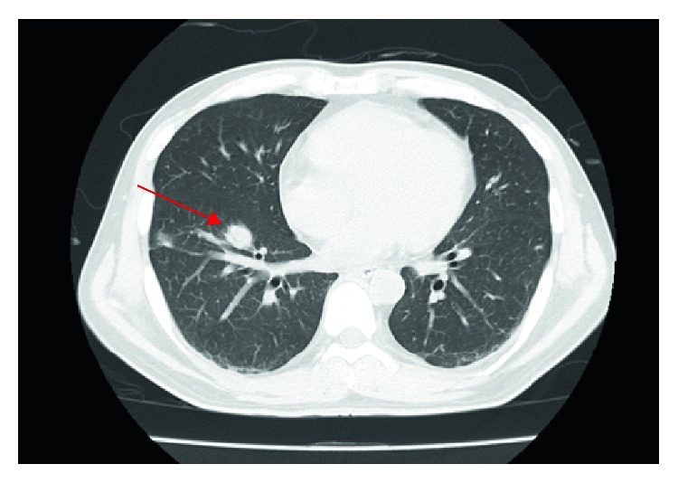
Blood tests revealed a white blood cell count of 18900/μL. His hemoglobin was 10.8 g/dL and platelet count was 2.6 × 10⁴/μL, and no schizocytes were observed. Aspartate aminotransferase was 31 U/L, alanine aminotransferase 18 U/L, lactate dehydrogenase 234 U/L, alkaline phosphatase 340 U/L, γ-glutamyltransferase 41 U/L, total bilirubin 12.4 mg/dL, and direct bilirubin 10.5 mg/dL, which is above the threshold for hyperbilirubinemia. Blood urea nitrogen was 85.6 mg/dL, creatinine was 8.3 mg/dL, and C-reactive protein was elevated to 29.26 mg/dL (normal range 0.0–0.3 mg/dL). In urine qualitative assays, positive results were observed for proteins, urobilinogen, bilirubin, and occult blood. In addition, urine sugar was positive despite not having diabetes. Chest CT showed multiple nodules in both lungs (Figure 1). Abdominal CT showed no abnormality that could explain the jaundice and kidney failure.
Figure 1.

The chest computed tomography on the 1st day showing multiple nodules (red arrow).
We initially strongly suspected leptospirosis. However, other infectious diseases could not be excluded because the multiple nodules in the lungs were atypical for leptospirosis.
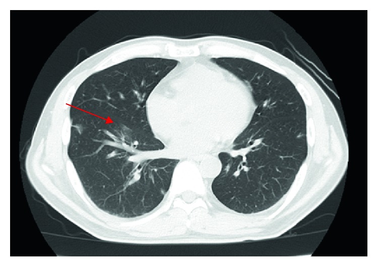
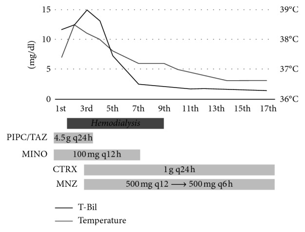
Because there was a possibility of various diseases, we avoided IV penicillin and started IV piperacillin-tazobactam and minocycline instead. He became anuric and was started on urgent hemodialysis on the second hospital day. Later on, antibody and PCR assays for Leptospira were negative. F. necrophorum was isolated in blood cultures on the second day. Further late, Streptococcus intermedius were isolated in blood cultures on the seventh day. We diagnosed his main disease as F. necrophorum rather than S. intermedius bacteremia based on the past case reports and the speed of positive blood cultures. Since F. necrophorum is famous for Lemierre's syndrome, we performed ultrasound in his jugular vein but found no thrombus. In addition, the dentist diagnosed a minor periodontal infection. F. necrophorum is thought to spread from periodontal infections. The main pathogen was F. necrophorum, but other anaerobic bacteria were also suspected to be involved. In addition, we were not able to test the susceptibility of F. necrophorum to penicillin. His treatment was thus de-escalated to IV ceftriaxone (CTRX) 1 g every 24 h and metronidazole (MNZ) 500 mg every 12 h on the 3rd day. Later, MNZ was increased up to 500 mg every 6 h. These treatments were continued up to the 17th day. The multiple lung nodules formed cavities and later disappeared after definitive therapy (Figure 2). He responded well to the treatment, was taken off hemodialysis, and discharged on the 18th hospital day. After discharge, he continued the treatment with amoxicillin and MNZ orally for three weeks (Figure 3). He had no relapse after three months of follow-up.
Figure 2.

The chest computed tomography on the 38th day showing multiple nodules disappeared (red arrow).
Figure 3.

The clinical course. T-Bil: total bilirubin; PIPC/TAZ: piperacillin-tazobactam; MINO: minocycline; CTRX: ceftriaxone; MNZ: metronidazole.
3. Discussion
This was a case of F. necrophorum infection without Lemierre's mimicking leptospirosis. Differentiating between these two diseases is important from the viewpoint of early antibacterial drug selection and identification of complications.
Leptospirosis is a zoonosis caused by the pathogenic spirochete Leptospira spp. Its occurrence is distributed throughout the world but is especially frequent in tropical areas [8]. The main vectors are rodents. Leptospira infects humans via contact of the skin, mucous membranes, and conjunctiva with animal urine and contaminated water and soil. After the incubation period, which is usually ten days, fever, muscle pain, and headache appear in 75–100% of patients [9]. When leptospirosis becomes severe, it complicates with symptoms of jaundice and renal failure [1]. That is also known as Weil's disease. Conjunctival hyperemia has also been observed in 55% of cases and is a symptom useful for differentiation from other infectious diseases [2].
F. necrophorum is an anaerobic Gram-negative bacillus that is resident in the oral cavity [10]. F. necrophorum infection decreased after the spread of penicillin [11]; however, F. necrophorum infection, and especially pharyngitis, has increased in recent years [12]. In addition, F. necrophorum infection has a 5–9% mortality rate even with antibiotics [10].
F. necrophorum infection is sometimes accompanied by jaundice. Especially, most cases of frank jaundice or hyperbilirubinemia due to F. necrophorum infection had a thrombus in the internal jugular vein, which progressed to Lemierre's syndrome [13]. This is a new point from this case: this case had no thrombus in the internal jugular vein. The exact frequency of jaundice by F. necrophorum is unknown. Moreover, there are completely different reports: frank jaundice is rare [14] or up to 49% [15]. In addition, the exact mechanism of jaundice by F. necrophorum remains unclear. We assume the lipopolysaccharide of F. necrophorum may affect jaundice because hyperbilirubinemia in sepsis is due to cholestasis by the lipopolysaccharide of bacteria [16], and the main toxicity of F. necrophorum is also from its lipopolysaccharide [17]. In addition, hemophagocytic lymphohistiocytosis can also be a differential diagnosis of jaundice and liver damage. In general, diagnosis of hemophagocytic lymphohistiocytosis in adults is due to multiple findings: fever ≥38.5°C, splenomegaly, pancytopenia, hypertriglyceridemia, and hyperferritinemia [18]. In this case, we did not measure triglyceride and ferritin, nor do a bone marrow puncture; therefore, we cannot deny hemophagocytic lymphohistiocytosis completely.
F. necrophorum sometimes has remote infection sites; in particular, septic embolism in the lung is found in 97% of F. necrophorum infections [13]. Septic embolism in the lung is an important feature of F. necrophorum infection and is not usually seen in leptospirosis. As the presence or absence of septic embolism can also affect the treatment period, it is important to differentiate between the two diseases. Incidentally, in this case, septic embolism in the lung was observed, and it was thought to be a disease condition close to Lemierre's syndrome, which is a severe clinical disease type of F. necrophorum infection characterized by pharyngitis and purulent thrombophlebitis in the jugular vein accompanied with bacteremia. However, thrombophlebitis of the jugular vein was not found by ultrasound or CT scan in this case.
This case showed the potential risks of using penicillin before diagnosis of leptospirosis. Penicillin is common for the treatment of leptospirosis [3]. F. necrophorum infection often causes a mixed infection with other anaerobic bacteria. In fact, our case was a mixed infection with S. intermedius. Antimicrobial resistance of anaerobic bacteria continues to progress; some anaerobic bacteria is resistance to penicillin. In addition, F. necrophorum itself occasionally produce β-lactamase [7]; that is, 2% of F. necrophorum is resistance to penicillin [19]. Unfortunately, we did not test susceptibility of F. necrophorum to PCG. Therefore, it is not exactly known whether this case has failed with penicillin or not; however, it remains a possibility that it may fail in a case of F. necrophorum and mixed infection. F. necrophorum is sensitive to CTRX and β-lactamase inhibitor combination drugs [2], and CTRX is equivalent to the penicillin group in the therapeutic effect against Leptospira [20, 21]. Therefore, initial treatment with cephalosporin or a β-lactamase inhibitor combination drug may be preferred when the cause of an infection has not been or cannot be distinguished.
We experienced a case of F. necrophorum infection mimicking leptospirosis. This case suggests the potential risks of using penicillin before a diagnosis of leptospirosis.
Conflicts of Interest
The authors declare that there are no conflicts of interest regarding the publication of this article.
Authors' Contributions
RY and SS were responsible for writing the manuscript and the literature search. TY was involved in reviewing the manuscript and the literature search.
References
- 1.Schreier S., Doungchawee G., Chadsuthi S., Triampo D., Triampo W. Leptospirosis: current situation and trends of specific laboratory tests. Expert Review of Clinical Immunology. 2013;9(3):263–280. doi: 10.1586/eci.12.110. [DOI] [PubMed] [Google Scholar]
- 2.Vanasco N. B., Schmeling M. F., Lottersberger J., Costa F., Ko A. I., Tarabla H. D. Clinical characteristics and risk factors of human leptospirosis in Argentina (1999–2005) Acta Tropica. 2008;107(3):255–258. doi: 10.1016/j.actatropica.2008.06.007. [DOI] [PubMed] [Google Scholar]
- 3.Baber M. D., Stuart R. D. Leptospirosis canicola a case treated with penicillin. The Lancet. 1946;248(6426):594–596. doi: 10.1016/s0140-6736(46)91055-0. [DOI] [PubMed] [Google Scholar]
- 4.Lin D., Suwantarat N., Young R. S. Lemierre’s syndrome mimicking leptospirosis. Hawaii Medical Journal. 2010;69(7):161–163. [PMC free article] [PubMed] [Google Scholar]
- 5.Wingfield T., Blanchard T. J., Ajdukiewicz K. M. B. Severe pneumonia and jaundice in a young man: an atypical presentation of an uncommon disease. Journal of Medical Microbiology. 2011;60(9):1391–1394. doi: 10.1099/jmm.0.029942-0. [DOI] [PubMed] [Google Scholar]
- 6.Han Y. W. Fusobacterium nucleatum: a commensal-turned pathogen. Current Opinion in Microbiology. 2015;23:141–147. doi: 10.1016/j.mib.2014.11.013. [DOI] [PMC free article] [PubMed] [Google Scholar]
- 7.Ahkee S., Srinath L., Raff M. J., Huang A., Ramirez J. A. Lemierre’s syndrome: postanginal sepsis due to anaerobic oropharyngeal infection. Annals of Otology, Rhinology & Laryngology. 1994;103(3):208–210. doi: 10.1177/000348949410300307. [DOI] [PubMed] [Google Scholar]
- 8.Hartskeerl R. A., Collares-Pereira M., Ellis W. A. Emergence, control and re-emerging leptospirosis: dynamics of infection in the changing world. Clinical Microbiology and Infection. 2011;17(4):494–501. doi: 10.1111/j.1469-0691.2011.03474.x. [DOI] [PubMed] [Google Scholar]
- 9.Katz A. R., Ansdell V. E., Effler P. V., Middleton C. R., Sasaki D. M. Assessment of the clinical presentation and treatment of 353 cases of laboratory-confirmed leptospirosis in Hawaii, 1974–1998. Clinical Infectious Diseases. 2001;33(11):1834–1841. doi: 10.1086/324084. [DOI] [PubMed] [Google Scholar]
- 10.Riordan T. Human infection with Fusobacterium necrophorum (necrobacillosis), with a focus on Lemierre’s syndrome. Clinical Microbiology Reviews. 2007;20(4):622–659. doi: 10.1128/cmr.00011-07. [DOI] [PMC free article] [PubMed] [Google Scholar]
- 11.Weesner C. L., Cisek J. E. Lemierre syndrome: the forgotten disease. Annals of Emergency Medicine. 1993;22(2):256–258. doi: 10.1016/s0196-0644(05)80216-1. [DOI] [PubMed] [Google Scholar]
- 12.Centor R. M. Expand the pharyngitis paradigm for adolescents and young adults. Annals of Internal Medicine. 2009;151(11):812–815. doi: 10.7326/0003-4819-151-11-200912010-00011. [DOI] [PubMed] [Google Scholar]
- 13.Sinave C. P., Hardy G. J., Fardy P. W. The Lemierre syndrome: suppurative thrombophlebitis of the internal jugular vein secondary to oropharyngeal infection. Medicine. 1989;68(2):85–94. doi: 10.1097/00005792-198903000-00002. [DOI] [PubMed] [Google Scholar]
- 14.Chirinos J. A., Lichtstein D. M., Garcia J., Tamariz L. J. The evolution of Lemierre syndrome: report of 2 cases and review of the literature. Medicine. 2002;81(6):458–465. doi: 10.1097/00005792-200211000-00006. [DOI] [PubMed] [Google Scholar]
- 15.Leugers C. M., Clover R. Lemierre syndrome: postanginal sepsis. The Journal of the American Board of Family Practice. 1995;8(5):384–391. [PubMed] [Google Scholar]
- 16.Chand N., Sanyal A. J. Sepsis-induced cholestasis. Hepatology. 2007;45(1):230–241. doi: 10.1002/hep.21480. [DOI] [PubMed] [Google Scholar]
- 17.Brazier J. S. Human infections with Fusobacterium necrophorum. Anaerobe. 2006;12(4):165–172. doi: 10.1016/j.anaerobe.2005.11.003. [DOI] [PubMed] [Google Scholar]
- 18.Jordan M. B., Allen C. E., Weitzman S., Filipovich A. H., McClain K. L. How I treat hemophagocytic lymphohistiocytosis. Blood. 2011;118(15):4041–4052. doi: 10.1182/blood-2011-03-278127. [DOI] [PMC free article] [PubMed] [Google Scholar]
- 19.Brazier J. S., Duerden B. I., Yusuf E., Hall V. Fusobacterium necrophorum infections in England and Wales 1990-2000. Journal of Medical Microbiology. 2002;51(3):269–272. doi: 10.1099/0022-1317-51-3-269. [DOI] [PubMed] [Google Scholar]
- 20.Panaphut T., Domrongkitchaiporn S., Vibhagool A., Thinkamrop B., Susaengrat W. Ceftriaxone compared with sodium penicillin G for treatment of severe leptospirosis. Clinical Infectious Diseases. 2003;36(12):1507–1513. doi: 10.1086/375226. [DOI] [PubMed] [Google Scholar]
- 21.Suputtamongkol Y., Niwattayakul K., Suttinont C., et al. An open, randomized, controlled trial of penicillin, doxycycline, and cefotaxime for patients with severe leptospirosis. Clinical Infectious Diseases. 2004;39(10):1417–1424. doi: 10.1086/425001. [DOI] [PubMed] [Google Scholar]


