Abstract
Spin-orbit coupling (SOC) has gained much attention for its rich physical phenomena and highly promising applications in spintronic devices. The Rashba-type SOC in systems with inversion symmetry breaking is particularly attractive for spintronics applications since it allows for flexible manipulation of spin current by external electric fields. Here, we report the discovery of a giant anisotropic Rashba-like spin splitting along three momentum directions (3D Rashba-like spin splitting) with a helical spin polarization around the M points in the Brillouin zone of trigonal layered PtBi2. Due to its inversion asymmetry and reduced symmetry at the M point, Rashba-type as well as Dresselhaus-type SOC cooperatively yield a 3D spin splitting with αR ≈ 4.36 eV Å in PtBi2. The experimental realization of 3D Rashba-like spin splitting not only has fundamental interests but also paves the way to the future exploration of a new class of material with unprecedented functionalities for spintronics applications.
Subject terms: Electronic properties and materials, Spintronics
Rashba type spin splitting – relevant for spintronics applications - is driven by inversion symmetry breaking but could so far not be realized in all momentum directions in a crystal. Here, the authors report on PtBi2 that exhibits Rashba spin splitting in all three momentum directions.
Introduction
The generation, manipulation, and detection of spin currents are the fundamental aspects of spintronics. The present material realizations for potential spintronics applications are based on spin–orbit coupling (SOC)1,2, which is a relativistic interaction of the electron spin with its motion in a potential. It allows for the removal of spin degeneracy in a non-magnetic material without the use of an external magnetic field. Among different types of SOC, Rashba-type SOC3,4 has attracted the most attention, since it can be manipulated by external electric fields, which is of central importance for applications in spintronics. In a system with Rashba SOC, each spin-degenerate parabolic band splits into two parabolic bands with opposite spin polarization. The band dispersions can be described by E±(k) = (ħ2k2/2m*) ± αR|k|, where m* is the effective mass of the electron and the Rashba parameter αR represents the strength of the SOC. The Rashba-type SOC is usually caused by structure inversion asymmetry (SIA) which stems from the inversion asymmetry of the confining potential2,3.
By contrast, there is other asymmetry in real materials called bulk inversion asymmetry (BIA). It originates from lack of inversion symmetry in the bulk and yields Dresselhaus-type SOC5,6. In many materials, these two kinds of SOC couple together, resulting in an anisotropy of spin splitting. Such anisotropy can lead to many interesting phenomena, such as a persistent spin helix observed in GaAs low-dimensional systems7,8, long spin relaxation times9–14, new device proposals, and more15–19.
Rashba splitting was first observed by spin-resolved and angle-resolved photoemission spectroscopy (Spin-ARPES) in the Shockley surface state of Au(111)20–22. Many phenomena related to Rashba-type SOC have been studied in a variety of systems23–50. In particular, BiTeI and GeTe gained attention for their giant Rashba splitting in bulk states35–45. However, in all known instances, the Rashba bands remain spin degenerate along out-of-plane direction kz for kx = ky = 0, while they show spin splitting along the in-plane momentum directions, namely, kx and ky directions. While there is no principal symmetry exclusion argument against Rashba spin splitting along three momentum directions (3D Rashba spin splitting) induced by inversion symmetry breaking, its realization in an explicit material framework persists to be a challenging task.
In this work, we report the discovery of a giant anisotropic 3D Rashba-like spin splitting directly observed by spin-ARPES in a binary compound: PtBi2. The spin-resolved ARPES employed the recently developed multichannel VLEED-type detector enabling fast mapping of a complete spin-polarized E(k) map51. The magnitude of the spin splitting is even stronger than that of BiTeI35. The observed spin splitting originates from the breaking of inversion symmetry of the PtBi2 crystal structure(space group P31m). In particular, the large spin splitting emerges at the M points of the Brillouin zone instead of the Γ point, where Rashba-type splitting is usually found. This bears crucial implications, rendering PtBi2 unique among related material classes. The reduced point group symmetry at M along with the BIA allow the Dresselhaus-type and Rashba-type SOC terms to cooperatively yield a large 3D anisotropic spin splitting with a helical texture around the M point.
Results
The crystal structure and Fermi surface of PtBi2
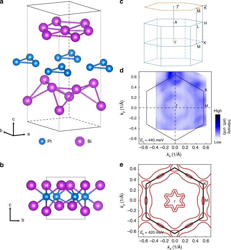
For trigonal layered PtBi2, two structural phases have been reported: space group 52 and P31m53,54. Both phases are layered structures with each layer consisting of edge-sharing PtBi6 octahedra, resembling the 1H-polytype of CdI2. ARPES results on the phase have been reported very recently55,56 motivated by the discovery of giant linear magneto-resistance57. On the other hand, in the P31m case, the distortion breaks inversion symmetry but the mirror symmetry is retained. Our X-ray single-crystal structure analysis demonstrates that the PtBi2 grown and studied in this work belongs to the P31m space group with lattice parameters a = b = 6.5705 Å and c = 6.1707 Å as shown in Fig. 1a, b (for XRD results, see supplementary). From the crystal structure in Fig. 1b, we can see that the Bi atoms in the layer above Pt atoms are coplanar while those below Pt are not. This leads to ABC stacking of three inequivalent layers, clearly breaking inversion symmetry. The corresponding bulk Brillouin zone is drawn in Fig. 1c in blue, and the projected surface Brillouin zone is sketched in orange. Figure 1d shows the experimental constant energy contour of PtBi2 at Eb = 440 meV and around kz = 0, which is in reasonable agreement with the calculation for Eb = 420 meV as shown in Fig. 1e. There are two six-pointed-star-shaped pockets around the Γ point and one triangular pocket at the K point. The elliptical contour around the M point and large closed pocket encircling much of the Brillouin zone arise from anisotropic spin splitting. In the following, we will focus on the band structure around the M point.
Fig. 1.
The crystal structure and Fermi surface of PtBi2. a, b Crystal structure of trigonal layered PtBi2 with space group P31m. c Bulk Brillouin zone (blue) and surface Brillouin zone (orange). d Constant-energy contour at Eb = 440 meV and around kz = 0 measured by angle-resolved photoemission spectroscopy (ARPES) (hν = 8.8 eV). The color scale is linear as in all other images. e Calculated constant energy contour at Eb = 420 meV and kz = 0
Rashba-like band splitting at the M point
The band dispersion along the Γ–M–Γ direction as measured by ARPES is displayed in Fig. 2a. It is evident that the bulk states exhibit large Rashba-type splitting with the crossing point at about Eb = 520 meV. This observed splitting is consistent with theoretical calculations as shown in Fig. 2b. The calculated band structure of PtBi2 along other special k lines can be found in Supplementary Fig. 2a. The peak positions of the momentum distribution curves (MDCs) corresponding to these split bands are compared against band calculations in Fig. 2b—we find good agreement between the experimental and theoretical band dispersions. From the experimental band dispersion, we can evaluate the characteristic parameters quantifying the strength of the Rashba splitting. Along the M–Γ direction, the momentum offset k0 = 0.045 ± 0.001 Å−1 and the Rashba energy ER = 98 ± 2 meV. Using ER = ħ2k02/2m* and k0 = m*αR/ħ2, we have obtained αR = 2ER/k0 = 4.36 ± 0.14 eV Å in PtBi2. This value is even larger than that in BiTeI (αR = 3.85 eV Å)35 and is one of the largest bulk Rashba states occurring at the lower symmetry M points among previous publications.
Fig. 2.
Rashba-like band splitting around the M point. a Rashba-like band splitting in PtBi2 at the M point, shown along the Γ–M–Γ direction (indicated by the blue line in the inset of b) measured by angle-resolved photoemission spectroscopy (ARPES) (hν = 9 eV). b Black circles show peak positions of the momentum distribution curves (MDCs) extracted from the ARPES data in a, while red lines represent the corresponding calculated bulk bands. The left inset shows the surface Brillouin zone (BZ) of PtBi2. c–k Constant-energy contours obtained by integrating the photoemission spectral weight over a 10 meV energy window at binding energies as labeled
Constant energy contours around the M point for selected binding energies are displayed in Fig. 2c–k to demonstrate the dispersion of the split band. We show constant energy contours starting from Eb = 400 meV instead of the Fermi level to examine details near the crossing point at the M point. Two contours appear at Eb = 400 meV: the inner contour is a closed ellipse but the outer one is open, in sharp contrast to the typical two concentric circles from pure Rashba splitting. With increasing binding energy, both the inner and outer contours tend to shrink gradually. The inner one shrinks to a point at Eb = 520 meV and the outer open one becomes two ellipses along kx (Γ–M direction). At higher binding energies, the ellipses continue to shrink until they completely disappear for binding energy about 620 meV.
Spin polarizaion of the Rashba-like splitting
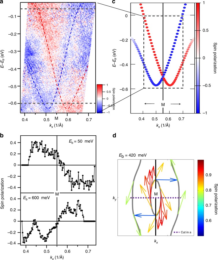
One characteristic feature of the Rashba splitting is a helical spin structure, which we confirm through spin-resolved ARPES measurements. The spin polarization Py along Γ–M–Γ (kx) obtained by substraction of spin-down (sy < 0) and spin-up (sy > 0) intensities is shown in Fig. 3a. Here sy denotes the spin component along the direction perpendicular to the slit of the electron analyzer (which is parallel to Γ–M–Γ or kx) and the surface normal. Red represents Py > 0 and blue represents Py < 0. Below Eb = 400 meV, it is apparent that the left(right) band dispersion is dominated by spin-down(up) intensity. These features are even more clearly demonstrated in Fig. 3b, which shows the momentum distribution curves (MDCs) of spin polarization at binding energies of 50 and 600 meV. The calculated spin-resolved band structure is displayed in Fig. 3c, which shows good agreement between the theoretical calculation and experimental data. Figure 3d shows the calculated spin texture on the constant-energy contour at Eb = 420 meV, where the arrows show the in-plane orientation of spin and their color indicates the degree of spin polarization. Both the inner ellipse and outer open contours around M exhibit a helical spin structure. The spin polarization in experiments is by a factor of 2–3 lower than in the calculations. This was usually observed and might be related to the k-space resolution in experiments, influence of the inelastic background, and sample inhomogeneity.
Fig. 3.
Spin texture of the Rashba-like splitting. a Spin-resolved band image around the M point along Γ–M–Γ (kx) measured by spin-resolved and angle-resolved photoemission spectroscopy (SARPES) (hν = 8.4 eV). The red and blue lines indicate the locations of the Rashba-split bands, while the horizontal gray dashed lines indicate the energy positions of the momentum distribution curves (MDCs) in b. b MDCs of spin polarization at Eb = 50 and 600 meV extracted from the data in a. c Calculated spin polarization of the Rashba-split bands (ky = kz = 0). d Spin texture on the constant-energy contours around M point at Eb = 420 meV. Arrows indicate the spin direction while their color indicates the degree of spin polarization. It has been checked that the experimental data in b exhibits the same spin chirality as the calculated data in d. The scale bars in a, c, and d represent spin polarization
Origination of the Rashba-like splitting
Comparing the band splitting along M–Γ with M–K in theoretical calculations, we find that the splitting has a significant anisotropy, which results in the observed elliptic contours. While an anisotropic effective mass could lead to anisotropic splitting, anisotropy in the underlying band structure is unable to explain why there are minima only along kx. Only including pure Rashba SOC cannot explain the observed anisotropy at all. The band splitting in PtBi2 occurs around the M point, whose little group has lower symmetry than that of the Γ point, where splitting is more commonly encountered. The mirror planes do not pass through M, and furthermore there are three inequivalent M points. The lower symmetry allows the coexistence of Rashba-type and Dresselhaus-type SOC terms, while the additional degrees of freedom can increase the carrier concentration and introduce additional hybridization between the bands from different M points. The little group is Cs at the M point and by using the theory of invariants the three-dimensional (3D) effective model around the M point can be obtained. The effective model up to second order in k reads
| 1 |
where the first three terms are the quadratic and the other terms correspond to the Rashba (α) and Dresselhaus (β) SOC in the kx − ky (α1, β1) and kx − kz planes (α2, β2). The absence of rotational symmetry in the little group allows the coexistence of the Rashba and Dresselhaus terms. It is usually forbidden when the Rashba splitting occurs around Γ, taking BiTeI as an example35.
For the kz = 0 plane, the effective model is reduced to . The density functional theory (DFT) calculated Rashba parameter along ΓM αR is 3.15 eV Å, which is smaller than the experimental value. By fitting the DFT bands, we obtain 1/mx = 45.0 eV Å2 (mx = 0.17me), 1/my = 9.0 eV Å2 (my = 0.85me), α1 = 1.33 eV Å and β1 = −0.7 eV Å. Therefore, Dresselhaus-type SOC cannot be neglected. In our trigonal lattice, each of the three M points will contribute to elliptic Rashba-like bands. When the outer bands from different M points meet, they can hybridize and form a large pocket around Γ (open curves around M point, see Supplementary Fig. 5). This explains the observed outer open contours around M at binding energies of ~400–440 meV in Fig. 2c–e. In our calculations, we also find a small but non-negligible out-of-plane spin component, which is attributable to the SOC terms in the kx–kz plane in the effective model.
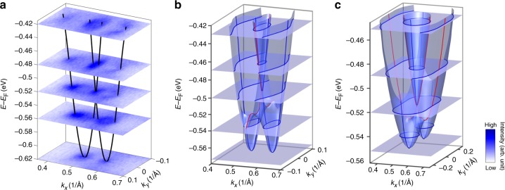
We also show a 3D E(kx, ky) map of the Rashba-like spin split bands as determined by ARPES in Fig. 4a and the 3D electronic structure calculated within DFT in Fig. 4b. Selecting constant-energy contours at certain binding energies for comparison, we find reasonable agreement. To further demonstrate the effect of Dresselhaus-type SOC, we calculated the band dispersions considering only the Rashba-type SOC term but taking the anisotropic effective masses determined by ARPES. The obtained bands are shown in Fig. 4c. The most pronounced feature is that the band splitting along kx (red) is smaller than that along ky (blue). The two elliptic contours at large binding energy are located along the M–K direction (not M–Γ as in experiments). In our fit using both Rashba and Dresselhaus components, α1 and β1 have the opposite sign. The Dresselhaus term will reduce the effective Rashba parameter (α1 + β1) along M–K but enhance it as (−α1 + β1) along M–Γ. This enhancement overcomes the effect of effective mass anisotropy, leading to a large band splitting along M–Γ. Therefore, we conclude that the splitting around the M point in trigonal layered PtBi2 is a result of the cooperation of Rashba-type and Dresselhaus-type SOC. The reasonable agreement between the measured and calculated bands indicates that the splitting originates from bulk inversion symmetry breaking, not surface effects.
Fig. 4.
3D electronic structure of the Rashba-like splitting. a 3D E(kx, ky) map from angle-resolved photoemission spectroscopy (ARPES) data (hν = 9 eV). The black lines indicate the Rashba-split bands. Different surfaces correspond to constant-energy contours at different binding energies. b Calculated 3D electronic structure of PtBi2 at the M point. The red lines indicate the Rashba-split dispersion along M–K (ky) direction, while the blue lines indicate the Rashba-split dispersion along M–Γ (kx) direction. c Calculated 3D electronic structure considering only pure Rashba SOC, taking effective masses from the ARPES data. The blue lines indicate the Rashba-split dispersion along M–K (ky) direction, while the red lines indicate the Rashba-split dispersion along M–Γ (kx) direction. Both red (blue) lines in b, c indicate dispersions with smaller (larger) band splitting. The gray surfaces with blue lines on them in b, c sketch out the constant-energy contours at different binding energies
Three-dimensional nature of the Rashba-like splitting
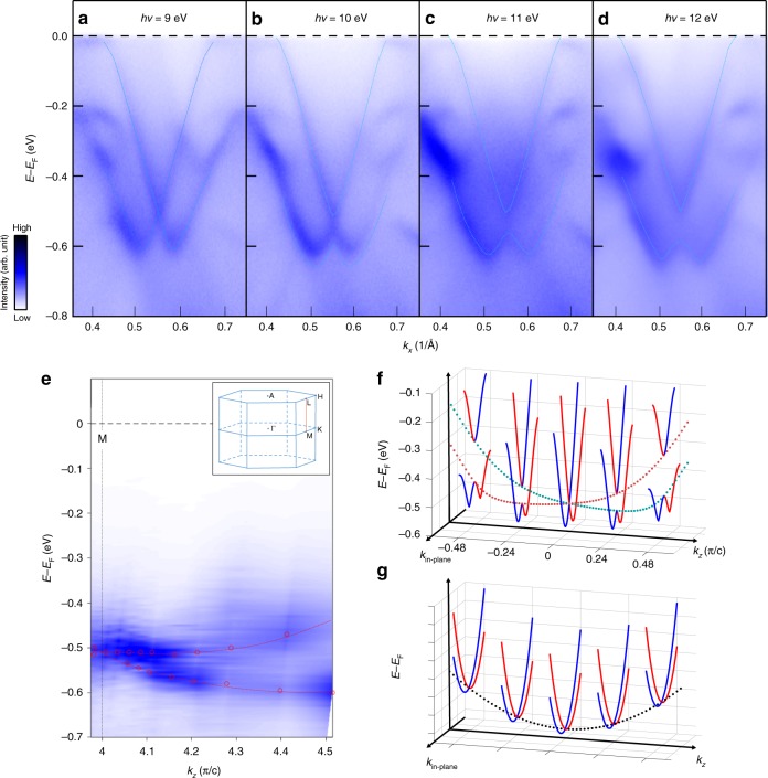
In order to investigate whether the Rasha-type spin splitting band is a bulk band or a surface state, we performed photon energy-dependent ARPES measurements with more than 15 different photon energies. In Fig. 5a–d, we selectively present bands taken along Γ–M direction with photon energy of 9, 10, 11, 12 eV. These bands show strong photon energy dependence, indicating strong kz dispersion. Based on the photon energy-dependent ARPES measurements, we can obtain the band dispersion along M–L direction in the three-dimensional Brillouin zone sketched in the inset of Fig. 5e. In order to obtain band dispersion along M–L direction, the extracted energy distribution curves at the M point taken at different photon energies are plotted as function of kz in Fig. 5e. In Fig. 5e, the band along M–L direction also shows similar band splitting comparable to that along Γ–M direction in Fig. 2. The observed maximum energy splitting is around 160 meV, which is smaller than the 384 meV along Γ–M direction. It is worth noting that the value of 160 meV along M–L direction is already lager than that of Rashba splitting observed in the surface state of Au(111)20. The obtained band splitting along kz direction can be qualitatively captured by the calculated results as shown in Fig. 5f. The specific shape of the dispersions is a little different, which may be attributed to the poor momentum resolution along out-of-plane direction in ARPES measurements. More importantly, our calculated results also verify that the split band is also spin polarized. Our results indicate that the band around the M point not only shows spin splitting in the kx–ky momentum plane but also in the kx–kz plane, which is also consistent with the effective model in Eq. (1). The smaller potential asymmetry with respect to the xz plane, compared with the xy plane, leads to a smaller strength of Rashba splitting along kz. Therefore, the observed Rashba-like spin splitting in PtBi2 is of three-dimensional nature: a consequence of coexistence of both Rashba-type and Dresselhaus-type SOC effects in xy and xz planes. So far, almost all of the reported Rashba bands only show spin splitting along the in-plane momentum directions. Although the Rashba bands of BiTeI and GeTe show dispersion along the out-of-plane direction (kz)36–38,40–42,45, they remain spin degenerate along kz for kx = ky = 0 as sketched in Fig. 5g, indicating a 2D Rashba spin splitting. Our results therefore represent the first realization of the three-dimensional Rashba-like spin splitting in a real material and potentially unfold numerous promising applications.
Fig. 5.
Three-dimensional nature of Rashba-like bands around M point. a–d Energy–momentum image mapped by angle-resolved photoemission spectroscopy (ARPES) taken at different photon energies. e Band dispersion along M–L direction obtained by plotting energy distribution curves at the M point taken at different photon energies as function of kz (the M–L direction as sketched in the three-dimensional Brillouin zone shown in the inset). f Calculated band structure of PtBi2 along the in-plane and out-of-plane momentum directions. The red and blue solid lines show the in-plane band structures at different kz. The red and green dashed lines show the band dispersions along kz direction, indicating Rashba splitting along kz. g Sketch of Rashba bands which have dispersion along kz but remain spin degenerate along kz for kx = ky = 0
Discussion
We have discovered a giant anisotropic 3D Rashba-like spin texture around the M points in a binary compound: PtBi2. The band splitting and its helical spin polarization measured by spin-ARPES are in good agreement with DFT calculations which also confirm the bulk nature of the effect. Due to the low point group symmetry Cs at the M point, the conspiring effects of Rashba-type and Dressselhaus-type SOC are essential to explain the observed 3D spin splitting. These results hence enrich our understanding of Rashba physics and inspire future exploration of new materials systems, which may host 3D Rashba spin texture and hold potential applications in spintronics.
Methods
Sample preparation
Single crystals of noncentrosymmetric trigonal layered PtBi2 were synthesized by the Bi self-flux method. Elemental Platinum (pieces, Alfa Aesar, 99.99%) and bismuth (grains, PRMat, 99.9999%) were mixed in the molar ratio Pt:Bi = 1:6 and loaded into an alumina crucible in an argon-filled glovebox. The crucible was sealed in a quartz tube under high vacuum. The tube was heated to 1073 K and maintained for 20 h, then cooled slowly to 723 K at a rate of 3 K h−1. The samples were then separated from the flux in a centrifuge and quenched immediately in cold water. An X-ray diffraction (XRD) pattern from the large surface of a PtBi2 single crystal is shown in Supplementary Fig. 1a, indicating the crystal surface is parallel to the ab plane. A low-energy electron diffraction (LEED) pattern of a cleaved PtBi2 single crystal surface is shown in Supplementary Fig. 1b, confirming the trigonal symmetry and high quality of the PtBi2 crystal. The structure is determined by performing single-crystal X-ray diffraction measurements(see supplementary).
The ARPES measurements
ARPES measurements were carried out on beamline BL-9A of the Hiroshima Synchrotron Radiation Center (HiSOR) using both synchrotron radiation (photon energy hν = 8–12 eV) and a Xenon lamp (photon energy hν = 8.4 eV) for photoelectron excitation sources. The total energy resolution was set to 20 meV and the base pressure was better than 5 × 10⁻¹¹ mbar. Spin-resolved ARPES measurements were carried out at the Shanghai Institute of Microsystem and Information Technology using a Xenon lamp and an imaging-type multichannel VLEED spin polarimeter51. The spin-resolved energy resolution was set to about 27 meV and the Sherman function Seff was ~0.25. For all ARPES measurements, the samples were cleaved in situ and measured at a temperature of about 40 K. The Fermi level was determined by the measurement of a polycrystalline gold sample in electrical contact with the sample. All the experimental results were reproducible in multiple samples.
Theoretical calculation
Theoretically, we employed relativistic first-principle calculations based on the density-functional theory as implemented in the QUANTUM ESPRESSO code58. The core electrons were treated using the projector augmented wave method59, and the exchange correlation energy was described by the generalized gradient approximation (GGA) using the PBE functional60. The cutoff energy for expanding the wave functions into a plane-wave basis was set to 50 Ry. The Brillouin zone was sampled in k space within the Monkhorst–Pack scheme61 with a k mesh of 8 × 8 × 8, which achieved reasonable convergence of electronic structures. The calculations were done for nonmagnetic PtBi2 with SOC. We used the maximally localized Wannier functions (MLWFs) to construct a tight-binding model by fitting the DFT band structure62, where 72 MLWFs were included. The spin polarization was calculated using this tight-binding model. For all calculations, we used the experimentally determined crystal structure (space group P31m, a = b = 6.5705 Å, c = 6.1707 Å).
Supplementary information
Acknowledgements
The authors would like to thank Darren Peets and Hongxin Yang for helpful discussion. This work is supported by the National Key Research and Development Program of China (Nos. 2017YFA0303600, 2016YFA0300600, and 2017YFA0302901), the National Natural Science Foundation of China (Grant Nos. 11674367,11227902,11927807, U1632266, and 11774399), the Natural Science Foundation of Zhejiang, China (LZ18A040002), the Ningbo Science and Technology Bureau (Grant No. 2018B10060), the German Science Foundation through DFG-SFB 1170 “TOCOTRONICS” (project B04). We acknowledge further financial support from the DFG through the Würzburg–Dresden Cluster of Excellence on Complexity and Topology in Quantum Matter—ct.qmat (EXC 2147, project-id 39085490). S.L.H. would like also to acknowledge the Ningbo 3315 program. We thank the Hiroshima Synchrotron Radiation Center for providing beamtime (project nos. 18AU013, 18AU017, and 18AG018).
Author contributions
S.L.H., S.Z.X., X.X.W., Y.F., and S.Q. conceived the project. M.Y. and Y.G.S. grew the samples. Y.F. and S.Z.X. carried out the ARPES measurements with the assistance from B.J.F., Q.J., T.X., W.J.L., X.F.Y., M.A., E.F.S. and K.S. S.Q. led the spin-resolved ARPES measurements with the assistance from Q.J., T.X., and W.J.L. X.X.W., H.O.J. and R.T. performed the theoretical calculations. S.L.H., Y.F., S.Z.X., X.X.W. and S.Q. analyzed the data and wrote the paper. All authors discussed the results and commented on the manuscript.
Data availability
The data that support the findings of this study are available from the corresponding authors upon reasonable request.
Competing interests
The authors declare no competing interests.
Footnotes
Peer review information Nature Communications thanks Aldo Romero and Markus Morgenstern for their contribution to the peer review of this work. Peer reviewer reports are available.
Publisher’s note Springer Nature remains neutral with regard to jurisdictional claims in published maps and institutional affiliations.
These authors contributed equally: Ya Feng, Qi Jiang, Baojie Feng, Meng Yang.
Contributor Information
Xianxin Wu, Email: xianxinwu@gmail.com.
Shaozhu Xiao, Email: xiaoshaozhu@nimte.ac.cn.
Shaolong He, Email: shaolonghe@nimte.ac.cn.
Supplementary information
Supplementary information is available for this paper at 10.1038/s41467-019-12805-2.
References
- 1.Soumyanarayanan A, et al. Emergent phenomena induced by spin-orbit coupling at surfaces and interfaces. Nature. 2016;539:509–517. doi: 10.1038/nature19820. [DOI] [PubMed] [Google Scholar]
- 2.Bychkov YA, Rashba EI. Properties of a 2D electron gas with lifted spectral degeneracy. JETP Lett. 1984;39:78–81. [Google Scholar]
- 3.Rashba EI. Properties of semiconductors with an extremum loop. 1. Cyclotron and combinational resonance in a magnetic field perpendicular to the plane of the loop. Sov. Phys. Solid State. 1960;2:1109–1122. [Google Scholar]
- 4.Casella RC. Toroidal energy surfaces in crystals with wurtzite symmetry. Phys. Rev. Lett. 1960;5:371–373. doi: 10.1103/PhysRevLett.5.371. [DOI] [Google Scholar]
- 5.Dresselhaus G. Spin-orbit coupling effects in zinc blende structures. Phys. Rev. 1955;100:580–586. doi: 10.1103/PhysRev.100.580. [DOI] [Google Scholar]
- 6.Dyakonov MI, Kachorovskii VYu. Spin relaxation of two-dimensional electrons in noncentrosymmetric semiconductors. Sov. Phys. Semicond. 1986;20:110–112. [Google Scholar]
- 7.Koralek JD, et al. Emergence of the persistent spin helix in semiconductor quantum wells. Nature. 2009;458:610–613. doi: 10.1038/nature07871. [DOI] [PubMed] [Google Scholar]
- 8.Walser MP, et al. Direct mapping of the formation of a persistent spin helix. Nat. Phys. 2012;8:757–762. doi: 10.1038/nphys2383. [DOI] [Google Scholar]
- 9.Ohno Y, et al. Spin relaxation in GaAs(110) quantum wells. Phys. Rev. Lett. 1999;83:4196. doi: 10.1103/PhysRevLett.83.4196. [DOI] [Google Scholar]
- 10.Karimov OZ, et al. High temperature gate control of quantum well spin memory. Phys. Rev. Lett. 2003;91:246601. doi: 10.1103/PhysRevLett.91.246601. [DOI] [PubMed] [Google Scholar]
- 11.Müller GM, et al. Spin noise spectroscopy in GaAs (110) quantum wells: access to intrinsic spin lifetimes and equilibrium electron dynamics. Phys. Rev. Lett. 2008;101:206601. doi: 10.1103/PhysRevLett.101.206601. [DOI] [PubMed] [Google Scholar]
- 12.Griesbeck M, et al. Strongly anisotropic spin relaxation revealed by resonant spin amplification in (110) GaAs quantum wells. Phys. Rev. B. 2012;85:085313. doi: 10.1103/PhysRevB.85.085313. [DOI] [Google Scholar]
- 13.Ye HQ, et al. Growth direction dependence of the electron spin dynamics in (111) GaAs quantum wells. Appl. Phys. Lett. 2012;101:032104. doi: 10.1063/1.4737007. [DOI] [Google Scholar]
- 14.Wang G, et al. Temperature dependent electric field control of the electron spin relaxation in (111)A GaAs quantum wells. Appl. Phys. Lett. 2013;102:242408. doi: 10.1063/1.4811555. [DOI] [Google Scholar]
- 15.Schliemann J, et al. Nonballistic spin-field-effect transistor. Phys. Rev. Lett. 2003;90:146801. doi: 10.1103/PhysRevLett.90.146801. [DOI] [PubMed] [Google Scholar]
- 16.Cartoixa X, et al. A resonant spin lifetime transistor. Appl. Phys. Lett. 2003;83:1462. doi: 10.1063/1.1601693. [DOI] [Google Scholar]
- 17.Hall KC, et al. Nonmagnetic semiconductor spin transistor. Appl. Phys. Lett. 2003;83:2937. doi: 10.1063/1.1609656. [DOI] [Google Scholar]
- 18.Cartoixa X, et al. Heterostructures for spin devices. J. Supercond. 2005;18:163–168. doi: 10.1007/s10948-005-3353-z. [DOI] [Google Scholar]
- 19.Kunihashi Y, et al. Proposal of spin complementary field effect transistor. Appl. Phys. Lett. 2012;100:113502. doi: 10.1063/1.3689753. [DOI] [Google Scholar]
- 20.LaShell S, et al. Spin splitting of an Au(111) surface state band observed with angle resolved photoelectron spectroscopy. Phys. Rev. Lett. 1996;77:3419. doi: 10.1103/PhysRevLett.77.3419. [DOI] [PubMed] [Google Scholar]
- 21.Hochstrasser M, et al. Spin-resolved photoemission of surface states of W(110)-(1 × 1)H. Phys. Rev. Lett. 2002;89:216802. doi: 10.1103/PhysRevLett.89.216802. [DOI] [PubMed] [Google Scholar]
- 22.Hoesch M, et al. Spin structure of the Shockley surface state on Au(111) Phys. Rev. B. 2004;69:241401(R). doi: 10.1103/PhysRevB.69.241401. [DOI] [Google Scholar]
- 23.Koroteev YM, et al. Strong spin–orbit splitting on Bi surfaces. Phys. Rev. Lett. 2004;93:046403. doi: 10.1103/PhysRevLett.93.046403. [DOI] [PubMed] [Google Scholar]
- 24.Krupin O, et al. Rashba effect at magnetic metal surfaces. Phys. Rev. B. 2005;71:201403(R). doi: 10.1103/PhysRevB.71.201403. [DOI] [Google Scholar]
- 25.Ast CR, et al. Giant spin splitting through surface alloying. Phys. Rev. Lett. 2007;98:186807. doi: 10.1103/PhysRevLett.98.186807. [DOI] [PubMed] [Google Scholar]
- 26.He K, et al. Spin polarization of quantum well states in Ag films induced by the Rashba effect at the surface. Phys. Rev. Lett. 2008;101:107604. doi: 10.1103/PhysRevLett.101.107604. [DOI] [PubMed] [Google Scholar]
- 27.Hugo Dil J, et al. Rashba-type spin–orbit splitting of quantum well states in ultrathin Pb films. Phys. Rev. Lett. 2008;101:266802. doi: 10.1103/PhysRevLett.101.266802. [DOI] [PubMed] [Google Scholar]
- 28.Gierz I, et al. Silicon surface with giant spin splitting. Phys. Rev. Lett. 2009;103:046803. doi: 10.1103/PhysRevLett.103.046803. [DOI] [PubMed] [Google Scholar]
- 29.Eremeev SV, et al. Ideal two-dimensional electron systems with a giant Rashba-type spin splitting in real materials: surfaces of bismuth tellurohalides. Phys. Rev. Lett. 2012;108:246802. doi: 10.1103/PhysRevLett.108.246802. [DOI] [PubMed] [Google Scholar]
- 30.Sakano M, et al. Strongly spin-orbit coupled two-dimensional electron gas emerging near the surface of polar semiconductors. Phys. Rev. Lett. 2013;110:107204. doi: 10.1103/PhysRevLett.110.107204. [DOI] [PubMed] [Google Scholar]
- 31.Chen W, et al. Significantly enhanced giant Rashba splitting in a thin film of binary alloy. New J. Phys. 2015;17:083015. doi: 10.1088/1367-2630/17/8/083015. [DOI] [Google Scholar]
- 32.Yao W, et al. Direct observation of spin-layer locking by local Rashba effect in monolayer semiconducting PtSe2 film. Nat. Commun. 2017;8:14216. doi: 10.1038/ncomms14216. [DOI] [PMC free article] [PubMed] [Google Scholar]
- 33.Singh S, et al. Giant tunable Rashba spin splitting in a two-dimensional BiSb monolayer and in BiSb/AlN heterostructures. Phys. Rev. B. 2017;95:165444. doi: 10.1103/PhysRevB.95.165444. [DOI] [Google Scholar]
- 34.Yang H, et al. Significant Dzyaloshinskii–Moriya interaction at graphene –ferromagnet interfaces due to the Rashba effect. Nat. Mater. 2018;17:605–609. doi: 10.1038/s41563-018-0079-4. [DOI] [PubMed] [Google Scholar]
- 35.Ishizaka K, et al. Giant Rashba-type spin splitting in bulk BiTeI. Nat. Mater. 2011;10:521–526. doi: 10.1038/nmat3051. [DOI] [PubMed] [Google Scholar]
- 36.Sakano M, et al. Three-dimensional bulk band dispersion in polar BiTeI with giant Rashba-type spin splitting. Phys. Rev. B. 2012;109:085204. doi: 10.1103/PhysRevB.86.085204. [DOI] [Google Scholar]
- 37.Landolt G, et al. Disentanglement of surface and bulk Rashba spin splittings in noncentrosymmetric BiTeI. Phys. Rev. Lett. 2012;86:116403. doi: 10.1103/PhysRevLett.109.116403. [DOI] [PubMed] [Google Scholar]
- 38.Crepaldi A, et al. Giant ambipolar Rashba effect in the semiconductor BiTeI. Phys. Rev. Lett. 2012;109:096803. doi: 10.1103/PhysRevLett.109.096803. [DOI] [PubMed] [Google Scholar]
- 39.Maaß H, et al. Spin-texture inversion in the giant Rashba semiconductor BiTeI. Nat. Commun. 2016;7:11621. doi: 10.1038/ncomms11621. [DOI] [PMC free article] [PubMed] [Google Scholar]
- 40.Sante D, et al. Electric control of the giant Rashba effect in bulk GeTe. Adv. Mater. 2013;25:509–513. doi: 10.1002/adma.201203199. [DOI] [PubMed] [Google Scholar]
- 41.Liebmann M, et al. Giant Rashba-type spin splitting in ferroelectric GeTe(111) Adv. Mater. 2016;28:560–565. doi: 10.1002/adma.201503459. [DOI] [PubMed] [Google Scholar]
- 42.Krempaský J, et al. Disentangling bulk and surface Rashba effects in ferroelectric α-GeTe. Phys. Rev. B. 2016;94:205111. doi: 10.1103/PhysRevB.94.205111. [DOI] [Google Scholar]
- 43.Elmers H, et al. Spin mapping of surface and bulk Rashba states in ferroelectric α-GeTe(111) films. Phys. Rev. B. 2016;94:201403. doi: 10.1103/PhysRevB.94.201403. [DOI] [Google Scholar]
- 44.Krempaský J, et al. Entanglement and manipulation of the magnetic and spin–orbit order in multiferroic Rashba semiconductors. Nat. Commun. 2016;7:13071. doi: 10.1038/ncomms13071. [DOI] [PMC free article] [PubMed] [Google Scholar]
- 45.Krempaský, J. et al. Spin-resolved electronic structure of ferroelectric α-GeTe and multiferroic Ge1−xMnxTe. J. Phys. Chem. Sol. 128, 237–244 (2017).
- 46.Niesner D, et al. Giant Rashba splitting in CH3NH3PbBr3 organic–inorganic Perovskite. Phys. Rev. Lett. 2016;117:126401. doi: 10.1103/PhysRevLett.117.126401. [DOI] [PubMed] [Google Scholar]
- 47.Kim M, et al. Switchable S = 1/2 and J = 1/2 Rashba bands in ferroelectric halide perovskites. PNAS. 2014;111:6900–6904. doi: 10.1073/pnas.1405780111. [DOI] [PMC free article] [PubMed] [Google Scholar]
- 48.Barke I, et al. Experimental evidence for spin-split bands in a one-dimensional chain structure. Phys. Rev. Lett. 2006;97:226405. doi: 10.1103/PhysRevLett.97.226405. [DOI] [PubMed] [Google Scholar]
- 49.Nakamura T, et al. Giant Rashba splitting of quasi-one-dimensional surface states on Bi/InAs(110)-(2 × 1) Phys. Rev. B. 2018;98:075431. doi: 10.1103/PhysRevB.98.075431. [DOI] [Google Scholar]
- 50.Frohna K, et al. Inversion symmetry and bulk Rashba effect in methylammonium lead iodide perovskite single crystals. Nat. Commun. 2018;9:1829. doi: 10.1038/s41467-018-04212-w. [DOI] [PMC free article] [PubMed] [Google Scholar]
- 51.Ji F, et al. Multichannel exchange-scattering spin polarimetry. Phys. Rev. Lett. 2016;116:177601. doi: 10.1103/PhysRevLett.116.177601. [DOI] [PubMed] [Google Scholar]
- 52.Biswas T, et al. Structural investigation of alloys Pt–Tl–Pb and Pt–Pb–Bi. J. Less-Common Met. 1969;19:223. doi: 10.1016/0022-5088(69)90099-X. [DOI] [Google Scholar]
- 53.Gao W, et al. A possible candidate for triply degenerate point fermions in trigonal layered PtBi2. Nat. Commun. 2018;9:3249. doi: 10.1038/s41467-018-05730-3. [DOI] [PMC free article] [PubMed] [Google Scholar]
- 54.Martin K, et al. Bi2Pt(hP9) by low-temperature reduction of Bi13Pt3I7: reinvestigation of the crystal structure and chemical bonding analysis. Z. Anorg. Allg. Chem. 2014;640:2742–2746. doi: 10.1002/zaac.201400331. [DOI] [Google Scholar]
- 55.Yao Q, et al. Bulk and surface electronic structure of hexagonal structured studied by angle-resolved photoemission spectroscopy. Phys. Rev. B. 2016;94:235140. doi: 10.1103/PhysRevB.94.235140. [DOI] [Google Scholar]
- 56.Thirupathaiah S, et al. Possible origin of linear magnetoresistance: observation of Dirac surface states in layered PtBi2. Phys. Rev. B. 2018;97:035133. doi: 10.1103/PhysRevB.97.035133. [DOI] [Google Scholar]
- 57.Yang X, et al. Giant linear magneto-resistance in nonmagnetic PtBi2. Appl. Phys. Lett. 2016;108:252401. doi: 10.1063/1.4954272. [DOI] [Google Scholar]
- 58.Giannozzi P, et al. QUANTUM ESPRESSO: a modular and open-source software project for quantum simulations of materials. J. Phys. Condens. Matter. 2009;21:395502. doi: 10.1088/0953-8984/21/39/395502. [DOI] [PubMed] [Google Scholar]
- 59.Blöchl PE. Projector augmented-wave method. Phys. Rev. B. 1994;50:17953. doi: 10.1103/PhysRevB.50.17953. [DOI] [PubMed] [Google Scholar]
- 60.Perdew JP, et al. Generalized gradient approximation made simple. Phys. Rev. Lett. 1996;77:3865. doi: 10.1103/PhysRevLett.77.3865. [DOI] [PubMed] [Google Scholar]
- 61.Monkhorst HJ, Pack JD. Special points for Brillouin-zone integrations. Phys. Rev. B. 1976;13:5188. doi: 10.1103/PhysRevB.13.5188. [DOI] [Google Scholar]
- 62.Mostofi AA, et al. An updated version ofwannier90: a tool for obtaining maximally-localised Wannier functions. Comput. Phys. Commun. 2014;185:2309–2310. doi: 10.1016/j.cpc.2014.05.003. [DOI] [Google Scholar]
Associated Data
This section collects any data citations, data availability statements, or supplementary materials included in this article.
Supplementary Materials
Data Availability Statement
The data that support the findings of this study are available from the corresponding authors upon reasonable request.


