Abstract
Estradiol is an estrogenic steroid that can undergo glucuronidation at two different sites, which results in two estradiol glucuronide regioisomers. These isomers can be produced by different enzymes and can have different biological activities before being eliminated from the body. Although there have been previous methods that can distinguish the two isomers, these methods often have long acquisition times or high cost per analysis. In this study, traveling wave ion mobility spectrometry (TWIMS) coupled to mass spectrometry (MS) was employed to separate estradiol glucuronides using alkali metal adduction in positive ion mode, where the sodiated dimer adduct provided adequate separation both in single-component standards and in two-component mixtures. Additionally, in negative mode tandem mass spectrometry (MS/MS) was used to quantitatively determine the relative composition of the two isomers. This was possible due to differences in the energetic requirements for loss of the glucuronic acid, which was characterized by energy-resolved collision-induced dissociation (CID). This work demonstrated that the intensity of the glucuronic acid neutral loss product as compared to the intensity of the intact precursor ion can be used to determine the percentage of each isomer present in a mixture. Overall, TWIMS successfully separated estradiol glucuronide isomers in positive ion mode and MS/MS via CID enables relative quantitation of each isomer in negative ion mode.
Keywords: Steroid Glucuronide Isomers, Traveling Wave Ion Mobility Spectrometry, Tandem Mass Spectrometry, Metal Ion Adduction
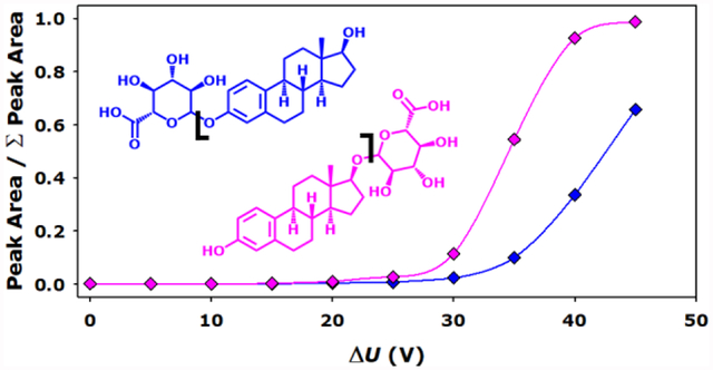
Graphical Abstract.

Introduction
Glucuronidation is a process that adds a glucuronic acid residue to non-polar substances, such as steroids, which allows them to be more easily transported and eliminated from the body [1]. Estradiol is an estrogenic steroid that can undergo glucuronidation creating estradiol glucuronides (EG) at two sites: the alcohol on carbon 3 near the aromatic ring, resulting in estradiol-3-glucuronide (E3G); or the alcohol on carbon 17 near the five membered ring, resulting in estradiol-17-glucuronide (E17G). These two isomers result from preferential glucuronidation by different enzymes: UDP-glucuronosyltransferases (UGTs) UGT1A3, UGT2B7 (H268), and human liver microsomes HL-15 [1]. Furthermore, E17G and E3G act as substrates for different enzymes resulting in different biological activities [2–5].
Previously, there have been reports of analytical techniques to distinguish isomeric steroids and steroid glucuronides. The primary method to separate various steroids and their respective conjugates has been liquid chromatography (LC); however, LC requires long acquisition times [2, 3, 6]. Additionally, sensitive immunoassays have been used to determine estradiol conjugates. Immunoassays tend to be relatively expensive and are subject to several potential confounding factors in complex matrices [7, 8]. More recently, ion mobility spectrometry (IMS) coupled to mass spectrometry (MS) has been employed to rapidly analyze steroid glucuronides.
IMS is a rapid gas phase separation method that uses an electric field and a buffer gas to separate molecules by size, shape, and charge [9]. IMS has been coupled to LC to separate testosterone and epitestosterone glucuronides [6]. Additionally, ion mobility has been used solely in conjunction with mass spectrometry to analyze EGs, where their deprotonated ions exhibited a slight difference in their ion-neutral collision cross sections (CCS), a quantitative property of the analyte and drift gas partner [10]. Nevertheless, the arrival time distributions of the deprotonated EG isomers were nearly overlapped [11]. Chouinard et al. and Rister et al. previously reported the use of alkali metal adducts and the formation of dimeric species to increase the separation of steroids achievable with IMS-MS [12–15].
Additionally, tandem mass spectrometry (MS/MS) has been used as a means of distinguishing isomers. The most common MS/MS method is collision-induced dissociation (CID), wherein analyte ions are accelerated into neutral gas atoms or molecules. The resulting collisions convert kinetic energy into internal energy, ultimately resulting in fragmentation of the analyte ion. The fragmentation spectra can rapidly provide invaluable structural information, and in some cases can reveal which of a number of possible isomeric structures is most likely [16].
This report illustrates two methods, one in positive ion mode and one in negative ion mode, that allow for the distinction and quantitation of the EG isomers. In positive ion mode, travelling wave IMS (TWIMS) separates dimeric species sufficiently for quantitation by peak height. In negative ion mode, MS/MS by CID is successfully applied to allow for relative quantitation of the EG isomers in a mixture. Overall, this work expands on the currently available methods for EG isomer discrimination by providing very rapid and sensitive IMS-MS and MS/MS approaches.
Methods
Material sources are provided in Table S1 in the Supplementary Material. In short, solutions consisting of one EG isomer or a mixture of the two were mixed with group I metal acetate salts at a concentration of 10 μM total EG to 20 μM of salt in 50% water / methanol. These solutions were then directly infused through a nano-electrospray ionization source into a Waters Synapt G2-S (Milford, MA) undergoing TWIMS separation in positive mode. The TWIMS parameters of wave height, wave velocity, and gas flow was maintained at 40 V, 600 m/s, and 60 mL/min of nitrogen, respectively. CCSs were calibrated using known drift tube values from polyalanine in nitrogen drift gas using a quadratic fit (td′ = aΩ’2 + bΩ’ + c) [17]. CID was performed in negative ion mode in the transfer cell using argon as the collision partner at collision energies ranging from 0 V to 45 V. All analyses were performed in four replicates on different days. The peak to peak resolution was calculated based on width at half-height as shown in Equation 1:
| Equation 1 |
Data was analyzed and visualized through the use of Drift Scope 2.7 and MassLynx 4.0 (Waters), Igor Pro 7.0 (WaveMetrics, Lake Oswego, OR), and SigmaPlot 13.0 (Systat, Chicago, IL).
Results and Discussion
Ion Mobility Spectrometry Separation.
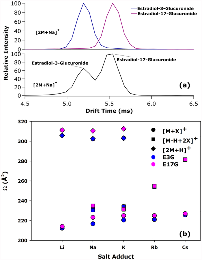
The EG isomers were separated as standards and in a mixture with a resolution value greater than 1.0 as [2M+Na]+ adducts. As seen in Figure 1a, the sodiated dimer adduct arrival time distributions (ATD) as single-component standards showed near-baseline separation, while a mixture of the isomers resulted in a decreased resolution (R = 1.23). The apparent decrease in resolution could be the result of heterodimer formation. If so, this could complicate the ability to perform quantitative analyses through this method. We also note that, while both components were infused at equivalent concentrations in the mixture, there were differences in their signal intensities. This could be caused by differing ionization efficiencies and / or potential post ionization fragmentation. CCS values were measured and plotted against the metal in the variety of adduct formations in Figure 1b. The resolution values, CCSs, and comparison of the standard and mixture CCSs are available in Tables S2–S4 in Supplementary Material. Notably, monomers in both positive and negative mode were poorly separated in mixtures, which can be seen in the resolution values provided in Table S2 and Figure S1 in the Supplementary Material. Overall, the separation of E3G and E17G as sodiated dimers was successfully demonstrated.
Figure 1.
Arrival time distributions for estradiol glucuronides standards (top) and mixtures (bottom) as their respective sodium adduct dimer in positive ion mode (a). Scatter plot of nitrogen collision cross sections for E3G (blue) and E17G (pink) as a function of the salt adduct (X = Li, Na, etc.) for [M+X] (circle), [M-H+2X] (square), and [2M+X] (diamond) in positive ion mode (b).
Tandem Mass Spectrometry.
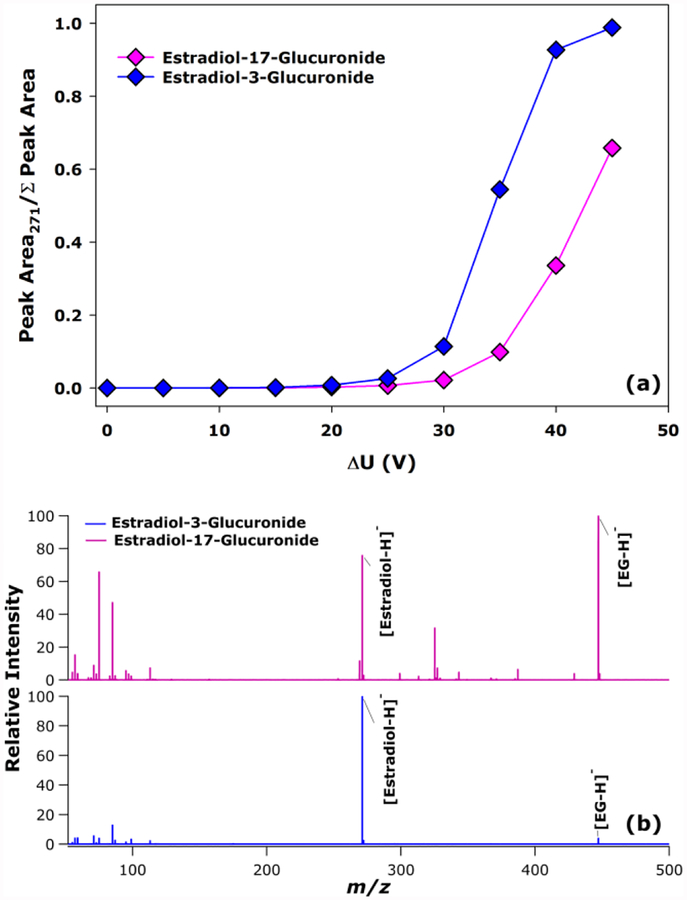
As an alternative and complementary method of distinguishing the EG isomers, negative ion mode MS/MS was performed by applying CID to the deprotonated ions at m/z 447. Upon fragmentation of the deprotonated ions, neutral loss of the glucuronic acid resulting in m/z 271 was a major product ion for both isomers. However, the fraction of this loss increases with increased collision energy more sharply for E3G than E17G, as illustrated in Figure 2a (the corresponding raw values are presented in Table S5 of the Supplemental Material). The fraction of this loss is distinct between the isomers starting at collision energy 30 V. Additionally, the tandem mass spectrum of both isomers is shown at CE 40 V in Figure 2b, where the ratio of m/z 271 to m/z 447 is clearly higher in E3G’s spectrum compared to E17G’s spectrum. This difference was proposed to be a result of the aromatic ring of E3G stabilizing the glycosidic bond thus mitigating the loss of glucuronic acid, where E17G does not have access to this stabilization effect. As a result, the distinct energetics for glucuronic acid loss could be a tool for determining steroid glucuronide isomers in mixtures.
Figure 2.
Scatter plot of the fraction of the peak area of the fragment m/z 271 as a function of transfer collision energy (a). Tandem mass spectra of deprotonated E17G (pink, top) and E3G (blue, bottom) at collision energy 40 V (b).
Relative Quantitation through Tandem Mass Spectrometry.
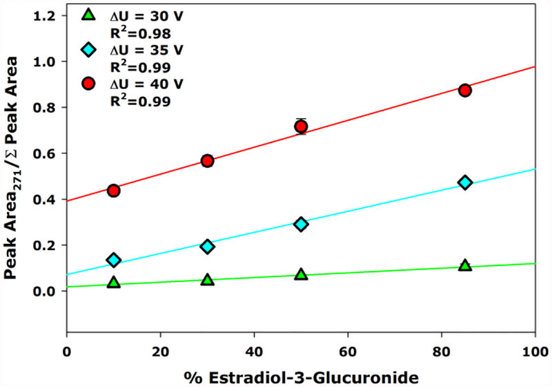
The isomer-specific energetics of glucuronic acid neutral loss was exploited for relative quantitation of each isomer in a variety of mixtures where the relative ratio of the isomers was systematically altered. Each mixture was subjected to MS/MS with CID energies of 30 V, 35 V, and 40 V. A scatter plot illustrating the fractional abundance of m/z 271 compared to m/z 271 + m/z 447 as a function of the percentage of E3G in the mixture is shown in Figure 3. From these results, a calibration curve was established to allow measurement of the relative amount of each compound in the mixture with high coefficients of determination ranging from 0.98 to 0.99. Additionally, due to the increasingly distinct dissociation spectra as collision energy increased, the higher collision energies resulted in a more sensitive method (i.e., the plot for 40 V collision energy has a steeper slope than 30 V collision energy). As a result, the extent of glucuronic acid loss can provide relative quantitation of the E3G and E17G isomers.
Figure 3.
Scatter plots with regression lines for the fraction of the peak area of m/z 271 as a function of the percentage of E3G compared to E17G in the solution at the collision energies of 30 V (green triangle), 35 V (cyan diamond), and 40 V (red circle).
Conclusions
TWIMS-MS and MS/MS were shown to separate and relatively quantitate EG isomers, respectively. The sodiated dimeric adduct allowed resolution of R = 1.23 in positive ion mode analysis of a mixture. However, shifts in drift time may suggest the formation of a heterodimer that could potentially limit the ability to perform quantitation. In negative ion mode, distinct energy-resolved CID behavior of the EG isomers provided a method to determine the relative quantities of each isomer. We also note that, while both techniques employed here are post-ionization and thus susceptible to signal suppression in complex matrices, these techniques can also be coupled to LC for enhanced analysis. Ultimately, this work demonstrated the ability to apply IMS and MS/MS to the rapid analysis of the E3G and E17G isomers in a mixture.
Supplementary Material
Acknowledgements
This work was supported in part by funding from the National Science Foundation, Division of Chemistry, through the Chemical Measurement and Imaging Program (Award Number 1507989). Funding from the National Institutes of Health, National Institute of General Medical Sciences, was received through a fellowship to A.L.R. from the Molecular Mechanisms of Disease Predoctoral Training Program (Award Number T32GM107001). Finally, the authors thank Jessica Minnick for constructive comments on a draft of the manuscript.
Footnotes
Supplementary Material
The online version of this article contains supplementary material, which is available to authorized users.
References
- 1.Gall WE, Zawada G, Mojarrabi B, Tephly TR, Green MD, Coffman BL, Mackenzie PI, Radominska-Pandya A: Differential Glucuronidation of Bile Acids, Androgens and Estrogens by Human UGT1A3 and 2B7. J. Steroid Biochem. Mol. Biol 70, 101–108 (1999) [DOI] [PubMed] [Google Scholar]
- 2.Andreolini F, Borra C, Caccamo F, Corcia AD, Samperi R: Estrogen Conjugates in Late-Pregnancy Fluids: Extraction and Group Separation by a Graphitized Carbon Black Cartridge and Quantification by High-Performance Liquid Chromatography. Anal. Chem 59,1720–1725 (1987) [DOI] [PubMed] [Google Scholar]
- 3.Belanger A, Labrie F: Unconjugated and Glucuronide Steroid Levels in Human Breast Cyst Fluid. Ann. NY Acad. Sci 586, 93–100 (1990) [DOI] [PubMed] [Google Scholar]
- 4.Kanai N, Lu R, Bao Y, Wolkoff AW, Vore M, Schuster VL: Estradiol 17 Beta-D-Glucuronide is a High-Affinity Substrate for oatp Organic Anion Transporter. Am. J. Physiol. - Renal Physiol 270, F326–F331 (1996) [DOI] [PubMed] [Google Scholar]
- 5.Yeoh C-G, Schreck CB, Fitzpatrick MS: Endogenous Steroid Metabolism Is Indicated by Fluctuations of Endogenous Steroid and Steroid Glucuronide Levels in Early Development of the Steelhead Trout (Oncorhynchus mykiss). Gen. Comp. Endocrinol 103, 107–114 (1996) [DOI] [PubMed] [Google Scholar]
- 6.Kaur-Atwal G, Reynolds JC, Mussell C, Champarnaud E, Knapman TW, Ashcroft AE, O’Connor G, Christie SD, Creaser CS: Determination of Testosterone and Epitestosterone Glucuronides in Urine by Ultra Performance Liquid Chromatography-Ion Mobility-Mass Spectrometry. Analyst 136, 3911–3916 (2011) [DOI] [PubMed] [Google Scholar]
- 7.Tanaka T, Takeda H, Ueki F, Obata K, Tajima H, Takeyama H, Goda Y, Fujimoto S, Matsunaga T: Rapid and Sensitive Detection of 173-Estradiol in Environmental Water using Automated Immunoassay System with Bacterial Magnetic Particles. J. Biotechnol 108, 153–159 (2004) [DOI] [PubMed] [Google Scholar]
- 8.Zhao L, Lin J-M, Li Z, Ying X: Development of a Highly Sensitive, Second Antibody Format Chemiluminescence Enzyme Immunoassay for the Determination of 173-Estradiol in Wastewater. Anal. Chim. Acta 558, 290–295 (2006) [Google Scholar]
- 9.Kanu AB, Dwivedi P, Tam M, Matz L, Hill HH Jr.: Ion Mobility-Mass Spectrometry. J. Mass Spectrom 43, 1–22 (2008) [DOI] [PubMed] [Google Scholar]
- 10.Shimizu A, Ohe T, Chiba M: A Novel Method for the Determination of the Site of Glucuronidation by Ion Mobility Spectrometry-Mass Spectrometry. Drug Metab. Dispos 40, 1456–1459 (2012) [DOI] [PubMed] [Google Scholar]
- 11.Reading E, Munoz-Muriedas J, Roberts AD, Dear GJ, Robinson CV, Beaumont C: Elucidation of Drug Metabolite Structural Isomers Using Molecular Modeling Coupled with Ion Mobility Mass Spectrometry. Anal. Chem 88, 2273–2280 (2016) [DOI] [PubMed] [Google Scholar]
- 12.Chouinard CD, Beekman CR, Kemperman RHJ, King HM, Yost RA: Ion Mobility-Mass Spectrometry Separation of Steroid Structural Isomers and Epimers. Int. J. Ion Mobil. Spectrom 20, 31–39 (2016) [Google Scholar]
- 13.Chouinard CD, Cruzeiro VWD, Roitberg AE, Yost RA: Experimental and Theoretical Investigation of Sodiated Multimers of Steroid Epimers with Ion Mobility-Mass Spectrometry. J. Am. Soc. Mass Spectrom 28, 323–331 (2017) [DOI] [PMC free article] [PubMed] [Google Scholar]
- 14.Rister A,L, Martin TL, Dodds ED: Application of Group I Metal Adduction to the Separation of Steroids by Traveling Wave Ion Mobility Spectrometry. J. Am. Soc. Mass Spectrom 30, 248–255 (2018) [DOI] [PMC free article] [PubMed] [Google Scholar]
- 15.Rister AL, Martin TL, Dodds ED: Formation of Multimeric Steroid Metal Adducts and Implications for Isomer Mixture Separation by Traveling Wave Ion Mobility Spectrometry. J. Mass Spectrom, DOI: 10.1002/jms.4350 (2019) [DOI] [PMC free article] [PubMed] [Google Scholar]
- 16.Johnson AR, Carlson EE: Collision-Induced Dissociation Mass Spectrometry: A Powerful Tool for Natural Product Structure Elucidation. Anal. Chem 87, 10668–10678 (2015) [DOI] [PubMed] [Google Scholar]
- 17.Bush MF, Campuzano ID, Robinson CV: Ion Mobility Mass Spectrometry of Peptide Ions: Effects of Drift Gas and Calibration Strategies. Anal. Chem 84, 7124–7130 (2012) [DOI] [PubMed] [Google Scholar]
Associated Data
This section collects any data citations, data availability statements, or supplementary materials included in this article.


