Abstract
Plants must continuously react to the ever-fluctuating nature of their environment. Repeated exposure to stressful conditions can lead to priming, whereby prior encounters heighten a plant’s ability to respond to future events. A clear example of priming is provided by the model plant Arabidopsis thaliana (Arabidopsis), in which photosynthetic and photoprotective responses are enhanced following recurring light stress. While there are various post-translational mechanisms underpinning photoprotection, an unresolved question is the relative importance of transcriptional changes toward stress priming and, consequently, the potential contribution from DNA methylation – a heritable chemical modification of DNA capable of influencing gene expression. Here, we systematically investigate the potential molecular underpinnings of physiological priming against recurring excess-light (EL), specifically DNA methylation and transcriptional regulation: the latter having not been examined with respect to EL priming. The capacity for physiological priming of photosynthetic and photoprotective parameters following a recurring EL treatment was not impaired in Arabidopsis mutants with perturbed establishment, maintenance, or removal of DNA methylation. Importantly, no differences in development or basal photoprotective capacity were identified in the mutants that may confound the above result. Little evidence for a causal transcriptional component of physiological priming was identified; in fact, most alterations in primed plants presented as a transcriptional ‘dampening’ in response to an additional EL exposure, likely a consequence of physiological priming. However, a set of transcripts uniquely regulated in primed plants provide preliminary evidence for a novel transcriptional component of recurring EL priming, independent of physiological changes. Thus, we propose that physiological priming of recurring EL in Arabidopsis occurs independently of DNA methylation; and that the majority of the associated transcriptional alterations are a consequence, not cause, of this physiological priming.
Keywords: DNA methylation, priming, gene expression, photoprotection
Plants must respond to various stresses imposed by their environment. Abiotic stresses may occur over long periods of development with unfavorable conditions persisting stably as is characteristic of extreme climates. Alternatively, environments can be highly dynamic and comprise of transient, often recurring, stressful events. Information processing is key for effective physiological and developmental responses to specific environmental factors. Indeed, there is growing evidence that plants can ‘remember’ past experiences (Hilker et al. 2016). In addition to long-term acclimation to sustained environmental changes, short-term plant stress responses are modified by prior exposure to a transient, and often recurring, specific environmental stimulus (referred to as priming). Here, the future fitness of a primed individual is increased by reducing the damage of stressful events, while the costs of initiating and maintaining priming are outweighed by the costs of stress exposure in an ‘un-primed’ (or naive) state (Hilker et al. 2016).
A variety of mechanisms have been reported to contribute toward stress priming including transcriptional memory underpinned by stalled RNA Pol II and elevated H3K4me3 (Ding et al. 2012), and fractionation of H3K27me3 patterns (Sani et al. 2013). It has also been reported that the activity of the HSFA2 transcription factor can result in H3K4me2 and H3K4me3 changes, in response to recurring heat stress, to convey transcriptional priming (Lämke et al. 2016). Additionally, HDA6-mediated histone H4 de-acetylation has been linked to prime jasmonic acid signaling leading to enhanced drought tolerance (Kim et al. 2017). These various examples highlight the importance of chromatin variation in facilitating of plant stress priming. Another chromatin mark speculated to promote priming is DNA methylation, variations in which could, theoretically, be stably inherited over mitotic cell divisions to convey persistent transcriptional control (Johannes and Schmitz 2019). The targeting of the RNA-directed DNA methylation (RdDM) pathway toward promoter regions of genes, and the occurrence of gene body methylation (gbM), suggests that differential methylation could arise within genes or their regulatory elements to cause functional differences (Matzke and Mosher 2014; Bewick et al. 2016). Such observations highlight the potential regulatory capacity for DNA methylation.
An extant question is the exact regulatory potential of DNA methylation. Canonically, DNA methylation is considered a mechanism for transcriptional repression, for example through steric hindrance of RNA polymerase II (Molloy 1986). This silencing is most pronounced at transposable elements (TEs) (Cokus et al. 2008). On the other hand, gbM is often found within constitutively expressed genes although there is conflicting evidence for an effect on transcription (Bewick et al. 2016; Muyle and Gaut 2019). Other reports implicate the involvement of DNA methylation in alternative splicing and modulation of transcription factor binding capacity (Shukla et al. 2011; O’Malley et al. 2016; Yin et al. 2017). Given these various mechanisms, and placing DNA methylation in the broader context as being one of many chromatin modifications, it might be unsurprising that efforts to quantify the contribution of DNA methylation, at an organism level, toward transcription have shown a weak relationship (Meng et al. 2016). Instead, changes in the methylome may be a consequence of gene expression changes rather than a driver (Secco et al. 2015). The ability to identify causative changes in the methylome are further complicated as the effects of DNA methylation can be in both cis and trans (Rowley et al. 2017).
Given these complications, various tools exist to quantify the effects of variable methylation (epi-alleles) on, and in response to, gene expression and physiological traits. The utilization of epigenetic recombinant inbred lines that display variable methylation patterns, but are isogenic, demonstrated that epi-alleles could contribute toward quantifiable phenotypic differences (Johannes et al. 2009; Cortijo et al. 2014). Mutants with defective methylation machinery that exhibit a variety of methylome variations, depending on the severity of the mutation, have also been used to correlate a relationship between DNA methylation and plant stress responses (Boyko et al. 2010; Le et al. 2014; Wibowo et al. 2016). Mutants are also described to be developmentally or morphologically aberrant, however, it is not clear whether this is the direct result of methylation changes or an indirect effect of TE de-regulation and genomic instability (Finnegan et al. 1996; Reinders et al. 2009; Stroud et al. 2014; Williams and Gehring 2017). Indeed, traits attributed toward methylome variants could equally be tied to underlying TE activity (Ong-Abdullah et al. 2015; Wibowo et al. 2016; He et al. 2018).
We previously demonstrated that Arabidopsis is primed by a recurring EL regime, evident by altered non-photochemical quenching (NPQ) and improved photosystem II (PSII) efficiency (Ganguly et al. 2018). However, we detected no associated changes in DNA methylation. While this demonstrated that the Arabidopsis methylome was impervious to recurring EL, it does not preclude transcriptional regulation of EL priming to which appropriate methylome maintenance may be important. In fact, EL-exposed tissue can promote the induction of EL-responsive transcripts in naive leaves for added photoprotection through the process of systemic acquired acclimation (SAA) (Karpinski 1999; Rossel et al. 2007; Gordon et al. 2012). As the methylation machinery was operational during previous analyses, any light-induced changes may have been reset prior to tissue harvesting. Indeed, the disruption of methylation pathways has revealed transgenerational effects that were otherwise reset (Iwasaki and Paszkowski 2014). Thus, we sought to clarify these unknowns by testing whether a range of mutants, which are unable to maintain or reset their methylome, were capable of priming against recurring EL; and subsequently characterizing differences in the transcriptome of EL primed plants. We report that DNA methylation mutants display functional photoprotection and EL priming to an equivalent extent as wild-type plants (WT; Col-0). Furthermore, while primed plants demonstrate a completely reset transcriptome, they also displayed attenuated responses to further EL, which we refer to as “dampening”, potentially reflecting the reduced generation of stress signaling molecules due to enhanced photoprotection.
Materials and Methods
Plant growth and germplasm
All Arabidopsis germplasm utilized were in the Columbia (Col-0) background. Plant lines comprised of wild-type Col-0 (WT), ddc (CS16384; drm1-2 drm2-2 cmt3-11), strs2 (SALK_028850), and rdd (derived from ros1-4, SALK_045303; dml2, SALK_131712; dml3-2, SALK_056440). All seeds were obtained from the Arabidopsis Biological Research Centre (Ohio State University, Columbus, OH, USA), with the exception of rdd which was kindly provided by Dr. Ming-Bo Wang (CSIRO, Canberra, Australia) (Le et al. 2014). Primers used for genotyping are listed in Supplementary Dataset 8.
For plant growth, Arabidopsis seeds were sown onto individual pots containing moist Seed Raising Mix (Debco, NSW, Australia). Soil was supplemented with Osmocote Exact Mini slow release fertilizer (Scotts, NSW, Australia) at a concentration of 1 g/L dry volume of soil and treated with 1 L of 0.3% (v/v) AzaMax (OCP, NSW, Australia) prior to sowing to prevent insect infection. Seeds were covered with clear plastic wrap and stratified at 4° in the dark for at least 72 hr to break dormancy and coordinate germination. Stratified seeds were transferred to a temperature controlled Conviron S10H growth chamber (Conviron, Winnipeg, MB, Canada) for cultivation under standard growth conditions: 12-hour photoperiod (08:00-20:00), 100-150 μ mol photons m-2 s-1, 20° (± 2°), 55% (± 5%) relative humidity. Upon germination, clear plastic wrap was slowly removed over 7-10 days, to maintain high humidity until seedlings were well-established and to avoid humidity shock. Plants were watered every 2-3 days depending on soil moisture, avoiding pooling of water to prevent algal and fungal growth. Each of three daily EL treatments consisted of 60 min of 1000 μ mol photons m-2 s-1 using a mixture of metal halide and high-pressure sodium lamps as previously described (Crisp et al. 2017; Ganguly et al. 2018).
For MethylC- and mRNA-seq experiments, developmentally equivalent leaves were harvested in biological triplicate (three independent plants per time-point or genotype) to make comparisons (true leaves 4 - 9 in order of emergence). Harvested tissue was flash-frozen in liquid N2 and ground into a fine powder using a 1/8″ steel ball bearing, in a 1.5 ml Eppendorf tube, with 1 min shaking at 25 Hz in the Tissue Lyser II (Qiagen, Hilden, Germany). Ground tissue was stored at -80°. Approximately 20 and 30 mg of ground tissue was used for extracting total RNA and genomic DNA, respectively. MethylC-seq of WT and strs2 was performed on paired tissue samples by using aliquots of ground frozen tissue from the same harvested plant.
High-throughput phenotyping
For all genotypes the development of 10-day old (post-germination) Arabidopsis seedlings was followed for 20 days (until 4-weeks of age) by measuring plant area and rosette compactness. The PlantScreen Compact System (Photon Systems Instruments, Brno, Czech Republic), a high-throughput platform for digital plant phenotyping, was used to measure plant area and rosette compactness at 08:00 daily. Images were analyzed using the PSI RGB-IR Analyzer software (version 1.0.0.2; Photon Systems Instruments, Brno, Czech Republic). Program settings were adjusted as needed to achieve well-defined plant areas and minimize background noise. The total number of macroscopically visible true leaves (cotyledons excluded) per rosette were also manually counted daily.
Monitoring PSII performance using chlorophyll fluorescence measurements
PSII photochemistry was probed in vivo using measures of chlorophyll fluorescence (Baker 2008) using a PSI FluorCam (Photon System Instruments; Brno, Czech Republic). Measures were taken between 11:00-14:00 across the adaxial side of 30 min dark adapted rosettes as performed previously (Ganguly et al. 2018). In this study, intermittent measures of chlorophyll fluorescence, specifically Ft, Fm, and Fm′, were taken after a saturating pulse (3000 µ mol photons m-2 sec-1) across 10-minutes in actinic light (700 μ mol photons m-2 sec-1) followed by a 4-minute dark period (https://goo.gl/RuYDE2). The resulting fluorescence images were analyzed using the FluorCam 7 software (v1.1.1.4; Photon System Instruments, Brno, Czech Republic). Chlorophyll fluorescence signals were analyzed across whole rosettes. To better quantify light energy partitioning between photochemistry, light-regulated thermal dissipation, and other non-light induced quenching, such as chlorophyll fluorescence, we adopted the yield terms of ΦPSII, ΦNPQ, and ΦNO, which sum to unity and do not require measures of Fo (Hendrickson et al. 2004; Kramer et al. 2004).
MethylC sequencing
Genomic DNA was extracted from ground tissue using the DNeasy Plant Mini Kit (Qiagen, Netherlands) according to the manufacturer’s instructions, and quantified using a ND-1000 Spectrophotometer. 70 ng of Covaris sheared gDNA (average fragment size = 200 bp) was bisulfite converted using the EZ DNA Methylation-Gold Kit (Zymo Research, CA, USA) according to the manufacturer’s instructions. Bisulfite converted DNA was used to create dual-indexed MethylC-seq libraries using the Accel-NGS Methyl-Seq DNA Library Kit paired with the Accel-NGS Methyl-Seq Dual Indexing Kit (Swift Biosciences, MI, USA) according to manufacturers’ instructions. All libraries were amplified in a 7-cycle indexing PCR reaction. All clean-ups were performed using either AMPure XP beads (Beckman Coulter, CA, USA) or Sera-mag SpeedBeads (GE Healthcare, Buckinghamshire, UK). A LabChip GXII (Perkin Elmer, MA, USA) was used to determine library molarity and fragment size distribution. Libraries were pooled in equimolar ratios and sequenced on one HiSeq2500 flow cell (100 bp single-end) at the ACRF Biomolecular Research Facility (Australian National University, ACT, Australia). In-depth details of library preparation are also described on protocols.io (https://goo.gl/vfwtEU).
Raw reads were quality controlled using FastQC (v0.11.2) with reads filtered and trimmed using Cutadapt (v1.9) and Trim Galore! (v.0.3.7) under default parameters. Single-end alignments of trimmed raw reads were aligned to the TAIR10 reference using Bismark (v0.14.5) (Krueger and Andrews 2011) and Bowtie2 (v2.2.9) (Langmead and Salzberg 2012) with the flags -N 0 and -L 20. Per cytosine methylation levels were calculated using Bismark methylation extractor with default settings. Only cytosines with read depth > 3X were retained for further analysis. Bisulfite conversion efficiency was calculated as the proportion of methylated cytosines in the CHH context within the chloroplast genome, which itself should be fully unmethylated. Alignment metrics are provided in Supplementary Dataset 1. Weighted methylation levels were used to calculate the proportion of CG, CHG, and CHH methylation to account for sequencing depth (Schultz et al. 2012). This output was binned into 50 kbp regions (filtered for read depth > 15X) across the genome to construct chromosomal level metaplots of methylation levels using BEDTools (Quinlan and Hall 2010).
Weighted methylation levels at single cytosines was utilized in DMR identification using DSS (v2.28.0) with default settings, including smoothing (smoothing span = 100) to improve methylation estimates (Feng et al. 2014; Wu et al. 2015). Differentially methylated cytosines (DMCs) were called (DMLtest) based on the posterior probabilities (q-value < 0.05) for a threshold in methylation difference (delta) at each cytosine in a context-specific manner: 0.5 CG, 0.2 CHG, and 0.1 CHH. Subsequently, DMRs are called based on adjacent statistically significant DMCs (callDMR with default parameters). These were refined by removing regions with a merged test statistic (areaStat) and estimated methylation difference in the lowest quartile, per sequence context, to minimize stochastic DMRs and those containing DMCs of opposite direction. The final list of DMRs were assigned genomic positions, or overlapped between comparisons, using BEDTools and the Araport11 annotation (Quinlan and Hall 2010; Cheng et al. 2017). Code used for analyses are available on Github (https://goo.gl/wsQrJT).
DRM1/2-dependent RdDM sites were determined as the CHH hypo-DMRs identified in common between drm1drm2 and three independent WT samples using DSS with the same parameters used herein (Stroud et al. 2015).
mRNA sequencing
Total RNA was extracted with TRIzol (Life Technologies) using an adapted protocol (Allen et al. 2010). Briefly, ground tissue was lysed in 1 ml TRIzol and mixed by gentle inversion and incubated at room temperature for 5 min. Subsequently, 200 μl chloroform was added and shaken vigorously to mix. Samples were centrifuged at 14,000 rcf for 10 min at 4° to separate the resulting upper aqueous phase from the organic phase. Chloroform extraction was repeated twice, transferring 400-600 μl then 300-400 μl aqueous phase to a new microfuge tube following each extraction. RNA was precipitated by adding an equal volume of 100% isopropanol and mixing by inversion before incubating at -20° overnight. RNA was recovered by 4° Centrifugation at 20,000 rcf for 20 min, the supernatant discarded, and the pellet washed with 80% ethanol and centrifuged at 7,500 rcf for a further 3 min. The supernatant was discarded and the pellet air-dried prior to resuspension in 50 μl DEPC-treated H2O. All purified RNA was stored at -80°. RNA quantity was assessed using ND-1000 spectrophotometer (NanoDrop Technologies). RNA quality was assessed using the LabChip GXII (Perkin-Elmer) for RIN > 6.5.
Poly(A)-enriched RNA-sequencing (mRNA-seq) libraries were prepared using the Illumina TruSeq Stranded mRNA Sample Preparation Kit (Illumina, CA, USA) using 1.3 μg input of extracted total RNA. The following modifications to the manufacturer’s instructions were made: reagent volumes were adjusted for 1/3 reactions; and Invitrogen SuperScript III Reverse Transcriptase (Invitrogen, Life Technologies Australia Pty Ltd) was used for first strand synthesis with adjusted reaction temperature of 50°. Libraries were constructed using Illumina TruSeq RNA Single Indexes (Set A and B; Illumina, CA, USA) in a 14-cycle indexing PCR reaction; all clean-ups were performed using RNAClean XP beads (Beckman Coulter, CA, USA). A LabChip GXII (Perkin Elmer, MA, USA) was used to determine library concentration and fragment size distribution, using a DNA High Sensitivity Kit. mRNA-seq libraries were pooled in equal molar ratios and sequenced (75 bp single-end) on a NextSeq500 at the ACRF Biomolecular Research Facility (Australian National University, ACT, Australia).
Raw reads were diagnosed using FastQC (v0.11.2). Due to strong nucleotide sequence content bias Trim Galore! and Cutadapt were used to trim low-quality reads with PHRED score < 20 (-q 20) and to make a hard clip of 10 bp and 1 bp from the 5′ and 3′ ends, respectively. Single-end alignments of trimmed raw reads were aligned to the TAIR10 reference genome using Subread (v1.6.2) (Liao et al. 2013) with the flags -t 0 and -u to report uniquely mapping reads, prior to sorting, indexing and compressing using Samtools (v1.2). Alignment metrics are provided Supplementary Dataset 3. Transcript quantification was performed at the gene-level with the Araport11 annotation (Cheng et al. 2017) using featureCounts (with flag -s 2 for reverse strand specificity).
Differential gene expression analyses were performed using the edgeR quasi-likelihood pipeline (Chen et al. 2016). Reads mapping to ribosomal RNA and organellar transcripts were removed; only loci containing counts per million (CPM) > 1 in at least three samples were examined. After this filtering, 17,509 loci were retained for analysis. The trimmed mean of M-values (TMM) method was used to normalize transcript abundance between libraries to account for sequencing depth and composition. Diagnostic multidimensional scaling plots (plotMDS) revealed a lack of sample clustering into treatment groups, which suggested possible outlier samples (Figure S1 A; samples N-r3 and N+T-r3). These were omitted for subsequent analyses. Generalized linear models were fitted to estimate dispersion (glmQLFit) allowing for differential expression testing, employing quasi-likelihood F-tests (glmQLTest) and controlling for false discovery rates due to multiple hypothesis testing (FDR adjusted p-value < 0.05). Gene ontology enrichments were examined using the statistical overrepresentation test (Binomial test with FDR correction) from the PANTHER classification suite using the Complete GO annotation datasets (Mi et al. 2019). Code used for RNA-seq analyses are available on Github (https://goo.gl/b7x5rc).
Statistical analyses
Statistical analyses and data visualization was performed using R (v 3.5.0) with the tidyverse package (v 1.2.1). Linear mixed-effects models were fitted using the lme4 package (v 1.1-17) (Bates et al. 2015) to account for both fixed (e.g., genotype, condition, time) and random effects (e.g., experimental design, blocking factors). Model fit was assessed using the conditional R2 value calculated using the piecewiseSEM package (v 2.0.2) (Lefcheck 2016). Fitted models allowed estimation of marginal means and 95% confidence intervals using the emmeans package including post hoc contrasts between factors with FDR p-value correction (v 1.2.1). All analyses were performed on single data points, representing individual biological replicates (independent plants). Hypergeometric testing was performed using the phyper function to test for significant overlaps, taking into account the 17,509 detected transcripts and the number of stress signaling-associated genes overlapped.
Data availability
All code used for analyses are available at GitHub (https://goo.gl/wsQrJT). All sequencing data generated for this study are accessible at the NCBI GEO repository (GSE121150, https://www.ncbi.nlm.nih.gov/geo/query/acc.cgi?acc=GSE121150). Independent WT and drm1drm2 MethylC-seq profiling was accessed from GEO accessions GSE39901 and GSE38286. Supplemental material available at FigShare: https://doi.org/10.25387/g3.9724019.
Results
Mutant characterization and the novel strs2 methylome
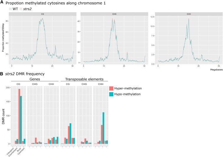
We utilized three Arabidopsis T-DNA insertion mutants targeting components of the DNA methylation machinery to test for stress priming with disrupted methylome maintenance. These include the triple methyltransferase mutant drm1drm2cmt3 (ddc) (Henderson and Jacobsen 2008), the triple demethylase mutant ros1dml2dml3 (rdd) (Le et al. 2014), and the novel putative RdDM mutant strs2 (Khan et al. 2014). While ddc and rdd are well-characterized mutants displaying gross methylome changes, reports suggest that strs2 displays subtler effects by fine-tuning DNA methylation levels at stress-associated genes (Khan et al. 2014). As previous studies investigating strs2 relied on a targeted analysis using chop-PCR, we performed MethylC-seq to confirm the full extent of methylome changes. Global levels of CG, CHG, and CHH methylation in strs2 were highly comparable to WT (Figure 1 A, Supplementary Dataset 1). However, employing DSS (Feng et al. 2014) to identify differentially methylated regions (DMRs) revealed moderate levels of local changes, predominantly in the CG context, with comparable numbers of hyper- and hypo-DMRs (Figure 1 B, Table 1, Supplementary Dataset 2). Nonetheless, many CHH DMRs were located within TEs consistent with the implication of STRS2 in RdDM. Furthermore, 130/199 (65.6%) strs2 CHH hypo-DMRs were in regions also targeted by DRM1 and DRM2 (7,393 CHH hypo-DMRs in drm1drm2), whereas CG and CHG hypo-DMRs demonstrated weaker overlaps (CG: 7/318, 2.2%; CHG: 8/34, 23.5%). Paired with previous observations (Khan et al. 2014), our analysis confirms partial involvement of STRS2 in the RdDM pathway potentially contributing in a manner akin to prior descriptions of “weak” RdDM mutants (Stroud et al. 2015). However, the bias toward CG DMRs and comparable numbers of hyper- and hypo-DMRs suggests broader roles in DNA methylation patterning. Nonetheless, the strs2 mutant fulfilled our criteria as displaying milder aberrations in DNA methylation.
Figure 1.
Subtle methylome perturbation in strs2 A Mean weighted methylation levels, binned into 50 kbp rolling windows, along Arabidopsis chromosome 1 for WT and strs2. B DMR frequency in strs2 grouped by genomic location relative to annotated genes or transposable elements. DMRs were classified as either occurring directly within (body), <1 kbp away from the 5` (upstream) or 3`end of (downstream), or >1 kbp (intergenic) away from genomic features.
Table 1. DMR analysis in strs2 (vs WT).
| DMR type | Methylation context | |||
|---|---|---|---|---|
| CG | CHG | CHH | Total | |
| Hyper-methylation | 322 | 64 | 140 | 526 |
| Hypo-methylation | 318 | 34 | 199 | 551 |
| Total | 640 | 98 | 339 | 1,077 |
Development and basal photoprotection in methylome mutants
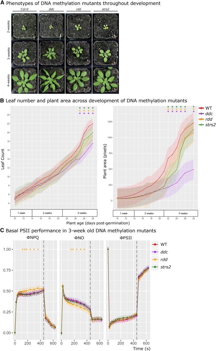
Aberrant DNA methylation is considered to result in developmental differences due to transcriptional misregulation and TE activation; ddc displays curled leaves and reduced stature due to SDC misexpression (Henderson and Jacobsen 2008), while strs2 shows a slight early flowering phenotype (Kant et al. 2007). To ensure that developmental abnormalities would not confound observations of priming, developmental traits were monitored until 4-weeks of age starting from 10-day old seedlings (Figure 2). While no unusual phenotypes were evident for rdd and strs2 (Figure 2 A), ddc displayed the expected curled leaves after 3-weeks of age. Quantitative measures of plant area reflect these visible observations, whereby the rosette area of ddc was reduced compared to WT (Figure 2 B). Minor differences in leaf number and plant area were observed between 3 and 4 weeks for rdd and strs2. Therefore, priming measurements were performed on 3 week-old plants prior to the onset of substantial morphological differences.
Figure 2.
DNA methylation mutant development and basal PSII function A Representative morphology of WT, ddc, rdd, and strs2 from 2-4 weeks of age. B Leaf number (left) and plant area (right) in 10-day old WT (red, n = 8), ddc (purple, n = 8), rdd (orange, n = 8), and strs2 (green, n = 8) seedlings, monitored for 20 days until 4-weeks of age. C Measures of PSII performance in 3-week old WT (red, n = 60), ddc (purple, n = 10), rdd (orange, n = 10), strs2 (green, n = 15) plants grown under standard conditions. Points denote estimated marginal means based on a fitted linear mixed-effect model for each genotype. Bars and shaded regions denote 95% confidence intervals; * indicates statistical significance (adjusted p-value < 0.05) from WT.
In addition to developmental abnormalities, aberrant methylome maintenance may also affect the capacity for basal photoprotective responses. Thus, functional PSII capacity was tested in 3-week old mutants (Figure 2 C). Photosynthetic efficiency (ΦPSII) was largely consistent across genotypes. Similarly, the capacity for, or activation of, actively regulated (ΦNPQ) and constitutively (ΦNO) dissipative quenching was largely consistent across genotypes. An exception to this was rdd that demonstrated elevated ΦNPQ with a concomitant reduction in ΦNO but unperturbed ΦPSII. However, the difference observed in rdd is minor compared to traditional mutants with perturbed photosystems (Niyogi et al. 2001) and is more reflective of natural variation (Jung and Niyogi 2009). Thus, no major disruptions to basal PSII performance is evident in these methylation mutants allowing for comparisons of EL priming.
Excess light priming evident despite aberrant methylome patterning
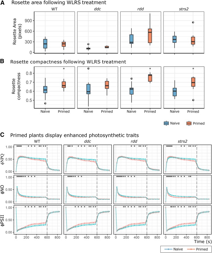
To investigate whether a perturbed methylome would impair priming to recurring EL, we repeated the WLRS time-course experiment (Ganguly et al. 2018) on WT, ddc, rdd, and strs2 (Figure 3). The week of recurring EL led to the expected morphological differences across all genotypes, namely increased rosette compaction but consistent leaf area (Figure 3 A-B). However, there was an attenuated difference in ddc area, which we attribute to its naturally curled leaf phenotype. Alongside morphological measures were changes in PSII traits indicative of physiological priming to recurring EL (Figure 3 C). The adoption of ΦNPQ and ΦNO also revealed distinct patterns in thermal dissipation not observed previously. Particularly, that the enhanced NPQ was underpinned by actively regulated pH-dependent thermal dissipation (ΦNPQ) rather than due to constitutive forms (ΦNO). Indeed, ΦNO was lower in primed plants. While ΦNPQ is rapidly activated in primed plants, it is deactivated concomitantly with an increase in ΦPSII. A key result here was that all methylation mutants demonstrated a WT-like priming response, for all three PSII traits, to recurring EL thus demonstrating appropriate light-acclimatory processes despite having perturbed methylomes.
Figure 3.
DNA methylation mutants exhibit priming to recurring EL A-B Boxplots of rosette area and compactness measured in all genotypes either exposed to recurring EL (primed, n = 12-15) or not (naive, 21-23). * denotes statistical significance determined using independent Student’s t-tests for each genotype (adjusted p-value < 0.05). C PSII performance traits in naive (WT n = 22; ddc n = 16, rdd n = 21, strs2 n = 23) and primed (WT n = 16; ddc n = 13, rdd n = 12, strs2 n = 15) plants for all genotypes. Points denote estimated marginal means based on a fitted linear mixed-effect model for each genotype. Bars and shading denote 95% confidence intervals; * indicates statistical significance (adjusted p-value < 0.05) from WT.
Primed plants exhibit transcriptional ‘dampening’ despite a reset transcriptome
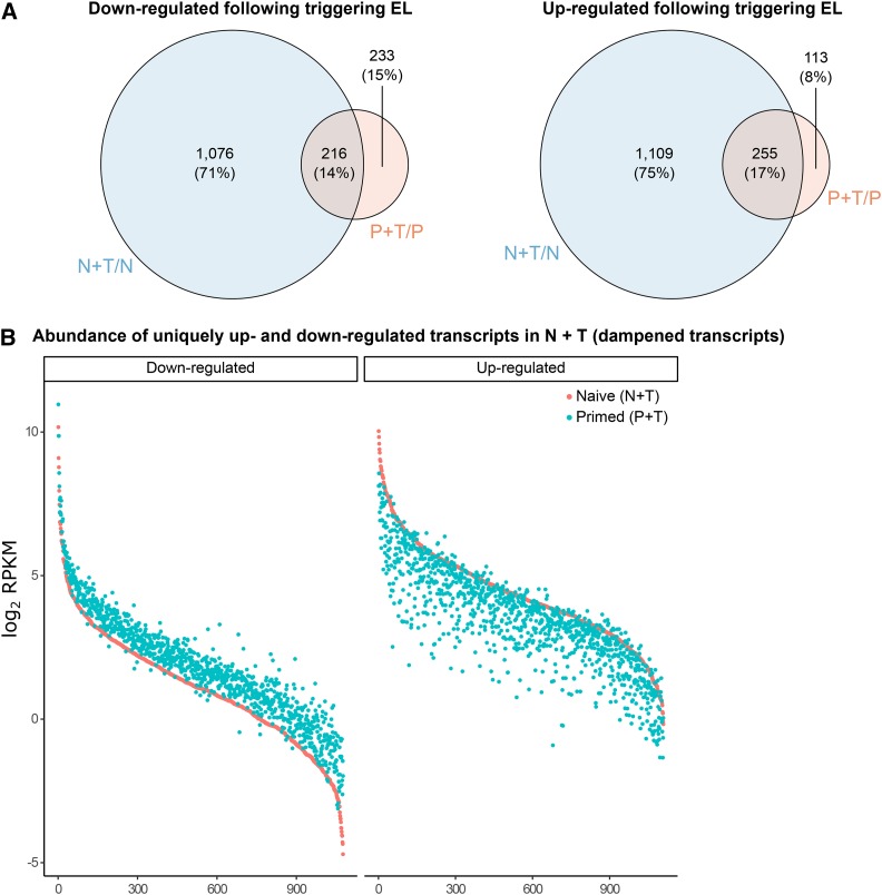
Persistent changes in gene expression are hypothesized to convey a primed state (Hilker et al. 2016). To test this, we searched for constitutive changes in gene expression between naïve (N) and primed (P) plants after one-week of recurring EL using mRNA-seq. Despite the observation of physiological priming in these tissues, we observed a completely reset transcriptome exemplified by close sample clustering and the absence of differentially expressed genes (DEGs) between naïve and primed samples (Figure S1 A-B). We subsequently investigated the transcriptional response of primed plants to an additional EL treatment (triggering stress, P+T). Application of an EL triggering stress to naive plants (N+T) elicited the differential expression of hundreds of genes, however, primed plants showed a vastly attenuated response (Figure S1 C-D, Supplementary Datasets 4 - 5). While the majority of EL-induced transcripts in P+T plants overlapped with those in N+T plants, a striking proportion of both up- (75%) and down-regulated (71%) transcripts were unique to N+T plants (Figure 4 A). The abundance of nearly all 1,076 uniquely down-regulated transcripts in N+T plants was greater in P+T plants (Figure 4 B, left). Equivalently, abundance of the 1,109 uniquely up-regulated transcripts in N+T plants was decreased in P+T plants (Figure 4 B, right). Together, these results evoke a dampened transcriptional response to EL triggering stimuli in primed plants, rather than reflecting differences between their basal (un-triggered) transcriptomes. The dampening effect is, however, subtle in nature - no DEGs were identified upon direct comparison of N+T and P+T samples.
Figure 4.
Transcriptional ‘dampening’ evident in EL primed plants A Overlap between up- and down-regulated transcripts in naive and primed plants in response to an additional triggering EL treatment. B Transcript abundance (log2 RPKM) of up- (left) and down-regulated (right) transcripts, in N+T plants, for both N+T and P+T plants.
We hypothesized that the transcriptional dampening observed in primed plants may be a consequence of reduced stress signaling because of enhanced dissipative quenching (Rossel et al. 2007; Suzuki et al. 2013; Carmody et al. 2016). A gene ontology analysis of the 2,185 dampened transcripts (Supplementary Dataset 6) revealed a strong enrichment for terms associated with various catabolic processes (e.g., protein, cellulose, and fatty acid catabolism) and, pertinent to our hypothesis, numerous stress factors, including salt, heat, water deprivation, and metal ion (Supplementary Dataset 7). Subsequently, genes induced by well-established stress signals including ROS (hydrogen peroxide [H2O2], singlet oxygen [1O2], and superoxide [O2-] (Gadjev et al. 2006)), salicylic acid (SA) (Blanco et al. 2009), β-cyclocitral (Ramel et al. 2012), ABA (Pornsiriwong et al. 2017), and SAA (Rossel et al. 2007) were overlapped with the dampened transcripts identified here. In total, 635/2,185 (29.1%) dampened transcripts were overlapped with the collated gene list. Performing hypergeometric testing on a per signal basis revealed statistically significant overlaps with β-cyclocitral and SAA induced genes (Table 2).
Table 2. Hypergeometric testing of 2,185 dampened transcripts overlapped with genes responsive to stress signals.
| Gene list | Total responsive genes | Overlap with dampened transcripts | Hypergeometric test |
|---|---|---|---|
| ABA | 2,471 | 252 | P[X ≥ 252] = 0.99 |
| SA | 217 | 21 | P[X ≥ 21] = 0.88 |
| O2- | 209 | 7 | P[X ≥ 7] = 0.99 |
| H2O2 | 325 | 36 | P[X ≥ 36] = 0.75 |
| 1O2 | 295 | 19 | P[X ≥ 19] = 0.99 |
| β-cyclocitral | 1,145 | 191 | P[X ≥ 191] < 0.001 |
| SAA | 703 | 109 | P[X ≥ 109] < 0.001 |
Discussion
The popular notion that DNA methylation may regulate plant stress responses is confounded by both supporting and conflicting investigations spanning a range of species and stressors. Light stress is one such example for which physiological priming and memory have been demonstrated in Arabidopsis (Szechyńska-Hebda et al. 2010; Gordon et al. 2012), yet we did not observe co-occurring stress-induced DNA methylation changes (Ganguly et al. 2018). Consequently, we refined the experimental design to address a distinct hypothesis: that physiological priming of recurring EL is underpinned by transcriptional regulation for which active maintenance of DNA methylation is important. We present evidence for recurring EL priming in DNA methylation mutants using a full-factorial experimental design, which facilitated systematic testing of two closely related potential priming mechanisms: 1. DNA methylation-mediated transcriptional regulation; and 2. altered transcription (independent of DNA methylation).
DNA methylation, development, and priming
Loss of DNA methylation leads to abnormal plant growth due to its importance for genome integrity (Reinders et al. 2009). Abnormal leaf development could in turn lead to confounding changes in light acclimation that are not a direct result of DNA methylation changes to light stress-responsive genes. Thus, we performed detailed physiological characterization of DNA methylation mutants to identify any constitutive changes in photosynthetic rate or capacity that could confound our interpretation of the contribution of DNA methylation to priming. Irrespective of the nature of methylome perturbance, we found negligible changes in photosynthesis at the developmental stage examined here.
We utilized two well-characterized methylation mutants, ddc and rdd, which display genome-wide hypomethylation and hypermethylation, respectively (Stroud et al. 2014, 2015). These relatively severe mutants were complemented by strs2, included based on reports of disrupted RdDM leading to targeted methylation changes at stress-responsive genes (Kant et al. 2007; Khan et al. 2014). While our focused analysis confirmed some contribution of STRS2 toward RdDM, our global analysis suggests a broader function and confirmed the relatively mild nature of the strs2 methylome in comparison to ddc and rdd.
Light stress induces multiple physiological, post-transcriptional and extensive transcriptional responses (Dietz 2015). Thus, it was hypothesized that changes in DNA methylation might impact the transcriptional component of the acclimation response, thereby impairing the ability for EL priming. Contradictory to this hypothesis perturbation of the methylome, regardless of severity (e.g., strs2 vs. ddc) or type (e.g., hyper- or hypo-methylation), did not impair basal photoprotective capacity nor the ability to be primed by recurring EL. Instead, the EL priming observed herein likely reflects post-transcriptional changes of the photosynthetic machinery. The reduction in ΦNO disputes constitutive changes in PSII conformation. Rather there appears to be greater electron flow (increased ΦPSII) in primed plants that may reflect an increased concentration of components facilitating photosynthetic electron transport, such as PSII core proteins, cytochrome b6f, and chloroplastic ATPases (Foyer et al. 2012; Dwyer et al. 2012; Zhu et al. 2017). Pertinent to this study is the observation that these processes remain functional under a range of methylome perturbations (Table 3).
Table 3. Diagnostic PSII parameters and their biological relevance.
| Parameter | Equation | Interpretation |
|---|---|---|
| ΦPSII | 1 - Ft/Fm′ | Fraction of light absorbed by PSII for photochemistry. |
| ΦNPQ | Ft/Fm′ - Ft/Fm′ | Fraction of light absorbed by PSII that is lost via thermal dissipation (ΔpH- and xanthophyll-regulated processes). |
| ΦNPQ | Ft/Fm | Fraction of light absorbed by PSII that is lost via chlorophyll fluorescence. |
Priming occurs independently of transcriptional changes
Transcriptional up-regulation of light-responsive genes is indicative of an acclimatory response (Gordon et al. 2012; Carmody et al. 2016), however, it remains an untested hypothesis that EL priming is underpinned by constitutive transcriptional changes. No persistent changes in gene expression were observed in primed plants, consistent with previous reports of transcriptome resetting following EL exposure (Crisp et al. 2017). Instead, an attenuated transcriptional response (“transcriptional dampening”) was observed in primed plants responding to a final triggering stress. This contrasts enhanced transcriptional responses observed upon a triggering stimulus following single stress treatments (Ding et al. 2012; Lämke et al. 2016; Crisp et al. 2017). The transcriptional dampening observed likely reflects a greater capacity of primed plants, after longer-term recurring EL exposure, to deal with increased light flux. That is, enhanced photosynthetic and photoprotective capacity in primed plants may alter the extent of EL-mediated signaling leading to an attenuated response. Indeed, nuclear gene expression can be modulated by changes in photosynthetic electron flow that can subsequently affect cellular metabolism and acclimatory responses (Gollan et al. 2017; Dickinson et al. 2018). Taken together, we propose that the dampened transcriptional response might be a consequence of altered stress signaling due to physiological priming. In support of this hypothesis was the observation that almost one-third of dampened transcripts overlapped with genes responsive to signaling pathways, specifically β-cyclocitral and SAA. However, limitations of the analysis performed here are that these signals were not directly assayed and they do not account for all dampened transcripts. Nonetheless, these results suggest that the gene expression changes observed in primed plants are consequential of physiological priming, rather than the cause.
Conclusion
Through the analysis of Arabidopsis mutants impaired in active methylome maintenance, we have conducted a systematic investigation of the hypothesis that DNA methylation may contribute to stress priming. Specifically, we address the hypothesis that physiological priming to recurring EL is underpinned by transcriptional regulation and active methylome maintenance. We found no evidence to suggest that aberrant maintenance or removal of DNA methylation impaired the capacity for physiological priming against recurring EL. In fact, primed plants demonstrated completely reset transcriptomes. Instead, an attenuation in transcriptional response to EL was observed in primed plants and is likely to be a consequence rather than a cause of physiological priming.
Acknowledgments
We would like to acknowledge the advice received from Prof. Wah Soon Chow on chlorophyll fluorescence based measures of photosynthetic performance. We would also like to acknowledge the Biomolecular Resource Facility at the ANU for performing Illumina sequencing, and Dr. Terry Neeman for advice on statistical analyses. This project was supported with the provision of plant growth facilities by the Australian Plant Phenomics Facility and computational infrastructure by the National Computational Infrastructure, both supported under the National Collaborative Research Infrastructure Strategy of the Australian Government.
Footnotes
Supplemental material available at FigShare: https://doi.org/10.25387/g3.9724019.
Communicating editor: B. Gregory
Literature Cited
- Allen R. S., Li J., Alonso-Peral M. M., White R. G., Gubler F. et al. , 2010. MicroR159 regulation of most conserved targets in Arabidopsis has negligible phenotypic effects. Silence 1: 18 10.1186/1758-907X-1-18 [DOI] [PMC free article] [PubMed] [Google Scholar]
- Baker N. R., 2008. Chlorophyll fluorescence: a probe of photosynthesis in vivo. Annu. Rev. Plant Biol. 59: 89–113. 10.1146/annurev.arplant.59.032607.092759 [DOI] [PubMed] [Google Scholar]
- Bates D., Mächler M., Bolker B., and Walker S., 2015. Fitting Linear Mixed-Effects Models Using lme4. J. Stat. Softw. 67: 1–48. [Google Scholar]
- Bewick A. J., Ji L., Niederhuth C. E., Willing E.-M., Hofmeister B. T. et al. , 2016. On the origin and evolutionary consequences of gene body DNA methylation. Proc. Natl. Acad. Sci. USA 113: 9111–9116. 10.1073/pnas.1604666113 [DOI] [PMC free article] [PubMed] [Google Scholar]
- Blanco F., Salinas P., Cecchini N. M., Jordana X., Van Hummelen P. et al. , 2009. Early genomic responses to salicylic acid in Arabidopsis. Plant Mol. Biol. 70: 79–102. 10.1007/s11103-009-9458-1 [DOI] [PubMed] [Google Scholar]
- Boyko A., Blevins T., Yao Y., Golubov A., Bilichak A. et al. , 2010. Transgenerational Adaptation of Arabidopsis to Stress Requires DNA Methylation and the Function of Dicer-Like Proteins. PLoS One 5: e9514. [DOI] [PMC free article] [PubMed] [Google Scholar]
- Carmody M., Crisp P. A., d’Alessandro S., Ganguly D., Gordon M. et al. , 2016. Uncoupling High Light Responses from Singlet Oxygen Retrograde Signaling and Spatial-Temporal Systemic Acquired Acclimation. Plant Physiol. 171: 1734–1749. 10.1104/pp.16.00404 [DOI] [PMC free article] [PubMed] [Google Scholar]
- Cheng C.-Y., Krishnakumar V., Chan A. P., Thibaud-Nissen F., Schobel S. et al. , 2017. Araport11: a complete reannotation of the Arabidopsis thaliana reference genome. Plant J. 89: 789–804. 10.1111/tpj.13415 [DOI] [PubMed] [Google Scholar]
- Chen Y., Lun A. T. L., and Smyth G. K., 2016. From reads to genes to pathways: differential expression analysis of RNA-Seq experiments using Rsubread and the edgeR quasi-likelihood pipeline. F1000 Res. 5: 1438. [DOI] [PMC free article] [PubMed] [Google Scholar]
- Cokus S. J., Feng S., Zhang X., Chen Z., Merriman B. et al. , 2008. Shotgun bisulphite sequencing of the Arabidopsis genome reveals DNA methylation patterning. Nature 452: 215–219. 10.1038/nature06745 [DOI] [PMC free article] [PubMed] [Google Scholar]
- Cortijo S., Wardenaar R., Colomé-Tatché M., Gilly A., Etcheverry M. et al. , 2014. Mapping the epigenetic basis of complex traits. Science 343: 1145–1148. 10.1126/science.1248127 [DOI] [PubMed] [Google Scholar]
- Crisp P. A., Ganguly D. R., Smith A. B., Murray K. D., Estavillo G. M. et al. , 2017. Rapid Recovery Gene Downregulation during Excess-Light Stress and Recovery in Arabidopsis. Plant Cell 29: 1836–1863. 10.1105/tpc.16.00828 [DOI] [PMC free article] [PubMed] [Google Scholar]
- Dickinson P. J., Kumar M., Martinho C., Yoo S. J., Lan H. et al. , 2018. Chloroplast Signaling Gates Thermotolerance in Arabidopsis. Cell Reports 22: 1657–1665. 10.1016/j.celrep.2018.01.054 [DOI] [PMC free article] [PubMed] [Google Scholar]
- Dietz K.-J., 2015. Efficient high light acclimation involves rapid processes at multiple mechanistic levels. J. Exp. Bot. 66: 2401–2414. 10.1093/jxb/eru505 [DOI] [PubMed] [Google Scholar]
- Ding Y., Fromm M., and Avramova Z., 2012. Multiple exposures to drought “train” transcriptional responses in Arabidopsis. Nat. Commun. 3: 740 10.1038/ncomms1732 [DOI] [PubMed] [Google Scholar]
- Dwyer S. A., Chow W. S., Yamori W., Evans J. R., Kaines S. et al. , 2012. Antisense reductions in the PsbO protein of photosystem II leads to decreased quantum yield but similar maximal photosynthetic rates. J. Exp. Bot. 63: 4781–4795. 10.1093/jxb/ers156 [DOI] [PMC free article] [PubMed] [Google Scholar]
- Feng H., Conneely K. N., and Wu H., 2014. A Bayesian hierarchical model to detect differentially methylated loci from single nucleotide resolution sequencing data. Nucleic Acids Res. 42: e69 10.1093/nar/gku154 [DOI] [PMC free article] [PubMed] [Google Scholar]
- Finnegan E. J., Peacock W. J., and Dennis E. S., 1996. Reduced DNA methylation in Arabidopsis thaliana results in abnormal plant development. Proc. Natl. Acad. Sci. USA 93: 8449–8454. 10.1073/pnas.93.16.8449 [DOI] [PMC free article] [PubMed] [Google Scholar]
- Foyer C. H., Neukermans J., Queval G., Noctor G., and Harbinson J., 2012. Photosynthetic control of electron transport and the regulation of gene expression. J. Exp. Bot. 63: 1637–1661. 10.1093/jxb/ers013 [DOI] [PubMed] [Google Scholar]
- Gadjev I., Vanderauwera S., Gechev T. S., Laloi C., Minkov I. N. et al. , 2006. Transcriptomic footprints disclose specificity of reactive oxygen species signaling in Arabidopsis. Plant Physiol. 141: 436–445. 10.1104/pp.106.078717 [DOI] [PMC free article] [PubMed] [Google Scholar]
- Ganguly D. R., Crisp P. A., Eichten S. R., and Pogson B. J., 2018. Maintenance of pre-existing DNA methylation states through recurring excess-light stress. Plant Cell Environ. 41: 1657–1672. 10.1111/pce.13324 [DOI] [PubMed] [Google Scholar]
- Gollan P. J., Lima-Melo Y., Tiwari A., Tikkanen M., and Aro E.-M., 2017. Interaction between photosynthetic electron transport and chloroplast sinks triggers protection and signalling important for plant productivity. Philos. Trans. R. Soc. Lond. B Biol. Sci. 372: 1730 10.1098/rstb.2016.0390 [DOI] [PMC free article] [PubMed] [Google Scholar]
- Gordon M. J., Carmody M., Albrecht V., and Pogson B., 2012. Systemic and Local Responses to Repeated HL Stress-Induced Retrograde Signaling in Arabidopsis. Front. Plant Sci. 3: 303. [DOI] [PMC free article] [PubMed] [Google Scholar]
- Henderson I. R., and Jacobsen S. E., 2008. Tandem repeats upstream of the Arabidopsis endogene SDC recruit non-CG DNA methylation and initiate siRNA spreading. Genes Dev. 22: 1597–1606. 10.1101/gad.1667808 [DOI] [PMC free article] [PubMed] [Google Scholar]
- Hendrickson L., Furbank R. T., and Chow W. S., 2004. A simple alternative approach to assessing the fate of absorbed light energy using chlorophyll fluorescence. Photosynth. Res. 82: 73–81. 10.1023/B:PRES.0000040446.87305.f4 [DOI] [PubMed] [Google Scholar]
- He L., Wu W., Zinta G., Yang L., Wang D. et al. , 2018. A naturally occurring epiallele associates with leaf senescence and local climate adaptation in Arabidopsis accessions. Nat. Commun. 9: 460 10.1038/s41467-018-02839-3 [DOI] [PMC free article] [PubMed] [Google Scholar]
- Hilker M., Schwachtje J., Baier M., Balazadeh S., Bäurle I. et al. , 2016. Priming and memory of stress responses in organisms lacking a nervous system. Biol. Rev. Camb. Philos. Soc. 91: 1118–1133. 10.1111/brv.12215 [DOI] [PubMed] [Google Scholar]
- Iwasaki M., and Paszkowski J., 2014. Identification of genes preventing transgenerational transmission of stress-induced epigenetic states. Proc. Natl. Acad. Sci. USA 111: 8547–8552. 10.1073/pnas.1402275111 [DOI] [PMC free article] [PubMed] [Google Scholar]
- Johannes F., Porcher E., Teixeira F. K., Saliba-Colombani V., Simon M. et al. , 2009. Assessing the impact of transgenerational epigenetic variation on complex traits. PLoS Genet. 5: e1000530 10.1371/journal.pgen.1000530 [DOI] [PMC free article] [PubMed] [Google Scholar]
- Johannes F., and Schmitz R. J., 2019. Spontaneous epimutations in plants. New Phytol. 221: 1253–1259. 10.1111/nph.15434 [DOI] [PubMed] [Google Scholar]
- Jung H.-S., and Niyogi K. K., 2009. Quantitative genetic analysis of thermal dissipation in Arabidopsis. Plant Physiol. 150: 977–986. 10.1104/pp.109.137828 [DOI] [PMC free article] [PubMed] [Google Scholar]
- Kant P., Kant S., Gordon M., Shaked R., and Barak S., 2007. STRESS RESPONSE SUPPRESSOR1 and STRESS RESPONSE SUPPRESSOR2, two DEAD-box RNA helicases that attenuate Arabidopsis responses to multiple abiotic stresses. Plant Physiol. 145: 814–830. 10.1104/pp.107.099895 [DOI] [PMC free article] [PubMed] [Google Scholar]
- Karpinski S., 1999. Systemic Signaling and Acclimation in Response to Excess Excitation Energy in Arabidopsis. Science 284: 654–657. 10.1126/science.284.5414.654 [DOI] [PubMed] [Google Scholar]
- Khan A., Garbelli A., Grossi S., Florentin A., Batelli G. et al. , 2014. The Arabidopsis STRESS RESPONSE SUPPRESSOR DEAD-box RNA helicases are nucleolar- and chromocenter-localized proteins that undergo stress-mediated relocalization and are involved in epigenetic gene silencing. Plant J. 79: 28–43. 10.1111/tpj.12533 [DOI] [PubMed] [Google Scholar]
- Kim J.-M., To T. K., Matsui A., Tanoi K., Kobayashi N. I. et al. , 2017. Acetate-mediated novel survival strategy against drought in plants. Nat. Plants 3: 17097 Erratum: 17097. 10.1038/nplants.2017.97 [DOI] [PubMed] [Google Scholar]
- Kramer D. M., Johnson G., Kiirats O., and Edwards G. E., 2004. New Fluorescence Parameters for the Determination of QARedox State and Excitation Energy Fluxes. Photosynth. Res. 79: 209–218. 10.1023/B:PRES.0000015391.99477.0d [DOI] [PubMed] [Google Scholar]
- Krueger F., and Andrews S. R., 2011. Bismark: a flexible aligner and methylation caller for Bisulfite-Seq applications. Bioinformatics 27: 1571–1572. 10.1093/bioinformatics/btr167 [DOI] [PMC free article] [PubMed] [Google Scholar]
- Lämke J., Brzezinka K., Altmann S., and Bäurle I., 2016. A hit-and-run heat shock factor governs sustained histone methylation and transcriptional stress memory. EMBO J. 35: 162–175. 10.15252/embj.201592593 [DOI] [PMC free article] [PubMed] [Google Scholar]
- Langmead B., and Salzberg S. L., 2012. Fast gapped-read alignment with Bowtie 2. Nat. Methods 9: 357–359. 10.1038/nmeth.1923 [DOI] [PMC free article] [PubMed] [Google Scholar]
- Lefcheck J. S., 2016. PIECEWISESEM: Piecewise structural equation modelling in r for ecology, evolution, and systematics. Methods Ecol. Evol. 7: 573–579. [Google Scholar]
- Le T.-N., Schumann U., Smith N. A., Tiwari S., Au P. C. K. et al. , 2014. DNA demethylases target promoter transposable elements to positively regulate stress responsive genes in Arabidopsis. Genome Biol. 15: 458 10.1186/s13059-014-0458-3 [DOI] [PMC free article] [PubMed] [Google Scholar]
- Liao Y., Smyth G. K., and Shi W., 2013. The Subread aligner: fast, accurate and scalable read mapping by seed-and-vote. Nucleic Acids Res. 41: e108 10.1093/nar/gkt214 [DOI] [PMC free article] [PubMed] [Google Scholar]
- Matzke M. A., and Mosher R. A., 2014. RNA-directed DNA methylation: an epigenetic pathway of increasing complexity. Nat. Rev. Genet. 15: 394–408. Erratum: 570. 10.1038/nrg3683 [DOI] [PubMed] [Google Scholar]
- Meng D., Dubin M., Zhang P., Osborne E. J., Stegle O. et al. , 2016. Limited Contribution of DNA Methylation Variation to Expression Regulation in Arabidopsis thaliana. PLoS Genet. 12: e1006141 10.1371/journal.pgen.1006141 [DOI] [PMC free article] [PubMed] [Google Scholar]
- Mi H., Muruganujan A., Huang X., Ebert D., Mills C. et al. , 2019. Protocol Update for large-scale genome and gene function analysis with the PANTHER classification system (v.14.0). Nat. Protoc. 14: 703–721. 10.1038/s41596-019-0128-8 [DOI] [PMC free article] [PubMed] [Google Scholar]
- Molloy P. L., 1986. Effects of DNA methylation on specific transcription by RNA polymerase II in vitro. Mol. Biol. Rep. 11: 13–17. 10.1007/BF00417589 [DOI] [PubMed] [Google Scholar]
- Muyle A., and Gaut B. S., 2019. Loss of gene body methylation in Eutrema salsugineum is associated with reduced gene expression. Mol. Biol. Evol. 36: 155–158. 10.1093/molbev/msy204 [DOI] [PMC free article] [PubMed] [Google Scholar]
- Niyogi K. K., Shih C., Soon Chow W., Pogson B. J., DellaPenna D. et al. , 2001. Photosynth. Res. 67: 139–145. 10.1023/A:1010661102365 [DOI] [PubMed] [Google Scholar]
- O’Malley R. C., Huang S.-S. C., Song L., Lewsey M. G., Bartlett A. et al. , 2016. Cistrome and Epicistrome Features Shape the Regulatory DNA Landscape. Cell 165: 1280–1292. Erratum: 1598. 10.1016/j.cell.2016.04.038 [DOI] [PMC free article] [PubMed] [Google Scholar]
- Ong-Abdullah M., Ordway J. M., Jiang N., Ooi S.-E., Kok S.-Y. et al. , 2015. Loss of Karma transposon methylation underlies the mantled somaclonal variant of oil palm. Nature 525: 533–537. 10.1038/nature15365 [DOI] [PMC free article] [PubMed] [Google Scholar]
- Pornsiriwong W., Estavillo G. M., Chan K. X., Tee E. E., Ganguly D. et al. , 2017. A chloroplast retrograde signal, 3′-phosphoadenosine 5′-phosphate, acts as a secondary messenger in abscisic acid signaling in stomatal closure and germination. eLife 6: 1–34. 10.7554/eLife.23361 [DOI] [PMC free article] [PubMed] [Google Scholar]
- Quinlan A. R., and Hall I. M., 2010. BEDTools: a flexible suite of utilities for comparing genomic features. Bioinformatics 26: 841–842. 10.1093/bioinformatics/btq033 [DOI] [PMC free article] [PubMed] [Google Scholar]
- Ramel F., Birtic S., Ginies C., Soubigou-Taconnat L., Triantaphylidès C. et al. , 2012. Carotenoid oxidation products are stress signals that mediate gene responses to singlet oxygen in plants. Proc. Natl. Acad. Sci. USA 109: 5535–5540. 10.1073/pnas.1115982109 [DOI] [PMC free article] [PubMed] [Google Scholar]
- Reinders J., Wulff B. B. H., Mirouze M., Marí-Ordóñez A., Dapp M. et al. , 2009. Compromised stability of DNA methylation and transposon immobilization in mosaic Arabidopsis epigenomes. Genes Dev. 23: 939–950. 10.1101/gad.524609 [DOI] [PMC free article] [PubMed] [Google Scholar]
- Rossel J. B., Wilson P. B., Hussain D., Woo N. S., Gordon M. J. et al. , 2007. Systemic and intracellular responses to photooxidative stress in Arabidopsis. Plant Cell 19: 4091–4110. 10.1105/tpc.106.045898 [DOI] [PMC free article] [PubMed] [Google Scholar]
- Rowley M. J., Rothi M. H., Böhmdorfer G., Kuciński J., and Wierzbicki A. T., 2017. Long-range control of gene expression via RNA-directed DNA methylation. PLoS Genet. 13: e1006749 10.1371/journal.pgen.1006749 [DOI] [PMC free article] [PubMed] [Google Scholar]
- Sani E., Herzyk P., Perrella G., Colot V., and Amtmann A., 2013. Hyperosmotic priming of Arabidopsis seedlings establishes a long-term somatic memory accompanied by specific changes of the epigenome. Genome Biol. 14: R59 10.1186/gb-2013-14-6-r59 [DOI] [PMC free article] [PubMed] [Google Scholar]
- Schultz M. D., Schmitz R. J., and Ecker J. R., 2012. “Leveling” the playing field for analyses of single-base resolution DNA methylomes. Trends Genet. 28: 583–585. 10.1016/j.tig.2012.10.012 [DOI] [PMC free article] [PubMed] [Google Scholar]
- Secco D., Wang C., Shou H., Schultz M. D., Chiarenza S. et al. , 2015. Stress induced gene expression drives transient DNA methylation changes at adjacent repetitive elements. eLife 4: 1– 26 10.7554/eLife.09343 [DOI] [PMC free article] [PubMed] [Google Scholar]
- Shukla S., Kavak E., Gregory M., Imashimizu M., Shutinoski B. et al. , 2011. CTCF-promoted RNA polymerase II pausing links DNA methylation to splicing. Nature 479: 74–79. 10.1038/nature10442 [DOI] [PMC free article] [PubMed] [Google Scholar]
- Stroud H., Do T., Du J., Zhong X., Feng S. et al. , 2014. Non-CG methylation patterns shape the epigenetic landscape in Arabidopsis. Nat. Struct. Mol. Biol. 21: 64–72. 10.1038/nsmb.2735 [DOI] [PMC free article] [PubMed] [Google Scholar]
- Stroud H., Greenberg M. V. C., Feng S., Bernatavichute Y. V., and Jacobsen S. E., 2015. Comprehensive Analysis of Silencing Mutants Reveals Complex Regulation of the Arabidopsis Methylome. Cell 161: 1697–1698. Erratum: 1697–1698. 10.1016/j.cell.2015.06.020 [DOI] [PMC free article] [PubMed] [Google Scholar]
- Suzuki N., Miller G., Salazar C., Mondal H. A., Shulaev E. et al. , 2013. Temporal-spatial interaction between reactive oxygen species and abscisic acid regulates rapid systemic acclimation in plants. Plant Cell 25: 3553–3569. 10.1105/tpc.113.114595 [DOI] [PMC free article] [PubMed] [Google Scholar]
- Szechyńska-Hebda M., Kruk J., Górecka M., Karpińska B., and Karpiński S., 2010. Evidence for light wavelength-specific photoelectrophysiological signaling and memory of excess light episodes in Arabidopsis. Plant Cell 22: 2201–2218. 10.1105/tpc.109.069302 [DOI] [PMC free article] [PubMed] [Google Scholar]
- Wibowo A., Becker C., Marconi G., Durr J., Price J. et al. , 2016. Hyperosmotic stress memory in Arabidopsis is mediated by distinct epigenetically labile sites in the genome and is restricted in the male germline by DNA glycosylase activity. eLife 5: e13546 10.7554/eLife.13546 [DOI] [PMC free article] [PubMed] [Google Scholar]
- Williams B. P., and Gehring M., 2017. Stable transgenerational epigenetic inheritance requires a DNA methylation-sensing circuit. Nat. Commun. 8: 2124 10.1038/s41467-017-02219-3 [DOI] [PMC free article] [PubMed] [Google Scholar]
- Wu H., Xu T., Feng H., Chen L., Li B. et al. , 2015. Detection of differentially methylated regions from whole-genome bisulfite sequencing data without replicates. Nucleic Acids Res. 43: e141. [DOI] [PMC free article] [PubMed] [Google Scholar]
- Yin Y., Morgunova E., Jolma A., Kaasinen E., Sahu B. et al. , 2017. Impact of cytosine methylation on DNA binding specificities of human transcription factors. Science 356: eaaj2239 10.1126/science.aaj2239 [DOI] [PMC free article] [PubMed] [Google Scholar]
- Zhu H., Zeng L.-D., Yi X.-P., Peng C.-L., Zhang W.-F. et al. , 2017. The half-life of the cytochrome bf complex in leaves of pea plants after transfer from moderately-high growth light to low light. Funct. Plant Biol. 44: 351 10.1071/FP16222 [DOI] [PubMed] [Google Scholar]
Associated Data
This section collects any data citations, data availability statements, or supplementary materials included in this article.
Data Availability Statement
All code used for analyses are available at GitHub (https://goo.gl/wsQrJT). All sequencing data generated for this study are accessible at the NCBI GEO repository (GSE121150, https://www.ncbi.nlm.nih.gov/geo/query/acc.cgi?acc=GSE121150). Independent WT and drm1drm2 MethylC-seq profiling was accessed from GEO accessions GSE39901 and GSE38286. Supplemental material available at FigShare: https://doi.org/10.25387/g3.9724019.


