This cross-sectional study examines the results of diffusion tensor imaging, cognitive testing, and a screen time survey to identify the implications of screen-based media use for the development of language and literacy skills in early childhood.
Key Points
Question
Is screen-based media use associated with differences in the structural integrity of brain white matter tracts that support language and literacy skills in preschool-aged children?
Findings
In this cross-sectional study of 47 healthy prekindergarten children, screen use greater than that recommended by the American Academy of Pediatrics guidelines was associated with (1) lower measures of microstructural organization and myelination of brain white matter tracts that support language and emergent literacy skills and (2) corresponding cognitive assessments.
Meaning
These findings suggest a need for further study into the association between screen-based media use and the developing brain, particularly during early childhood.
Abstract
Importance
The American Academy of Pediatrics (AAP) recommends limits on screen-based media use, citing its cognitive-behavioral risks. Screen use by young children is prevalent and increasing, although its implications for brain development are unknown.
Objective
To explore the associations between screen-based media use and integrity of brain white matter tracts supporting language and literacy skills in preschool-aged children.
Design, Setting, and Participants
This cross-sectional study of healthy children aged 3 to 5 years (n = 47) was conducted from August 2017 to November 2018. Participants were recruited at a US children’s hospital and community primary care clinics.
Exposures
Children completed cognitive testing followed by diffusion tensor imaging (DTI), and their parent completed a ScreenQ survey.
Main Outcomes and Measures
ScreenQ is a 15-item measure of screen-based media use reflecting the domains in the AAP recommendations: access to screens, frequency of use, content viewed, and coviewing. Higher scores reflect greater use. ScreenQ scores were applied as the independent variable in 3 multiple linear regression models, with scores in 3 standardized assessments as the dependent variable, controlling for child age and household income: Comprehensive Test of Phonological Processing, Second Edition (CTOPP-2; Rapid Object Naming subtest); Expressive Vocabulary Test, Second Edition (EVT-2; expressive language); and Get Ready to Read! (GRTR; emergent literacy skills). The DTI measures included fractional anisotropy (FA) and radial diffusivity (RD), which estimated microstructural organization and myelination of white matter tracts. ScreenQ was applied as a factor associated with FA and RD in whole-brain regression analyses, which were then narrowed to 3 left-sided tracts supporting language and emergent literacy abilities.
Results
Of the 69 children recruited, 47 (among whom 27 [57%] were girls, and the mean [SD] age was 54.3 [7.5] months) completed DTI. Mean (SD; range) ScreenQ score was 8.6 (4.8; 1-19) points. Mean (SD; range) CTOPP-2 score was 9.4 (3.3; 2-15) points, EVT-2 score was 113.1 (16.6; 88-144) points, and GRTR score was 19.0 (5.9; 5-25) points. ScreenQ scores were negatively correlated with EVT-2 (F2,43 = 5.14; R2 = 0.19; P < .01), CTOPP-2 (F2,35 = 6.64; R2 = 0.28; P < .01), and GRTR (F2,44 = 17.08; R2 = 0.44; P < .01) scores, controlling for child age. Higher ScreenQ scores were correlated with lower FA and higher RD in tracts involved with language, executive function, and emergent literacy abilities (P < .05, familywise error–corrected), controlling for child age and household income.
Conclusions and Relevance
This study found an association between increased screen-based media use, compared with the AAP guidelines, and lower microstructural integrity of brain white matter tracts supporting language and emergent literacy skills in prekindergarten children. The findings suggest further study is needed, particularly during the rapid early stages of brain development.
Introduction
In a single generation, through what has been described as a vast “uncontrolled experiment,”1 the landscape of childhood has been digitized, affecting how children play, learn, and form relationships. In addition to traditional programming, rapidly emerging technologies, particularly portable electronic devices, provide unprecedented access to a wide range of media.2,3 Use begins in infancy4 and increases with age, and it was recently estimated at more than 2 hours per day in children younger than 9 years, aside from use during childcare and school.3 Accompanying this rise are variables with potential risks and benefits, including access to screens (eg, in bedrooms), frequency of use, content, and grownup-child interaction (eg, coviewing).5,6 The American Academy of Pediatrics (AAP) recommends limits on screen-based media, citing developmental and health risks with excessive and inopportune use.3 These risks include language delay7,8; poor sleep2,9; impaired executive function10 and general cognition11; and decreased parent-child engagement, including reading together.12,13,14 The World Health Organization recently released even more restrictive recommendations for children younger than 5 years, discouraging screen time and advocating greater study of its implications for health and development.15
Recent evidence suggests that screen-based media use poses neurobiological risks in children,16,17,18,19 yet its associations with early brain development are largely unknown, particularly during the dynamic span of development before kindergarten.10 Although sensory networks mature relatively early,11 those sensory networks for higher-order skills, such as language,12 executive function,20,21 multimodal association,13 and reading,22,23 exhibit protracted development11,14 and are dependent on constructive stimulation in the home. Specifically, the organization and myelination of white matter tracts, which enhance the efficiency of signal conduction within and between these networks, are highly sensitive to environmental factors.21,24,25,26
Diffusion tensor imaging (DTI) is a powerful means to quantify white matter integrity in the brain and its various factors.27 Parameters of DTI include fractional anisotropy (FA) and radial diffusivity (RD), scalar values associated with microstructural organization (eg, bundling, packing), and myelination of white matter tracts.28 The aim of this study was to use DTI to explore the association between composite screen-based media use in the context of the domains cited in the AAP recommendations5 (access, frequency, content, and coviewing) and the indexes of white matter integrity in preschool-aged children, particularly major tracts involved with language, executive functions, and emergent literacy (arcuate fasciculus, inferior longitudinal fasciculus, and uncinate fasciculus).29,30,31,32,33,34 Given the evidence of risks associated with screen time,5,7 we administered assessments of expressive language, speed of processing, and emergent literacy skills to serve as cognitive-behavioral correlates. Our hypothesis was that higher use would be associated with lower integrity in these tracts (ie, lower FA and higher RD) and with lower scores on corresponding cognitive measures.
Methods
Participants
A total of 69 parent-child dyads were recruited through advertisement at a children’s medical center and surrounding primary care clinics. Inclusion criteria were as follows: children aged 3 to 5 years, born at at least 36 weeks’ gestation, living in a household of native English speakers, without a history of neurodevelopmental disorder conferring risk of language delays, no previous or current kindergarten attendance, and no contraindications to magnetic resonance imaging (MRI), such as metal implants. Written informed consent was obtained from a custodial parent, and families were compensated for their time and travel expenses. This study was approved by the institutional review board of Cincinnati Children's Hospital Medical Center and was conducted from August 2017 to November 2018.
Screen-Based Media Use Assessment
Research coordinators administered the ScreenQ35 survey to a custodial parent in a private room before or during the child’s MRI, and responses were entered into a REDCap (Research Electronic Data Capture) database.36 ScreenQ is a recently developed, 15-item composite measure of screen-based media use of children aged 3 to 9 years, reflecting the AAP recommendations for this age range.5,6 The conceptual model for ScreenQ is derived from the 4 domains of the AAP recommendations: access to screens, frequency of use, content viewed, and interactivity or coviewing. A preliminary version of the ScreenQ was tested and psychometrically refined in a previous study.37 Psychometric analyses for the current version included modern-theory Rasch methods and were favorable, including a range of item difficulty, reliability, internal consistency (Cronbach α = .74), and criterion-related validity referenced to external standards of child cognitive skills and home cognitive environment (StimQ-P38).35 The ScreenQ scoring range was 0 to 26 points, with higher scores reflecting greater use than the AAP recommendations.
Behavioral Measures and Analyses
Three standardized assessments of language and emergent literacy abilities were administered to the children before their MRI: (1) Expressive Vocabulary Test, Second Edition (EVT-2), a norm-referenced measure for children aged 2.5 years or older39; (2) Comprehensive Test of Phonological Processing, Second Edition (CTOPP-2), a comprehensive, norm-referenced instrument for assessing phonological processing abilities as prerequisites to reading fluency,40,41 with a Rapid Object Naming subtest evaluating the retrieval of information from memory and execution of operations quickly (speed of processing) by young children who have not yet mastered letters, numbers, or colors; and (3) Get Ready to Read! (GRTR), a norm-referenced assessment of core emergent literacy skills for children 3 to 6 years of age that is a factor associated with reading outcomes.42 ScreenQ total scores were applied as variables in a series of multiple linear regression models, with CTOPP-2, EVT-2, and GRTR scores as the dependent variables. Child age and household income level were applied in each model as covariates, with a threshold of statistical significance of P < .05. We used SAS software, version 9.4 (SAS Institute) for behavioral analyses.
Diffusion Tensor Imaging
Details of play-based acclimatization techniques used with children before the MRI are described by Vannest et al.43 All children were awake and not sedated during the MRI. The MRI protocol involved a T1-weighted anatomical scan that lasted approximately 6 minutes, 4 BOLD (blood oxygen level–dependent) functional MRI sequences that lasted approximately 5 minutes each (resting state and 3 active tasks), and DTI that lasted approximately 8 minutes; each procedure was separated by approximately 2 minutes to allow the child to rest. Children were allowed to watch a video of their choice during DTI.
The MRI was conducted using a scanner equipped with a 32-channel head coil (3 Tesla Achieva; Philips). For DTI, 61 diffusion-weighted (b-value = 2000 seconds/mm2) images were acquired with a spin echo-planar imaging sequence with the following parameters: repetition time/echo time = 5000/88 ms, acquisition matrix: 96 × 96, 66 slices, slice thickness = 2 mm, 2 × 2 mm in-plane resolution, in-plane parallel-reduction factor = 1.5, and multiband factor = 3. Data were visually assessed for quality issues (eg, pervasive motion or artifact, inadequate coverage) before processing and analyses conducted using the FMRIB (Functional MRI of the Brain) Software Library (FSL), version 5.0.11. Diffusion-weighted images were corrected for subject movement and other artifacts using the eddy function of the FSL.44 Mean (SD) framewise movement across children was 0.54 (0.29) mm, and mean (SD) frequency of outlier slices was 1.3% (1.1%). Slices identified as outliers were replaced with estimates by the gaussian process (eddy’s “–repol” option with default parameters, except single-slice and groupwise outlier detection for multiband acquisition).45 The eddy-corrected diffusion-weighted images and rotated b-vectors were applied to fit a standard diffusion tensor model at each voxel using the dtifit function of the FSL. Maps of FA and RD were calculated from the principal eigenvalues of the diffusion tensor using standard methods.46
Tract-Based Spatial Statistics
We performed 2 analyses using FSL’s tract-based spatial statistics47 pipeline to test for associations between DTI measures and ScreenQ scores: (1) whole-brain analysis and (2) tract-specific analysis. First, individual FA maps were nonlinearly warped into alignment with the FMRIB58_FA template, and the same warp fields were applied to the RD maps to bring them into alignment with standard space. For the whole-brain analysis, an analysis mask was created by skeletonizing the group mean FA map after thresholding at a value of 0.2. Second, a new analysis mask was created by intersecting the whole-brain mask with binarized probability maps48 of the left arcuate fasciculus, inferior longitudinal fasciculus, and uncinate fasciculus, which are 3 tracts associated with language and literacy abilities assessed through the selected cognitive measures (language, emergent literacy, rapid naming).29,30,31,32,33,34
Left-sided tracts were selected given well-described structural and functional lateralization supporting these abilities at this age.49,50,51 A general linear model approach was used to test for correlations between ScreenQ scores and each of the 2 DTI measures at each voxel, with ScreenQ scores, demeaned age, and demeaned household income level composing the design matrix. The randomize function52 of the FSL was used to perform 10 000 random permutations of this multiple regression analysis, and familywise error (FWE) rate was controlled with threshold-free cluster enhancement.53 Substantial clusters in the whole-brain analysis were cross-referenced to known white matter tracts using the Johns Hopkins University white matter tractography atlas,54 including commissural tracts.
Results
Demographics
A total of 47 of 69 children (68%) completed DTI (20 [43%] boys and 27 [57%] girls; mean [SD] age, 54.3 [7.5] months). Median household income category was $50 001 to $100 000 per year, and 37 mothers (78%) were college graduates or had higher educational status, reflecting a sample with middle to high socioeconomic status.55 Participant demographics are summarized in Table 1.
Table 1. Participant Demographics .
| Variable | No. (%) | No. Who Completed Measure | Mean (SD) [Range] |
|---|---|---|---|
| Child age, y | |||
| ≥3 | 10 (21) | NA | NA |
| ≥4 | 22 (47) | NA | NA |
| ≥5 | 15 (32) | NA | NA |
| Child sex | |||
| Male | 20 (43) | NA | NA |
| Female | 27 (57) | NA | NA |
| Annual household income, US $ | |||
| ≤25 000 | 8 (17) | NA | NA |
| 25 001-50 000 | 6 (13) | NA | NA |
| 50 001-100 000 | 13 (28) | NA | NA |
| 100 001-150 000 | 11 (23) | NA | NA |
| >150 000 | 9 (19) | NA | NA |
| Maternal educational level | |||
| ≤High school | 3 (7) | NA | NA |
| Some college/associate degree | 7 (15) | NA | NA |
| College graduate | 19 (40) | NA | NA |
| >College | 18 (38) | NA | NA |
| ScreenQ total scores | NA | 47 | 8.6 (4.8) [1-19] |
| CTOPP-2 Rapid Object Naming subtest scaled score | NA | 40 | 9.4 (3.3) [2-15] |
| EVT-2 scaled score | NA | 46 | 113.1 (16.6) [88-144] |
| GRTR total score | NA | 47 | 19.0 (5.9) [5-25] |
Abbreviations: CTOPP-2, Comprehensive Test of Phonological Processing, Second Edition; EVT-2, Expressive Vocabulary Test, Second Edition; GRTR, Get Ready to Read! assessment; NA, not applicable.
ScreenQ Survey and Cognitive Test Scores
Mean (SD; range) ScreenQ score for children completing DTI was 8.6 (4.8; 1-19) points. Median (range) age for initiating screen use was 18 (0-36) months, and median (range) use was 1.5 (0-12) hours per day. Twenty-eight children (60%) had their own portable device, and 19 (41%) had a television or a portable device in their bedroom. Mean (SD; range) CTOPP-2 score was 9.4 (3.3; 2-15) points. Mean (SD; range) EVT-2 scaled score was 113.1 (16.6; 88-144) points, with 64% of scores in the mean range for age. Mean (SD; range) GRTR score was 19.0 (5.9; 5-25) points. Scores are summarized in Table 1.
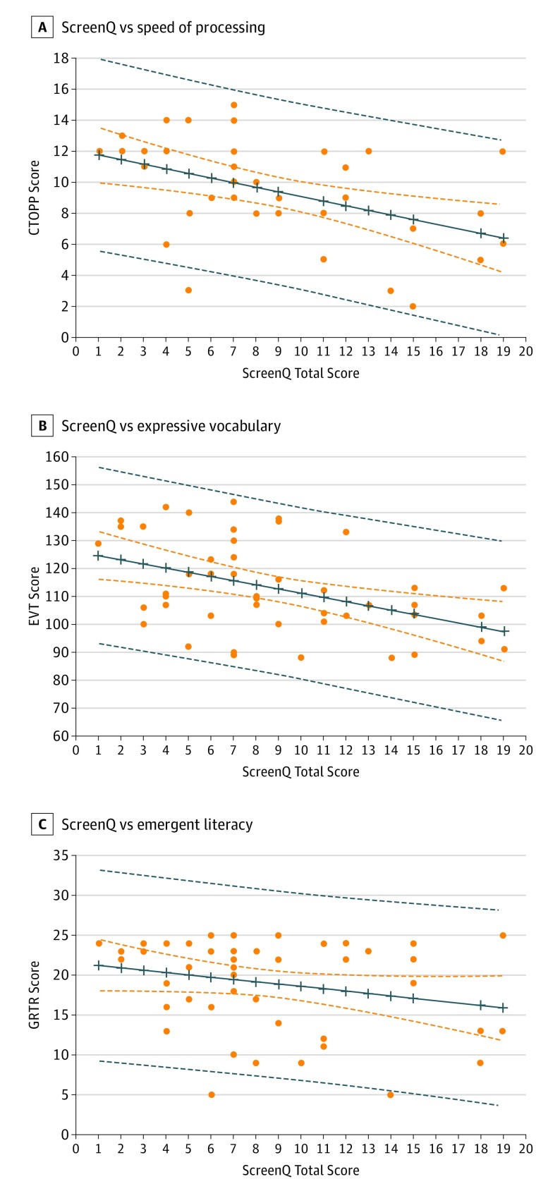
ScreenQ scores were negatively correlated with EVT-2 (F2,43 = 5.14; R2 = 0.19; P < .01), CTOPP-2 (F2,35 = 6.64; R2 = 0.28; P < .01), and GRTR (F2,44 = 17.08; R2 = 0.44; P < .01) scores, controlling for child age. These associations did not reach statistical significance when household income was included as a covariate, and this may be associated with the moderate collinearity between income level and ScreenQ scores (rρ = –0.54). Scatterplots of ScreenQ scores in comparison with EVT-2, CTOPP-2, and GRTR scores are shown in Figure 1.
Figure 1. Scatterplots of ScreenQ Survey Total Scores vs Cognitive Assessment Scores.
Solid blue line represents least squares fit; dashed blue lines, 95% CI bounds of slope; dashed orange lines, 95% prediction interval for new observations. CTOPP-2 indicates Comprehensive Test of Phonological Processing, Second Edition, Rapid Object Naming subtest; EVT, Expressive Vocabulary Test, Second Edition; GRTR, Get Ready to Read! assessment.
Diffusion Tensor Imaging
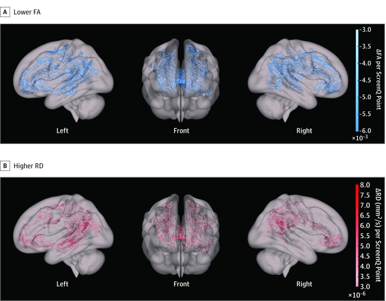
Higher ScreenQ scores were statistically significantly correlated with lower FA and higher RD in extensive aspects of white matter tracts in the whole-brain analysis, controlling for child age and household income (P < .05, FWE-corrected). Figure 2 illustrates the extent and degree of these correlations, with parameter estimates from multiple regression analyses showing the adjusted change in FA or RD per unit increase in ScreenQ score. Correlations for FA (35% vs 28%) and RD (33% vs 21%) were generally more extensive in the left hemispheric tracts compared with their contralateral counterparts, as described in Table 2. Tracts with the most extensive associations were involved with language, visual processing or imagery, executive functions, and multimodal association. The arcuate fasciculus exhibited the greatest degree of left laterality (35% vs 10% of tract for FA; 44% vs 6% for RD), consistent with its well-described role in language.56
Figure 2. Diffusion Tensor Imaging (DTI) Parameter Maps for Whole-Brain Analysis.
White matter voxels exhibit a statistically significant correlation between screen-based media use (ScreenQ scores) and lower fractional anisotropy (FA; A) as well as higher radial diffusivity (RD; B) in whole-brain analysis, controlling for child age and household income level (P < .05, familywise error–corrected). Color indicates the slope or magnitude of correlation (ie, change in the DTI parameter for every point increase in ScreenQ score).
Table 2. Extent of Tracts With Statistically Significant Correlations Between ScreenQ Scores and Diffusion Tensor Imaging Parameters in Whole-Brain Analysis.
| White Matter Tract | Regions Connected | Major Functions | FA Decrease, % | RD Increase, % | ||
|---|---|---|---|---|---|---|
| Left | Right | Left | Right | |||
| Anterior thalamic radiation | Thalamus-prefrontal | Executive | 33a | 13 | 18a | 10 |
| Cingulum (cingulate gyrus) | Limbic | Emotion, learning, memory | 28a | 18 | 25 | 22 |
| Cingulum (hippocampus) | Limbic | Emotion, learning, memory | 4 | 20a | 4 | 27a |
| Corticospinal tract | Cortex–spinal cord | Sensory-motor | 29 | 21 | 26a | 15 |
| Forceps major | Occipital-occipital | Visual processing/imagery | 33 | 39 | 38 | 33 |
| Forceps minor | Frontal-frontal | Executive | 55 | 56 | 34 | 33 |
| Inferior fronto-occipital fasciculus | Frontal-occipital/temporal | Association, language | 51 | 44 | 48a | 24 |
| Inferior longitudinal fasciculus | Occipital-temporal | Visual association/imagery, language | 38a | 21 | 48a | 6 |
| Superior longitudinal fasciculus | Frontal-occipital-temporal-parietal | Association, working memory, language | 24 | 40a | 29 | 37 |
| Arcuate fasciculus | Frontal-temporal-parietal | Language | 35a | 10 | 44a | 6 |
| Uncinate fasciculus | Orbitofrontal-temporal | Association, language (rapid naming) | 55a | 35 | 53a | 20 |
Abbreviations: FA, fractional anisotropy; RD, radial diffusivity.
Substantial lateralization of correlation (extent >50% greater than contralateral tract).
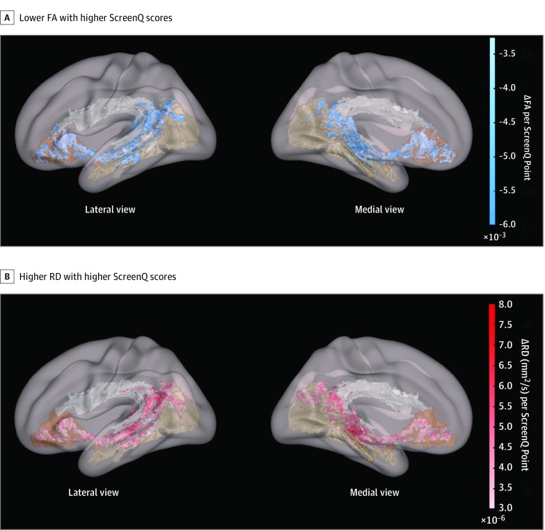
In the hypothesis-driven analysis, higher ScreenQ scores were correlated with lower FA, covering 16% of the arcuate fasciculus, 29% of the inferior longitudinal fasciculus, and 55% of the uncinate fasciculus, controlling for child age and household income (P < .05, FWE-corrected). Higher ScreenQ scores were correlated with higher RD, spanning 20% of the arcuate fasciculus, 36% of the inferior longitudinal fasciculus, and 39% of the uncinate fasciculus, controlling for age and income (P < .05, FWE-corrected). These associations are shown in Figure 3.
Figure 3. Tract-Based Spatial Statistical Analysis in the Left Hemisphere.
The tract-based spatial statistical analysis involves these 3 language- and literacy-associated tracts in the left hemisphere: arcuate fasciculus (white), inferior longitudinal fasciculus (tan), uncinate fasciculus (brown). A, Blue represents statistically significantly lower fractional anisotropy (FA) with higher ScreenQ scores. B, Red represents statistically significantly higher radial diffusivity (RD) with higher ScreenQ scores. Both analyses controlled for child age and household income level (P < .05, familywise error–corrected).
Discussion
Unprecedented technological upheaval over the past few decades has largely outpaced the ability of scientists to characterize its fueling controversy and anxiety among parents, educators, and clinicians. To our knowledge, this study is the first to describe structural neurobiological correlates of screen-based media use in preschool-aged children. The findings are in the context of well-described measures of white matter microstructural integrity, FA and RD.57 Fractional anisotropy is most clearly associated with organization of white matter in parallel bundles, whereas RD is inversely associated with degree of myelination of such bundles57 as well as axonal packing and other microstructural processes. Increases in FA and decreases in RD are consistently observed across development in older children and in response to constructive environmental stimulation, such as language exposure.21,58,59,60 This study found higher FA and lower RD associated with more screen time in major fiber tracts supporting core language and executive and emergent literacy skills, raising questions as to whether at least some aspects of screen-based media use may provide suboptimal neurodevelopmental stimulation during early childhood.
Structural markers for language performance involving FA and RD have been described in full-term and preterm children as young as 3 years.31,61 The arcuate fasciculus, which connects receptive (Wernicke) and expressive (Broca) language brain areas,29,30 is associated with language skills,31,62,63 including phonological processing and vocabulary.31,59,64 In this study, increased screen time was associated with lower FA in 16% and higher RD in 20% of the arcuate fasciculus and also with significantly lower EVT-2 and CTOPP-2 scores, which are concordant neurocognitive results. Similar associations were also seen involving the uncinate fasciculus and inferior longitudinal fasciculus, which support other aspects of language and literacy, such as semantic processing, emotional integration, and incorporation of visual imagery.30,31,33,65,66 That these associations were left-lateralized (Table 2) is consistent with reliance on language areas in the left hemisphere that increases with age.51 Lower FA and higher RD in other tracts (eg, superior longitudinal fasciculus, anterior thalamic radiation, and forceps minor) are also consistent with the reliance of language processing on a distributed, bilateral network that becomes more specialized with age.31,59,61 Although these findings are correlational in nature and do not identify causality or long-term developmental risks, they align with findings in previous studies describing the detrimental outcomes of screen time for language abilities in children,7,67 warranting further investigation.
Rapid automatized naming is a critical language and literacy skill that supports orthographic-phonological translation (eg, nonword reading) and semantic processing (eg, deriving meaning from objects, symbols, or letters) and is highly indicative of reading outcomes.68,69,70 Lesion studies have described major deficits in word and object naming involving the uncinate fasciculus, particularly in the left hemisphere.33,34 The inferior fronto-occipital fasciculus also plays a major role in rapid automatized naming, with higher FA estimating better performance.71 Substantial decreases in FA in the uncinate fasciculus (55% of the left, and 35% of the right) and fronto-occipital fasciculus (44% of the extent of the left, and 34% of the right) in the context of lower CTOPP-2 scores are concordant neurocognitive findings associated with greater screen use. In addition, rapid automatized naming is intimately associated with broader executive functions, including speed of processing and working memory,72,73 which are supported by other tracts associated with greater use in the whole-brain analyses (superior longitudinal fasciculus, forceps minor, and anterior thalamic tracts).74,75 Although broader executive functions were not directly assessed in this study, the associations between higher screen use and lower FA and higher RD for each of these tracts are consistent with deleterious associations that have been previously described in young children.10,19,76
The children in this study were not yet attending school and not yet reading independently, but we believe these findings are highly relevant within the construct of emergent literacy, an incremental process beginning in infancy that involves skills, knowledge, and attitudes required for reading and writing.22,77 Emergent literacy development has been associated with increases in FA and decreases in RD in the arcuate fasciculus,62 whereas lower FA in the arcuate fasciculus, inferior longitudinal fasciculus, and superior longitudinal fasciculus has been associated with lower prereading (particularly phonological) skills.49,78 Neural microstructure involving frontal, thalamic, parietal, and temporal areas (eg, through arcuate fasciculus or superior longitudinal fasciculus) has also been shown to be a factor in reading outcomes.79,80,81 Associations between lower FA and higher RD in the arcuate fasciculus, inferior longitudinal fasciculus, and other tracts with higher ScreenQ scores, in the context of negative correlation between ScreenQ and GRTR scores, are concordant findings related to this multifaceted skill set.
Although establishing causation is beyond the scope of this study, a critical question is whether neurobiological differences are directly associated with properties of screen-based media itself or indirectly associated with differences in human interactive (eg, shared reading) time, which tends to decrease with greater use.12,13,14 Aspects of both associations seem likely, as suggested by a recent MRI study of children aged 8 to 12 years that found lower functional connectivity within the emerging reading network was correlated with higher screen time and higher functional connectivity was correlated with more reading time.18 Given the prevalence of “learning” applications marketed for young children and the emphasis on technology in early childhood educational curricula,3 multiyear studies of emergent literacy outcomes are warranted to determine long-term implications.
Limitations and Strengths
This study has several limitations. The sample largely comprised families with high socioeconomic status, yet household income was accounted for in the DTI analyses. Associations between ScreenQ survey and cognitive testing scores did not meet the threshold for statistical significance when income was included in the model, which may be associated with the moderate collinearity between income level and ScreenQ scores that negatively affected statistical power, particularly given the small sample size. Both behavioral and DTI findings may have been even more robust in families with higher socioeconomic status compared with families with lower socioeconomic status, for whom language, literacy, and other cognitive disparities are well documented.82,83
Although exhibiting strong psychometric properties,35 ScreenQ is a new measure reliant on parent reporting and thus is subject to social desirability bias. However, no similar composite measure or direct assessment currently exists, and ScreenQ reflects domains in the current AAP recommendations.5 Although DTI is a powerful method for studying white matter in the brain, it is vulnerable to artifacts attributable to nonuniform directionality of fiber tracts, such as crossing fibers.28 However, the analyses involved well-defined tracts and a hypothesis-driven approach, and FA and RD findings were consistent with results in the current behavioral and neuroimaging literature and with each other. Diffusion-weighted images were acquired at the end of the MRI session, resulting in a lower success rate (68%) than from recent work,17 possibly conferring bias toward older children with higher attention skills. However, DTI is a task-free paradigm that is not affected by performance factors. Although important, the cross-sectional nature of this study precludes the discernment of causation or whether the results are attributable to screen time or an indirect effect of differences in reading, language, or other constructive experiences.3,28
This study also has strengths. The sample of preschool-aged children is remarkable given the difficulty of successfully conducting MRI at this age. The DTI results survived stringent correction for motion and multiple comparisons, controlling for child age and household income, and were concordant with behavioral results controlling for child age. Rather than apply a single screen time variable, such as minutes per day or violent content as is often the case,84,85 ScreenQ incorporated domains of use into a coherent composite, providing a more holistic view of use.35 The analyses were driven by a hypothesis based on recent neuroimaging evidence and AAP guidelines, including characterization of associations for a broad range of tracts supporting relevant cognitive abilities and focused analyses with increased statistical power. The DTI findings aligned with those on validated cognitive measures, providing compelling insights into the implications for child development, preschool education, and further research.
Although preliminary, this study fills a gap in evidence and provides a novel, neurobiological lens through which to view the recommendations of the AAP1,3 and World Health Organization15 in terms of readiness of brain networks to constructively process screen-based media and the marketing of such media to children. Given the results of this study, we can speculate that before age 5 years, when brain networks are developing rapidly,86,87 caution is warranted, noting the postulate attributed to Donald Hebb: “neurons that fire together, wire together.”42 We believe multiyear studies are needed to explore whether the associations observed in this study, related to language and literacy abilities, reflect longer-term risks and outcomes. Such studies are critical in the context of a child’s early cognitive ecosystem, in which unprecedented technological shifts may change how children live, connect, and learn.
Conclusions
In this study of 47 preschool-aged children, increased use of screen-based media in the context of the AAP guidelines was associated with lower microstructural integrity of brain white matter tracts that support language, executive functions, and emergent literacy skills, controlling for child age and household income. Screen use was also associated with lower scores on corresponding behavioral measures, controlling for age. Given that screen-based media use is ubiquitous and increasing in children in home, childcare, and school settings, these findings suggest the need for further study to identify the implications for the developing brain, particularly during stages of dynamic brain growth in early childhood.
References
- 1.Groundbreaking study examines effects of screen time on kids [transcript]. 60 Minutes CBS television. December 9, 2018.
- 2.Carter B, Rees P, Hale L, Bhattacharjee D, Paradkar MS. Association between portable screen-based media device access or use and sleep outcomes: a systematic review and meta-analysis. JAMA Pediatr. 2016;170(12):-. doi: 10.1001/jamapediatrics.2016.2341 [DOI] [PMC free article] [PubMed] [Google Scholar]
- 3.Rideout V. The Common Sense Census: Media Use by Kids Age Zero to Eight. San Francisco, CA: Common Sense Media; 2017. [Google Scholar]
- 4.Hutton JS, Lin L, Gruber R, et al. Shared reading and television across the perinatal period in low-SES households. Clin Pediatr (Phila). 2018;57(8):904-912. doi: 10.1177/0009922817737077 [DOI] [PubMed] [Google Scholar]
- 5.AAP Council on Communications and Media Media and Young Minds. Elk Grove Village, IL: American Academy of Pediatrics; 2016. [Google Scholar]
- 6.Moreno MA, Chassiakos YR, Cross C; Council on Communications and Media. Media use in school-aged children and adolescents Pediatrics. 2016;138(5). [DOI] [PubMed] [Google Scholar]
- 7.Anderson DR, Subrahmanyam K; Cognitive Impacts of Digital Media Workgroup . Digital screen media and cognitive development. Pediatrics. 2017;140(suppl 2):S57-S61. doi: 10.1542/peds.2016-1758C [DOI] [PubMed] [Google Scholar]
- 8.Ma J, Birken C Handheld screen time linked with speech delays in young children. Paper presented at: Pediatric Academic Societies Meeting; May 6, 2017; San Francisco, CA. https://www.aappublications.org/news/2017/05/04/PASScreenTime050417. Accessed April 2019 [Google Scholar]
- 9.Garrison MM, Liekweg K, Christakis DA. Media use and child sleep: the impact of content, timing, and environment. Pediatrics. 2011;128(1):29-35. doi: 10.1542/peds.2010-3304 [DOI] [PMC free article] [PubMed] [Google Scholar]
- 10.Lillard AS, Li H, Boguszewski K. Television and children’s executive function. Adv Child Dev Behav. 2015;48:219-248. doi: 10.1016/bs.acdb.2014.11.006 [DOI] [PubMed] [Google Scholar]
- 11.Walsh JJ, Barnes JD, Cameron JD, et al. Associations between 24 hour movement behaviours and global cognition in US children: a cross-sectional observational study. Lancet Child Adolesc Health. 2018;2(11):783-791. doi: 10.1016/S2352-4642(18)30278-5 [DOI] [PMC free article] [PubMed] [Google Scholar]
- 12.Choi JH, Mendelsohn AL, Weisleder A, et al. Real-world usage of educational media does not promote parent-child cognitive stimulation activities. Acad Pediatr. 2018;18(2):172-178. doi: 10.1016/j.acap.2017.04.020 [DOI] [PMC free article] [PubMed] [Google Scholar]
- 13.Tomopoulos S, Valdez PT, Dreyer BP, et al. Is exposure to media intended for preschool children associated with less parent-child shared reading aloud and teaching activities? Ambul Pediatr. 2007;7(1):18-24. doi: 10.1016/j.ambp.2006.10.005 [DOI] [PubMed] [Google Scholar]
- 14.Mendelsohn AL, Berkule SB, Tomopoulos S, et al. Infant television and video exposure associated with limited parent-child verbal interactions in low socioeconomic status households. Arch Pediatr Adolesc Med. 2008;162(5):411-417. doi: 10.1001/archpedi.162.5.411 [DOI] [PMC free article] [PubMed] [Google Scholar]
- 15.World Health Organization WHO Guidelines on Physical Activity, Sedentary Behaviour and Sleep for Children Under 5 Years of Age. Geneva, Switzerland: World Health Organization; 2019. [PubMed] [Google Scholar]
- 16.Paulus MP, Squeglia LM, Bagot K, et al. Screen media activity and brain structure in youth: evidence for diverse structural correlation networks from the ABCD study. Neuroimage. 2019;185:140-153. doi: 10.1016/j.neuroimage.2018.10.040 [DOI] [PMC free article] [PubMed] [Google Scholar]
- 17.Hutton JS, Dudley J, Horowitz-Kraus T, DeWitt T, Holland SK. Differences in functional brain network connectivity during stories presented in audio, illustrated, and animated format in preschool-age children [published online October 30, 2018]. Brain Imaging Behav. doi: 10.1007/s11682-018-9985-y [DOI] [PubMed] [Google Scholar]
- 18.Horowitz-Kraus T, Hutton JS. Brain connectivity in children is increased by the time they spend reading books and decreased by the length of exposure to screen-based media. Acta Paediatr. 2018;107(4):685-693. doi: 10.1111/apa.14176 [DOI] [PubMed] [Google Scholar]
- 19.Zivan M, Bar S, Jing X, Hutton J, Farah R, Horowitz-Kraus T. Screen-exposure and altered brain activation related to attention in preschool children: An EEG study. Trends Neurosci Educ. 2019;17: 1-5. doi: 10.1016/j.tine.2019.100117 [DOI] [PubMed] [Google Scholar]
- 20.Rohr CS, Arora A, Cho IYK, et al. Functional network integration and attention skills in young children. Dev Cogn Neurosci. 2018;30:200-211. doi: 10.1016/j.dcn.2018.03.007 [DOI] [PMC free article] [PubMed] [Google Scholar]
- 21.Sousa SS, Amaro E Jr, Crego A, Gonçalves ÓF, Sampaio A. Developmental trajectory of the prefrontal cortex: a systematic review of diffusion tensor imaging studies. Brain Imaging Behav. 2018;12(4):1197-1210. doi: 10.1007/s11682-017-9761-4 [DOI] [PubMed] [Google Scholar]
- 22.Horowitz-Kraus T, Hutton JS. From emergent literacy to reading: how learning to read changes a child's brain. Acta Paediatr. 2015;104(7):648-656. doi: 10.1111/apa.13018 [DOI] [PubMed] [Google Scholar]
- 23.Dehaene S, Cohen L, Morais J, Kolinsky R. Illiterate to literate: behavioural and cerebral changes induced by reading acquisition. Nat Rev Neurosci. 2015;16(4):234-244. doi: 10.1038/nrn3924 [DOI] [PubMed] [Google Scholar]
- 24.Yeatman JD, Dougherty RF, Ben-Shachar M, Wandell BA. Development of white matter and reading skills. Proc Natl Acad Sci U S A. 2012;109(44):E3045-E3053. doi: 10.1073/pnas.1206792109 [DOI] [PMC free article] [PubMed] [Google Scholar]
- 25.Keller TA, Just MA. Altering cortical connectivity: remediation-induced changes in the white matter of poor readers. Neuron. 2009;64(5):624-631. doi: 10.1016/j.neuron.2009.10.018 [DOI] [PMC free article] [PubMed] [Google Scholar]
- 26.Forbes TA, Gallo V. All wrapped up: environmental effects on myelination. Trends Neurosci. 2017;40(9):572-587. doi: 10.1016/j.tins.2017.06.009 [DOI] [PMC free article] [PubMed] [Google Scholar]
- 27.Qiu A, Mori S, Miller MI. Diffusion tensor imaging for understanding brain development in early life. Annu Rev Psychol. 2015;66:853-876. doi: 10.1146/annurev-psych-010814-015340 [DOI] [PMC free article] [PubMed] [Google Scholar]
- 28.Soares JM, Marques P, Alves V, Sousa N. A hitchhiker’s guide to diffusion tensor imaging. Front Neurosci. 2013;7:31. doi: 10.3389/fnins.2013.00031 [DOI] [PMC free article] [PubMed] [Google Scholar]
- 29.Friederici AD. White-matter pathways for speech and language processing. Handb Clin Neurol. 2015;129:177-186. doi: 10.1016/B978-0-444-62630-1.00010-X [DOI] [PubMed] [Google Scholar]
- 30.Friederici AD, Gierhan SM. The language network. Curr Opin Neurobiol. 2013;23(2):250-254. doi: 10.1016/j.conb.2012.10.002 [DOI] [PubMed] [Google Scholar]
- 31.Dodson CK, Travis KE, Borchers LR, Marchman VA, Ben-Shachar M, Feldman HM. White matter properties associated with pre-reading skills in 6-year-old children born preterm and at term. Dev Med Child Neurol. 2018;60(7):695-702. doi: 10.1111/dmcn.13783 [DOI] [PMC free article] [PubMed] [Google Scholar]
- 32.Jouen AL, Ellmore TM, Madden-Lombardi CJ, Pallier C, Dominey PF, Ventre-Dominey J. Beyond the word and image: II- structural and functional connectivity of a common semantic system. Neuroimage. 2018;166:185-197. doi: 10.1016/j.neuroimage.2017.10.039 [DOI] [PubMed] [Google Scholar]
- 33.Sierpowska J, Gabarrós A, Fernández-Coello A, et al. White-matter pathways and semantic processing: intrasurgical and lesion-symptom mapping evidence. Neuroimage Clin. 2019;22:101704. doi: 10.1016/j.nicl.2019.101704 [DOI] [PMC free article] [PubMed] [Google Scholar]
- 34.Papagno C. Naming and the role of the uncinate fasciculus in language function. Curr Neurol Neurosci Rep. 2011;11(6):553-559. doi: 10.1007/s11910-011-0219-6 [DOI] [PubMed] [Google Scholar]
- 35.Hutton JS, Huang G, Sahay R, DeWitt T. Ittenbach RF Assessment of screen-based media use in children: preliminary validation of the ScreenQ. Paper presented at: Pediatric Academic Societies Meeting; April 30, 2019; Baltimore, MD [Google Scholar]
- 36.Harris PA, Taylor R, Thielke R, Payne J, Gonzalez N, Conde JG. Research electronic data capture (REDCap)—a metadata-driven methodology and workflow process for providing translational research informatics support. J Biomed Inform. 2009;42(2):377-381. doi: 10.1016/j.jbi.2008.08.010 [DOI] [PMC free article] [PubMed] [Google Scholar]
- 37.Hutton JS, Xu Y, DeWitt T, Ittenbach RF Assessment of screen-based media use in children: development and psychometric refinement of the ScreenQ. Paper presented at: Pediatric Academic Societies Meeting; May 4, 2018; Toronto, Canada [Google Scholar]
- 38.Bellevue Project for Early Language Literacy and Education Success STIMQ Home Cognitive Environment. http://pediatrics.med.nyu.edu/developmental/research/the-belle-project/stimq-cognitive-home-environment. Accessed March 2019.
- 39.Williams KT. Expressive Vocabulary Test, Second Edition (EVT-2) Minneapolis, MN: Pearson Assessments; 2007. [Google Scholar]
- 40.Mitchell J. Comprehensive test of phonological processing (CTOPP). Assess Eff Interv. 2001;26(3):57-63. doi: 10.1177/073724770102600305 [DOI] [Google Scholar]
- 41.Wagner RK, Torgesen JK, Rashotte CA, Pearson NA. CTOPP-2: Comprehensive Test of Phonological Processing–Second Edition Austin, TX: Pro-Ed; 2013. https://www.proedinc.com/Products/13080/ctopp2-comprehensive-test-of-phonological-processingsecond-edition.aspx. Accessed March 2019.
- 42.Hebb D. The Organization of Behavior. A Neuropsychological Theory. New York, NY: Wiley; 1949. [Google Scholar]
- 43.Vannest J, Rajagopal A, Cicchino ND, et al. ; CMIND Authorship Consortium . Factors determining success of awake and asleep magnetic resonance imaging scans in nonsedated children. Neuropediatrics. 2014;45(6):370-377. doi: 10.1055/s-0034-1387816 [DOI] [PubMed] [Google Scholar]
- 44.Andersson JLR, Sotiropoulos SN. An integrated approach to correction for off-resonance effects and subject movement in diffusion MR imaging. Neuroimage. 2016;125:1063-1078. doi: 10.1016/j.neuroimage.2015.10.019 [DOI] [PMC free article] [PubMed] [Google Scholar]
- 45.Andersson JLR, Graham MS, Zsoldos E, Sotiropoulos SN. Incorporating outlier detection and replacement into a non-parametric framework for movement and distortion correction of diffusion MR images. Neuroimage. 2016;141:556-572. doi: 10.1016/j.neuroimage.2016.06.058 [DOI] [PubMed] [Google Scholar]
- 46.Basser PJ, Mattiello J, LeBihan D. MR diffusion tensor spectroscopy and imaging. Biophys J. 1994;66(1):259-267. doi: 10.1016/S0006-3495(94)80775-1 [DOI] [PMC free article] [PubMed] [Google Scholar]
- 47.Smith SM, Jenkinson M, Johansen-Berg H, et al. Tract-based spatial statistics: voxelwise analysis of multi-subject diffusion data. Neuroimage. 2006;31(4):1487-1505. doi: 10.1016/j.neuroimage.2006.02.024 [DOI] [PubMed] [Google Scholar]
- 48.Rojkova K, Volle E, Urbanski M, Humbert F, Dell’Acqua F, Thiebaut de Schotten M. Atlasing the frontal lobe connections and their variability due to age and education: a spherical deconvolution tractography study. Brain Struct Funct. 2016;221(3):1751-1766. doi: 10.1007/s00429-015-1001-3 [DOI] [PubMed] [Google Scholar]
- 49.Yeatman JD, Dougherty RF, Rykhlevskaia E, et al. Anatomical properties of the arcuate fasciculus predict phonological and reading skills in children. J Cogn Neurosci. 2011;23(11):3304-3317. doi: 10.1162/jocn_a_00061 [DOI] [PMC free article] [PubMed] [Google Scholar]
- 50.Panesar SS, Yeh FC, Jacquesson T, Hula W, Fernandez-Miranda JC. A quantitative tractography study into the connectivity, segmentation and laterality of the human inferior longitudinal fasciculus. Front Neuroanat. 2018;12:47. doi: 10.3389/fnana.2018.00047 [DOI] [PMC free article] [PubMed] [Google Scholar]
- 51.Holland SK, Vannest J, Mecoli M, et al. Functional MRI of language lateralization during development in children. Int J Audiol. 2007;46(9):533-551. doi: 10.1080/14992020701448994 [DOI] [PMC free article] [PubMed] [Google Scholar]
- 52.Winkler AM, Ridgway GR, Webster MA, Smith SM, Nichols TE. Permutation inference for the general linear model. Neuroimage. 2014;92:381-397. doi: 10.1016/j.neuroimage.2014.01.060 [DOI] [PMC free article] [PubMed] [Google Scholar]
- 53.Smith SM, Nichols TE. Threshold-free cluster enhancement: addressing problems of smoothing, threshold dependence and localisation in cluster inference. Neuroimage. 2009;44(1):83-98. doi: 10.1016/j.neuroimage.2008.03.061 [DOI] [PubMed] [Google Scholar]
- 54.Hua K, Zhang J, Wakana S, et al. Tract probability maps in stereotaxic spaces: analyses of white matter anatomy and tract-specific quantification. Neuroimage. 2008;39(1):336-347. doi: 10.1016/j.neuroimage.2007.07.053 [DOI] [PMC free article] [PubMed] [Google Scholar]
- 55.United States Census Bureau Income and Poverty in the United States: 2017. Washington, DC: US Department of Commerce; 2018. [Google Scholar]
- 56.Skeide MA, Brauer J, Friederici AD. Brain functional and structural predictors of language performance. Cereb Cortex. 2016;26(5):2127-2139. doi: 10.1093/cercor/bhv042 [DOI] [PubMed] [Google Scholar]
- 57.Winklewski PJ, Sabisz A, Naumczyk P, Jodzio K, Szurowska E, Szarmach A. Understanding the physiopathology behind axial and radial diffusivity changes—what do we know? Front Neurol. 2018;9:92. doi: 10.3389/fneur.2018.00092 [DOI] [PMC free article] [PubMed] [Google Scholar]
- 58.Krogsrud SK, Fjell AM, Tamnes CK, et al. Changes in white matter microstructure in the developing brain—a longitudinal diffusion tensor imaging study of children from 4 to 11 years of age. Neuroimage. 2016;124(pt A):473-486. doi: 10.1016/j.neuroimage.2015.09.017 [DOI] [PMC free article] [PubMed] [Google Scholar]
- 59.Romeo RR, Segaran J, Leonard JA, et al. Language exposure relates to structural neural connectivity in childhood. J Neurosci. 2018;38(36):7870-7877. doi: 10.1523/JNEUROSCI.0484-18.2018 [DOI] [PMC free article] [PubMed] [Google Scholar]
- 60.Reynolds JE, Grohs MN, Dewey D, Lebel C. Global and regional white matter development in early childhood. Neuroimage. 2019;196(Apr):49-58. doi: 10.1016/j.neuroimage.2019.04.004 [DOI] [PubMed] [Google Scholar]
- 61.Walton M, Dewey D, Lebel C. Brain white matter structure and language ability in preschool-aged children. Brain Lang. 2018;176:19-25. doi: 10.1016/j.bandl.2017.10.008 [DOI] [PubMed] [Google Scholar]
- 62.Thiebaut de Schotten M, Cohen L, Amemiya E, Braga LW, Dehaene S. Learning to read improves the structure of the arcuate fasciculus. Cereb Cortex. 2014;24(4):989-995. doi: 10.1093/cercor/bhs383 [DOI] [PubMed] [Google Scholar]
- 63.Smits M, Jiskoot LC, Papma JM. White matter tracts of speech and language. Semin Ultrasound CT MR. 2014;35(5):504-516. doi: 10.1053/j.sult.2014.06.008 [DOI] [PubMed] [Google Scholar]
- 64.Lebel C, Beaulieu C. Lateralization of the arcuate fasciculus from childhood to adulthood and its relation to cognitive abilities in children. Hum Brain Mapp. 2009;30(11):3563-3573. doi: 10.1002/hbm.20779 [DOI] [PMC free article] [PubMed] [Google Scholar]
- 65.Teichmann M, Rosso C, Martini JB, et al. A cortical-subcortical syntax pathway linking Broca’s area and the striatum. Hum Brain Mapp. 2015;36(6):2270-2283. doi: 10.1002/hbm.22769 [DOI] [PMC free article] [PubMed] [Google Scholar]
- 66.Von Der Heide RJ, Skipper LM, Klobusicky E, Olson IR. Dissecting the uncinate fasciculus: disorders, controversies and a hypothesis. Brain. 2013;136(Pt 6):1692-1707. doi: 10.1093/brain/awt094 [DOI] [PMC free article] [PubMed] [Google Scholar]
- 67.Zimmerman FJ, Christakis DA, Meltzoff AN. Associations between media viewing and language development in children under age 2 years. J Pediatr. 2007;151(4):364-368. doi: 10.1016/j.jpeds.2007.04.071 [DOI] [PubMed] [Google Scholar]
- 68.Norton ES, Wolf M. Rapid automatized naming (RAN) and reading fluency: implications for understanding and treatment of reading disabilities. Annu Rev Psychol. 2012;63:427-452. doi: 10.1146/annurev-psych-120710-100431 [DOI] [PubMed] [Google Scholar]
- 69.Hulme C, Snowling MJ. Reading disorders and dyslexia. Curr Opin Pediatr. 2016;28(6):731-735. doi: 10.1097/MOP.0000000000000411 [DOI] [PMC free article] [PubMed] [Google Scholar]
- 70.Lohvansuu K, Hämäläinen JA, Ervast L, Lyytinen H, Leppänen PHT. Longitudinal interactions between brain and cognitive measures on reading development from 6 months to 14 years. Neuropsychologia. 2018;108:6-12. doi: 10.1016/j.neuropsychologia.2017.11.018 [DOI] [PubMed] [Google Scholar]
- 71.Rollans C, Cheema K, Georgiou GK, Cummine J. Pathways of the inferior frontal occipital fasciculus in overt speech and reading. Neuroscience. 2017;364:93-106. doi: 10.1016/j.neuroscience.2017.09.011 [DOI] [PubMed] [Google Scholar]
- 72.Papadopoulos TC, Spanoudis GC, Georgiou GK. How is RAN related to reading fluency? a comprehensive examination of the prominent theoretical accounts. Front Psychol. 2016;7:1217. doi: 10.3389/fpsyg.2016.01217 [DOI] [PMC free article] [PubMed] [Google Scholar]
- 73.McCabe DP, Roediger HL, McDaniel MA, Balota DA, Hambrick DZ. The relationship between working memory capacity and executive functioning: evidence for a common executive attention construct. Neuropsychology. 2010;24(2):222-243. doi: 10.1037/a0017619 [DOI] [PMC free article] [PubMed] [Google Scholar]
- 74.Smolker HR, Friedman NP, Hewitt JK, Banich MT. Neuroanatomical correlates of the unity and diversity model of executive function in young adults. Front Hum Neurosci. 2018;12:283. doi: 10.3389/fnhum.2018.00283 [DOI] [PMC free article] [PubMed] [Google Scholar]
- 75.Mamiya PC, Richards TL, Kuhl PK. Right forceps minor and anterior thalamic radiation predict executive function skills in young bilingual adults. Front Psychol. 2018;9:118. doi: 10.3389/fpsyg.2018.00118 [DOI] [PMC free article] [PubMed] [Google Scholar]
- 76.Nathanson AI, Aladé F, Sharp ML, Rasmussen EE, Christy K. The relation between television exposure and executive function among preschoolers. Dev Psychol. 2014;50(5):1497-1506. doi: 10.1037/a0035714 [DOI] [PubMed] [Google Scholar]
- 77.Whitehurst GJ, Lonigan CJ. Child development and emergent literacy. Child Dev. 1998;69(3):848-872. doi: 10.1111/j.1467-8624.1998.tb06247.x [DOI] [PubMed] [Google Scholar]
- 78.Saygin ZM, Norton ES, Osher DE, et al. Tracking the roots of reading ability: white matter volume and integrity correlate with phonological awareness in prereading and early-reading kindergarten children. J Neurosci. 2013;33(33):13251-13258. doi: 10.1523/JNEUROSCI.4383-12.2013 [DOI] [PMC free article] [PubMed] [Google Scholar]
- 79.Borchers LR, Bruckert L, Dodson CK, et al. Microstructural properties of white matter pathways in relation to subsequent reading abilities in children: a longitudinal analysis. Brain Struct Funct. 2019;224(2):891-905. doi: 10.1007/s00429-018-1813-z [DOI] [PMC free article] [PubMed] [Google Scholar]
- 80.Lebel C, Shaywitz B, Holahan J, Shaywitz S, Marchione K, Beaulieu C. Diffusion tensor imaging correlates of reading ability in dysfluent and non-impaired readers. Brain Lang. 2013;125(2):215-222. doi: 10.1016/j.bandl.2012.10.009 [DOI] [PubMed] [Google Scholar]
- 81.Rimrodt SL, Peterson DJ, Denckla MB, Kaufmann WE, Cutting LE. White matter microstructural differences linked to left perisylvian language network in children with dyslexia. Cortex. 2010;46(6):739-749. doi: 10.1016/j.cortex.2009.07.008 [DOI] [PMC free article] [PubMed] [Google Scholar]
- 82.Hart B, Risley T. Meaningful Differences in the Everyday Experience of Young American Children. Baltimore, MD: Paul Brookes Publishing Co; 1995. [Google Scholar]
- 83.Larson K, Russ SA, Nelson BB, Olson LM, Halfon N. Cognitive ability at kindergarten entry and socioeconomic status. Pediatrics. 2015;135(2):e440-e448. doi: 10.1542/peds.2014-0434 [DOI] [PubMed] [Google Scholar]
- 84.Lissak G. Adverse physiological and psychological effects of screen time on children and adolescents: literature review and case study. Environ Res. 2018;164:149-157. doi: 10.1016/j.envres.2018.01.015 [DOI] [PubMed] [Google Scholar]
- 85.Christakis DA, Zimmerman FJ. Violent television viewing during preschool is associated with antisocial behavior during school age. Pediatrics. 2007;120(5):993-999. doi: 10.1542/peds.2006-3244 [DOI] [PubMed] [Google Scholar]
- 86.Knudsen EI. Sensitive periods in the development of the brain and behavior. J Cogn Neurosci. 2004;16(8):1412-1425. doi: 10.1162/0898929042304796 [DOI] [PubMed] [Google Scholar]
- 87.Petrican R, Taylor MJ, Grady CL. Trajectories of brain system maturation from childhood to older adulthood: implications for lifespan cognitive functioning. Neuroimage. 2017;163:125-149. doi: 10.1016/j.neuroimage.2017.09.025 [DOI] [PubMed] [Google Scholar]


