Significance
Human brain function is often studied through group-averaging, yielding valuable insight into typical organization and differences across special populations. However, cortical organization is known to vary across individuals. Here, we characterize brain regions that differ from group network organization (“network variants”). We find that network variants are present in all individuals, are stable across time within a person, and are related to functional variation in tasks. We further observe that there are separable subgroups of individuals who share common distributions of variants and differ in behavior. Our study suggests that distributions of individual differences in brain networks have a trait-like nature and sets up future investigations of their neurobiological sources and their link to stable behavioral traits.
Keywords: functional connectivity, resting-state, individual differences, networks
Abstract
Resting-state functional magnetic resonance imaging (fMRI) has provided converging descriptions of group-level functional brain organization. Recent work has revealed that functional networks identified in individuals contain local features that differ from the group-level description. We define these features as network variants. Building on these studies, we ask whether distributions of network variants reflect stable, trait-like differences in brain organization. Across several datasets of highly-sampled individuals we show that 1) variants are highly stable within individuals, 2) variants are found in characteristic locations and associate with characteristic functional networks across large groups, 3) task-evoked signals in variants demonstrate a link to functional variation, and 4) individuals cluster into subgroups on the basis of variant characteristics that are related to differences in behavior. These results suggest that distributions of network variants may reflect stable, trait-like, functionally relevant individual differences in functional brain organization.
Identifying the nature of individual variability in human brain function is a central question in many fields of study, including psychology, psychiatry, neurology, and neuroscience. Many human neuroimaging studies have identified stable, meaningful individual differences in functional activations during task performance (1–5) or volumetric differences (e.g., refs. 6 and 7) within specific brain regions. However, a number of recent investigations have revealed substantial individual variability while subjects are at rest not only in single regions, but also in large-scale networks throughout the brain (8–16). Here, we examine the characteristics of these individual differences in brain networks, asking if they are stable and systematic features of individual brain organization. Furthermore, we investigate if the distributions of these differences within an individual have trait-like aspects that might be linked to trait-like individual differences in behavior.
Large-scale functional brain networks are composed of distributed brain areas that demonstrate correlated fluctuations in their spontaneous (resting-state) activity measured using functional magnetic resonance imaging (fMRI). Over the last decade convergent descriptions of canonical functional network organization of the human brain have emerged from fMRI studies (17, 18). These efforts have revealed that functional networks map onto known large-scale brain systems, including the motor (19), auditory (20), and visual systems (21), as well as higher-level systems, such as those for executive control (22). Furthermore, regions within the same functional network tend to coactivate during tasks (23).
Most of the aforementioned studies have analyzed data from large groups of typical adults averaged together in order to delineate group-level descriptions of network organization (17, 18). However, several recent investigations have revealed variability in functional network organization across individuals (8, 9, 11, 12), including observations that highly sampled individuals show focal deviations from the group-level description (10, 13, 14). We refer to these individual-specific deviations in functional network organization as “network variants.”
Natural questions raised by the observation of variants are whether individual differences in functional brain organization relate systematically to individual differences in function. Here, we ask specifically whether: 1) Network variants exhibit stability over time within an individual, 2) network variants have systematic spatial distributions or functional network associations, 3) individuals separate into subgroups with different distributions of variants, and 4) aspects of network variants relate to individual differences in brain function and behavior. These questions seek to address the trait-like nature of distributions of individual differences in brain organization.
We investigate these questions using 3 datasets, one composed of 10 highly sampled individuals from the Midnight Scan Club (MSC) (13), a second of a single individual scanned over the course of a year called the MyConnectome dataset (24), and the third including 384 unrelated individuals with high-quality data from the Human Connectome Project (HCP) (see Materials and Methods for exclusion criteria) (25). Furthermore, we split the HCP dataset into 2 matched samples for within-study replication. Together, these datasets allow us to examine both the within-individual stability of network variants as well as the distribution of network variants across larger samples.
Results
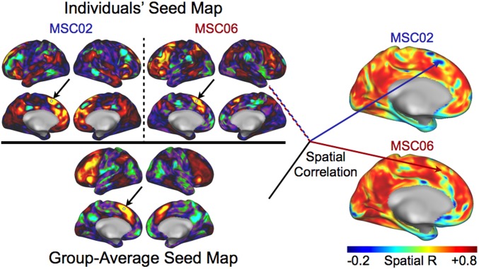
We compared individual resting-state functional correlations (rsFC) to a group average across the entire cortex. We found that most of the brain in individuals shows moderate to high correspondence with group-average rsFC, with a few locations showing large deviations, as in ref. 10. We defined network variants as the locations where individuals’ rsFC differs substantially from the group average (Fig. 1). Our goal was to examine the nature of these network variants and to determine if they relate to brain function.
Fig. 1.
Identification of network variants. We computed a spatial correlation between an individual’s seed map and the group-average seed map at every vertex on the cortical surface. An example is shown here for a seed in the dorsal medial frontal cortex (white seed indicated by the black arrows). We compare the pattern of correlations for subject MSC02 with the group average and the pattern of correlations for subject MSC06 with the group average. Notably, MSC02’s seed map differs substantially from the group average, while MSC06’s seed map agrees well. Hence, the spatial correlation at that vertex is low in MSC02 (blue arrow, top brain, Right) and high in MSC06 (red arrow, bottom brain, Right). Network variants are defined as contiguous cortical regions where this spatial correlation measure is low (dark blue areas on the brains, Right), excluding brain areas with low signal (see Materials and Methods for additional details).
Network Variants Are Present and Reliable in Individuals.
Network variants (Fig. 1) were observed in all individuals included in the study, in the MSC, MyConnectome, and HCP datasets (SI Appendix, Fig. S1). All individuals have at least one brain region with low similarity to the group average (defined as r < 0.15 rather than lowest decile for this analysis only). Thus, network variants appear to be a common phenomenon, not just an idiosyncrasy of a few individuals. However, the location, size, and network assignments of variants differed across individuals, as will be described in more detail below.
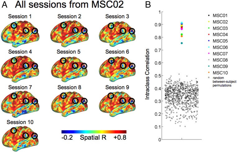
Next, we asked if network variants were stable within an individual, rather than reflecting measurement noise, state change, or sampling variability. We examined session-to-session variability of variants in the MSC dataset. For each individual, 10 separate 30-min resting-state sessions were available (collected over 3 wk). The spatial correlation map was robust across sessions (see example from MSC02 in Fig. 2A), with high (>0.75) intraclass correlations (ICC) across sessions for 9 of the 10 individuals, and the distribution of randomly sampled between-subject ICCs was substantially lower (Fig. 2B; similar results were found with binarized network variants, as shown in SI Appendix, Fig. S2). The individual with a relatively low ICC (0.44 for MSC08) had a substantial amount of high-motion data and self-reported sleeping during extensive portions of data acquisition, as previously described (13, 26). Thus, this subject was excluded from all further analyses. Furthermore, we found that network variants were stable over a year in the individual from the MyConnectome dataset (SI Appendix, Fig. S3), which is a more ecologically valid timeframe. Finally, we examined the amount of data required to identify network variants reliably (SI Appendix, Fig. S4), and demonstrated that ∼40 min of high-quality (low-motion) data are needed (10, 13).
Fig. 2.
Within-subject reliability of network variants. (A) The spatial correlation values at each cortical surface vertex for all 10 independent resting-state fMRI sessions from subject MSC02 are shown. Locations with low spatial correlations correspond to network variants (e.g., black circles; similar results were seen for the medial surface and right hemisphere). (B) The ICC of the spatial correlation maps (for the entire cerebral cortex) computed across each session within each individual in the MSC dataset is shown. The ICC reflects the test–retest reliability of network variants identified via data from each session independently for an individual. The open black circles represent the correlation between 2 randomly selected spatial correlation maps from different subjects (one session per subject; 1,000 random permutations performed). Subject MSC08 (the excluded high-motion subject) is the only individual with a relatively low ICC that overlaps the distribution of between-subject correlations.
Variants Occur Mostly in Frontal and Temporo-Parietal Cortex and Often Associate with Higher-Level Functional Networks.
The location, size, and network associations of variants differed across individuals. If variants relate to a limited number of trait-like features, we might expect them to show characteristic patterns of variation across the population. Thus, in our next analysis we examined the characteristic spatial distribution and functional network associations of variants across individuals. We expanded on previous measurements of individual variability in brain networks (8, 11, 12) by characterizing the distribution of network variants across both highly sampled (MSC) and large-group (HCP) datasets.
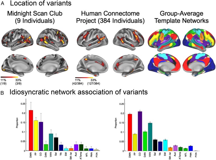
In both datasets we found common locations for network variants near the temporo–occipito–parietal junction and in the lateral frontal cortex, especially in the right hemisphere, with overlaps peaking around 33% of subjects in both datasets (3 of 9 highly sampled individuals, 127 of 384 HCP subjects). In the group average, these regions overlap with association networks, including the frontoparietal (FP) and ventral attention networks. Conversely, network variants occur rarely in the insula, superior parietal lobe, posterior cingulate, and primary sensory and motor cortical areas, with an exception around the occipital pole (Fig. 3A). Thus, there appears to be a characteristic distribution of network variants across individuals, with more network variants occurring in specific regions of the association cortex. Notably, this common distribution was found using separate datasets collected from 2 different scanners (3T Trio vs. custom 3T HCP Skyra) with different acquisition parameters (e.g., spatial resolution of 4-mm isotropic voxels vs. 2-mm isotropic voxels, temporal resolution of 2.5 s vs. 0.72 s, anterior-to-posterior vs. left-to-right and right-to-left phase encoding, and single-band vs. multiband acquisition).
Fig. 3.
Distribution of network variants across individuals. (A) The overlap of network variant locations across individuals is displayed, with brighter colors indicating increasing levels of overlap for the MSC (Left) and HCP (Center) datasets. Network variants occur commonly in lateral frontal cortex and near the temporo–occipito–parietal junction, and are rarely found in primary sensorimotor areas, the insula, superior parietal lobule, or posterior cingulate cortex. (B) In addition to occurring in characteristic locations, network variants were also typically associated with a characteristic set of networks. The mean proportion of variant functional network assignments to 14 canonical networks (12) across individuals in the MSC (Left) and HCP (Right) datasets (error bars = SEM) is displayed. A plurality of variants was assigned to the DMN (red) and CO (purple) networks across individuals in both datasets.
To determine whether network variants are driven by individual differences in gross anatomical features, we examined the overlap between network variants and deformations that occurred during surface registration for each individual, following Gordon et al. (11). We observed extremely low overlap between network variants and deformations due to surface registration (SI Appendix, Fig. S5) (mean dice overlap = 0.0001).
In addition to their location, we examined the functional network with which each variant was associated (i.e., idiosyncratically “assigned to”). After identifying the location of the variant, we implemented a modified winner-take-all template matching approach to determine the resting-state functional network to which the variant is most similar (see Materials and Methods for details) (12). For example, consider that the canonical (group-average) FP network is the network in which variants are most often located (e.g., dorsolateral prefrontal cortex in Fig. 3A). Thus, variants in this part of the brain are non-FP by definition (e.g., the default mode variants shown in Fig. 4B). We observed that variants are often assigned to the default mode, cingulo-opercular (CO), and other attention/control networks and infrequently assigned to networks related to sensorimotor and memory functions (Fig. 3B). Thus, variants often “switch” from one association network to another.
Fig. 4.
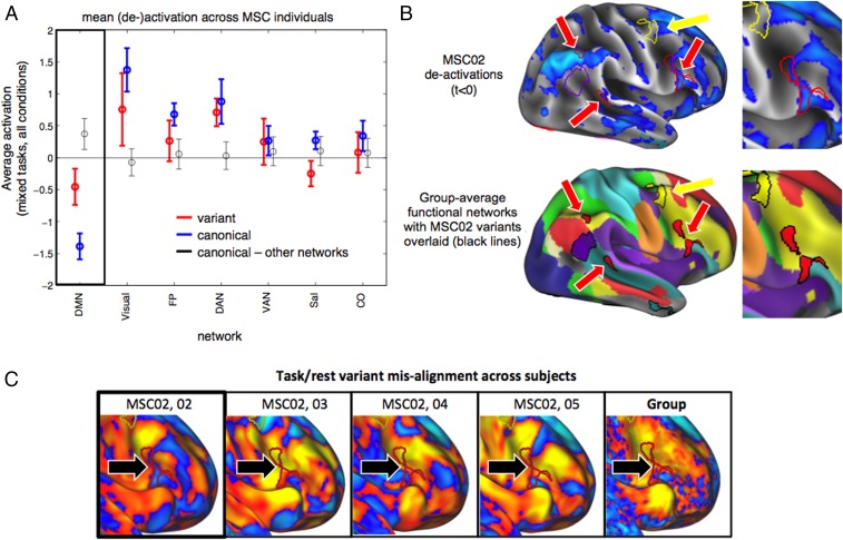
Functional activation of network variants. (A) The average task-evoked activations are displayed for variants (red) assigned to different networks (x axis) and contrasted with the average activation for canonical regions in each network (blue) and for canonical regions in other networks (black). Mean de-activations are significantly stronger for DMN variants than in non-DMN canonical regions, and approach the levels of deactivation seen for canonical DMN locations (error bars = SEM across individuals). De-activations were not present across all variants; variants from task-activated networks, like visual, FP, and DAN, show activations during the task, approaching levels for canonical regions in each network. (B) Example de-activations (t < 0) are displayed for subject MSC02 with outlines of the individual’s network variants overlaid. Note that there is strong de-activation in DMN variants (red arrows), whereas there is no deactivation in other variants (e.g., FP variant, yellow arrow). The group-average networks with the same variants overlaid are displayed below for reference, and the righthand image shows an enlarged view of 2 DMN variants in right lateral frontal cortex. (C) The same variant from MSC02 (red outline) is overlaid on an activation map from MSC02 (mostly de-activated), as well as other example subjects (MSC03-05) and the group average. In other individuals, this location exhibits activations. Indeed, across DMN variants in all subjects, activations were significantly lower for DMN variants than the matched location in other subjects. See SI Appendix, Fig. S6 for more extended examples.
Altogether, network variants’ anatomical distribution and typical functional network assignments show characteristic and systematic distributions, largely related to alterations in association systems, suggesting that they may be particularly linked to individual differences in higher-level functions.
Task-Evoked Signals in Variants Correspond to Network Association, Not Location.
To further validate whether network variants are related to changes in task function, next we asked if variants exhibit task fMRI activations consistent with their novel network assignments. To address this question, we focused on default mode network (DMN) variants as a test case because 1) all MSC subjects had examples of DMN variants and 2) the activation profile of the DMN is well described and distinct from other networks, with a robust propensity to show de-activations during most tasks (27). Thus, we examined whether DMN variants follow the expected patterns of de-activations during task performance, despite being located in regions outside of the canonical DMN.
To this end, we measured the average blood-oxygen level-dependent (BOLD) activations across all task conditions in the set of mixed-design tasks (semantic, visual coherence) collected in the MSC dataset. We found that DMN variants show significantly stronger de-activations than canonical nondefault regions of the brain [t(8) = 3.33, P = 0.01] (Fig. 4A, red vs. gray lines), approaching the level of de-activation shown by canonical regions of the DMN (Fig. 4A, blue lines). This pattern of de-activations in variants is notable, given that variants, per our working definition, occur in locations remote from canonical DMN locations (see Fig. 4B for an example of variants and task de-activations from one individual). Indeed, DMN variants in a given individual show significantly lower activations than the same location in other subjects [t(8) = 7.86, P < 0.001] (SI Appendix, Fig. S6C). Fig. 4C and SI Appendix, Fig. S6 A and B show examples of DMN variant alignments to de-activations within and across subjects, including in a region of the dorsolateral prefrontal cortex that is typically associated with positive activations in these tasks.
Importantly, de-activation was not a generic characteristic of all variants, as variants associated with many other networks show activations (e.g., variants assigned to task-activated networks, such as the FP, dorsal attention [DAN], and visual) (Fig. 4A), approaching the activations shown by canonical regions in each network with these contrasts. To supplement this finding, we conducted a related analysis on sustained task activations in CO network variants. Group studies have suggested that sustained activations are fairly selective to the CO network, rather than other control-related networks, like the FP network (22, 28–30). We found a descriptive result for sustained activation in CO network variants (SI Appendix, Fig. S7). Since only a small number of participants exhibited CO variants in the MSC dataset (6 of 9), we describe the result without formal statistics. These findings provide initial evidence that network variants carry task-evoked variations in their functional signals related to their idiosyncratic network identity at locations not expected from group-activation maps.
Distinct Subgroups of Individuals Clustered by Properties of Network Variants.
An additional hypothesis regarding the trait-like nature of network variants is that common distributions of variants may be present across individuals, much as eye color or blood type present in common clusters across individuals. To address this question, we examined whether individuals could be clustered into separate subgroups on the basis of the distributions of network variants using a data-driven approach (InfoMap) (Materials and Methods) (31). Given the exploratory nature of this analysis, we first examined different clustering possibilities in the MSC dataset, and then used 2 matched split-halves in the independent HCP dataset to validate the MSC results.
No clustering via anatomical location of variants.
First, we examined whether individuals could be clustered according to the anatomical locations of their network variants (irrespective of functional identity). We constructed a binary map of variant locations for each subject. Then, we computed the spatial similarity of the anatomical distribution of variants between all pairs of individuals, and applied InfoMap to this spatial similarity matrix (see Materials and Methods for details). Across InfoMap thresholds, individuals generally grouped into a single large cluster or were unassigned to a group (e.g., for the full 384 HCP subjects the average number of individuals in the large cluster was 201 ± 130) (SI Appendix, Fig. S8). Thus, individuals were not classified into large subgroups of common anatomical locations of network variants.
Distinct clusters of individuals via functional network of variants.
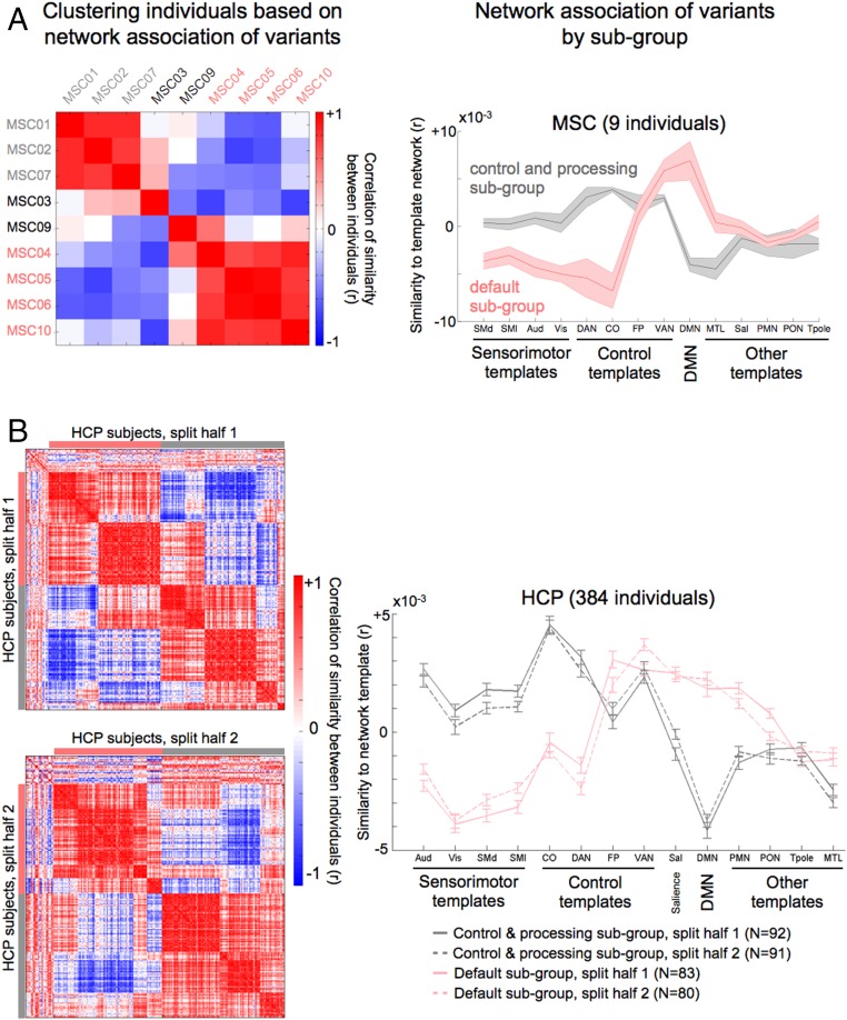
Next, we tested if individuals could be clustered according to the functional properties (network assignment) of their variants. To this end, we examined the similarity between the seed map of each variant to standard templates of canonical functional networks (14 template functional networks were derived from a separate dataset, the WashU 120 group average; see ref. 12 for more details). This procedure produced 14 correlation coefficients per network variant, conveying the extent to which a variant is default-like, visual-like, and so forth. The mean template similarity across variants (averaged across all variants within an individual) revealed 2 distinct patterns across individuals in the MSC dataset (Fig. 5A). Importantly, we replicated the result in 2 independent, matched HCP split-halves (Fig. 5B). Again, this is notable given the differences in the subjects [e.g., IQs are much higher in the MSC dataset (13)], scanner, and acquisition parameters. Furthermore, we validated the 2-group clustering solution via a modularity-based null model as well as hierarchical clustering (SI Appendix, Fig. S9). The 2-subgroup solution was the most robust across datasets, with some evidence for a 4-subgroup solution (SI Appendix, Fig. S10).
Fig. 5.
Separable groups of individuals via network associations of variants. The figure displays groups of individuals in the (A) MSC and (B) HCP datasets clustered by the network associations of their variants. Network associations were computed for each variant as their similarity to templates of 14 canonical functional networks (Materials and Methods) (11). The matrices on the left show the correlation between pairs of individuals in terms of variant network associations; each row/column represents a single individual’s correlation to all other individuals. The matrices were clustered in a data-driven fashion using InfoMap. The gray and pink colors or bars along the edges of the matrices denote individuals in the same subgroup. These groupings were used to create the averages (line graphs) on the right. The line graphs show the average similarity of variants to each functional network template for individuals within the control and processing subgroup (gray) and the default subgroup (pink; error bars = SEM across individuals).
The first subgroup consisted of individuals (NMSC = 3 of 9, NHCP,1 = 92 of 192, NHCP,2 = 91 of 192) whose variants exhibited stronger correlations to the CO, DAN, and sensorimotor networks (Fig. 5, gray), suggesting that network variants in these individuals associated more strongly with control and processing systems. The second subgroup consisted of individuals whose variants exhibited stronger correlations to the DMN, among others (NMSC = 4 of 9, NHCP,1 = 83 of 192, NHCP,2 = 80 of 192) (Fig. 5, pink). The 2 subgroups were strongly anticorrelated (see the matrices on the left in Fig. 5), indicating that functional characteristics of variants in these subgroups differed substantially from one another. We observed a similar but weaker pattern of subgroups when individuals were clustered based on the overall size of each functional network relative to the group average (SI Appendix, Fig. S11).
Moreover, we observed a small but significant difference between the 2 HCP subgroups in terms of neuropsychological measures of behavior (SI Appendix, Fig. S12). We found that individuals in the control and processing subgroup had a higher score [t(344) = 2.04, P < 0.05] in the positive life-experience factor and a lower score [t(344) = 2.04, P < 0.05] in the history of drug abuse factor. Both differences were significant after false-discovery rate-correction (see SI Appendix for full details).
Jointly, these findings suggest that individuals cluster into subgroups based on the network assignment of each variant, with one subgroup exhibiting more control and processing-like variants and the other subgroup exhibiting more default-like variants. Importantly, these findings provide evidence for systematic variation of network variants across individuals that replicated across 3 independent samples, with potential implications for behavior.
As noted above, when we examined cluster solutions across alternate thresholds (Materials and Methods and SI Appendix, Fig. S9), we observed subpatterns within each of the 2 primary clusters in the large HCP samples at lower (sparser) thresholds. There was some evidence for a 4-subgroup solution (SI Appendix, Fig. S10), but it was less reliable than the 2-subgroup solution across HCP split-halves. The presence of more fine-grained subgroups suggests that greater sample size, as well as the inclusion of additional measures and data from clinical populations, might yield further clusters of individuals not yet characterized, and potentially more fine-grained relationships between variants and behavior.
Discussion
The present study deepens our understanding of individual differences in the systems-level organization of the human brain by demonstrating that these differences reflect stable, trait-like features with systematic properties that cluster across individuals. Specifically, our results demonstrate that network variants: 1) Show high session-to-session stability in highly sampled individuals, suggesting that they are trait-like; 2) occur commonly in lateral frontal and temporoparietal regions and often associate with the DMN, CO, and other control networks, suggesting a systematic linkage to higher-level functions; 3) are related to functional variations during tasks, displaying brain (de-)activations consistent with their novel network reassignment and validating their putative network function; and 4) have a systematic patterning across individuals, allowing for the clustering of individuals into subgroups, with small differences in behavior between subgroups. Jointly, these findings suggest that network variants are promising candidates for endo-phenotypic markers of systems-level brain variability.
Network Variants Are Stable, Trait-Like Components of Individual Functional Brain Organization.
Our primary goal was to investigate properties of network variants. We hypothesized that they might show trait-like differences, including stability over time within individuals and systematic variation across individuals.
We found that all individuals across 2 independent datasets (with separate scanners and scan parameters, n = 393) showed characteristic focal deviations from the group-level description of functional brain organization. This indicates that network variants are standard components of typical adult functional network organization, as hinted at by the strong individual variation reported in previous research (10–15). Moreover, we expand upon these findings by showing that network variants are stable within an individual, appearing consistently across 10 independent resting-state fMRI sessions in highly sampled individuals. These findings extend previous evidence that resting-state correlations are sensitive to individual differences in brain organization, given sufficient data and adequate control for nuisance sources of variance (10, 15, 32–34).
Our results also indicate that group-average functional networks represent a mixture of individuals from distinct subgroups (Fig. 5). However, we demonstrate that network variants are highly localized to particular portions of the cortex. In other words, individuals showed substantial similarity to the group-average networks at most cortical locations. Since the maximum spatial overlap of network variants is ∼33%, the group average may be a reasonable description for the majority of individuals in most brain locations. Thus, group-average functional networks provide an adequate description of the expected pattern of brain organization, but the group average is not a good representation of any given person and is, therefore, limited in inferences that can be drawn about brain–behavior relationships.
Taken together, the presence of network variants in all individuals and their robustness over sessions provides compelling evidence that they act as stable variations in the systems-level organization of the human brain. These features may prove to be useful substrates in understanding individual differences in brain function and behavior across many domains.
Network Variants Have Characteristic Distributions and Functional Network Associations Across Individuals.
We observed that network variants are found commonly near the temporo–occipito–parietal junction and in the lateral prefrontal cortex. We rarely detected network variants in primary sensorimotor cortical areas, the insula, superior parietal lobe, or posterior cingulate cortex. This finding not only replicates across independent datasets, but also converges with previous studies of individual differences in functional network organization reporting high individual differences in association networks (8, 11, 34, 35), with a few specific differences. For example, compared to Mueller et al. (8), we found more network variants near the inferior frontal gyrus and fewer near angular gyrus and supramarginal gyrus. Differences in data processing and registration (surface-based, here) may have contributed to some of these discrepancies. This idea is supported by the similarity of the results here and those reported by Kong et al. (35).
Interestingly, the distribution of common locations for network variants does not appear to be symmetric between the 2 hemispheres, as we found generally more variants in the right hemisphere. Lateralization in the brain is a well-established phenomenon, in terms of both anatomy and function (e.g., refs. 36 and 37), even at the level of individual rsFCs (9). The significance and implications of potential network variant lateralization is a topic for future work to explore.
Furthermore, we found that variants tend to associate with the DMN, CO, and other association networks more often than other functional networks. Networks like the CO and FP network are thought to be important for control functions and performance monitoring (22, 28, 38–41), and the DMN has been proposed to be involved in numerous domains, including autobiographical memory, internal monitoring, and theory of mind (42–44). Network variants occur most often in these “association” networks, and they tend to reassign from one higher-level functional network to another. Together, these results suggest that flexibility in functional network organization may relate more closely to higher-level functions typically associated with these regions.
Network Variants Exhibit Functional Variations during Tasks.
We observed that network variants coincide with locations of functional variations during tasks in the MSC dataset. Specifically, we demonstrated that variants that associate with the DMN exhibit decreases in activity during task performance, as has been robustly observed for canonical DMN regions in most externally directed tasks (27). This was the case even in DMN variants in the dorsolateral prefrontal cortex, a brain region that canonically shows robust positive activity during tasks in most individuals. Moreover, this de-activation was not a general property of all variants; variants associated with task-activated systems like the visual, DAN, and FP networks showed (positive) activations. Similarly, CO network variants showed a trend toward higher levels of sustained activation, consistent with a role of the CO network in the stable maintenance of task set (22, 28–30). This finding provides initial validation that network variants shift toward the response characteristics of their functional network assignment during task performance, providing corroborating evidence that variants reflect true deviations in the functional organization of individual human brains that impact task function.
This result converges with work from Tavor et al. (45), who built a model that was able to predict individual differences in task activations on the basis of individual differences in resting-state data. Their model did not specifically operate on network variants, although the presence of network variants would certainly impact the training of the model. Similarly, Gordon et al. (13) demonstrated that individual specific task-related activation patterns map onto that individual’s resting-state functional networks better than they map onto different individuals’ networks. Here, we build on these findings by showing that network variants specifically show improved task–rest alignment in individuals compared with canonical network assignments. Finally, seminal work from Miller and colleagues revealed stable, meaningful individual differences in brain activations during task performance (1–4). These investigations led to the idea that individual-specific activation patterns reflect, or are potentially determined by, subject-specific information-processing strategies. The results presented herein suggest that if these hypotheses are true, this trait-like brain activity may be localized to network variant regions, specifically.
Individuals Cluster into Discrete Groups on the Basis of Network Variant Characteristics.
We found evidence for subgroups of individuals within a normative sample with similar forms of network variants, suggesting that variants demonstrate systematic variation across individuals. Intriguingly, it was the network assignment of variants, rather than their anatomical location, that appeared to be the driving force behind these distinct subgroups. That is, it appears as though group-level variation of network variants is more related to functional assignment than location.
We observed 2 subgroups across individuals that were consistent in both datasets. The 2 subgroups were composed of individuals with more control and processing-like variants and individuals with more default-like variants. The strong distinction between the subgroups may relate to the specific functional networks onto which the variants map, which generally activate and de-activate, respectively, during externally directed tasks (46, 47). A related possibility is that the distinction between the subgroups may be due to changes in the relative size of the aforementioned networks. In other words, individuals with more default-like variants may have an expanded DMN, which could be achieved via “trading” anatomical space canonically occupied by control and processing functional networks for DMN network variants (and vice-versa). Any of these possibilities relates to the trait-like status of the distributions of variants across individuals.
While our work provides initial evidence for groups of individuals with similar network variants across 2 different samples, it appears likely that additional subgroups will be found in future studies. Some additional subgroups may be associated with other properties of network variants (e.g., specific locations, networks, and their interactions), while others may emerge with a more behaviorally diverse range of individuals (e.g., those with neurologic or psychiatric disorders). Notably, in the larger HCP dataset we observed some evidence of further clustering, with the 2 initial groups dividing further into 4 subgroups of individuals. The current work presents a starting point for future investigations into systematic variation in individual functional brain organization, an area that merits substantial additional exploration.
Network Variants May Relate to Behavior.
The trait-like nature of network variant distributions raises the question of whether or not network variants relate to individual differences in behavior. It is possible that these individual differences in brain organization reflect different manners of instantiating the same behavior, a behavioral phenocopy (48), or functional degeneracy (49, 50). In other words, network variants may reflect individual differences in processing organization that ultimately lead to similar functional outcome. Conversely, there may be systematic relationships between network variants and measures of behaviors, either in a categorical (e.g., differences between network variant subgroups and behavior) or continuous fashion.
We observed a small but significant relationship between network variant subgroups and behavior. Individuals in the DMN subgroup had a lower life satisfaction and higher history of drug abuse, on average. Previous investigations revealed that individual differences in rsFCs are related to a positive and negative “mode” of lifestyle (51, 52) as well as measures of executive function (35, 53).
In addition to the subgroup analysis presented herein, there may be continuous relationships between network variants and measures of behavior, such as those observed by Bijsterbosch et al. (52). Connections between network variants and behavior should be pursued by future studies with more specialized behavioral measures and a broader range of network variant properties and subgroups. By extending behavioral relationships to network variants specifically, future investigations should have more precise targets: That is, variant locations, for both basic experimentation and potential medical intervention (e.g., via stimulation-based methods). It is possible that our approach of identifying network variants may provide a rich source of targets to better understand the neurobiological sources of individual differences in behavior.
Neurobiological Interpretations of Network Variants.
In the present study, we demonstrate that individuals stably vary in specific elements of their functional network organization. This observation raises the question of what neural mechanisms underlie these individual differences. Any network variant could represent: 1) A regional border shift, in which a neighboring brain area is enlarged, contracted, or displaced; 2) a relative shift in the functional and connectivity properties of an existing area, leading to reassignment to a distinct network; or 3) a de novo brain area unique to an individual or small group.
The appearance of completely novel cortical areas in individuals seems unlikely. Most studied cortical areas (i.e., visual areas, motor areas, attention-related areas) are found reliably in essentially every individual primate (54–56). Variations in the size of brain areas have been observed previously, such as a 2-fold difference in the size of some visual areas (57). In cases of perturbations, larger changes can be seen. For example, area V1 in congenitally blind individuals is significantly decreased in size (58–60) and genetic manipulations can affect the size and position of areas; for example, primary sensory and motor areas are expanded and shifted rostro-laterally in mice that overexpress Emx2 (61, 62). Cortical areas may also be displaced along the cortex, as was observed for area 55b by Glasser et al. (63), leading to the appearance of variant pieces in nonoverlapping locations.
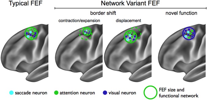
As a conceptual example (Fig. 6), consider the frontal eye fields (FEF), an area that lies close to regions where network variants are frequently found across individuals (in the right hemisphere, at least). Essentially every human likely has at least one FEF per hemisphere (64). If an individual has a larger (or smaller) FEF than the average, the expanded (or contracted) portion of the cortex will appear as a network variant. Similarly, if the FEF is displaced in an individual relative to the typical cortical location of the FEF, it will be identified as a network variant. While these more local variations likely occur (and account for some network variants), previous work has demonstrated that individually variable network assignments can occur at regions remote from network borders (12).
Fig. 6.
Schematic of potential neural mechanisms underlying network variants. A schematic of the typical (group-average) FEF is displayed on the left. Neurons coding for saccades, attention in space, and visual stimuli are color-coded (light blue, green, and dark blue, respectively). An individual’s FEF may be identified as a network variant if its border has shifted relative to its typical location, via either contraction/expansion in size (contraction displayed Center Left) or displacement along the cortex (Center Right). Another possibility is that the underlying functional and connectivity properties of the individual’s FEF are different from the group average, for example, more neurons that code for visual stimuli (Right).
One potential explanation for remote network variants is altered functional response and connectivity properties of those areas in certain individuals. For example, individual FEF neurons code for saccades, visual stimuli, attention in space, and combinations of these 3 properties (65). Depending on the relative proportion of these various types of FEF neurons, individuals may have different functional connectivity and task-evoked BOLD signals in this area relative to the group average. Whereas a typical individual’s FEF may have a high proportion of eye movement and attention neurons, thus producing the usual association with the DAN, another individual’s FEF might contain an unusually large number of neurons coding visual stimuli (e.g., due to genetics and accumulated experience) and, thus, associate with the visual functional network. Therefore, one possibility is that an area may appear like a network variant not because it is truly a novel area, but because the distribution or function of its neurons is shifted systematically in that individual.
This last idea is supported by the observed task-related activations in network variants. For several functional networks, we showed that the level of (de-)activation in those network variants is between the level of (de-)activation seen typically for that location and what would be expected given the network variant’s reassignment (Fig. 4A). Moreover, a related paper by Arcaro et al. (66) provided an example of functional reassignment based on lifetime experience, such that the portion of the inferotemporal cortex that is typically face-selective becomes body- and hand-selective in monkeys reared without exposure to faces. The neurons in this region of the cortex are innately retinotopic and biased toward the scale and curvature of visual stimuli (i.e., biased to respond to faces). However, due to the monkeys’ atypical environment and experience, the response properties of the region changed (even for face stimuli), suggesting that the area may appear as a network variant compared to typical monkeys.
Each network variant may be due to one or more of the abovementioned mechanisms, and future work is necessary to determine the consequences of these different types of network variants. While the mechanisms are difficult to disambiguate precisely in humans, studies with animal models may be well equipped to examine this question and to expand our understanding of the sources of individual variability in large-scale brain networks.
Conclusion
We find that network variants are stable components of typical adult functional network organization. The organization and arrangement of network variants across individuals appears to be systematic. Specifically, they tend to occur in the lateral prefrontal cortex and near the temporo–occiptio–parietal junction and are often reassigned to association networks, suggesting a link to higher-level cognitive functions. Moreover, network variants are related to functional variations during tasks. Finally, individuals cluster into subgroups on the basis of these variants and these subgroups demonstrate small differences in behavior. Taken in sum, our data support the idea that network variant distributions are trait-like and their patterning across individuals is functionally relevant.
Materials and Methods
Datasets, Acquisition Parameters, and Exclusion Criteria.
Three datasets are analyzed in this report: The MSC (13), MyConnectome (24), and HCP (25) datasets. In addition, for group-average comparisons, a previously collected dataset of 120 typical adults was used as the group-level referent (17), referred to in the text as the WashU 120. All data collection was approved by the Washington University and University of Texas Internal Review Boards and all procedures complied with ethical regulations for studies involving human research participants. Dataset composition, acquisition parameters, and exclusion criteria have been described in detail previously (10, 13, 24, 25) for all datasets (see SI Appendix for a brief description).
Data and Code Availability.
All data and data-processing code used in the manuscript are publicly available (MSC and code: https://openneuro.org/datasets/ds000224/versions/00002 MyConnectome: myconnectome.org/wp/; HCP: https://db.humanconnectome.org/app/template/Login.vm; WashU 120: https://legacy.openfmri.org/dataset/ds000243/). Code for network variant analyses (custom MATLAB scripts) are available at https://github.com/MidnightScanClub.
Resting-State Data Processing.
All data processing has been described in detail previously for each dataset. For extended details, see ref. 13 for MSC, ref. 10 for MyConnectome, ref. 67 for HCP, and ref. 68 for WashU 120. We briefly review relevant details for each type of processing below.
Anatomical processing.
First, FreeSurfer 5.3 automatic segmentation was applied to the T1-weighted images to create masks of the gray matter, white matter, and ventricles for each subject (69). Then, FreeSurfer’s default recon-all pipeline was used to reconstruct each subject’s native anatomical surface. These native surfaces were aligned to the fsaverage surface using a shape-based spherical registration (70–73). The 2 hemispheres were registered to each other using a landmark-based algorithm (74, 75). The final resolution of each subject’s surface was 32,492 vertices per hemisphere.
Functional processing.
For each subject, standard preprocessing procedures were applied (slice-timing correction, functional realignment, mode 1,000 normalization, atlas registration and resampling, and distortion correction) in addition to further preprocessing to remove motion-related artifacts (frame-wise displacement for frame censoring, regression of nuisance signals, including the whole-brain mean, interpolation over censored frames, and bandpass filtering) (68). See SI Appendix for full details.
Volume-to-surface mapping and functional connectivity processing.
After preprocessing, a CIFTI was created for each subject. Preprocessed BOLD time series data were mapped to the surface following the procedure of Gordon et al. (76). Before computing the correlations, all previously censored frames were discarded to account for distance-dependent motion artifacts (68). Pairwise correlations between time series from every pair of cortical surface vertices from both hemispheres (59,412 × 59,412) were computed to construct an individual-specific vertex-to-vertex correlation matrix, which was then Fisher-transformed. For the WashU 120 dataset, the individual correlation matrices were averaged together. See SI Appendix for full details.
Task Data Processing.
In this study, we focus on activations in the 2 mixed design (77) tasks from the MSC dataset: The semantic task and the coherence task. Tasks and their analyses are described in detail in Gordon et al. (13). Task activations were modeled with in-house imaging analysis software (IDL) using a general linear model approach, as previously described (13, 15). See SI Appendix for a brief description.
The DMN has been consistently linked to task-deactivations. To determine whether network variants (see next section) associated with the DMN also show the same functional profile, we examined network variant activations in all conditions (cues, trials, and sustained activations) vs. implicit baseline. We conducted this comparison for variants associated with each network, canonical (i.e., nonvariant, group) regions associated with each network, and the average of canonical regions associated with all other networks. In addition, the average activation of each DMN variant in a single individual was compared with the average activation of that same location in other individuals. In all cases, statistical comparisons were carried out using paired 2-sided t tests.
In addition, we added a complementary supplementary analysis of task activations associated with the CO system. The CO network has been consistently linked to sustained activations, especially during resource-limited tasks, unlike other control systems such as the FP network (22, 29, 30). To examine whether variants associated with the CO network displayed sustained activations, we examined activations associated with the sustained block regressor during a resource-limited semantic task (28).
Identification of Network Variants.
To identify network variants, individual subject correlation matrices were compared (independently) to a group-average correlation matrix generated from the WashU 120. For each individual, the spatial similarity between the individual’s and the group’s pattern of correlations (seed map) at each cortical surface vertex was computed. More precisely, each row of an individual’s matrix was correlated with the corresponding row in the group-average matrix, resulting in one spatial correlation per vertex. Susceptibility regions were masked out using a vertex-wise measure of signal quality derived from the group-average data. All vertices with a mean BOLD signal less than 750 (as computed in ref. 78) were set to 0. Then, the spatial similarity was binarized such that all cortical vertices with a spatial correlation value in the lowest decile of the individual’s distribution were considered for further analyses (these vertices were set to 1, and all others were set to 0). Network variants were defined as regions of the cortex in which sets of at least 50 contiguous vertices were below the spatial correlation threshold. As an alternative to allowing the threshold for network variants to vary across individuals (lowest decile of the individual’s spatial correlation distribution), the threshold was fixed at a spatial correlation value of 0.3. Results were extremely consistent between analysis procedures, given that the mean lowest decile cutoff value is 0.32 ± 0.03.
Functional Network Assignment of Network Variants.
A winner-take-all procedure was implemented to assign functional networks to each network variant in each individual. To do so, 14 template networks were created from the 14 group-average networks, as described previously (12). The templates are the group-average resting-state correlation pattern (seed map) of each canonical functional network in the WashU 120 (e.g., the group-average DMN seed map, the group-average visual network seed map, and so forth). Then, for each unique network variant, the following matching procedure was applied: 1) A seed map was computed from the average BOLD time series from all vertices within the network variant; 2) the similarity between that variant seed map and each template network was computed (i.e., the spatial correlation between the template seed map and the variant seed map); 3) the template network with the highest similarity was assigned to the network variant; 4) any network variants where the winning template system had low similarity (i.e., r < 0.3) were reassigned as “unknown system”; and finally, 5) we ensured that the variant did not match the group-average network at that cortical location. In other words, we removed the variant if it overlapped (spatially) with its assigned group-average functional network by 50% or more (this occurred infrequently: 5 of 129 = 4% in the MSC dataset, 276 of 7,498 = 3.7% in the HCP dataset).
Overlap of Network Variants Across Individuals.
In order to examine the spatial overlap of network variants across individuals, binary versions of the final maps of network variants (after functional network assignment) were summed across individuals to create an overlap map within each dataset. These were divided by the number of people within each dataset, to express the frequency of network variants at each cortical vertex.
Within-Subject Reliability of Network Variants.
To measure within-subject reliability of network variants, we compared variants across different days from the same participant. Each MSC subject had 10 independent 30-min resting-state sessions collected on separate days. We processed each session separately (as described above) in order to assess within-subject session-to-session variability of network variants. For each session, we generated the spatial correlation map used for identifying network variants (seed maps from the session vs. seed maps from the group average). Then, we measured the ICC of each map within an individual. In addition, this entire analysis was repeated with binarized network variant maps, but computing the mean dice coefficient instead of ICCs (since the maps are binary). The latter analysis allowed for a focused reliability measure of variant regions only. For the binarized variants analysis, we generated a null model of between-subject variant overlaps for comparison. We performed 1,000 random permutations of pairs of sessions drawn from 2 different MSC individuals (with replacement) and computed the mean dice coefficient of the binary network variant maps from those sessions. For the MyConnectome dataset, we compared the stability of network variants from sequential 3-wk blocks of data (i.e., variants identified from 6 sessions concatenated together for each 3-wk block). All possible pairs of variant spatial correlation maps (from each 3-wk block of time) were correlated with one another.
Patterns of Network Variants Across Individuals.
Next, we turned our attention to whether similar types of network variants were seen across subgroups of individuals. We tested 2 options: 1) Whether subgroups of individuals exhibited variants at similar anatomical locations and, 2) whether subgroups of individuals exhibited variants with similar network associations.
Anatomical.
To determine whether subgroups of individuals exhibited variants at similar anatomical locations, we compared the binary maps of (final) network variants between each pair of individuals using dice coefficients. This resulted in a symmetric dice overlap matrix with a size of N by N (9 × 9 for the MSC dataset and 192 × 192 for each HCP split-half), with each entry representing the degree to which a given pair of individuals covaries in terms of the spatial distribution of network variants (i.e., the degree to which the pair both have variants 1, 2, and 3 and they both do not have variants 4, 5, and 6). A clustering algorithm (InfoMap) (31) was applied to this dice overlap matrix. Before clustering, we applied a threshold to the matrix to create a sparse network on which to operate. We examined a wide range of density thresholds from the top 2 to 30% of correlations in increments of 1%.
Functional.
To determine whether subgroups of individuals exhibited variants with similar network associations, we compared their match to 14 standard network templates. Specifically, during functional network assignment of variants, we compute the similarity (i.e., spatial correlation) between each variant and each template functional network. This results in a 14 × 1 vector of correlations (to the 14 template networks) for each variant. This measure represents the degree to which a variant is default mode-like, visual-like, and so forth. Then, we computed the variant-size-weighted mean similarity for all variants within an individual to each template network (indicating the degree to which all of that individual’s variants are default mode-like, visual-like, and so forth). This mean measure was correlated across subjects, and the same clustering algorithm (InfoMap) was implemented to identify groups of individuals with similar patterns of network variants. We used a range of thresholds from 2 to 10% in increments of 1%.
Conceptually, the functional measure discussed above calculates the average similarity of variants to canonical networks, producing a quantitative estimate of the (for example) DMN-like characteristics of all variants in an individual. To complement this measure, we also clustered individuals based on the amount of the cortex (number of surface vertices) assigned to each functional network. The WashU 120 group-average functional networks were used as a referent to compare the relative expansion or reduction of each individual’s functional networks. Thus, we calculated the number of expanded or contracted surface vertices for each network (relative to the group-average) using the variants’ network assignments; for example, a given individual may have +1,000 CO vertices, −75 default mode vertices, and so on.
Analysis of Behavior.
Arguably, an important aspect of network variants is their relation to behavior. As a proof-of-concept, and given network variants’ distributions and reassignment to association networks, we examined relationships to the HCP behavioral measures (79). We used exploratory factor analysis (EFA) for data reduction and to identify latent constructs in the HCP data. We focused on behavior categories that included multiple instruments or that did not already have summary measures available, which included demographics, cognition, emotion, and substance use variables. Age, sex, and handedness were not considered in the EFA to allow flexibility to include or exclude these variables in analyses of brain–behavior relationships (e.g., as covariates). Data from all HCP subjects (n = 1,206) were included in the EFA. EFA factors were then compared across subgroups using multiple linear regression. Details about the results of the EFA and the regression analysis between HCP subgroups of individuals are in SI Appendix, Supplemental Methods and Fig. S12.
Supplementary Material
Acknowledgments
We thank Joshua S. Siegel and Deanna M. Barch for assistance with the Human Connectome Project data. This research was supported by NIH Grant T32NS073547 (to B.A.S.), NIH Grant F32NS092290 (to C.G.), NIH Grant R01MH118370 (to C.G.), NIH Grant R25MH112473 (to T.O.L.), NIH Grant T32NS047987 (to B.T.K.), National Science Foundation Graduate Research Fellowship Program Award DGE-1143954 (to A.W.G.), an American Psychological Association Dissertation Research Award (to A.W.G.), NIH Grant K01MH104592 (to D.J.G.), a Dart Neuroscience, LLC Grant (to K.B.M.), a McDonnell Foundation Collaborative Activity Award (to S.E.P.), NIH Grant R01NS32979 (to S.E.P.), NIH Grant R01NS06424 (to S.E.P.), and Career Development Award 1IK2CX001680 (to E.M.G.) from the US Department of Veterans Affairs Clinical Sciences Research and Development Service. The contents of this manuscript do not represent the views of the US Department of Veterans Affairs or the United States Government.
Footnotes
The authors declare no competing interest.
This article is a PNAS Direct Submission. M.B.M. is a guest editor invited by the Editorial Board.
Data deposition: All data and data processing code used in the manuscript are publicly available (Midnight Scan Club and code: https://openneuro.org/datasets/ds000224/versions/00002; MyConnectome: myconnectome.org/wp/; Human Connectome Project: https://db.humanconnectome.org/app/template/Login.vm; WashU 120: https://legacy.openfmri.org/dataset/ds000243/). Code for network variant analyses (custom MATLAB scripts) is available at https://github.com/MidnightScanClub.
See Commentary on page 22432.
This article contains supporting information online at www.pnas.org/lookup/suppl/doi:10.1073/pnas.1902932116/-/DCSupplemental.
References
- 1.Miller M. B., et al. , Unique and persistent individual patterns of brain activity across different memory retrieval tasks. Neuroimage 48, 625–635 (2009). [DOI] [PMC free article] [PubMed] [Google Scholar]
- 2.Miller M. B., Donovan C. L., Bennett C. M., Aminoff E. M., Mayer R. E., Individual differences in cognitive style and strategy predict similarities in the patterns of brain activity between individuals. Neuroimage 59, 83–93 (2012). [DOI] [PubMed] [Google Scholar]
- 3.Van Horn J. D., Grafton S. T., Miller M. B., Individual variability in brain activity: A nuisance or an opportunity? Brain Imaging Behav. 2, 327–334 (2008). [DOI] [PMC free article] [PubMed] [Google Scholar]
- 4.Congdon E., et al. , Engagement of large-scale networks is related to individual differences in inhibitory control. Neuroimage 53, 653–663 (2010). [DOI] [PMC free article] [PubMed] [Google Scholar]
- 5.Neta M., Whalen P. J., Individual differences in neural activity during a facial expression vs. identity working memory task. Neuroimage 56, 1685–1692 (2011). [DOI] [PMC free article] [PubMed] [Google Scholar]
- 6.Kanai R., Rees G., The structural basis of inter-individual differences in human behaviour and cognition. Nat. Rev. Neurosci. 12, 231–242 (2011). [DOI] [PubMed] [Google Scholar]
- 7.Filipek P. A., et al. , Volumetric MRI analysis comparing subjects having attention-deficit hyperactivity disorder with normal controls. Neurology 48, 589–601 (1997). [DOI] [PubMed] [Google Scholar]
- 8.Mueller S., et al. , Individual variability in functional connectivity architecture of the human brain. Neuron 77, 586–595 (2013). [DOI] [PMC free article] [PubMed] [Google Scholar]
- 9.Wang D., et al. , Parcellating cortical functional networks in individuals. Nat. Neurosci. 18, 1853–1860 (2015). [DOI] [PMC free article] [PubMed] [Google Scholar]
- 10.Laumann T. O., et al. , Functional system and areal organization of a highly sampled individual human brain. Neuron 87, 657–670 (2015). [DOI] [PMC free article] [PubMed] [Google Scholar]
- 11.Gordon E. M., Laumann T. O., Adeyemo B., Petersen S. E., Individual variability of the system-level organization of the human brain. Cereb. Cortex 27, 386–399 (2017). [DOI] [PMC free article] [PubMed] [Google Scholar]
- 12.Gordon E. M., et al. , Individual-specific features of brain systems identified with resting state functional correlations. Neuroimage 146, 918–939 (2017). [DOI] [PMC free article] [PubMed] [Google Scholar]
- 13.Gordon E. M., et al. , Precision functional mapping of individual human brains. Neuron 95, 791–807.e7 (2017). [DOI] [PMC free article] [PubMed] [Google Scholar]
- 14.Braga R. M., Buckner R. L., Parallel interdigitated distributed networks within the individual estimated by intrinsic functional connectivity. Neuron 95, 457–471.e5 (2017). [DOI] [PMC free article] [PubMed] [Google Scholar]
- 15.Gratton C., et al. , Functional brain networks are dominated by stable group and individual factors, not cognitive or daily variation. Neuron 98, 439–452.e5 (2018). [DOI] [PMC free article] [PubMed] [Google Scholar]
- 16.Marek S., et al. , Spatial and temporal organization of the individual human cerebellum. Neuron 100, 977–993.e7 (2018). [DOI] [PMC free article] [PubMed] [Google Scholar]
- 17.Power J. D., et al. , Functional network organization of the human brain. Neuron 72, 665–678 (2011). [DOI] [PMC free article] [PubMed] [Google Scholar]
- 18.Yeo B. T. T., et al. , The organization of the human cerebral cortex estimated by intrinsic functional connectivity. J. Neurophysiol. 106, 1125–1165 (2011). [DOI] [PMC free article] [PubMed] [Google Scholar]
- 19.Biswal B., Yetkin F. Z., Haughton V. M., Hyde J. S., Functional connectivity in the motor cortex of resting human brain using echo-planar MRI. Magn. Reson. Med. 34, 537–541 (1995). [DOI] [PubMed] [Google Scholar]
- 20.Cordes D., et al. , Mapping functionally related regions of brain with functional connectivity MR imaging. AJNR Am. J. Neuroradiol. 21, 1636–1644 (2000). [PMC free article] [PubMed] [Google Scholar]
- 21.Lowe M. J., Mock B. J., Sorenson J. A., Functional connectivity in single and multislice echoplanar imaging using resting-state fluctuations. Neuroimage 7, 119–132 (1998). [DOI] [PubMed] [Google Scholar]
- 22.Dosenbach N. U. F., et al. , Distinct brain networks for adaptive and stable task control in humans. Proc. Natl. Acad. Sci. U.S.A. 104, 11073–11078 (2007). [DOI] [PMC free article] [PubMed] [Google Scholar]
- 23.Smith S. M., et al. , Correspondence of the brain’s functional architecture during activation and rest. Proc. Natl. Acad. Sci. U.S.A. 106, 13040–13045 (2009). [DOI] [PMC free article] [PubMed] [Google Scholar]
- 24.Poldrack R. A., et al. , Long-term neural and physiological phenotyping of a single human. Nat. Commun. 6, 8885 (2015). [DOI] [PMC free article] [PubMed] [Google Scholar]
- 25.Van Essen D. C., et al. ; WU-Minn HCP Consortium , The Human Connectome Project: A data acquisition perspective. Neuroimage 62, 2222–2231 (2012). [DOI] [PMC free article] [PubMed] [Google Scholar]
- 26.Laumann T. O., et al. , On the stability of BOLD fMRI correlations. Cereb. Cortex 27, 4719–4732 (2017). [DOI] [PMC free article] [PubMed] [Google Scholar]
- 27.Shulman G. L., et al. , Common blood flow changes across visual tasks: II. Decreases in cerebral cortex. J. Cogn. Neurosci. 9, 648–663 (1997). [DOI] [PubMed] [Google Scholar]
- 28.Dubis J. W., Siegel J. S., Neta M., Visscher K. M., Petersen S. E., Tasks driven by perceptual information do not recruit sustained BOLD activity in cingulo-opercular regions. Cereb. Cortex 26, 192–201 (2016). [DOI] [PMC free article] [PubMed] [Google Scholar]
- 29.Dosenbach N. U. F., et al. , A core system for the implementation of task sets. Neuron 50, 799–812 (2006). [DOI] [PMC free article] [PubMed] [Google Scholar]
- 30.Dosenbach N. U. F., Fair D. A., Cohen A. L., Schlaggar B. L., Petersen S. E., A dual-networks architecture of top-down control. Trends Cogn. Sci. 12, 99–105 (2008). [DOI] [PMC free article] [PubMed] [Google Scholar]
- 31.Rosvall M., Bergstrom C. T., Maps of random walks on complex networks reveal community structure. Proc. Natl. Acad. Sci. U.S.A. 105, 1118–1123 (2008). [DOI] [PMC free article] [PubMed] [Google Scholar]
- 32.Braun U., et al. , Test-retest reliability of resting-state connectivity network characteristics using fMRI and graph theoretical measures. Neuroimage 59, 1404–1412 (2012). [DOI] [PubMed] [Google Scholar]
- 33.Birn R. M., et al. , The effect of scan length on the reliability of resting-state fMRI connectivity estimates. Neuroimage 83, 550–558 (2013). [DOI] [PMC free article] [PubMed] [Google Scholar]
- 34.Chen B., et al. , Individual variability and test-retest reliability revealed by ten repeated resting-state brain scans over one month. PLoS One 10, e0144963 (2015). [DOI] [PMC free article] [PubMed] [Google Scholar]
- 35.Kong R., et al. , Spatial topography of individual-specific cortical networks predicts human cognition, personality, and emotion. Cereb. Cortex 29, 2533–2551 (2019). [DOI] [PMC free article] [PubMed] [Google Scholar]
- 36.Broca P., Remarques sur le siège de la faculté du langage articulé, suivies d’une observation d’aphémie (perte de la parole). Bull la Société Anat 6, 330–357 (1861). [Google Scholar]
- 37.Sperry R. W., Cerebral organization and behavior: The split brain behaves in many respects like two separate brains, providing new research possibilities. Science 133, 1749–1757 (1961). [DOI] [PubMed] [Google Scholar]
- 38.Seeley W. W., et al. , Dissociable intrinsic connectivity networks for salience processing and executive control. J. Neurosci. 27, 2349–2356 (2007). [DOI] [PMC free article] [PubMed] [Google Scholar]
- 39.Sadaghiani S., D’Esposito M., Functional characterization of the cingulo-opercular network in the maintenance of tonic alertness. Cereb. Cortex 25, 2763–2773 (2015). [DOI] [PMC free article] [PubMed] [Google Scholar]
- 40.Neta M., Schlaggar B. L., Petersen S. E., Separable responses to error, ambiguity, and reaction time in cingulo-opercular task control regions. Neuroimage 99, 59–68 (2014). [DOI] [PMC free article] [PubMed] [Google Scholar]
- 41.Gratton C., et al. , Distinct stages of moment-to-moment processing in the cinguloopercular and frontoparietal networks. Cereb. Cortex 27, 2403–2417 (2017). [DOI] [PMC free article] [PubMed] [Google Scholar]
- 42.Buckner R. L., Andrews-Hanna J. R., Schacter D. L., The brain’s default network: Anatomy, function, and relevance to disease. Ann. N. Y. Acad. Sci. 1124, 1–38 (2008). [DOI] [PubMed] [Google Scholar]
- 43.Spreng R. N., Mar R. A., Kim A. S. N., The common neural basis of autobiographical memory, prospection, navigation, theory of mind, and the default mode: A quantitative meta-analysis. J. Cogn. Neurosci. 21, 489–510 (2009). [DOI] [PubMed] [Google Scholar]
- 44.Raichle M. E., The brain’s default mode network. Annu. Rev. Neurosci. 38, 433–447 (2015). [DOI] [PubMed] [Google Scholar]
- 45.Tavor I., et al. , Task-free MRI predicts individual differences in brain activity during task performance. Science 352, 216–220 (2016). [DOI] [PMC free article] [PubMed] [Google Scholar]
- 46.Fox M. D., et al. , The human brain is intrinsically organized into dynamic, anticorrelated functional networks. Proc. Natl. Acad. Sci. U.S.A. 102, 9673–9678 (2005). [DOI] [PMC free article] [PubMed] [Google Scholar]
- 47.Margulies D. S., et al. , Situating the default-mode network along a principal gradient of macroscale cortical organization. Proc. Natl. Acad. Sci. U.S.A. 113, 12574–12579 (2016). [DOI] [PMC free article] [PubMed] [Google Scholar]
- 48.Schlaggar B. L., McCandliss B. D., Development of neural systems for reading. Annu. Rev. Neurosci. 30, 475–503 (2007). [DOI] [PubMed] [Google Scholar]
- 49.Tononi G., Sporns O., Edelman G. M., Measures of degeneracy and redundancy in biological networks. Proc. Natl. Acad. Sci. U.S.A. 96, 3257–3262 (1999). [DOI] [PMC free article] [PubMed] [Google Scholar]
- 50.Friston K. J., Price C. J., Degeneracy and redundancy in cognitive anatomy. Trends Cogn. Sci. 7, 151–152 (2003). [DOI] [PubMed] [Google Scholar]
- 51.Smith S. M., et al. , A positive-negative mode of population covariation links brain connectivity, demographics and behavior. Nat. Neurosci. 18, 1565–1567 (2015). [DOI] [PMC free article] [PubMed] [Google Scholar]
- 52.Bijsterbosch J. D., et al. , The relationship between spatial configuration and functional connectivity of brain regions. eLife 7, e32992 (2018). [DOI] [PMC free article] [PubMed] [Google Scholar]
- 53.Finn E. S., et al. , Functional connectome fingerprinting: Identifying individuals using patterns of brain connectivity. Nat. Neurosci. 18, 1664–1671 (2015). [DOI] [PMC free article] [PubMed] [Google Scholar]
- 54.Tusa R. J., Palmer L. A., Rosenquist A. C., “Multiple cortical visual areas” in Multiple Visual Areas, Woolsey C. N., Ed. (Cortical Sensory Organization, Humana Press, 1981), vol. 2, pp. 1–31. [Google Scholar]
- 55.Bowker R. M., Coulter J. D., “Intracortical connectivities of somatic sensory and motor areas” in Multiple Somatic Areas, Woolsey C. N., Ed. (Cortical Sensory Organization, Humana Press, 1981), vol. 1, pp. 205–242. [Google Scholar]
- 56.Brugge J. F., “Auditory cortical areas in primates” in Multiple Auditory Areas, Woolsey C. N., Ed. (Cortical Sensory Organization, Humana Press, 1982), vol. 3, pp. 59–70. [Google Scholar]
- 57.Dougherty R. F., et al. , Visual field representations and locations of visual areas V1/2/3 in human visual cortex. J. Vis. 3, 586–598 (2003). [DOI] [PubMed] [Google Scholar]
- 58.Park H. J., et al. , Morphological alterations in the congenital blind based on the analysis of cortical thickness and surface area. Neuroimage 47, 98–106 (2009). [DOI] [PubMed] [Google Scholar]
- 59.Jiang J., et al. , Thick visual cortex in the early blind. J. Neurosci. 29, 2205–2211 (2009). [DOI] [PMC free article] [PubMed] [Google Scholar]
- 60.Noppeney U., Friston K. J., Ashburner J., Frackowiak R., Price C. J., Early visual deprivation induces structural plasticity in gray and white matter. Curr. Biol. 15, R488–R490 (2005). [DOI] [PubMed] [Google Scholar]
- 61.Bishop K. M., Goudreau G., O’Leary D. D. M., Regulation of area identity in the mammalian neocortex by Emx2 and Pax6. Science 288, 344–349 (2000). [DOI] [PubMed] [Google Scholar]
- 62.Hamasaki T., Leingärtner A., Ringstedt T., O’Leary D. D. M., EMX2 regulates sizes and positioning of the primary sensory and motor areas in neocortex by direct specification of cortical progenitors. Neuron 43, 359–372 (2004). [DOI] [PubMed] [Google Scholar]
- 63.Glasser M. F., et al. , A multi-modal parcellation of human cerebral cortex. Nature 536, 171–178 (2016). [DOI] [PMC free article] [PubMed] [Google Scholar]
- 64.Paus T., Location and function of the human frontal eye-field: A selective review. Neuropsychologia 34, 475–483 (1996). [DOI] [PubMed] [Google Scholar]
- 65.Tehovnik E. J., Sommer M. A., Chou I. H., Slocum W. M., Schiller P. H., Eye fields in the frontal lobes of primates. Brain Res. Brain Res. Rev. 32, 413–448 (2000). [DOI] [PubMed] [Google Scholar]
- 66.Arcaro M. J., Schade P. F., Vincent J. L., Ponce C. R., Livingstone M. S., Seeing faces is necessary for face-domain formation. Nat. Neurosci. 20, 1404–1412 (2017). [DOI] [PMC free article] [PubMed] [Google Scholar]
- 67.Glasser M. F., et al. ; WU-Minn HCP Consortium , The minimal preprocessing pipelines for the Human Connectome Project. Neuroimage 80, 105–124 (2013). [DOI] [PMC free article] [PubMed] [Google Scholar]
- 68.Power J. D., et al. , Methods to detect, characterize, and remove motion artifact in resting state fMRI. Neuroimage 84, 320–341 (2014). [DOI] [PMC free article] [PubMed] [Google Scholar]
- 69.Fischl B., et al. , Whole brain segmentation: Automated labeling of neuroanatomical structures in the human brain. Neuron 33, 341–355 (2002). [DOI] [PubMed] [Google Scholar]
- 70.Dale A. M., Fischl B., Sereno M. I., Cortical surface-based analysis. I. Segmentation and surface reconstruction. Neuroimage 9, 179–194 (1999). [DOI] [PubMed] [Google Scholar]
- 71.Fischl B., Sereno M. I., Dale A. M., Cortical surface-based analysis. II: Inflation, flattening, and a surface-based coordinate system. Neuroimage 9, 195–207 (1999). [DOI] [PubMed] [Google Scholar]
- 72.Dale A. M., Sereno M. I., Improved localizadon of cortical activity by combining EEG and MEG with MRI cortical surface reconstruction: A linear approach. J. Cogn. Neurosci. 5, 162–176 (1993). [DOI] [PubMed] [Google Scholar]
- 73.Ségonne F., Grimson E., Fischl B., A genetic algorithm for the topology correction of cortical surfaces. Inf. Process. Med. Imaging 19, 393–405 (2005). [DOI] [PubMed] [Google Scholar]
- 74.Anticevic A., et al. , Automated landmark identification for human cortical surface-based registration. Neuroimage 59, 2539–2547 (2012). [DOI] [PMC free article] [PubMed] [Google Scholar]
- 75.Van Essen D. C., Glasser M. F., Dierker D. L., Harwell J., Coalson T., Parcellations and hemispheric asymmetries of human cerebral cortex analyzed on surface-based atlases. Cereb. Cortex 22, 2241–2262 (2012). [DOI] [PMC free article] [PubMed] [Google Scholar]
- 76.Gordon E. M., et al. , Generation and evaluation of a cortical area parcellation from resting-state correlations. Cereb. Cortex 26, 288–303 (2016). [DOI] [PMC free article] [PubMed] [Google Scholar]
- 77.Petersen S. E., Dubis J. W., The mixed block/event-related design. Neuroimage 62, 1177–1184 (2012). [DOI] [PMC free article] [PubMed] [Google Scholar]
- 78.Ojemann J. G., et al. , Anatomic localization and quantitative analysis of gradient refocused echo-planar fMRI susceptibility artifacts. Neuroimage 6, 156–167 (1997). [DOI] [PubMed] [Google Scholar]
- 79.Barch D. M., et al. ; WU-Minn HCP Consortium , Function in the human connectome: Task-fMRI and individual differences in behavior. Neuroimage 80, 169–189 (2013). [DOI] [PMC free article] [PubMed] [Google Scholar]
Associated Data
This section collects any data citations, data availability statements, or supplementary materials included in this article.
Supplementary Materials
Data Availability Statement
All data and data-processing code used in the manuscript are publicly available (MSC and code: https://openneuro.org/datasets/ds000224/versions/00002 MyConnectome: myconnectome.org/wp/; HCP: https://db.humanconnectome.org/app/template/Login.vm; WashU 120: https://legacy.openfmri.org/dataset/ds000243/). Code for network variant analyses (custom MATLAB scripts) are available at https://github.com/MidnightScanClub.


