

Abstract
With the advancement of high‐throughput DNA/RNA sequencing and computational analysis techniques, commensal bacteria are now considered almost as important as pathological ones. Understanding the interaction between these bacterial microbiota, host and asthma is crucial to reveal their role in asthma pathophysiology. Several airway and/or gut microbiome studies have shown associations between certain bacterial taxa and asthma. However, challenges remain before gained knowledge from these studies can be implemented into clinical practice, such as inconsistency between studies in choosing sampling compartments and/or sequencing approaches, variability of results in asthma studies, and not taking into account medication intake and diet composition especially when investigating gut microbiome. Overcoming those challenges will help to better understand the complex asthma disease process. The therapeutic potential of using pro‐ and prebiotics to prevent or reduce risk of asthma exacerbations requires further investigation. This review will focus on methodological issues regarding setting up a microbiome study, recent developments in asthma bacterial microbiome studies, challenges and future therapeutic potential.
Keywords: asthma, clinical immunology, omics‐ and systems biology, regulatory aspects
1. INTRODUCTION
Asthma is a chronic inflammatory airway disease characterized by reversible airway obstruction and hyperresponsiveness leading to recurrent episodes of exacerbations, breathlessness, chest tightness, and coughing. According to the Global Burden of Disease Study, asthma was the most prevalent chronic respiratory disease world‐wide, affecting 358.2 million people and causing 0.4 million deaths in 2015.1 The increasing prevalence of asthma1, 2 and its heterogeneous nature3 increases the disease burden and introduces some challenges for the appropriate diagnosis and treatment.
The human body harbours trillions of symbiotic microbiota.4 The genetic constituent of these microbiota is termed as the “human microbiome”.5, 6 World‐wide microbiome projects have been conducted to understand the role of the microbiome in health and disease.7, 8 One of the investigated disorders in relation to the microbiome is asthma. In the past, studies have focused on identifying pathogenic micro‐organisms, such as bacteria, viruses and fungi that can be potentially associated with the disease process of asthma.9, 10, 11, 12 The link between microbiome and allergic disorders was further elucidated with the introduction of the “hygiene hypothesis”,13 which was based on the observation that children who were more exposed to infectious agents during early childhood were less prone to get allergic disorders. Studying the role of the microbiome and how it interacts with the host will provide opportunities to better understand the complex disease process of asthma.
The development of micro‐organism identification technologies from culture‐dependent to DNA/RNA sequencing technologies has made more in‐depth microbiome research possible. The focus shifted to a wider prospective, including both commensal and pathogenic micro‐organisms and their interaction with the human body and their role in asthma. To date, several cross‐sectional and longitudinal microbiome studies have been conducted in asthmatics with different phenotypes, which have revealed many associations between bacterial composition and asthma. However, further research is still needed to reach definitive conclusions.
In this review, we aim to provide an overview of methodological aspects of conducting asthma microbiome studies. In addition, we will explore the associations and role of bacterial microbiota in asthma and discuss the challenges that hinder appropriate interpretation of the results and transfer of knowledge into clinical practice.
2. METHODOLOGICAL ASPECTS OF ASTHMA MICROBIOME STUDIES
2.1. Sampling of the microbiome
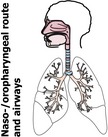
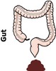
The first step in the analysis of the microbiome is to obtain samples. Samples for analysis of the microbiome in relation to asthma either originate from the naso‐/oropharyngeal route (such as nose, throat), airways or from the large intestinal tract (gut). Table 1 represents commonly used sampling compartments, and their main advantages and disadvantages.
Table 1.
Microbiome sampling compartments, advantages and disadvantages
| Route | Sampling compartment | Advantages | Disadvantages |
|---|---|---|---|
|
Nasal swab or wash | Non‐invasive, high patient acceptability, easy to sample frequently |
Patients may feel slightly uncomfortable As it only represents the upper respiratory tract, it may not be suitable if the study aim was to characterize the lower respiratory microbiome |
| Saliva, buccal swab or wash | Non‐invasive, high patient acceptability, easy to sample frequently | Differences related to gender, pH and diet intake should be accounted for | |
| Sputum (spontaneous or induced) | Relatively non‐invasive, can represent microbiota from the lower respiratory tract | May be cross‐contaminated from bacteria from the saliva or oral cavity, patient's cooperation is required to assure the quality of sample | |
| Bronchoalveolar lavage and bronchial aspirates | Good representation of the microbiota from the lower respiratory tract | Invasive, less patient acceptability, risk of cross‐contamination during aspiration | |
| Bronchial brushings and biopsies | Good representation of the microbiota from the lower respiratory tract, including mucosa‐associated microbiota | Invasive, less patient acceptability | |
|
Stool | Non‐invasive | Patient might feel uncomfortable collecting stool samples |
| Rectal swab | Non‐invasive, easy to sample frequently | Patients may feel more discomfort than stool samples, patient acceptability may be low, may be cross‐contaminated by bacteria from the skin |
Since many different sampling compartments and methods are currently used,14, 15, 16, 17, 18 obtained results are difficult to compare. For example, a study conducted in 39 asthmatics and 19 healthy controls revealed that statistically significant differences in bacterial α‐diversity (diversity in individual site or sample) and β‐diversity (intervariability, between sites or samples) were found between bronchial brushings and bronchoalveolar lavage (BAL) fluid within individuals for both asthmatics and control groups.14 In a study of 30 asthmatic children, significant differences in bacterial α‐ and β‐diversity were found between nasal brushes and nasal washes within individuals.15 In another study comparing bacterial composition in bronchial aspirates and bronchial brushings in 13 adults with severe asthma, the composition of the microbiota in the bronchial brushings showed a specific pattern that was not represented in bronchial aspirates.16 Induced sputum was found to be more compositionally similar to bronchial brushings than oral washes or nasal brushings in adult atopic asthmatics (n = 22).17 A study conducted in 60 individuals comparing stool samples with rectal swabs found that certain bacterial taxa are highly specific to the sample site, while other taxa can be found in both sample types and are specific for the individual.18
Although the choice of the sampling compartments in studies depends on several factors such as patients’ age, disease state and available facilities, it is of importance to note that microbial communities can change between sampling compartments within the same individual, for example mucosa‐associated or luminal bacteria. In addition, it is very important to consider the effect introduced by treatment with drugs on the microbiome present in different sampling compartments. For instance, studies have shown that medications such as proton pump inhibitors, antibiotics, antidepressants, statins, antidiabetics and probably others can influence gut microbiota.19, 20
2.2. Available microbiome analysis techniques
Various techniques are available to analyse the microbiome (Table S1). In the past, microbial studies in asthma were dependent on the growth of the micro‐organisms on laboratory culture media, which was limited to certain pathogenic micro‐organisms.
More recent studies have shown that culture‐based methods are relatively insensitive to detect certain micro‐organisms, such as atypical bacteria.21, 22 Therefore, other techniques have been used such as serologic testing, which depends on identifying antibodies against a pathogen, for example microimmunofluorescence or ELISA.23, 24 Newer methods, which target DNA of a pathogen, such as PCR and reverse transcriptase PCR, have been used in microbiome asthma studies,25, 26, 27 with some commercially available PCR kits for detecting certain pathogens.28, 29
With the emergence of novel nucleotide sequencing technologies, microbiome research shifted from a single pathogenic micro‐organism to a microbial community‐oriented approach, studying commensal and pathogenic bacteria and their interactions. Culture‐independent or sequencing techniques start with the extraction of the nucleic acid (DNA and/or RNA) of the collected samples and can be subdivided into amplicon sequencing or whole‐genome shotgun (WGS) sequencing.
Amplicon sequencing of the 16S ribosomal RNA (rRNA) of the microbiome is one of the most common techniques used recently in microbiome asthma studies. 16S rRNA is found in all bacteria and archaea, and is ideal for classifying micro‐organisms. There are nine separate hypervariable regions (V1‐V9) within the 16S rRNA gene30 that are targeted by the sequencing platforms. On the other hand, WGS sequencing depends on fragmentation and random sequencing of the totally extracted microbiome DNA. It has been reported that WGS sequencing has improved accuracy and provides better detection of microbial species when compared to 16S amplicon sequencing.31 Moreover, WGS sequencing provides the opportunity to assess the functional characteristics of the microbiome because it targets the whole genome instead of particular regions. However, it is more expensive and requires higher computational power than amplicon sequencing, and is therefore less used.
Various sequencing platforms are available which differ based on the read length, sequencing depth and error rates.32 The most frequently used platforms in the literature are Illumina platforms (eg, MiSeq and HiSeq) and 454 pyrosequencing. Other platforms are also available such as Ion Torrent, PacBio and SOLiD. It is important to note that different sequencing platforms may produce variable results for the same samples.32, 33 For a clear comparison between sequencing approaches and platforms, see recent articles written by Liu et al 34 and Tessler et al 35
In addition to the methodological aspects we discussed, we recommend reading a recently published article that highlights the best methodological practices that should be taken into account in the microbiome respiratory research such as quality control, data handling, microbial contamination, low biomass samples and standardized protocols.36
3. LATEST INSIGHTS INTO MICROBIAL DYSBIOSIS AND ASTHMA PHENOTYPES (SEQUENCING STUDIES)
Samples for high‐throughput microbiome sequencing studies in asthma patients are either taken at a certain time‐point of patients’ life (cross‐sectional studies) or at two or more time‐points (longitudinal studies).
3.1. Cross‐sectional studies
Various cross‐sectional studies have studied associations between airway or gut bacterial dysbiosis at single time‐points in relation to asthma phenotypes, for example mild‐moderate, severe, and uncontrolled asthma or inflammatory phenotypes, such as eosinophilic and neutrophilic asthma. Most of the studies compared the microbial compositions between asthmatics with different severity levels and healthy controls, and sometimes with other airway disorders, such as cystic fibrosis (CF) and chronic obstructive pulmonary disease (COPD). Table 2 summarizes the main findings of airway microbiome cross‐sectional studies conducted in asthmatic patients.
Table 2.
Cross‐sectional microbiome asthma studies
| Authors | Number and type of participants | Age group | Samples collected | Technique used to assign bacterial taxa | Outcome | Key finding |
|---|---|---|---|---|---|---|
| Pang et al 53, 2019 | 24 mild‐to‐moderate asthma patients (12 eosinophilic and 12 non‐eosinophilic) and 12 healthy controls | Adults | Induced sputum | 16S ribosomal RNA‐based method (targeting V3‐V4 region) | Differences in microbiota composition between eosinophilic, non‐eosinophilic asthma patients and healthy controls | Asthmatics especially the non‐eosinophilic group showed decreased diversity compared to healthy controls |
| Pérez‐Losada et al 57, 2018 | 163 children with asthma | Children (6‐18 y) | Nasal washes | 16S ribosomal RNA‐based method (targeting V4 region) | Characteristics of nasal microbiota between clusters of paediatric asthma phenotypes |
Clustering on patients clinical and biochemical characteristics revealed three distinct asthma phenotypes Bacterial diversity was significantly different between the three asthma phenotypic clusters. At phyla level, relative abundances of Proteobacteria, Actinobacteria and Bacteroidetes differed significantly within the three clusters, while at genera level, Moraxella, Corynebacterium, Dolosigranulum and Prevotella were significantly different |
| Lee et al 43, 2018 | 60 young adults and elderly with asthma and 20 healthy controls | Young adults and elderly | Nasopharyngeal swabs | 16S ribosomal RNA‐based method and shotgun metagenomics | Differences in microbiota composition between young adults and elderly asthmatic and non‐asthmatic patients |
No significant differences in bacterial diversity between asthmatics and healthy controls in both young adults and elderly patients In young adults, the relative abundance of Proteobacteria phylum was higher in asthmatics compared with healthy controls, while in the elderly, the relative abundance of Moraxella was higher in healthy controls compared with asthmatics In young adults, FEV1 predicted was inversely correlated with Prevotella, Neisseria and Fusobacterium. In contrast to the elderly, where FEV1 predicted was positively correlated with Burkholderia and Psychrobacter |
| Durack et al 17, 2018 | 22 atopic asthma patients, 12 atopic non‐asthma patients and 11 healthy controls | Adults | Protected bronchial epithelial brushes (PBs), induced sputum, oral wash and nasal brushes (NB) | 16S ribosomal RNA‐based method (targeting V4 region) | Microbiota compositional similarity between different sampling compartments |
Bacterial composition similarity was greatest between bronchial brushes and induced sputum samples especially in atopic asthma and atopic non‐asthma patients Number of bacterial taxa shared between induced sputum‐bronchial brushes and between nasal brushes‐bronchial brushes was highest in atopic asthma patients compared with healthy controls |
| Begley et al 143, 2018 | 24 adults with asthma and 8 healthy controls | Adults | Stool | 16S ribosomal RNA‐based method (targeting V4 region) | Relationship of microbiota composition to asthma characteristics and phenotypes |
Beta diversity indices were inversely correlated with FEV1 and positive specific IgE to aeroallergen, respectively Clustering on bacterial composition of asthma patients showed three clusters which differed mainly in FEV1 Bifidobacterium and Lachnospiraceae bacterial families were more abundant in asthmatics, while Bacteroides and Enterobacteriaceae families were more abundant in healthy controls |
| Buendía et al 144, 2018 | 42 asthma patients with fixed airway obstruction, 74 with reversible airway obstruction and 66 with no airway obstruction | Children and adults (8‐70 y) | Stool | 16S ribosomal RNA‐based method (targeting V4 region) | Relationship of gut microbiota composition to airway obstruction in asthma patients living in the tropics |
No significant differences in bacterial richness and diversity were found between the three phenotypes Bacterial families Streptococcaceae, Enterobacteriaceae and Veillonellaceae which corresponds to genera Streptococcus, Escherichia, Shigella and Megasphaera discriminated between patients with fixed airway obstruction and other groups |
| Yang et al 145, 2018 | 111 adults with chronic rhinosinusitis (CRS) | Adults | Nasal swabs | 16S ribosomal RNA‐based method (targeting V4 region) | Differences in microbiota composition between CRS patients with or without asthma |
No significant differences in alpha diversity between asthmatic CRS patients and others. In addition, alpha diversity was not related to emergency room visits, ACT or FEV1 Streptococcus genus was significantly higher in relative abundance in asthmatic CRS patients compared to others Proteobacteria phylum was significantly higher in asthmatic CRS patients with emergency room visits compared to other asthmatics |
| Wang et al 52, 2018 | 36 adults with asthma, and 185 healthy controls | Adults | Stool | Shotgun metagenomics | Differences in microbiota composition between asthmatics and healthy controls |
Lower bacterial richness and diversity were found in asthmatics compared to healthy controls Clostridium bolteae, Clostridium ramosum, Clostridium spiroforme and Eggerthella lenta were more abundant in asthmatic patients, while Faecalibacterium prausnitzii, Sutterella wadsworthensis and Bacteroides stercoris were more abundant in healthy controls Functional analysis shows that modules related to short‐chain fatty acid (SCFA) production such as acetate and butyrate are more enriched in healthy controls |
| Taylor et al 54, 2018 | 167 adults with stable asthma | Adults | Induced sputum | 16S ribosomal RNA‐based method (targeting V1‐V3 region) | Relationship of airway microbiota composition to asthma inflammatory phenotypes |
Patients with neutrophilic asthma had lower bacterial diversity and higher dissimilarity compared with eosinophilic asthma patients Neutrophilic asthma patients had higher relative abundances of Haemophilus and Moraxella and lower relative abundances of Streptococcus, Gemella and Porphyromonas compared with eosinophilic and paucigranulocytic asthma patients |
| Yang et al 55, 2018 | 20 adults with neutrophilic asthma and 34 with non‐neutrophilic asthma | Adults | Induced sputum | 16S ribosomal RNA‐based method (targeting V3‐V4 region) | Differences in microbiota composition between neutrophilic and non‐neutrophilic asthma |
Neutrophilic asthma is associated with lower bacterial richness and diversity compared with non‐neutrophilic asthma Proteobacteria phylum especially Haemophilus and Moraxella were more abundant in neutrophilic asthma patients. In contrast, Firmicutes, Actinobacteria and Saccharibacteria phyla were more abundant in non‐neutrophilic asthma patients |
| Fazlollahi et al 60, 2018 | 31 adults with non‐exacerbated asthma, 20 with exacerbated asthma and 21 healthy controls | Adults | Nasal swabs | 16S ribosomal RNA‐based method (targeting V3‐V4 region) | Differences in microbiota composition between exacerbated, non‐exacerbated asthma and healthy controls |
At phyla level, Bacteroidetes and Proteobacteria were enriched in asthma patients compared to healthy controls At genera level, Prevotella, Alkanindiges and Gardnerella were more abundant in exacerbated asthma patients, while Dialister was more abundant in non‐exacerbated asthma patients No bacteria were more enriched in healthy controls compared to the asthma groups |
| Goldman et al 146, 2018 | 15 children with severe asthma, 11 non‐asthmatic children and 5 patients with CF | Children (no defined age range) | Bronchoalveolar lavage (BAL) | 16S ribosomal RNA‐based method (targeting V4 region) | Differences in microbiome profiles between the three groups |
Similar bacterial taxa were found in both severe asthma and non‐asthmatic children. However, CF patients had lower richness and diversity Firmicutes, Proteobacteria and Cateriodetes were the most predominant bacterial phyla across all groups Severe asthma and non‐asthmatic groups differed significantly in 15 bacterial genera. Bacteroides, Faecalibacterium, Roseburia and 10 others were enriched in severe asthma samples, whereas Proteus and Capnocytophaga were more abundant in non‐asthma samples |
| Boutin et al 147, 2017 | 27 children with asthma, 57 with CF and 62 healthy controls | Children (6‐12 y) | Oropharyngeal swabs | 16S ribosomal RNA‐based method (targeting V4 region) | Differences in microbiome profiles between the three groups. |
Children with CF had lower bacterial diversity and total abundance compared to asthmatic children and healthy controls Asthmatics had higher abundance of Haemophilus compared to CF and healthy controls |
| Li et al 50, 2017 | 25 adults with severe asthma, 24 non‐severe asthma and 15 healthy controls | Adults | Induced sputum | 16S ribosomal RNA‐based method (targeting V3‐V5 region) | Relationship between airway microbiota, asthma severity and inflammatory type |
No significant different in microbial richness and diversity and at phyla level between severe, non‐severe asthmatics and healthy controls At the family level, severe asthmatics had higher abundance in Pseudomonadaceae and Enterobacteriaceae compared to non‐severe asthmatics and healthy controls Eosinophilic asthma patients had higher abundance of Actinomycetaceae compared to non‐eosinophilic asthma patients Healthy subjects had the highest abundance of Porphyromonadaceae compared to other groups |
| Durack et al 47, 2017 | 42 atopic steroid‐naïve asthma patients, 21 atopic non‐asthma patients and 21 non‐atopic healthy controls | Adults | PBs | 16S ribosomal RNA‐based methods (targeting V4 region) | Differences in airway microbiota in steroid‐naïve atopic asthma patients, atopic non‐asthma patients and non‐atopic healthy controls | Asthmatic patients had higher abundances of Haemophilus, Neisseria, Fusobacterium, and Porphyromonas, and Sphingomonodaceae family, and decreased abundances of Mogibacteriaceae family, and Lactobacillales order compared to other groups |
| Ruokolainen et al 59, 2017 | 196 randomly selected children from; Finnish (n = 98) and Russian (n = 98) Karelia | Children (7‐11 y and followed again at 15‐20 y) | Skin and nasal swabs | DNA sequencing | Changes in allergy in children from school age until young adulthood from two different regions, Finland and Russian Karelia, with corresponding microbiome changes in skin and nasal cavity |
Allergic disorders, such as asthma, atopic eczema, hay fever, rhinitis and atopic sensitization, were three‐ to ten‐fold more common in the Finland when compared to Russian Karelia Microbiota from skin and nasal cavity showed that overall microbial diversity and abundance of Acinetobacter genus were higher in Russian Karelia |
| Millares et al 16, 2017 | 13 patients with severe asthma | Adults | Bronchial biopsies and aspirates | 16S ribosomal RNA‐based methods | Differences in bronchial bacterial composition and their functional capacities between bronchial biopsies (BB) and bronchial aspirates (BA) in patients with severe IgE‐mediated asthma patients |
Bacteriodetes, Firmicutes, Proteobacteria and Actinobacteria were the most abundant phyla in both BB and BA samples, while prevotella and streptococcus are the most predominant genera Fusobacteria showed significantly higher relative abundances in BA contrast to Proteobacteria was significantly lower in BA when compared with BB |
| Sverrild et al 48, 2016 | 23 steroid‐free asthma patients and 10 healthy controls | Adults | BAL | 16S ribosomal RNA‐based methods (V4 region) | Relationship between airway microbiome and airway inflammation in steroid‐free asthmatics and healthy controls | Asthmatic patients with low levels of eosinophils had different microbial profiles compared with asthmatic patients with high levels of eosinophils and healthy controls; they had more abundance of Neisseria, Bacteroides and Rothia species and less abundance of Sphingomonas, Halomonas and Aeribacillus species |
| Depner et al 51, 2016 | A total of 327 rural farm and non‐farm children; 125 children with asthma and 202 controls | Children (6‐12 y) | Nasal and throat swabs | 16S ribosomal RNA‐based methods (V3‐V5 region) | Differences in nasal and throat microbiota between children with asthma and healthy controls in both farm and non‐farm children |
Children with asthma had lower nasal bacterial diversity compared with healthy controls. No difference was found in throat samples Asthmatic children had increased abundance of Moraxella bacterial genus (Proteobacteria phylum), but this was only evident in non‐farm children |
| Jung et al 44, 2016 | 89 steroid‐naïve asthma patients and 36 healthy subjects | Adults | Induced whole sputum | 16S ribosomal RNA gene terminal restriction fragment length polymorphism (T‐RFLP) | Differences in nasal microbiota between adult steroid‐naïve asthmatics and healthy controls |
No significant differences in microbial diversity and composition at phylum level were found between asthmatics and healthy controls Slight significant differences in the OTUs between the two groups were found |
| Pérez‐Losada et al 15, 2016 | 30 asthmatic children | Children (6‐17 y) | Nasal brushes (NB) and nasal washes (NW) | 16S ribosomal RNA‐based methods (V3‐V5 region) | Spatial variations in microbiome between NB and NW |
The most predominant nasopharyngeal bacterial genera in both NB and NW were Moraxella, Staphylococcus, Corynebacterium, Haemophilus, Fusobacterium, Prevotella and Dolosigranulum NB microbiome had higher α‐diversity when compared to NW Both samples showed significant differences in abundances and community composition at genera level |
| Zhang et al 42, 2016 | 26 severe asthma, 18 non‐severe asthma and 12 healthy subjects | Adults | Induced sputum | 16S ribosomal RNA‐based method (targeting V3‐V5 region) | Differences in microbiome profile between severe and non‐severe asthma patients and healthy controls |
Patients with severe and non‐severe asthma had a reduced prevalence of Bacteriodetes and Fusobacteria when compared to healthy controls There was a high increase in the prevalence of Firmicutes and a minor increase in Proteobacteria in severe asthmatics when compared to healthy controls Also, Firmicutes was increased in severe asthmatics when compared to non‐severe asthmatics Non‐severe asthmatics showed an increase in Proteobacteria compared to healthy controls and severe asthmatics |
| Simpson et al 56, 2016 | 46 patients with poorly controlled asthma | Adults | Induced sputum | 16S ribosomal RNA‐based method, and PCR | Sputum microbiome profile in adults with poorly controlled asthma |
Patients with neutrophilic asthma had lower bacterial diversity and high prevalence of Haemophilus influenza compared with non‐neutrophilic (eosinophilic) asthma, while patients with eosinophilic asthma had high prevalence of Tropheryma whipplei Neutrophilic asthma patients had lower abundance of Actinobacteria and Firmicutes, and higher abundance of Proteobacteria compared with patient with non‐neutrophilic asthma |
| Denner et al 14, 2015 | 39 asthmatic patients and 19 control | Adults | Endobronchial brushings (EB) and BAL | 16S ribosomal RNA‐based method (targeting V4 region) | Lower airway microbiome profile in relation to clinical characteristic of asthma, corticosteroid medications and airway eosinophilia |
Brush samples of asthmatic patients had higher abundance of Lactobacillus, Pseudomonas and Rickettsia species compared to controls, in contrast, healthy controls had higher abundance of Prevotella, Streptococcus and Veillonella Relative abundances of bacterial taxa were significantly associated with corticosteroid use. There was a decrease in relative abundance of Bacteriodetes and Fusobacteria and increase in Proteobacteria based on oral corticosteroid use FEV1 levels were found to influence EB microbial diversity and profile |
| Huang et al 41, 2015 |
40 severe asthma patients A comparison was further made on 41 mild‐moderate asthma subjects, and 7 healthy controls |
Adults | PBs | 16S ribosomal RNA‐based methods, followed by in silico predictive metagenomic analysis of bacterial groups of interest | Bronchial bacterial composition and its association with disease‐related features, such as BMI, ACQ scores, sputum total leucocytes, bronchial biopsy eosinophils |
Proteobacteria associated with worsening ACQ and sputum leucocyte count; in contrast, Actinobacteria associated with improving ACQ scores Protecobacteria and Firmicutes were negatively correlated with biopsy eosinophils; in contrast, Actinobacteria was positively correlated Greater bacterial burden associated with less variation in asthma control and less eosinophil infiltration in bronchial tissue Severe asthmatics were enriched with Klebsiella compared with health controls, and Actinobacteria, Gammaproteobacteria and Klebsiella compared with mild‐moderate asthmatics In contrast, several families of Proteobacteria were more abundant in mild‐moderate asthmatics compared with severe patients, with the exception of Entrobacteriaceae which was more enriched in severe asthmatics |
| Ogorodova et al 148, 2015 (Russian article) | 50 patients with bronchial asthma (23 with mild‐moderate asthma and 27 with severe uncontrolled asthma) and 88 patients with COPD (57 with mild‐moderate severity and 31 with severe course of disease) | Adults | Oropharyngeal swabs | 16S ribosomal RNA‐based method (targeting V3‐V4 region) | Differences in oropharyngeal microbiota composition between patients with bronchial asthma and chronic obstructive pulmonary disease with different severity levels |
There was a decrease in prevalence of Prevotella and increase of species Bifidobacterium longum, Prevotella nanceiensis, Neisseria cinerea, Aggregatibacter segnis and genera Odoribacter, Alloiococcus, Lactobacillus, Megasphaera, Parvimonas and Sneathia in severe uncontrolled asthma patients in comparison with patients which have mild persistent asthma In asthma patients compared to COPD patients, there was an increase in prevalence of Prevotella melaninogenica and genera Selenomonas, Granulicatella and Gemella, and decrease of Prevotella nigrescens, Haemophilus influenza and genera Aggregatibacter, Alloiococcus, Catonella, Mycoplasma, Peptoniphilus and Sediminibacterium |
| Park et al 40, 2014 | 18 asthma patients, 17 COPD patients and 12 healthy controls | Adults | Oropharyngeal swabs | 16S ribosomal RNA‐based method (targeting V1‐V3 region) | Differences in oropharyngeal microbiota composition between asthma, COPD patients and healthy controls | Asthma and COPD patients had higher abundance of Proteobacteria and Firmicutes (particularly Pseudomonas and Lactobacillus species) compared to healthy controls; in contrast, healthy controls had higher abundance of Streptococcus, Veillonella, Prevotella and Neisseria species |
| Green et al 149, 2014 | 28 severe treatment‐resistant neutrophilic asthma subjects | Adults | Induced sputum | 16S rRNA gene T‐RFLP | Abundance of bacterial taxa in severe treatment‐resistant asthmatics and their association with clinical characteristics and airway inflammatory markers | Airway colonization with Haemophilus spp, Streptococcus spp or Moraxella catarrhalis (members of the phyla Proteobacteria and Firmicutes) positively correlates with sputum neutrophilia and Il‐8, lower FEV1, and longer disease duration, with M catarrhalis the bacterial species most associated with neutrophilic disease |
| Goleva et al 49, 2013 | 39 subjects with asthma (29 corticosteroid‐resistant and 10 corticosteroid‐sensitive) and 12 healthy controls | Adults | BAL | 16S ribosomal RNA‐based method | Differences in airway microbial composition between corticosteroid‐resistant, corticosteroid‐sensitive asthmatics and normal control subjects | No difference in microbial phyla composition was found between corticosteroid‐resistant, corticosteroid‐sensitive individuals, and healthy controls in terms of richness, diversity, evenness and community composition. However, significant variations in the percentage of sequences of different bacterial genera were found |
| Marri et al 39, 2013 | 10 patients with mild asthma and 10 healthy controls | Adults | Induced sputum | 16S ribosomal RNA‐based method (targeting V6 region) | Differences in airway microbiota between asthmatics and controls | Higher bacterial diversity was found in samples of asthmatic patients compared to controls. Proteobacteria were significantly higher in asthmatics compared to controls. In contrast, Firmicutes and Actinobacteria were non‐significantly higher in controls |
| Haung et al 38, 2011 | 65 sub‐optimally controlled asthma patients, and 10 healthy subjects | Adults | PBs | 16S ribosomal RNA microarray | Differences in bronchial bacterial composition between suboptimal controlled asthmatics and controls |
Bacterial diversity was significantly higher in asthmatic subjects compared to controls Methacholine P20 concentrations were negatively correlated with bacterial diversity, suggesting that bacterial diversity is positively correlated with bronchial hyperresponsiveness. The most predominant bacterial phyla associated with bronchial hyperresponsiveness were Proteobacteria |
| Hilty et al 37, 2010 | 24 adults patients, 11 with asthma, 5 with COPD and 5 healthy controls 20 children, 13 with difficult asthma and 7 controls | Adults, and children (up to 17 y) |
Adults: Naso‐oropharyngeal swabs, bronchial duplicate cytology brushings Children: BAL |
16S ribosomal RNA‐based method | Differences in airway microbiota between asthma, COPD patients and controls |
Microbial community from the nose was distinct from the oropharyngeal and bronchial brushings Proteobacteria, especially Haemophilus spp, were more common in diseased patients (asthmatics and COPD) compared to controls; in contrast, Bacteriodetes, especially Prevotella spp, were more common in controls than asthmatics and COPD patients |
Abbreviations: ACQ, Asthma Control Questionnaire; BA, bronchial aspirates; BAL, bronchoalveolar lavage; BB, bronchial biopsies; BMI, body mass index; CF, cystic fibrosis; COPD, chronic obstructive pulmonary disease; CRS, chronic rhinosinusitis; EB, endobronchial brushings; FEV1, forced expiratory volume in 1 second; IgE, immunoglobulin E; NB, nasal brushes; NW, nasal washes; OTU, operational taxonomic unit; PBs, protected bronchial epithelial brushes; T‐RFLP, terminal restriction fragment length polymorphism.
Regarding airway bacterial composition, various studies have shown that Proteobacteria, particularly Haemophilus species, are more common in asthmatic patients, while Bacteroidetes and Actinobacteria are more common in healthy controls.37, 38, 39, 40, 41, 42, 43 However, it is not clear whether this difference in bacterial composition is a feature of asthma itself, related to corticosteroid use, or both. For example, there was an increase in the relative abundance of Proteobacteria and decrease in the abundance of Bacteroidetes and Fusobacteria in endobronchial brushings samples with increasing corticosteroid use.14 Furthermore, it was reported that there were no differences in microbial compositions at phyla level between healthy controls and corticosteroid‐free asthma patients.44 A possible explanation for the effect of corticosteroids on microbiota is that they suppress or disturb mucosal immune system and therefore lead to overgrowth of bacteria. This overgrowth probably leads to harmful health effects if opportunistic or pathogenic micro‐organisms became abundant such as fungal candidiasis associated with the use of inhaled corticosteroids.45 However, it may still lead to beneficial effects by increasing abundances of beneficial commensal bacteria.46 The results of other studies may reflect that corticosteroids are not the only influencers of the bacterial compositions.47, 48, 49 For instance, in a study of 42 atopic corticosteroid‐naïve asthmatics, 21 atopic non‐asthmatics and 21 healthy controls, corticosteroid‐naïve asthmatics had a distinct airway bacterial profile when compared to the other groups.47 In the same study, atopic asthmatic patients were further randomized into fluticasone arm versus placebo which later revealed that corticosteroids may cause changes in certain bacteria.47 In another study of 23 corticosteroid‐free asthma patients and 10 healthy controls, bacterial dysbiosis was found between asthma patients with a low level of eosinophils when compared with asthma patients with high levels of eosinophils and healthy controls.48 This suggests that asthma disease by itself, different phenotypes, and medications used (including corticosteroids) play a significant role in shaping the microbiome profiles.
Regarding bacterial diversity, some studies have shown that asthma patients had higher bacterial diversity in the airways when compared to healthy controls.38, 39 Again, this might be related to the effect of corticosteroids, which by suppressing immunity would enhance the growth of diverse microbiota. However, in other studies, no significant differences in bacterial diversity were found between asthma patients and healthy controls.43, 44, 50 Other studies showed that asthmatic patients had lower airway and gut microbiome diversity when compared to healthy controls.51, 52, 53
Inflammatory phenotypes of asthma have been shown to be associated with airway bacterial dysbiosis. Three studies have reported that neutrophilic asthma patients had lower sputum bacterial richness and diversity and showed higher abundances of Proteobacteria phylum especially Moraxella and Haemophilus compared with non‐neutrophilic, eosinophilic or paucigranulocytic asthma phenotypes.54, 55, 56 In contrast, eosinophilic asthma patients had higher sputum abundances of Actinobacteria phylum especially Actinomycetaceae family compared with non‐eosinophilic or neutrophilic asthma patients.50, 56 The microbial dysbiosis observed between different asthma phenotypes has driven some research groups to phenotype cluster their patients based on asthma clinical and biochemical characteristics before exploring microbial dysbiosis between these phenotypes. Two studies have shown that microbial composition can differ between the identified phenotypic clusters.57, 58 Observed microbial differences between different asthma phenotypes may help later to better diagnose them and provide an opportunity to use the microbiome as a biomarker in precision medicine approaches of asthma.
However, it is important to note that the above‐mentioned studies measured microbiota in different sampling compartments which might induce variability of the obtained results and limit the ability to directly apply microbiome results in clinical care. In addition, some variations in results are still observed when comparing studies originating from the same sampling compartment. It should be noted that other factors can play a role in results dissimilarities. For instance, individuals originating from different populations might contribute to this variability because of the exposure to different environments and risk factors. A study shown that children from Russian Karelia had higher overall bacterial diversity and abundance of genus Acinetobacter and were less likely to have allergic disorders including asthma, compared to children from Finland Karelia.59 In addition to different populations and environments, other factors related to multiple asthma phenotypes studied,50, 53, 55, 60 asthma and non‐asthma medications used besides corticosteroids,61 dietary habits62 and age groups.43 Moreover, it should be noted that sample size is a huge determining factors for inconsistencies found in reported microbiome statistical differences which should be considered while designing the microbiome asthma studies.
In conclusion, there is no distinct microbiome profile that can be said to be responsible for or associated with the asthma disease process. Under the umbrella of inconsistencies in reported results, several factors might hold responsible such as different populations, environments, age groups, drugs, diet and study design.
3.2. Longitudinal studies
In longitudinal microbiome studies, patients have been followed prospectively to study associations between early life microbial dysbiosis and asthma development later in life or changes in the microbiome composition between two time‐points (Table 3). Often the gut microbiome in children has been investigated in this type of studies. A recent study showed that an immature gut microbiome profile at 1 year of age in children born to asthmatic mothers was associated with asthma development at 5 years of age.63 However, another study showed that children who developed asthma at 7 years of age (n = 8) had distinct gut bacterial profiles at one week and one month, but not at one year of age.64 Similarly, it was shown that during the first three months of life, children with high risk of asthma had lower abundances of certain bacteria genera in their stool compared to others.65, 66 A different study showed that there was an association between gut bacterial diversity at 1 month and 1 year of age and allergic sensitization, but not with asthma development until 6 years of age.67
Table 3.
Longitudinal microbiome asthma studies
| Authors | Number and type of participants | Samples | Time of collection | Technique used to assign bacterial taxa | Outcomes | Key finding |
|---|---|---|---|---|---|---|
| Dzidic et al 150, 2018 | 80 children (47 allergic and 33 healthy) | Saliva | 3, 6, 12, 24 mo and 7 y of age | 16S ribosomal RNA‐based method (targeting V1‐V5 region) | Allergic symptoms and sensitization at 7 y of age |
Allergic children, particularly asthmatics, had lower bacterial diversity compared to healthy children at 7 y of age During early infancy, there was an increase in relative abundances of Gemella haemolysans in allergic children, while healthy children showed increased abundances of Lactobacillus gasseri and Lactobacillus crispatus |
| Stokholm et al 63, 2018 | 690 children | Stool | 1 wk, 1 mo and 1 y of age | 16S ribosomal RNA‐based method (targeting V4 region) | Asthma at 5 y of age | Children at 1 y of age who have an immature gut microbiome profile have increased risk of asthma at 5 y of age, but this effect is only evident in children who are born to asthmatic mothers |
| Pérez‐Losada et al 151, 2017 | 40 asthmatic children (6‐18 y) | Nasopharyngeal washes | 2 samples collected 5.5‐6.5 mo apart | 16S ribosomal RNA‐based method (targeting V4 region) | Temporal dynamics of airway microbiome in asthmatic children and their stability over time |
No significant differences in α‐ and β‐diversity were found between seasons There were significant differences in relative abundances of Haemophilus, Staphylococcus, Moraxella and Corynebacterium between summer and fall season and between age groups |
| Fujimura et al 152, 2016 | 298 children | Stool | 1 and 6 mo | 16S ribosomal RNA‐based method (targeting V4 region) | Atopy diagnosis at 2 y of age and asthma diagnosis at 4 y of age |
Neonates with highest risk of atopy and asthma diagnosis showed reduced relative abundances of Bifidobacterium, Akkermansia and Faecalibacterium bacterial taxa and high relative abundances of Candida and Rhodotorula fungi They also showed enriched fecal pro‐inflammatory metabolites |
| Stiemsma et al 66, 2016 | 76 children (39 preschool asthmatic children and 37 matched healthy control) | Stool | 3 mo and 1 y of age | 16S ribosomal RNA‐based method (targeting V3 region) | Asthma diagnosis by 4 y of age |
Asthma patients at 3 mo of age showed decreased abundance of the genus Lachnospira and a trend for increased abundance of the species Clostridium neonatale The ratio of these two bacteria (L/C) was negatively associated with asthma risk by 4 y of age |
| Arrieta et al 65, 2015 | 319 children (136 with wheezing, 87 with atopy, 22 both, and 74 controls) | Stool | 3 mo and 1 y of age | 16S ribosomal RNA‐based method (targeting V3 region) | High risk of asthma development (asthma diagnosis at 3 y of age) |
No significant difference was found in microbiome diversity among the four groups During the first 100 d of life, children with high risk of asthma had lower abundance of the bacteria genera; Lachnospira, Veillonella, Faecalibacterium and Rothia |
| Teo et al 68, 2015 | 234 children | Nasopharyngeal aspirates | 2, 6 and 12 mo of age during two states; healthy condition and episodes of acute respiratory illness | 16S ribosomal RNA‐based method | Impact of dynamics of nasopharyngeal microbiome, during the first year of life during both healthy condition and episodes of acute respiratory infections, on allergic sensitization at 2 y and chronic wheeze at 5 and 10 y of age | Children who had atopy by age of 2 y and developed chronic wheeze at 5 y of age were twice likely to have early colonization with Streptococcus, and lower respiratory tract infection during the first year of life compared to those who did not develop wheezing |
| Abrahamson et al 64, 2013 | 47 children | Stool | 1 wk, 1 mo and 1 y of age | 16S ribosomal RNA‐based method (targeting V3‐V4 region) | Allergic diseases at school age (7 y), such as; asthma, eczema and allergic rhinitis |
8 children who developed asthma had lower microbial diversity compared to non‐asthmatic children at 1 wk and 1 mo of age, but no significant difference at 1 y of age No significant difference in bacterial relative abundances was found between children with asthma, eczema or allergic rhinitis and others at all age groups |
| Bisgaard et al 67, 2011 | 411 children | Stool | 1 mo and 1 y of age | PCR of 16s ribosomal RNA and bacterial cultures | Allergic diseases until age of 6 y | Bacterial diversity at 1 mo and 1 y of age was inversely associated with allergic rhinitis, allergic sensitization (skin prick test and serum specific IgE) and blood eosinophils. No association was found with asthma development or atopic dermatitis |
Abbreviations: IgE, immunoglobulin E; PCR, polymerase chain reaction.
Only limited studies are available studying early life airway bacterial composition and the risk of developing asthma. One study showed that chronic wheeze at 5 years of age was associated with early airway colonization with Streptococcus during the first year of life.68 In another study, cultures from the hypopharyngeal region aspirates of 321 infants at 1 month showed that colonization with Streptococcus pneumoniae, Haemophilus influenzae or Moraxella catarrhalis increased the risk for recurrent wheeze and asthma at 5 years of age.69
Despite that there was inconsistency about the most important time period (whether the first 3 months or the first year of life) to shape asthma risk later in life, it is apparent that gut or airway microbiota disturbances at an early age, especially the first year of life, are risk factors for asthma development. This bacterial dysbiosis may explain why early life antibiotic exposure may increase risk of later asthma development.70, 71, 72
In conclusion, longitudinal microbiome studies may help to uncover the shifts that occur in microbiome profiles over time. Studying the time‐associated changes in the microbiome is essential because the microbiome is dynamic by nature and is influenced by several environmental and biological conditions. In addition to the studies that investigate early life microbial dysbiosis and its relation to asthma risk, more longitudinal investigation should be also conducted in patients with already established asthma disease to uncover the dynamic relation between the microbiome and the asthma disease process.
4. HOW CAN THE MICROBIOME INFLUENCE ASTHMA DEVELOPMENT?
Various studies have found associations between microbial dysbiosis in the airways and/or intestinal tract and the risk of asthma development; however, the causal relationship remains unclear. In this section, we discuss two different processes, which may explain how the microbiome might influence asthma risk and the evidence supporting them.
4.1. From hygiene hypothesis to old friends theory
The “hygiene hypothesis” that was first developed by Strachan et al assumes that excessive cleanliness, vaccination and antibiotic use can lead to an over‐reactive immune system.73, 74 It was suggested that too much hygiene lowers the threshold of immune adaptation, it becomes sensitive and it over‐reacts to substances that are normally not recognized by the immune system, like pollen and other allergens. In contrast, more exposure to micro‐organisms may lead to immune tolerance and hence less recognition to allergens. Various studies have shown that more exposure to micro‐organisms during childhood has protective effects against asthma development, for example; growing up in a farm environment, exposure to livestock animals and dogs, drinking unpasteurized milk and breastfeeding all have protective effects on asthma development.75, 76, 77, 78, 79
However, the limitation to this hypothesis is that it cannot be generalized to all populations. For instance, Japanese individuals are less prone to develop asthma than individuals living in the United States and Australia,80 despite that they are exposed to a more hygienic environment.81 The Japanese people have different dietary habits, eat more fermented products and possibly have a microbiome profile that protects them from allergy. Due to the inconsistencies to the hygiene hypothesis found in various studies, a revision has been suggested by the introduction of the “old friend” theory by Rook et al.82 This theory proposed that micro‐organisms co‐evolved with humans (old friends) which have been essential for the development of the immune system.82 Exposure to industrialized environments diminishes these micro‐organisms with subsequent defective immune regulation which results in predisposition of the human to chronic inflammatory diseases, such as asthma. Therefore, exposure to natural environments, such as breastfeeding, natural delivery, outdoor activities, healthy‐balanced diets and suitable use of antibiotics can re‐establish the healthy microbiome environment and consequently reduces risk of allergic disorders.83
4.2. The gut‐lung axis and effects on immunity
Gut microbiota produce different types and amounts of short‐chain fatty acids (SCFAs) by fermentation of dietary carbohydrates depending on the food type.84 High‐fibre diets produce the largest amount of SCFAs. Acetate, proprionate and butyrate are the most abundant SCFAs.85, 86 SCFAs go to the liver via the portal vein where they are metabolized and those who escape metabolism pass into the systemic circulation and consequently reach other organs such as the lungs. A case‐control study conducted in 476 children found that high vegetable fibre intake was protective against moderate to severe airway hyperresponsiveness,87 which may suggest that low SCFAs in the gut may be associated with asthma development. A recent prospective birth cohort study conducted on 301 children has shown that the highest levels of butyrate and propionate (>95th percentile) found in the stool of one‐year‐old infants were later associated with reduced risk of asthma and other allergic disorders at 3 and 6 years of age which might be linked to dietary intake.88 In a mice study, a high‐fibre diet increased levels of circulating SCFAs which decreased susceptibility to allergic airway inflammation, in contrast to low fibre diet which showed the reverse effect.89
Short‐chain fatty acids have affinity for G‐protein‐coupled receptors such as GPR41, GPR43 and GPR109a, which are known to regulate mucosal immunity.89, 90, 91, 92 In preclinical and clinical studies, high‐fibre diet and SCFAs were found to reduce inflammatory biomarkers and improve lung function through an action mediated through these receptors.89, 93 In addition to the effects of SCFAs on receptors, butyrate has been shown to act as histone deacetylase inhibitor (HDACi).94 HDACis have been shown to inhibit airway hyperresponsiveness and have anti‐inflammatory effect with therapeutic potential in patients with asthma.95, 96 Figures S1 and S2 show the proposed mechanisms by which SCFAs interacts with airway environment through a gut‐lung axis–dependent pathway in both healthy and diseased states.
In addition to the effect of SCFAs, certain bacterial taxa, whether commensal or pathogenic, have an influence on immunity. Mice studies have shown that commensal bacteria, such as Clostridium, Lactobacillus, Bifidobacterium, Bacteroides (or their capsular polysaccharides), have been found to induce Treg cell production, and/or reduce TH1, TH2 and TH17 responses and inflammatory process in some tissues including the airways.97, 98, 99, 100, 101, 102, 103, 104 Bifidobacterium longum was found in higher prevalence in the gut of healthy children (n = 102) compared to those with high risk of allergic asthma and atopic dermatitis (n = 99).105 Increased abundance of nasopharyngeal lactobacillus was associated with reduced risk of wheezing at two years of age in 118 infants.106 In the respiratory tract, nasopharyngeal colonization with S pneumoniae and H influenzae leads to increased production of CXCL8 and CXCL2 in nasal lavage fluid of mice, and amplification of asthma‐like pro‐inflammatory responses.107 A study conducted on the bronchial epithelial brushings of severe asthmatics (n = 40), mild‐moderate asthmatics (n = 41) and healthy controls (n = 7) showed that Actinobacteria was correlated with gene expression of FK506 binding protein (FKBP5), which is a marker of corticosteroid responsiveness, while Proteobacteria was correlated with Th17 gene expression.41 In a study conducted in 8 asthmatics and 6 healthy controls, asthma patients had higher abundance of Proteobacteria, and particularly M catarrhalis was associated with increased expression of inflammatory mediators (eg CCL20, IL1A and IRAK2) and apoptosis (eg TNF and C8orf4).108, 109 A mice study confirmed the pathogenic role of Proteobacteria inhaled through the nostrils, which induced severe lung inflammation and immunopathology characterized by prevalent airway neutrophilia, and expression of cytokine/chemokine profile.110 These results are in line with the cross‐sectional asthma studies which were discussed previously and imply that increased abundances of certain bacterial taxa, for example Proteobacteria and Firmicutes, and decreased abundances of others, for example Bacteroidetes and Actinobacteria, in asthmatics are probably influencers of asthma disease development, risk and severity.
5. THERAPEUTIC VALUE OF THE MICROBIOME INSIGHTS FOR ASTHMA: pro‐BIOTIC, PREBIOTIC AND SYNBIOTIC INTERVENTIONS
The importance of the microbiota in the asthma disease process has raised interest in the possibility of using dietary supplements influencing the intestinal bacteria, such as pro‐biotics111 and prebiotics112 and their combinations synbiotics to treat or control asthma. Pro‐biotics are “live micro‐organisms that, when administered in adequate amounts, confer a health benefit to the host”,111 while prebiotics are “substrates that are selectively utilized by host micro‐organisms conferring a health benefit”.112 Synbiotics are combinations of both pro‐ and prebiotics.
Most evidence of beneficial effects of pro‐, pre‐ and synbiotics comes from animal studies. Mice studies showed that administration of Bifidobacterium breve or adolescentis strains and Lactobacillus plantarum or rhamnosus strains significantly decreased allergic airway inflammation in OVA‐ or birch pollen‐induced asthma mice models.113, 114, 115, 116 It is worthy to note that the effect of pro‐biotics in mice asthma models is more prominent in neonates than adults.117
One of the available therapeutic options is CpG oligodeoxynucleotides (CpG‐ODN) which are synthetic single‐stranded DNA that have affinity for TLR9. Mice and other animal (eg cats and rhesus monkeys) studies have shown that CpG‐ODN can significantly reduce airway inflammation, hyperreactivity and remodelling 118, 119, 120, 121, 122, 123, 124 and the effect was more pronounced when it was combined with the antigen or used as nanoparticle formulations.119, 121, 122, 125, 126
In a house dust mite (HDM)‐induced asthma mice model, dietary galacto‐oligosaccharides were found to prevent airway hyperresponsiveness, airway eosinophilia and TH2 response, in a comparable way to budesonide127 and the effect was eliminated in a Treg depleted model which might suggest that it is a Treg cell–mediated response.128 Synbiotic combination of B breve and non‐digestible oligosaccharides was found to have a strong airway anti‐inflammatory properties in mice asthma models,129, 130 and the anti‐allergic effect was greater than their individual effects.131
In contrast to mice studies, little evidence has been found for the beneficial effect of pro‐biotics in human asthma. A meta‐analysis conducted on 3257 infants from nine randomized clinical trials found that the risk ratio (RR) of asthma in infants received pro‐biotics was 0.99 (95% confidence interval [CI] 0.81‐1.21) and RR of wheeze was 0.97 (95% CI 0.87‐1.09).132 Another meta‐analysis conducted on 25 randomized clinical trials showed that pro‐biotics significantly reduced total IgE levels (−7.59 U/mL mean reduction, 95% CI: −14.96 to −0.22); however, they did not significantly reduce risk of asthma or wheeze (relative risk = 0.96, 95% CI: 0.85‐1.07).133 A randomized controlled trial was conducted on 1,302 asthmatic subjects older than 5 years of age to assess whether pro‐biotic intake can reduce antibiotic prescribing for winter respiratory tract infections.134 In this trial, no significant difference between the intervention group (n = 652) and control group (n = 650) was seen regarding the proportion of participants who were prescribed new courses of antibiotics (odds ratio = 1.04; 95% CI, 0.82‐1.34).134 Despite the diminished effects of pro‐biotics shown in human controlled clinical trials, a recent study showed that the delayed gut microbial development in infants with high risk of asthma can be altered with daily oral supplementation of Lactobacillus rhamnosus GG compared with placebo, and this effect was lost 6 months after stopping the supplementation.135 By changing gut microbial maturation early in infancy, there is a possibility to modify asthma risk later in life. This study provides a new potential strategy for using pro‐biotics as an early preventive measure in high‐risk infants.
In an open‐label trial of 20 adult human asthmatics allergic to HDM, A‐type CpG‐ODN given as adjuvant to subcutaneous immunotherapy with HMD allergen extract was found to be safe and produce nearly complete alleviation of allergic symptoms after 10 weeks of immunotherapy.136 In a double‐blind trial, where 63 asthmatic patients were treated with 7 injections of either QbG10 (bacteriophage with CpG‐motif G10 inside) or placebo, it was found that two‐thirds of the intervention group had their asthma well controlled after 12 weeks of treatment compared to one‐third of the placebo group.137 For the effect of prebiotics on humans, a meta‐analysis conducted on 249 infants from 2 trials showed that prebiotics reduce the risk of asthma/wheezing compared to the control group (RR = 0.37, 95% CI: 0.17‐0.8); however, low number of events made the precision questionable.138
In summary, mice studies show promising results of using pro‐, pre‐ and synbiotics in asthma models, while evidence in humans is scarce, especially for pro‐biotics. One possibility for decreased therapeutic outcomes in infants is that the intended therapeutic outcomes are long term (for years) which can be influenced by different factors, such as environmental exposure, different dietary habits and dynamicity of microbiome over time. Whether the individual should receive high‐fibre diet or making certain diet restrictions should be also considered in clinical trials of pro‐biotics. New strategies for using products targeting the microbiota to prevent or treat asthma should be further investigated. Moreover, the role of precision medicine is very important to define certain individuals or patients’ groups who can benefit most from the potential therapeutic “‐biotics” options.
6. THE ASTHMA MICROBIOME FROM BENCH TO BEDSIDE: CHALLENGES
Results of microbiome studies in patients with asthma convey the importance of the role of microbiome either in the intestinal tract or in the airways in shaping asthma. However, using combined information from different studies to elaborate the asthma‐microbiome interaction in humans is hindered by methodological, design and interpretations issues/challenges in microbiome studies (Table 4). We suggest some recommendations to overcome them.
Table 4.
Challenges that interfere for optimum delivery of gained knowledge and therapeutic potential of microbiome and “‐biotics” products from bench to bedside
| From bench to bedside | Challenges | Recommendations |
|---|---|---|

|
Different sampling compartments and whether they are mucosal or luminal should be considered while studying microbiome in asthma | It is important to investigate the microbiome from various sampling compartments in the same studied population and whether drugs (both asthma and non‐asthma medications) are associated with microbial changes in certain compartments. Therefore, it might be wise to collect samples from multiple compartments while conducting microbiome asthma studies and adjust for effects introduced by certain medications |
| Different sequencing approaches and platforms might produce variability in reported results | The choice of desired method depends on various factors, such as costs, quality and error rates of the produced sequencing reads. However, investigators should take into account that some of the variability can be introduced by the choice of the sequencing platforms and techniques. Therefore, this is important to consider when comparing results from different studies | |
| Some inconsistencies in the reported results have been found in microbiome asthma studies, which might influence clinical applicability and relevance | Large multi‐centre international microbiome studies, accounting for the effect of multiple possible confounders, should be conducted to reach more definitive conclusions | |
| Different ways of reporting studies results, in terms of bacterial taxa, whether at phylum, genus or species level and using specialized microbiological terms can make hindrance for non‐expert healthcare professional to see clinical relevance and can produce difficulty to directly compare results between studies | An agreement of clinical experts, stakeholders and healthcare organizations on standardized structure or pattern (as possible) for reporting results of microbiome studies will help to facilitate comparison, interpretation and systematic analysis of the combined work of world‐wide research groups | |
| Some of the published microbiome asthma studies have investigated the gut microbiome profile without reference to the diet composition or the type of meal the patients usually consume which is an important determinant for the production of SCFAs | Accounting for diet while investigating the microbiome should be undertaken in the asthma studies | |
| There is hindrance in the optimum transfer of the therapeutic potential of products targeting microbiota(‐biotics) from animal studies to humans | A thorough investigation is required to adequately enclose barriers of transmission, and whether personalizing or tailoring “‐biotics” to certain patient/groups of patients will show more beneficial effect |
More efforts are required from healthcare professionals and researchers to conduct large‐scale microbiome asthma studies, if possible, accounting for and/or minimizing sources of variability, to obtain a clearer overview of the microbiome role in asthma. We still do not clearly know the type of link between reported microbial dysbiosis and different asthma characteristics; whether is it an association or causation? And what is the direction in case of the latter? The finding that early life microbial dysbiosis was associated with increased risk of asthma later in life might suggest that the microbiome is responsible to some extent to asthma disease development. However, this does not negate that the link might be bidirectional. More research is needed to investigate the relationship behind just “associations.”
Translating the results of microbiome asthma studies from sequencing reads into applicable clinical guidelines requires a systematic approach and cooperation of multi‐disciplinary research groups. One strategy is to use the microbiome to better refine asthma phenotypes (eg neutrophilic asthma) diagnosis, for which certain therapeutics targets might be directed. Another strategy is to detect groups of asthma patients with clear microbial dysbiosis for whom interventions (eg pre‐, pro‐ and synbiotics) to reshape the gut and/or airway microbiome might be more beneficial.
The microbiome is thought to have host genetic background,139 cause epigenetic modulations,140 affect host metabolism141 and be influenced by environmental exposures.142 This necessitates integrating the microbiome with other omics layers, such as metabolomics, proteomics, transcriptomics, genomics, epigenomics and exposomics (environmental factors), which will help to provide more insight into the mechanisms of complex disease processes such as asthma. Hopefully, this will aid to discover new biomarkers for better diagnosis and/or new therapeutic targets in asthmatic patients.
CONFLICT OF INTEREST
AHM has been reimbursed for visiting the ATS by Chiesi, AHM received a fee for participating in advisory boards for Boehringer lngelheim and Astra Zeneca, and AHM received an unrestricted research grant from GSK. ADK received grants/research support from several companies ao Janssen, GSK, Nutricia Research, Friesland Campina and NTRC.
Supporting information
Abdel‐Aziz MI, Vijverberg SJH, Neerincx AH, Kraneveld AD, Maitland‐van der Zee AH. The crosstalk between microbiome and asthma: Exploring associations and challenges. Clin Exp Allergy. 2019;49:1067–1086. 10.1111/cea.13444
REFERENCES
- 1. Soriano JB, Abajobir AA, Abate KH, et al. Global, regional, and national deaths, prevalence, disability‐adjusted life years, and years lived with disability for chronic obstructive pulmonary disease and asthma, 1990–2015: a systematic analysis for the Global Burden of Disease Study 2015. Lancet Respir Med. 2017;5(9):691‐706. [DOI] [PMC free article] [PubMed] [Google Scholar]
- 2. Loftus PA, Wise SK. Epidemiology of asthma. Curr Opin Otolaryngol Head Neck Surg. 2016;24(3):245‐249. [DOI] [PubMed] [Google Scholar]
- 3. Borish L, Culp JA. Asthma: a syndrome composed of heterogeneous diseases. Ann Allergy Asthma Immunol. 2008;101(1):1–8; quiz 8‐11, 50. [DOI] [PubMed] [Google Scholar]
- 4. Sender R, Fuchs S, Milo R. Revised estimates for the number of human and bacteria cells in the body. PLoS Biol. 2016;14(8):e1002533. [DOI] [PMC free article] [PubMed] [Google Scholar]
- 5. Turnbaugh PJ, Ley RE, Hamady M, Fraser‐Liggett C, Knight R, Gordon JI. The human microbiome project: exploring the microbial part of ourselves in a changing world. Nature. 2007;449(7164):804–810. [DOI] [PMC free article] [PubMed] [Google Scholar]
- 6. Ursell LK, Metcalf JL, Parfrey LW, Knight R. Defining the human microbiome. Nutr Rev. 2012;70(Suppl 1):S38–S44. [DOI] [PMC free article] [PubMed] [Google Scholar]
- 7. Peterson J, Garges S, Giovanni M, et al. The NIH human microbiome project. Genome Res. 2009;19(12):2317–2323. [DOI] [PMC free article] [PubMed] [Google Scholar]
- 8. Qin J, Li R, Raes J, et al. A human gut microbial gene catalogue established by metagenomic sequencing. Nature. 2010;464(7285):59–65. [DOI] [PMC free article] [PubMed] [Google Scholar]
- 9. Busse WW, Lemanske RF, Gern JE. The role of viral respiratory infections in asthma and asthma exacerbations. Lancet. 2010;376(9743):826–834. [DOI] [PMC free article] [PubMed] [Google Scholar]
- 10. Johnston SL, Martin RJ. Chlamydophila pneumoniae and Mycoplasma pneumoniae: a role in asthma pathogenesis? Am J Respir Crit Care Med. 2005;172(9):1078–1089. [DOI] [PubMed] [Google Scholar]
- 11. Kraft M. The role of bacterial infections in asthma. Clin Chest Med. 2000;21(2):301–313. [DOI] [PubMed] [Google Scholar]
- 12. Denning DW, O'Driscoll BR, Hogaboam CM, Bowyer P, Niven RM. The link between fungi and severe asthma: a summary of the evidence. Eur Respir J. 2006;27(3):615–626. [DOI] [PubMed] [Google Scholar]
- 13. Strachan DP. Hay fever, hygiene, and household size. BMJ. 1989;299(6710):1259–1260. [DOI] [PMC free article] [PubMed] [Google Scholar]
- 14. Denner DR, Sangwan N, Becker JB, et al. Corticosteroid therapy and airflow obstruction influence the bronchial microbiome, which is distinct from that of bronchoalveolar lavage in asthmatic airways. J Allergy Clin Immunol. 2016;137(5):1398–1405.e1393. [DOI] [PMC free article] [PubMed] [Google Scholar]
- 15. Perez‐Losada M, Crandall KA, Freishtat RJ. Two sampling methods yield distinct microbial signatures in the nasopharynges of asthmatic children. Microbiome. 2016;4(1):25. [DOI] [PMC free article] [PubMed] [Google Scholar]
- 16. Millares L, Bermudo G, Pérez‐Brocal V, et al. The respiratory microbiome in bronchial mucosa and secretions from severe IgE‐mediated asthma patients. BMC Microbiol. 2017;17:20. [DOI] [PMC free article] [PubMed] [Google Scholar]
- 17. Durack J, Huang YJ, Nariya S, et al. Bacterial biogeography of adult airways in atopic asthma. Microbiome. 2018;6(1):104. [DOI] [PMC free article] [PubMed] [Google Scholar]
- 18. Jones RB, Zhu X, Moan E, et al. Inter‐niche and inter‐individual variation in gut microbial community assessment using stool, rectal swab, and mucosal samples. Sci Rep. 2018;8(1):4139. [DOI] [PMC free article] [PubMed] [Google Scholar]
- 19. Imhann F, Vich Vila A, Bonder MJ, et al. The influence of proton pump inhibitors and other commonly used medication on the gut microbiota. Gut Microbes. 2017;8(4):351–358. [DOI] [PMC free article] [PubMed] [Google Scholar]
- 20. Jackson MA, Goodrich JK, Maxan M‐E, et al. Proton pump inhibitors alter the composition of the gut microbiota. Gut. 2016;65(5):749–756. [DOI] [PMC free article] [PubMed] [Google Scholar]
- 21. She RC, Thurber A, Hymas WC, et al. Limited utility of culture for Mycoplasma pneumoniae and Chlamydophila pneumoniae for diagnosis of respiratory tract infections. J Clin Microbiol. 2010;48(9):3380–3382. [DOI] [PMC free article] [PubMed] [Google Scholar]
- 22. Daxboeck F, Krause R, Wenisch C. Laboratory diagnosis of Mycoplasma pneumoniae infection. Clin Microbiol Infect. 2003;9(4):263–273. [DOI] [PubMed] [Google Scholar]
- 23. Dowell SF, Peeling RW, Boman J, et al. Standardizing Chlamydia pneumoniae assays: recommendations from the Centers for Disease Control and Prevention (USA) and the Laboratory Centre for Disease Control (Canada). Clin Infect Dis. 2001;33(4):492–503. [DOI] [PubMed] [Google Scholar]
- 24. Narita M. Evaluation of ELISA kits for detection of Mycoplasma pneumoniae–specific IgG, IgA, IgM antibodies on the diagnosis of Mycoplasma pneumoniae infection in children. Kansenshogaku Zasshi. 2005;79(7):457–463. [DOI] [PubMed] [Google Scholar]
- 25. Biscione GL, Corne J, Chauhan AJ, Johnston SL. Increased frequency of detection of Chlamydophila pneumoniae in asthma. Eur Respir J. 2004;24(5):745–749. [DOI] [PubMed] [Google Scholar]
- 26. Kraft M, Cassell GH, Henson JE, et al. Detection of Mycoplasma pneumoniae in the airways of adults with chronic asthma. Am J Respir Crit Care Med. 1998;158(3):998–1001. [DOI] [PubMed] [Google Scholar]
- 27. Martin RJ, Kraft M, Chu HW, Berns EA, Cassell GH. A link between chronic asthma and chronic infection. J Allergy Clin Immunol. 2001;107(4):595–601. [DOI] [PubMed] [Google Scholar]
- 28. Dumke R, Jacobs E. Evaluation of five real‐time PCR assays for detection of Mycoplasma pneumoniae . J Clin Microbiol. 2014;52(11):4078–4081. [DOI] [PMC free article] [PubMed] [Google Scholar]
- 29. Gullsby K, Storm M, Bondeson K. Simultaneous detection of Chlamydophila pneumoniae and Mycoplasma pneumoniae by use of molecular beacons in a duplex real‐time PCR. J Clin Microbiol. 2008;46(2):727–731. [DOI] [PMC free article] [PubMed] [Google Scholar]
- 30. Van de Peer Y, Chapelle S, De Wachter R. A quantitative map of nucleotide substitution rates in bacterial rRNA. Nucleic Acids Res. 1996;24(17):3381–3391. [DOI] [PMC free article] [PubMed] [Google Scholar]
- 31. Ranjan R, Rani A, Metwally A, McGee HS, Perkins DL. Analysis of the microbiome: advantages of whole genome shotgun versus 16S amplicon sequencing. Biochem Biophys Res Commun. 2016;469(4):967–977. [DOI] [PMC free article] [PubMed] [Google Scholar]
- 32. Amato KR. An introduction to microbiome analysis for human biology applications. Am J Hum Biol. 2017;29(1):e22931. [DOI] [PubMed] [Google Scholar]
- 33. Salipante SJ, Kawashima T, Rosenthal C, et al. Performance comparison of Illumina and ion torrent next‐generation sequencing platforms for 16S rRNA‐based bacterial community profiling. Appl Environ Microbiol. 2014;80(24):7583–7591. [DOI] [PMC free article] [PubMed] [Google Scholar]
- 34. Liu L, Li Y, Li S, et al. Comparison of next‐generation sequencing systems. J Biomed Biotechnol. 2012;2012:251364. [DOI] [PMC free article] [PubMed] [Google Scholar]
- 35. Tessler M, Neumann JS, Afshinnekoo E, et al. Large‐scale differences in microbial biodiversity discovery between 16S amplicon and shotgun sequencing. Sci Rep. 2017;7(1):6589. [DOI] [PMC free article] [PubMed] [Google Scholar]
- 36. Watson RL, de Koff EM, Bogaert D. Characterising the respiratory microbiome. Eur Respir J. 2019;53(2):1801711. [DOI] [PubMed] [Google Scholar]
- 37. Hilty M, Burke C, Pedro H, et al. Disordered microbial communities in asthmatic airways. PLoS ONE. 2010;5(1):e8578. [DOI] [PMC free article] [PubMed] [Google Scholar]
- 38. Huang YJ, Nelson CE, Brodie EL, et al. Airway microbiota and bronchial hyperresponsiveness in patients with suboptimally controlled asthma. J Allergy Clin Immunol. 2011;127(2):372–381.e1‐3. [DOI] [PMC free article] [PubMed] [Google Scholar]
- 39. Marri PR, Stern DA, Wright AL, Billheimer D, Martinez FD. Asthma‐associated differences in microbial composition of induced sputum. J Allergy Clin Immunol. 2013;131(2):346–352.e1‐3. [DOI] [PMC free article] [PubMed] [Google Scholar]
- 40. Park H, Shin JW, Park SG, Kim W. Microbial communities in the upper respiratory tract of patients with asthma and chronic obstructive pulmonary disease. PLoS ONE. 2014;9(10):e109710. [DOI] [PMC free article] [PubMed] [Google Scholar]
- 41. Huang YJ, Nariya S, Harris JM, et al. The airway microbiome in patients with severe asthma: associations with disease features and severity. J Allergy Clin Immunol. 2015;136(4):874–884. [DOI] [PMC free article] [PubMed] [Google Scholar]
- 42. Zhang Q, Cox M, Liang Z, et al. Airway microbiota in severe asthma and relationship to asthma severity and phenotypes. PLoS ONE. 2016;11(4):e0152724. [DOI] [PMC free article] [PubMed] [Google Scholar]
- 43. Lee J‐J, Kim S‐H, Lee M‐J, et al. Different upper airway microbiome and their functional genes associated with asthma in young adults and elderly individuals. Allergy. 2019;74(4):709–719. [DOI] [PubMed] [Google Scholar]
- 44. Jung J‐W, Choi J‐C, Shin J‐W, et al. Lung microbiome analysis in steroid‐naїve asthma patients by using whole sputum. Tuberc Respir Dis (Seoul). 2016;79(3):165–178. [DOI] [PMC free article] [PubMed] [Google Scholar]
- 45. Fukushima C, Matsuse H, Tomari S, et al. Oral candidiasis associated with inhaled corticosteroid use: comparison of fluticasone and beclomethasone. Ann Allergy Asthma Immunol. 2003;90(6):646–651. [DOI] [PubMed] [Google Scholar]
- 46. Huang EY, Inoue T, Leone VA, et al. Using corticosteroids to reshape the gut microbiome: implications for inflammatory bowel diseases. Inflamm Bowel Dis. 2015;21(5):963–972. [DOI] [PMC free article] [PubMed] [Google Scholar]
- 47. Durack J, Lynch SV, Nariya S, et al. Features of the bronchial bacterial microbiome associated with atopy, asthma, and responsiveness to inhaled corticosteroid treatment. J Allergy Clin Immunol. 2017;140(1):63–75. [DOI] [PMC free article] [PubMed] [Google Scholar]
- 48. Sverrild A, Kiilerich P, Brejnrod A, et al. Eosinophilic airway inflammation in asthmatic patients is associated with an altered airway microbiome. J Allergy Clin Immunol. 2017;140(2):407–417.e411. [DOI] [PubMed] [Google Scholar]
- 49. Goleva E, Jackson LP, Harris JK, et al. The effects of airway microbiome on corticosteroid responsiveness in asthma. Am J Respir Crit Care Med. 2013;188(10):1193–1201. [DOI] [PMC free article] [PubMed] [Google Scholar]
- 50. Li N, Qiu R, Yang Z, et al. Sputum microbiota in severe asthma patients: relationship to eosinophilic inflammation. Respir Med. 2017;131:192–198. [DOI] [PubMed] [Google Scholar]
- 51. Depner M, Ege MJ, Cox MJ, et al. Bacterial microbiota of the upper respiratory tract and childhood asthma. J Allergy Clin Immunol. 2017;139(3):826–834.e813. [DOI] [PubMed] [Google Scholar]
- 52. Wang QI, Li F, Liang B, et al. A metagenome‐wide association study of gut microbiota in asthma in UK adults. BMC Microbiol. 2018;18(1):114. [DOI] [PMC free article] [PubMed] [Google Scholar]
- 53. Pang Z, Wang G, Gibson P, et al. Airway microbiome in different inflammatory phenotypes of asthma: a cross‐sectional study in Northeast China. Int J Med Sci. 2019;16(3):477–485. [DOI] [PMC free article] [PubMed] [Google Scholar]
- 54. Taylor SL, Leong L, Choo JM, et al. Inflammatory phenotypes in patients with severe asthma are associated with distinct airway microbiology. J Allergy Clin Immunol. 2018;141(1):94–103.e115. [DOI] [PubMed] [Google Scholar]
- 55. Yang X, Li H, Ma Q, Zhang Q, Wang C. Neutrophilic asthma is associated with increased airway bacterial burden and disordered community composition. Biomed Res Int. 2018;2018:9230234. [DOI] [PMC free article] [PubMed] [Google Scholar]
- 56. Simpson JL, Daly J, Baines KJ, et al. Airway dysbiosis: Haemophilus influenzae and Tropheryma in poorly controlled asthma. Eur Respir J. 2016;47(3):792–800. [DOI] [PubMed] [Google Scholar]
- 57. Perez‐Losada M, Authelet KJ, Hoptay CE, Kwak C, Crandall KA, Freishtat RJ. Pediatric asthma comprises different phenotypic clusters with unique nasal microbiotas. Microbiome. 2018;6(1):179. [DOI] [PMC free article] [PubMed] [Google Scholar]
- 58. Ghebre MA, Pang PH, Diver S, et al. Biological exacerbation clusters demonstrate asthma and chronic obstructive pulmonary disease overlap with distinct mediator and microbiome profiles. J Allergy Clin Immunol. 2018;141(6):2027–2036.e2012. [DOI] [PMC free article] [PubMed] [Google Scholar]
- 59. Ruokolainen L, Paalanen L, Karkman A, et al. Significant disparities in allergy prevalence and microbiota between the young people in Finnish and Russian Karelia. Clin Exp Allergy. 2017;47(5):665–674. [DOI] [PubMed] [Google Scholar]
- 60. Fazlollahi M, Lee TD, Andrade J, et al. The nasal microbiome in asthma. J Allergy Clin Immunol. 2018;142(3):834–843.e832. [DOI] [PMC free article] [PubMed] [Google Scholar]
- 61. Maier L, Pruteanu M, Kuhn M, et al. Extensive impact of non‐antibiotic drugs on human gut bacteria. Nature. 2018;555(7698):623–628. [DOI] [PMC free article] [PubMed] [Google Scholar]
- 62. Singh RK, Chang H‐W, Yan DI, et al. Influence of diet on the gut microbiome and implications for human health. J Transl Med. 2017;15(1):73. [DOI] [PMC free article] [PubMed] [Google Scholar]
- 63. Stokholm J, Blaser MJ, Thorsen J, et al. Maturation of the gut microbiome and risk of asthma in childhood. Nat Commun. 2018;9(1):141. [DOI] [PMC free article] [PubMed] [Google Scholar]
- 64. Abrahamsson TR, Jakobsson HE, Andersson AF, Bjorksten B, Engstrand L, Jenmalm MC. Low gut microbiota diversity in early infancy precedes asthma at school age. Clin Exp Allergy. 2014;44(6):842–850. [DOI] [PubMed] [Google Scholar]
- 65. Arrieta M‐C, Stiemsma LT, Dimitriu PA, et al. infancy microbial and metabolic alterations affect risk of childhood asthma. Sci Transl Med. 2015;7(307):307ra152. [DOI] [PubMed] [Google Scholar]
- 66. Stiemsma LT, Arrieta M‐C, Dimitriu PA, et al. Shifts in Lachnospira and Clostridium sp. in the 3‐month stool microbiome are associated with preschool age asthma. Clin Sci (Lond). 2016;130(23):2199–2207. [DOI] [PubMed] [Google Scholar]
- 67. Bisgaard H, Li N, Bonnelykke K, et al. Reduced diversity of the intestinal microbiota during infancy is associated with increased risk of allergic disease at school age. J Allergy Clin Immunol. 2011;128(3):646–652.e1‐5. [DOI] [PubMed] [Google Scholar]
- 68. Teo S, Mok D, Pham K, et al. The infant nasopharyngeal microbiome impacts severity of lower respiratory infection and risk of asthma development. Cell Host Microbe. 2015;17(5):704–715. [DOI] [PMC free article] [PubMed] [Google Scholar]
- 69. Bisgaard H, Hermansen MN, Buchvald F, et al. Childhood asthma after bacterial colonization of the airway in neonates. N Engl J Med. 2007;357(15):1487–1495. [DOI] [PubMed] [Google Scholar]
- 70. Marra F, Marra CA, Richardson K, et al. Antibiotic use in children is associated with increased risk of asthma. Pediatrics. 2009;123(3):1003–1010. [DOI] [PubMed] [Google Scholar]
- 71. Yamamoto‐Hanada K, Yang L, Narita M, Saito H, Ohya Y. Influence of antibiotic use in early childhood on asthma and allergic diseases at age 5. Ann Allergy Asthma Immunol. 2017;119(1):54–58. [DOI] [PubMed] [Google Scholar]
- 72. Ahmadizar F, Vijverberg S, Arets H, et al. Early life antibiotic use and the risk of asthma and asthma exacerbations in children. Pediatr Allergy Immunol. 2017;28(5):430–437. [DOI] [PubMed] [Google Scholar]
- 73. Strachan DP. Family size, infection and atopy: the first decade of the 'hygiene hypothesis'. Thorax. 2000;55(Suppl 1):S2–S10. [DOI] [PMC free article] [PubMed] [Google Scholar]
- 74. Okada H, Kuhn C, Feillet H, Bach JF. The 'hygiene hypothesis' for autoimmune and allergic diseases: an update. Clin Exp Immunol. 2010;160(1):1–9. [DOI] [PMC free article] [PubMed] [Google Scholar]
- 75. Riedler J, Braun‐Fahrländer C, Eder W, et al. Exposure to farming in early life and development of asthma and allergy: a cross‐sectional survey. Lancet. 2001;358(9288):1129–1133. [DOI] [PubMed] [Google Scholar]
- 76. Fall T, Lundholm C, Örtqvist AK, et al. Early exposure to dogs and farm animals and the risk of childhood asthma. JAMA Pediatr. 2015;169(11):e153219. [DOI] [PubMed] [Google Scholar]
- 77. Silvers KM, Frampton CM, Wickens K, et al. Breastfeeding protects against current asthma up to 6 years of age. J Pediatr. 2012;160(6):991–996.e991. [DOI] [PubMed] [Google Scholar]
- 78. Waser M, Michels KB, Bieli C, et al. Inverse association of farm milk consumption with asthma and allergy in rural and suburban populations across Europe. Clin Exp Allergy. 2007;37(5):661–670. [DOI] [PubMed] [Google Scholar]
- 79. Sozanska B, Pearce N, Dudek K, Cullinan P. Consumption of unpasteurized milk and its effects on atopy and asthma in children and adult inhabitants in rural Poland. Allergy. 2013;68(5):644–650. [DOI] [PubMed] [Google Scholar]
- 80. Ichinose M, Sugiura H, Nagase H, et al. Japanese guidelines for adult asthma 2017. Allergol Int. 2017;66(2):163–189. [DOI] [PubMed] [Google Scholar]
- 81. Maslowski KM, Mackay CR. Diet, gut microbiota and immune responses. Nat Immunol. 2011;12(1):5–9. [DOI] [PubMed] [Google Scholar]
- 82. Rook GA, Lowry CA, Raison CL. Microbial 'Old Friends', immunoregulation and stress resilience. Evol Med Public Health. 2013;2013(1):46–64. [DOI] [PMC free article] [PubMed] [Google Scholar]
- 83. Bloomfield SF, Rook GA, Scott EA, Shanahan F, Stanwell‐Smith R, Turner P. Time to abandon the hygiene hypothesis: new perspectives on allergic disease, the human microbiome, infectious disease prevention and the role of targeted hygiene. Perspect Public Health. 2016;136(4):213–224. [DOI] [PMC free article] [PubMed] [Google Scholar]
- 84. Macfarlane S, Macfarlane GT. Regulation of short‐chain fatty acid production. Proc Nutr Soc. 2003;62(1):67–72. [DOI] [PubMed] [Google Scholar]
- 85. Cummings JH, Macfarlane GT. Role of intestinal bacteria in nutrient metabolism. JPEN J Parenter Enteral Nutr. 1997;21(6):357–365. [DOI] [PubMed] [Google Scholar]
- 86. Topping DL, Clifton PM. Short‐chain fatty acids and human colonic function: roles of resistant starch and nonstarch polysaccharides. Physiol Rev. 2001;81(3):1031–1064. [DOI] [PubMed] [Google Scholar]
- 87. Protudjer JL, Sevenhuysen GP, Ramsey CD, Kozyrskyj AL, Becker AB. Low vegetable intake is associated with allergic asthma and moderate‐to‐severe airway hyperresponsiveness. Pediatr Pulmonol. 2012;47(12):1159–1169. [DOI] [PubMed] [Google Scholar]
- 88. Roduit C, Frei R, Ferstl R, et al. High levels of butyrate and propionate in early life are associated with protection against atopy. Allergy. 2019;74(4):799–809. [DOI] [PubMed] [Google Scholar]
- 89. Trompette A, Gollwitzer ES, Yadava K, et al. Gut microbiota metabolism of dietary fiber influences allergic airway disease and hematopoiesis. Nat Med. 2014;20(2):159–166. [DOI] [PubMed] [Google Scholar]
- 90. den Besten G, van Eunen K, Groen AK, Venema K, Reijngoud D‐J, Bakker BM. The role of short‐chain fatty acids in the interplay between diet, gut microbiota, and host energy metabolism. J Lipid Res. 2013;54(9):2325–2340. [DOI] [PMC free article] [PubMed] [Google Scholar]
- 91. Maslowski KM, Vieira AT, Ng A, et al. Regulation of inflammatory responses by gut microbiota and chemoattractant receptor GPR43. Nature. 2009;461(7268):1282–1286. [DOI] [PMC free article] [PubMed] [Google Scholar]
- 92. Macia L, Tan J, Vieira AT, et al. Metabolite‐sensing receptors GPR43 and GPR109A facilitate dietary fibre‐induced gut homeostasis through regulation of the inflammasome. Nat Commun. 2015;6:6734. [DOI] [PubMed] [Google Scholar]
- 93. Halnes I, Baines KJ, Berthon BS, MacDonald‐Wicks LK, Gibson PG, Wood LG. Soluble fibre meal challenge reduces airway inflammation and expression of GPR43 and GPR41 in asthma. Nutrients. 2017;9(1):57. [DOI] [PMC free article] [PubMed] [Google Scholar]
- 94. Waldecker M, Kautenburger T, Daumann H, Busch C, Schrenk D. Inhibition of histone‐deacetylase activity by short‐chain fatty acids and some polyphenol metabolites formed in the colon. J Nutr Biochem. 2008;19(9):587–593. [DOI] [PubMed] [Google Scholar]
- 95. Royce SG, Karagiannis TC. Histone deacetylases and their role in asthma. J Asthma. 2012;49(2):121–128. [DOI] [PubMed] [Google Scholar]
- 96. Royce SG, Karagiannis TC. Histone deacetylases and their inhibitors: new implications for asthma and chronic respiratory conditions. Curr Opin Allergy Clin Immunol. 2014;14(1):44–48. [DOI] [PubMed] [Google Scholar]
- 97. O'Mahony C, Scully P, O'Mahony D, et al. Commensal‐induced regulatory T cells mediate protection against pathogen‐stimulated NF‐kappaB activation. PLoS Pathog. 2008;4(8):e1000112. [DOI] [PMC free article] [PubMed] [Google Scholar]
- 98. Karimi K, Inman MD, Bienenstock J, Forsythe P. Lactobacillus reuteri‐induced regulatory T cells protect against an allergic airway response in mice. Am J Respir Crit Care Med. 2009;179(3):186–193. [DOI] [PubMed] [Google Scholar]
- 99. Atarashi K, Tanoue T, Oshima K, et al. Treg induction by a rationally selected mixture of Clostridia strains from the human microbiota. Nature. 2013;500(7461):232–236. [DOI] [PubMed] [Google Scholar]
- 100. Tang CE, Kamiya T, Liu Y, et al. Inhibition of dectin‐1 signaling ameliorates colitis by inducing lactobacillus‐mediated regulatory T cell expansion in the intestine. Cell Host Microbe. 2015;18(2):183–197. [DOI] [PubMed] [Google Scholar]
- 101. Di Giacinto C, Marinaro M, Sanchez M, Strober W, Boirivant M. Probiotics ameliorate recurrent Th1‐mediated murine colitis by inducing IL‐10 and IL‐10‐dependent TGF‐beta‐bearing regulatory cells. J Immunol. 2005;174(6):3237–3246. [DOI] [PubMed] [Google Scholar]
- 102. Konieczna P, Groeger D, Ziegler M, et al. Bifidobacterium infantis 35624 administration induces Foxp3 T regulatory cells in human peripheral blood: potential role for myeloid and plasmacytoid dendritic cells. Gut. 2012;61(3):354–366. [DOI] [PubMed] [Google Scholar]
- 103. Schiavi E, Gleinser M, Molloy E, et al. The surface‐associated exopolysaccharide of bifidobacterium longum 35624 plays an essential role in dampening host proinflammatory responses and repressing local TH17 responses. Appl Environ Microbiol. 2016;82(24):7185–7196. [DOI] [PMC free article] [PubMed] [Google Scholar]
- 104. Johnson JL, Jones MB, Cobb BA. Bacterial capsular polysaccharide prevents the onset of asthma through T‐cell activation. Glycobiology. 2015;25(4):368–375. [DOI] [PMC free article] [PubMed] [Google Scholar]
- 105. Akay HK, Bahar Tokman H, Hatipoglu N, et al. The relationship between bifidobacteria and allergic asthma and/or allergic dermatitis: a prospective study of 0–3 years‐old children in Turkey. Anaerobe. 2014;28:98–103. [DOI] [PubMed] [Google Scholar]
- 106. Rosas‐Salazar C, Shilts MH, Tovchigrechko A, et al. Nasopharyngeal lactobacillus is associated with a reduced risk of childhood wheezing illnesses following acute respiratory syncytial virus infection in infancy. J Allergy Clin Immunol. 2018;142(5):1447–1456.e9. [DOI] [PMC free article] [PubMed] [Google Scholar]
- 107. Ratner AJ, Lysenko ES, Paul MN, Weiser JN. Synergistic proinflammatory responses induced by polymicrobial colonization of epithelial surfaces. Proc Natl Acad Sci USA. 2005;102(9):3429–3434. [DOI] [PMC free article] [PubMed] [Google Scholar]
- 108. Castro‐Nallar E, Shen Y, Freishtat RJ, et al. Integrating microbial and host transcriptomics to characterize asthma‐associated microbial communities. BMC Med Genomics. 2015;8:50. [DOI] [PMC free article] [PubMed] [Google Scholar]
- 109. Perez‐Losada M, Castro‐Nallar E, Bendall ML, Freishtat RJ, Crandall KA. Dual transcriptomic profiling of host and microbiota during health and disease in pediatric asthma. PLoS ONE. 2015;10(6):e0131819. [DOI] [PMC free article] [PubMed] [Google Scholar]
- 110. Larsen JM, Musavian HS, Butt TM, Ingvorsen C, Thysen AH, Brix S. Chronic obstructive pulmonary disease and asthma‐associated Proteobacteria, but not commensal Prevotella spp., promote Toll‐like receptor 2‐independent lung inflammation and pathology. Immunology. 2015;144(2):333–342. [DOI] [PMC free article] [PubMed] [Google Scholar]
- 111. Hill C, Guarner F, Reid G, et al. Expert consensus document. The International Scientific Association for Probiotics and Prebiotics consensus statement on the scope and appropriate use of the term probiotic. Nat Rev Gastroenterol Hepatol. 2014;11(8):506–514. [DOI] [PubMed] [Google Scholar]
- 112. Gibson GR, Hutkins R, Sanders ME, et al. Expert consensus document: The International Scientific Association for Probiotics and Prebiotics (ISAPP) consensus statement on the definition and scope of prebiotics. Nat Rev Gastroenterol Hepatol. 2017;14(8):491–502. [DOI] [PubMed] [Google Scholar]
- 113. Hougee S, Vriesema A, Wijering SC, et al. Oral treatment with probiotics reduces allergic symptoms in ovalbumin‐sensitized mice: a bacterial strain comparative study. Int Arch Allergy Immunol. 2010;151(2):107–117. [DOI] [PubMed] [Google Scholar]
- 114. Sagar S, Morgan ME, Chen SI, et al. Bifidobacterium breve and Lactobacillus rhamnosus treatment is as effective as budesonide at reducing inflammation in a murine model for chronic asthma. Respir Res. 2014;15:46. [DOI] [PMC free article] [PubMed] [Google Scholar]
- 115. Spacova I, Petrova MI, Fremau A, et al. Intranasal administration of probiotic Lactobacillus rhamnosus GG prevents birch pollen‐induced allergic asthma in a murine model. Allergy. 2019;74:100–110. [DOI] [PubMed] [Google Scholar]
- 116. Casaro MC, Crisma AR, Vieira AT, et al. Prophylactic Bifidobacterium adolescentis ATTCC 15703 supplementation reduces partially allergic airway disease in Balb/c but not in C57BL/6 mice. Benef Microbes. 2018;9(3):465–476. [DOI] [PubMed] [Google Scholar]
- 117. Nunes CF, Nogueira JS, Vianna P, et al. Probiotic treatment during neonatal age provides optimal protection against experimental asthma through the modulation of microbiota and T cells. Int Immunol. 2018;30(4):155–169. [DOI] [PubMed] [Google Scholar]
- 118. Sur S, Wild JS, Choudhury BK, Sur N, Alam R, Klinman DM. Long term prevention of allergic lung inflammation in a mouse model of asthma by CpG oligodeoxynucleotides. J Immunol. 1999;162(10):6284–6293. [PubMed] [Google Scholar]
- 119. Shirota H, Sano K, Kikuchi T, Tamura G, Shirato K. Regulation of murine airway eosinophilia and Th2 cells by antigen‐conjugated CpG oligodeoxynucleotides as a novel antigen‐specific immunomodulator. J Immunol. 2000;164(11):5575–5582. [DOI] [PubMed] [Google Scholar]
- 120. Jain VV, Kitagaki K, Businga T, et al. CpG‐oligodeoxynucleotides inhibit airway remodeling in a murine model of chronic asthma. J Allergy Clin Immunol. 2002;110(6):867–872. [DOI] [PubMed] [Google Scholar]
- 121. Kline JN, Kitagaki K, Businga TR, Jain VV. Treatment of established asthma in a murine model using CpG oligodeoxynucleotides. Am J Physiol Lung Cell Mol Physiol. 2002;283(1):L170–179. [DOI] [PubMed] [Google Scholar]
- 122. Kitagaki K, Businga TR, Kline JN. Oral administration of CpG‐ODNs suppresses antigen‐induced asthma in mice. Clin Exp Immunol. 2006;143(2):249–259. [DOI] [PMC free article] [PubMed] [Google Scholar]
- 123. Reinero CR, Cohn LA, Delgado C, Spinka CM, Schooley EK, DeClue AE. Adjuvanted rush immunotherapy using CpG oligodeoxynucleotides in experimental feline allergic asthma. Vet Immunol Immunopathol. 2008;121(3–4):241–250. [DOI] [PubMed] [Google Scholar]
- 124. Fanucchi MV, Schelegle ES, Baker GL, et al. Immunostimulatory oligonucleotides attenuate airways remodeling in allergic monkeys. Am J Respir Crit Care Med. 2004;170(11):1153–1157. [DOI] [PMC free article] [PubMed] [Google Scholar]
- 125. Ballester M, Jeanbart L, de Titta A, et al. Nanoparticle conjugation enhances the immunomodulatory effects of intranasally delivered CpG in house dust mite‐allergic mice. Sci Rep. 2015;5:14274. [DOI] [PMC free article] [PubMed] [Google Scholar]
- 126. Takahashi Y, Maezawa T, Araie Y, Takahashi Y, Takakura Y, Nishikawa M. In vitro and in vivo stimulation of Toll‐like receptor 9 by CpG oligodeoxynucleotides incorporated into polypod‐like DNA nanostructures. J Pharm Sci. 2017;106(9):2457–2462. [DOI] [PubMed] [Google Scholar]
- 127. Verheijden K, Willemsen L, Braber S, et al. Dietary galacto‐oligosaccharides prevent airway eosinophilia and hyperresponsiveness in a murine house dust mite‐induced asthma model. Respir Res. 2015;16:17. [DOI] [PMC free article] [PubMed] [Google Scholar]
- 128. Verheijden K, Braber S, Leusink‐Muis T, et al. Regulatory T cell depletion abolishes the protective effect of dietary galacto‐oligosaccharides on eosinophilic airway inflammation in house dust mite‐induced asthma in mice. J Nutr. 2015;146(4):831–837. [DOI] [PubMed] [Google Scholar]
- 129. Sagar S, Vos AP, Morgan ME, et al. The combination of Bifidobacterium breve with non‐digestible oligosaccharides suppresses airway inflammation in a murine model for chronic asthma. Biochim Biophys Acta. 2014;1842(4):573–583. [DOI] [PubMed] [Google Scholar]
- 130. Verheijden K, Willemsen L, Braber S, et al. The development of allergic inflammation in a murine house dust mite asthma model is suppressed by synbiotic mixtures of non‐digestible oligosaccharides and Bifidobacterium breve M‐16V. Eur J Nutr. 2016;55(3):1141–1151. [DOI] [PMC free article] [PubMed] [Google Scholar]
- 131. Schouten B, van Esch B, Hofman GA, et al. Cow milk allergy symptoms are reduced in mice fed dietary synbiotics during oral sensitization with whey. J Nutr. 2009;139(7):1398–1403. [DOI] [PubMed] [Google Scholar]
- 132. Azad MB, Coneys JG, Kozyrskyj AL, et al. Probiotic supplementation during pregnancy or infancy for the prevention of asthma and wheeze: systematic review and meta‐analysis. BMJ. 2013;347:f6471. [DOI] [PMC free article] [PubMed] [Google Scholar]
- 133. Elazab N, Mendy A, Gasana J, Vieira ER, Quizon A, Forno E. Probiotic administration in early life, atopy, and asthma: a meta‐analysis of clinical trials. Pediatrics. 2013;132(3):e666–676. [DOI] [PubMed] [Google Scholar]
- 134. Smith TD, Watt H, Gunn L, Car J, Boyle RJ. Recommending oral probiotics to reduce winter antibiotic prescriptions in people with asthma: a pragmatic randomized controlled trial. Ann Fam Med. 2016;14(5):422–430. [DOI] [PMC free article] [PubMed] [Google Scholar]
- 135. Durack J, Kimes NE, Lin DL, et al. Delayed gut microbiota development in high‐risk for asthma infants is temporarily modifiable by Lactobacillus supplementation. Nat Commun. 2018;9(1):707. [DOI] [PMC free article] [PubMed] [Google Scholar]
- 136. Senti G, Johansen P, Haug S, et al. Use of A‐type CpG oligodeoxynucleotides as an adjuvant in allergen‐specific immunotherapy in humans: a phase I/IIa clinical trial. Clin Exp Allergy. 2009;39(4):562–570. [DOI] [PubMed] [Google Scholar]
- 137. Beeh K‐M, Kanniess F, Wagner F, et al. The novel TLR‐9 agonist QbG10 shows clinical efficacy in persistent allergic asthma. J Allergy Clin Immunol. 2013;131(3):866–874. [DOI] [PubMed] [Google Scholar]
- 138. Cuello‐Garcia C, Fiocchi A, Pawankar R, et al. Prebiotics for the prevention of allergies: A systematic review and meta‐analysis of randomized controlled trials. Clin Exp Allergy. 2017;47(11):1468–1477. [DOI] [PubMed] [Google Scholar]
- 139. Goodrich JK, Davenport ER, Waters JL, Clark AG, Ley RE. Cross‐species comparisons of host genetic associations with the microbiome. Science. 2016;352(6285):532–535. [DOI] [PMC free article] [PubMed] [Google Scholar]
- 140. Qin Y, Wade PA. Crosstalk between the microbiome and epigenome: messages from bugs. J Biochem. 2018;163(2):105–112. [DOI] [PMC free article] [PubMed] [Google Scholar]
- 141. Visconti A, Le Roy CI, Rosa F, et al. Interplay between the human gut microbiome and host metabolism. 2019:561787. [DOI] [PMC free article] [PubMed]
- 142. Sbihi H, Boutin RC, Cutler C, Suen M, Finlay BB, Turvey SE. Thinking bigger: How early‐life environmental exposures shape the gut microbiome and influence the development of asthma and allergic disease. Allergy. 2019: 1–13. 10.1111/all.13812 [DOI] [PubMed] [Google Scholar]
- 143. Begley L, Madapoosi S, Opron K, et al. Gut microbiota relationships to lung function and adult asthma phenotype: a pilot study. BMJ Open Respir Res. 2018;5(1):e000324. [DOI] [PMC free article] [PubMed] [Google Scholar]
- 144. Buendia E, Zakzuk J, San‐Juan‐Vergara H, Zurek E, Ajami NJ, Caraballo L. Gut microbiota components are associated with fixed airway obstruction in asthmatic patients living in the tropics. Sci Rep. 2018;8(1):9582. [DOI] [PMC free article] [PubMed] [Google Scholar]
- 145. Yang HJ, LoSavio PS, Engen PA, et al. Association of nasal microbiome and asthma control in patients with chronic rhinosinusitis. Clin Exp Allergy. 2018. [DOI] [PMC free article] [PubMed] [Google Scholar]
- 146. Goldman DL, Chen Z, Shankar V, Tyberg M, Vicencio A, Burk R. Lower airway microbiota and mycobiota in children with severe asthma. J Allergy Clin Immunol. 2018;141(2):808–811.e807. [DOI] [PubMed] [Google Scholar]
- 147. Boutin S, Depner M, Stahl M, et al. Comparison of oropharyngeal microbiota from children with asthma and cystic fibrosis. Mediators Inflamm. 2017;2017:1–10. [DOI] [PMC free article] [PubMed] [Google Scholar]
- 148. Ogorodova LM, Fedosenko SV, Popenko AS, et al. Comparison study of oropharyngeal microbiota in case of bronchial asthma and chronic obstructive pulmonary disease in different severity levels. Vestn Ross Akad Med Nauk. 2015;70(6):669–678. [DOI] [PubMed] [Google Scholar]
- 149. Green BJ, Wiriyachaiporn S, Grainge C, et al. Potentially pathogenic airway bacteria and neutrophilic inflammation in treatment resistant severe asthma. PLoS ONE. 2014;9(6):e100645. [DOI] [PMC free article] [PubMed] [Google Scholar]
- 150. Dzidic M, Abrahamsson TR, Artacho A, Collado MC, Mira A, Jenmalm MC. Oral microbiota maturation during the first 7 years of life in relation to allergy development. Allergy. 2018;73(10):2000–2011. [DOI] [PubMed] [Google Scholar]
- 151. Perez‐Losada M, Alamri L, Crandall KA, Freishtat RJ. Nasopharyngeal microbiome diversity changes over time in children with asthma. PLoS ONE. 2017;12(1):e0170543. [DOI] [PMC free article] [PubMed] [Google Scholar]
- 152. Fujimura KE, Sitarik AR, Havstad S, et al. Neonatal gut microbiota associates with childhood multisensitized atopy and T cell differentiation. Nat Med. 2016;22(10):1187–1191. [DOI] [PMC free article] [PubMed] [Google Scholar]
Associated Data
This section collects any data citations, data availability statements, or supplementary materials included in this article.
Supplementary Materials