Abstract
Chronic myelomonocytic leukemia (CMML) is a myeloid neoplasm characterized by dysplasia, abnormal production and accumulation of monocytic cells and an elevated risk of transforming into acute leukemia. Over the past two decades, our knowledge about the pathogenesis and molecular mechanisms in CMML has increased substantially. In parallel, better diagnostic criteria and therapeutic strategies have been developed. However, many questions remain regarding prognostication and optimal therapy. In addition, there is a need to define potential pre-phases of CMML and special CMML variants, and to separate these entities from each other and from conditions mimicking CMML. To address these unmet needs, an international consensus group met in a Working Conference in August 2018 and discussed open questions and issues around CMML, its variants, and pre-CMML conditions. The outcomes of this meeting are summarized herein and include diag nostic criteria and a proposed classification of pre-CMML conditions as well as refined minimal diagnostic criteria for classical CMML and special CMML variants, including oligomonocytic CMML and CMML associated with systemic mastocytosis. Moreover, we propose diagnostic standards and tools to distinguish between ‘normal’, pre-CMML and CMML entities. These criteria and standards should facilitate diagnostic and prognostic evaluations in daily practice and clinical studies in applied hematology.
Introduction
Chronic myelomonocytic leukemia (CMML) is a myeloid stem cell disease characterized by an abnormal production and accumulation of monocytic cells, often in association with other signs of myeloproliferation, substantial dysplasia in one or more hematopoietic cell lineages, and an increased risk of transformation into secondary acute myeloid leukemia (AML).1-5 As per definition, the Philadelphia chromosome and its related BCR-ABL1 fusion gene are absent in CMML. Other disease-related drivers, such as the JAK2 mutation V617F or the KIT mutation D816V, may be detected and may indicate a special variant of CMML, such as CMML associated with systemic mastocytosis (SM-CMML).6-8 However, most somatic mutations identified in CMML patients, such as mutations in SRSF2, TET2, or RAS, are not disease-specific, but are also detected in myelodysplastic syndromes (MDS), myeloproliferative neoplasms (MPN), or AML.8-11
For many years, CMML was listed as a separate variant among the MDS in the classification of the French-American-British (FAB) working group.2,12 However, in 2001, the World Health Organization (WHO) reclassified CMML into a newly created MDS/MPN overlap group, defined by the presence of both MDS-related and MPN-related morphological and clinical features.13 Depending on the leukocyte count, CMML can be divided into a ‘dysplastic’ variant (leukocyte count ≤13x109/L) and a ‘proliferative’ variant (leukocyte count >13x109/L).2 In 2001 and 2008, the WHO also proposed a split into CMML-1 and CMML-2, based on the percentage of blast cells in the blood and bone marrow (BM).13,14 In the most recent updates of the WHO 2016 classification, CMML is again listed amongst the MDS/MPN overlap disorders.15,16 Based on the percentage of blasts, CMML is now divided into CMML-0, CMML-1, and CMML-2.15-19 Moreover, contrasting the 2008 WHO classification, the diagnosis of CMML now requires both an absolute monocytosis (≥1×10⁹/L) and relative monocytosis (≥10% of leukocytes) in the peripheral blood (PB).15-19 In the 2008 and 2016 update of the WHO classification, CMML can only be diagnosed per definition when rearrangements in PDGFRA, PDGFRB or FGFR1 genes have been excluded, and in the 2016 update, the PCM1-JAK2 fusion gene was added as an excluding criterion.14-16,19 These molecular aberrations are commonly found in eosinophilia-associated neoplasms such as chronic eosinophilic leukemia.20,21 However, CMML is also listed as an underlying variant in these molecular ‘entities’ in the WHO classification system.20,21
Over the past two decades, our knowledge about the molecular features and mechanisms in CMML has increased substantially.4-11,22-26 Moreover, new diagnostic criteria, prognostic markers, and therapeutic concepts have been developed.26-29 Nevertheless, a number of questions remain concerning basic diagnostic standards, prognostication, optimal management and therapeutic options. Furthermore, there is a need to define clinically relevant pre-phases of CMML and distinct CMML variants by clinical variables, histomorphological features, flow cytometric phenotypes, molecular markers and cytogenetic findings. It is also important to separate CMML and pre-CMML conditions from diverse mimickers. To address these unmet needs, an international consensus group discussed open questions and issues around CMML, its variants and pre-CMML entities in a Working Conference held in August 2018. The outcomes of this meeting are summarized in this article and include proposed diagnostic criteria and a classification of pre-CMML conditions as well as updated minimal diagnostic criteria for CMML and its variants. In addition, diagnostic standards and diagnostic algorithms are proposed. Details concerning the conference format, pre- and post-conference discussion and consensus-finding are described in the Online Supplement.
Definition of CMML and minimal diagnostic criteria
The diagnostic criteria of CMML, as defined by the WHO,15,16 are depicted in Online Supplementary Table S1. Our faculty is of the opinion that these criteria are valid in general for the classical form of CMML, but need adjustments for special variants of CMML. Based on consensus discussion, the following concept is proposed.
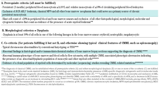
The classical form of CMML is defined by the following pre-requisite criteria: (i) persistent (at least 3 months) absolute PB monocytosis (≥1×10⁹/L) and relative monocytosis (≥10% of PB leukocytes); (ii) exclusion of BCR-ABL1+ leukemia, classical MPN and all other hematologic neoplasms that may serve as a primary source of monocytosis; and (iii) a blast cell count of 0-19% in PB and/or BM smears and exclusion of all (other) histopathological, morphological, phenotypic, molecular and cytogenetic signs that qualify as evidence of AML. In addition, morphological and/or histopathological evidence for diagnostic dysplasia in one or more of the three major BM cell lineages (≥10% of megakaryocytes and/or erythroid precursor cells and/or neutrophilic cells) must be present. If dysplasia is absent or not diagnostic (<10%), the presence of cytogenetic or molecular lesions (mutations) typically found in CMML and/or the presence of CMML-related flow cytometry abnormalities may be employed as co-criteria and may lead to the diagnosis of CMML, provided that the pre-requisite criteria listed above are fulfilled. Pre-requisite criteria and co-criteria of the classical form of CMML are presented in Table 1.
Table 1.
Minimal diagnostic criteria for classical chronic myelomonocytic leukemia.*
The exclusion of various reactive states producing monocytosis (and sometimes even dysplasia) was also discussed and regarded as being of great importance. However, these mimickers cannot a priori exclude the presence of a concomitant CMML, but may indeed occur in CMML patients in the context of certain infections. Furthermore, most of these mimickers do not produce persistent monocytosis. Proof of clonality by molecular and cytogenetic studies, and other disease-specific parameters, together with global and specific laboratory (e.g., microbial screen) tests should easily lead to the conclusion that the patient is suffering from reactive monocytosis but not from (or also from) CMML.
The a priori exclusion of AML as a criterion should apply to both the classical and the special variants of CMML, whereas the a priori exclusion of other indolent hematopoietic neoplasms should only apply to the classical variant of CMML and oligomonocytic CMML but not to other special CMML variants. This is because several previous and more recent studies have shown that CMML may be accompanied by (or may accompany) other myeloid or lymphoid neoplasms, such as systemic mastocytosis. In several of these patients, the CMML clone is dominant and the additional sub-clone is smaller in size and usually not relevant clinically, even if these smaller clones express certain driver mutations, such as KIT D816V or a rearranged PDGFRA or PDGFRB. Rarely, a Philadelphia chromosome-positive chronic myeloid leukemia may develop as an additional small-sized (sub)clone in a patient with CMML. Our faculty is of the opinion that the presence of additional (chronic) myeloid, mast cell, or lymphoid neoplasms does not exclude a diagnosis of CMML, provided that diagnostic WHO criteria for CMML are fulfilled. Moreover, these concomitant neoplasms should not exclude a diagnosis of CMML even when the driver of the concomitant disease (e.g., KIT D816V) is detectable in CMML monocytes. Thus, whereas the occurrence of AML is always regarded as transformation of CMML, the occurrence of indolent myeloid, mast cell, or lymphoid neoplasms should be regarded as concomitant disorders. Co-existing myeloid neoplasms and CMML may be derived from the same original founder clone.
There are also patients in whom a certain driver of another BM neoplasm is present, such as a mutated JAK2, PDGFRA/B, or FGFR1, but only the diagnostic criteria for CMML (not those of the other BM neoplasm) are fulfilled. Our faculty concludes that these cases should also be regarded and diagnosed as special variants of CMML. This strategy is in line with the current WHO classification. In fact, whereas the primary molecular diagnosis is often based on a mutated form of JAK2, PDGFRA/B or other classical driver, the underlying or additional diagnosis may well be CMML.20,21
Grading of CMML
The grading system of CMML proposed by the WHO is regarded as standard in clinical hematology. Our faculty recommends the use of this grading system as the initial prognostic tool in classical CMML. In fact, classical CMML should be split into CMML-0, CMML-1 and CMML-2 based on the blast cell count (Online Supplementary Table S2).15-19 In addition, CMML can be divided into a dysplastic variant and a proliferative variant based on leukocyte counts (threshold: 13x109/L) (Online Supplementary Table S2). The resulting grading system defines six distinct CMML variants with variable clinical outcome.17 However, grading may sometimes be challenging. For example, blast cell counts obtained from BM smears may differ from those obtained in the PB so that the grade is questionable. Our faculty recommends that in patients in whom results from BM and PB smears would not fit into one distinct grade of CMML (e.g., BM blasts 4% and PB blasts 6%) grading should be based on the higher blast cell percentage (Online Supplementary Table S2). It is worth noting that initial prognostication by grading does not include all essential prognostic parameters. We therefore recommend that in each case, deeper (full) prognostication should follow using multiparametric scoring systems (see later). It should be noted, however, that grading of CMML has only been validated in the classical form of CMML, not in special CMML variants. Therefore, although grading is also recommended for special CMML entities, it is not regarded standard and the result must be interpreted with caution in these patients.
Special variants of CMML: overview
As mentioned before, the classical form of CMML meets all pre-requisite criteria, and no signs (including molecular features) of an additional, concomitant BM neoplasm are detected. The special variants of CMML form a heterogeneous group of neoplasms comprising distinct clinical and biological entities. In one group of patients, the relative monocyte count (≥10%) is fulfilled without resulting in an absolute count ≥1×10⁹/L, precluding the diagnosis of ‘classical CMML’. Most of these patients are diagnosed as having MDS or MPN/MDS-unclassified by WHO criteria. In another group of patients, a molecular signature suggestive of a different type of myeloid neoplasm is detected but only the criteria for CMML (not those for the other neoplasm) are met. Such an example is CMML with JAK2 V617F (without definitive evidence of a concomitant MPN). In a third group, CMML co-exists with another BM neoplasm, such as MPN or mastocytosis. In these patients, additional blood count abnormalities (e.g., eosinophilia), an elevated serum tryptase level and/or BM fibrosis, may be detected.
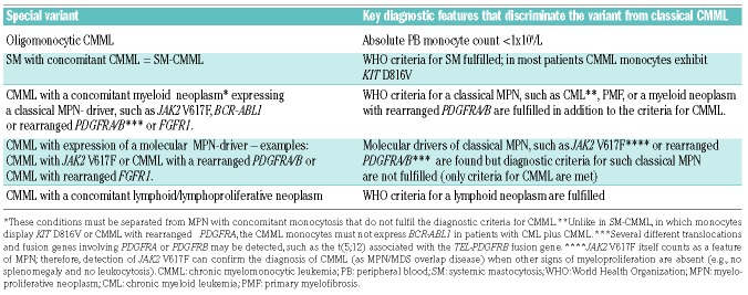
All variants of CMML (classical and special) can occur as a primary CMML or as a secondary CMML following a ‘mutagenic’ event, such as chemotherapy (therapy-related CMML). In addition, our faculty is of the opinion, that the term secondary CMML may also be appropriate for those patients who develop CMML (months or years) after another indolent myeloid neoplasm, such as a MDS or systemic (indolent or aggressive) mastocytosis, had been diagnosed. In the following paragraphs, the clinical features and diagnostic criteria of special (atypical) variants of CMML are proposed and discussed. An overview of the special variants of CMML is provided in Table 2.
Table 2.
Overview of special variants of chronic myelomonocytic leukemia.
Oligomonocytic CMML
Over the past few years, more and more cases of cytopenic patients exhibiting relative monocytosis (≥10%) and moderately increased absolute blood monocytes not reaching the required threshold to diagnose classical CMML (1.0x10⁹/L) have been described. These cases have recently been referred to as oligomonocytic CMML.30 According to the WHO classification most of these patients would be classified as having MDS (with monocytosis) or perhaps MPN/MDS-unclassifiable. However, most of these patients exhibit typical features of CMML, including a typical morphology of PB and BM cells, splenomegaly, and CMML-related molecular features (e.g. mutations in TET2 and SRSF2).30-32 Some of these patients have prominent BM monocytosis without diagnostic PB monocytosis at diagnosis.30,32
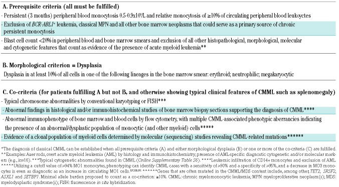
Whereas several of these cases remain stable without progression, the majority will develop ‘overt’ CMML or, eventually, secondary AML during follow-up. Therefore, oligomonocytic CMML may also be regarded as a potential pre-phase of classical CMML. Our faculty is of the opinion that the term oligomonocytic CMML should be used in clinical practice. Diagnostic pre-requisite criteria for oligomonocytic CMML are: (i) persistent (lasting at least 3 months) absolute peripheral monocytosis of 0.5-0.9×10⁹/L and relative blood monocytosis (≥10% of blood leukocytes); (ii) exclusion of BCR-ABL1+ leukemia, classical MPN and all other myeloid neoplasms that can explain monocytosis; and (iii) a blast cell count of 0-19% in PB and/or BM smears and exclusion of all histopathological, morphological, phenotypic, molecular and cytogenetic signs that count as proof of AML. Diagnostic dysplasia in one or more of the three major BM lineages (≥10%) must also be documented. If dysplasia is lacking or ‘sub-diagnostic’ (<10%), the presence of cytogenetic or molecular lesions (mutations) typically found in CMML and/or the presence of CMML-related flow cytometry abnormalities, may also lead to the conclusion that the patient has oligomonocytic CMML provided that the other diagnostic criteria described above are fulfilled and all other myeloid neoplasms have been excluded. The proposed criteria for oligomonocytic CMML are listed in Table 3. Patients with oligomonocytic CMML should be managed and followed clinically in the same way as patients with classical CMML.
Table 3.
Proposed minimal diagnostic criteria for oligomonocytic chronic myelomonocytic leukemia.*
CMML associated with KIT D816V+ systemic mastocytosis
According to WHO criteria, systemic mastocytosis (SM) can be divided into: (i) indolent SM (ISM), which is assocaiated with a normal life expectancy; (ii) smoldering SM (SSM), in which signs of BM dysplasia, myeloproliferation and/or splenomegaly are found but survival and prognosis are still favorable; and (iii) advanced SM, defined by a poor prognosis.33-36 Advanced SM is further divided into aggressive SM (ASM), SM with an associated hematologic neoplasm (SM-AHN) and mast cell leukemia (MCL).33-36 The most frequent AHN detected in patients with SM-AHN is CMML.6-8,36 In these patients the SM component of the diseases may present as ISM, ASM or, rarely, as MCL. Our faculty concludes that diagnostic WHO criteria for SM and diagnostic criteria for classical CMML (except exclusion of SM) must be fulfilled to diagnose SM-CMML.
Patients with SM may present with monocytosis resembling oligomonocytic CMML. However, the clinical features of SSM and advanced SM overlap largely with those found in patients with oligomonocytic CMML. Especially in SSM, myeloproliferation, dysplasia and splenomegaly are diagnostic criteria.33-35 Therefore, our faculty is of the opinion that such patients should be classified as having ISM, SSM or ASM with monocytosis rather than SM with oligomonocytic CMML.
In patients with CMML, a concomitant SM is often overlooked, especially when the disease does not present with cutaneous lesions. In other patients, CMML is diagnosed long before SM is detected by chance or after KIT D816V is identified: even though it is tempting to call these conditions CMML-SM, our faculty agreed that the classical terminology should be SM-CMML which is also in line with the WHO classification34,35 and that the subtype of SM and of CMML should be defined in the final diagnosis (e.g., ISM-CMML-1 or ASM-CMML-2) with recognition that in the SM-context, CMML is always regarded as a secondary neoplasm.6,36 Furthermore our faculty is of the opinion that it should be standard practice to examine BM and blood leukocytes for the presence of KIT D816V in all patients with (suspected) CMML. In almost all patients with SM-CMML, neoplastic monocytes display KIT D816V.7 In these monocytes, mutated KIT is not expressed on the cell surface but acts as a cytoplasmic driver. In line with this hypothesis drugs targeting KIT D816V can sometimes induce a major decrease in monocyte counts in patients with ASM-CMML.37
Therapy of SM-CMML should be based on a bi-directional strategy: in fact the SM component of the disease should be treated as if no CMML was diagnosed and CMML should be treated as if no SM had been found, with recognition of drug-drug interactions and the possibility of drug-induced anaphylaxis.33-35 In many cases (ISM-CMML) the SM component of the disease is only treated symptomatically.33-35
CMML associated with mutated JAK2, rearranged PDGFRA/B or other drivers
Patients with CMML may present with the JAK2 mutation V617F, a rearranged PDGFRA or PDGFRB, often in the context of hypereosinophilia, or other drivers related to distinct hematopoietic neoplasms as defined by the WHO.5,9-11,38-43
CMML with rearranged PDGFRA, PDGFRB, FGFR1 or PCM1-JAK2
In these patients, persistent substantial monocytosis (≥1.0x10⁹/L) is detected and all other consensus criteria for classical CMML (see previous paragraphs) are also met, except the following specific exclusion criteria: CMML to be excluded in the presence of a well-characterized diagnosis of myeloid/lymphoid neoplasm with rearranged PDGFRA, PDGFRB, FGFR1 or PCM1-JAK2 (Table 2). Except for neglecting the above-mentioned criteria, our proposal is otherwise fully in agreement with all of the other tenets postulated by the WHO classification.14,15 In relation to neoplasms with rearranged PDGFRA/B, FGFR1 or PCM1-JAK2, the WHO’s definition of ‘myeloid/lymphoid neoplasms’ is too generic and there is a clinical need to know whether the underlying myeloid neoplasm is an aggressive disease, like AML, or a chronic neoplasm such as CMML or chronic eosinophilic leukemia.20,21 Our faculty is of the opinion that (unlike in previous times) the presence of one criterion-confirmed myeloid neoplasm should not a priori exclude the presence of another (second concomitant) myeloid or lymphoid neoplasm. Hence, when CMML is encountered in the context of another molecularly defined myeloid/lymphoid neoplasm (as a final diagnosis), it should be delineated as a specific subtype of the myeloid/lymphoid neoplasm with eosinophilia along with the specific associated gene rearrangement (PDGFRA/B or FGFR1 or PCM1-JAK2).
CMML with JAK2 V617F
In these patients the situation is different. First, JAK2 V617F itself may be considered as a criterion of myeloproliferation in MDS/MPN, e.g. in cases with MDS/MPN with ring sideroblasts and thrombocytosis. In the context of CMML, the JAK2 mutation is also typically associated with other signs of myeloproliferation (including BM fibrosis) and with the ‘myeloproliferative variant’ of CMML.39,42,43 Therefore, our faculty concludes that JAK2 V617F should also count as a molecular co-criterion of MDS/MPN and thus for CMML. Second, the presence of a JAK2-mutated MPN does not exclude the presence of a concomitant CMML if diagnostic criteria for both neoplasms are fulfilled. If this is not the case because the size of the MPN-like clone carrying JAK2 V617F is too small and/or other MPN features are clearly missing, the final diagnosis will be CMML with JAK2 V617F. On the other hand, in patients in whom the JAK2 allelic burden is high and clinical and laboratory features argue for an overt MPN rather than CMML (e.g., polycythemia and/or BM fibrosis without dysplasia and without molecular or flow cytometry-based signs of CMML) the final diagnosis will be JAK2 V617F+ MPN with monocytosis.43 In a third group of patients, diagnostic criteria for both a distinct MPN and CMML are fulfilled and the mutation status confirms the presence of an overt JAK2-mutated MPN (usually with high allelic burden). These patients are suffering from both MPN and CMML or from a gray zone disease displaying hybrid features between MPN and CMML.44,45 Our faculty concludes that it is therefore important to measure the JAK2 V617F allele burden in all patients with CMML.39,42,43 Other drivers, such as BCR-ABL1, are rarely found in patients with CMML. However, although in classical CMML, the presence of BCR-ABL1 must be excluded, it may be detected in rare patients, suggesting the existence of a special variant of CMML (defined by a co-existing chronic myeloid leukemia). In some of these cases, the chronic myeloid leukemia clone may be small. In other patients, however, the chronic myeloid leukemia may even mask the CMML at the initial diagnosis.46
The management and therapy of patients with special variants of CMML depend on the subtype of the disease and the molecular driver involved, e.g., FIP1L1/PDGFRA, other gene abnormalities involving PDGFRA or PDGFRB, KIT D816V or JAK2 V617F. Therefore, it is of crucial importance to screen for all these drivers in all patients with CMML. The type of therapy to consider in these patients depends on clinical features, the histopathological diagnosis, the size of the mutated clone(s) and the type of driver. The type of driver is of considerable importance since novel treatments directed against these drivers, are often extremely effective.47-50 For example, imatinib can induce long-lasting molecular and hematologic complete remissions in patients with FIP1L1/PDGFRA-rearranged myeloid neoplasms with features of CMML or MPN.47-49 Even in patients who develop CMML and secondary AML in the context of FIP1L1/PDGFRA, the disease may respond to imatinib.50 It is, therefore, important to diagnose all patients based on molecular markers and to define the major drivers and therapeutic targets expressed by malignant cells in order to provide optimal management and therapy.
CMML associated with lymphoid neoplasms
In a small group of patients with CMML, a co-existing lymphoproliferative neoplasm is diagnosed, such as a lymphocytic leukemia, non-Hodgkin lymphoma or multiple myeloma.51-60 In most patients, the lymphoid neoplasm is detected first, and CMML is considered to develop as treatment-induced, secondary, leukemia.51,57 In other patients, CMML is diagnosed first and later a lymphoid neoplasm is detected during follow-up.52-56 It is worth noting that in patients with CMML, polyclonal hypergammaglobulinemia is often recorded: this must be distinguished from the monoclonal gammopathy of concomitant myeloma, monoclonal gammopathies of undetermined significance and both low-count and high-count monoclonal B lymphocytoses which represent pre-malignant conditions.
The management and treatment of lymphoid neoplasms presenting with concomitant (secondary) CMML is a clinical challenge. In non-transplantable cases, both diseases require separate treatment plans. Because of the high risk of transformation to AML, allogeneic hematopoietic stem cell transplantation should be considered in young, fit patients, especially when it can be expected that the lymphoid neoplasm will also be eradicated by this approach.
Treatment-related CMML and other secondary forms of CMML
Our faculty concluded that both the classical form of CMML and the special variants of CMML should be divided into primary (de novo) CMML and secondary CMML. The latter group includes patients who (i) received chemotherapy and/or radiation therapy in the past (therapy-related CMML) or (ii) have a history of a preceding MDS, MPN or another indolent myeloid or mast cell neoplasm prior to their diagnosis of CMML.51,57,58,61-64 Recent data suggest that patients with therapy-related secondary CMML may have shorter overall survival compared to that of patients with primary (de novo) CMML.65 Although progression-free survival may not be different in these patients compared to those with de novo CMML, some of these patients progress rapidly to secondary AML. It is also worth noting that patients with therapy-related secondary CMML have a higher frequency of karyotypic abnormalities compared to patients with de novo CMML.66 Eligible patients in this group should be offered allogeneic hematopoietic stem cell transplantation.
Potential pre-phases of CMML
During the past few years evidence has accumulated suggesting that hematopoietic neoplasms, including MDS, MPN and MDS/MPN, develop in a step-wise manner. In the earliest phases of clonal development, patients present without overt signs or symptoms of a hematopoietic neoplasm but their leukocytes carry one or more somatic mutations, usually (early, passenger-type) mutations otherwise also found in overt myeloid neoplasms (for example TET2 mutations).67-70 In the context of MDS and other myeloid neoplasms, these cases have been referred to as clonal hematopoiesis of indeterminate potential (CHIP), or, when accompanied by cytopenia, as clonal cytopenia of unknown significance (CCUS).69-73 Since these mutations are frequently detected in older individuals, the condition is also called age-related clonal hematopoiesis (ARCH).70,73 In a few healthy individuals, bona fide oncogenic drivers (such as BCR-ABL1) are detected in a small subset of leukocytes. Because of the oncogenic potential of these drivers, these conditions are termed clonal hematopoiesis with oncogenic potential (CHOP).71,73 CHIP, CCUS and CHOP may also be the earliest clonal conditions preceding CMML. For these cases, the definitions recently proposed for CHIP, CCUS and CHOP should apply.69,71,73
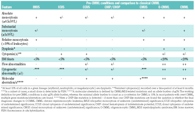
Apart from somatic mutations, other factors, such as epigenetic modifications, chronic inflammation or aging-related processes, may also trigger the selection and expansion of pre-malignant neoplastic clones in myeloid neoplasms including CMML.74-76 Some of these conditions may present with persistent monocytosis without signs of an overt myeloid neoplasm and may represent pre-phases of overt CMML. In other patients, however, no or another hematopoietic neoplasm develops during follow-up. Therefore, our faculty concluded that this pre-phase should be termed idiopathic monocytosis of unknown significance, provided that the following criteria are met: (i) persistent (at least 3 months) relative (≥10%) and absolute (>0.5x10⁹/L) monocytosis; (ii) no diagnostic dysplasia and no signs of myeloproliferation; (iii) no signs and criteria of a myeloid or other hematopoietic neoplasm fulfilled; (iv) no flow cytometric abnormalities or somatic mutations related to a myeloid, mast cell or lymphoid neoplasm detected in leukocytes; and (v) no reactive condition that would explain reactive monocytosis is detected (Table 4 and Online Supplementary Table S3). If CHIP-like mutations are found in such patients, but no hematopoietic neoplasm can be diagnosed using the WHO criteria, the final diagnosis changes to clonal monocytosis of unknown significance (Online Supplementary Table S3). It is also worth noting that idiopathic cytopenias of unknown significance can precede CMML.64,77-79 Especially in patients with idiopathic thrombocytopenia of unknown significance, a CMML may be detected upon deeper investigations or during follow-up.77-79 Finally, as mentioned before, oligomonocytic CMML, although proposed as a special variant of CMML, must also be regarded as a potential pre-phase of classical CMML. In this regard it is important to note that these patients should have a regular follow-up with repeated investigations of all disease-related parameters. A summary of non-clonal and clonal conditions potentially preceding CMML is shown in Table 4. With regard to criteria delineating non-clonal pre-diagnostic conditions, like idiopathic cytopenia of undetermined significance from the clonal conditions described above (CHIP, CCUS, CHOP), we refer the reader to the pertinent literature.69,71,73
Table 4.
Overview of non-clonal and clonal conditions that may precede chronic myelomonocytic leukemia.
Peripheral blood and bone marrow smears: proposed standards and recommendations
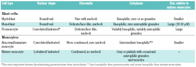
As in other myeloid neoplasms, a thorough examination of appropriately prepared and stained BM and PB smears is a crucial diagnostic approach in suspected CMML. It is standard to examine and count at least 100 leukocytes in the PB film and 200-500 nucleated cells in well-prepared thin BM films. BM cellularity, the erythroid-to-myeloid (E:M) ratio, and the percentage of blast cells (including monoblasts and promonocytes), monocytes, mast cells, and other myeloid cells must be recorded (reported) in each case. As in patients with MDS, at least 10% of cells in one of the major BM lineages (erythroid and/or neutrophilic and/or megakaryocytic) must be dysplastic to meet the dysplasia criterion for CMML.13-18 It is also standard to study well-prepared and appropriately stained PB smears in CMML and to report the percentage of circulating monocytes, including normal (mature) and abnormal (immature) monocytes, blast cells, other immature myeloid cells, dysplastic (hypogranulated) neutrophils and other cell types in the PB. Overall, the same standards and recommendations that count for the evaluation of MDS by morphology (BM and PB stains)12,80-83 also apply in cases with (suspected) CMML.13-18 An important point is the classification of blast cells and monocytic cells in CMML (Table 5).16,84 Blast cell types detectable in CMML include myeloblasts, monoblasts and also promonocytes (even if not named blast cells) (Table 5). Monocytes should be classified as normal (mature) or abnormal (immature).16,84 The morphological criteria used to distinguish between these cell types are presented in Table 5. Together with morphology, cytochemical staining for non-specific esterase can also assist in the cytological delineation between monocytes, monoblasts and promononcytes.16 An important aspect is that in many patients, megakaryocyte dysplasia is better documented and quantified in BM histology sections than in BM smears. Therefore, megakaryocyte dysplasia should only be recorded in BM smears when a sufficient number of these cells can be detected. Finally, the morphology of mast cells, when detected, should always be reported using established criteria and standards.85
Table 5.
Classification of blast cells and monocytes in patients with chronic myelomonocytic leukemia.
Bone marrow histology and immunohistochemistry in CMML
A thorough investigation of an appropriately processed and stained BM biopsy section by histology and immunhistochemistry is standard in all cases with known or suspected CMML or a suspected pre-CMML condition.14-16,30,86 Notably, BM histology and immunhistochemistry are essential approaches to confirm the diagnosis of CMML and to exclude AML and other CMML-mimickers. Moreover, BM histology and immunhistochemistry may provide important additional information, including that on BM fibrosis, focal accumulations of blast cells, increased angiogenesis, atypical (dysplastic) megakaryocytes, a hypocellular BM or concomitant mastocytosis (Online Supplementary Table S4).33-35,86 The evaluation and enumeration of CD14+ monocytes, CD34+ progenitor cells and CD117+/KIT+ cells (progenitors and mast cells) by immunhistochemistry in BM biopsy sections represent an integral part of the diagnostic assessment. These approaches can also prevent diagnostic errors. For example, when the smear is of suboptimal quality, a preliminary diagnosis of CMML may change to AML based on BM histology and CD34 immunhistochemistry.
BM biopsy specimens are usually taken from the iliac crest and should be of adequate length (≥2 cm). The specimen should be fixed in neutral formalin (or alternative standard fixation), decalcified in EDTA (for at least 8 h) or by alternative standard decalcification, and embedded in paraffin-wax. Ideally 2-3 μm thin sections should be prepared. Routine stains include hematoxylin-eosin, Giemsa, Prussian blue, AS-D chloroacetate esterase, toluidine blue and silver impregnation (Gömöri’s stain). BM cellularity should be measured and reported according to published standards.87,88 For routine purposes, the pathologist should determine the cellularity as ‘normocellular’, ‘hypocellular’, or ‘hypercellular’, based on an age-adapted estimate.89 The presence of variable degrees of BM fibrosis (usually mild to moderate) has been reported in CMML cases, with several recent studies attempting to determine the prognostic value of this finding.42,90,91 Indeed, although the data are not yet conclusive, the presence of marrow fibrosis in CMML seems to be of prognostic importance.42,90,91
The application of immunhistochemical markers is recommended in all patients with (suspected) CMML. The minimal immunohistochemistry panel includes CD14 (monocytes), CD34 (progenitors), CD117/KIT (progenitors and mast cells), tryptase (mast cells), and a megakaryocyte marker (CD41, CD42 or CD61) (Online Supplementary Table S5).86,92,93 In unclear cases or when a co-existing (second) BM neoplasm is suspected, additional lineage-specific antibodies such as CD3, CD20, or CD25 (suspected mastocytosis) should be applied (Online Supplementary Table S4). When employing CD34 as a progenitor-related immunhistochemical marker, it is important to know that endothelial cells also express this antigen. Another important point is that blasts may sometimes be CD34-negative. In such cases, KIT/CD117 is applied as an alternative marker (Online Supplementary Table S4). For the immunohistochemical detection of monocytic cells, CD14 is a preferred antigen.71,86 Tryptase and CD117 are useful immunhistochemistry markers for detecting and quantifying mast cells.92,93 When spindle-shaped mast cells form compact clusters in the BM and express CD25, these cells usually also display KIT D816V – in these cases the final diagnosis is always SM-CMML.93 In other cases, the pathologist will ask for JAK2 V617F, based on an abnormal morphology and distribution of megakaryocytes. As in MDS, megakaryocytes may also express CD34 in patients with CMML.
Karyotyping in CMML: current recommendations and standards
Clonal cytogenetic abnormalities are detected in 20-30% of all patients with CMML. The most frequently identified aberrations are trisomy 8, abnormalities of chromosome 7 (especially monosomy 7 and deletion of 7q), and loss of the Y chromosome (-Y) (Online Supplementary Table S6).94-97 Compared to MDS, isolated del(5q) and complex abnormal karyotypes are rarely detected in CMML. Our faculty is of the opinion that conventional karyotyping of BM cells should be performed in all patients with known or suspected CMML or a suspected pre-CMML condition. At least 20 metaphases should be examined.98 In the case of a clear-cut result, even 10-20 metaphases may be sufficient to define the karyogram. Reporting of karyotypes should be performed using the International System for Human Cytogenetic Nomenclature (ISCN) guidelines.99 A clone is defined by two or more metaphases showing the same gain or structural rearrangement (deletion, inversion, translocation) of chromosomal material or at least three metaphases showing a monosomy of the same chromosome.99 Several of the cytogenetic anomalies in CMML may be difficult to detect by conventional karyotyping. Therefore, we are of the opinion that fluorescence in situ hybridization (FISH) should be performed in all patients with (suspected) CMML, at least in those in whom no karyotype anomaly was detected by conventional karyotyping. The FISH probes should cover all relevant regions, including 5q31, cep7, 7q31, 20q, cep8, cepY and p53. Special consideration should be directed to cryptic deletions of TET2 (in 4q24), NF1 (17q11), and ETV6 (12p13) which can occur in up to 10% of CMML patients10 and are only detectable by interphase FISH (Online Supplementary Table S6). It is worth noting that NF1 deletions may occur during progression/karyotype evolution in CMML. The limitation of FISH is that is does not detect all karyotypic abnormalities. In some patients with CMML, clonal evolution is found. Subclones are defined by additional chromosomal defects (apart from the primary chromosomal defect) in at least two cells (or 3 cells for monosomies) and the absence of these additional chromosomal defects in the other clonal cells.99 A complex karyotype is defined by at least three chromosome defects in one clone.99 As in MDS, a complex karyotype in CMML is indicative of a poor prognosis. Overall, cytogenetic studies are of prognostic significance in CMML and have been used to optimize prognostic scoring systems.97,100-102 In some patients with CMML, clonal evolution is observed over time and may then also be an adverse prognostic sign. Therefore, we recommend that chromosome analyses are performed each time a BM investigation is done in the follow-up in order to detect (or exclude) clonal evolution.
Mutation profiles in CMML: current standards and limitations
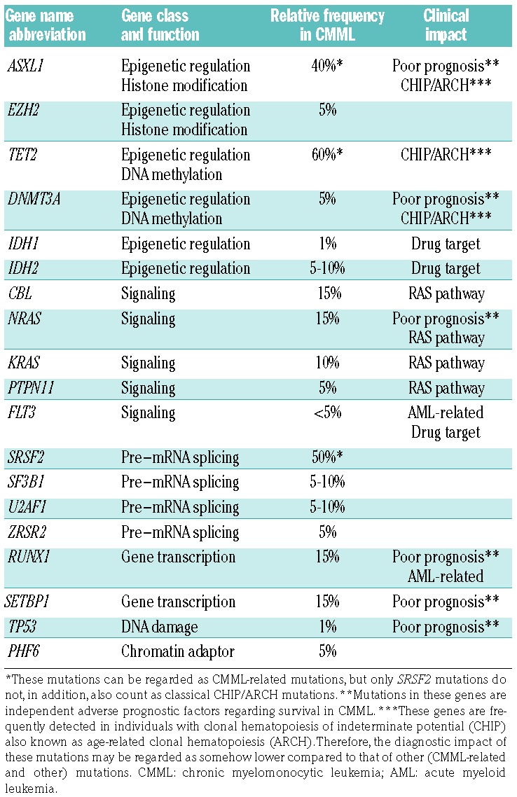
Somatic mutations are detectable in the vast majority of patients with CMML.8,11,103-106 The clonal architecture, clone sizes and clonal evolution patterns vary from patient to patient.106-108 In some cases, initially small clones expand over time. It is, therefore, standard to apply next-generation sequencing assays with sufficient sensitivity to identify bona fide somatic mutations associated with CMML. The most frequently detected somatic mutations in CMML are mutations in TET2 (60%), SRSF2 (50%), and ASXL1 (40%) (Table 6).31,103-110 The presence of a SRSF2 mutation, particularly in combination with mutated TET2, correlates strongly with a CMML phenotype.31,109,110 It is also worth noting that two of these mutations (TET2, ASXL1) are also known as CHIP/ARCH-related mutations. However, only mutated ASXL1 has been associated with a poor prognosis in CMML.104,109 An overview of somatic mutations recurrently detected in CMML is provided in Table 6. Somatic mutations with independent prognostic impact include several RAS-pathway mutations as well as mutations in ASXL1, RUNX1 and SETBP1 (Table 6).31,103-111 RAS-pathway mutations trigger cell signaling and proliferation and have been associated with cytokine-independent growth of CMML progenitor cells, the proliferative variant of CMML, AML transformation and poor survival.10,22,23,112-116 Other driver mutations involved in cell signaling, such as JAK2 V617F or KIT D816V, are also major triggers of cellular differentiation (Online Supplementary Table S7). These drivers alone cannot induce transformation, but they may act together with other (e.g., ‘RAS pathway’) mutations to cause disease progression. Whereas JAK2 V617F is a strong indicator of MPN-like differentiation, the presence of KIT D816V is almost always associated with concomitant mast cell differentiation and mastocytosis (SM-CMML).6-8,32-36,39,42,43 The other mutations found in CMML act as modulators of epigenetic events and transcription (e.g., ASXL1) or DNA methylation (e.g., TET2), as regulators of the spliceosome machinery (e.g., SRSF2), or as modulators of the DNA damage response, such as TP53 (Table 6). During progression of CMML to secondary AML and especially during therapy, the mutational landscape(s) and clonal architecture(s) may change.109-113 For example initially small clones may expand and may be selected because of resistance-mediating molecular features. It is worth noting that several mutated gene products also serve as potential targets of therapy (Table 6).
Table 6.
Commonly mutated genes detectable in patients with classical chronic myelomonocytic leukemia.
Our faculty recommends that next-generation sequencing studies should be regarded as a standard approach in all patients with suspected or known CMML as well as in patients with idiopathic monocytosis of unknown significance and in those with persistent reactive monocytosis (in order to exclude an additional clonal component). When a CMML-related mutation is found in an individual with idiopathic monocytosis of unknown significance or reactive monocytosis, the diagnosis may change to clonal monocytosis of unknown significance or oligomonocytic CMML, depending on additional findings.
Our faculty also recommends that the next-generation sequencing assay should have sufficient sensitivity (to detect 2-5% clonal cells) and should cover all relevant lesions shown in Table 6. In the context of CHIP/ARCH, a cutoff variant allele frequency of 2% is considered diagnostic,69 whereas in the context of CMML, we propose 10% as the variant allele frequency diagnostic cut off and thus marker to count as a co-criterion of CMML when, for example, no diagnostic morphological dysplasia can be documented (Tables 1 and 3), similar to the definition in MDS.71,73 Determining the variant allele frequency is also useful for documenting the clinical impact of certain driver lesions in special CMML variants (e.g., with JAK2 V617F or KIT D816V) and clone expansion during follow-up. Therefore, our faculty recommends that molecular studies in CMML should report variant allele frequencies with sufficient precision and sufficient sensitivity – in the same way as in MDS.71,73 Finally, our faculty recommends that molecular markers should increasingly be used to optimize prognostic scoring systems in CMML.117-120
Flow cytometry in CMML: standards and limitations
Flow cytometry studies are an essential diagnostic tool in patients with (suspected) classical CMML, pre-CMML conditions and special CMML variants.121-132 Therefore, our faculty is of the opinion that it is standard practice to perform multi-color flow cytometry (MFC) in the PB and BM in all cases with suspected or known CMML or a suspected pre-CMML condition. MFC studies are helpful to confirm the monocyte and blast cell counts in these patients and to exclude AML. In addition, MFC is useful to confirm the presence of distinct monocyte populations. Monocytes are defined as CD14+ cells in these analyses. Based on the expression of CD14 and CD16, monocytes are further divided into classical (MO1) monocytes (CD14bright/CD16−), intermediate (MO2) monocytes (CD14bright/CD16+) and non-classical (MO3) monocytes (CD14dim/CD16+) (Table 7).127,128,132 Compared to age-matched healthy donors133 and patients with reactive monocytosis, but also myeloid neoplasms other than CMML (even MDS), the percentages of MO1 monocytes in the PB are higher and the percentage of MO3 monocytes is lower in patients with CMML.127,131,132 When the absolute monocyte count is increased in the PB, a cutoff value of >94% MO1 monocytes, based on their immunophenotype, can identify CMML with a sensitivity of >90% and a specificity of >95%.127,129,131 Moreover, during successful therapy, the distribution of MO1, MO2, and MO3 monocytes changes back to near normal or normal.128 Therefore, our faculty recommends that the percentages of MO1 monocytes are quantified in the PB by MFC in all cases with suspected or known CMML at diagnosis and during follow-up.
Table 7.
Phenotypic classification of monocytes and distribution of monocyte subsets in patients with chronic myelomonocytic leukemia and in controls.*
In many cases with CMML, neoplastic monocytes aberrantly display CD2, CD5, CD10, CD23, and/or CD56.121-124 Of all aberrantly expressed surface markers, CD56 is most commonly detected on CMML monocytes.121-124 CD5 is only (very) weakly expressed on neoplastic monocytes in most cases with CMML. The most frequently underexpressed antigens may be CD14 and CD15. Overall, however, the use of decreased expression of these markers as a diagnostic test in CMML is limited by a relatively low sensitivity. An abnormal monocyte immunophenotype is also seen in other myeloid neoplasms, including MDS. On the other hand, phenotypically aberrant monocytes (as described above) are typically neoplastic cells (unless the patient has been treated with growth factors). Therefore, our faculty recommends that MFC studies in patients with (suspected) CMML employ antibodies directed against aberrantly expressed surface markers, including CD2 and CD56. Additionally, as mentioned before, several surface markers are ‘under-expressed’ on CMML monocytes compared to their levels on normal blood monocytes. These antigens include, among others, CD13, CD14, CD15, CD33, CD38, CD45, and CD64.121-124,129,131
Other cell types may also express aberrant markers detectable by MFC in CMML. For example, myeloid progenitor cells may express CD56 in CMML and often exhibit the same phenotypic abnormalities as in MDS; this also holds true for neutrophils and erythroid cells (Online Supplementary Table S8). Other cell types that may show aberrant phenotypes are dendritic cells and mast cells. Mast cells are of particular importance as these cells may be indicative of the presence of a concomitant mastocytosis (SM-CMML). In these cases, mast cells almost invariably express CD25 in MFC analyses (Online Supplementary Table S8).134 Overall, our faculty is of the opinion that MFC studies should be performed on monocyte subsets, myeloid progenitors, neutrophils, erythroid cells and mast cells in (suspected) CMML. An overview of immunophenotypic aberrancies detectable in CMML is given in Online Supplementary Table S8.
Differential diagnoses of CMML: reactive and clonal mimickers
A number of conditions can mimick CMML and must be taken into account when patients with unexplained monocytosis are evaluated. Reactive disorders mimicking CMML include certain chronic bacterial infections (examples: tuberculosis or subacute endomyocarditis), fungal infections, chronic auto-immune processes and non-hematologic neoplasms. There are also hematologic malignancies that may present as a CMML-like disease. For example, Philadelphia chromosome-positive chronic myeloid leukemia usually presents with (absolute) monocytosis and can also show signs of dysplasia. Particularly high monocyte counts are recorded in chronic myeloid leukemia cases expressing BCR-ABL1p190. When cryptic variants of BCR-ABL1 are expressed by leukemic cells, it can be difficult to exclude CMML. Myeloid neoplasms (MDS or MPN) in progression and myelomonocytic or monocytic AML may also resemble CMML. The reactive and clonal mimickers of CMML are listed in Online Supplementary Table S9.
Scoring systems in CMML: recommended standards
Although several prognostic variables have been identified in CMML regarding survival and AML evolution, accurate prediction of the clinical course and survival remains a clinical challenge. A first step in prognostication is grading into CMML-0, CMML-1 and CMML-2. To delineate the prognosis in CMML more accurately, a number of scoring systems have been developed in the past.29,117-121,135-138 Until 2012, the International Prognostic Scoring System (IPSS) served as the gold standard for prognostication in MDS and (dysplastic) CMML.135
However, a number of more specific scoring systemic taking CMML-related features into account have also been proposed.117-120,136-138 During the past few years, researchers have successfully started to integrate cytogenetic and molecular variables into these scoring models.117-121 Our faculty concludes that these novel approaches should be followed and developed into clinical application.
Management strategies and therapeutic options in CMML
Several new treatment strategies for CMML have been developed during the past 15 years. A detailed description of therapeutic options is beyond the scope of this article. The reader is referred to a series of excellent published review articles.139-146 A disappointing fact is that all drug therapies are still non-curative. The only curative therapy in CMML remains allogeneic hematopoietic stem cell transplantation.147,148 For most young and eligible patients with acceptable transplant-related risk, allogeneic hematopoietic stem cell transplantation is therefore recommended. All other forms of treatment are cytoreductive, experimental or palliative in nature. Some of these drugs, such as the hypomethylating agents (5-azacytidine, decitabine) may induce long-term disease control in a subset of patients with classical CMML.139-145 In general, cytoreductive and palliative drugs should be used according to available recommendations provided by major societies.145,148 Similarly, treatment response assessment should be performed in line with available (accepted) guidelines.146,150
Specific therapy may work in those patients who suffer from a special variant of CMML. For example, in CMML patients with a transforming PDGFRA/B mutation, treatment with imatinib or other similar tyrosine kinase inhibitor usually induces major responses or even long-lasting remissions.47-49,151 In patients with SM-CMML, midostaurin may result in disease control, especially when the CMML portion of the disease exhibits KIT D816V. However, in many cases, relapses occur. Treatment options in CMML and its variants are summarized in Online Supplementary Table S10.
Concluding remarks and future perspectives
CMML is a unique and rare hematopoietic neoplasm with a complex biology and pathology. In the past 10 years, several different pre-CMML conditions and sub-variants of CMML have been defined. In the current article, we propose minimal diagnostic criteria for classical CMML and for special CMML variants. These criteria should help in the diagnosis of pre-CMML conditions, classical CMML, special CMML variants, and conditions that mimick CMML. In addition, we propose standards and tools for the diagnosis, prognostication and management of CMML. Contemporary assays define all major histopathological, molecular, cytogenetic and flow cytometry-based features of neoplastic cells, and thereby cover all CMML variants, including oligomonocytic CMML and CMML associated with certain drivers or a concomitant myeloid neoplasm, such as mastocytosis. Different aberration profiles may also be found, resulting in a quite heterogeneous clinical picture and a variable clinical course. Although the course is often unpredictable, initial grading and consecutive application of CMML-directed prognostic scores are standard tools that support the prognostication of patients with CMML concerning survival and AML evolution. The application of criteria, tools and standards proposed herein should assist in the diagnosis, prognostication and management of patients with CMML.
Supplementary Material
Acknowledgments
We thank Sabine Sonnleitner, Sophia Rammler, Susanne Gamperl, Emir Hadzijusofovic and all other members of Peter Valent’s research group involved for their excellent support in the organization of the Working Conference. This study was supported by Austrian Science Fund (FWF) grants F4701-B20 and F4704-B20, the Ludwig Boltzmann Institute for Hematology and Oncology (LBI HO) and a Stem Cell Research grant from the Medical University of Vienna.
Footnotes
Check the online version for the most updated information on this article, online supplements, and information on authorship & disclosures: www.haematologica.org/content/104/10/1935
References
- 1.Storniolo AM, Moloney WC, Rosenthal DS, Cox C, Bennett JM. Chronic myelomonocytic leukemia. Leukemia. 1990;4(11):766-770. [PubMed] [Google Scholar]
- 2.Bennett JM, Catovsky D, Daniel MT, et al. The chronic myeloid leukaemias: guidelines for distinguishing chronic granulocytic, atypical chronic myeloid, and chronic myelomonocytic leukaemia. Proposals by the French-American-British Cooperative Leukaemia Group. Br J Haematol. 1994;87(4):746-754. [DOI] [PubMed] [Google Scholar]
- 3.Bennett JM. Chronic myelomonocytic leukemia. Curr Treat Options Oncol. 2002;3(3):221-223. [DOI] [PubMed] [Google Scholar]
- 4.Patnaik MM, Parikh SA, Hanson CA, Tefferi A. Chronic myelomonocytic leukaemia: a concise clinical and pathophysiological review. Br J Haematol. 2014;165(3):273-286. [DOI] [PubMed] [Google Scholar]
- 5.Itzykson R, Duchmann M, Lucas N, Solary E. CMML: clinical and molecular aspects. Int J Hematol. 2017;105(6):711-719. [DOI] [PubMed] [Google Scholar]
- 6.Sperr WR, Horny HP, Lechner K, Valent P. Clinical and biologic diversity of leukemias occurring in patients with mastocytosis. Leuk Lymphoma. 2000;37(5-6):473-486. [DOI] [PubMed] [Google Scholar]
- 7.Sotlar K, Fridrich C, Mall A, et al. Detection of c-kit point mutation Asp-816 –> Val in microdissected pooled single mast cells and leukemic cells in a patient with systemic mastocytosis and concomitant chronic myelomonocytic leukemia. Leuk Res. 2002; 26(11):979-984. [DOI] [PubMed] [Google Scholar]
- 8.Patnaik MM, Vallapureddy Rangit, Lasho TL, et al. A comparison of clinical and molecular characteristics of patients with systemic mastocytosis with chronic myelomonocytic leukemia to CMML alone. Leukemia. 2018;32(8):1850-1856. [DOI] [PubMed] [Google Scholar]
- 9.McCullough KB, Patnaik MM. Chronic myelomonocytic leukemia: a genetic and clinical update. Curr Hematol Malig Rep. 2015;10(3):292-302. [DOI] [PubMed] [Google Scholar]
- 10.Kohlmann A, Grossmann V, Klein HU, et al. Next-generation sequencing technology reveals a characteristic pattern of molecular mutations in 72.8% of chronic myelomonocytic leukemia by detecting frequent alterations in TET2, CBL, RAS, and RUNX1. J Clin Oncol. 2010;28(24):3858-3865. [DOI] [PubMed] [Google Scholar]
- 11.Patnaik MM, Tefferi A. Cytogenetic and molecular abnormalities in chronic myelomonocytic leukemia. Blood Cancer J. 2016;6:e393. [DOI] [PMC free article] [PubMed] [Google Scholar]
- 12.Bennett JM, Catovsky D, Daniel MT, et al. Proposals for the classification of the myelodysplastic syndromes. Br J Haematol. 1982;51(2):189-199. [PubMed] [Google Scholar]
- 13.Vardiman JW, Imbert M, Pierre R, et al. Chronic myelomonocytic leukemia. In: World Health Organization Classification of Tumours – Pathology & Genetics: Tumours of the Haematopoietic and Lymphoid Tissues: Eds. Jaffe ES, Harris NL, Stein H, Vardiman JW. IARC Press; Lyon: 2001, pp 49-52. [Google Scholar]
- 14.Orazi A, Bennett JM, Germing U, Brunning RD, Bain B, Thiele J. Chronic myelomonocytic leukemia. In: WHO Classification of Tumours of Haematopoietic and Lymphoid Tissues: Eds. Swerdlow SH, Campo E, Harris NL, et al. International Agency for Research on Cancer - IARC Press; Lyon: 2008, pp 76-79. [Google Scholar]
- 15.Arber DA, Orazi A, Hasserjian R, et al. The 2016 revision to the World Health Organization classification of myeloid neoplasms and acute leukemia. Blood. 2016;127(20):2391-2405. [DOI] [PubMed] [Google Scholar]
- 16.Orazi A, Bain B, Bennett JM, et al. Chronic myelomonocytic leukemia. In: WHO Classification of Tumours of Haematopoietic and Lymphoid Tissues: Eds. Swerdlow SH, Campo E, Harris NL, et al. International Agency for Research on Cancer – IARC Press; Lyon: 2017, pp 82-86. [Google Scholar]
- 17.Schuler E, Schroeder M, Neukirchen J, et al. Refined medullary blast and white blood cell count based classification of chronic myelomonocytic leukemias. Leuk Res. 2014;38(12):1413-1419. [DOI] [PubMed] [Google Scholar]
- 18.Bennett JM. Changes in the updated 2016: WHO classification of the myelodysplastic syndromes and related myeloid neoplasms. Clin Lymphoma Myeloma Leuk. 2016;16(11):607-609. [DOI] [PubMed] [Google Scholar]
- 19.Moon Y, Kim MH, Kim HR, et al. The 2016 WHO versus 2008 WHO criteria for the diagnosis of chronic myelomonocytic leukemia. Ann Lab Med. 2018;38(5):481-483. [DOI] [PMC free article] [PubMed] [Google Scholar]
- 20.Bain BJ, Gilliland DG, Horny HP, Vardiman JW. Myeloid and lymphoid neoplasms with eosinophilia and abnormalities of PDGFRA, PDGFRB or FGFR1. In: WHO Classification of Tumours of Haematopoietic and Lymphoid Tissues: Eds. Swerdlow SH, Campo E, Harris NL, et al. International Agency for Research on Cancer - IARC Press; Lyon: 2008, pp 68-73. [Google Scholar]
- 21.Bain BJ, Horny HP, Arber DA, Tefferi A, Hasserjian RP. Myeloid/lymphoid neoplasms with eosinophilia and rearrangement of PDGFRA, PDGFRB or FGFR1, or with PCM1-JAK2. In: WHO Classification of Tumours of Haematopoietic and Lymphoid Tissues: Eds. Swerdlow SH, Campo E, Harris NL, et al. International Agency for Research on Cancer – IARC Press; Lyon: 2017, pp 72-79. [Google Scholar]
- 22.Parikh C, Subrahmanyam R, Ren R. Oncogenic NRAS rapidly and efficiently induces CMML- and AML-like diseases in mice. Blood. 2006;108(7):2349-2357. [DOI] [PMC free article] [PubMed] [Google Scholar]
- 23.Gelsi-Boyer V, Trouplin V, Adélaïde J, et al. Genome profiling of chronic myelomonocytic leukemia: frequent alterations of RAS and RUNX1 genes. BMC Cancer. 2008; 8:299. [DOI] [PMC free article] [PubMed] [Google Scholar]
- 24.Reinig E, Yang F, Traer E, et al. Targeted next-generation sequencing in myelodysplastic syndrome and chronic myelomonocytic leukemia aids diagnosis in challenging cases and identifies frequent spliceosome mutations in transformed acute myeloid leukemia. Am J Clin Pathol. 2016;145(4): 497-506. [DOI] [PubMed] [Google Scholar]
- 25.Benton CB, Nazha A, Pemmaraju N, Garcia-Manero G. Chronic myelomonocytic leukemia: forefront of the field in 2015. Crit Rev Oncol Hematol. 2015;95(2):222-242. [DOI] [PMC free article] [PubMed] [Google Scholar]
- 26.Sallman DA, Padron E. Transformation of the clinical management of CMML patients through in-depth molecular characterization. Clin Lymphoma Myeloma Leuk. 2015;15S:S50-55. [DOI] [PubMed] [Google Scholar]
- 27.Patnaik MM, Tefferi A. Chronic myelomonocytic leukemia: 2016 update on diagnosis, risk stratification, and management. Am J Hematol. 2016;91(6):631-642. [DOI] [PubMed] [Google Scholar]
- 28.Onida F. Models of prognostication in chronic myelomonocytic leukemia. Curr Hematol Malig Rep. 2017;12(6):513-521. [DOI] [PubMed] [Google Scholar]
- 29.Nazha A, Patnaik MM. Making sense of prognostic models in chronic myelomonocytic leukemia. Curr Hematol Malig Rep. 2018;13(5):341-347. [DOI] [PubMed] [Google Scholar]
- 30.Geyer JT, Tam W, Liu YC, et al. Oligomonocytic chronic myelomonocytic leukemia (chronic myelomonocytic leukemia without absolute monocytosis) displays a similar clinicopathologic and mutational profile to classical chronic myelomonocytic leukemia. Mod Pathol. 2017;30(9):1213-1222. [DOI] [PubMed] [Google Scholar]
- 31.Malcovati L, Papaemmanuil E, Ambaglio I, et al. Driver somatic mutations identify distinct disease entities within myeloid neoplasms with myelodysplasia. Blood. 2014;124(9):1513-1521. [DOI] [PMC free article] [PubMed] [Google Scholar]
- 32.Schuler E, Frank F, Hildebrandt B, et al. Myelodysplastic syndromes without peripheral monocytosis but with evidence of marrow monocytosis share clinical and molecular characteristics with CMML. Leuk Res. 2018;65:1-4. [DOI] [PubMed] [Google Scholar]
- 33.Valent P, Horny HP, Escribano L, et al. Diagnostic criteria and classification of mastocytosis: a consensus proposal. Leuk Res. 2001;25(7):603-625. [DOI] [PubMed] [Google Scholar]
- 34.Valent P, Akin C, Metcalfe DD. Mastocytosis: 2016 updated WHO classification and novel emerging treatment concepts. Blood. 2017;129(11):1420-1427. [DOI] [PMC free article] [PubMed] [Google Scholar]
- 35.Valent P, Akin C, Hartmann K, et al. Advances in the classification and treatment of mastocytosis: current status and outlook toward the future. Cancer Res. 2017;77(6): 1261-1270. [DOI] [PMC free article] [PubMed] [Google Scholar]
- 36.Sperr WR, Horny HP, Valent P. Spectrum of associated clonal hematologic non-mast cell lineage disorders occurring in patients with systemic mastocytosis. Int Arch Allergy Immunol. 2002;127(2):140-142. [DOI] [PubMed] [Google Scholar]
- 37.Gotlib J, Kluin-Nelemans HC, George TI, et al. Efficacy and safety of midostaurin in advanced systemic mastocytosis. N Engl J Med. 2016;374(26):2530-2541. [DOI] [PubMed] [Google Scholar]
- 38.Tefferi A, Gilliland DG. Oncogenes in myeloproliferative disorders. Cell Cycle. 2007;6(5):550-566. [DOI] [PubMed] [Google Scholar]
- 39.Pich A, Riera L, Sismondi F, et al. JAK2V617F activating mutation is associated with the myeloproliferative type of chronic myelomonocytic leukaemia. J Clin Pathol. 2009;62(9):798-801. [DOI] [PubMed] [Google Scholar]
- 40.Bacher U, Haferlach T, Schnittger S, Kreipe H, Kröger N. Recent advances in diagnosis, molecular pathology and therapy of chronic myelomonocytic leukaemia. Br J Haematol. 2011;153(2):149-167. [DOI] [PubMed] [Google Scholar]
- 41.Bell GC, Padron E. Detection of a PDGFRB fusion in refractory CMML without eosinophilia: a case for broad spectrum tumor profiling. Leuk Res Rep. 2015;4(2):70-71. [DOI] [PMC free article] [PubMed] [Google Scholar]
- 42.Gur HD, Loghavi S, Garcia-Manero G, et al. Chronic myelomonocytic leukemia with fibrosis is a distinct disease subset with myeloproliferative features and frequent JAK2 p.V617F mutations. Am J Surg Pathol. 2018;42(6):799-806. [DOI] [PubMed] [Google Scholar]
- 43.Ramos Hu Z, Medeiros CBLJ, et al. Utility of JAK2 V617F allelic burden in distinguishing chronic myelomonocytic leukemia from primary myelofibrosis with monocytosis. Hum Pathol. 2019;85:209-298. [DOI] [PubMed] [Google Scholar]
- 44.Chapman J, Geyer JT, Khanlari M, et al. Myeloid neoplasms with features intermediate between primary myelofibrosis and chronic myelomonocytic leukemia. Mod Pathol. 2018;31(3):429-441. [DOI] [PubMed] [Google Scholar]
- 45.Patnaik MM, Pophali PA, Lasho TL, et al. Clinical correlates, prognostic impact and survival outcomes in chronic myelomonocytic leukemia patients with the JAK2V617F mutation. Haematologica. 2019;104(6):e236-239. [DOI] [PMC free article] [PubMed] [Google Scholar]
- 46.Khorashad JS, Tantravahi SK, Yan D, et al. Rapid conversion of chronic myeloid leukemia to chronic myelomonocytic leukemia in a patient on imatinib therapy. Leukemia. 2016;30(11):2275-2279. [DOI] [PMC free article] [PubMed] [Google Scholar]
- 47.Magnusson MK, Meade KE, Nakamura R, Barrett J, Dunbar CE. Activity of STI571 in chronic myelomonocytic leukemia with a platelet-derived growth factor beta receptor fusion oncogene. Blood. 2002;100(3):1088-1091. [DOI] [PubMed] [Google Scholar]
- 48.Apperley JF, Gardembas M, Melo JV, et al. Response to imatinib mesylate in patients with chronic myeloproliferative diseases with rearrangements of the platelet-derived growth factor receptor beta. N Engl J Med. 2002;347(7):481-487. [DOI] [PubMed] [Google Scholar]
- 49.Reiter A, Walz C, Cross NC. Tyrosine kinases as therapeutic targets in BCR-ABL negative chronic myeloproliferative disorders. Curr Drug Targets. 2007;8(2):205-216. [DOI] [PubMed] [Google Scholar]
- 50.Shah S, Loghavi S, Garcia-Manero G, Khoury JD. Discovery of imatinib-responsive FIP1L1-PDGFRA mutation during refractory acute myeloid leukemia transformation of chronic myelomonocytic leukemia. J Hematol Oncol. 2014;7:26. [DOI] [PMC free article] [PubMed] [Google Scholar]
- 51.Ueki K, Sato S, Tamura J, et al. Three cases of multiple myeloma developing into melphalan-related chronic myelomonocytic leukemia. J Med. 1991;22(3):157-161. [PubMed] [Google Scholar]
- 52.Kouides PA, Bennett JM. Transformation of chronic myelomonocytic leukemia to acute lymphoblastic leukemia: case report and review of the literature of lymphoblastic transformation of myelodysplastic syndrome. Am J Hematol. 1995;49(2):157-162. [DOI] [PubMed] [Google Scholar]
- 53.Yamamoto M, Nakagawa M, Ichimura N, et al. Lymphoblastic transformation of chronic myelomonocytic leukemia in an infant. Am J Hematol. 1996;52(3):212-214. [DOI] [PubMed] [Google Scholar]
- 54.Gaulier A, Jary-Bourguignat L, Serna R, Pulik M, Davi F, Raphaël M. Occurrence of angioimmunoblastic T cell lymphoma in a patient with chronic myelomonocytic leukemia features. Leuk Lymphoma. 2000; 40(1-2):197-204. [DOI] [PubMed] [Google Scholar]
- 55.Robak T, Urbańska-Ryś H, Smolewski P, et al. Chronic myelomonocytic leukemia coexisting with B-cell chronic lymphocytic leukemia. Leuk Lymphoma. 2003;44(11): 2001-2008. [DOI] [PubMed] [Google Scholar]
- 56.Menter T, Schlageter M, Bastian L, Haberthür R, Rätz Bravo AE, Tzankov A. Development of an Epstein-Barr virus-associated lymphoproliferative disorder in a patient treated with azacitidine for chronic myelomonocytic leukaemia. Hematol Oncol. 2014;32(1):47-51. [DOI] [PubMed] [Google Scholar]
- 57.Pemmaraju N, Shah D, Kantarjian H, et al. Characteristics and outcomes of patients with multiple myeloma who develop therapy-related myelodysplastic syndrome, chronic myelomonocytic leukemia, or acute myeloid leukemia. Clin Lymphoma Myeloma Leuk. 2015;15(2):110-114. [DOI] [PMC free article] [PubMed] [Google Scholar]
- 58.Hagihara M, Inoue M, Kodama K, Uchida T, Hua J. Simultaneous manifestation of chronic myelomonocytic leukemia and multiple myeloma during treatment by prednisolone and eltrombopag for immune-mediated thrombocytopenic purpura. Case Rep Hematol. 2016;2016: 4342820. [DOI] [PMC free article] [PubMed] [Google Scholar]
- 59.Saillard C, Guermouche H, Derrieux C, et al. Response to 5-azacytidine in a patient with TET2-mutated angioimmunoblastic T-cell lymphoma and chronic myelomonocytic leukaemia preceded by an EBV-positive large B-cell lymphoma. Hematol Oncol. 2017;35(4):864-868. [DOI] [PubMed] [Google Scholar]
- 60.Soriano PK, Stone T, Baqai J, Sana S. A case of synchronous bone marrow chronic myelomonocytic leukemia (CMML) and nodal marginal zone lymphoma (NMZL). Am J Case Rep. 2018;19:1135-1139. [DOI] [PMC free article] [PubMed] [Google Scholar]
- 61.Wang SA, Galili N, Cerny J, et al. Chronic myelomonocytic leukemia evolving from preexisting myelodysplasia shares many features with de novo disease. Am J Clin Pathol. 2006;126(5):789-797. [DOI] [PubMed] [Google Scholar]
- 62.Breccia M, Cannella L, Frustaci A, Stefanizzi C, D’Elia GM, Alimena G. Chronic myelomonocytic leukemia with antecedent refractory anemia with excess blasts: further evidence for the arbitrary nature of current classification systems. Leuk Lymphoma. 2008;49(7):1292-1296. [DOI] [PubMed] [Google Scholar]
- 63.Ahmed F, Osman N, Lucas F, et al. Therapy related CMML: a case report and review of the literature. Int J Hematol. 2009;89(5):699-703. [DOI] [PubMed] [Google Scholar]
- 64.Singh ZN, Post GR, Kiwan E, Maddox AM. Cytopenia, dysplasia, and monocytosis: a precursor to chronic myelomonocytic leukemia or a distinct subgroup? Case reports and review of literature. Clin Lymphoma Myeloma Leuk. 2011;11(3):293-297. [DOI] [PubMed] [Google Scholar]
- 65.Subari S, Patnaik M, Alfakara D, et al. Patients with therapy-related CMML have shorter median overall survival than those with de novo CMML: Mayo Clinic long-term follow-up experience. Clin Lymphoma Myeloma Leuk. 2015;15(9):546-549. [DOI] [PubMed] [Google Scholar]
- 66.Patnaik MM, Vallapureddy R, Yalniz FF, et al. Therapy related-chronic myelomonocytic leukemia (CMML): molecular, cytogenetic, and clinical distinctions from de novo CMML. Am J Hematol. 2018;93(1):65-73. [DOI] [PubMed] [Google Scholar]
- 67.Busque L, Patel JP, Figueroa ME, et al. Recurrent somatic TET2 mutations in normal elderly individuals with clonal hematopoiesis. Nat Genet. 2012;44:1179-1181. [DOI] [PMC free article] [PubMed] [Google Scholar]
- 68.Genovese G, Kähler AK, Handsaker RE, et al. Clonal hematopoiesis and blood-cancer risk inferred from blood DNA sequence. N Engl J Med. 2014;371(26):2477-2487. [DOI] [PMC free article] [PubMed] [Google Scholar]
- 69.Steensma DP, Bejar R, Jaiswal S, et al. Clonal hematopoiesis of indeterminate potential and its distinction from myelodysplastic syndromes. Blood. 2015;126(1):9-16. [DOI] [PMC free article] [PubMed] [Google Scholar]
- 70.Jaiswal S, Fontanillas P, Flannick J, et al. Age-related clonal hematopoiesis associated with adverse outcomes. N Engl J Med. 2014;371(26):2488-2498. [DOI] [PMC free article] [PubMed] [Google Scholar]
- 71.Valent P, Orazi A, Steensma DP, et al. Proposed minimal diagnostic criteria for myelodysplastic syndromes (MDS) and potential pre-MDS conditions. Oncotarget. 2017;8(43):73483-73500. [DOI] [PMC free article] [PubMed] [Google Scholar]
- 72.Gibson CJ, Lindsley RC, Tchekmedyian V, et al. Clonal hematopoiesis associated with adverse outcomes after autologous stem cell transplantation for lymphoma. J Clin Oncol. 2017;35(14):1598-1605. [DOI] [PMC free article] [PubMed] [Google Scholar]
- 73.Valent P, Akin C, Arock M, et al. Proposed terminology and classification of pre-malignant neoplastic conditions: a consensus proposal. EBioMedicine. 2017;26:17-24. [DOI] [PMC free article] [PubMed] [Google Scholar]
- 74.Elbæk MV, Sørensen AL, Hasselbalch HC. Chronic inflammation and autoimmunity as risk factors for the development of chronic myelomonocytic leukemia? Leuk Lymphoma. 2016;57(8):1793-1799. [DOI] [PubMed] [Google Scholar]
- 75.Grignano E, Mekinian A, Braun T, et al. Autoimmune and inflammatory diseases associated with chronic myelomonocytic leukemia: a series of 26 cases and literature review. Leuk Res. 2016;47:136-141. [DOI] [PubMed] [Google Scholar]
- 76.Deininger MWN, Tyner JW, Solary E. Turning the tide in myelodysplastic/myeloproliferative neoplasms. Nat Rev Cancer. 2017;17(7):425-440. [DOI] [PubMed] [Google Scholar]
- 77.Mainwaring CJ, Shutt J, James CM. Not all cases of idiopathic thrombocytopenic purpura (correction of pupura) are what they might first seem. Clin Lab Haematol. 2002;24(4):261-262. [DOI] [PubMed] [Google Scholar]
- 78.Cai Y, Teng R, Lin Z, Zhang Y, Liu H. Chronic myelomonocytic leukemia presenting as relapsing thrombotic thrombocytopenic purpura. Aging Clin Exp Res. 2013;25(3):349-350. [DOI] [PubMed] [Google Scholar]
- 79.Hadjadj J, Michel M, Chauveheid MP, Godeau B, Papo T, Sacre K. Immune thrombocytopenia in chronic myelomonocytic leukemia. Eur J Haematol. 2014;93(6):521-526. [DOI] [PubMed] [Google Scholar]
- 80.Valent P, Horny HP, Bennett JM, et al. Definitions and standards in the diagnosis and treatment of the myelodysplastic syndromes: consensus statements and report from a working conference. Leuk Res. 2007;31(6):727-736. [DOI] [PubMed] [Google Scholar]
- 81.Mufti GJ, Bennett JM, Goasguen J, et al. Diagnosis and classification of myelodysplastic syndrome: International Working Group on Morphology of Myelodysplastic Syndrome (IWGM-MDS) consensus proposals for the definition and enumeration of myeloblasts and ring sideroblasts. Haematologica. 2008;93(11):1712-1717. [DOI] [PubMed] [Google Scholar]
- 82.Germing U, Strupp C, Giagounidis A, et al. Evaluation of dysplasia through detailed cytomorphology in 3156 patients from the Düsseldorf Registry on myelodysplastic syndromes. Leuk Res. 2012;36(6):727-734. [DOI] [PubMed] [Google Scholar]
- 83.Della Porta MG, Travaglino E, Boveri E, et al. Minimal morphological criteria for defining bone marrow dysplasia: a basis for clinical implementation of WHO classification of myelodysplastic syndromes. Leukemia. 2015;29(1):66-75. [DOI] [PubMed] [Google Scholar]
- 84.Goasguen JE, Bennett JM, Bain BJ, Vallespi T, Brunning R, Mufti GJ, International Working Group on Morphology of Myelodysplastic Syndrome Morphological evaluation of monocytes and their precursors. Haematologica. 2009;94(7):994-997. [DOI] [PMC free article] [PubMed] [Google Scholar]
- 85.Sperr WR, Escribano L, Jordan JH, et al. Morphologic properties of neoplastic mast cells: delineation of stages of maturation and implication for cytological grading of mastocytosis. Leuk Res. 2001;25(7):529-536. [DOI] [PubMed] [Google Scholar]
- 86.Orazi A, Chiu R, O’Malley DP, et al. Chronic myelomonocytic leukemia: the role of bone marrow biopsy immunohistology. Mod Pathol. 2006;19(12):1536-1545. [DOI] [PubMed] [Google Scholar]
- 87.Tuzuner N, Bennett JM. Reference standards for bone marrow cellularity. Leuk Res. 1994;18(8):645-647. [DOI] [PubMed] [Google Scholar]
- 88.Tuzuner N, Cox C, Rowe JM, Bennett JM. Bone marrow cellularity in myeloid stem cell disorders: impact of age correction. Leuk Res. 1994;18(8):559-564. [DOI] [PubMed] [Google Scholar]
- 89.Schemenau J, Baldus S, Anlauf M, et al. Cellularity, characteristics of hematopoietic parameters and prognosis in myelodysplastic syndromes. Eur J Haematol 2015;95(3):181-189. [DOI] [PubMed] [Google Scholar]
- 90.Petrova-Drus K, Chiu A, Margolskee E, et al. Bone marrow fibrosis in chronic myelomonocytic leukemia is associated with increased megakaryopoiesis, splenomegaly and with a shorter median time to disease progression. Oncotarget. 2017;8(61):103274-103282. [DOI] [PMC free article] [PubMed] [Google Scholar]
- 91.Khan M, Muzzafar T, Kantarjian H, et al. Association of bone marrow fibrosis with inferior survival outcomes in chronic myelomonocytic leukemia. Ann Hematol. 2018;97(7):1183-1191. [DOI] [PMC free article] [PubMed] [Google Scholar]
- 92.Horny HP, Sotlar K, Valent P. Diagnostic value of histology and immunohistochemistry in myelodysplastic syndromes. Leuk Res. 2007;31(12):1609-1616. [DOI] [PubMed] [Google Scholar]
- 93.Horny HP, Sotlar K, Sperr WR, Valent P. Systemic mastocytosis with associated clonal haematological non-mast cell lineage diseases: a histopathological challenge. J Clin Pathol. 2004;57(6):604-608. [DOI] [PMC free article] [PubMed] [Google Scholar]
- 94.Fugazza G, Bruzzone R, Dejana AM, et al. Cytogenetic clonality in chronic myelomonocytic leukemia studied with fluorescence in situ hybridization. Leukemia. 1995;9(1):109-114. [PubMed] [Google Scholar]
- 95.Haase D, Germing U, Schanz J, et al. New insights into the prognostic impact of the karyotype in MDS and correlation with subtypes: evidence from a core dataset of 2124 patients. Blood. 2007;110(13):4385-4395. [DOI] [PubMed] [Google Scholar]
- 96.Wassie EA, Itzykson R, Lasho TL, et al. Molecular and prognostic correlates of cytogenetic abnormalities in chronic myelomonocytic leukemia: a Mayo Clinic-French Consortium Study. Am J Hematol. 2014;89(12):1111-1115. [DOI] [PubMed] [Google Scholar]
- 97.Palomo L, Xicoy B, Garcia O, et al. Impact of SNP array karyotyping on the diagnosis and the outcome of chronic myelomonocytic leukemia with low risk cytogenetic features or no metaphases. Am J Hematol. 2016;91(2):185-192. [DOI] [PubMed] [Google Scholar]
- 98.Steidl C, Steffens R, Gassmann W, et al. Adequate cytogenetic examination in myelodysplastic syndromes: analysis of 529 patients. Leuk Res. 2005;29(9):987-993. [DOI] [PubMed] [Google Scholar]
- 99.ISCN: an International System for Human Cytogenetic Nomenclature (2016). Eds. McGowan-Jordan J, Simons A, Schmid M. Karger Basel, New York, 2016. [Google Scholar]
- 100.Such E, Cervera J, Costa D, et al. Cytogenetic risk stratification in chronic myelomonocytic leukemia. Haematologica. 2011;96(3):375-383. [DOI] [PMC free article] [PubMed] [Google Scholar]
- 101.Nomdedeu M, Calvo X, Pereira A, et al. ; Spanish Group of Myelodysplastic Syndromes. Prognostic impact of chromosomal translocations in myelodysplastic syndromes and chronic myelomonocytic leukemia patients. A study by the Spanish Group of Myelodysplastic Syndromes. Genes Chromosomes Cancer. 2016;55(4): 322-327. [DOI] [PubMed] [Google Scholar]
- 102.Hirsch-Ginsberg C, LeMaistre AC, Kantarjian H, et al. RAS mutations are rare events in Philadelphia chromosome-negative/bcr gene rearrangement-negative chronic myelogenous leukemia, but are prevalent in chronic myelomonocytic leukemia. Blood. 1990;76(6):1214-1219. [PubMed] [Google Scholar]
- 103.Kohlmann A, Grossmann V, Haferlach T. Integration of next-generation sequencing into clinical practice: are we there yet? Semin Oncol. 2012;39(1):26-36. [DOI] [PubMed] [Google Scholar]
- 104.Smith AE, Mohamedali AM, Kulasekararaj A, et al. Next-generation sequencing of the TET2 gene in 355 MDS and CMML patients reveals low-abundance mutant clones with early origins, but indicates no definite prognostic value. Blood. 2010;116(19):3923-3932. [DOI] [PubMed] [Google Scholar]
- 105.Patnaik MM, Tefferi A. Chronic myelomonocytic leukemia: 2018 update on diagnosis, risk stratification and management. Am J Hematol. 2018;93(6):824-840. [DOI] [PMC free article] [PubMed] [Google Scholar]
- 106.Jankowska AM, Makishima H, Tiu RV, et al. Mutational spectrum analysis of chronic myelomonocytic leukemia includes genes associated with epigenetic regulation: UTX, EZH2, and DNMT3A. Blood. 2011;118(14): 3932-3941. [DOI] [PMC free article] [PubMed] [Google Scholar]
- 107.Itzykson R, Kosmider O, Renneville A, et al. Clonal architecture of chronic myelomonocytic leukemias. Blood. 2013;121(12):2186-2198. [DOI] [PubMed] [Google Scholar]
- 108.Patel BJ, Przychodzen B, Thota S, et al. Genomic determinants of chronic myelomonocytic leukemia. Leukemia. 2017; 31(12):2815-2823. [DOI] [PMC free article] [PubMed] [Google Scholar]
- 109.Gelsi-Boyer V, Trouplin V, Roquain J, et al. ASXL1 mutation is associated with poor prognosis and acute transformation in chronic myelomonocytic leukaemia. Br J Haematol. 2010;151(4):365-375. [DOI] [PubMed] [Google Scholar]
- 110.Federmann B, Abele M, Rosero Cuesta DS, et al. The detection of SRSF2 mutations in routinely processed bone marrow biopsies is useful in the diagnosis of chronic myelomonocytic leukemia. Hum Pathol. 2014;45(12):2471-2479. [DOI] [PubMed] [Google Scholar]
- 111.Bally C, Adès L, Renneville A, et al. Prognostic value of TP53 gene mutations in myelodysplastic syndromes and acute myeloid leukemia treated with azacitidine. Leuk Res. 2014;38(7):751-755. [DOI] [PubMed] [Google Scholar]
- 112.Padua RA, Guinn BA, Al-Sabah AI, et al. RAS, FMS and p53 mutations and poor clinical outcome in myelodysplasias: a 10-year follow-up. Leukemia. 1998;12(6):887-892. [DOI] [PubMed] [Google Scholar]
- 113.Ricci C, Fermo E, Corti S, et al. RAS mutations contribute to evolution of chronic myelomonocytic leukemia to the proliferative variant. Clin Cancer Res. 2010;16(8):2246-2256. [DOI] [PubMed] [Google Scholar]
- 114.Wang J, Liu Y, Li Z, et al. Endogenous oncogenic Nras mutation promotes aberrant GM-CSF signaling in granulocytic/monocytic precursors in a murine model of chronic myelomonocytic leukemia. Blood. 2010;116(26):5991-6002. [DOI] [PMC free article] [PubMed] [Google Scholar]
- 115.Padron E, Painter JS, Kunigal S, et al. GM-CSF-dependent pSTAT5 sensitivity is a feature with therapeutic potential in chronic myelomonocytic leukemia. Blood. 2013;121(25):5068-5077. [DOI] [PMC free article] [PubMed] [Google Scholar]
- 116.Geissler K, Jäger E, Barna A, et al. Chronic myelomonocytic leukemia patients with RAS pathway mutations show high in vitro myeloid colony formation in the absence of exogenous growth factors. Leukemia. 2016;30(11):2280-2281. [DOI] [PMC free article] [PubMed] [Google Scholar]
- 117.Itzykson R, Kosmider O, Renneville A, et al. Prognostic score including gene mutations in chronic myelomonocytic leukemia. J Clin Oncol. 2013;31(19):2428-2436. [DOI] [PubMed] [Google Scholar]
- 118.Elena C, Gallì A, Such E, et al. Integrating clinical features and genetic lesions in the risk assessment of patients with chronic myelomonocytic leukemia. Blood. 2016;128(10):1408-1417. [DOI] [PMC free article] [PubMed] [Google Scholar]
- 119.Palomo L, Garcia O, Arnan M, et al. Targeted deep sequencing improves outcome stratification in chronic myelomonocytic leukemia with low risk cytogenetic features. Oncotarget. 2016;7(35):57021-57035. [DOI] [PMC free article] [PubMed] [Google Scholar]
- 120.Onida F. Models of Prognostication in chronic myelomonocytic leukemia. Curr Hematol Malig Rep. 2017;12(6):513-521. [DOI] [PubMed] [Google Scholar]
- 121.Xu Y, McKenna RW, Karandikar NJ, Pildain AJ, Kroft SH. Flow cytometric analysis of monocytes as a tool for distinguishing chronic myelomonocytic leukemia from reactive monocytosis. Am J Clin Pathol. 2005;124(5):799-806. [DOI] [PubMed] [Google Scholar]
- 122.Lacronique-Gazaille C, Chaury MP, Le Guyader A, Faucher JL, Bordessoule D, Feuillard J. A simple method for detection of major phenotypic abnormalities in myelodysplastic syndromes: expression of CD56 in CMML. Haematologica. 2007;92(6):859-860. [DOI] [PubMed] [Google Scholar]
- 123.Kern W, Bacher U, Haferlach C, Schnittger S, Haferlach T. Acute monoblastic/monocytic leukemia and chronic myelomonocytic leukemia share common immunophenotypic features but differ in the extent of aberrantly expressed antigens and amount of granulocytic cells. Leuk Lymphoma. 2011;52(1):92-100. [DOI] [PubMed] [Google Scholar]
- 124.Shen Q, Ouyang J, Tang G, et al. Flow cytometry immunophenotypic findings in chronic myelomonocytic leukemia and its utility in monitoring treatment response. Eur J Haematol. 2015;95(2):168-176. [DOI] [PubMed] [Google Scholar]
- 125.Selimoglu-Buet D, Wagner-Ballon O, Saada V, et al. Francophone Myelodysplasia Group Characteristic repartition of monocyte subsets as a diagnostic signature of chronic myelomonocytic leukemia. Blood. 2015;125(23):3618-3626. [DOI] [PMC free article] [PubMed] [Google Scholar]
- 126.Harrington AM, Schelling LA, Ordobazari A, Olteanu H, Hosking PR, Kroft SH. Immunophenotypes of chronic myelomonocytic leukemia (CMML) subtypes by flow cytometry: a comparison of CMML-1 vs CMML-2, myeloproliferative vs dysplastic, de novo vs therapy-related, and CMML-specific cytogenetic risk subtypes. Am J Clin Pathol. 2016;146(2):170-181. [DOI] [PubMed] [Google Scholar]
- 127.Selimoglu-Buet D, Badaoui B, Benayoun E, et al. Groupe Francophone des Myélodysplasies Accumulation of classical monocytes defines a subgroup of MDS that frequently evolves into CMML. Blood. 2017;130(6):832-835. [DOI] [PubMed] [Google Scholar]
- 128.Picot T, Aanei CM, Flandrin Gresta P, et al. Evaluation by flow cytometry of mature monocyte subpopulations for the diagnosis and follow-up of chronic myelomonocytic leukemia. Front Oncol. 2018;8:109. [DOI] [PMC free article] [PubMed] [Google Scholar]
- 129.Hudson CA, Burack WR, Leary PC, Bennett JM. Clinical utility of classical and nonclassical monocyte percentage in the diagnosis of chronic myelomonocytic leukemia. Am J Clin Pathol. 2018;150(4):293-302. [DOI] [PubMed] [Google Scholar]
- 130.Feng R, Bhatt VR, Fu K, Pirruccello S, Yuan J. Application of immunophenotypic analysis in distinguishing chronic myelomonocytic leukemia from reactive monocytosis. Cytometry B Clin Cytom. 2018;94(6):901-909. [DOI] [PubMed] [Google Scholar]
- 131.Hudson CA, Burack WR, Bennett JM. Emerging utility of flow cytometry in the diagnosis of chronic myelomonocytic leukemia. Leuk Res. 2018;73:12-15. [DOI] [PubMed] [Google Scholar]
- 132.Talati C, Zhang L, Shaheen G, et al. Monocyte subset analysis accurately distinguishes CMML from MDS and is associated with a favorable MDS prognosis. Blood. 2017;129(13):1881-1883. [DOI] [PubMed] [Google Scholar]
- 133.Damasceno D, Teodosio C, van den Bossche WBL, et al. on behalf of the TiMaScan Study Group Distribution of subsets of blood monocytic cells throughout life. J Allergy Clin Immunol. 2019;144(1):320-323.e5. [DOI] [PubMed] [Google Scholar]
- 134.Escribano L, Garcia Montero AC, Núñez R, Orfao A, Red Española de Mastocitosis Flow cytometric analysis of normal and neoplastic mast cells: role in diagnosis and follow-up of mast cell disease. Immunol Allergy Clin North Am. 2006;26(3):535-547. [DOI] [PubMed] [Google Scholar]
- 135.Greenberg P, Cox C, LeBeau MM, et al. International scoring system for evaluating prognosis in myelodysplastic syndromes. Blood. 1997;89(6):2079-2088. [PubMed] [Google Scholar]
- 136.Onida F, Kantarjian HM, Smith TL, et al. Prognostic factors and scoring systems in chronic myelomonocytic leukemia: a retrospective analysis of 213 patients. Blood. 2002;99(3):840-809. [DOI] [PubMed] [Google Scholar]
- 137.Such E, Germing U, Malcovati L, et al. Development and validation of a prognostic scoring system for patients with chronic myelomonocytic leukemia. Blood. 2013;121(15):3005-3015. [DOI] [PubMed] [Google Scholar]
- 138.Padron E, Garcia-Manero G, Patnaik MM, et al. An international data set for CMML validates prognostic scoring systems and demonstrates a need for novel prognostication strategies. Blood Cancer J. 2015;5:e333. [DOI] [PMC free article] [PubMed] [Google Scholar]
- 139.Padron E, Komrokji R, List AF. The clinical management of chronic myelomonocytic leukemia. Clin Adv Hematol Oncol. 2014;12(3):172-178. [PubMed] [Google Scholar]
- 140.Pleyer L, Germing U, Sperr WR, et al. Azacitidine in CMML: matched-pair analyses of daily-life patients reveal modest effects on clinical course and survival. Leuk Res. 2014;38(4):475-483. [DOI] [PubMed] [Google Scholar]
- 141.Padron E, Steensma DP. Cutting the cord from myelodysplastic syndromes: chronic myelomonocytic leukemia-specific biology and management strategies. Curr Opin Hematol. 2015;22(2):163-170. [DOI] [PubMed] [Google Scholar]
- 142.Solary E, Itzykson R. How I treat chronic myelomonocytic leukemia. Blood. 2017;130(2):126-136. [DOI] [PubMed] [Google Scholar]
- 143.Moyo TK, Savona MR. Therapy for chronic myelomonocytic leukemia in a new era. Curr Hematol Malig Rep. 2017;12(5):468-477. [DOI] [PubMed] [Google Scholar]
- 144.Hunter AM, Zhang L, Padron E. Current management and recent advances in the treatment of chronic myelomonocytic leukemia. Curr Treat Options Oncol. 2018;19(12):67. [DOI] [PubMed] [Google Scholar]
- 145.Diamantopoulos PT, Kotsianidis I, Symeonidis A, et al. Hellenic MDS Study Group Chronic myelomonocytic leukemia treated with 5-azacytidine - results from the Hellenic 5-Azacytidine Registry: proposal of a new risk stratification system. Leuk Lymphoma. 2018;14:1-10. [DOI] [PubMed] [Google Scholar]
- 146.Itzykson R, Fenaux P, Bowen D, et al. on behalf of the European Hematology Association, the European LeukemiaNet Diagnosis and treatment of chronic myelomonocytic leukemias in adults: recommendations from the European Hematology Association and the European LeukemiaNet. HemaSphere. 2018;2(6): e150. [DOI] [PMC free article] [PubMed] [Google Scholar]
- 147.Eissa H, Gooley TA, Sorror ML, et al. Allogeneic hematopoietic cell transplantation for chronic myelomonocytic leukemia: relapse-free survival is determined by karyotype and comorbidities. Biol Blood Marrow Transplant. 2011;17(6):908-915. [DOI] [PMC free article] [PubMed] [Google Scholar]
- 148.de Witte T, Bowen D, Robin M, et al. Allogeneic hematopoietic stem cell transplantation for MDS and CMML: recommendations from an international expert panel. Blood. 2017;129(13):1753-1762. [DOI] [PMC free article] [PubMed] [Google Scholar]
- 149.Onida F, Barosi G, Leone G, et al. Management recommendations for chronic myelomonocytic leukemia: consensus statements from the SIE, SIES, GITMO groups. Haematologica. 2013;98(9):1344-1352. [DOI] [PMC free article] [PubMed] [Google Scholar]
- 150.Savona MR, Malcovati L, Komrokji R, et al. MDS/MPN International Working Group An international consortium proposal of uniform response criteria for myelodysplastic/myeloproliferative neoplasms (MDS/MPN) in adults. Blood. 2015;125(12): 1857-1865. [DOI] [PMC free article] [PubMed] [Google Scholar]
- 151.Drechsler M, Hildebrandt B, Kündgen A, Germing U, Royer-Pokora B. Fusion of H4/D10S170 to PDGFRbeta in a patient with chronic myelomonocytic leukemia and long-term responsiveness to imatinib. Ann Hematol. 2007;86(5):353-354. [DOI] [PubMed] [Google Scholar]
Associated Data
This section collects any data citations, data availability statements, or supplementary materials included in this article.


