Abstract
Hypoxic environments are generally undesirable for most plants, but for astringent persimmon, high CO2 treatment (CO2 > 90%), also termed artificial high-CO2 atmosphere (AHCA), causes acetaldehyde accumulation and precipitation of soluble tannins and could remove astringency. The multiple transcriptional regulatory linkages involved in persimmon fruit deastringency have been advanced significantly by characterizing the ethylene response factors (ERFs), WRKY and MYB; however, the involvement of zinc finger proteins for deastringency has not been investigated. In this study, five genes encoding C2H2-type zinc finger proteins were isolated and designed as DkZF1-5. Phylogenetic and sequence analyses suggested the five DkZFs could be clustered into two different subgroups. qPCR analysis indicated that transcript abundances of DkZF1/4 were significantly upregulated during AHCA treatment (1% O2 and 95% CO2) at day 1, DkZF2/5 at both day 1 and 2, while DkZF3 at day 2. Dual-luciferase assay indicated DkZF1 and DkZF2 as the activators of deastringency-related structural genes (DkPDC2 and DkADH1) and transcription factors (DkERF9/10). Moreover, combinative effects between various transcription factors were investigated, indicating that DkZF1 and DkZF2 synergistically showed significantly stronger activations on the DkPDC2 promoter. Further, both bimolecular fluorescence complementation (BiFC) and yeast two hybrid (Y2H) assays confirmed that DkZF2 had protein–protein interactions with DkZF1. Thus, these findings illustrate the regulatory mechanisms of zinc finger proteins for persimmon fruit deastringency under AHCA.
Keywords: persimmon, deastringency, zinc finger, C2H2, hypoxia stress
1. Introduction
For plants, low oxygen concentration leads to drastic metabolic rearrangements and causes rapid molecular and anaerobic responses to endure such conditions, which are mainly termed abiotic stress [1]. These oxygen levels are directly measured by the cell through sensor proteins and their target genes, and many of these sensor genes are required to maintain energy production through glycolysis, such as pyruvate decarboxylase (PDC) and alcohol dehydrogenase (ADH) [2,3]. The significant roles of both ADH and PDC for hypoxia survival have been demonstrated in many plant species, such as maize, rice, and Arabidopsis [4,5,6]. By contrast, considerable progress has been made in using these oxygen deprivation responses to increase shelf lives and relieve physiological disorders of various fruits to enhance their quality and consumer acceptance [7,8,9]. For example, a specific advantage for fruit quality conferred by low-O2 levels has been reported for astringent-type persimmon (Diospyros kaki) [6,10].
Astringency mainly arises from tannins, which is the key factor to determine the degree of astringency in fruit. Peel of apple [11], pear [12], peach [13], mango [14], pomegranate [15], hardy kiwifruit [16], and quince [17] contain more phenolic and tannins than pulp, but in the case of astringent persimmon, high levels of tannins were also found in its pulp [18]. The accumulation of phenolic at higher concentrations in skin or flesh may adversely affect palatability by inducing bitterness or astringency in the fruit [19]. Apart from other astringency removing technologies, artificial high-CO2 atmosphere (AHCA, 1% O2 and 95% CO2) [20] was found to be most effective in lowering the level of tannins in persimmon due to the activation of ADH and PDC coding genes and their enzyme activities [6]. Due to its economic importance and the dramatic physiological changes, the regulatory mechanisms of AHCA on persimmon fruit deastringency were extensively investigated, especially on the transcription factors (TFs) and their regulatory mechanisms. Among them, some TFs (DkERF9/10/19) showed direct regulation of target genes (DkADH/DkPDC) [21], while other TFs constituted networks, such as transcriptional cascades [22] or TF complexes [23]. In addition to these dominant TFs, other TFs showed responsive expression patterns and limited transactivations, such as DkNAC7/16 [21,24,25]. However, the involvement of TFs in AHCA driven persimmon deastringency were not fully addressed, as most of the reported TFs belonged to ERF (especially ERF-VII group), NAC, MYB, WRKY, while other TFs (e.g., zinc finger proteins) remained unclear.
Zinc finger proteins are defined by their protein domains, which consist of a zinc atom bonded by cysteine (Cys) and/or histidine (His) residues [26]. Zinc finger proteins are classified into several types based on location and number of Cys and His residues, i.e., C2H2, C2HC, C2HC5, C2C2, CCCH, C3HC4, C4, C4HC3, C6, and C8 [27,28]. Due to diverse structures, RNA metabolism, high transcriptional regulation, and differential biological functions, C2H2–ZF proteins have a chief position among other classified zinc finger proteins ( ZFPs) [29]. To date, some transcription factors in this subclass-family are reported to have oxidative stress responsiveness. For instance, ZAT7, ZAT10, ZAT12, ZAT18, AZF1, AZF2, AZF3, and ZFP3 in Arabidopsis were involved in oxidative stress of oxygen deprivation [30,31,32,33,34,35]. Similarly, in rice, ZFP36 [36], ZFP245 [37], and ZFP182 [38] improved oxidative stress tolerance. Hence, these findings of ZF proteins for oxidative stress led to the exploration of the potential role of ZFs in regulating persimmon fruit deastringency during hypoxic conditions, which has not previously been touched on.
In the present study, five DkZF (DkZF1-5) transcription factors were isolated, and phylogenetic analysis was performed with zinc finger of Arabidopsis thaliana (ZAT). The expression pattern of DkZFs in response to the application of AHCA treatment (1% O2 and 95% CO2) was analyzed by real-time PCR. Further, regulatory roles of DkZFs to deastringency-related genes (both TFs and structural genes) were investigated using dual-luciferase assay. Protein–protein interactions of DkZFs were investigated by bimolecular fluorescence complementation (BiFC), and yeast two hybrid (Y2H) assays and their synergistic effects were also analyzed.
2. Results
2.1. Phylogeny and Sequence Analyses of DkZFs
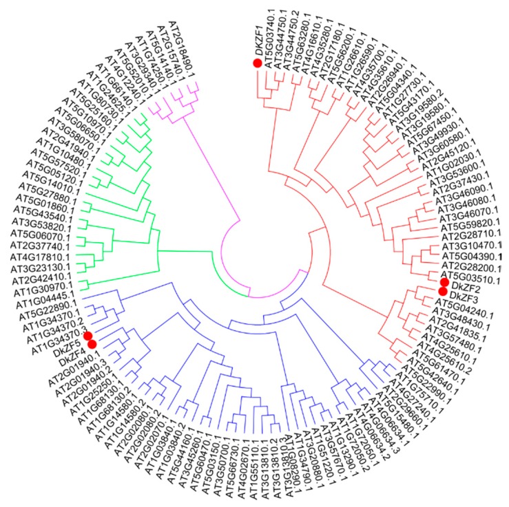
Five DkZFs (DkZF1-5, GenBank accession numbers MN158717-21) were isolated from persimmon fruit. Pairwise sequence identities among isolated DkZFs ranged from 0.143 (DkZF2 vs. DkZF5) to 0.319 (DkZF1 vs. DkZF4) (Table S1). The phylogenetic analysis indicated that DkZFs were mainly clustered into two main clades, with DkZF1-3 in clade I and DkZF4 and DkZF5 in clade II (Figure 1).
Figure 1.
Phylogenetic analysis of DkZFs. Phylogenetic analysis was conducted with DkZFs and zinc finger of Arabidopsis thaliana (ZAT). Red, blue, green, and purple colors indicate clades/subtypes I, II, III, and IV of ZFs respectively, while red circles indicate DkZF transcription factors.
2.2. Expression of DkZFs in Response to AHCA Treatment
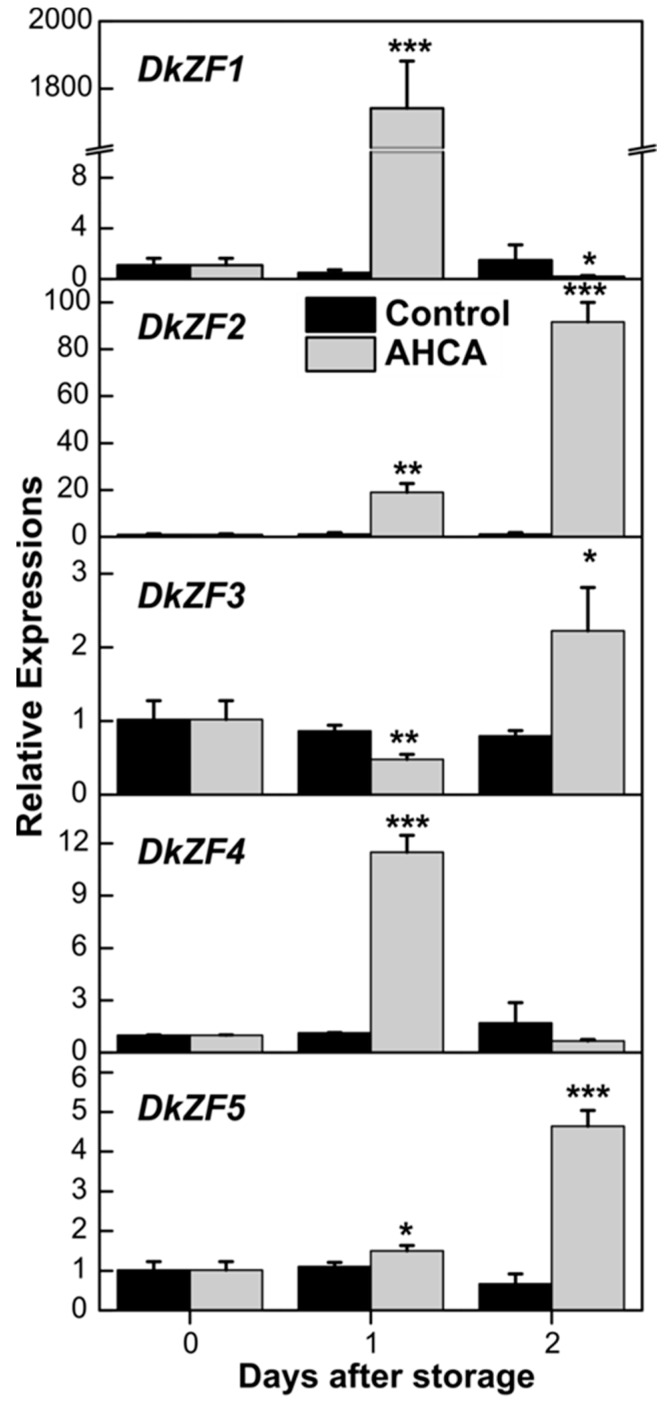
Our previous report indicated that artificial high-CO2 atmosphere (AHCA, 1% O2 and 95% CO2) was effective in astringency removal from “Gongcheng-shuishi” fruit [24], which was used to analyze the expression of DkZFs. qRT-PCR analysis revealed that the transcripts of DkZF1/2/4 were induced by AHCA treatment, while DkZF3/5 showed fewer responses to AHCA treatment at day 1 (Figure 2). Expression of DkZF3/5 mainly accumulated after removal of AHCA treatment, which was after fruit deastringency. Among the three AHCA responsive DkZFs, the relative abundance of DkZF1 was much higher (1743-fold at day 1) than DkZF2 (92-fold at day 2) and DkZF4 (12-fold at day 2) (Figure 2).
Figure 2.
Expression analysis of DkZF genes in response to high-CO2 atmosphere (AHCA) treatment in ‘Gongcheng-shuishi’ fruit. Transcripts of DkZF genes were measured by real time PCR and day 0 fruit values were set as 1. Error bars indicate standard errors from three biological replicates (* p < 0.05, ** p < 0.01, *** p < 0.001).
2.3. Transcriptional Effects of DkZFs on Promoters of Deastringency-Related Genes
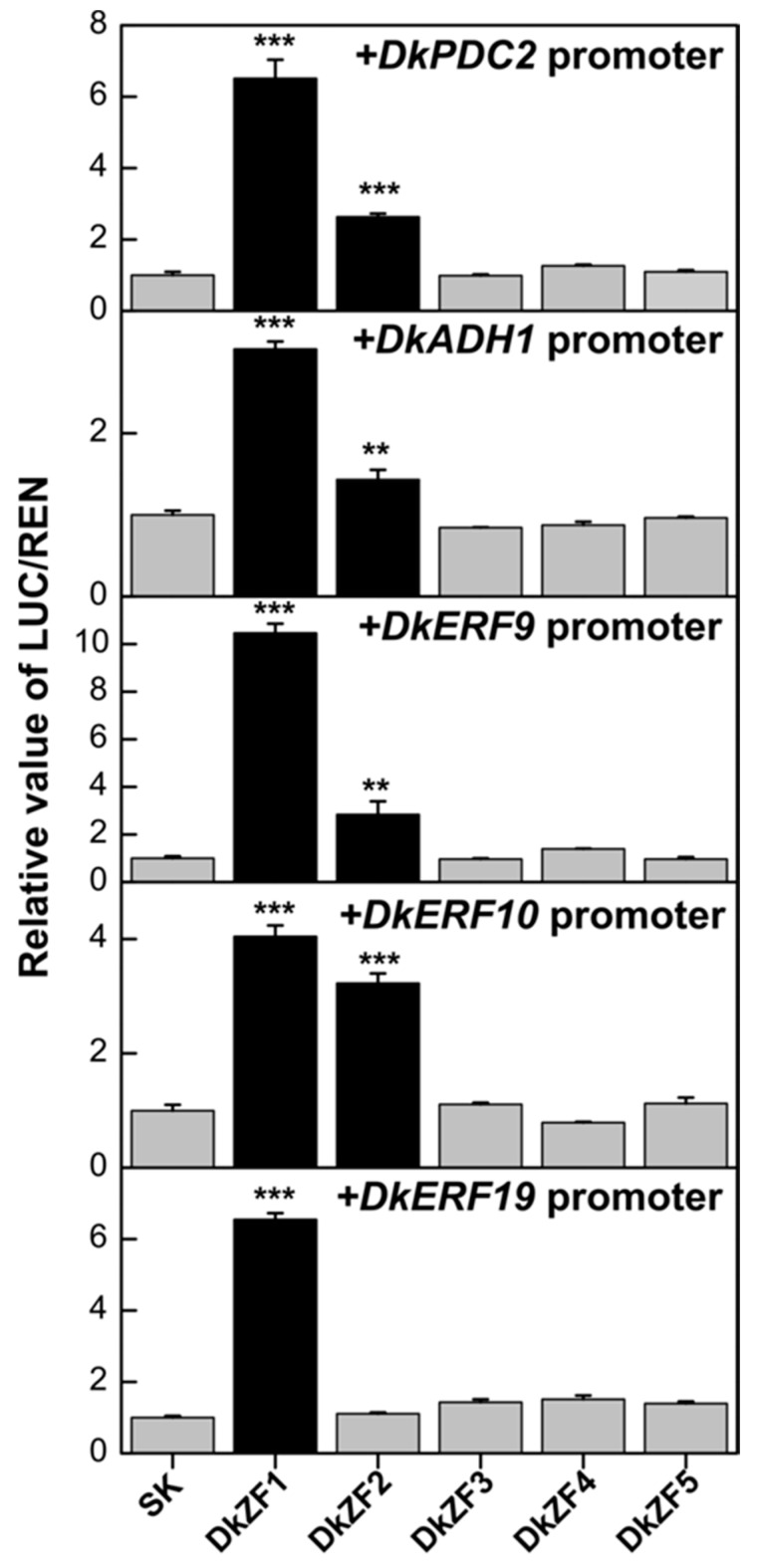
Dual-luciferase assay indicated the regulations of DkZF1 and DkZF2 on the promoters of multiple deastringency-related genes (Figure 3). Here, DkZF1 showed transactivations on all five examined promoters (DkPDC2, DkADH1, DkERF9, DkERF10 and DkERF19), while DkZF2 was an activator for four of them (except for the DkERF19 promoter) (Figure 3). The maximum regulation of DkZF1 was found on the DkERF9 promoter (10.5-fold), while DkZF2 was most effective on the DkERF10 promoter (3.2-fold). Moreover, DkZF3-5 did not show significant regulatory effects on any examined promoters.
Figure 3.
Regulatory effects of DkZF1-5 on the promoters of deastringency-related genes (DkERF9/10/19, DkADH1, and DkPDC2) using the dual-luciferase assay. The ratio of II 0800-LUC vector (LUC)/REN in the empty vector (SK) plus promoter was used as calibrator (set as 1). Values are means (+SE) from four biological replicates (** p < 0.01, *** p < 0.001).
2.4. Synergistic Regulations of DkZF2 and DkZF1 on DkPDC2 Promoter
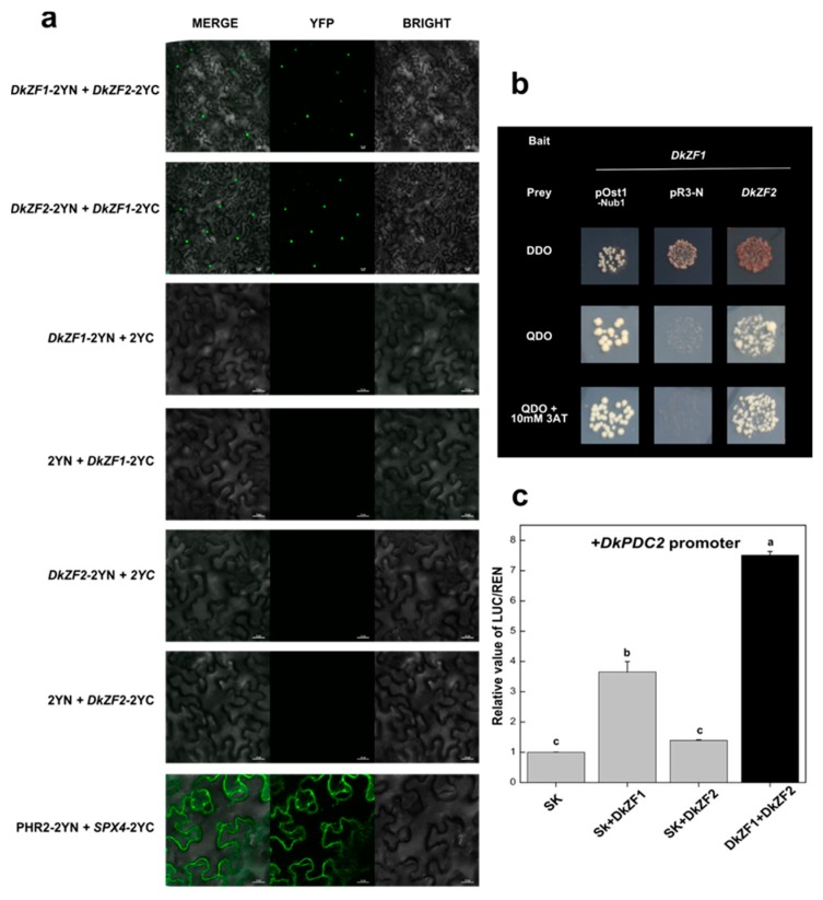
The dual-luciferase assay indicated both DkZF1 and DkZF2 were effective on most of the promoters of deastringency-related genes (Figure 3), which forced us to investigate the relations between these two DkZFs. In order to test protein–protein interaction between DkZF1 and DkZF2, BiFC and Y2H assays were employed (Figure 4). For BiFC, DkZF1 and DkZF2 were fused with both the N-terminal of yellow fluorescent protein (p2YN) and C-terminal of YFP (p2YC) and then transformed together into tobacco leaves. Co-injection of DkZF1 and DkZF2 showed green florescence signals in the nucleus, indicating their protein–protein interactions (Figure 4a), which was further confirmed by Y2H (Figure 4b).
Figure 4.
Protein–protein interactions of DkZF1 and DkZF2 and their synergistic relationship with the DkPDC2 promoter. (a) bimolecular fluorescence complementation (BiFC) assay of DkZF1 and DkZF2 with all possible combinations while merge and YFP florescence indicate protein–protein interactions. Scale bar 25 µm. (b) Y2H assay showed in vivo protein–protein interactions of DkZF1 and DkZF2. The positive control is pOst1–Nub1, while pPR3-N is negative control. (c) Synergistic transactivation effect of combination of DkZF1 and DKZF2 genes on the DkPDC2 promoter. Means with different letters had significant differences (p < 0.05).
Subsequently, the synergistic effectd of DkZF1 and DkZF2 transcription factors on promoters of deastringency-related genes were analyzed. The combination of DkZF1 and DkZF2 significantly enhanced the DkPDC2 promoter compared to that of the individual DkZF (Figure 4c).
3. Discussion
The mechanisms of AHCA [6] were investigated long-term, due to its importance for the persimmon industry and because it is the ideal model for fruit hypoxia research. In recent years, studies have moved beyond physiological and biochemical analyses [16] to molecular aspects [6,22], with characterization of a few key TFs, such as DkERF9/10/19 [6], DkMYB6/10 [22], and DkWRKY1 [23]. However, the involvement of DkZFs on deastringency regulation remain unclear. Here, five DkZFs were isolated from persimmon fruit and distributed into two main clades (Figure 1). Clade I had DkZF1-3 along with reported oxidative stress responsive At5g4340.1 (ZAT6), AT3G46090.1 (ZAT7), AT1G27730.1 (ZAT10) and AT5G59820.1 (ZAT12) [30,31,33]. For instance, ZAT6 caused oxidative stress-induced anthocyanin synthesis in Arabidopsis by directly activating transcription levels of several genes involved in anthocyanin biosynthetic pathway, i.e., TT5, TT7, TT3, TT18, TT4, TT6, MYB12, and MYB111 [39]. ZAT7, ZAT10, and ZAT18 responded during oxidative stress at low oxygen levels [30,34,40]; ZAT12 has been shown to be regulated by several stresses (including oxidative signal), and its regulon contained 42 genes that were involved in the response to oxidative stresses [41,42,43]. Thus, based on the phylogenetic analysis, DkZF1-3 were more likely to be involved in AHCA-driven deastringency for persimmon fruit.
Persimmon astringency removal imperiled by AHCA treatment is widely considered hypoxia-dependent because high CO2/low O2 treatment stimulates anaerobic fermentation, increasing acetaldehyde concentration, which precipitates soluble tannins and ultimately causes an astringency elimination [6,44]. Moreover, AHCA treatment could rapidly decrease soluble tannins to basal level at day 1 in different cultivars [22,24]. Thus, from the RNA-seq data, the increasing expression of DkZF1/2/4 at day 1 in AHCA was considered as the correlation of deastringency, while DkZF3/5 was not. Of these five genes, DkZF1 was the most responsive to the deastringent treatment, especially to AHCA treatment (greater than 1500-fold increase, Figure 2). More direct evidence was provided by dual-luciferase assays, which indicated the transactivation of DkZF1 and DkZF2 on the promoters of deastringency-related genes. Based on the results from phylogenetic analysis, gene expression, and dual-luciferase analysis, DkZF1 and DkZF2 were proposed as two novel regulators for AHCA-driven persimmon fruit deastringency. It is worth emphasizing that the previously characterized TFs showed specificity to limited target genes (e.g., DkERF9 for the DkPDC2 promoter and DkERF10 for the DkADH1 promoter [6]), but DkZF1/2 can regulate five and four promoters, respectively. We reluctantly claim the importance of DkZF1/2, but these findings at least reflect the existence of multidirectional regulation by deastringency-related TFs.
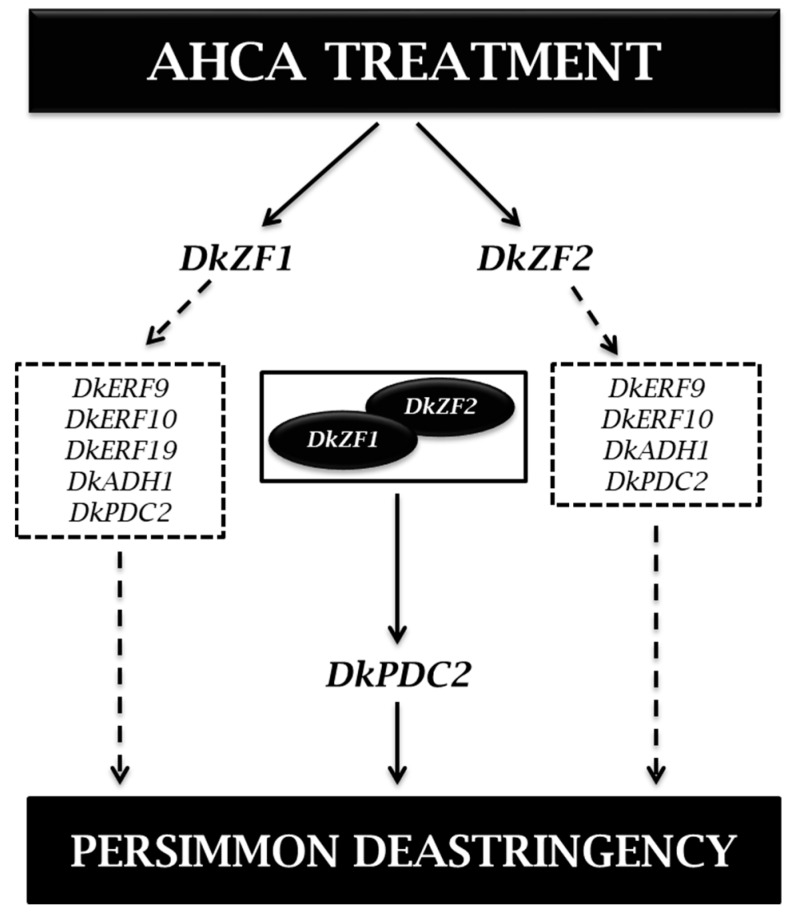
Furthermore, DkZF1 and DkZF2 could interact with each other at the protein levelm, and such an interaction could generate stronger transactivations on the DkPDC2 promoter (Figure 4), which may explain the multidirectional regulations of both DkZF1 and DkZF2. Actually, the TF complexes were widely reported in plants, such as the well-known MYB-bHLH-WD40 in anthocyanin biosynthesis [45,46]. For persimmon deastringency, DkWRKY1 and DkERF24 could also form the complex, which also showed synergistic effects on the DkPDC2 promoter [47]. In conclusion, the present study firstly focused on DkZFs in persimmon fruit deastringency regulation. DkZF1/2 showed significant transactivations on promoters of deastringency-related genes, and their interaction showed synergistic effects on the DkPDC2 promoter (Figure 5). Thus, these findings indicate the involvement of C2H2-type zinc finger proteins (DkZF1/2) in synergistically controlling persimmon fruit deastringency driven by AHCA treatment.
Figure 5.
A proposed model of DkZF transcription factors (TFs) in response to AHCA treatment. AHCA treatment triggers the expressions of DkZF1 and DkZF2 transcription factors that transcriptionally regulate deastringency-related genes represented in dashed boxes respectively. On the other hand, DkZF1 and DkZF2 also form a protein complex and synergistically interact with the DkPDC2 promoter, and ultimately aid in persimmon fruit deastringency.
4. Materials and Methods
4.1. Plant Materials and Treatments
Mature fruit of astringent type persimmon, “Gongcheng-shuishi” (Diospyros kaki, “Gongcheng-shuishi”) were collected in 2015 from a commercial orchard at Gongcheng (Guilin, China) with mean color index and firmness of 8.27 and 60.05 N, respectively. Only those fruits that were disease-free, uniform in shape, and had no mechanical wounds were carefully selected. The fruits were transported to Zhejiang University (Hangzhou, Zhejiang, China) on the second day after harvest. Further, 180 fruits were divided into two 90-fruit lots. Treated fruit were exposed to high-CO2 atmosphere (AHCA, 95% CO2 and 1% O2) to accelerate insolubilization of soluble tannins (deastringency); control fruit were exposed to air, and both were placed in airtight containers for 1 day. After treatment, the fruits were held in air at 20 °C until the end of the experiment. For each sampling point, fruit flesh samples (without skin and core) were taken from three replicates and immediately frozen in liquid nitrogen and stored at –80 °C for further experiments. The physiological data and sampling information are described in [24].
4.2. Gene Isolation and Sequence Analysis
Five unigenes that encoded DkZF were obtained from the RNA-seq database [20], and all of them were putative full-length. The sequences of full-length TFs were confirmed and translated with the ExPASy software (http://web.expasy.org/translate) while amplified with primers (listed in Table S2), spanning the start and stop codons. All DkZFs were named after BLAST analysis in NCBI. For phylogenetic tree analysis, the zinc finger transcription factors in Arabidopsis were obtained from The Arabidopsis Information Resource (https://www.arabidopsis.org/. 15-04-2019). Newly-isolated DkZFs were firstly aligned with ZATs using ClustalW, and then a combined phylogenetic tree of amino acids sequences was constructed by MEGA7.0 (Molecular Evolutionary Genetics Analysis) program.
4.3. RNA Extraction and cDNA Synthesis
Total RNA was prepared, using a cetyltrimethylammonium bromide (CTAB) method [48]. The TURBO DNA free kit (Ambion) was used to digest the trace amount of genomic DNA in total RNA. First strand cDNA synthesis was initiated from 1.0 µg DNA-free RNA, using iScript cDNA Synthesis Kit (Bio-Rad, Hercules, CA, USA). Three biological replicates were used at each sampling point for RNA extraction and subsequent cDNA synthesis.
4.4. Oligonucleotide Primers and Real-Time PCR
Oligonucleotide primers were designed with primer3 (v. 0.4.0, http://frodo.wi.mit.edu/cgibin/primer3/primer3_www.cgi) for real-time PCR and listed in Table S3. The specificity of the real-time PCR primers was tested by sequencing the qRT-PCR products and melting curves. For real-time PCR, CFX96 instrument (Bio-Rad) was used and the PCR mixtures and reactions were the same as in our previous report [49]. The abundance of cDNA templates was measured as 2−ΔΔCt while normalized against the transcript levels of DkActin, a housekeeping gene.
4.5. Dual-Luciferase Assay
To detect in vivo transactivation effects of TFs on promoters, dual-luciferase assay was performed [6]. Full-length CDS of DkZF1-5 and the selected promoter sequences were inserted into pGreen II 0029 62-SK vector (SK) and pGreen II 0800-LUC vector (LUC), respectively. The full-length DkZFs were amplified using primers, as listed in Table S2. The construction of promoters to LUC vector was previously conducted (DkADH1 and DkPDC2 promoters [6]; DkERF9/10/19 promoters [22]). All constructs were electroporated into Agrobacterium tumefaciens GV1301. The dual-luciferase assay was carried out in Nicotiana benthamiana leaves following the same protocol as described in our previous report [6]. The Agrobacterium was suspended in infiltration buffer (10 mM MES, 10 mM MgCl2, 150 mM acetosyringone, pH 5.6) to an OD600 of ~0.75. TFs and promoter were combined in a v/v ratio of 10:1 and infiltrated into N. benthamiana leaves by needle-free syringe. A dual-luciferase assay kit (Promega) was used to analyze the transient expression in N. benthamiana leaves after 3 d of infiltration. Absolute LUC and REN were measured in a GLOMAX 96 Microplate Luminometer (Promega, Madison, WI, USA). Three independent experiments with at least four biological replicates were performed to verify the luciferase activities.
4.6. Bimolecular Fluorescence Complementation (BiFC) Assays
For bimolecular fluorescence complementation (BiFC) assays, the coding sequences (CDSs) without the stop codon were inserted into C-terminal of yellow fluorescent protein (p2YC) and N-terminal of yellow fluorescent protein (p2YN). Both constructs were individually transformed into A. tumefaciens GV3101. Agrobacterium-infiltration was carried out with a needle-free syringe and transiently co-expressed in all possible combinations of p2YN and p2YC fusion proteins in N. benthamiana leaves. Fluorescence was observed by confocal laser scanning microscopy (A1, Nikon, Japan), as described previously [25].
4.7. Yeast Two-Hybrid Assays
The yeast two-hybrid assays were performed using the DUAL hunter system (Dual-Systems Biotech). Full-length coding sequences of DkZF1 were cloned into the pDHB1 vector as bait, and the full-length DkZF2 was cloned into pPR3-N vector as prey. All constructs were transformed into the yeast strain NMY51. The assays were performed with different media: (1) DDO (SD medium lacking Trp and Leu); (2) QDO (SD medium lacking Trp, Leu, His, and Ade); and (3) QDO+3AT (QDO with 10 mM 3-amino-1,2,4-triazole). Auto-activations were tested with empty pPR3-N vectors and target genes with pDHB1, which were co-transformed into NMY51 and plated on QDO. Autoactivations were indicated by the presence of colonies. Protein–protein interaction assays were performed with co-transformation of DkZF1 in pDHB1 and DkZF2 in pPR3-N. The presence of colonies in QDO and QDO+3AT indicated protein–protein interaction.
4.8. Statistical Analysis
Analysis of variance followed by Duncan’s multiple range test was used to test the overall significance of differences among treatments (p < 0.05). Significant differences between treatments were assessed by Student’s t-test at p < 0.05, p < 0.01, and p < 0.001. All data were analyzed in SPSS v25 (SPSS Inc., Chicago, IL, USA).
5. Conclusions
In conclusion, two C2H2-type zinc finger proteins involved in persimmon fruit de-astringency by synergistically trans-activated the DkPDC2 promoter.
Abbreviations
| CTAB | Cetyltrimethyl Ammonium Bromide |
| MEGA | Molecular Evolutionary Genetics Analysis |
| RNA-seq | Ribose nucleic acid sequencing |
| Y2H | Yeast two hybrid |
Supplementary Materials
Supplementary materials can be found at https://www.mdpi.com/1422-0067/20/22/5611/s1.
Author Contributions
X.-R.Y., Q.-G.Z. and X.-F.L. participated in the design of the study; W.J., W.W., H.G., J.-W.H. and M.A. participated in carrying out the experiments; Q.-G.Z., R.J. and X.-F.L. participated in data analysis; W.J. and M.A. participated in drafting the manuscript; X.-R.Y. revised the manuscript and approved the final version.
Funding
This research was supported by the National Natural Science Foundation of China (31722042; 31672204), the Fok Ying Tung Education Foundation, China (161028), the Fundamental Research Funds for the Central Universities (2018XZZX002-03), and the 111 Project (B17039).
Conflicts of Interest
The authors declare no conflict of interest.
References
- 1.Banti V., Giuntoli B., Gonzali S., Loreti E., Magneschi L., Novi G., Paparelli E., Parlanti S., Pucciariello C., Santaniello A. Low oxygen response mechanisms in green organisms. Int. J. Mol. Sci. 2013;14:4734–4761. doi: 10.3390/ijms14034734. [DOI] [PMC free article] [PubMed] [Google Scholar]
- 2.Ismond K.P., Dolferus R., De Pauw M., Dennis E.S., Good A.G. Enhanced low oxygen survival in Arabidopsis through increased metabolic flux in the fermentative pathway. Plant Physiol. 2003;132:1292–1302. doi: 10.1104/pp.103.022244. [DOI] [PMC free article] [PubMed] [Google Scholar]
- 3.Rocha M., Licausi F., Araújo W.L., Nunes Nesi A., Sodek L., Fernie A.R., Van Dongen J.T. Glycolysis and the tricarboxylic acid cycle are linked by alanine aminotransferase during hypoxia induced by waterlogging of Lotus japonicus. Plant Physiol. 2010;152:1501–1513. doi: 10.1104/pp.109.150045. [DOI] [PMC free article] [PubMed] [Google Scholar]
- 4.Dolferus R., Klok E.J., Delessert C., Wilson S., Ismond K.P., Good A.G., Peacock W.J., Dennis E.S. Enhancing the anaerobic response. Ann. Bot. 2003;91:111–117. doi: 10.1093/aob/mcf048. [DOI] [PMC free article] [PubMed] [Google Scholar]
- 5.Loreti E., Poggi A., Novi G., Alpi A., Perata P. A genome wide analysis of the effects of sucrose on gene expression in Arabidopsis seedlings under anoxia. Plant Physiol. 2005;137:1130–1138. doi: 10.1104/pp.104.057299. [DOI] [PMC free article] [PubMed] [Google Scholar]
- 6.Min T., Yin X.R., Shi Y.N., Luo Z.R., Yao Y.C., Grierson D., Ferguson I.B., Chen K.S. Ethylene-responsive transcription factors interact with promoters of ADH and PDC involved in persimmon (Diospyros kaki) fruit de-astringency. J. Exp. Bot. 2012;63:6393–6405. doi: 10.1093/jxb/ers296. [DOI] [PMC free article] [PubMed] [Google Scholar]
- 7.Ali S., Khan A.S., Malik A.U., Shahid M. Effect of controlled atmosphere storage on pericarp browning, bioactive compounds and antioxidant enzymes of litchi fruits. Food Chem. 2016;206:18–29. doi: 10.1016/j.foodchem.2016.03.021. [DOI] [PubMed] [Google Scholar]
- 8.Bekele E.A., Ampofo A.J., Alis R., Hertog M.L., Nicolai B.M., Geeraerd A.H. Dynamics of metabolic adaptation during initiation of controlled atmosphere storage of ‘Jonagold’apple: Effects of storage gas concentrations and conditioning. Postharvest Biol. Technol. 2016;117:9–20. doi: 10.1016/j.postharvbio.2016.02.003. [DOI] [Google Scholar]
- 9.Matityahu I., Marciano P., Holland D., Ben-Arie R., Amir R. Differential effects of regular and controlled atmosphere storage on the quality of three cultivars of pomegranate (Punica granatum L.) Postharvest Biol. Technol. 2016;115:132–141. doi: 10.1016/j.postharvbio.2015.12.018. [DOI] [Google Scholar]
- 10.Taira S., Ikeda K., Ohkawa K. Comparison of insolubility of tannins induced by acetaldehyde vapor in fruits of three types of astringent persimmon. J. Jpn. Soc. Hort. Sci. Food Tech. 2001;48:684–687. doi: 10.3136/nskkk.48.684. [DOI] [Google Scholar]
- 11.Wolfe K.L., Liu R.H. Apple peels as a value-added food ingredient. J. Agric. Food Chem. 2003;51:1676–1683. doi: 10.1021/jf025916z. [DOI] [PubMed] [Google Scholar]
- 12.Galvis Sánchez A.C., Gil I.A., Gil M.I. Comparative study of six pear cultivars in terms of their phenolic and vitamin C contents and antioxidant capacity. J. Sci. Food Agric. 2003;83:995–1003. doi: 10.1002/jsfa.1436. [DOI] [Google Scholar]
- 13.Remorini D., Tavarini S., Degl I.E., Loreti F., Massai R., Guidi L. Effect of rootstocks and harvesting time on the nutritional quality of peel and flesh of peach fruits. Food Chem. 2008;110:361–367. doi: 10.1016/j.foodchem.2008.02.011. [DOI] [PubMed] [Google Scholar]
- 14.Kim H., Moon J.Y., Kim H., Lee D.-S., Cho M., Choi H.K., Kim Y.S., Mosaddik A., Cho S.K. Antioxidant and antiproliferative activities of mango (Mangifera indica L.) flesh and peel. Food Chem. 2010;121:429–436. doi: 10.1016/j.foodchem.2009.12.060. [DOI] [Google Scholar]
- 15.Li Y., Guo C., Yang J., Wei J., Xu J., Cheng S. Evaluation of antioxidant properties of pomegranate peel extract in comparison with pomegranate pulp extract. Food Chem. 2006;96:254–260. doi: 10.1016/j.foodchem.2005.02.033. [DOI] [Google Scholar]
- 16.Kim J.G., Beppu K., Kataoka I. Varietal differences in phenolic content and astringency in skin and flesh of hardy kiwifruit resources in Japan. Sci. Hortic. 2009;120:551–554. doi: 10.1016/j.scienta.2008.11.032. [DOI] [Google Scholar]
- 17.Silva B.M., Andrade P.B., Ferreres F., Domingues A.L., Seabra R.M., Ferreira M.A. Phenolic profile of quince fruit (Cydonia oblonga Miller) (pulp and peel) J. Agric. Food Chem. 2002;50:4615–4618. doi: 10.1021/jf0203139. [DOI] [PubMed] [Google Scholar]
- 18.Akagi T., Ikegami A., Suzuki Y., Yoshida J., Yamada M., Sato A., Yonemori K. Expression balances of structural genes in shikimate and flavonoid biosynthesis cause a difference in proanthocyanidin accumulation in persimmon (Diospyros kaki Thunb.) fruit. Planta. 2009;230:899–915. doi: 10.1007/s00425-009-0991-6. [DOI] [PubMed] [Google Scholar]
- 19.Lesschaeve I., Noble A.C. Polyphenols: Factors influencing their sensory properties and their effects on food and beverage preferences. Am. J. Clin. Nutr. 2005;81:330S–335S. doi: 10.1093/ajcn/81.1.330S. [DOI] [PubMed] [Google Scholar]
- 20.Min T., Fang F., Ge H., Shi Y.N., Luo Z.R., Yao Y.C., Grierson D., Yin X.R., Chen K.S. Two novel anoxia-induced ethylene response factors that interact with promoters of deastringency-related genes from persimmon. PLoS ONE. 2014;9:e97043. doi: 10.1371/journal.pone.0097043. [DOI] [PMC free article] [PubMed] [Google Scholar]
- 21.Min T., Wang M.M., Wang H., Liu X., Fang F., Grierson D., Yin X.R., Chen K.S. Isolation and expression of NAC genes during persimmon fruit postharvest astringency removal. Int. J. Mol. Sci. 2015;16:1894–1906. doi: 10.3390/ijms16011894. [DOI] [PMC free article] [PubMed] [Google Scholar]
- 22.Zhu Q.G., Gong Z.Y., Wang M.M., Li X., Grierson D., Yin X.R., Chen K.S. A transcription factor network responsive to high CO2/hypoxia is involved in deastringency in persimmon fruit. J. Exp. Bot. 2018;69:2061–2070. doi: 10.1093/jxb/ery028. [DOI] [PMC free article] [PubMed] [Google Scholar]
- 23.Zhu Q.G., Gong Z.Y., Huang J., Grierson D., Chen K.S., Yin X.R. High-CO2/hypoxia-responsive transcription factors DkERF24 and DkWRKY1 interact and activate DkPDC2 promoter. Plant Physiol. 2019;180:621–633. doi: 10.1104/pp.18.01552. [DOI] [PMC free article] [PubMed] [Google Scholar]
- 24.Jamil W., Wu W., Ahmad M., Zhu Q.G., Liu X.F., Jin R., Yin X.R. High-CO2/hypoxia-modulated NAC transcription factors involved in de-astringency of persimmon fruit. Sci. Hortic. 2019;252:201–207. doi: 10.1016/j.scienta.2019.03.018. [DOI] [Google Scholar]
- 25.Jin R., Zhu Q.G., Shen X.Y., Wang M.M., Jamil W., Grierson D., Yin X.R., Chen K.S. DkNAC7, a novel high CO2/hypoxia-induced NAC transcription factor, regulates persimmon fruit de-astringency. PLoS ONE. 2018;13:e0194326. doi: 10.1371/journal.pone.0194326. [DOI] [PMC free article] [PubMed] [Google Scholar]
- 26.Takatsuji H. Zinc-finger proteins: The classical zinc finger emerges in contemporary plant science. Plant Mol. Biol. 1999;39:1073–1078. doi: 10.1023/A:1006184519697. [DOI] [PubMed] [Google Scholar]
- 27.Jenkins T.H., Li J., Scutt C.P., Gilmartin P.M. Analysis of members of the Silene latifolia Cys 2/His 2 zinc-finger transcription factor family during dioecious flower development and in a novel stamen-defective mutant ssf1. Planta. 2005;220:559–571. doi: 10.1007/s00425-004-1365-8. [DOI] [PubMed] [Google Scholar]
- 28.Schumann U., Prestele J., OGeen H., Brueggeman R., Wanner G., Gietl C. Requirement of the C3HC4 zinc RING finger of the Arabidopsis PEX10 for photorespiration and leaf peroxisome contact with chloroplasts. Proc. Natl. Acad. Sci. USA. 2007;104:1069–1074. doi: 10.1073/pnas.0610402104. [DOI] [PMC free article] [PubMed] [Google Scholar]
- 29.Moore M., Ullman C. Recent developments in the engineering of zinc finger proteins. Brief. Func. Genom. 2003;1:342–355. doi: 10.1093/bfgp/1.4.342. [DOI] [PubMed] [Google Scholar]
- 30.Ciftci Y.S., Morsy M.R., Song L., Coutu A., Krizek B.A., Lewis M.W., Warren D., Cushman J., Connolly E.L., Mittler R. The EAR-motif of the Cys2/His2-type zinc finger protein ZAT7 plays a key role in the defense response of Arabidopsis to salinity stress. J. Biol. Chem. 2007;282:9260–9268. doi: 10.1074/jbc.M611093200. [DOI] [PubMed] [Google Scholar]
- 31.Kim S.H., Hong J.K., Lee S.C., Sohn K.H., Jung H.W., Hwang B.K. CAZFP1, Cys 2/His 2-type zinc-finger transcription factor gene functions as a pathogen-induced early-defense gene in Capsicum annuum. Plant Mol. Biol. 2004;55:883–904. doi: 10.1007/s11103-005-2151-0. [DOI] [PubMed] [Google Scholar]
- 32.Sakamoto H., Maruyama K., Sakuma Y., Meshi T., Iwabuchi M., Shinozaki K., Yamaguchi-Shinozaki K. Arabidopsis Cys2/His2-type zinc-finger proteins function as transcription repressors under drought, cold, and high-salinity stress conditions. Plant Physiol. 2004;136:2734–2746. doi: 10.1104/pp.104.046599. [DOI] [PMC free article] [PubMed] [Google Scholar]
- 33.Vogel J.T., Zarka D.G., Van Buskirk H.A., Fowler S.G., Thomashow M.F. Roles of the CBF2 and ZAT12 transcription factors in configuring the low temperature transcriptome of Arabidopsis. Plant J. 2005;41:195–211. doi: 10.1111/j.1365-313X.2004.02288.x. [DOI] [PubMed] [Google Scholar]
- 34.Yin M., Wang Y., Zhang L., Li J., Quan W., Yang L., Wang Q., Chan Z. The Arabidopsis Cys2/His2 zinc finger transcription factor ZAT18 is a positive regulator of plant tolerance to drought stress. J. Exp. Bot. 2017;68:2991–3005. doi: 10.1093/jxb/erx157. [DOI] [PMC free article] [PubMed] [Google Scholar]
- 35.Zhang S., Zhang D., Fan S., Du L., Shen Y., Xing L., Li Y., Ma J., Han M. Effect of exogenous GA3 and its inhibitor paclobutrazol on floral formation, endogenous hormones, and flowering-associated genes in ‘Fuji’ apple (Malus Domestica Borkh.) Plant Physiol. Biochem. 2016;107 doi: 10.1016/j.plaphy.2016.06.005. [DOI] [PubMed] [Google Scholar]
- 36.Pandey D.M., Kim S.R. Identification and expression analysis of hypoxia stress inducible CCCH-type zinc finger protein genes in rice. J. Plant Biol. 2012;55:489–497. doi: 10.1007/s12374-012-0384-4. [DOI] [Google Scholar]
- 37.Huang J., Sun S.J., Xu D.Q., Yang X., Bao Y.-M., Wang Z.F., Tang H.J., Zhang H. Increased tolerance of rice to cold, drought and oxidative stresses mediated by the overexpression of a gene that encodes the zinc finger protein ZFP245. Biochem. Biophys. Res. Commun. 2009;389:556–561. doi: 10.1016/j.bbrc.2009.09.032. [DOI] [PubMed] [Google Scholar]
- 38.Zhang H., Ni L., Liu Y., Wang Y., Zhang A., Tan M., Jiang M. The C2H2-type Zinc Finger Protein ZFP182 is Involved in Abscisic Acid-Induced Antioxidant Defense in Rice F. J. Integr. Plant Biol. 2012;54:500–510. doi: 10.1111/j.1744-7909.2012.01135.x. [DOI] [PubMed] [Google Scholar]
- 39.Shi H., Liu G., Wei Y., Chan Z. The zinc-finger transcription factor ZAT6 is essential for hydrogen peroxide induction of anthocyanin synthesis in Arabidopsis. Plant Mol. Biol. 2018;97:165–176. doi: 10.1007/s11103-018-0730-0. [DOI] [PubMed] [Google Scholar]
- 40.Ciftci Y.S., Mittler R. The zinc finger network of plants. Cell. Mol. Life Sci. 2008;65:1150–1160. doi: 10.1007/s00018-007-7473-4. [DOI] [PMC free article] [PubMed] [Google Scholar]
- 41.Davletova S., Schlauch K., Coutu J., Mittler R. The zinc-finger protein Zat12 plays a central role in reactive oxygen and abiotic stress signaling in Arabidopsis. Plant Physiol. 2005;139:847–856. doi: 10.1104/pp.105.068254. [DOI] [PMC free article] [PubMed] [Google Scholar]
- 42.Le C.T.T., Brumbarova T., Ivanov R., Stoof C., Weber E., Mohrbacher J., Fink-Straube C., Bauer P. Zinc finger of Arabidopsis thaliana12 (ZAT12) interacts with FER-like iron deficiency-induced transcription factor (FIT) linking iron deficiency and oxidative stress responses. Plant Physiol. 2016;170:540–557. doi: 10.1104/pp.15.01589. [DOI] [PMC free article] [PubMed] [Google Scholar]
- 43.Miller G., Shulaev V., Mittler R. Reactive oxygen signaling and abiotic stress. Physiol. Plant. 2008;133:481–489. doi: 10.1111/j.1399-3054.2008.01090.x. [DOI] [PubMed] [Google Scholar]
- 44.Salvador A., Arnal L., Besada C., Larrea V., Quiles A., Pérez M.I. Physiological and structural changes during ripening and deastringency treatment of persimmon fruit cv.‘Rojo Brillante’. Postharvest Biol. Technol. 2007;46:181–188. doi: 10.1016/j.postharvbio.2007.05.003. [DOI] [Google Scholar]
- 45.Espley R.V., Hellens R.P., Putterill J., Stevenson D.E., Kutty A.S., Allan A.C. Red colouration in apple fruit is due to the activity of the MYB transcription factor, MdMYB10. Plant J. 2007;49:414–427. doi: 10.1111/j.1365-313X.2006.02964.x. [DOI] [PMC free article] [PubMed] [Google Scholar]
- 46.Liu X., Feng C., Zhang M., Yin X., Xu C., Chen K. The MrWD40-1 gene of Chinese bayberry (Myrica rubra) interacts with MYB and bHLH to enhance anthocyanin accumulation. Plant Mol. Biol. Rep. 2013;31:1474–1484. doi: 10.1007/s11105-013-0621-0. [DOI] [Google Scholar]
- 47.Zhu Q.G., Wang M.M., Gong Z.Y., Fang F., Sun N.J., Li X., Grierson D., Yin X.R., Chen K.S. Involvement of DkTGA1 transcription factor in anaerobic response leading to persimmon fruit postharvest de-astringency. PLoS ONE. 2016;11:e0155916. doi: 10.1371/journal.pone.0155916. [DOI] [PMC free article] [PubMed] [Google Scholar]
- 48.Wang M.M., Zhu Q.G., Deng C.L., Luo Z.R., Sun N.J., Grierson D., Yin X.R., Chen K.S. Hypoxia-responsive ERF s involved in postdeastringency softening of persimmon fruit. Plant Biotechnol. J. 2017;15:1409–1419. doi: 10.1111/pbi.12725. [DOI] [PMC free article] [PubMed] [Google Scholar]
- 49.Yin X.R., Shi Y.N., Min T., Luo Z.R., Yao Y.C., Xu Q., Ferguson I., Chen K.S. Expression of ethylene response genes during persimmon fruit astringency removal. Planta. 2012;235:895–906. doi: 10.1007/s00425-011-1553-2. [DOI] [PubMed] [Google Scholar]
Associated Data
This section collects any data citations, data availability statements, or supplementary materials included in this article.


