Abstract
This mini-review examines the crucial importance of transcription factors as a first line of defense in the detoxication of xenobiotics. Key transcription factors that recognize xenobiotics or xenobiotic-induced stress such as reactive oxygen species (ROS), include AhR, PXR, CAR, MTF, Nrf2, NF-κB, and AP-1. These transcription factors constitute a significant portion of the pathways induced by toxicants as they regulate phase I-III detoxication enzymes and transporters as well as other protective proteins such as heat shock proteins, chaperones, and anti-oxidants. Because they are often the first line of defense and induce phase I-III metabolism, could these transcription factors be considered the phase 0 of xenobiotic response?
Introduction:
The term phase I and phase II detoxication have been a part of the lexicon of the toxicology vocabulary since first coined by R.T. Williams in 1959 [1]. Phase I and II enzymes include the cytochrome P450s (CYPs), flavin-containing mono-oxygenases (FMOs), peroxidases, dismutases, and conjugases [glutathione S-transferase (GST), uridine diphospho-glucuronosyltransferases (UDPGT; UGT), and sulfotransferases (SULTs)] that hydroxylate, oxidize, reduce, desulfurize, epoxidate, and conjugate xenobiotics [2]. Phase I refers to the early oxidative metabolism of the xenobiotic, and phase II typically refers to conjugation but potentially to a second oxidative reaction such as de-epoxidation by epoxide hydrolase [3].
Later the discovery of xenobiotic transporters led to the term phase III and refers to proteins that eliminate xenobiotics from cells through membrane transport pumps [4]. Phase III transporters include key members of ATP-binding cassette (ABC) transporters primarily in groups ABCB and ABCC such as multidrug resistance associated protein 2 (MRP2), multidrug resistance protein (MDR1), and bile salt export pump (BSEP) [5–8] (Fig. 1). Additional phase III transporters can also be found in groups such as ABCG (ABCG2; breast cancer resistance protein)[9]. Phase III transport can occur first, prior to transcription factor activation or phase I metabolism, as some xenobiotics are pumped out shortly after entering the cell by transporters such as MDR1 (also known as P-glycoprotein (PGP)). Therefore, phase III has been recently referred to as “phase 0 transport” because these transporters eliminate chemicals from the cell without prior phase I and II metabolism [10, 11]. However, for clarity and recognition of “transport”, I propose that phase III be used whether or not metabolism occurs prior to membrane transport. Similarly, conjugation of xenobiotics (phase II) can also occur prior to phase I metabolism if the proper leaving group is available and yet conjugation is still called phase II [12–14].
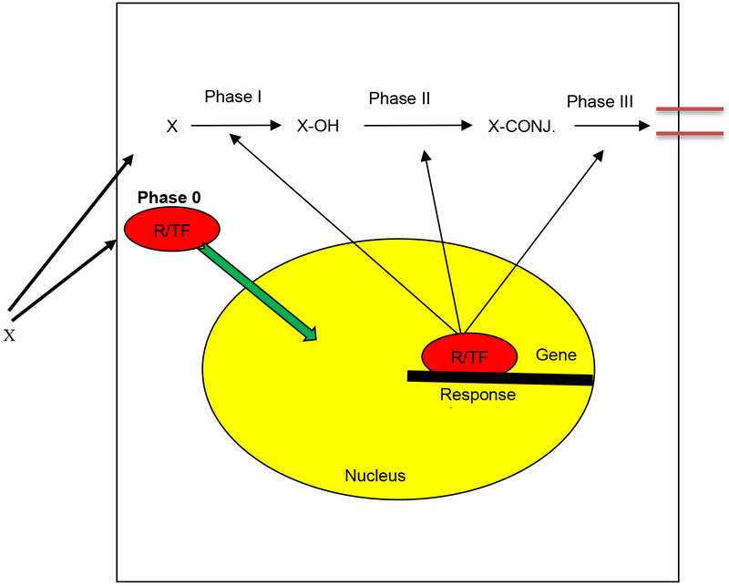
Figure 1. Phase 0 response to xenobiotics is activation of a transcriptional response by xenobiotic responsive transcription factors.
Phase I-III detoxication is well documented and relatively well defined as oxidative metabolism, conjugation, and transport, respectively. Phase 0 xenobiotic response is defined as the transcriptional response of and initial acclimation of the cell to xenobiotics leading to increased phase I-III detoxication through gene regulation. R/TF = receptor/transcription factor.
Most of these phase I-III detoxification enzymes and transporters are inducible and elegantly regulated by a suite of transcription factors. We often refer to specific pathways in transcriptomics based on the transcription factor activated. Thus, given that transcription factors are often our first responders following chemical exposure, they could be considered our first phase of detoxication. However, the term phase I is already taken and well established in the literature. Therefore, transcription factors that initiate our molecular response to chemical intrusion and help individuals acclimate to xenobiotic insults be identified as “phase 0”, “phase 0 detoxication” or “phase 0 xenobiotic response” because these transcription factors act as the initial response that increases phase I-III metabolism (Fig. 1)?
Xenobiotic-responsive transcription factors:
Transcription factors are any number of proteins that can help initiate or regulate transcription by binding DNA at specific promoter or enhancer sites [15]. The transcription factors crucial in toxicology can respond directly to xenobiotic exposure or respond to adverse metabolites or reactions caused by the chemicals such as increased ROS or perturbations in mitochondrial viability [16, 17]. The list of transcription factors presented below is not exhaustive, but includes the most prominent transcription factors in acclimating to chemical stress.
Transcription factors typically perturbed by endo- or xenobiotic-mediated stress are trans-acting elements that can be activated by ROS, hormones, xenobiotics or any number of extracellular stress signals. In turn, the transcription factor activates transcription by binding to DNA at specific cis-elements (i.e. response elements; consensus sequences) sometimes called a Xenobiotic Response Element (XRE) when dealing with the response element is unique to a xenobiotic responsive transcription factor. There are several groups of transcription factors often classified based on the type of DNA binding motif that they contain such as zinc fingers (nuclear receptors), basic leucine zippers (bZIP), or basic helix-loop-helix (bHLH). An example is the bHLH group of transcription factors containing a basic region adjacent to a helix-loop-helix (HLH) domain [15]. HLH members include the aryl hydrocarbon receptor (AhR), a key transcription factor in toxicology [18].
AhR:
The aryl hydrocarbon receptor (AhR), also known as the dioxin receptor, is a bHLH transcription factor in the Per-Arnt-Sim (PAS) family. It is an established xenobiotic receptor, as it is bound and activated by polycyclic aromatic hydrocarbons (PAHs), dioxins, and other coplanar aromatic compounds. It has additional functions based on the phenotype of untreated AhR-null mice, which include roles in the immune system, cardiac hypertrophy, cardiac development, hepatic growth, and oocyte development [19–23]. Cardiac toxicity is common in animals exposed to TCDD and other AhR agonists during development [24, 25] and recent work shows mice treated with AhR agonists have a propensity towards obesity [26] and non-alcoholic fatty liver disease [27, 28]. AhR is also involved in regulating tryptophan metabolism through the kynurenine pathway [29].
The AhR binds to a number of xenobiotics such as the halogenated aromatic hydrocarbons (HAHs), (i.e the dibenzo-p-dioxins, dibenzofurans, and polychlorinated biphenyls (PCBs)), and the polycyclic aromatic hydrocarbons (i.e. pyrene, 3-methylcholanthrene, benzo[a]pyrene) [30]. HAHs have higher affinities for the AhR than PAHs and this difference is strongly associated with toxicity [31, 32]. It is assumed that the toxicity of HAHs and PAHs is due to the inappropriate expression of specific genes induced by AhR.
Following ligand activation, the AhR translocates to the nucleus, and mediates the transcription of a number of genes. Many of the proteins induced by AhR activation are detoxication enzymes and include CYP1A, CYP1B, NADPH:quinone oxidoreductase 1, and UDP-glucuronosyltransferase 1 [32–34] of which several are necessary for the metabolism of the xenobiotic and protection from its adverse effects. There is also data implicating AhR activity in the down-regulation of the estrogen receptor, glucocorticoid receptor, epidermal growth factor receptor, and CYP2C11 in mammals [35, 36].
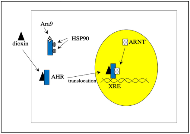
Activation of AhR in the cytosol displaces it from heat shock proteins (HSP90), HSP23 and the immunophilin chaperone Ara9 (also known as XAP2) [29, 37]. This allows for translocation to the nucleus and association with the aryl hydrocarbon receptor nuclear translocator (ARNT). The AhR/ARNT complex binds to the xenobiotic response element (XRE, also known as the dioxin responsive element, DRE) and in turn activates basal transcription factors, the transcription of CYP1A, and other target genes (Fig. 2). In addition, ligand binding to AhR also releases proto-oncogene tyrosine kinase (SRC), which is a tyrosine kinase that phosphorylates and activates the ERK1/2 pathway. SRC can also phosphorylate AhR, increasing nuclear translocation [38].
Figure 2. The AhR is activated by ligands such as dioxin.
Ligand activation induces the release of HSP90 and Ara9 and allows for translocation of the AhR into the nucleus. In the nucleus, the AhR binds ARNT and this complex binds the xenobiotic response element (XRE) and regulates transcription of CYP1A and other detoxification genes.
Interestingly, reported cardiotoxicity mediated by benzo(a)pyrene is significantly reduced in zebrafish lacking AhR activity and in turn Cyp1a induction; however, this is not true for all PAHs tested [39]. 2,3,7,8-tetrachlorodibenzo-p-dioxin’s (TCDD) teratogenicity is significantly ameliorated in AhR-null mice demonstrating a role for the AhR and its transcriptional response in mediatiating toxicity [40, 41]. Furthermore, a poor response to AhR agonists is associated with tolerance to dioxin and dioxin-like chemicals [18, 42, 43]. For example, Fundulus heteroclitus resistant to PAHs, polychlorinated biphenyls, and dioxins have been found in New Bedford Harbor, MA, Newark Bay, NJ, and the Elizabeth River, VA, and each of these populations demonstrate weak induction of CYP1A following chemical exposures primarily due to mutant AhRs [43–49].
Nuclear Receptors (PXR, CAR, HR96):
The nuclear receptor superfamily contains several transcription factors of which most are activated by small lipophilic ligands such as steroids, bile acids, bilirubin, fatty acids, heme, and xenobiotics [50–52]. Several nuclear receptors have a role in protecting individuals from the build-up of toxic endobiotics, including farnesoid X-receptor (FXR) liver X-receptor (LXR), and the peroxisome proliferator activated receptors (PPARs); however, their role in xenobiotic metabolism and elimination is limited [53–62]. We will not consider these nuclear receptors for this review but it should be noted that they are crucial in the detoxication of endobiotics including bile acids, bilirubin, fatty acids, and oxysterols [53, 58, 60, 63–65].
PPARs, Vitamin D receptor (VDR), GR, and the retinoid receptors; RAR and RXR, are considered new targets for endocrine disruption by xenobiotics, in addition to the traditional endocrine targets such as estrogen receptors, thyroid hormone receptors and androgen receptors (all within the nuclear receptor family)[58]. In addition, new nuclear receptors have been discovered recently in several invertebrate species including some within the xeno-sensing clade [51, 66–68]. Of these “non-toxicology” nuclear receptors, the PPARs (PPARα/δ/γ) are of special interest in toxicology because several chemicals activate various PPARs including tributyltin, perfluoronated compounds such as PFOS, mono-2-ethylhexylphthalate, and atrazine [54, 58, 69–72]. Activation of PPARα causes peroxisome proliferation in the mouse liver, which is associated with rodent liver cancer [73, 74]. However, PPARα is highly expressed in rodent liver, but weakly expressed in human liver and this difference in expression is thought to be the underlying cause of specie’s differences in PPARα-mediated liver cancer, which is not observed in humans [75]. PPARγ activation is associated with obesity in multiple species. Current research suggests that PPARγ-mediated obesogens work in conjunction with RXR activation [54, 76]. However, there is little data indicating that activation of the PPARs provides a toxicokinetic effect by altering the metabolism, distribution, or clearance of most chemicals.
Confirmed xenobiotic sensing nuclear receptors include the pregnane X-receptor (PXR), its relative the constitutive androstane receptor (CAR), and their invertebrate ortholog, HR96 [68, 77–83]. Because of their impact on toxicology there are extensive reviews available on the wide range of ligands and indirect activators that mediate transcriptional activation through these nuclear receptors. For an overall review of these nuclear receptors and their ligands see the following manuscripts [84–87].
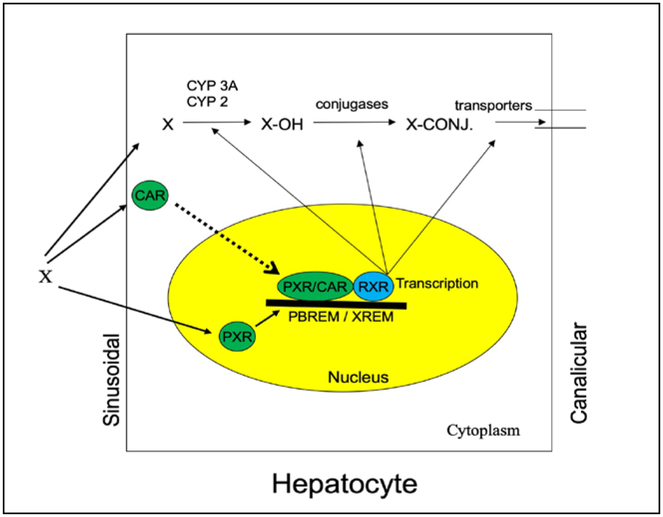
PXR and CAR are primarily expessed in the liver but show limited expression in other tissues such as the intestines, urinary tract, brain, and fish gills [78, 81, 88–92]. PXR and CAR act as master regulators of phase I and phase II metabolism enzymes and phase III transporters (Fig. 3). This includes CYP3A, CYP2B, UDPGT, GSTA2, SULT2A, and MRP2 [79, 93–98] with significant overlapping ligand specificities and gene regulation [86, 99]. There are several manuscripts and reviews that have taken a comprehensive look at environmental chemicals, nutrients, and pharmaceuticals that activate CAR and PXR [86, 100–104].
Figure 3. Function of CAR and PXR in xenobiotic metabolism.
CAR and PXR are activated by specific xenobiotics (X). In turn they dimerize with RXR, act on consensus sequences (XREM/PBREM), and initiate transcription of phase I and II enzymes involved in the hydroxylation and conjugation of xenobiotics, and phase III transporters that eliminate the xenobiotics. Indirect activatin of CAR and its subsequent translocation to the nucleus is key in its transcriptional activity.
PXR is considered promiscuous because it binds a variety of bile acids, steroids and xenobiotics in mammals, including a large number of endocrine disrupting chemicals [105–109], providing circumstantial evidence that the PXR and its close relative, CAR, are protectors of the endocrine system [3]. Xenobiotics that bind and activate PXR in mammals include rifampicin, hyperforin, bisphenol A, 4-nonylphenol, phthalic acid, 1,1-dichloro-2,2-bis (p-chlorophenyl)ethylene (DDE), methoxychlor, vinclozolin, alachlor, cyperterone acetate, trifluralin, and non-planar polychlorinated biphenyls (PCBs) [3, 79, 86, 104, 107–114].
PXR binds to a larger number of diverse chemicals than other receptors [86, 115]. PXR’s promiscuity is attributed to its large and flexible ligand binding domain (LBD). PXR only needs to use a portion of its ligand binding pocket for chemical activation, and it has polar residues spaced through a smooth, multi-regional hydrophobic ligand-binding domain [112, 116, 117]. In addition, PXR has alpha helices that are flexible or can unwind, which allows for PXR to contract or expand in order to bind ligands of different sizes [117]. PXR also forms homodimers and a PXR-RXR heterotetramer complex that is crucial in the recruitment of coactivators and stabilization of the AF-2 domain [118, 119].
CAR is also promiscuous, however it is for reasons related to phosphorylation rather than its binding domain. CAR’s ligand binding pocket is smaller and less flexible than PXR’s, and in turn CAR is less promiscuous than PXR [3, 116, 120, 121]. TCPOBOP is one of a small number of true ligands for CAR. [122]. However, CAR can also be activated indirectly through changes in phosphorlylation status (i.e. phenobarbital) leading to translocation to the nucleus [16, 123, 124]. Under normal (inactivated) conditions, CAR is phosphorylated at Thr38. Dephosphorylation occurs through a Receptor for Activated C Tyrosine Kinase 1 (RACK1) - Protein Phosphatase 2 A (PP2A) cascade [125] that is inhibited by Epidermal Growth Factor (EGF) signaling [126]. Phenobarbital binds the EGFR near the EGF binding site and reduces bound EGF [127] thus inhibiting downstream actions of EGF on SRC and subsequent phosphorylation of RACK-1 allowing for increased dephosphorylation by PPA2 [126]. This blocks EGF action and increases CAR activity.
Another putative mechanism for CAR activation by phenobarbital involves AMP activated protein Kinase (AMPK) [128]. However, downstream functions such as translocation of CAR or induction of CYP2B have varied depending on the system. Inhibition of CAR translocation and CYP2B expression have also been observed following AMPK activation providing contradictory results [129]. In summary, removal of the phosphate group from Thr38 triggers nuclear translocation of CAR. CAR in turn forms a heterodimer with RXR in the nucleus and binds PBREM/DR4 enhancer modules that induce gene transcription of CYP2B6 in humans, Cyp2b10 in mice [80], along with a host of other genes required for cell growth, metabolic activity, and detoxification [86, 130, 131].
Given CAR and PXR’s role in the induction of crucial phase I-III detoxication proteins, it is not surprising that CAR and PXR are associated with protection from anthropogenic pollutants and endobiotics. For example, CAR or PXR activation increases the metabolism, detoxication, and clearance of bile acids [132, 133]. PXR expression increases in response to higher steroid levels during pregnancy presumably to protect the mother from the increased steroid load [134]. Lycopene is in part protective from atrazine toxicity due to its actions on CAR and PXR, and in turn increased metabolism of atrazine [70]. PXR also protects from benzo(a)pyrene toxicity by repressing AhR-mediated transcription [135]. CAR-null mice show significantly increased sensitivity to parathion due to decreased metabolism of parathion to p-nitrophenol, the key detoxication product of parathion [136]. In addition, PXR-null mice showed greater hepatic damage following nonylphenol exposure than wildtype mice, presumably due to a repressed transcriptional response and lower hepatic detoxication enzyme levels in PXR-null mice. PXR-null mice also had greater serum nonylphenol concentrations than wildtype mice, providing further evidence that PXR-null mice were unable to transcriptionally respond and detoxify nonylphenol [137]. Interestingly, human PXR (hPXR) mice showed responses in between wildtype and PXR-null mice indicating that hPXR is less responsive to nonylphenol than murine PXR [137]. Overall, these xenobiotics showed greater toxic effects probably due to the lack of the proper regulation or induction of key detoxification enzymes [136–139].
However, sometimes PXR or CAR activation increases metabolic activation and the toxicity of the chemical. For example, thalidomide activation of PXR increases the production of its teratogenic metabolite, 5-hydroxy-thalidomide through CYP induction [140]. PXR also mediates rifampin toxicity through its actions on aldosterone [141]. CAR-null and PXR-null mice are also less sensitive to acetaminophen, and activation of these receptors and subsequent CYP induction increases NAPQI production and toxicity [142, 143]. PXR activation has also been associated with drug-drug interactions. For example, PXR activation by hyperforin increases clearance of warfarin, estradiol, and other drugs leading to reduced efficacy and poor clinical outcomes [3, 111, 144]. Overall, the discovery of PXR (and to a lesser extent CAR) has provided crucial information on drug-drug interactions and therefore saved lives.
HR96, as well as the C. elegans receptors, NRH48 and NHR8, are the invertebrate homologs to CAR, PXR, and VDR [51, 77, 145]. Similar to CAR and PXR, HR96 regulates lipid homeostasis [146–150]. HR96 also mediates the induction of phase I-III detoxication genes [68, 151, 152] following activation by endogenous or exogenous chemicals [68, 77, 83]. Recent studies indicate that activation of HR96 by atrazine provides protection from some chemicals such as docosahexaenoic acid and triclosan, but increases toxicity to others such as endosulfan and parathion [83, 152]. Thus, many invertebrates also contain nuclear receptors responsive to anthropogenic stress that induce phase I-III metabolism.
Nrf2, AP-1 and NF-kB:
Nrf2, AP-1, and NF-κB are transcription factors that respond to oxidative stress and are important regulators of GSTs, superoxide dismutase (SOD), catalase, and other protective proteins involved in the detoxification of ROS. The toxicant-induced formation of reactive oxygen species (ROS) has been associated with reduced fitness, apoptosis, cancer, and death. For example, pulp and paper mill and sewage effluents induce ROS and fatty acyl-CoA oxidase activity (an enzyme that produces the oxidant H2O2 from O2) in exposed Longnose sucker (Catostomus catostomus) [153]. Several metals including iron, copper, nickel, chromium, cadmium and possibly arsenic mediate toxic effects through oxidative mechanisms and alter redox-sensitive signaling through Nrf2, AP-1 and NF-κB [154–159]. Of these metals, chromium and arsenic have also been shown to block AP-1 and NF-κB activity by binding their respective response elements [160], and the lack of Nrf2 increases sensitivity of cells to metals and a variety of other pro-oxidants [155]. Furthermore, cadmium-induced oxidative stress increases cytochrome c and activated caspase 3, 8 and 9, causing apoptosis through the mitochondrial pathway. Anti-oxidants were able to alleviate the effects of cadmium on apoptosis demonstrating the role of ROS in cadmium-induced apoptosis [161].
The induction of antioxidant enzymes is also associated with protection from toxicants in vivo; a concept supported by the presence of a resistant population of Fundulus heteroclitus from the Elizabeth River, VA, USA that exhibits high glutathione peroxidase and reductase activities, along with high glutathione concentrations [162]. In addition, this population demonstrates both induction and a heritable increase in manganese superoxide dismutase (MnSOD) and glutathione concentrations, which may play a key role in their tolerance to PAHs [163]. Overall, the induction of anti-oxidant defenses is key in protecting individuals from chemical stress and there are three key transcription factors involved in their induction with Nrf2 as the primary protective sensor for ROS.
NF-κB:
Nuclear factor-kappa B (NF-κB) is a transcription factor complex that can be activated by a number of different external signals, including several cytokines, bacterial and viral products, ultraviolet irradiation, oxidative stress, environmental chemicals such as arsenic, chromium, and diesel exhaust, and some therapeutic pharmaceuticals [164–172]. NF-κB in turn regulates the transcription of cytokines, cell adhesion molecules, stress response proteins, acute phase proteins, and regulators of apoptosis. Genes regulated by NF-κB include GSTP 1–1, COX-2, IL-1, C-reactive protein, phospholipase A2, DT-diaphorase, superoxide dismutase, α−1-antichymotrypsin, caspase 10, and IGF-BP1 [173–181] Comprehensive reviews on NF-κB can be found at [164, 173], or a website maintained by Dr. Thomas Gilmore, Boston University (www.NF-kB.org).
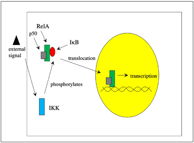
NF-κB is typically found in the cytosol in its inactive state where it is bound to inhibitory IκB proteins such as IκBα. Extracellular stimuli cause the activation of IKK, a kinase that phosphorylates IκB and targets it for ubiquination and proteosomal degradation [182]. This releases the NF-κB and allows it to enter the nucleus, bind DNA and activate transcription. Interestingly, NF-κB induces the transcription of IkBα that causes an inhibitory auto-regulatory cascade. IkBα enters the nucleus following translation, binds and inactivates NF-κB, and removes it from the nucleus [183, 184]. Thus, the transcriptional activation of the NF-κB pathway is often a short, transient process in cells. Figure 4 provides an overview of NF-κB action.
Figure 4. Activation of transcription factors by external and internal stress reponses – NF-κB.
External stimuli such as environmental toxicants and oxidative stress activate IKK which phosphorylates IkB, targeting it for proteosomal degradation. The release of the inhibitory IkB allows for the NF-kB complex (RelA/p50) to enter the nucleus and initiate transcription. Interestingly, one of the genes transcribed following NF-kB activation is IkBa, which can in turn inhibit NF-kB.
NF-κB refers to a family of proteins that control transcription and are involved in development, immune system functions, inflammation, cellular growth and apoptosis. Many of these proteins are referred to as Rel proteins and include RelA, Rel2, c-Rel, p105/p50, and p100/p52. The p105/p50 and p100/p52 proteins are inactive in the cell in their larger form and when the C-terminus is cleaved (p105 cleaved to p50) they become active, shorter DNA-binding proteins. The p105/p50 and p100/p52 proteins are not generally active in transcription unless bound to the Rel proteins. Rel/NF-κB proteins can regulate a large number of different genes because Rel proteins can form homodimers or heterodimers and the individual dimers have distinct DNA-binding sites. The most studied and most common of these dimers is the p50-RelA heterodimer [185].
Interestingly, NF-κB increases the transcription of several genes such as elk-1 and c-fos involved in the Activation Protein-1 (AP-1) transcriptional pathway, another sensor of oxidative stress. Thus, NF-kB can increase stress responses by activating transcription factors that bind other responses elements such as the antioxidant response element (ARE) and tetradecanoyl-phorbol-13-acetate (TPA)-response element [186]. This cross activation increases the number of genes transcribed following a stress event.
AP-1:
Activation Protein-1 (AP-1) belongs to a family of transcription factors characterized by a basic domain and a region of leucine and hydrophobic residue repeats (bZIP family) [187]. AP-1 is a complex comprised of two main proteins, a Jun and a Fos, where Jun may include c-Jun, JunB, or JunD and Fos may include c-Fos, FosB, Fra1, or Fra2 [188]. These proteins may form homo- or heterodimers among themselves such as Jun-Jun or Jun-Fos dimers, and in turn interact with additional proteins to initiate transcription at sites containing an AP-1 consensus sequence. There are several different AP-1 DNA binding sites, including a “classical” AP-1 (TPA-response element) binding site and the antioxidant response element (ARE) [189, 190].
Like NF-κB, AP-1 plays a vital role in increasing the expression of antioxidant enzymes, phase II detoxification enzymes, and cytoprotective genes in order to protect the cell from ROS. Genes thought to be regulated by AP-1 binding to the ARE include GST Ya subunits, NAD(P)H:quinine oxidoreductase (NQO1), Cu/ZnSOD, MnSOD, glutathione peroxidase, catalase, glutathione reductase, and heme oxygenase [187, 191].
Nrf2:
NF-E2 p45-related factor 2 (Nrf2) is a bZIP transcription factor with a cap’n’collar (CNC) structure that also binds several AREs [192]. Similar to NF-κB, Nrf2 is activated by the release of its inhibitor, in this case Keap-1, in the presence of ROS and then heterodimerizes with bZIP proteins such as Fos, Jun, Activating Transcription Factor-4 (ATF4), and most likely musculoaponeurotic fibrosarcoma (Maf) proteins at a variety of AREs [193–195]. AREs include classical antioxidant response elements, electrophile-response elements, β-napthoflavone-response elements, Maf-recognition elements, and AP-1 sites found within the AREs. Greater insight into all of these AREs is available in a recent review [195].
Mice lacking Nrf2 (Nrf2 −/−) show decreased mRNA transcript levels of catalase, NQO1, SOD1, heme oxygenase, stress protein A170, GST alpha and mu, and peroxiredoxin MSP 23. Furthermore, hyperoxia induced levels of NQO1, GST Ya, and glucuronosyltransferase were significantly lower in Nrf2 −/− mice compared with Nrf +/+ mice [192, 196, 197]. Mechanistic studies demonstrate the role of Nrf2 in the regulation of phase I-III detoxication enzymes, primarily conjugases and anti-oxidant defenses, but also MRP transporters [194, 198–200]. These studies and others [155, 156, 201–203] have made it increasingly obvious that Nrf2 is a key transcriptional regulation of oxidative balance. For example, acetaminophen is tolerated in wildtype mice at doses that kill Nrf2-null mice due to their inability to respond to oxidative stress [204].
Nrf2 is activated endogenously by a number of polyunsaturated fatty acid (PUFA) metabolites such as the oxylipins [205], including key oxo-DHA metabolites [158]. DHA, EPA, and other PUFAs are metabolized to several different oxylipins of which some activate Nrf2 such as 15-J2-IsoP [158, 205]. The production of these oxylipins and subsequent activation of Nrf2 may play a protective role in several diseases including mitochondrial disfunction and cardiovascular disease [206]. Other diseases in which Nrf2 plays a protective role because of transcriptional regulation of anti-oxidant defenses include fatty liver disease, cancer, diabetes, emphysema, and chronic obstructive pulmonary disease [207–209].
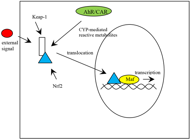
Nrf2 is also activated exogenously by a host of chemicals that perturb redox status. These include several metals, PFOS, paraquat, MPTP, and other chemicals [155, 156, 201, 210]. Nrf2 also crosstalks with the AhR and nuclear receptors such as CAR. Thus, the activation of AhR and CAR causes the subsequent activation of Nrf2 for protection of oxidative stress [194, 203]. AhR or CAR activation probably activates Nrf2 due to the formation of reactive metabolites produced by CYPs following AhR/CAR-mediated CYP induction [194, 203] (Fig. 5). It has been hypothesized, but not definitely demonstrated, that ROS may be directly produced by specific CYPs such as Cyp2b or Cyp2e in a substrate-independent manner and this in turn activates Nrf2 [203]. More likely, Nrf2 activation following AhR or CAR-mediated CYP induction occurs due to increased ROS due to reactive metabolites or CYP-mediated oxylipin formation. The potential role of CYP induction in the activation of anti-oxidant defenses following activation by traditional xeno-sensing receptors is an interesting concept in need of more research [203]. Overall, most toxicology studies would indicate that Nrf2 is the most important of the anti-oxidant transcription factors.
Figure 5. Activation of transcription factors by external and internal stress reponses – Nrf2.
Reactive oxygen species modify central cysteine species on Keap-1 that leads to the decoupling of Keap-1 and Nrf2. Alternatively, Nrf2 can be phosphorylated by kinases. In turn, Nrf2 is decoupled from Keap-1 and translocates to the nucleus where it binds Maf, JunD, or ATF4 and initiates transcription of a variety of antioxidant enzymes and transporters.
Metal-responsive transcription factor-1 (MTF-1):
Metallothionein is primarily regulated by metal-responsive transcription factor-1 (MTF-1) [211]. Metalllthioneins (MT) are ubiquitous, low molecular weight, cysteine-rich proteins that bind and regulate the available concentrations of many metals. The primary role of MTs is to regulate concentrations of the essential trace metals, copper and zinc. At high concentrations, even essential trace metals can bind macromolecules and elicit toxicity and MT ensures a stable bioavailable population of these metals by binding excess essential metals. MT also provides protection from similar toxic metals such as Cd and Hg. For example, Cd-resistant populations of fish express high levels of metallothionein [212], and MT −/− mice show increased sensitivity to many different metals [213].
Zinc and other divalent metals bind MTF-1, which in turn binds DNA at the metal responsive element (MRE) and promotes transcription of MT. MTF-1 is also activiated by oxidative stress [214]. The promoter region of the zebrafish MT gene contains four MREs, three AP-1s and a SP-1 site. However, only the MREs and in particular the distal MRE is required for induction of MT by Zn+2, Cd+2, Cu+2, or Hg+2. MT was not induced by Ni+2, Pb+2, and Co+2 in cell culture [215]. Interestingly, while cadmium is a potent inducer of MT, it does not appear to bind MTF-1 in mammals or yeast, indicating that cadmium indirectly activates MTF-1 [216, 217].
Biomarkers of Exposure are Often Regulated Through Transcription Factors:
The transcription factors described above regulate the expression of genes involved in xenobiotic responses, including several established biomarkers of chemical exposure (Table 1). It is the transcriptional regulation by chemical stress that provides the basis for many of the biomarkers. The toxicant binds to the appropriate receptor, which when bound to the promoter region of DNA, initiates the transcription of genes that can be used as biomarkers. Some of biomarkers are indicative of exposure to a specific toxicant or class of toxicants, while others are much more general and suggest oxidative or physiological stress. MT, for example, is a well established biomarker of exposure to metals due to activation of MTF-1 [211, 218, 219]. CYP1A induction is a well established biomarker of exposure to PAHs and HAHs and has been used as a biomarker in multiple species (AhR activation) [92, 220, 221]. Cyp2b and Cyp3a are biomarkers of CAR and PXR activation, respectively [78, 81, 222, 223]. Cyp4a is induced by PPAR and vitellogenin induction provides a specific biomarker for estrogenic chemicals (estrogen receptor; ER activation)[224–226]. Although altered expression of GSTs, SOD, and other antioxidant enzymes provides information about the general physiological state or stress level of the organism, they typically do not indicate exposure to a particular toxicant, but instead production of ROS (Nrf2; other ROS sensors) [203, 209]. Taken together, transcription factors provide the basis for the biomarker responses toxicologists have been measuring for decades and will continue to use.
Table 1:
Some currently used molecular biomarkers of exposure and the transcription factors that govern their response.
| Biomarker | Chemical | Transcription factor |
|---|---|---|
| CYP1A | PAHs, HAHs | AhR |
| CYP2B | Pharmaceuticals, pesticides | CAR |
| CYP3A | Pharmaceuticals, some EDCs | PXR |
| Vitellogenin | estrogens | ER |
| CYP4A | lipids, some organic pollutants | PPAR |
| Peroximsome proliferation | lipids, some organic pollutants | PPAR |
| Metalliothionein | Zn, Cd, Hg, Cu | MTF-1 |
| GSTs | ROS, metals, HAHs, PAHs | Nrf2, AP-1, AhR, PXR/CAR |
| C-reactive protein | stress | NF-κB |
| Superoxide dismutase | ROS, physiological stress | Nrf2,NF-κb, AP-1 |
| Heat shock proteins | stress, metals, ROS | HSF |
In conclusion, there are a number of crucial transcription factors that activate detoxication pathways through their regulation of key phase I-III detoxication enzymes and transporters as well as other protective proteins such as heat shock proteins, chaperones, and anti-oxidants. These transcription factors induce enzymes that protect individuals from xeno- and endobiotic stressors, including activation of AhR by members of the tryptophan-kynurenine pathway [29, 227], activation of Nrf2 by oxylipins [158, 205, 228], activation and inactivation of PXR and CAR by steroids, steroid precursors, and bile acids [78, 101, 134, 229], and of course numerous xenobiotic chemicals that activate all of the transcription factors mentioned previously. In conclusion, transcription factors are often an initial line of defense from toxic xeno- and endobiotics because their activation leads to a response to chemical stress that allows individuals to acclimate to the chemical insult, and therefore are the phase 0 xenobiotic response.
Acknowledgements:
Research support was provided in part by National Institutes of Environmental Health Sciencs grant R15ES017321. The author would like to thank Melissa Heintz, Matt Hamilton, Emily Gessner, and Emily Olack for reading over the manuscript and providing valuable insight.
References:
- 1.Williams RT, Detoxication Mechanisms: The metabolism and detoxication of drugs, toxic substances, and other organic compounds. 2nd edition ed. 1959: John Wiley & Sons, Inc; New York, NY. [Google Scholar]
- 2.Eaton DL and Klaassen CD, Principles of Toxicology, in Casarett and Doull’s Toxicology: The Basic Science of Poisons., Klaassen C, Editor. 1996, McGraw-Hill Co. Inc.: New York: p. 13–34. [Google Scholar]
- 3.Kretschmer XC and Baldwin WS, CAR and PXR: Xenosensors of Endocrine Disrupters? Chem-Biol. Interac, 2005. 155: p. 111–128. [DOI] [PubMed] [Google Scholar]
- 4.Ishikawa T, The ATP-dependent glutathione S-conjugate export pump. Trends Biochem Sci, 1992. 17: p. 463–468. [DOI] [PubMed] [Google Scholar]
- 5.Bain LJ and LeBlanc GA, Interaction of structurally diverse pesticides with the human MDR1 gene product P-glycoprotein. Toxicol Appl Pharmacol, 1996. 141: p. 288–298. [DOI] [PubMed] [Google Scholar]
- 6.Karthikeyan S and Hoti SL, Development of Fourth Generation ABC Inhibitors from Natural Products: A Novel Approach to Overcome Cancer Multidrug Resistance. Anticancer Agents Med Chem, 2015. 15(5): p. 605–615. [DOI] [PubMed] [Google Scholar]
- 7.Belinsky MG, et al. , Characterization of MOAT-C and MOAT-D, new members of the MRP/cMOAT subfamily of transporter proteins. J Natl Cancer Inst, 1998. 90(22): p. 1735–1741. [DOI] [PubMed] [Google Scholar]
- 8.Mandal A, et al. , Transporter effects on cell permeability in drug delivery. Expert Opin Drug Deliv, 2017. 14(3): p. 385–401. [DOI] [PubMed] [Google Scholar]
- 9.Woodward OM, Köttgen A, and Köttgen M, ABCG transporters and disease. FEBS J, 2011. 278(3215–3225). [DOI] [PMC free article] [PubMed] [Google Scholar]
- 10.Döring B and Petzinger E, Phase 0 and phase III transport in various organs: combined concept of phases in xenobiotic transport and metabolism. Drug Metab Rev, 2014. 46(3): p. 261–282. [DOI] [PubMed] [Google Scholar]
- 11.Dietrich CG, Götze O, and Geier A, Molecular changes in hepatic metabolism and transport in cirrhosis and their functional importance. World J Gastroenterol 2016. 22: p. 72–88. [DOI] [PMC free article] [PubMed] [Google Scholar]
- 12.Baldwin WS and LeBlanc GA, In vivo biotransformation of testosterone by phase I and II detoxication enzymes and their modulation by 20-hydroxyecdysone in Daphnia magna. Aquatic Toxicol., 1994. 29: p. 103–117. [Google Scholar]
- 13.Hayes JD and Pulford DJ, The glutathione S-transferase supergene family: regulation of GST and the contribution of the isoenzymes to cancer chemoprotection and drug resistance. Crit Rev Biochem Mol Biol, 1995. 30(6): p. 445–600. [DOI] [PubMed] [Google Scholar]
- 14.Wu J. l., Liu J, and Cai Z, Determination of triclosan metabolites by using in-source fragmentation from high-performance liquid chromatography/negative atmospheric pressure chemical ionization ion trap mass spectrometry. Rapid Comm in Mass Spectrometry, 2010. 24: p. 1828–1834. [DOI] [PubMed] [Google Scholar]
- 15.Lewin B, Essential Genes. 2006: Pearson Prentice Hall, Upper Saddle River, NJ: 594. [Google Scholar]
- 16.Blattler SM, et al. , In the regulation of cytochrome P450 genes, phenobarbital targets LKB1 for necessary activation of AMP-activated protein kinase. Proc Natl Acad Sci U S A, 2007. 104(3): p. 1045–1050. [DOI] [PMC free article] [PubMed] [Google Scholar]
- 17.Zhou Y, et al. , The Bach Family of Transcription Factors: A Comprehensive Review. Clin Rev Allergy Immunol, 2016. 50: p. 345–356. [DOI] [PubMed] [Google Scholar]
- 18.Ohi H, et al. , Molecular cloning and expression analysis of the aryl hydrocarbon receptor of Xenopus laevis. Biochem Biophys Res Commun, 2003. 307(4): p. 595–599. [DOI] [PubMed] [Google Scholar]
- 19.Schmidt JV, et al. , Characterization of a murine Ahr null allele: involvement of the Ah receptor in hepatic growth and development. Proc Natl Acad Sci U S A, 1996. 93(13): p. 6731–6736. [DOI] [PMC free article] [PubMed] [Google Scholar]
- 20.Robles R, et al. , The aryl hydrocarbon receptor, a basic helix-loop-helix transcription factor of the PAS gene family, is required for normal ovarian germ cell dynamics in the mouse. Endocrinology, 2000. 141(1): p. 450–453. [DOI] [PubMed] [Google Scholar]
- 21.Thackaberry EA, et al. , Insulin regulation in AhR-null mice: embryonic cardiac enlargement, neonatal macrosomia, and altered insulin regulation and response in pregnant and aging AhR-null females. Toxicol Sci, 2003. 76(2): p. 407–417. [DOI] [PubMed] [Google Scholar]
- 22.Xu T, et al. , Aryl Hydrocarbon Receptor Protects Lungs from Cockroach Allergen-Induced Inflammation by Modulating Mesenchymal Stem Cells. J Immunol, 2015. 192(12): p. 5539–5550. [DOI] [PMC free article] [PubMed] [Google Scholar]
- 23.Carreira VS, et al. , Ah Receptor Signaling Controls the Expression of Cardiac Development and Homeostasis Genes. Toxicol Sci, 2015. 147(2): p. 425–435. [DOI] [PMC free article] [PubMed] [Google Scholar]
- 24.Handley-Goldstone HM, Grow MW, and Stegeman JJ, Cardiovascular gene expression profiles of dioxin exposure in zebrafish embryos. Toxicol Sci, 2005. 85(1): p. 683–693. [DOI] [PubMed] [Google Scholar]
- 25.Lanham KA, et al. , Cardiac myocyte-specific AHR activation phenocopies TCDD-induced toxicity in zebrafish. Toxicol Sci, 2014. 141(1): p. 141–154. [DOI] [PMC free article] [PubMed] [Google Scholar]
- 26.Chang JW, et al. , Abdominal Obesity and Insulin Resistance in People Exposed to Moderate-to-High Levels of Dioxin. PLoS One, 2016. 11(1): p. e0145818. [DOI] [PMC free article] [PubMed] [Google Scholar]
- 27.Lu P, et al. , Activation of aryl hydrocarbon receptor dissociates fatty liver from insulin resistance by inducing fibroblast growth factor 21. Hepatology, 2015. 61(6): p. 1908–1919. [DOI] [PMC free article] [PubMed] [Google Scholar]
- 28.Wahlang B, et al. , Polychlorinated biphenyl 153 is a diet-dependent obesogen that worsens nonalcoholic fatty liver disease in male C57BL6/J mice. J Nutr Biochem, 2013. 24: p. 1587–1595. [DOI] [PMC free article] [PubMed] [Google Scholar]
- 29.Noakes R, The aryl hydrocarbon receptor: a review of its role in the physiology and pathology of the integument and its relationship to the tryptophan metabolism. In J Tryptophan Res, 2015. 8: p. 7–18. [DOI] [PMC free article] [PubMed] [Google Scholar]
- 30.Machala M, et al. , Aryl hydrocarbon receptor-mediated activity of mutagenic polycyclic aromatic hydrocarbons determined using in vitro reporter gene assay. Mutat Res, 2001. 497: p. 49–62. [DOI] [PubMed] [Google Scholar]
- 31.Safe S, Polychlorinated biphenyl (PCBs), dibenzo-0-dioxins (PCDDs), dibenzofurans (PCDFs), and related compounds: environmental and mechanistic considerations which support the development of toxic equivalency factors (TEFs). Crit Rev Toxicol, 1990. 21: p. 51–58. [DOI] [PubMed] [Google Scholar]
- 32.Denison MS and Phelan D, The Ah Receptor Signal Transduction Pathway, in Toxicant-Receptor Interactions, Denison MS and Helferich WG, Editors. 1998, Taylor and Francis: Philadelphia, PA: p. 243. [Google Scholar]
- 33.Ernest S and Bello-Reuss E, P-glycoprotein functions and substrates: possible roles of MDR1 gene in the kidney. Kidney Int. Suppl, 1998. 65: p. S11–7. [PubMed] [Google Scholar]
- 34.Bock KW and Kohle C, Ah receptor- and TCDD-mediated liver tumor promotion: clonal selection and expansion of cells evading growth arrest and apoptosis. Biochem Pharmacol, 2005. 69(10): p. 1403–1408. [DOI] [PubMed] [Google Scholar]
- 35.Sunahara GI, et al. , Characterization of 2,3,7,8-tetrachlorodibenzo-p-dioxin-mediated decreases in dexamethasone binding to rat hepatic cytosolic glucocorticoid receptor. Mol Pharmacol, 1989. 36(239–247). [PubMed] [Google Scholar]
- 36.DeVito MJ, et al. , Antiestrogenic action of 2,3,7,8-tetrachlorodibenzo-p-dioxin: tissue-specific regulation of estrogen receptor in CD1 mice. Toxicol Appl Pharmacol, 1992. 113: p. 284–292. [DOI] [PubMed] [Google Scholar]
- 37.Petrulis JR, Hord NG, and Perdew GH, Subcellular localization of the aryl hydrocarbon receptor is modulated by the immunophilin homolog hepatitis B virus X-associated protein 2. J Biol Chem, 2000. 275(48): p. 37448–37453. [DOI] [PubMed] [Google Scholar]
- 38.Vázquez-Gómez G, et al. , Benzo[a]pyrene activates an AhR/Src/ERK axis that contributes to CYP1A1 induction and stable DNA adducts formation in lung cells. Toxicol Lett, 2018. 289: p. 54–62. [DOI] [PubMed] [Google Scholar]
- 39.Incardona JP, Linbo TL, and Scholz NL, Cardiac toxicity of 5-ring polycyclic aromatic hydrocarbons is differentially dependent on the aryl hydrocarbon receptor 2 isoform during zebrafish development. Toxicol Appl Pharmacol, 2011. 257: p. 242–249. [DOI] [PubMed] [Google Scholar]
- 40.Mimura J, et al. , Loss of teratogenic response to 2,3,7,8-tetrachlorodibenzo-p-dioxin (TCDD) in mice lacking the Ah (dioxin) receptor. Genes Cells, 1997. 2(10): p. 645–654. [DOI] [PubMed] [Google Scholar]
- 41.Peters JM, et al. , Toxicol Sci. 1999. January;47(1):86–92. [DOI] [PubMed] [Google Scholar]; Amelioration of TCDD-induced teratogenesis in aryl hydrocarbon receptor (AhR)-null mice. Toxicol Sci, 1999. 47(1): p. 86–92. [DOI] [PubMed] [Google Scholar]
- 42.Lavine JA, et al. , Aryl Hydrocarbon Receptors in the frog Xenopus laevis: Two AHR1 paralogs exhibit low affinity for 2,3,7,8-tetrachlorodibenzo-p-dioxin (TCDD). Toxicol Sci, 2005. 88(1): p. 60–72. [DOI] [PMC free article] [PubMed] [Google Scholar]
- 43.Reitzel AM, et al. , Genetic variation at aryl hydrocarbon receptor (AHR) loci in populations of Atlantic killifish (Fundulus heteroclitus) inhabiting polluted and reference habitats. BMC Evol Biol, 2014. 14: p. 6. [DOI] [PMC free article] [PubMed] [Google Scholar]
- 44.Arzuaga X, Calcano W, and Elskus A, The DNA de-methylating agent 5-azacytidine does not restore CYP1A induction in PCB resistant Newark Bay killifish (Fundulus heteroclitus). Mar Environ Res, 2004. 58: p. 517–520. [DOI] [PubMed] [Google Scholar]
- 45.Hahn ME, et al. , Aryl hydrocarbon receptor polymorphisms and dioxin resistance to Atlantic killifish (Fundulus heroclitus). Pharmacogenetics, 2004. 14: p. 131–143. [DOI] [PubMed] [Google Scholar]
- 46.Bello SM, et al. , Acquired resistance to Ah receptor agonists in a population of atlantic killifish (Fundulus heteroclitus) inhabting a marine superfund site: in vivo and in vitro studies on the inducibility of xenobiotic metabolizing enzymes. Toxicol Sci, 2001. 60: p. 77–91. [DOI] [PubMed] [Google Scholar]
- 47.Meyer JN, Nacci DE, and Di Giulio RT, Cytochrome P4501A (CYP1A) in killifish (Fundulus heteroclitus): heritability of altered expression and relationship to survival in contaminated sediments. Toxicol Sci, 2002. 68(1): p. 69–81. [DOI] [PubMed] [Google Scholar]
- 48.Nacci DE, et al. , Effects of benzo[a]pyrene exposure on a fish population resistant to the toxic effects of dioxin-like compounds. Aquat Toxicol, 2002. 57: p. 203–215. [DOI] [PubMed] [Google Scholar]
- 49.Aluru N, et al. , Targeted mutagenesis of aryl hydrocarbon receptor 2a and 2b genes in Atlantic killifish (Fundulus heteroclitus). Aquat Toxicol, 2015. 158: p. 291–201. [DOI] [PMC free article] [PubMed] [Google Scholar]
- 50.Zhao Y, et al. , Families of Nuclear Receptors in Vertebrate Models: Characteristic and Comparative Toxicological Perspective. Sci Rep, 2015. 5: p. 8554. [DOI] [PMC free article] [PubMed] [Google Scholar]
- 51.Litoff EJ, et al. , Annotation of the Daphnia magna nuclear receptors: Comparison to Daphnia pulex. Gene 2014. 552: p. 116–125. [DOI] [PMC free article] [PubMed] [Google Scholar]
- 52.Evans RM, The nuclear receptor superfamily: A rosetta stone for physiology. Mol Endocrinol, 2005. 19(6): p. 1429–1434. [DOI] [PubMed] [Google Scholar]
- 53.Echeverría F, et al. , Long-chain polyunsaturated fatty acids regulation of PPARs, signaling: Relationship to tissue development and aging. Prostaglandins Leukot Essent Fatty Acids, 2016. 114: p. 28–34. [DOI] [PubMed] [Google Scholar]
- 54.Shoucri BM, et al. , Retinoid X Receptor Activation Alters the Chromatin Landscape To Commit Mesenchymal Stem Cells to the Adipose Lineage. Endocrinology, 2017. 158(10): p. 3109–3125. [DOI] [PMC free article] [PubMed] [Google Scholar]
- 55.le Maire A, et al. , Activation of RXR-PPAR heterodimers by organotin environmental endocrine disruptors. EMBO Reports, 2009. 10(4): p. 367–373. [DOI] [PMC free article] [PubMed] [Google Scholar]
- 56.Yue L, et al. , Ligand-binding regulation of LXR/RXR and LXR/PPAR heterodimerizations: SPR technology-based kinetic analysis correlated with molecular dynamics simulation. Protein Sci, 2005. 14(3): p. 812–822. [DOI] [PMC free article] [PubMed] [Google Scholar]
- 57.Jeske J, et al. , Ligand-dependent and -independent regulation of human hepatic sphingomyelin phosphodiesterase acid-like 3A expression by pregnane X receptor and crosstalk with liver X receptor. Biochem Pharmacol, 2017. 136: p. 122–135. [DOI] [PubMed] [Google Scholar]
- 58.LeBlanc GA, et al. , Detailed Review Paper on the State of the Science on Novel In Vitro and In Vivo Screening and Testing Methods and Endpoints for Evaluating Endocdrine Disruptors in Series on Testing & Assessment: No. 178. 2012, Organisation for Economic Co-operation and Development: Paris: p. 213. [Google Scholar]
- 59.Urquhart BL, Tirona RG, and Kim RB, Nuclear receptors and the regulation of drug-metabolizing enzymes and drug transporters: implications for interindividual variability in response to drugs. J Clin Pharmacol, 2007. 47(5): p. 566–578. [DOI] [PubMed] [Google Scholar]
- 60.Malerod L, et al. , Bile acids reduce SR-BI expression in hepatocytes by a pathway involving FXR/RXR, SHP, and LRH-2. Biochem Biophys Res Commun, 2005. 336: p. 1096–1105. [DOI] [PubMed] [Google Scholar]
- 61.Howarth DL, et al. , Two farnesoid X receptor alpha isoforms in Japanese medaka (Oryzias latipes) are differentially activated in vitro. Aquat Toxicol, 2010. 98(3): p. 245–255. [DOI] [PMC free article] [PubMed] [Google Scholar]
- 62.Reschly EJ, et al. , Evolution of the bile salt nuclear receptor FXR in vertebrates. J Lipid Res, 2008. 49: p. 1577–1587. [DOI] [PMC free article] [PubMed] [Google Scholar]
- 63.Matsubara T, Li F, and Gonzalez FJ, FXR signaling in the enterohepatic system. Mol Cell Endocrinol, 2013. 368(1–2): p. 17–29. [DOI] [PMC free article] [PubMed] [Google Scholar]
- 64.Kim KH, et al. , Xenobiotic Nuclear Receptor Signaling Determines Molecular Pathogenesis of Progressive Familial Intrahepatic Cholestasis. Endocrinology, 2018. 159: p. 2435–2446. [DOI] [PMC free article] [PubMed] [Google Scholar]
- 65.Li X, Wang Z, and Klaunig JE, The effects of perfluorooctanoate on high fat diet induced non-alcoholic fatty liver disease in mice. Tociology, 2019. 416: p. 1–14. [DOI] [PubMed] [Google Scholar]
- 66.Lindblom TH, Pierce GJ, and Sluder AE, A C. elegans orphan nuclear receptor contributes to xenobiotic resistance. Curr Biol, 2001. 11(11): p. 864–868. [DOI] [PubMed] [Google Scholar]
- 67.Li Y, Ginjupalli GK, and Baldwin WS, The HR97 (NR1L) Group of Nuclear Receptors: A New Group of Nuclear Receptors Discovered in Daphnia species Gen Comp Endocrinol, 2014. 206: p. 30–42. [DOI] [PMC free article] [PubMed] [Google Scholar]
- 68.King-Jones K, et al. , The DHR96 nuclear receptor regulates xenobiotic responses in Drosophila. Cell Metab, 2006. 4: p. 37–48. [DOI] [PubMed] [Google Scholar]
- 69.Maloney EK and Waxman DJ, trans-Activation of PPARalpha and PPARgamma by structurally diverse environmental chemicals. Toxicol Appl Pharmacol, 1999. 161(2): p. 209–218. [DOI] [PubMed] [Google Scholar]
- 70.Xia J, et al. , Atrazine-induced environmental nephrosis was mitigated by lycopene via modulating nuclear xenobiotic receptors-mediated response. J Nutr Biochem, 2018. 51: p. 80–90. [DOI] [PubMed] [Google Scholar]
- 71.Hurst CH and Waxman DJ, Activation of PPARa and PPARg by environmental phthalate monesters. Toxicol Sci, 2003. 74: p. 297–308. [DOI] [PubMed] [Google Scholar]
- 72.Yanik SC, et al. , Organotins are potent activators of PPARgamma and adipocyte differentiation in bone marrow multipotent mesenchymal stromal cells. Toxicol Sci, 2011. 122: p. 476–488. [DOI] [PMC free article] [PubMed] [Google Scholar]
- 73.Holden PR and Tugwood JD, Peroxisome proliferator-activated receptor alpha: role in rodent liver cancer and species differences. J Mol Endocrinol, 1999. 22: p. 1–8. [DOI] [PubMed] [Google Scholar]
- 74.Hatch EE, et al. , Cancer risk in women exposed to diethylstilbestrol in utero. JAMA, 1998. 280(7): p. 630–634. [DOI] [PubMed] [Google Scholar]
- 75.Gonzalez FJ and Shah YM, PPARalpha: Mechanism of species differences and hepatocarcinogenesis of peroxisome proliferators. Toxicology, 2007. 246: p. 2–8. [DOI] [PubMed] [Google Scholar]
- 76.Wang YH, et al. , Tributyltin synergizes with 20-hydroxyecdysone to produce endocrine toxicity. Toxicol Sci, 2011. 123(1): p. 71–79. [DOI] [PubMed] [Google Scholar]
- 77.Karimullina E, et al. , HR96 is a promiscuous endo- and xeno-sensing nuclear receptor. Aquat Toxicol, 2012. 116–117: p. 69–78. [DOI] [PMC free article] [PubMed] [Google Scholar]
- 78.Kliewer SA, et al. , An orphan nuclear receptor activated by pregnanes defines a novel steroid signaling pathway. Cell, 1998. 92(1): p. 73–82. [DOI] [PubMed] [Google Scholar]
- 79.Blumberg B, et al. , SXR, a novel steroid and xenobiotic-sensing nuclear receptor. Genes Dev, 1998. 12: p. 3195–3205. [DOI] [PMC free article] [PubMed] [Google Scholar]
- 80.Honkakoski P, et al. , The nuclear orphan-receptor CAR-retinoid X receptor heterodimer activates the phenobarbital-responsive module of the CYP2B gene. Mol Cell Biol, 1998. 18: p. 5652–5658. [DOI] [PMC free article] [PubMed] [Google Scholar]
- 81.Wei P, et al. , The nuclear receptor CAR mediates specific xenobiotic induction of drug metabolism. Nature, 2000. 407(6806): p. 920–923. [DOI] [PubMed] [Google Scholar]
- 82.Afschar S, et al. , Nuclear hormone receptor DHR96 mediates the resistance to xenobiotics but not the increased lifespan of insulin-mutant Drosophila. Proc Natl Acad Sci U S A, 2016. 113(5): p. 1321–1326. [DOI] [PMC free article] [PubMed] [Google Scholar]
- 83.Schmidt AM, et al. , RNA sequencing indicates that atrazine induces multiple detoxification genes in Daphnia magna and this is a potential sources of its mixtures interactions with other chemicals. Chemosphere, 2017. 189: p. 699–708. [DOI] [PMC free article] [PubMed] [Google Scholar]
- 84.Chawla A, et al. , Nuclear receptors and lipid physiology: opening the X-files. Science, 2001. 294(5548): p. 1866–1870. [DOI] [PubMed] [Google Scholar]
- 85.Tien ES and Negishi M, Nuclear receptors CAR and PXR in the regulation of hepatic metabolism. Xenobiotica, 2006. 36: p. 1152–1163. [DOI] [PMC free article] [PubMed] [Google Scholar]
- 86.Hernandez JP, Mota LC, and Baldwin WS, Activation of CAR and PXR by dietary, environmental and occupational chemicals alters drug metabolism, intermediary metabolism, and cell proliferation. Curr Pharmacog Personal Med, 2009. 7: p. 81–105. [DOI] [PMC free article] [PubMed] [Google Scholar]
- 87.Xu C, Huang M, and Bi H, PXR- and CAR-mediated herbal effect on human diseases. Biochim Biophys Acta, 2016. 1859: p. 1121–1129. [DOI] [PubMed] [Google Scholar]
- 88.Dauchy S, et al. , ABC transporters, cytochromes P450 and their main transcription factors: expression at the human blood-brain barrier. J Neurochem, 2008. 107(6): p. 1518–1528. [DOI] [PubMed] [Google Scholar]
- 89.Frye CA, et al. , Motivated behaviors and levels of 3α,5α-THP in the midbrain are attenuated by knocking down expression of pregnane xenobiotic receptor in the midbrain ventral tegmental area of proestrous rats. J Sex Med, 2013. 10(7): p. 1692–1706. [DOI] [PMC free article] [PubMed] [Google Scholar]
- 90.Maglich JM, et al. , The first complete genome sequence from a teleost fish (Fugu rubripes) adds significant diversity to the nuclear receptor superfamily. Nucleic Acids Res, 2003. 31(14): p. 4051–4058. [DOI] [PMC free article] [PubMed] [Google Scholar]
- 91.Depaz IMB, et al. , Differential Expression of Human Cytochrome P450 Enzymes from the CYP3A Subfamily in the Brains of Alcoholic Subjects and Drug-Free Controls. Drug Metab Dispos, 2013. 41: p. 1187–1194. [DOI] [PubMed] [Google Scholar]
- 92.Toselli F, et al. , Gene expression profiling of cytochromes P450, ABC transporters and their principal transcription factors in the amygdala and prefrontal cortex of alcoholics, smokers and drug-free controls by qRT-PCR. Xenobiotica, 2015. 45(12): p. 1129–1137. [DOI] [PubMed] [Google Scholar]
- 93.LeCluyse EL, Pregnane X receptor: molecular basis for species differences in CYP3A induction by xenobiotics. Chem Biol Interact, 2001. 134(3): p. 283–289. [DOI] [PubMed] [Google Scholar]
- 94.Kauffman HM, et al. , Influence of redox-active compounds and PXR-activators on human MRP1 and MRP2 gene expression. Toxicology, 2002. 171: p. 137–146. [DOI] [PubMed] [Google Scholar]
- 95.Falkner KC, et al. , Regulation of the rat glutathione S-transferase A2 gene by glucocorticoids: involvement of both the glucocorticoid and pregnane X receptors. Mol Pharmacol, 2001. 60(3): p. 611–619. [PubMed] [Google Scholar]
- 96.Sonoda J, et al. , Regulation of a xenobiotic sulfonation cascade by nuclear pregnane X receptor (PXR). Proc Natl Acad Sci U S A, 2002. 99(21): p. 13801–13806. [DOI] [PMC free article] [PubMed] [Google Scholar]
- 97.Duanmu Z, et al. , Effects of dexamethasone on aryl (SULT1A1)- and hydroxysteroid (SULT2A1)-sulfotransferase gene expression in primary cultured human hepatocytes. Drug Metab Dispos, 2002. 30(9): p. 997–1004. [DOI] [PubMed] [Google Scholar]
- 98.Chen C, Staudinger JL, and Klaassen CD, Nuclear receptor, pregname X receptor, is required for induction of UDP-glucuronosyltranferases in mouse liver by pregnenolone-16 alpha-carbonitrile. Drug Metab Dispos, 2003. 31(7): p. 908–915. [DOI] [PubMed] [Google Scholar]
- 99.Moore LB, et al. , Orphan nuclear receptors constitutive androstane receptor and pregnane X receptor share xenobiotic and steroid ligands. J Biol Chem, 2000. 275(20): p. 15122–15127. [DOI] [PubMed] [Google Scholar]
- 100.Milnes MR, et al. , Activation of steroid and xenobiotic receptor (SXR, NR1I2) and its orthologs in laboratory, toxicologic, and genome model species. Environ Health Perspect, 2008. 116: p. 880–885. [DOI] [PMC free article] [PubMed] [Google Scholar]
- 101.Baldwin WS and Roling JA, A concentration addition model for the activation of the constitutive androstane receptor by xenobiotic mixtures. Toxicol Sci, 2009. 107: p. 93–105. [DOI] [PMC free article] [PubMed] [Google Scholar]
- 102.Yamada H, et al. , Induction of the hepatic cytochrome P450 2B subfamily by xenobiotics: Research history, evolutionary aspect, relation to tumorigenesis, and mechanism. Curr Drug Metab, 2006. 7: p. 397–409. [DOI] [PubMed] [Google Scholar]
- 103.Sinz M, et al. , Evaluation of 170 xenobioitics as transactivators of human pregnane X receptor (hPXR) and correlation to known CYP3A4 drug interactions. Curr Drug Metab, 2006. 7: p. 375–388. [DOI] [PubMed] [Google Scholar]
- 104.Shukla SJ, et al. , Identification of Clinically Used Drugs That Activate Pregnane X Receptors. Drug Metab Dispos, 2011. 39(1): p. 151–159. [DOI] [PMC free article] [PubMed] [Google Scholar]
- 105.Xie W, et al. , An essential role for nuclear receptors SXR/PXR in detoxification of cholestatic bile acids. Proc Natl Acad Sci USA, 2001. 98: p. 3375–3380. [DOI] [PMC free article] [PubMed] [Google Scholar]
- 106.Sonoda J, et al. , Pregnane X receptor prevents hepatorenal toxicity from cholesterol metabolites. Proc Natl Acad Sci U S A, 2005. 102(6): p. 2198–2203. [DOI] [PMC free article] [PubMed] [Google Scholar]
- 107.Mikamo E, et al. , Endocrine disruptors induce cytochrome P450 by affecting transcriptional regulation via pregnane X receptor. Toxicol Appl Pharmacol, 2003. 193(1): p. 66–72. [DOI] [PubMed] [Google Scholar]
- 108.Wyde ME, et al. , The environmental pollutant 1,1-dichloro-2,2-bis (p-chlorophenyl)ethylene induces rat hepatic cytochrome P450 2B and 3A expression through the constitutive androstane receptor and pregnane X receptor. Mol Pharmacol, 2003. 64(2): p. 474–481. [DOI] [PubMed] [Google Scholar]
- 109.Masuyama H, et al. , Endocrine disrupting chemicals, phthalic acid and nonylphenol, activate Pregnane X Receptor-mediated transcription. Mol Endocrinol, 2000. 14: p. 421–428. [DOI] [PubMed] [Google Scholar]
- 110.Schuetz EG, Brimer C, and Schuetz JD, Environmental xenobiotics and the antihormones cyproterone acetate and spironolactone use the nuclear hormone pregnenolone X receptor to activate the CYP3A23 hormone response element. Mol Pharmacol, 1998. 54(6): p. 1113–1117. [DOI] [PMC free article] [PubMed] [Google Scholar]
- 111.Vogel G, Pharmacology. A worrisome side effect of an antianxiety remedy. Science, 2001. 291: p. 37. [DOI] [PubMed] [Google Scholar]
- 112.Ekins S and Erickson JA, A pharmacophore for human pregnane X receptor ligands. Drug Metab Dispos, 2002. 30: p. 96–99. [DOI] [PubMed] [Google Scholar]
- 113.Hernandez JP, et al. , The environmental estrogen, nonylphenol, activates the constitutive androstane receptor (CAR). Toxicol Sci, 2007. 98: p. 416–426. [DOI] [PMC free article] [PubMed] [Google Scholar]
- 114.Jacobs MN, Nolan GT, and Hood SR, Lignans, bacteriocides and organochlorine compounds activate the human pregnane X receptor (PXR). Toxicol Appl Pharmacol, 2005. 209(2): p. 123–33. [DOI] [PubMed] [Google Scholar]
- 115.Wang J, et al. , Biology of PXR: Role in drug-hormone interactions. EXCLI J, 2014. 13: p. 728–739. [PMC free article] [PubMed] [Google Scholar]
- 116.Watkins RE, et al. , The human nuclear xenobiotic receptor PXR: Structural determinants of directed promiscuity. Science, 2001. 292: p. 2329–2333. [DOI] [PubMed] [Google Scholar]
- 117.Xue Y, et al. , Crystal structure of the pregane X receptor-estradiol complex provides insights into endobiotic recognition. Mol Endocrinol, 2007. 21(5): p. 1028–1038. [DOI] [PubMed] [Google Scholar]
- 118.Wallace BD, et al. , Structural and functional analysis of the human nuclear xenobiotic receptor PXR in complex with RXRa. J Mol Biol, 2013. 425(14): p. 2561–2577. [DOI] [PMC free article] [PubMed] [Google Scholar]
- 119.Teotico DG, et al. , Active nuclear receptors exhibit highly correlated AF-2 domain motions. PLoS Comp Biol, 2008. 4(7): p. e1000111. [DOI] [PMC free article] [PubMed] [Google Scholar]
- 120.Suino K, et al. , The nuclear xenobioitic receptor CAR: structural determinants of constitutive activation and heterodimerization Mol Cell, 2004. 16: p. 893–905. [DOI] [PubMed] [Google Scholar]
- 121.Gao J and Xie W, Pregnane X receptor and constitutive androstane receptor at the crossroads of drug metabolism and energy metabolism. Drug Metab Dispos, 2010. 38(12): p. 2091–2095. [DOI] [PMC free article] [PubMed] [Google Scholar]
- 122.Tzameli I, et al. , The xenobiotic compound 1,4-bis[2-(3,5-dichloropyridyloxy)]benzene is an agonist ligand for the nuclear receptor CAR. Mol Cell Biol, 2000. 20(9): p. 2951–2958. [DOI] [PMC free article] [PubMed] [Google Scholar]
- 123.Yoshinori K, et al. , Identification of the nuclear receptor CAR: HSP90 complex in mouse liver and recruitment of protein phosphotase 2A in response to phenobarbital. FEBS Lett, 2003. 548: p. 17–20. [DOI] [PubMed] [Google Scholar]
- 124.Hosseinpour F, et al. , Serine 202 regulates the nuclear translocation of constitutive active/androstane receptor. Mol Pharmacol, 2006. 69(4): p. 1095–1102. [DOI] [PubMed] [Google Scholar]
- 125.Yoshinari K, et al. , Identification of the nuclear receptor CAR:HSP90 complex in mouse liver and recruitment of protein phosphatase 2A in response to phenobarbital. FEBS Lett, 2003. 548: p. 17–20. [DOI] [PubMed] [Google Scholar]
- 126.Mutoh S, et al. , Phenobarbital Indirectly Activates the Constitutive Active Androstane Receptor (CAR) by Inhibition of Epidermal Growth Factor Receptor Signaling. Sci Signal, 2013. 6(274): p. ra31. [DOI] [PMC free article] [PubMed] [Google Scholar]
- 127.Meyer SA and Jirtle RL, Old dance with a new partner: EGF receptor as the phenobarbital receptor mediating Cyp2b expression. Sci Signal, 2013. 6(274): p. pe16. [DOI] [PubMed] [Google Scholar]
- 128.Rencurel F, et al. , AMP-activated protein kinase mediates phenobarbital induction of CYP2B gene expression in hepatocytes and a newly derived human hepatoma cell line. J Biol Chem, 2005. 280(6): p. 4367–4373. [DOI] [PubMed] [Google Scholar]
- 129.Yang H and Wang H, Signaling control of the constitutive androstane receptor (CAR). Protein Cell, 2014. 5(2): p. 113–123. [DOI] [PMC free article] [PubMed] [Google Scholar]
- 130.Ueda A, et al. , Diverse roles of the nuclear orphan receptor CAR in regulating hepatic genes in response to phenobarbital. Mol Pharmacol, 2002. 61: p. 1–6. [DOI] [PubMed] [Google Scholar]
- 131.Niu B, et al. , In vivo genome-wide binding interactions of mouse and human constitutive androstane receptors reveal novel gene targets Nucl Acids Res, 2018. 46(16): p. 8385–8403. [DOI] [PMC free article] [PubMed] [Google Scholar]
- 132.Wang X, et al. , Semi-quantitative profiling of bile acids in serum and liver reveals the dosage-related effects of dexamethasone on bile acid metabolism in mice. J Chromatogr B Analyt Technol Biomed Life Sci, 2018. 1095: p. 65–74. [DOI] [PubMed] [Google Scholar]
- 133.Saini SP, et al. , A novel constitutive androstane receptor-mediated and CYP3A-independent pathway of bile acid detoxification. Mol Pharmacol, 2004. 65(2): p. 292–300. [DOI] [PubMed] [Google Scholar]
- 134.Masuyama H, et al. , The expression of pregnane X receptor and its target gene, cytochrome P450 3A1, in perinatal mouse. Mol Cell Endocrinol, 2001. 172: p. 47–56. [DOI] [PubMed] [Google Scholar]
- 135.Cui H, et al. , Pregnane X receptor regulates the AhR/Cyp1A1 pathway and protects liver cells from benzo-[α]-pyrene-induced DNA damage. Toxicol Lett, 2017. 275: p. 67–76. [DOI] [PubMed] [Google Scholar]
- 136.Mota LC, Hernandez JP, and Baldwin WS, CAR-null mice are sensitive to the toxic effects of parathion: Association with reduced Cytochrome P450-mediated parathion metabolism. Drug Metab Dispos, 2010. 38(9): p. 1582–1588. [DOI] [PMC free article] [PubMed] [Google Scholar]
- 137.Mota LC, et al. , Nonylphenol-mediated CYP induction is PXR-dependent: The use of humanized mice and human hepatocytes suggests that hPXR is less sensitive than mouse PXR to nonylphenol treatment. Toxicol Appl Pharmacol, 2011. 252(3): p. 259–67. [DOI] [PMC free article] [PubMed] [Google Scholar]
- 138.Hernandez JP, et al. , Sexually dimorphic regulation and induction of P450s by the constitutive androstane receptor (CAR). Toxicology, 2009. 256: p. 53–64. [DOI] [PMC free article] [PubMed] [Google Scholar]
- 139.Kumar R, et al. , Compensatory changes in CYP expression in three different toxicology mouse models: CAR-null, Cyp3a-null, and Cyp2b9/10/13-null mice. PLOS ONE, 2017. 12(3): p. e0174355. [DOI] [PMC free article] [PubMed] [Google Scholar]
- 140.Murayama N, et al. , Induction of human cytochrome P450 3A enzymes in cultured placental cells by thalidomide and relevance to bioactivation and toxicity. J Toxicol Sci, 2017. 42(3): p. 343–348. [DOI] [PMC free article] [PubMed] [Google Scholar]
- 141.Zhai Y, et al. , Activation of pregnane X receptor disrupts glucocorticoid and mineralocorticoid homeostasis. Mol Endocrinol, 2007. 21(1): p. 138–147. [DOI] [PubMed] [Google Scholar]
- 142.Wang C, et al. , Poly(ADP-ribosyl)ated PXR is a critical regulator of acetominophen-induced hepatotoxicity. Cell Death Dis, 2018. 9(8): p. 819. [DOI] [PMC free article] [PubMed] [Google Scholar]
- 143.Zhang J, et al. , Modulation of acetaminophen-induced hepatotoxicity by the xenobiotic receptor CAR. Science, 2002. 298: p. 422–424. [DOI] [PubMed] [Google Scholar]
- 144.Xie W Induction of P450s through PXR in 34th Gordon Research Conference on Drug Metabolism. 2004. Holderness School, Plymouth NH. [Google Scholar]
- 145.Thomson SA, et al. , Annotation, phylogenetics, and expression of the nuclear receptors in Daphnia pulex. BMC Genomics, 2009. 10: p. 500. [DOI] [PMC free article] [PubMed] [Google Scholar]
- 146.Horner MA, et al. , The Drosophila DHR96 nuclear receptor binds cholesterol and regulates cholesterol homeostasis. Genes Dev, 2009. 23(23): p. 2711–2716. [DOI] [PMC free article] [PubMed] [Google Scholar]
- 147.Sieber MH and Thummel CS, The DHR96 nuclear receptor controls triacylglycerol homeostasis in Drosophila. Cell Metab, 2009. 10(6): p. 481–490. [DOI] [PMC free article] [PubMed] [Google Scholar]
- 148.Sieber MH and Thummel C, Coordination of Triacylglycerol and Cholesterol Homeostasis by DHR96 and the Drosophila LipA Homolog magro. Cell Metab, 2012. 15: p. 122–127. [DOI] [PMC free article] [PubMed] [Google Scholar]
- 149.Sengupta N, Gerard PD, and Baldwin WS, Perturbations in polar lipids, starvation survival and reproduction following exposure to unsaturated fatty acids or environmental toxicants in Daphnia magna. Chemosphere, 2016. 144: p. 2302–2311. [DOI] [PMC free article] [PubMed] [Google Scholar]
- 150.Sengupta N, et al. , Exchange of polar lipids from adults to neonates in Daphnia magna: Perturbations in sphingomyelin allocation by dietary lipids and environmental toxicants. PLoS ONE 2017. 12(5): p. e0178131. [DOI] [PMC free article] [PubMed] [Google Scholar]
- 151.Lin GG, Kozaki T, and Scott JG, Hormone receptor-like in 96 and Broad-Complex modulate phenobarbital induced transcription of cytochrome P450 CYP6D1 in Drosophila S2 cells. Insect Mol Biol, 2011. 20(1): p. 87–95. [DOI] [PMC free article] [PubMed] [Google Scholar]
- 152.Sengupta N, Litoff EJ, and Baldwin WS, The HR96 activator, atrazine, reduces sensitivity of D. magna to triclosan and DHA. Chemosphere, 2015. 128: p. 299–306. [DOI] [PMC free article] [PubMed] [Google Scholar]
- 153.Oakes KD, McMaster ME, and Van der Kraak GJ, Oxidative stress responses in longnose sucker (Catostomus catostomus) exposed to pulp and paper mill and municipal sewage effluents. Aquat Toxicol, 2004. 67(3): p. 255–271. [DOI] [PubMed] [Google Scholar]
- 154.Buzard GS and Kasprzak KS, Possible roles of nitric oxide and redox cell signaling in metal-induced toxicity and carcinogenesis: a review. J Environ Pathol Toxicol Oncol, 2000. 19(3): p. 179–199. [PubMed] [Google Scholar]
- 155.Tebay LE, et al. , Mechanisms of activation of the transcription factor Nrf2 by redox stressors, nutrient cues, and energy status and the pathways through which it attenuates degenerative disease. Free Radic Biol Med, 2015. 88: p. 108–146. [DOI] [PMC free article] [PubMed] [Google Scholar]
- 156.Xu J, et al. , PFOS induces adipogenesis and glucose uptake in association with activation of Nrf2 signaling pathway. Toxicol Appl Pharmacol, 2016. 290: p. 21–30. [DOI] [PMC free article] [PubMed] [Google Scholar]
- 157.Gruber F, et al. , Nrf2 deficiency causes lipid oxidation, inflammation, and matrix-protease expression in DHA-supplemented and UVA-irradiated skin fibroblasts. Free Radic Biol Med, 2015. 88(Pt B): p. 439–451. [DOI] [PubMed] [Google Scholar]
- 158.Bang HY, et al. , Docosahexaenoic Acid Induces Expression of Heme Oxygenase-1 and NAD(P)H:quinone Oxidoreductase through Activation of Nrf2 in Human Mammary Epithelial Cells. Molecules, 2017. 22(6): p. pii: E969. [DOI] [PMC free article] [PubMed] [Google Scholar]
- 159.Chapman LM, et al. , Construction of a subtractive library from hexavalent chromium treated winter flounder (Pseudopleuronectes americanus) reveals alterations in non-selenium glutathione peroxidases. Aquat Toxicol, 2004. 67(2): p. 181–194. [DOI] [PubMed] [Google Scholar]
- 160.Kaltreider RC, et al. , Differential effects of arsenic(III) and chromium(VI) on nuclear transcription factor binding. Mol Carcinogen, 1999. 25: p. 219–229. [PubMed] [Google Scholar]
- 161.Risso-de Faverney C, et al. , Cadmium-induced apoptosis through the mitochondrial pathway in rainbow trout hepatocytes: involvement of oxidative stress. Aquat Toxicol, 2004. 69(3): p. 247–258. [DOI] [PubMed] [Google Scholar]
- 162.Bacanskas LR, Whitaker J, and Di Giulio RT, Oxidative stress in two population of killifish (Fundulus heteroclitus) with differing contaminant exposure histories. Mar Environ Res, 2004. 58(2–5): p. 597–601. [DOI] [PubMed] [Google Scholar]
- 163.Meyer JN, et al. , Antioxidant defenses in killifish (Fundulus heteroclitus) exposed to contaminated sediments and model prooxidants: short-term and heritable responses. Aquat Toxicol, 2003. 65: p. 377–395. [DOI] [PubMed] [Google Scholar]
- 164.Gilmore TD, The Re1/NF-kappa B/I kappa B signal transduction pathway and cancer. Cancer Treat Res, 2003. 115: p. 241–265. [PubMed] [Google Scholar]
- 165.Hazan U, et al. , Stimulation of a human T-cell clone with anti-CD3 or tumor necrosis factor induces NF-kB translocation but not human immunodeficiency virus 1 enhancer-dependent transcription. Proc Natl Acad Sci USA, 1990. 87: p. 7861–7865. [DOI] [PMC free article] [PubMed] [Google Scholar]
- 166.Huang C, et al. , Arsenic-induced NFkB transactivation through ERKs- and JNKs-dependent pathways in mouse epidermal JB6 cells. Mol Cell Biochem, 2001. 222: p. 29–34. [PubMed] [Google Scholar]
- 167.Kim SH, et al. , Celecoxib induces apoptosis in cervical cancer cells independent of cyclooxygenase using NF-kB as a possible target. J Cancer Res Clin Oncol, 2004. 130: p. 551–560. [DOI] [PubMed] [Google Scholar]
- 168.Kim YS, et al. , The effects of thalidomide on the stimulation of NF-kB activity and TNF-a production by lipopolysaccharide in a human colonic epithelial cell line. Mol Cells, 2004. 17: p. 210–216. [PubMed] [Google Scholar]
- 169.Westendorp MO, et al. , HIV-1 Tat potentiates TNF-induced NF-kB activation and cytotoxicity by altering the redox state. EMBO Journal, 1995. 14: p. 546–554. [DOI] [PMC free article] [PubMed] [Google Scholar]
- 170.Ye J, et al. , Chromium(VI)-induced nuclear factor-kappa B activation in intact cells via free radical reactions. Carcinogenesis, 1995. 16(10): p. 2401–2405. [DOI] [PubMed] [Google Scholar]
- 171.Marano F, et al. , Human airway epithelial cells in culture for studying the molecular mechanisms of the inflammatory response triggered by diesel exhaust particles. Cell Biol Toxicol, 2002. 18: p. 315–320. [DOI] [PubMed] [Google Scholar]
- 172.Munroe DG, et al. , Novel intracellular signaling function of prostaglandin H synthase-1 in NF-kB activation. J Inflammation, 1995. 45: p. 260–268. [PubMed] [Google Scholar]
- 173.Gilmore TD, ed. Rel/NF-kB Seminars in Cancer Biology. Vol. Vol. 8–2 1997, Academic Press: Cambridge, England: 61–129. [Google Scholar]
- 174.Lieb K, et al. , Interleukin-1 beta and tumor necrosis facor-alpha induce expression of alpha 1-antichymotrypsin in human astrocytoma cell by activation of nuclear factor-kappa B. J Neurochem, 1996. 67(5): p. 2039–2044. [DOI] [PubMed] [Google Scholar]
- 175.Lang CH, Nystrom GJ, and Frost RA, Regulation of IGF binding protein-1 in hep G2 cells by cytokines and reactive oxygen species. Am J Physiol, 1999. 276: p. G719–27. [DOI] [PubMed] [Google Scholar]
- 176.Yao KS and O’Dwyer PJ, Involvement of NF-kappa B in the induction of NAD(P)H:quinone oxidoreductase (DT-diaphorase) by hypoxia, oltipraz and mitomycin C. Biochem Pharmacol, 1995. 49(3): p. 275–282. [DOI] [PubMed] [Google Scholar]
- 177.Yamamoto K, et al. , Transcriptional roles of nuclear factor kappa B and nuclear factor-interleukin-6 in the tumor necrosis factor alpha-dependent induction of cyclooxygenase-2 in MC3T3-E1 cells. J Biol Chem, 1995. 270: p. 31315–31320. [DOI] [PubMed] [Google Scholar]
- 178.Zhang G, Slaughter C, and Humphries EH, v-rel induces ectopic expression of an adhesion molecule, DM-GRASP, during B-lymphoma development. Mol Cell Biol, 1995. 15: p. 1806–1816. [DOI] [PMC free article] [PubMed] [Google Scholar]
- 179.Rojo AI, et al. , Regulation of Cu/Zn-superoxide dismutase expression via the phosphatidylinositol 3 kinase/Akt pathway and nuclear factor-kB. J Neurosci, 2004. 24: p. 9324–9334. [DOI] [PMC free article] [PubMed] [Google Scholar]
- 180.Das KC, Lewis-Molock Y, and White CW, Activation of NF-kB and elevation of MnSOD gene expression by thiol reducing agents in lung adenocarcinoma (A549) cells. Amer J Physiol, 1995. 5: p. L558–L602. [DOI] [PubMed] [Google Scholar]
- 181.Morri H, Ozaki M, and Watanabe Y, 5’-flanking region surrounding a human cytosolic phospholipase A2 gene. Biochem Biophys Res Commun, 1994. 205(1): p. 6–11. [DOI] [PubMed] [Google Scholar]
- 182.Magnani M, et al. , The ubiquitin-dependent proteolytic system and other potential targets for the modulation of nuclear factor-kB (NF-kB). Curr Drug Targets, 2000. 1(14): p. 387–399. [DOI] [PubMed] [Google Scholar]
- 183.Ito CY, Kazantsev AG, and Baldwin ASJ, Three NF-kappa B sites in the I kappa B-alpha promoter are required for induction of gene expression by TNF alpha. Nucleic Acids Res, 1994. 22(18): p. 3787–3792. [DOI] [PMC free article] [PubMed] [Google Scholar]
- 184.Sun S-C, et al. , NF-kB controls expression of inhibitor IkBa: evidence for an inducible autoregulatory pathway. Science, 1993. 259: p. 1912–1915. [DOI] [PubMed] [Google Scholar]
- 185.Oeckinghaus A and Ghosh S, The NF-κB Family of Transcription Factors and Its Regulation. Cold Spring Harb Perspect Biol, 2009. 1(4): p. a000034. [DOI] [PMC free article] [PubMed] [Google Scholar]
- 186.Fujioka S, et al. , NF-κB and AP-1 Connection: Mechanism of NF-κB-Dependent Regulation of AP-1 Activity. Mol Cell Biol, 2004. 24(17): p. 7806–7819. [DOI] [PMC free article] [PubMed] [Google Scholar]
- 187.Sen CK and Packer L, Antioxidant and redox regulation of gene transcription. Faseb J, 1996. 10: p. 709–720. [DOI] [PubMed] [Google Scholar]
- 188.Otterbein LE and Choi AMK, The saga of leucine zippers continues: In response to oxidative stress. Am J Respir Cell Mol Biol, 2002. 26: p. 161–163. [DOI] [PubMed] [Google Scholar]
- 189.Prestera T, et al. , Parallel induction of heme xoygenase-1 and chemoprotective phase 2 enzymes by electrophiles and antioxdants: regulation by upstream antioxidant-responsive elements (ARE). Mol Med, 1995. 1: p. 827–837. [PMC free article] [PubMed] [Google Scholar]
- 190.Rushmore TH, Morton MR, and Pickett CB, The antioxidant responsive element: activation by oxidative stress and identification of the DNA consensus sequence required for functional activity. J. Biol. Chem, 1991. 266: p. 11632–11639. [PubMed] [Google Scholar]
- 191.Lee PJ and Choi AMK, Serial review: Role of reactive oxygen and nitrogen species (ROS/RNS) in lung injury and diseases. Free Radic Biol Med, 2003. 35(4): p. 341–350.12899937 [Google Scholar]
- 192.McMahon M, et al. , The Cap’n’Collar basic leucine zipper transcription factor Nrf2 (NF-E2 p45-related factor 2) controls both constitutive and inducible expression of intestinal detoxification and glutathione biosynthetic enzymes. Cancer Res, 2001. 61(8): p. 3299–3307. [PubMed] [Google Scholar]
- 193.Itoh K, et al. , Keap1 represses nuclear activation of antioxidant responsive elements by Nrf2 through binding to the amino-terminal Neh2 domain. Genes Dev, 1999. 13: p. 76–86. [DOI] [PMC free article] [PubMed] [Google Scholar]
- 194.Nguyen T, Nioi P, and Pickett CB, The Nrf2-Antioxidant Response Element Signaling Pathway and Its Activation by Oxidative Stress. J Biol Chem, 2009. 284(13291–13295). [DOI] [PMC free article] [PubMed] [Google Scholar]
- 195.Raghunath A, et al. , Antioxidant response elements: Discovery, classes, regulation and potential applications. Redox Biol, 2018. 17: p. 297–314. [DOI] [PMC free article] [PubMed] [Google Scholar]
- 196.Chan K and Kan YW, Nrf2 is essential for protection against acute pulmonary injjury in mice. Proc Nat Acad Sci USA, 1999. 96: p. 12731–12736. [DOI] [PMC free article] [PubMed] [Google Scholar]
- 197.Ishii T, et al. , Transcription factor Nrf2 coordinately regulates a group of oxidative stress-inducible genes in macrophages. J Biol Chem, 2000. 275: p. 16023–16029. [DOI] [PubMed] [Google Scholar]
- 198.Hayes JD, et al. , The Nrf2 transcription factor contributes both to the basal expression of glutathione S-transferases in mouse liver and to their induction by the chemopreventive synthetic antioxidants, butylated hydroxyanisole and ethoxyquin. Biochem Soc Trans, 2000. 28(2): p. 33–41. [DOI] [PubMed] [Google Scholar]
- 199.Yueh MF and Tukey RH, Nrf2-Keap1 signaling pathway regulates human UGT1A1 expression in vitro and in transgenic UGT1 mice. J Biol Chem, 2007. 282(12): p. 8749–8758. [DOI] [PubMed] [Google Scholar]
- 200.Maher JM, et al. , Oxidative and electrophilic stress induces multidrug resistance-associated protein transporters via the nuclear factor-E2-related factor-2 transcriptional pathway. Hepatology, 2007. 46(5): p. 1597–1610. [DOI] [PubMed] [Google Scholar]
- 201.Fisher CD, et al. , Induction of drug-metabolizing enzymes by garlic and allyl sulfide compounds via activation of the CAR and NRF2. Drug Metab Dispos, 2007. 35: p. 995–1000. [DOI] [PubMed] [Google Scholar]
- 202.Petrick JS and Klaassen CD, Importance of hepatic induction of constitutive androstane receptor (CAR) and other transcription factors that regulate xenobiotic metabolism and transport. Drug Metab Dispos, 2007. 35: p. 1806–1815. [DOI] [PubMed] [Google Scholar]
- 203.Rooney JP, et al. , Chemical Activation of the Constitutive Androstane Receptor Leads to Activation of Oxidant-Induced Nrf2. Toxicol Sci, 2019. 167(1): p. 172–189. [DOI] [PMC free article] [PubMed] [Google Scholar]
- 204.Chan K, Han X-D, and Kan YW, An important function of Nrf2 in combating oxidative stress: Detoxification of acetaminophen. Proc Natl Acad Sci USA, 2001. 98(8): p. 4611–4616. [DOI] [PMC free article] [PubMed] [Google Scholar]
- 205.Gao L, et al. , Novel n-3 fatty acid oxidation products activate Nrf2 by destabilizing the association between Keap1 and Cullin3. J Biol Chem, 2007. 282(4): p. 2529–2537. [DOI] [PubMed] [Google Scholar]
- 206.Satta S, et al. , The role of Nrf2 in cardiovascular function and disease. Oxid Med Cell Longev, 2017. 2017: p. 9237263. [DOI] [PMC free article] [PubMed] [Google Scholar]
- 207.Meakin PJ, et al. , Susceptibility of Nrf2-Null Mice to Steatohepatitis and Cirrhosis upon Consumption of a High-Fat Diet Is Associated with Oxidative Stress, Perturbation of the Unfolded Protein Response, and Disturbance in the Expression of Metabolic Enzymes but Not with Insulin Resistance. Mol Cell Biol, 2014. 34(17): p. 3305–3320. [DOI] [PMC free article] [PubMed] [Google Scholar]
- 208.Uruno A, et al. , The Keap1-Nrf2 System Prevents Onset of Diabetes Mellitus. Mol Cell Biol, 2013. 33(15): p. 2996–3010. [DOI] [PMC free article] [PubMed] [Google Scholar]
- 209.Vomund S, et al. , Nrf2, the Master Regulator of Anti-Oxidative Responses. Int J Mol Sci, 2017. 18(12): p. 2772. [DOI] [PMC free article] [PubMed] [Google Scholar]
- 210.Wang Q, et al. , Paraquat and MPTP induce neurodegeneration and alteration in the expression profile of microRNAs: the role of transcription factor Nrf2. NP J Parkinsons, 2017. 3: p. 31. [DOI] [PMC free article] [PubMed] [Google Scholar]
- 211.Heuchel R, et al. , The transcription factor MTF-1 is essential for basal and heavy metal-induced metallothionein gene expression. EMBO J, 1994. 13(12): p. 2870–2875. [DOI] [PMC free article] [PubMed] [Google Scholar]
- 212.Xie L and Klerks PL, Metallothionein-like protein in the least killifish Heterandria formosa and its role in cadmium resistance. Environ Tox Chem, 2004. 23(1): p. 173–177. [DOI] [PubMed] [Google Scholar]
- 213.Satoh M, et al. , Enhanced renal toxicity by inorganic mercury in metallothionein-null mice. J Pharmacol Exp Ther, 1997. 283(3): p. 1529–1533. [PubMed] [Google Scholar]
- 214.Dalton TP, et al. , Oxidative stress activates metal-responsive transcription factor-1 binding activity. Occupancy in vivo of metal response elements in the metallothionein-I gene promoter. J Biol Chem, 1996. 271: p. 26233–26241. [DOI] [PubMed] [Google Scholar]
- 215.Yan CH and Chan KM, Cloning of zebrafish metallothionein gene and characterization of its gene promoter region in HepG2 cell line. Biochim Biophys Acta, 2004. 1679(1): p. 47–58. [DOI] [PubMed] [Google Scholar]
- 216.Wang Y, et al. , Metal-responsive transcription factor-1 (MTF-1) selected different types of metal response elements at low vs. high zinc concentration. Biol Chem, 2004. 385(7): p. 623–632. [DOI] [PubMed] [Google Scholar]
- 217.Daniels PJ, et al. , Mammalian metal response element-binding transcription factor-1 functions as a zinc sensor in yeast, but not as a sensor of cadmium or oxidative stress. Nucleic Acids Res, 2002. 30(14): p. 3130–3140. [DOI] [PMC free article] [PubMed] [Google Scholar]
- 218.Cheung AP, Lam TH, and Chan KM, Regulation of Tilapia metallothionein gene expression by heavy metal ions. Mar Environ Res, 2004. 58(2–5): p. 389–394. [DOI] [PubMed] [Google Scholar]
- 219.Asselman J, et al. , Functional characterization of four metallothionein genes in Daphnia pulex exposed to environmental stressors. Aquat Toxicol, 2012. 110–111: p. 54–65. [DOI] [PMC free article] [PubMed] [Google Scholar]
- 220.Roling JA, Bain LJ, and Baldwin WS, Differential gene expression in mummichogs (Fundulus heteroclitus) following treatment with pyrene: comparison to a creosote contaminated site. Mar Environ Res, 2004. 57: p. 377–395. [DOI] [PubMed] [Google Scholar]
- 221.Hahn ME, Aryl hydrocarbon receptors: diversity and evolution. Chem Biol Interact, 2002. 141: p. 131–160. [DOI] [PubMed] [Google Scholar]
- 222.Oshida K, et al. , Identification of chemical modulators of the constitutive activated receptor (CAR) in a gene expression compendium. Nucl Recept Signal, 2015. 13: p. e002. [DOI] [PMC free article] [PubMed] [Google Scholar]
- 223.Baldwin WS, et al. , Effects of nonylphenol on hepatic testosterone metabolism and the expression of acute phase proteins in winter flounder (Pleuronectes americanus): comparison to the effects of Saint John’s Wort. Comp Biochem Physiol C Toxicol Pharmacol, 2005. 140(1): p. 87–96. [DOI] [PubMed] [Google Scholar]
- 224.Espandiari P, et al. , The herbicide dicamba (2-methoxy-3,6-dichlorobenzoic acid) is a peroxisome proliferator in rats. Fund Appl Toxicol, 1995. 26(1): p. 85–90. [DOI] [PubMed] [Google Scholar]
- 225.Thomas M, et al. , Activating and Inhibitory Functions of WNT/β-Catenin in the Induction of Cytochromes P450 by Nuclear Receptors in HepaRG Cells. Mol Pharmacol, 2015. 87(6): p. 1013–1020. [DOI] [PubMed] [Google Scholar]
- 226.White R, et al. , Environmentally persistent alkylphenolic compounds are estrogenic. Endocrinology, 1994. 135(1): p. 175–182. [DOI] [PubMed] [Google Scholar]
- 227.Prendergast GC, et al. , IDO2 in Immunomodulation and Autoimmune Disease. Front Immunol, 2014. 5: p. 585. [DOI] [PMC free article] [PubMed] [Google Scholar]
- 228.Nagahora N, et al. , Nrf2 Activation by 5-lipoxygenase Metabolites in Human Umbilical Vascular Endothelial Cells. Nutrients, 2017. 9(9): p. 1001. [DOI] [PMC free article] [PubMed] [Google Scholar]
- 229.Staudinger J, et al. , Coordinate regulation of xenobiotic and bile acid homeostasis by pregnane X receptor. Drug Metab Dispos, 2001. 29(11): p. 1467–1472. [PubMed] [Google Scholar]


