Abstract
RNAs contain post-transcriptional modifications, which fulfill a variety of functions in translation, secondary structure stabilization and cellular stress survival. Here, 2-methylthiocytidine (ms2C) is identified in tRNA of E. coli and P. aeruginosa using NAIL-MS (nucleic acid isotope labeling coupled mass spectrometry) in combination with genetic screening experiments. ms2C is only found in 2-thiocytidine (s2C) containing tRNAs, namely tRNAArgCCG, tRNAArgICG, tRNAArgUCU and tRNASerGCU at low abundances. ms2C is not formed by commonly known tRNA methyltransferases. Instead, we observe its formation in vitro and in vivo during exposure to methylating agents. More than half of the s2C containing tRNA can be methylated to carry ms2C. With a pulse-chase NAIL-MS experiment, the repair mechanism by AlkB dependent sulfur demethylation is demonstrated in vivo. Overall, we describe ms2C as a bacterial tRNA modification and damage product. Its repair by AlkB and other pathways is demonstrated in vivo by our powerful NAIL-MS approach.
Subject terms: Chemical modification, RNA, RNA modification
Bacterial tRNA is modified by thiolation of nucleosides. Here the authors identify 2-methylthiocytidine in bacterial tRNA using nucleic acid isotope labeling coupled mass spectrometry. Exposure to methylating agents converts 2-thiocytidine to 2-methylthiocytidine, which is repaired by demethylase AlkB in vivo.
Introduction
Each Nucleic acid is composed of four canonical nucleobases, which are connected through ribose (RNA) or 2′-O-deoxyribose (DNA) and phosphate. In addition, both DNA and RNA are target of modifying enzymes which introduce methylations or other functionalities. To this day, over 160 modified nucleosides have been identified in RNA from organisms in all domains of life1.
Due to the nucleophilic character of the canonical nucleobases, both RNA and DNA are prone to direct methylation. Inside cells these electrophiles comprise molecules such as S-adenosylmethionine (SAM), which is the common methyl-donor for enzymatic methylation reactions but it is also known to directly alkylate nucleic acids2. In addition, bacteria use various electrophiles as chemical warheads (e.g., streptozotocin, azaserine or methylenchloride) to harm other bacteria by methylation and alkylation of their macromolecules3–6. Thus, bacteria are constantly forced to overcome these alkylating reagents and various adaptive mechanisms can be found. To study the impact of direct methylation, electrophiles such as methyl-methanesulfonate (MMS) or dimethyl sulfate (DMS) are used. Methylation damage of both RNA and DNA is repaired by direct demethylation through the alpha-ketoglutarate dependent dioxygenase AlkB in bacteria7,8. So far the methylated aromatic nitrogens of 3-methylcytidine (m3C), 1-methyladenosine (m1A) and 1-methylguanosine (m1G) are reported substrates of AlkB. In addition, the exocyclic amines of N2,N2-dimethylguanosine (m2,2G) can be demethylated by AlkB in vitro, which finds application in the RNA modification sequencing technique ARM-Seq9. So far, no non-nitrogen methylations have been described as substrates of AlkB in RNA.
In addition to the nucleobase methylations introduced by direct alkylation, cells in all domains of life use SAM for the enzymatic methylation of RNA. The occurrence of these natural methylated nucleosides makes the detection of damage-derived methylated nucleosides challenging. With the application of CD3-labeled methionine in a nucleic acid isotope labeling coupled mass spectrometry (NAIL-MS) assay, this discrimination can be achieved and, e.g., enzymatic 7-methylguanosine (m7G) and MMS-damage derived m7G can be quantified independently10. With NAIL-MS, the analysis of alkylation damage of canonical nucleosides was possible in the presence of natural methylated nucleosides. A key finding in this study was that m7G damage is as common as m1A, which was believed to be the only dominant RNA damage product upon direct methylation in vivo11. In addition, NAIL-MS was used in a pulse-chase setup to follow the in vivo demethylation kinetics of m1A and m3C during MMS recovery10. Especially m3C is quickly and efficiently (~80%) repaired within nine hours of recovery whereas only 20% of m1A damage sites are repaired within the same timeframe12. For m3C, the demethylation by AlkB is reported in vitro, but so far not in vivo13.
While methylation is a major modification of RNA, especially tRNA contains additional and more complex modifications. One group of unique tRNA modifications is the enzymatic thiolation. For example, bacteria thiolate cytidine at position 32 of the tRNA to form 2-thiocytidine (s2C). The thiolated nucleosides at position 32 and 37 of tRNA flank the anticodon (position 34–36) allowing for a more flexible anticodon loop and thus improve the binding of the codon during translation. s2C is incorporated into bacterial tRNAArgCCG, tRNAArgICG, tRNAArgUCU and tRNASerGCU by the thiotransferase TtcA at position 32. Its effects on tRNA structure and bacterial translation has been addressed in several studies14,15.
Here, through the use of genetic and analytical tools, we identify and confirm the structure of 2-methylthiocytidine as an endogenous modification in bacterial tRNA. In accordance with the nomenclature provided by the RNA community1 it is abbreviated as ms2C. The new modification is present in the same tRNA isoacceptors as its precursor modification s2C, but only 1% of all s2C modified tRNAs are methylated to ms2C. Due to the low abundance of ms2C in vivo and its formation by SAM in vitro, ms2C might present an endogenous tRNA lesion. During exposure of bacteria to the natural antibiotic streptozotocin and the alkylating reagent MMS, up to 50 % of the s2C modified tRNAs become methylated and ms2C is formed. Thus, ms2C is an additional damage product of bacterial RNA and of comparable abundance to the main methylation products m1A and m7G in tRNA10,11. With our unique NAIL-MS approach, we observe a fast and efficient repair of ms2C and regeneration of s2C in the damaged tRNAs. With NAIL-MS, we identify high demethylation activity of AlkB towards ms2C and m3C, and only slow demethylation of the described substrate m1A. In addition, we observe s2C from rethiolated cytidine after dethiomethylation by an AlkB independent mechanism. Overall, NAIL-MS is a tool which is useful for the identification of nucleoside structures and allows deeper insights into RNA demethylation mechanisms in vivo.
Results
2-methylthiocytidine is a bacterial tRNA modification
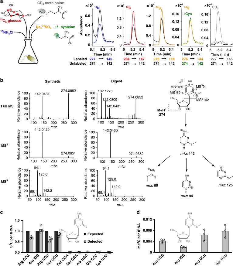
With the goal of discovering novel modified nucleosides in bacterial tRNA, Escherichia coli and Pseudomonas aeruginosa were grown in differentially stable isotope labeled growth media as recently reported16,17. The total tRNA were purified and analyzed by mass spectrometry. After data evaluation we found a promising candidate eluting at 5.2 min with an m/z of 274 from unlabeled tRNA, an m/z of 284 from [13C]-labeled tRNA, an m/z of 277 from [15N]-labeled tRNA and an m/z of 276 from [34S]-labeled tRNA (Fig. 1a, Supplementary Table 1). The sum formula of the nucleobase is C5HxN3OyS1 (Supplementary Fig. 1) and the nucleoside is thus C10HXN3OXS1. Through the addition of the heavy labeled amino acid CD3-methionine to the growth medium, we predicted the presence of a single methyl-group in the nucleoside candidate. Aiming at clarification of the analyte’s structure, we screened E. coli knockouts for its absence. TtcA is responsible for 2-thiolation of cytidine and in the ΔttcA strain, the m/z 274 signal is lost alongside 2-thiocytidine (s2C) (Supplementary Fig. 2a, Supplementary Tables 2 and 3)18. The mass difference of the novel nucleoside (m/z 274) and s2C (m/z 260) is 14 Dalton, which is a strong indicator of methylation of s2C.
Fig. 1. Verification of 2-methylthiocytidine (ms2C) structure and distribution in bacterial tRNA isoacceptors.
a Different isotope labeling in bacteria. Co-elution of the synthesized ms2C nucleoside (black) and ms2C from [15N], [13C] or [34S] labeled bacteria and products of metabolic labeling with [32S]-cysteine in [34S] medium and CD3-methionine labeling. The respective isotope is indicated above each chromatogram. b High resolution mass spectrometry and fragmentation of ms2C by direct injection of the synthetic standard or the natural bacterial digest and collision induced dissociation. c Expected (black)1 and experimentally detected (gray) abundance of s2C per respective tRNA isoacceptor. d Experimentally detected abundance of ms2C per respective s2C containing tRNA isoacceptor. All experiments are from n = 3 biol. replicates, error bars reflect standard deviation. Source data of (c) and (d) are provided as a Source Data file.
Due to the high nucleophilicity of thiols, we predicted that both the chemical synthesis and biosynthesis of 2-methylthiocytidine (ms2C) is feasible, and thus the methylation reaction at position 2 of s2C was performed. Interestingly, the synthetic route was already described in 2010, when the Suzuki lab19 used 2-methylthiocytidine as an intermediate of agmatidine synthesis. We followed their synthetic route under milder conditions and received pure ms2C in 28% yield (Supplementary Figs. 2b and 3). We mixed the synthesized ms2C with the heavy isotope labeled tRNA digests and observed perfect co-elution for the synthesized compound and its native isotopologues (Fig. 1a). Additional high resolution MS, MS² and MS³ spectra of the synthesized product and of native tRNA confirmed the structure of the novel nucleoside to be ms2C in E. coli (Fig. 1b) and P. aeruginosa (Supplementary Fig. 4).
With the synthetic standard of ms2C and an internal standard produced by metabolic isotope labeling20, we could now quantify the absolute abundance of ms2C in specific tRNAs from the unstressed E. coli BW25113 wild-type strain (WT). The precursor of ms2C, s2C is reported in three of the five tRNAArg isoacceptors and one of the five tRNASer isoacceptors1,14,21. Therefore, we designed DNA probes (Supplementary Table 4) against tRNAs with a reported s2C, namely tRNAArgCCG, tRNAArgICG, tRNAArgUCU and tRNASerGCU, and purified these isoacceptor tRNAs from total tRNA22. As a control we decided to isolate tRNAs with C32 (tRNASerGGA, tRNA LysUUU), a tRNA with 2’-O-methylcytidine at position 32 (Cm32, tRNASerUGA) and tRNAs without a C32, tRNAGlyCCC and tRNAAlaUGC. As expected, we found s2C in tRNAArgCCG, tRNAArgICG, tRNAArgUCU and tRNASerGCU (Fig. 1c). The complete modification profile of these tRNAs is shown in Supplementary Table 5 and Supplementary Fig. 5. ms2C was also found in these s2C-containing tRNA isoacceptors, however according to our data only 0.2–1.0% of actually s2C-modified tRNAs contain ms2C.
For exploration of the biosynthetic pathway, we quantified the abundance of ms2C in total tRNA from the E. coli WT and various isogenic knockout strains (Supplementary Table 2) in glucose-containing M9 minimal medium (Supplementary Fig. 6) and LB medium (Supplementary Fig. 7). In total tRNA from WT E. coli grown in M9 we found 0.08 s2C per average tRNA, and only ~0.00032 ms2C. In the ΔttcA mutant no s2C and ms2C were detectable (corresponding to less than 0.00005 modifications per tRNA). We observed decreased ms2C formation in various knockout strains grown in M9 medium but not in LB medium. To exclude an involvement of these enzymes in ms2C formation, we analyzed E. coli strains that overexpress tRNA modifying enzymes (Supplementary Fig. 7 and Supplementary Table 6). This confirmed that none of the investigated enzymes is involved in ms2C biosynthesis. The overall low abundance and high fluctuation of ms2C in unstressed bacteria hints towards its nature as an endogenous tRNA lesion. Indeed, in vitro experiments showed the formation of ms2C from s2C in the presence of SAM and MMS (Supplementary Fig. 8).
2-thiocytidine is a main substrate of MMS alkylation in vivo
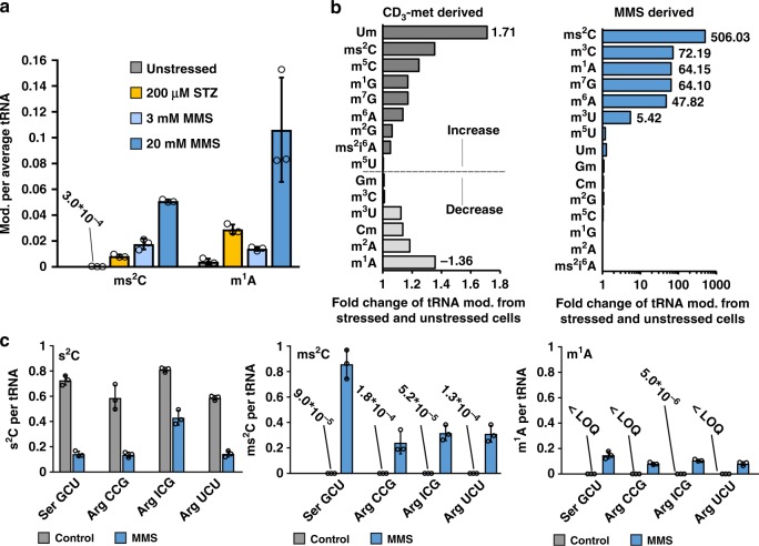
Intrigued by the possibility that s2C is methylated in vivo by naturally occurring methyl donors like SAM, we were wondering about its methylation during alkylation stress. We exposed WT E. coli to the LD50 dose of MMS (20 mM), non-lethal MMS doses (3 mM and 0.5 mM) and the LD50 dose of the natural antibiotic streptozotocin (STZ, 200 µM) in M9 medium (Supplementary Fig. 9). The total tRNA was isolated for absolute quantification by LC-MS/MS (Supplementary Table 7). The results for the main RNA damage product m1A and ms2C are shown in Fig. 2a. We observed ~0.1 m1A per tRNA and ~0.05 ms2C per tRNA in the 20 mM MMS exposed bacteria. With the non-lethal dose of 3 mM MMS, the formation of damage is reduced to 0.014 m1A and 0.018 ms2C per tRNA. With low doses of alkylating agent, the s2C damage was more prominent in total tRNA than the adenosine damage. The absolute abundance of other modified nucleosides is shown in Supplementary Figs. 10 and 11. STZ methylated both adenosine (0.03 m1A/tRNA) and s2C (0.008 ms2C/tRNA). Overall, ms2C formed readily during alkylation stress and in comparable extent to m1A.
Fig. 2. Absolute abundance of modified nucleosides in total tRNA and tRNA isoacceptors after methylation stress.
a Abundance of mod. (ms2C and m1A) per average tRNA after 0 mM (unstressed, gray), 200 µM Streptozotocin (STZ, natural methylating agent, 50% lethality, orange), 3 mM MMS (no lethality, light blue) and 20 mM MMS (50% lethality, dark blue). b Impact of MMS exposure (unstressed vs. 20 mM stressed) on E. coli total tRNA modifications (mod.). The origin of the methylated tRNA modifications is displayed in separate plots. Left: impact of MMS exposure on CD3-met (met = methionine) derived methylation displayed as the fold change ratio of stressed-to-unstressed. Right: impact of MMS exposure on directly methylated nucleosides (MMS derived methylation) displayed as the fold change of stressed-to-unstressed. c Absolute abundance of s2C (left), ms2C (middle) and m1A (right) in various tRNA isoacceptors without (gray) and with (blue) 20 mM MMS exposure. In the negative control (without MMS) E. coli were grown in 13C medium (gray bars, control) and the MMS exposed bacteria in non-labeled (blue bars, MMS) media. Co-purification of tRNA isoacceptors was done in a comparative NAIL-MS experiment as detailed in the text. All experiments are from n = 3 biol. replicates and error bars reflect standard deviation. Source data are provided as a Source Data file.
While m1A is known to be a direct methylation product, the increase in ms2C might be caused by either direct methylation or SAM-derived enzymatic methylation. To elucidate the origin of the methylation in ms2C under MMS stress, we used nucleic acid isotope labeling coupled mass spectrometry (NAIL-MS) to discriminate direct and SAM-derived methylation during MMS stress in vivo. Adapted from our recently established assay10, we cultured E. coli WT bacteria in the presence of CD3-methionine to allow complete CD3-labeling of all SAM dependent methylated nucleosides. One aliquot of these bacteria was exposed to the LD50 dose of MMS, while the other remained unstressed. After 1 h of MMS exposure the tRNA was purified and analyzed by LC-MS/MS. Enzymatically methylated nucleosides carry a + 3 label whereas the directly MMS methylated nucleosides are unlabeled and thus the two species are distinguishable by mass spectrometry. Absolute quantification of the enzymatically and directly methylated nucleosides in the unstressed and stressed cells revealed the abundance of methylated nucleosides in the samples. The fold changes between the stressed and unstressed samples were calculated and plotted in Fig. 2b. We observed only little adaptation of enzymatically introduced tRNA modifications under these growth conditions. The strongest increase was 1.7 fold, observed for Um (2′-O-methyluridine) from total tRNA. In both the stressed and unstressed samples, ms2C was again found to be labeled through CD3-methionine dependent pathways, with a slightly higher abundance in the stressed samples. This small increase in CD3-methylated ms2C does not reflect its substantial abundance in Fig. 2a. In contrast to the SAM dependent methylations, we found a high increase for the known products of direct methylation, namely m3U (5 fold), m6A (47 fold), m7G (64 fold) m1A (64 fold) and m3C (74 fold)10. However, the highest increase was for the product of direct s2C methylation, ms2C, which was over 500 fold increased in the stressed bacteria compared to the unstressed (absolute quantities in Supplementary Fig. 12). Thus, we prove that the ms2C observed in Fig. 2a originated from direct s2C methylation by MMS.
The previous experiments were done with total tRNA, but only four tRNA isoacceptors carry s2C. To study the impact of direct methylation on these s2C-containing tRNAs, we performed a comparative NAIL-MS experiment. For this purpose, the unstressed E. coli are grown in 13C6-glucose labeled medium and the MMS exposed bacteria (LD50) in unlabeled medium. After cell harvesting, the unstressed and stressed bacteria were mixed to avoid purification biases, as recently suggested12. From the resulting co-purified total tRNA, the tRNA isoacceptors SerGCU, ArgCCG, ArgICG and ArgUCU were isolated, digested and subjected to LC-MS/MS analysis. The mass spectrometer was set to distinguish the 13C-labeled nucleosides (unstressed bacteria) from the unlabeled nucleosides (stressed bacteria) and thus the abundance of each nucleoside can be determined (Supplementary Figs. 13 and 14, Supplementary Tables 8 and 9). On average, we found 0.8 s2C per respective unstressed tRNA, but after MMS exposure, the abundance of s2C drops substantially (Fig. 2c, left). For tRNA SerGCU, ArgCCG and ArgUCU we found a 4-fold decrease to <0.2 s2C per tRNA. tRNAArgICG was the only exception with a ~2-fold decrease to around 0.4 s2C per tRNA. As expected, the abundance of ms2C was increased in the stressed samples, which indicated that the lost s2C was directly methylated to ms2C in these tRNAs (Fig. 2c, middle). In all purified tRNAs, the ms2C abundance was higher than the m1A abundance (Fig. 2c, right).
This data demonstrates that s2C is a better substrate of the methylation agent MMS than adenosine.
2-methylthiocytidine may influence translation
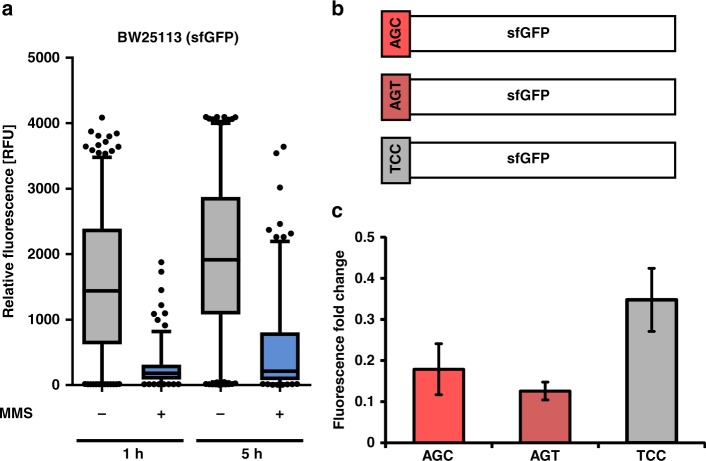
The s2C containing tRNAs are major targets of direct methylation by MMS. s2C is located at position 32 where it fulfils important roles in translation of the serine codons AGU and AGC by tRNASerGCU and the arginine codons CGG (tRNAArgCCG), AGA (tRNAArgUCU) and CGC, CGU and CGA (tRNAArgICG)14. The methylation of s2C and formation of ms2C might result in electronic and electrostatic shifts of the nucleobase, which potentially changes the properties of the whole anticodon loop of the affected tRNAs. Thus translation of the respective serine and arginine codons might be influenced by the methylation of s2C. To address this question, the effect of MMS on the synthesis of superfolder green fluorescent protein (sfGFP) in E. coli WT was determined. We decided to study sfGFP (for sequence and used primers see Supplementary Table 10), which contains one AGC (at position 2) and one AGT codon for serine, five GCU, two CGC and one CGG codon for arginine. 10 codons are translated by s2C modified tRNAs, which will be damaged to carry ms2C under MMS stress. Thus the translation of sfGFP could be effected by MMS due to the s2C methylation. Expression of sfgfp was under control of the PBAD promoter, and unstressed or stressed (exposures to 20 mM MMS for 1 h) cells of the mid-log growth phase were induced by arabinose. Fluorescence intensity of single E. coli cells of strain BW25113 was determined after 1 and 5 h (Supplementary Fig. 15). In the presence of 3 mM MMS, fluorescence was significantly lower under acute stress (1 h), but cells recovered after 5 h indicated by the increase in fluorescence. Similar results are seen for cells after exposure to 20 mM MMS (Fig. 3a). We mutated a single codon in the sfgfp gene to replace the AGC codon for serine with AGT and TCC (Fig. 3b). While AGC and AGT are read by the same s2C modified tRNASerGCU, the TCC codon is read by tRNASerGGA, which is not s2C modified and thus the anticodon is not affected by the MMS stress (see Fig. 1c). We tested the influence of these codons (AGC, AGT, TTC) at position 2 in sfGFP on the fluorescence in the WT strain. Fluorescence of stressed cells producing the AGT- and AGC-variants of sfGFP is more affected in comparison to the TCC construct (Fig. 3c). At the used MMS doses, we previously observed about 80% ms2C modified tRNA SerGCU while only 20% were s2C modified (Fig. 2c). Thus we see a direct correlation of ms2C abundance and decreased translational efficiency for the respective codons. Moreover, translation of the AGT-containing sfGFP was slightly more affected by the methylating agent than the AGC-containing construct. These results indicate a negative effect of ms2C on translation.
Fig. 3. Impact of s2C methylation on translation.
a Long-term effects of 20 mM MMS stress on sfGFP synthesis in E. coli WT cells. Single cell fluorescence was determined for a minimum of 300 E. coli cells producing sfGFP under control of the L-arabinose inducible promoter (pBAD24). Fluorescence was measured 1 and 5 h after addition of arabinose to unstressed cells or cells pre-exposed to 20 mM MMS stress for 1 h. (box plots represent the 5-95% percentile) b Illustration of the used sfGFP constructs. The serine on amino acid position 2 of the used sfGFP variant was encoded by different codons: AGC, AGT and TCC. c Influence of codons on sfGFP synthesis after exposure of cells to MMS stress. Fluorescence was measured after 1 h of induction with arabinose. The fold change was determined by dividing the mean fluorescence of stressed cells (20 mM MMS) by the mean fluorescence of unstressed cells. All experiments are from n = 3 biol. replicates and error bars reflect standard deviation. Source data are provided as a Source Data file.
2-methylthiocytidine is efficiently repaired by AlkB
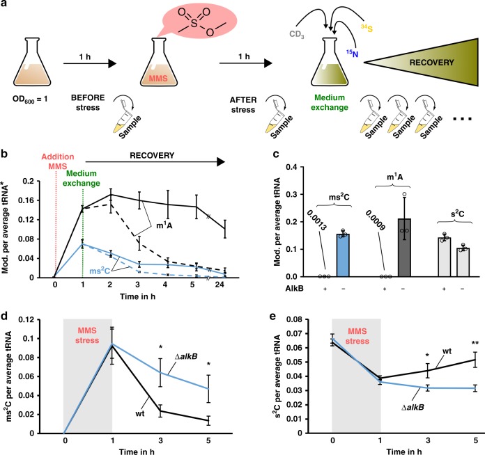
The ms2C damage has a potential effect on the translation of E. coli. It is highly probable that the methylation damage is recognized and repaired by the bacteria to adapt to the stress. This repair is either possible by direct demethylation by AlkB as shown for m1A23 or by tRNA degradation. To address the question whether ms2C is repaired and which mechanism dominates, we designed a pulse-chase assay involving stable isotope labeling. Figure 4a shows the principle of the assay. The goal of this assay is to discriminate the damaged tRNAs from the tRNAs transcribed during the recovery. Thus, we can follow the fate of the damaged tRNAs and their nucleosides independently from dilution by transcription. For this purpose, cells were grown in media containing only [14N]- and [32S]-nutrients. Consequently, the RNA was completely labeled with [14N] and all s2C have a [32S] label (original s2C), e.g. m/z (s2C) 260. All methylated nucleosides had a CH3 mark (e.g., original m1A m/z 282). In this medium, the cells were exposed to MMS (LD50) and s2C was converted to ms2C and, e.g., A to m1A. After exposure, MMS was removed by exchanging the media with heavy-isotope media. During the following recovery period, newly transcribed RNA were [15N] labeled, newly methylated nucleosides were [CD]3 labeled and new s2C had a [34S] label (new s2C, m/z 265 and new m1A, m/z 290).
Fig. 4. Principle and results of pulse-chase NAIL-MS experiments to determine the repair of ms2C in vivo.
a Principle of a pulse-chase NAIL-MS experiment. The bacteria are grown in unlabeled (n.l.) media before and after exposure to MMS (structure shown). After 1 h MMS exposure, the media is removed and fresh, [15N], [34S] and [CD3]-methionine containing media is added. Samples are drawn during the recovery time for tRNA isolation. b Formation of the nucleoside damage product ms2C (blue) and m1A (black) per average tRNA after 20 mM MMS exposure. *Dashed lines: abundance of mod. (ms2C and m1A) per all tRNAs (sum of original, unlabeled and new, [15N]-labeled transcripts). Solid lines: abundance of mod. (ms2C and m1A) per original tRNA. c Abundance of mod. (ms2C in blue, m1A in dark gray and s2C in light gray) per tRNA incubated with purified AlkB in vitro. The substrate tRNA is isolated from E. coli bacteria exposed to 20 mM MMS. d ms2C abundance of E. coli WT (black) in comparison to alkB deficient E. coli (ΔalkB, blue) after MMS stress (20 mM, 1 h) and during recovery in a pulse-chase NAIL-MS experiment, as described in a) and b). e s2C abundance of E. coli WT (black) in comparison to alkB deficient E. coli (ΔalkB, blue) after MMS stress (20 mM, 1 h) and during recovery. All experiments are from n = 3 biol. replicates and error bars reflect standard deviation. p-values from student t-test (equal distribution, two-sided): *p < 0.05 and **p < 0.01. Source data are provided as a Source Data file.
The dashed line in Fig. 4b, represents the decrease of m1A if referenced to the sum of all tRNAs (original + new transcripts). The fast decrease is caused by dilution due to ongoing tRNA transcription. In the past, this decrease could be misinterpreted as active tRNA demethylation, but with NAIL-MS we can follow the fate of modified nucleosides inside living cells by excluding transcription processes. By referencing m1A abundance to the original tRNAs, we saw a slow but steady decrease of m1A in the damaged tRNAs over time which reflects true demethylation (Fig. 4b, solid line). For ms2C, we saw a fast and nearly complete repair over the same observation period (Fig. 4b). This decrease was not caused by increased tRNA degradation as MMS exposed bacteria showed the same original-tRNA dilution pattern as unstressed bacteria (Supplementary Fig. 16). We isolated tRNAs SerGCU and ArgICG from the NAIL-MS pulse-chase experiments, and for these damaged tRNAs we saw no increased degradation either (Supplementary Fig. 17). Thus we conclude that the removal of ms2C from damaged tRNA is not caused by tRNA degradation.
m1A is known to be demethylated by AlkB13,23. To test whether AlkB also demethylates ms2C to s2C, we isolated total tRNA from MMS exposed bacteria and tested the AlkB activity in vitro. After addition of AlkB to the damaged tRNA, we could no longer detect ms2C and m1A in the tRNA (Fig. 4c). Instead, we could observe an increase of s2C, which indicates that AlkB demethylates ms2C to s2C. This finding is rather surprising as, until now, only nitrogen demethylation was observed for AlkB but never sulfur demethylation11,23.
To verify that AlkB demethylates ms2C containing tRNAs in vivo, we decided to repeat the NAIL-MS pulse-chase assay in an alkB deficient E. coli strain. After exposure to 20 mM MMS, we observed a fast decrease of ms2C in the WT strain and a slower decrease in the isogenic ΔalkB mutant (Fig. 4d). Interestingly, we observed an increase of s2C in the WT strain but not in the ΔalkB strain (Fig. 4e). Our data indicates that a portion of ms2C is demethylated by AlkB under recovery of the original [32S]-s2C. The other portion of ms2C seems to be lost by an AlkB-independent mechanism. For m3C we also observed an AlkB dependent demethylation, but surprisingly not for m1A (Supplementary Fig. 18). We repeated the experiment in the presence of 3 mM MMS and 0.5 mM MMS to ensure the viability of the cells. 3 mM MMS corresponds to 20% lethality in the ΔalkB strain (Supplementary Fig. 19) and no lethality in the WT strain. Directly after 3 mM MMS exposure, the abundance of ms2C, m1A and m3C per original tRNA was comparable in the WT and ΔalkB strain (Supplementary Fig. 20). After 2 h of recovery, the abundance of ms2C, m1A and m3C was more reduced in the WT strain compared to the ΔalkB mutant. This indicates repair of ms2C by AlkB in vivo as observed in Fig. 4d. For 0.5 mM MMS, we saw an AlkB independent reduction of m1A, m3C, and ms2C of comparable extent. On tRNAs with a damage abundance below a threshold level, AlkB appeared to be uninvolved in repair.
In summary, we demonstrate that ms2C damaged tRNAs are yet undescribed substrates of AlkB, both in vitro and in vivo.
Dethiomethylation as an alternative repair pathway
The in vitro as well as the in vivo analyses of AlkB convinced us of the role of AlkB as key player in ms2C demethylation. Based on our data shown in Fig. 4, we assume direct demethylation by AlkB, which results in the recovery of [32S]-s2C.
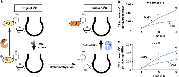
Chemically, the thiomethyl group is an acceptable leaving group in the presence of nucleophiles such as water. Dethiomethylation might occur inside the cell and would result in the formation of canonical cytidine. In theory, this cytidine should then be a substrate of TtcA and a rethiolation to s2C would result (Fig. 5a). Such an s2C turnover event is observable in vivo with our pulse-chase NAIL-MS experiment. During the design of the experiment, we chose two media with different sulfur isotopes. The stress medium contains the sulfur-32 isotope and thus all s2C of original tRNAs is [32S]-s2C with an m/z of 260. The recovery medium contains no sulfur-32, but sulfur-34 instead. Thus, s2C in new transcripts is not only three units heavier (nitrogen-15) but five units ([15N3/34S]-s2C, m/z 265). In case of the imagined scenario of spontaneous dethiomethylation of ms2C into cytidine followed by rethiolation into s2C, only sulfur-34 is available and thus the original [32S]-s2C would turn into [34S]-s2C with an m/z of 262. The original [32S]-s2C produced by AlkB demethylation and the hypothetical turnover [34S]-s2C formed after dethiomethylation/rethiolation can be distinguished by mass spectrometry (Supplementary Table 11).
Fig. 5. Pulse-chase NAIL-MS analysis of turnover s2C after 20 mM MMS exposure in WT BW25113 and E. coli ΔalkB knockout strain.
a Concept of turnover s2C generation and its detection by NAIL-MS. b Abundance of [34S] turnover s2C in WT BW25113 during recovery after MMS stress (20 mM) and unstressed control. c Abundance of [34S] turnover s2C in the ΔalkB strain during recovery after MMS stress (20 mM) and unstressed control. All experiments are from n = 3 biol. replicates and error bars reflect standard deviation. p-values from student t-test (equal distribution, two-sided): *p < 0.05, **p < 0.01 and ***p < 0.001. Source data are provided as a Source Data file.
The result for the unstressed and 20 mM MMS treated WT strain is shown in Fig. 5b. Immediately after the stress, the number of turnover [34S]-s2C was comparable and rose over time. The increase of [34S]-s2C in unstressed bacteria was most likely caused by regular s2C biosynthesis into original tRNAs, which were not fully mature at the time of medium exchange. The presence of only [34S] at these later time points led to the formation of [34S]-s2C in the control bacteria. In contrast, MMS exposure led to more [34S]-s2C. To test involvement of AlkB in the formation of turnover [34S]-s2C, we plotted the [34S]-s2C abundance observed in the ΔalkB strain (Fig. 5c). Here, we saw an increase in turnover s2C, which was comparable to the turnover in the WT strain. We thus conclude that the turnover is not a consequence of the AlkB repair process.
The increase in turnover s2C can be explained by dethiomethylation of ms2C followed by rethiolation of the formed cytidine. Currently it is unclear, whether the dethiomethylation occurs spontaneously after a nucleophilic attack of water or by an enzymatic pathway. From a chemical perspective, we believe that spontaneous dethiomethylation is the most likely.
Discussion
In this study, we identified s2C as a substrate of direct methylation in bacterial tRNAs, and the mechanisms of the subsequent repair by AlkB. We initially identified the new structure of the methylated nucleoside in total tRNA of E. coli and P. aeruginosa. The structure of the ms2C (2-methylthiocytidine) nucleoside was confirmed after synthesis by various analytical tools including fragmentation patterns in high resolution mass spectrometry. Methylated derivatives of other thiolated nucleosides like s4U or mnm5s2U might also form endogenously, but were not detected in our nucleoside discovery experiments.
ms2C was found at position 32 of tRNAArgICG, tRNAArgCCG, tRNAArgUCU and tRNASerGCU. Only 1% of the s2C modified tRNA isoacceptors carry ms2C under unstressed growth conditions. The endogenous formation of ms2C was not found to be connected to common tRNA modifying enzymes. At this stage we cannot determine whether the endogenous, but low abundant, ms2C modification of bacterial tRNA is introduced enzymatically or by direct methylation through, e.g., SAM. For low abundant nucleoside modifications it is often an open question whether they are lesions or functional entities24.
Intrigued by the observation that ms2C forms naturally in the presence of the methyl donor SAM, we were wondering whether ms2C is formed by direct methylation during alkylation stress after MMS exposure. MMS is a direct mono-methylating agent, which is known to methylate RNA nucleosides and DNA nucleosides. From our systematic study, we know that the main damage products of canonical nucleosides are m1A and m7G10. In the current study, we also focused on the quantities of ms2C formed during an LD50 MMS exposure of E. coli. We find 0.1 m1A damage sites per average tRNA and surprisingly 0.05 ms2C per average tRNA. At lower MMS doses (3 mM), the number of m1A damage sites is reduced to 0.014 per average tRNA and 0.018 ms2C per average tRNA. It is now clear that ms2C is an equally prominent damage product as m1A in total tRNA. If one considers that the average tRNA in bacteria is composed of 15 adenosines while only 0.1 s2C are found in the average tRNA, our results are rather intriguing. Although adenosine is 150 fold more abundant than s2C in total tRNA, the methylation products m1A and ms2C are of comparable quantities. From our data, we conclude that s2C is an at least equal or even better substrate of direct methylation than adenosine in the tRNA of the bacterium E. coli.
Although the abundance of modified nucleosides is low in comparison to the amount of canonical nucleosides in the total RNA pool, modified nucleosides fulfil a variety of important functions within the cell. The damage of a modified nucleoside will most likely interrupt the modification’s function and might be disadvantageous to the organism. We have tested how the methylation of s2C influences the translation in vivo. Serine can be translated by tRNASerGGA (no s2C) reading the TCC codon or tRNASerGCU reading the AGC or AGU codons. We found that sfgfp with the AGC or AGU codon was less efficiently translated than sfgfp with the TCC codon under MMS stress. This finding indicates that the translation of the AGC and AGT codon is disturbed after MMS stress, potentially due to the presence of ms2C.
The repair of damaged ribonucleosides might occur by two potential mechanisms. RNA is a transient molecule and constantly transcribed from DNA within the cell. Upon damage, it is possible to remove the damage by controlled degradation of the RNA. Due to the fact that tRNA maturation and modification is energy consuming, the second repair scenario of direct damage repair by, e.g., demethylation, is more attractive. In bacterial RNA, the repair of m1A and m3C by oxidative demethylation by the enzyme AlkB has been shown7. In our NAIL-MS studies, we use stable isotopes such as carbon-13, nitrogen-15 or sulfur-34 to distinguish RNA that was present during the stress event (here, MMS exposure) and new RNA transcribed during the recovery period. We can thus distinguish repair by RNA degradation and demethylation. With NAIL-MS, we observe AlkB dependent demethylation of ms2C, m3C and potentially m1A.
The AlkB dependent repair of m3C had been suggested in previous studies but has never been shown in vivo. Surprisingly, m1A repair was similar in the WT and ΔalkB mutant. Only for the 3 mM MMS dose, 3 h after stress exposure, the WT strain showed less m1A per tRNA compared to the ΔalkB strain. This is in accordance with the only other in vivo study, where m1A repair by AlkB was observed in radioactive tRNA 3 h after stress at a low dose of alkylating agent23.
In unstressed ΔalkB bacteria, we did not observe an accumulation of ms2C (Supplementary Fig. 21). This is to be expected if one considers the low copy number of AlkB in unstressed wildtype bacteria (1 copy of AlkB in log phase E. coli cells25). Only after exposure to alkylating agents, alkB is induced and six hours after 3 mM MMS exposure 6000 AlkB copies/cell have been found11.
Concerning ms2C repair, we found an increase of original [32S]-s2C in vivo, which reflects direct sulfur demethylation by AlkB. With our sophisticated NAIL-MS approach we could additionally observe an AlkB independent formation of s2C which originated from dethiomethylation of s2C to cytidine and subsequent rethiolation. The initial dethiolation step might be enzymatically catalyzed, but due to the electrophilic nature of the C2 in ms2C, a direct nucleophilic attack by water is also possible.
So far, m1A was thought to be the main methylation damage product in bacterial RNA and thus to be the main substrate of AlkB. Our data shows that ms2C is a methylation damage in bacterial tRNAs of comparable extent after MMS exposure. In addition, AlkB is faster during the repair of ms2C damaged tRNAs compared to m1A damaged tRNAs.
Since its discovery, AlkB has been an enzyme full of surprises. In this work, we present the next surprise, which is AlkBs extended substrate repertoire and its preference for the sulfur methylated nucleoside ms2C.
Methods
Salts, reagents, and nucleosides
All salts were obtained from Sigma-Aldrich (Munich, Germany) at molecular biology grade unless stated otherwise. The isotopically labeled compounds 15NH4Cl (>98 atom %) and [D3]-L-methionine (98 atom %) were obtained from Sigma-Aldrich. Isotopically labeled 13C6- glucose (≥99 atom %) and isotopically labeled Na234SO4 (99.11 atom %) were obtained from Eurisotope (Saarbruecken, Germany). All solutions and buffers were made with water from a Millipore device (Milli-Q, Merck, Darmstadt, Germany). The nucleosides adenosine, cytidine, guanosine, uridine, and N2-methylguanosine (m2G) were obtained from Sigma-Aldrich. 1-Methyladenosine (m1A), 2-methyladenosine (m2A), N3-methylcytidine (m3C), N6-methyladenosine (m6A), 7-methylguanosine (m7G), 5-methylcytidine (m5C), 5-methyluridine (m5U), 2’-O-methylcytidine (Cm), 2′-O-methylguanosine (Gm), 1-methylguanosine (m1G), and 3-methyluridine (m3U) were obtained from Carbosynth (Newbury, UK).
Synthesis of 2-methylthiocytidine
Twenty milligrams of s2C were stirred in 1 mL anhydrous ethanol under nitrogen atmosphere.7.4 mg of NaHCO3 and 24 µL of methyl iodide were added and the reaction was stirred overnight at room temperature. The clear yellow solution was evaporated and two times purified by silica gel chromatography in dichloromethane with 10 % methanol. The Rf value (TLC) in DCM/MeOH (10:1) was 0.46.
1H NMR (400 MHz, D2O):δ = 8.39 (d, J = 7.7 Hz, 1H, H-6), 6.60 (d, J = 7.7Hz, 1H, H-5), 5.98 (d, J = 3.0Hz, 1H, H-1′), 4.38 (t, J = 3.4 Hz, 1H, H-2′), 4.23-4.18 (m, 2H, H-3′, H-4′), 3.99 (d, J = 12.7Hz, 1H, H-5′a), 3.85 (dd, J = 2.7 Hz, 13.4 Hz, 1H, H-5′b), 2.67 (s, 3H, CH3). 13C NMR (500 MHz, D2O):δ = 166.4 (C-2), 162.1 (C-4), 141.5 (C-6), 102.1 (C-5), 92.7 (C-1′), 84.6 (C-3′), 74.9 (C-2′), 68.5 (C-4′), 59.8 (C-5′), 14.4 (C-7).
E. coli and P. aeruginosa strains
The used E. coli wild-type strain BW25113 and the isogenic knockout strains were purchased from the Keio database26. Pseudomonas aeruginosa PA14 was a generous gift of Prof. Peter Dedon. Knockout strains with kanamycin resistance were grown on kanamycin LB agar plates (50 µg/mL). The E. coli strain AG1 (ME5305) transformed with the indicated pCA24N-based vectors was used for gene overexpression and ordered from the ASKA library database27. Strains with chloramphenicol resistance were grown in the presence of chloramphenicol (30 µg/mL). All cultures were grown in a shaking incubator at 37 °C at 250 rpm (Orbit = 10 mm). Overnight cultures were grown in 5 mL of the media used for the respective experiment. The cells were grown starting with an OD600 of 0.1, 0.5 or 1 (as specified in the respective section) and grown until reaching stationary phase (OD600 ~ 4).
Growth media LB/ M9/ isotope labeled
For LB media, LB Broth (Luria Miller) from Roth (Karlsruhe, Germany) was used. 1.5% (wt/vol) agar plates were prepared with LB-broth and Agar-Agar (Kobe I from Roth) according to manufacturer’s protocol.
M9 minimal medium was used with and without the indicated isotopes. Unlabeled M9 was prepared by mixing a 10 × M9 stock solution with glucose, MgCl2, Na2SO4, and CaCl2 (as detailed below). For unlabeled 10 × M9 stock solution, Na2HPO4 (68 g/L), KH2PO4 (30 g/L), NaCl (2.5 g/L), and NH4Cl (10 g/L) were mixed and autoclaved. For 15N-labeled 10 × M9 stock solution, 15NH4Cl (10 g/L) was used. MgCl2 (0.1 M), CaCl2 (0.1 M), Na2SO4 (0.1 M), and 20% (wt%) glucose were prepared by sterile filtration. A 20% (wt%) 13C6-labeled glucose solution was prepared for 13C-labeled M9 media. For 34S-labeled M9 media a 0.1 M Na234SO4 solution was prepared. Final M9 media was prepared by mixing, e.g., 500 μL M9 stock solution with 100 μL glucose, 100 μL MgCl2, 100 μL Na2SO4, 5 μL CaCl2 and water to a final volume of 5 mL. For 15N-labeled cultures, the 15N-10 × M9 stock solution was used. For 13C-labeled cultures, the 20% (wt%) 13C6-labeled glucose solution and for 34S-labeled cultures, the 0.1 M Na234SO4 solution was used. For CD3-methylome labeling, 200 μL CD3-methionine (stock 5 g/L) was added to 5 mL of culture volume.
E. coli knockout/overexpression strain library
The knockout strains were cultured in LB or M9 media, starting from OD600 = 0.5. After 3 h the cells were harvested and the RNA was isolated and purified. The overexpression strains were cultured in LB media starting with an OD600 = 0.5 and an IPTG concentration of 0.1 mM. After 1 h incubation at 37 °C and 250 rpm (Orbit = 10 mm) the IPTG concentration was increased to 1 mM to induce full overexpression. The bacteria were incubated for further 2 h before the cells were harvested and RNA was isolated and purified
Survival assay
Unlabeled M9 media was used throughout the survival assay. A 5 mL culture with OD600 1.0 (or 0.1, respectively) were prepared from an E. coli overnight culture. After 60 min incubation (30 min with Streptozotocin), 100 µL of the culture were diluted 10−5 or 10−6 with sterile water. From this dilution 70 µL were plated on a prewarmed LB agar plate. The colony number of this plate represents 100% bacterial survival. MMS (Methyl-methanesulfonate, 99%) or Streptozotocin (STZ, 10 mM stock solution) was added to the remaining bacteria in defined concentrations. After 60 min (30 min) exposure, 100 µL of the culture were diluted and plated. The LB plates were incubated at 37 °C overnight and the colonies counted for determination of the survival at the respective MMS or STZ concentration.
E. coli incubation with Streptozotocin (STZ)
From an overnight culture of E. coli strain BW25113 (WT) the OD600 was brought to 1.0 and the culture was grown for 1 h. To 5 mL bacterial culture 125 µL of a 10 mM STZ solution was added (200 µM final conc.) and the culture was incubated for 30 min. Afterwards, the bacteria were centrifuged and the resulting pellet was used for RNA isolation and purification.
Cell lysis and tRNA purification
The bacteria culture was centrifuged at 1200 × g for 5 min. The supernatant was discarded and the cell pellet was resuspended in 1 mL TRI reagent (Sigma-Aldrich) per 5 mL bacteria culture. The total RNA was isolated according to the supplier’s manual. tRNA was purified by size exclusion chromatography (SEC) according to published procedures10. The tRNA was resuspended in water (30 μL).
Isoacceptor purification
The procedure was adapted from Hauenschild et al.22. One microgram of total tRNA was incubated with 100 pmol of the biotinylated DNA probe and purified using Dynabeads T1 (Thermo Fisher Scientific, Waltham, MA, USA) according to the manusfacturer’s protocol. For tRNA isoacceptor purification, pre-purified total tRNA was used. The sequences of the biotinylated 2′-deoxyoligonucleotide probes are listed in Supplementary Table 4.
Comparative NAIL-MS
One E. coli culture was grown overnight in an unlabeled M9 medium and another in a 13C labeled M9 medium. From these cultures, a labeled and unlabeled exposure culture with an OD600 of 1.0 and a volume of 5 mL were prepared. After 60 min growth, 8.5 μL MMS (final conc. 20 mM) were added to the unlabeled culture and 8.5 μL of water to the 13C-labeled culture (MOCK). After 60 min of exposure both cultures were mixed and the RNA was purified immediately. Total tRNA and tRNA isoacceptors were isolated as described above. For validation of the comparative NAIL-MS assay, the experiment was repeated by mixing and co-purifying RNA (total tRNA and tRNA isoacceptors) from an unlabeled MOCK treated culture and a 13C-labeled MOCK treated culture.
Methylome discrimination assay
For this assay, CD3-labeled M9 medium was used at all times. A 5 mL bacterial solution with an OD600 of 0.1 was prepared from an overnight culture. After 60 min growth, 8.5 μL MMS (final conc. 20 mM) were added. As a control, 8.5 µL water was added to a second culture. After 60 min of exposure, the RNA was isolated and total tRNA purified by SEC.
sfGFP reporter and single-cell fluorescence microscopy
For in vivo quantification of the effects of tRNA modifications after MMS stress on translation, the E. coli strains BW25113, BW25113 ΔalkB and BW25113 ΔttcA were transformed with pBAD24 containing a copy of sfgfp under the control of the arabinose-inducible PBAD promoter. To test the effects of different serine codons (AGC, AGT, TCC) the only serine codon of the used sfgfp sequence (corresponds to amino acid position 2) was exchanged by using the respective primers during the cloning process. To enhance the number of serines, the sfgfp sequence was extended at the 5′-end with multiple motifs coding for proline-alanine-serine (ASPAAPSASAPSAASAAPSAA)-sequence, modified from previous studies28. We used the primers PAS-gfp_for/ PAS-AGT_gfp_for / PAS-TCC_gfp_for plus gfp_rev to introduce different types of codons for serine (AGC, AGT or TCC) in the sfGFP variants extended with the proline-alanine-serine (ASPAAPSASAPSAASAAPSAA)-sequence or respectively gfp_for / AGT_gfp_for / TCC_gfp_for plus gfp_rev to produce the sfGFP variants with different types of codons for serine (AGC, AGT or TCC). The restrictions sites EcoRI and XbaI were used for integration of the sfgfp variants into the pBAD24 plasmid.
For each experiment cells of an overnight culture were inoculated into fresh M9 minimal medium supplemented with 0.5 % (vol/vol) glycerol as a sole carbon source and incubated under vigorous shaking at 37 °C. Cells were grown to mid-log phase, and the culture was divided. One half was stressed by the addition of MMS to final concentrations of 3 and 20 mM, respectively, the other half remained untreated. After 1 h of incubation in presence of MMS the medium was exchanged with fresh, pre-tempered M9 minimal media supplemented with 0.5% (vol/vol) glycerol and 0.2% (vol/vol) arabinose to induce sfGFP expression. As a control, cells were cultivated without addition of arabinose. To measure sfGFP fluorescence, cells were fixed on an agarose pad (1% wt/vol in phosphate-buffered saline) placed on a microscope slide with coverslip. Micrographs were taken on a Leica microscope DMI 6000B equipped with a Leica DFC 365Fx camera (Andor, 12 bit). sfGFP fluorescence was visualized using an excitation wavelength of 460 nm and a 512 nm emission filter with a 75-nm bandwidth. Fluorescence intensities of a minimum of 300 cells per transformant were collected and quantified using Fiji29.
Pulse-chase NAIL-MS experiment
A single colony of E. coli BW25113 or E. coli JW2200-KC (ΔalkB) was picked and grown in unlabeled M9 medium (5 mL) overnight. From the first overnight culture, a 50 mL culture was prepared in unlabeled M9 medium and grown overnight. From the second overnight culture, 120 mL culture (OD600 of 1.0) was prepared in unlabeled M9 medium. After 60 min growth, the first aliquot (7 mL) was taken for RNA isolation. The remaining culture was split into two flasks of 56.5 mL each. One was exposed to MMS (95.7 μL, 20 mM final concentration) the other to water (MOCK) and inverted before both cultures were cultivated for 60 min. An aliquot (7 mL) was drawn from each culture, and the RNA was isolated. The remaining bacteria were centrifuged (1200 × g, 5 min), and the MMS/MOCK-containing supernatants were discarded. The bacteria pellets were washed with 15N, 34S and CD3-methionine labeled M9 medium (5 mL), and each bacterial pellet was suspended in fresh 15N/34S/CD3 M9 medium (50 mL). The bacteria were allowed to grow and recover from the MMS/MOCK treatment. Seven millilitres of each bacterial culture were harvested after 1, 2, 3, 4, and 23 h. The RNA was isolated and the tRNA purified by SEC. The experiment was also done with lower volumes and different concentrations of MMS.
SAM and MMS incubation assay in vitro
4.3 μL of a 93 μM s2C synthetic standard solution (0.4 nmol) were mixed with 0, 500, 1000, 1500 or 2000 equivalents of SAM (32 mM stock) or MMS (20 mM stock) in a final volume of 100 μL containing 50 mM Na2HPO4/NaH2PO4 buffer (pH 7.0). The mixture was incubated at 37 °C for 60 min in a shaking heat block. After incubation, 900 µL water were added (1:10 dilution) and spiked with SILIS to be analyzed by quantitative LC-MS/MS.
AlkB in vitro assay
Five microlitres of purified tRNA (30 ng/µL) from 20 mM MMS exposed E. coli was mixed with 45 μL of AlkB. (45 µL AlkB was prepared with 7.5 μL KCl (100 mM), 1.5 μL freshly mixed sodium α-ketoglutarate (10 mM), 2.5 μL Tris buffer (1 M, pH 7.65), 5 μL freshly mixed sodium L-ascorbate (20 mM), 1.5 μL freshly mixed Fe(II)(NH4)2(SO4)2 (10 mM) and 1.5 μL of AlkB enzyme (39.1 μM, Peak Proteins, Cheshire, UK) and water.) The reaction mixture was incubated at 37 °C for 60 min. After incubation, 500 μL of LiClO4 in acetone (2 vol%) was added for tRNA precipitation and mixed thoroughly. After 10 min at room temperature, the sample was centrifuged at 5000 × g for 10 min. The RNA pellet was washed with 70 vol% ethanol and resuspended in 20 µL water before digestion for LC-MS/MS analysis.
tRNA digestion for mass spectrometry
tRNA (100 ng) in aqueous digestion mix (30 μL) was digested to single nucleosides by using 0.2 U alkaline phosphatase, 0.02 U phosphodiesterase I (VWR, Radnor, Pennsylvania, USA), and 0.2 U benzonase in Tris (pH 8, 5 mM) and MgCl2 (1 mM) containing buffer. Furthermore, 0.5 µg tetrahydrouridine (Merck, Darmstadt, Germany), 1 µM butylated hydroxytoluene, and 0.1 µg pentostatin were added to avoid deamination and oxidation of the nucleosides30. The mixture was incubated for 2 h at 37 °C and then filtered through 96-well filter plates (AcroPrep Advance 350 10 K Omega, PALL Corporation, New York, USA) at 3000 × g and 4 °C for 30 min. 1/10 Vol. of SILIS (stable isotope labeled internal standard) as prepared in31 was added to each filtrate before analysis by QQQ mass spectrometry.
High resolution mass spectrometry
The ribonucleosides were separated using a Dionex Ultimate 3000 HPLC system on an Interchim Uptisphere120-3HDO C18 or a Synergi, 2.5 μm Fusion-RP C18, 100 Å, 100 × 2 mm (Phenomenex®, Torrance, California, USA). Mobile phase A was 2 mM ammonium acetate and mobile phase B was 80% acetonitrile containing 2 mM ammonium acetate. Gradient elution started with 0% B and increased to 12% B after 10 min and to 80% after 12 min. After 4 min elution at 80% B and subsequently regeneration of starting conditions to 100% A after 5 min, the column was equilibrated at 100% A for 8 min. The flow rate was 0.2 mL/min and the column temperature 30 °C. High-resolution mass spectra of precursor and product ions were recorded by a ThermoFinnigan LTQ Orbitrap XL. The parameters of the mass spectrometer were tuned with a freshly mixed solution of adenosine (5 μM). The parameters were sheath gas flow rate, 16 arb; auxiliary gas flow rate, 11 arb; sweep gas flow rate, 4 arb; spray voltage, 5.0 kV; capillary temperature, 200 °C; capillary voltage, 20 V, tube lens 65 V.
QQQ mass spectrometry
For quantitative mass spectrometry an Agilent 1290 Infinity II equipped with a diode-array detector (DAD) combined with an Agilent Technologies G6470A Triple Quad system and electrospray ionization (ESI-MS, Agilent Jetstream) was used. Operating parameters: positive-ion mode, skimmer voltage of 15 V, cell accelerator voltage of 5 V, N2 gas temperature of 230 °C and N2 gas flow of 6 L/min, sheath gas (N2) temperature of 400 °C with a flow of 12 L/min, capillary voltage of 2500 V, nozzle voltage of 0 V, and nebulizer at 40 psi. The instrument was operated in dynamic MRM mode.
For separation a Core-Shell Technology column (Phenomenex, Torrance, CA, USA; Kinetex 1.7 μm EVO C18, 100 Å, 150 × 2.1 mm) at 35 °C and a flow rate of 0.35 mL/min were used in combination with a binary mobile phase of 5 mM NH4OAc aqueous buffer A, brought to pH 5.6 with glacial acetic acid (65 μL), and an organic buffer B of pure acetonitrile (Roth, LC-MS grade, purity ≥.99.95). The gradient started at 100% solvent A, followed by an increase to 10% over 10 min. From 10 to 15 min, solvent B was increased to 45% and was maintained for 3 min before returning to 10% solvent A and a 3 min re-equilibration period.
Calibration
For calibration, synthetic nucleosides were weighed and dissolved in water to a stock concentration of 1–10 mM. Due to an unknown content of water and salts in the synthesized ms2C standard, the concentration could not be determined through weighing. Therefore, the concentration of the stock solution was determined by comparison to s2C containing isoacceptors after MMS exposure. The calibration solutions range from 0.3 to 500 pmol for each canonical nucleoside and from 0.3 to 500 fmol for each modified nucleoside and were spiked with 10% SILIS20. The sample data were analyzed by the Quantitative and Qualitative MassHunter Software from Agilent. The areas of the MRM signals were integrated for each modification and their isotope derivatives.
The absolute amounts of the modifications were referenced to the absolute amounts of the respective canonical. In the case of the pulse-chase experiment, the different isotopomers were referenced to their respective labeled canonicals, so that original modifications were referenced to original canonicals and new modifications were referenced to new canonicals. See Eqs. (1) and (2) for s2C as an example in Supplementary Table 12.
Statistics
All experiments were performed at least three times (biological replicates) to allow student t-test analysis. p-values of student t-test (unpaired, two-tailed, equal distribution) were calculated using Excel or Graphpad Prism.
Reporting summary
Further information on research design is available in the Nature Research Reporting Summary linked to this article.
Supplementary information
Acknowledgements
V.R. and S.K. are grateful for funding from the Fonds der chemischen Industrie. The project is financially supported by the Deutsche Forschungsgemeinschaft [Exc114-2 (S.K. and K.J.), KE1943/3-1 (S.K.) and Projektnummer 325871075, SFB 1309 (S.K.)]. Funding for open access publication is provided by the LMU mentoring program (S.K.). V.R. and S.K. thank Thomas Carell and his group for instrument time (QQQ and high-resolution mass spectrometer), infrastructure for synthesis and advice. V.R. and S.K. thank Mark Helm for E. coli overexpression strains.
Source data
Author contributions
V.R., D.P., K.J., and S.K. planned the experiments and wrote the manuscript. V.R., D.P. and V.W. conducted the experiments. V.R., D.P., and V.W. performed data analysis.
Data availability
A reporting summary for this Article is available as a Supplementary Information file. The source data underlying Figs. 1c, d, 2a–c, 3a, c, 4b–e, 5b, c and Supplementary Figs. 2a, 5–8, 10–18, 20, 21 as well as Supplementary Table 10 are provided as a Source Data file. All relevant data are available from the corresponding author upon reasonable request.
Competing interests
The authors declare no competing interests.
Footnotes
Peer review information Nature Communications thanks the anonymous reviewers for their contribution to the peer review of this work. Peer reviewer reports are available.
Publisher’s note Springer Nature remains neutral with regard to jurisdictional claims in published maps and institutional affiliations.
Supplementary information
Supplementary information is available for this paper at 10.1038/s41467-019-13565-9.
References
- 1.Boccaletto P, et al. MODOMICS: a database of RNA modification pathways. 2017 update. Nucleic Acids Res. 2018;46:D303–D307. doi: 10.1093/nar/gkx1030. [DOI] [PMC free article] [PubMed] [Google Scholar]
- 2.Rydberg B, Lindahl T. Nonenzymatic methylation of DNA by the intracellular methyl group donor S-adenosyl-L-methionine is a potentially mutagenic reaction. EMBO J. 1982;1:211–216. doi: 10.1002/j.1460-2075.1982.tb01149.x. [DOI] [PMC free article] [PubMed] [Google Scholar]
- 3.Taverna P, Sedgwick B. Generation of an endogenous DNA-methylating agent by nitrosation in Escherichia coli. J. Bacteriol. 1996;178:5105–5111. doi: 10.1128/jb.178.17.5105-5111.1996. [DOI] [PMC free article] [PubMed] [Google Scholar]
- 4.Sedgwick B, Lindahl T. Recent progress on the Ada response for inducible repair of DNA alkylation damage. Oncogene. 2002;21:8886–8894. doi: 10.1038/sj.onc.1205998. [DOI] [PubMed] [Google Scholar]
- 5.Sedgwick B, Vaughan P. Widespread adaptive response against environmental methylating agents in microorganisms. Mutat. Res. 1991;250:211–221. doi: 10.1016/0027-5107(91)90178-Q. [DOI] [PubMed] [Google Scholar]
- 6.Wuosmaa AM, Hager LP. Methyl chloride transferase: a carbocation route for biosynthesis of halometabolites. Science. 1990;249:160–162. doi: 10.1126/science.2371563. [DOI] [PubMed] [Google Scholar]
- 7.Fedeles BI, Singh V, Delaney JC, Li D, Essigmann JM. The AlkB family of Fe(II)/alpha-Ketoglutarate-dependent dioxygenases: repairing nucleic acid alkylation damage and beyond. J. Biol. Chem. 2015;290:20734–20742. doi: 10.1074/jbc.R115.656462. [DOI] [PMC free article] [PubMed] [Google Scholar]
- 8.Yi C, He C. DNA repair by reversal of DNA damage. Cold Spring Harb. Perspect. Biol. 2013;5:a012575. doi: 10.1101/cshperspect.a012575. [DOI] [PMC free article] [PubMed] [Google Scholar]
- 9.Cozen AE, et al. ARM-seq: AlkB-facilitated RNA methylation sequencing reveals a complex landscape of modified tRNA fragments. Nat. Methods. 2015;12:879–884. doi: 10.1038/nmeth.3508. [DOI] [PMC free article] [PubMed] [Google Scholar]
- 10.Reichle VF, Weber V, Kellner S. NAIL-MS in E. coli determines the source and fate of methylation in tRNA. Chembiochem. 2018;19:2575–2583. doi: 10.1002/cbic.201800525. [DOI] [PMC free article] [PubMed] [Google Scholar]
- 11.Vagbo CB, Svaasand EK, Aas PA, Krokan HE. Methylation damage to RNA induced in vivo in Escherichia coli is repaired by endogenous AlkB as part of the adaptive response. DNA Repair. 2013;12:188–195. doi: 10.1016/j.dnarep.2012.11.010. [DOI] [PubMed] [Google Scholar]
- 12.Reichle VF, et al. Surpassing limits of static RNA modification analysis with dynamic NAIL-MS. Methods. 2018;156:91–101. doi: 10.1016/j.ymeth.2018.10.025. [DOI] [PubMed] [Google Scholar]
- 13.Aas PA, et al. Human and bacterial oxidative demethylases repair alkylation damage in both RNA and DNA. Nature. 2003;421:859–863. doi: 10.1038/nature01363. [DOI] [PubMed] [Google Scholar]
- 14.Cantara WA, et al. Modifications modulate anticodon loop dynamics and codon recognition of E. coli tRNA(Arg1,2) J. Mol. Biol. 2012;416:579–597. doi: 10.1016/j.jmb.2011.12.054. [DOI] [PubMed] [Google Scholar]
- 15.Jager G, Leipuviene R, Pollard MG, Qian Q, Bjork GR. The conserved Cys-X1-X2-Cys motif present in the TtcA protein is required for the thiolation of cytidine in position 32 of tRNA from Salmonella enterica serovar Typhimurium. J. Bacteriol. 2004;186:750–757. doi: 10.1128/JB.186.3.750-757.2004. [DOI] [PMC free article] [PubMed] [Google Scholar]
- 16.Kellner S, et al. Profiling of RNA modifications by multiplexed stable isotope labelling. Chem. Commun. 2014;50:3516–3518. doi: 10.1039/c3cc49114e. [DOI] [PubMed] [Google Scholar]
- 17.Dal Magro C, et al. A vastly increased chemical variety of RNA modifications containing a thioacetal structure. Angew. Chem. Int Ed. Engl. 2018;57:7893–7897. doi: 10.1002/anie.201713188. [DOI] [PubMed] [Google Scholar]
- 18.Carbon J, David H, Studier MH. Thiobases in Escherchia coli transfer RNA: 2-thiocytosine and 5-methylaminomethyl-2-thiouracil. Science. 1968;161:1146–1147. doi: 10.1126/science.161.3846.1146. [DOI] [PubMed] [Google Scholar]
- 19.Ikeuchi Y, et al. Agmatine-conjugated cytidine in a tRNA anticodon is essential for AUA decoding in archaea. Nat. Chem. Biol. 2010;6:277–282. doi: 10.1038/nchembio.323. [DOI] [PubMed] [Google Scholar]
- 20.Borland K, et al. Production and application of stable isotope-labeled internal standards for RNA modification analysis. Genes. 2019;10:E26. doi: 10.3390/genes10010026. [DOI] [PMC free article] [PubMed] [Google Scholar]
- 21.Juhling F, et al. tRNAdb 2009: compilation of tRNA sequences and tRNA genes. Nucleic Acids Res. 2009;37:D159–D162. doi: 10.1093/nar/gkn772. [DOI] [PMC free article] [PubMed] [Google Scholar]
- 22.Hauenschild R, et al. The reverse transcription signature of N-1-methyladenosine in RNA-Seq is sequence dependent. Nucleic Acids Res. 2015;43:9950–9964. doi: 10.1093/nar/gkv895. [DOI] [PMC free article] [PubMed] [Google Scholar]
- 23.Ougland R, et al. AlkB restores the biological function of mRNA and tRNA inactivated by chemical methylation. Mol. Cell. 2004;16:107–116. doi: 10.1016/j.molcel.2004.09.002. [DOI] [PubMed] [Google Scholar]
- 24.Pfaffeneder T, et al. Tet oxidizes thymine to 5-hydroxymethyluracil in mouse embryonic stem cell DNA. Nat. Chem. Biol. 2014;10:574–581. doi: 10.1038/nchembio.1532. [DOI] [PubMed] [Google Scholar]
- 25.Schmidt A, et al. The quantitative and condition-dependent Escherichia coli proteome. Nat. Biotechnol. 2016;34:104–110. doi: 10.1038/nbt.3418. [DOI] [PMC free article] [PubMed] [Google Scholar]
- 26.Baba T, et al. Construction of Escherichia coli K-12 in-frame, single-gene knockout mutants: the Keio collection. Mol. Syst. Biol. 2006;2:2006 0008. doi: 10.1038/msb4100050. [DOI] [PMC free article] [PubMed] [Google Scholar]
- 27.Kitagawa M, et al. Complete set of ORF clones of Escherichia coli ASKA library (a complete set of E. coli K-12 ORF archive): unique resources for biological research. DNA Res. 2005;12:291–299. doi: 10.1093/dnares/dsi012. [DOI] [PubMed] [Google Scholar]
- 28.Schlapschy M, et al. PASylation: a biological alternative to PEGylation for extending the plasma half-life of pharmaceutically active proteins. Protein Eng. Des. Sel. 2013;26:489–501. doi: 10.1093/protein/gzt023. [DOI] [PMC free article] [PubMed] [Google Scholar]
- 29.Schindelin J, et al. Fiji: an open-source platform for biological-image analysis. Nat. Methods. 2012;9:676–682. doi: 10.1038/nmeth.2019. [DOI] [PMC free article] [PubMed] [Google Scholar]
- 30.Cai WM, et al. A platform for discovery and quantification of modified ribonucleosides in RNA: application to stress-induced reprogramming of tRNA modifications. Methods Enzymol. 2015;560:29–71. doi: 10.1016/bs.mie.2015.03.004. [DOI] [PMC free article] [PubMed] [Google Scholar]
- 31.Kellner S, et al. Absolute and relative quantification of RNA modifications via biosynthetic isotopomers. Nucleic Acids Res. 2014;42:e142. doi: 10.1093/nar/gku733. [DOI] [PMC free article] [PubMed] [Google Scholar]
Associated Data
This section collects any data citations, data availability statements, or supplementary materials included in this article.
Supplementary Materials
Data Availability Statement
A reporting summary for this Article is available as a Supplementary Information file. The source data underlying Figs. 1c, d, 2a–c, 3a, c, 4b–e, 5b, c and Supplementary Figs. 2a, 5–8, 10–18, 20, 21 as well as Supplementary Table 10 are provided as a Source Data file. All relevant data are available from the corresponding author upon reasonable request.


