Abstract
Purpose
This study aimed to assess the effect of diurnal intermittent fasting (DIF) during and outside of the month of Ramadan on plasma levels of interleukin (IL)-1β, IL-6, and IL-8, while controlling for sleep/wake pattern, sleep length and quality, meal composition, energy consumption and expenditure, and light exposure. DIF outside of the month of Ramadan was performed to evaluate the effect of DIF in the absence of the way of life accompanying Ramadan.
Methods
Twelve healthy male volunteers with a mean age of 25.1 ± 2.5 years arrived to the sleep laboratory on 4 times: 1) adaptation, 5 weeks before Ramadan; 2) 4 weeks before Ramadan while performing DIF for 1 week (fasting outside of Ramadan; FOR); 3) 1 week before Ramadan (non-fasting baseline; non-fasting BL); and 4) After completing 2 weeks of Ramadan while performing DIF. Plasma levels of cytokines were assessed using enzyme-linked immunoassays at 22:00, 02:00, 04:00, 06:00, and 11:00.
Results
During DIF, there was a significant decrease in the levels of cytokines, particularly, IL-1β and IL-6, in most measurements compared to non-fasting BL. This reduction was more obvious during the FOR period. There were no significant changes in the circadian phase of the measured cytokines reflected by the acrophase of the measured variables during fasting (FOR and Ramadan) compared to non-fasting BL.
Conclusion
Under controlled conditions, DIF led to significantly decreased plasma levels of cytokines (IL-1β, IL-6, and IL-8), particularly IL-1β and IL-6 across 24 h. DIF had no effect on the circadian patterns of the measured cytokines as shown by cosinor analysis.
Introduction
Recently, great interest had been developed on the effect of intermittent fasting on cadriometabolic risk, particularly, diurnal intermittent fasting (DIF) [1]. DIF is practiced by hundreds of millions of Muslims during the month of Ramadan [2]. The effects of DIF on human health have attracted the attention of investigators in the past few years. DIF is not similar to caloric restriction (CR), in which caloric intake is reduced for long periods, but meal frequency is preserved [2]. During Ramadan DIF, fast performers abstain from food, drinks, and smoking from dawn to sunset for a period of one month. DIF during Ramadan fasting can be looked at as a time-restricted feeding (between dawn and sunset) protocol with no calorie restriction. Traditionally, 2 main meals are taken at night during Ramadan; the first after sunset and the second before dawn, where performers usually obtain adequate sleep at night before the pre-dawn meal [3]. This practice has a religious dimension that aims to motivate performers to rise early, before dawn, for the pre-dawn meal and dawn prayer. However, recent studies have shown that Ramadan month is associated with several lifestyle changes that might influence the health of fast performers, such as changes in sleep/wake schedule, reduced nocturnal sleep duration, reduced physical activity, increased caloric intake, and changes in meal contents [2]. Therefore, data from studies assessing the health effects of CR or other types of fasting cannot be extrapolated to the Ramadan DIF [1].
Cytokines are small glycoproteins produced by several cell types, especially white blood cells, which regulate immunity and response to inflammation. Their persistent elevation has negative effects on various organs and bodily functions, such as increasing the risk of diabetes, metabolic syndrome, and cardiovascular diseases [4]. Studies in animal models have shown that low-calorie intake reduces the levels of proinflammatory cytokines [5]. A few studies have examined inflammatory biomarkers during Ramadan DIF. A recent meta-analysis of 10 studies conducted on DIF performers during Ramadan demonstrated a small reduction in inflammatory biomarkers during fasting [6].
However, previous studies collected only a single morning blood sample for evaluation of proinflammatory cytokine levels. Additionally, previous studies that measured proinflammatory cytokines during DIF were performed without controlling for lifestyle changes that might affect the measured biomarkers, such as caloric intake [7], sleep duration [8], changes in the biologic clock [9], circadian variation of the levels of biomarkers [10], and energy expenditure [11]. Therefore, the obtained results might be influenced by these factors and not reflect the pure effect of DIF. In order to assess the effects of DIF during Ramadan on proinflammatory cytokine levels, an experimental design that has good control of the associated lifestyle changes is mandatory.
To account for the variation in the levels of the biomarkers with the biological clock, plasma levels of cytokines were measured at 22:00, 02:00, 04:00, 06:00, and 11:00 [10, 12, 13]. Interleukin (IL)-1β, IL-6, and Il-8 are major components of low-grade systemic inflammation and are important proinflammatory cytokines that predispose the development of cardiometabolic diseases [14]. IL-1β is a central mediator of inflammatory responses, involved in a range of cellular activities, including cell proliferation, differentiation, and apoptosis [15]. IL-6 is secreted by T-lymphocytes and macrophages and has a role in chronic inflammation and autoimmunity [16]. IL-8 is a proinflammatory cytokine that induces chemotaxis in target cells, mainly neutrophils and other granulocytes [17, 18].
Based on the above, we hypothesized that DIF reduces proinflammatory cytokine levels. Therefore, this study was designed to assess the effect of DIF during and outside of the month of Ramadan on plasma levels of IL-1β, IL-6, and IL-8 while controlling for the sleep/wake pattern, sleep length and quality, meal composition, energy consumption, and light exposure. DIF outside of the month of Ramadan was performed to evaluate the effect of DIF on plasma levels of cytokines in the absence of the lifestyle changes accompanying Ramadan. This study allowed the investigaotrs to assess the effects of DIF on proinflammatory cytokine levels.
Methods
Study subjects
The research project was approved by the institutional review board of the College of Medicine at King Saud University and written informed consent was obtained from all participants. Twelve non-smoking, healthy volunteer males between the ages of 20 and 30 years were included. Participants were recruited through printed and electronic advertisements on notice boards at the University campus. The advertisement indicated the research objectives, protocol, and duration. Exclusion criteria included taking any medications (prescription or over-the-counter), drinking alcohol, being on diet, body mass index (BMI) ≥ 30 kg/m2, and shift workers. All participants were interviewed, and their electronic medical files were reviewed to rule out medical, sleep, or psychiatric disorders. Before final recruitment, the sleep/wake schedule of the participants was monitored using wrist actigraphy for one week to confirm a regular sleep pattern and morning chronotype. A regular sleep/wake schedule was considered if the daily variability in bedtime and rise time were less than 1 hour [19].The work schedules of the participants during weekdays were from 07:30 to 16:30 outside of Ramadan and from 10:00 to 15:00 during Ramadan.
Study protocol
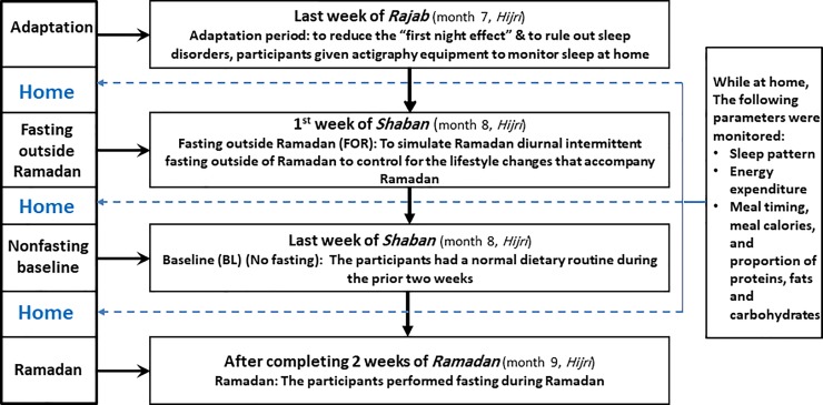
We have used and described the applied protocol in detail in previous studies [19, 20]. Fig 1 illustrates the protocol of the study. The study was conducted following the Hijri (Islamic) calendar, which follows the lunar system in which the year is 11 days shorter than the Gregorian year [21]. The study was conducted during the last week of month number 7 (Rajab), month number 8 (Shaban), and the first 2 weeks of month number 9 (Ramadan) of the Hijri year 1439, which in the Gregorian calendar corresponds to the period from April 9 to May 29, 2018.
Fig 1. Study protocol.
As per the study protocol, the volunteers reported to the laboratory at 18:00 and spent approximately 24 h in the laboratory on 4 occasions as detailed below. During each stay, an overnight polysomnography (PSG) was performed to measure sleep parameters:
The last week of month number 7 (Rajab): The aim of this visit was to ensure adaptation to the environment and equipment used in order to minimize the “first night effect” [22].
The first week of month number 8 (Shaban): Participants were asked to perform diurnal fasting (i.e., abstain from food and drink on the first week of Shaban from dawn to sunset) for one week and were admitted to the laboratory for monitoring on the last day of the week. This fasting protocol was labeled as “Fasting outside Ramadan” (FOR) and aimed to assess the effects of DIF on the measured bio-inflammatory markers outside of Ramadan to eliminate the effect of lifestyle changes that accompany Ramadan [23]. This fasting week was ended 3 weeks before Ramadan to ensure an adequate wash-out period and to avoid any carryover effects.
The last week of month number 8 (Shaban): During the last 3 weeks of the month of Shaban, participants were not fasting. This period was termed “non-fasting baseline (BL).” Participants were admitted to the laboratory for approximately 24 h for monitoring between days 21 and 28 of the month.
After completing 2 weeks of Ramadan (month 9, Hijri): Participants performed diurnal fasting (from dawn to sunset) during the first 2 weeks of Ramadan. This period was called “Ramadan fasting.” Participants were admitted to the laboratory for approximately 24 h for monitoring between days 14 and 17 of the month.
Monitoring at home
During the study period, participants were instructed to follow the same sleep/wake pattern and physical activity. The sleep/wake pattern at home was objectively monitored using actigraphy. Additionally, energy expenditure was monitored via the SenseWear Pro Armband™ (Body Media, Pittsburgh, PA, USA) (see below for details). Meals at home were specified to match the meals in the laboratory for the fasting (FOR and Ramadan) and non-fasting (BL) periods concerning mealtime and meal composition (proportion of proteins, carbohydrates, and fats) and calories, and a nutritionist worked with the participants to achieve that goal. A daily checklist was completed by the participants to ensure abiding by the set protocol.
Protocol in the laboratory
• Fasting and meal-timings protocol
During their stay in the laboratory, participants performed fasting from dawn to sunset. During non-fasting BL, participants received 3 meals; dinner at 20:30, breakfast at 07:15, and lunch at 12:00 (mid-day). During FOR and Ramadan, a light meal was served at sunset to break the fast, followed by a dinner meal at 21:00 and a pre-dawn (Suhur) meal 30 min before dawn time. During the study period, sunset and dawn times ranged from 18:23–18:35 and 04:02–03:35, respectively. A fixed caloric intake (35 Kcal/kg/24 h) and a fixed proportion of proteins, fats, and carbohydrates were served.
• Light intensity in the laboratory
From 18:00 until bedtime, the light level was maintained at 50 lux. During the pre-dawn meal, the light was kept < 30 lux [24]. From bedtime until rising time (PSG recording), all lights were switched off and light intensity was maintained at < 1 lux. Light intensity was determined using a Spectral Star Light Meter LX-1 (Japan).
• Energy expenditure in the laboratory
Energy expenditure in the laboratory was monitored using the SenseWear Pro Armband™ (Body Media, Pittsburgh, PA, USA). The armband is a portable device worn around the right arm.
The device has sensors that continuously measure skin temperature, galvanic skin response, heat flux, and movements and uses a validated advanced algorithm to estimate total energy expenditure [25, 26].
• Sleep monitoring in the laboratory
Participants were instructed to avoid napping during the day of admission to the laboratory. During non-fasting BL, bedtime was at 23:00 and rise time at 07:00. During FOR and Ramadan, bedtime was at 23:00. During Ramadan, participants were awakened at 03:05 for the pre-dawn meal, and participants resumed sleep from 03:50 until 07:45. During FOR, participants were awakened at 03:45 for the pre-dawn meal (to account for the shift in dawn time), and participants resumed sleep from 04:30 until 07:45.
Sleep monitoring was performed using Alice 6 diagnostic equipment (Philips/Respironics, Inc., Murrysville, PA, USA). The following parameters were reported in the results: sleep efficiency (the percentage of time spent asleep while in bed); arousal index, which is a measure of sleep disruption reporting on the number of arousals per hour of sleep; and “stage shifts,” which reports on the number of changes in sleep stages from lights out to lights on. Scoring was performed according to the American Academy of Sleep Medicine scoring criteria [27].
Blood samples collection and measurement of cytokine levels
At 20:00, a cannula was placed in a vein in the antecubital fossa of the arm, in order to draw blood without disturbing sleep. Five blood samples were collected from each participant when in the laboratory in the 3 periods: FOR, non-fasting BL, and Ramadan. Blood samples for IL-1β, IL-6, and IL-8 were collected at 22:00, 02:00, 04:00, 06:00, and 11:00 h. After blood samples were collected, they were immediately centrifuged at 4°C and then stored at -70°C.
Levels of interleukins were determined using enzyme-linked immunoassays (DIAsource ImmunoAssays S.A., Ottignies-Louvain-la-Neuve, Belgium). The assay sensitivity for IL-1β was 0.35 pg/mL. The intra-assay and inter-assay coefficients of variation were <2.4% and <5%, respectively. The assay sensitivity for IL-6 was 2 pg/mL. The intra-assay and inter-assay coefficients of variation were <4.3% and <5.4%, respectively. The assay sensitivity for IL-8 was 1.1 pg/mL. The intra-assay and inter-assay coefficients of variation were <3.6% and <13.1%, respectively. All measurement procedures were performed according to the manufacturer’s instructions.
Serum glucose level was measured for all participants at 15:30 when in the laboratory.
Statistical analysis
Continuous variables are presented in the text, tables, and figures as the mean ± standard deviation. Metabolic equivalents (METs) were used to express energy expenditure during day and night. Average METs were calculated to obtain an overall hourly average for each day.
Cosinor analysis for all measured cytokines was performed using a 24-h cosinor rhythmometry model
where M = mesor; A = amplitude; Ø = acrophase, and T = 24 h, to obtain the best estimates of the acrophase for IL-1β, IL-6, and IL-8 [28]. The model was executed for the data of each participant during the 3 periods. Comparisons of the non-fasting BL, FOR, and Ramadan fasting groups were performed using repeated measure analysis of variance (ANOVA). Repeated measures ANOVA on ranks test was used if the normality test failed. Post hoc analysis (Holm-Sidak) was used to estimate the significance of differences between individual groups if normality test was achieved and psot hoc analysis (Tukey) was sued with the One-way repeated measures ANOVA on ranks. Data of cytokines’ levels at each measured time were presenter as Box & Whisker Plot. This plot allows the presentation of lower extreme, lower quartile, median, upper quartile, and upper extreme. The upper part of the box represents the 75th percentile, the horizontal line in the box represents the median, and the lower part of the box represents the 25th percentile. The whiskers (lines) of the box represents the smallest and largest values that are not minor or extreme outlying values.
A p-value of < 0.05 was considered significant. SigmaStat version 3.5 software (Dundas Software LTD., wpcubed GmbH, Germany) was used for the analyses.
Results
The mean age of participants was 25.1 ± 2.5 years and the mean BMI was 23.4 ± 3.5 kg/m2. Body weight remained stable during the 3 study periods (69.3 ± 6.6 kg, 69.6 ± 5.7 kg, and 69.1 ± 6.8 kg for non-fasting BL, FOR, and Ramadan, respectively, p = 0.9). In addition, no significant variations were detected in serum glucose levels while in the laboratory, in the 3 study periods (5.8 ± 0.5, 5.6 ± 0.5, and 5.5 ± 0.6 mmol/L during non-fasting BL, FOR, and Ramadan, respectively, p = 0.4).
Table 1 displays the sleep pattern and energy expenditure at home. There was a significant delay in bedtime and rise time during Ramadan compared to the non-fasting BL and FOR periods. However, there were no significant changes observed in sleep duration between the 3 periods. Additionally, there was a significant reduction in energy expenditure during Ramadan when compared to the BL period. However, no significant changes in energy expenditure were noted during FOR. All participants followed the meal timing and composition protocol during FOR; however, only two of them followed the food protocol during Ramadan.
Table 1. Sleep/wake pattern while at home during the study periods, measured via actigraphy.
| Measured variables | Non-fasting BL | FOR | Ramadan | p-value |
|---|---|---|---|---|
| Mean Bedtime | 00:39 | 01:05 | 02:33* | 0.003 |
| Mean Wake-up time ± SD (in h) | 05:35 | 05:50 | 08:56* | 0.031 |
| nTST (h) | 5.9±1.3 | 5.7±1.4 | 6.3±1.6 | 0.6 |
| nTST + NAP (h) | 6.8±1.1 | 6.7±1.9 | 7.2±2.5 | 0.8 |
| METs | 1.89±0.18 | 1.77±0.19 | 1.71±0.15* | 0.02 |
nTST: nocturnal total sleep time; NAP: daytime naps
*Statistically significant difference between non-fasting BL and Ramadan. No difference observed between non-fasting BL and FOR.
Table 2 presents the measured sleep parameters and energy expenditure while in the laboratory. No differences were observed in sleep parameters reflecting sleep quality while in the laboratory between the 3 study periods, including, total sleep time, sleep efficiency, arousal index, stage shifts, and sleep latency. Sleep efficiency was slightly lower than normal during all periods which might reflect the effect of drawing blood samples with the intravenous cannula in the antecubital fossa 5 times during sleep.
Table 2. Sleep parameters and energy expenditure while in the laboratory during the 3 study periods.
| Measured variables | Non-fasting BL | Fasting outside Ramadan | Ramadan | p-value |
|---|---|---|---|---|
| Sleep parameters during monitoring at the laboratory using polysomnography | ||||
| Total sleep time (min) | 359.5 ± 41.9 | 351.5 ± 49.9 | 341.7 ± 52.3 | 0.7 |
| Sleep efficiency (%) | 79.3 ± 9.1 | 78.1 ± 8.4 | 75.8 ± 10.2 | 0. 6 |
| Sleep latency (min) | 31.3 ± 18.9 | 28.5 ± 16.7 | 30.7 ± 22.4 | 0.9 |
| Arousal index (arousal/hr) | 6.7 ± 3.5 | 7.1 ± 4.7 | 8.2 ± 3.9 | 0.6 |
| Stage shifts | 69.1 ± 19.7 | 72.1 ± 18.3 | 73.4 ± 20.7 | 0.9 |
| Energy expenditure based on the SenseWear Pro Armband | ||||
| METs | 1.5 ± 0.1 | 1.4 ± 0.2 | 1.4 ± 0.1 | 0.2 |
| METs (Day) | 1.5 ± 0.1 | 1.4 ± 0.2 | 1.4 ± 0.2 | 0.3 |
| METs (Night) | 1.4 ± 0.1 | 1.4 ± 0.2 | 1.4 ± 0.1 | 1 |
METs: metabolic equivalents; stage shifts: total number of changes in sleep stage from the sleep-onset to rise time.
No changes were noted in energy expenditure (expressed by METs) between day and night during the 3 monitoring periods in the laboratory.
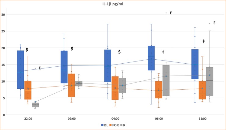
Fig 2 displays the pattern of IL-1β concentrations during non-fasting BL, FOR, and Ramadan. There was a significant decrease in IL-1β levels during FOR compared to the non-fasting BL period at 06:00, 11:00, 22:00, 02:00, and 04:00 (p<0.05). Additionally, there was a significant reduction in IL-1β levels during Ramadan compared to the non-fasting BL period at 22:00, 02:00 and 04:00 (p<0.05).
Fig 2. Circadian pattern of plasma IL-1β concentrations at non-fasting baseline (non-fasting BL), fasting outside Ramadan (FOR), and Ramadan.
Plasma levels were presented as box and whisker plot. The upper part of the box represents the 75th percentile, the horizontal line in the box represents the median, the lower part of the box represents the 25th percentile, and the blue circle represents the mean. The whiskers (lines) of the box represents the smallest and largest values that are not minor or extreme outlying values. $ indicates significance between both Ramadan and fasting outside of Ramadan and baseline measurements (BL), (p < 0.05). ǂ indicates significance between FOR and BL, (p < 0.05). E: extreme outlier.
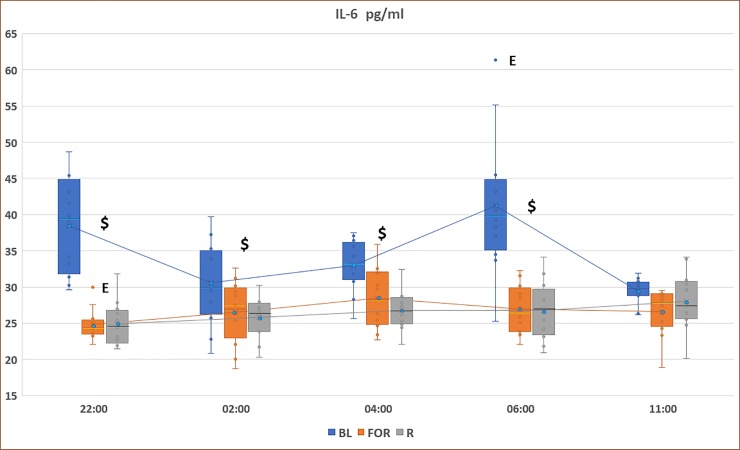
Fig 3 demonstrates the pattern of IL-6 concentrations during non-fasting BL, FOR, and Ramadan. There was a significant reduction in IL-6 levels during FOR and Ramadan compared to the non-fasting BL period at 06:00, 22:00, 02:00, and 04:00 (p<0.05).
Fig 3. Circadian pattern of plasma IL-6 concentrations at non-fasting BL, FOR, and Ramadan.
Plasma levels were presented as box and whisker plot. The upper part of the box represents the 75th percentile, the horizontal line in the box represents the median, the lower part of the box represents the 25th percentile, and the blue circle represents the mean. The whiskers (lines) of the box represents the smallest and largest values that are not minor or extreme outlying values. $ indicates significance between both Ramadan and fasting outside of Ramadan and baseline measurements (BL), (p < 0.05). ǂ indicates significance between FOR and BL, (p < 0.05). E: extreme outlier.
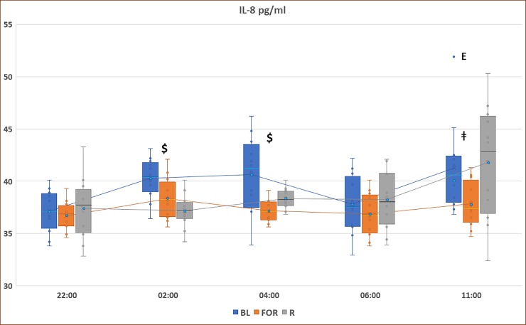
Fig 4 presents the pattern of IL-8 concentrations during non-fasting BL, FOR, and Ramadan. There was a significant reduction in IL-8 levels during FOR compared to the non-fasting BL period at 11:00, 02:00, and 04:00 (p<0.05). With regard to Ramadan, there was a significant reduction in IL-8 levels during Ramadan compared to the non-fasting BL period at 02:00 and 04:00 (p<0.05).
Fig 4. Circadian pattern of plasma IL-8 concentrations at non-fasting BL), FOR, and Ramadan.
Plasma levels were presented as box and whisker plot. The upper part of the box represents the 75th percentile, the horizontal line in the box represents the median, the lower part of the box represents the 25th percentile, and the blue circle represents the mean. The whiskers (lines) of the box represents the smallest and largest values that are not minor or extreme outlying values. $ indicates significance between both Ramadan and fasting outside of Ramadan and baseline measurements (BL), (p < 0.05). ǂ indicates significance between FOR and BL, (p < 0.05). E: extreme outlier.
Table 3 shows the cosinor analysis of IL-1β, IL-6, and IL-8 plasma levels. There were no significant changes in the circadian phase of the cytokines profile as reflected by acrophase. However, the amplitude of IL-6 during Ramadan was significantly lower than the value at BL (p = 0.038).
Table 3. Cosinor analysis summary of IL-1β, IL-6, and IL-8 circadian rhythms during baseline, baseline fasting, and Ramadan.
| BL | FOR | Ramadan | p-value | |
|---|---|---|---|---|
| IL-1β Circadian Rhythm | ||||
| Amplitude | 6.7 ± 5.3 | 8.3 ± 7 | 6.9 ± 2.9 | 0.941 |
| Acrophase | 4.1 ± 2.8 | 4 ± 2.5 | 4.1 ± 0.7 | 1.000 |
| IL-6 Circadian Rhythm | ||||
| Amplitude | 12 ± 7 | 4.4 ± 4.4 | 2 ± 0.6 | 0.038* |
| Acrophase | 1.7 ± 1.1 | 3.8 ± 2.9 | 3.9 ± 0.5 | 0.203 |
| IL-8 Circadian Rhythm | ||||
| Amplitude | 2.8 ± 1.7 | 2.3 ± 1.9 | 4.1 ± 0.5 | 0.242 |
| Acrophase | 4.8 ± 1.9 | 3.1 ± 3.3 | 3.6 ± 0.3 | 0.563 |
Tukey Post Hoc multiple comparison
* BL vs. Ramadan (p = 0.038)
BL: Baseline data; FOR: fasting outside of Ramadan
Discussion
This is the first study to assess the effects of DIF on the levels of selected proinflammatory cytokines during and outside of Ramadan using several readings at different times of day and night (24 h) to control for circadian rhythm effect. Additionally, this is the first study to objectively monitor both sleep and energy expenditure while at home as both factors may influence the levels of measured inflammatory cytokines. No changes were observed during FOR compared to BL in sleep and energy expenditure, which make the results during FOR more reflective of the true effect of DIF. However, during Ramadan, it is difficult to control for sleep pattern, physical activity, and meals while at home as working time and hours change and get shorter, and the whole system gets delayed in the whole society [29]. Additionally, during Ramadan, fasting performers stay awake until late at night to perform Ramadan night prayers [21]. The findings of the current study supported our hypothesis that DIF results in reduced levels of proinflammatory cytokines. Our results demonstrated a significant reduction of the levels of cytokines across 24 h during fasting compared to baseline, and particularly those of IL-1β and IL-6 during fasting outside of Ramadan. The greater reduction in cytokine levels during FOR compared to Ramadan suggested that DIF, per se, can lead to reduced proinflammatory cytokines in the absence of Ramadan-associated lifestyle changes.
Previous studies that assessed the effect of DIF on levels of proinflammatory cytokines during Ramadan reported only on the evaluation of a single morning sample of the measured biomarkers, not accounting for the diurnal rhythmicity of cytokine production [12, 30]. Moreover, currently available data have suggested that mealtime and meal composition might affect the levels of the measured inflammatory markers [31, 32]. For example, it has been shown in mice that postprandial IL-1β levels increase in response to a meal [33]. Similar findings have been demonstrated in humans, where an increase in plasma IL-1β levels after intake of a high fat content meal was reported [34]. Therefore, collecting a single sample in the early morning after a heavy pre-dawn meal may influence the levels of the measured markers. Additionally, shifting mealtimes to the night during DIF might have an effect on the biologic clock of the body and its physiological responses [1], and hence may affect the rhythmicity of the inflammatory biomarkers. To overcome this, taking several blood samples around the clock would be the most efficient way to assess the effects of DIF on the levels of serum cytokines. Additionally, all previous studies did not control for factors shown to affect the levels of the measured biomarkers, such as caloric intake [7], sleep duration [8], and energy expenditure [11].
Studies that assessed circadian rhythm and biological clock markers during Ramadan DIF without controlling for the above mentioned lifestyle changes that accompany Ramadan, demonstrated a shift delay in the body circadian rhythm [2]. Recent well-designed studies revealed that the body internal circadian clock influences the levels of cytokines [12]. Moreover, cytokines in human blood have been shown to display circadian rhythms. Therefore, diurnal rhythmicity of cytokine secretion has important implications for the timing of blood samples collection. Previous studies that assessed cytokine levels during DIF did not account for possible changes in the biologic clock of volunteers and did not assess the effects of DIF on the circadian pattern of cytokines. In the present study, we controlled for light exposure, as well as bedtime and rise time to avoid changes in the circadian clock of participants. Using the same protocol, we have previously demonstrated that there is no change in the circadian rhythm of melatonin [35]. Additionally, we used a cosinor model to assess the effect of DIF on the circadian pattern of the measured cytokines. The present study demonstrated no significant changes in the circadian phase (acrophase) of the measured cytokines during DIF. However, the amplitude of IL-6 was significantly lower during Ramadan compared to BL, which reflects a more reduction the circadian oscillation of IL-6 after 2 weeks of fasting compared to the reduction after 1 week of fasting (FOR period). This reduction in the amplitude of IL-6 rhythmicity may suggest that, at least in part, longer fasting might have a sustained inhibitory effect on inflammation [36]. Further studies are needed to support this hypothesis.
Weight loss has been proposed as a mechanism for the reduction of proinflammatory cytokine levels during DIF [37]. Therefore, it is important to monitor weight when assessing the impact of DIF on proinflammatory cytokine levels, as weight changes can be a major confounder affecting cytokine levels [38, 39]. In the present study, we enlisted volunteers with normal BMI and monitored their weight during the study period. Hence, weight loss could not be the sole mechanism explaining the reduction of cytokine levels. Additionally, both animal and human studies have suggested that CR results in a decrease in proinflammatory cytokine levels [5, 40]. However, in the present study, we maintained the same caloric intake for volunteers during monitoring in the 3 study periods. Another plausible mechanism for the reduction in proinflammatory cytokine levels during DIF could be through the insulin-like growth factor 1 (IGF-1), which has been shown to augment proinflammatory cytokine levels [41]. Fasting has been reported to lead to decreased IGF-1 levels in animals [42]. A recent study demonstrated a significant reduction in IGF-1 levels during Ramadan DIF compared to baseline [43].
The present study had strengths, as well as limitations. The strength of this study rested in that it was the first study to assess plasma levels and circadian patterns of selected proinflammatory cytokines in volunteers performing DIF during and outside of Ramadan while controlling for several confounders that might affect the levels of the measured cytokines. A good control of confounders was achieved during FOR, and this may explain the better results obtained during FOR. Nevertheless, the current study had also inherent limitations that need to be addressed. All experimental studies that have performed several physiological measurements under controlled conditions in a restricted time period (Ramadan month), had only a relatively small number of participants included. This has been an inherent limitation of all previous studies that used a similar design and objectively assessed physiological parameters under controlled conditions [9, 19, 44–47]. Another point that needs to be addressed is the fact that this study was conducted on healthy, non-obese young males; hence the current results cannot be extrapolated to females, different age groups or obese subjects with comorbidities. Females were not included in this study, because from a religious point of view, they have to break the fast during the menstrual cycle. Lastly, as the study group was enrolled from within a university, the studied sample may not represent the general public because University employees are more likely to have higher education than the general public.
In summary, in young healthy male volunteers on predetermined caloric intake, set proportions of carbohydrate, fat, and protein consumption and similar light exposure and sleep schedules during baseline and DIF exposure, DIF resulted in significantly decreased plasma levels of cytokines (IL-1β, IL-6, and IL-8), particularly IL-1β and IL-6, across 24 h. However, DIF had no effect on the circadian patterns of the measured cytokines as shown by cosinor analysis. The current findings suggest that DIF might aid in overall health promotion by reducing the levels of proinflammatory cytokines.
Future studies should examine the timely persistence of this reduction in basal cytokines expression levels after Ramadan and whether DIF has similar anti-inflammatory effects under conditions of chronic inflammation, such as obesity and other chronic inflammatory disorders.
Acknowledgments
We thank the participants who despite their busy schedules agreed to participate in this project. Without their dedication and involvement, this research project would not have been executed.
Data Availability
All relevant data are within the paper.
Funding Statement
This study was supported by a grant from the Researchers Supporting Project number (RSP-2019/51). King Saud University, Riyadh Saudi Arabia. The grant was obtained by Prof. Ahmed S. BaHammam. The study sponsors played no role in the study design, the collection, analysis, or interpretation of data, writing the manuscript, or the decision to submit the manuscript.
References
- 1.Almeneessier AS, Pandi-Perumal SR, BaHammam AS. Intermittent fasting, insufficient sleep, and circadian rhythm: Interaction and effects on the cardiometabolic system. Curr Sleep Med Rep. 2018;4:179–95. 10.1007/s40675-018-0124-5 [DOI] [Google Scholar]
- 2.Almeneessier AS, BaHammam AS. How does diurnal intermittent fasting impact sleep, daytime sleepiness, and markers of the biological clock? Current insights. Nat Sci Sleep. 2018;10:439–52. Epub 2018/12/24. 10.2147/NSS.S165637 [DOI] [PMC free article] [PubMed] [Google Scholar]
- 3.Mindikoglu AL, Opekun AR, Gagan SK, Devaraj S. Impact of Time-Restricted Feeding and Dawn-to-Sunset Fasting on Circadian Rhythm, Obesity, Metabolic Syndrome, and Nonalcoholic Fatty Liver Disease. Gastroenterol Res Pract. 2017;2017:3932491 Epub 2018/01/20. 10.1155/2017/3932491 [DOI] [PMC free article] [PubMed] [Google Scholar]
- 4.Yu SL, Kuan WP, Wong CK, Li EK, Tam LS. Immunopathological roles of cytokines, chemokines, signaling molecules, and pattern-recognition receptors in systemic lupus erythematosus. Clin Dev Immunol. 2012;2012:715190 Epub 2012/02/09. 10.1155/2012/715190 [DOI] [PMC free article] [PubMed] [Google Scholar]
- 5.Gonzalez O, Tobia C, Ebersole J, Novak MJ. Caloric restriction and chronic inflammatory diseases. Oral Dis. 2012;18(1):16–31. Epub 2011/07/14. 10.1111/j.1601-0825.2011.01830.x [DOI] [PMC free article] [PubMed] [Google Scholar]
- 6.Faris MAE, A.Jahrami H, Obaideen AA, Madkour MI. Impact of diurnal intermittent fasting during Ramadan on inflammatory and oxidative stress markers in healthy people: Systematic review and meta-analysis. J Nutrition Intermediary Metabolism. 2019;15:18–26. 10.1016/j.jnim.2018.11.005 [DOI] [Google Scholar]
- 7.Kiecolt-Glaser JK. Stress, food, and inflammation: psychoneuroimmunology and nutrition at the cutting edge. Psychosomatic medicine. 2010;72(4):365–9. Epub 2010/04/23. 10.1097/PSY.0b013e3181dbf489 [DOI] [PMC free article] [PubMed] [Google Scholar]
- 8.Buxton OM, Broussard JL, Zahl AK, Hall M. Effects of Sleep Defi ciency on Hormones, Cytokines, and Metabolism In: Redline S, Berger RA, editors. Impact of Sleep and Sleep Disturbances on Obesity and Cancer, Energy Balance and Cancer. New York: Springer Science+Business Media; 2014. p. 25–50. [Google Scholar]
- 9.BaHammam A, Alrajeh M, Albabtain M, Bahammam S, Sharif M. Circadian pattern of sleep, energy expenditure, and body temperature of young healthy men during the intermittent fasting of Ramadan. Appetite. 2010;54(2):426–9. Epub 2010/01/27. S0195-6663(10)00034-6 [pii] 10.1016/j.appet.2010.01.011 . [DOI] [PubMed] [Google Scholar]
- 10.Lange T, Dimitrov S, Born J. Effects of sleep and circadian rhythm on the human immune system. Ann N Y Acad Sci. 2010;1193:48–59. Epub 2010/04/20. 10.1111/j.1749-6632.2009.05300.x . [DOI] [PubMed] [Google Scholar]
- 11.Smart NA, Steele M. The effect of physical training on systemic proinflammatory cytokine expression in heart failure patients: a systematic review. Congest Heart Fail. 2011;17(3):110–4. Epub 2011/05/26. 10.1111/j.1751-7133.2011.00217.x . [DOI] [PubMed] [Google Scholar]
- 12.Nakao A. Temporal regulation of cytokines by the circadian clock. J Immunol Res. 2014;2014:614529 Epub 2014/05/09. 10.1155/2014/614529 [DOI] [PMC free article] [PubMed] [Google Scholar]
- 13.Dominguez Rodriguez A, Abreu Gonzalez P, Garcia MJ, de la Rosa A, Vargas M, Marrero F. [Circadian variations in proinflammatory cytokine concentrations in acute myocardial infarction]. Rev Esp Cardiol. 2003;56(6):555–60. Epub 2003/06/05. 10.1016/s0300-8932(03)76916-4 . [DOI] [PubMed] [Google Scholar]
- 14.Leon-Pedroza JI, Gonzalez-Tapia LA, del Olmo-Gil E, Castellanos-Rodriguez D, Escobedo G, Gonzalez-Chavez A. [Low-grade systemic inflammation and the development of metabolic diseases: from the molecular evidence to the clinical practice]. Cir Cir. 2015;83(6):543–51. Epub 2015/07/15. 10.1016/j.circir.2015.05.041 . [DOI] [PubMed] [Google Scholar]
- 15.Dinarello CA. Overview of the IL-1 family in innate inflammation and acquired immunity. Immunol Rev. 2018;281(1):8–27. Epub 2017/12/17. 10.1111/imr.12621 [DOI] [PMC free article] [PubMed] [Google Scholar]
- 16.Tanaka T, Narazaki M, Kishimoto T. IL-6 in inflammation, immunity, and disease. Cold Spring Harb Perspect Biol. 2014;6(10):a016295 Epub 2014/09/06. 10.1101/cshperspect.a016295 [DOI] [PMC free article] [PubMed] [Google Scholar]
- 17.Jiang WG, Sanders AJ, Ruge F, Harding KG. Influence of interleukin-8 (IL-8) and IL-8 receptors on the migration of human keratinocytes, the role of PLC-gamma and potential clinical implications. Exp Ther Med. 2012;3(2):231–6. Epub 2012/09/13. 10.3892/etm.2011.402 [DOI] [PMC free article] [PubMed] [Google Scholar]
- 18.Bickel M. The role of interleukin-8 in inflammation and mechanisms of regulation. J Periodontol. 1993;64(5 Suppl):456–60. Epub 1993/05/01. . [PubMed] [Google Scholar]
- 19.Alzoghaibi MA, Pandi-Perumal SR, Sharif MM, BaHammam AS. Diurnal intermittent fasting during Ramadan: the effects on leptin and ghrelin levels. PLoS One. 2014;9(3):e92214 Epub 2014/03/19. 10.1371/journal.pone.0092214 [DOI] [PMC free article] [PubMed] [Google Scholar]
- 20.Almeneessier AS, Alzoghaibi M, BaHammam AA, Ibrahim MG, Olaish AH, Nashwan SZ, et al. The effects of diurnal intermittent fasting on the wake-promoting neurotransmitter orexin-A. Ann Thorac Med. 2018;13(1):48–54. Epub 2018/02/02. 10.4103/atm.ATM_181_17 [DOI] [PMC free article] [PubMed] [Google Scholar]
- 21.Bahammam A. Does Ramadan fasting affect sleep? Int J Clin Pract. 2006;60(12):1631–7. Epub 2006/05/04. IJCP811 [pii] 10.1111/j.1742-1241.2005.00811.x . [DOI] [PubMed] [Google Scholar]
- 22.Agnew HW Jr., Webb WB, Williams RL. The first night effect: an EEG study of sleep. Psychophysiology. 1966;2(3):263–6. Epub 1966/01/01. 10.1111/j.1469-8986.1966.tb02650.x . [DOI] [PubMed] [Google Scholar]
- 23.Qasrawi SO, Pandi-Perumal SR, BaHammam AS. The effect of intermittent fasting during Ramadan on sleep, sleepiness, cognitive function, and circadian rhythm. Sleep Breath. 2017;21:577–86. Epub 2017/02/13. 10.1007/s11325-017-1473-x . [DOI] [PubMed] [Google Scholar]
- 24.Cajochen C, Zeitzer JM, Czeisler CA, Dijk DJ. Dose-response relationship for light intensity and ocular and electroencephalographic correlates of human alertness. Behavioural brain research. 2000;115(1):75–83. Epub 2000/09/21. 10.1016/s0166-4328(00)00236-9 . [DOI] [PubMed] [Google Scholar]
- 25.Malavolti M, Pietrobelli A, Dugoni M, Poli M, Romagnoli E, De Cristofaro P, et al. A new device for measuring resting energy expenditure (REE) in healthy subjects. Nutr Metab Cardiovasc Dis. 2007;17:338–43. 10.1016/j.numecd.2005.12.009 [DOI] [PubMed] [Google Scholar]
- 26.Mignault D, St Onge M, Karelis AD, Allison DB, Rabasa-Lhoret R. Evaluation of the portable health wear armband, a device to measure total daily energy expenditure in free-living type 2 diabetic individuals. Diabetes Care. 2005;28:225–7. [DOI] [PubMed] [Google Scholar]
- 27.Berry RB, Albertario CL, Harding SM, Lloyd RM, Plante DT, Quan SF, et al. The AASM Manual for the Scoring of Sleep and Associated Events: Rules, Terminology and Technical Specifications, Version 2.5. www.aasmnet.org, Darien, Illinois: American Academy of Sleep Medicine, 2018. 2018. [Google Scholar]
- 28.Nelson W, Tong YL, Lee JK, Halberg F. Methods for cosinor-rhythmometry. Chronobiologia. 1979;6(4):305–23. Epub 1979/10/01. . [PubMed] [Google Scholar]
- 29.BaHammam A. Assessment of sleep patterns, daytime sleepiness, and chronotype during Ramadan in fasting and nonfasting individuals. Saudi Med J. 2005;26(4):616–22. Epub 2005/05/19. . [PubMed] [Google Scholar]
- 30.Petrovsky N, Harrison LC. The chronobiology of human cytokine production. Int Rev Immunol. 1998;16(5–6):635–49. Epub 1998/07/01. 10.3109/08830189809043012 . [DOI] [PubMed] [Google Scholar]
- 31.Emerson SR, Kurti SP, Harms CA, Haub MD, Melgarejo T, Logan C, et al. Magnitude and Timing of the Postprandial Inflammatory Response to a High-Fat Meal in Healthy Adults: A Systematic Review. Adv Nutr. 2017;8(2):213–25. Epub 2017/03/17. 10.3945/an.116.014431 [DOI] [PMC free article] [PubMed] [Google Scholar]
- 32.Manning PJ, Sutherland WH, McGrath MM, de Jong SA, Walker RJ, Williams MJ. Postprandial cytokine concentrations and meal composition in obese and lean women. Obesity (Silver Spring). 2008;16(9):2046–52. Epub 2009/02/03. 10.1038/oby.2008.334 . [DOI] [PubMed] [Google Scholar]
- 33.Dror E, Dalmas E, Meier DT, Wueest S, Thevenet J, Thienel C, et al. Postprandial macrophage-derived IL-1beta stimulates insulin, and both synergistically promote glucose disposal and inflammation. Nat Immunol. 2017;18(3):283–92. Epub 2017/01/17. 10.1038/ni.3659 . [DOI] [PubMed] [Google Scholar]
- 34.Devaraj S, Wang-Polagruto J, Polagruto J, Keen CL, Jialal I. High-fat, energy-dense, fast-food-style breakfast results in an increase in oxidative stress in metabolic syndrome. Metabolism. 2008;57(6):867–70. Epub 2008/05/27. 10.1016/j.metabol.2008.02.016 [DOI] [PMC free article] [PubMed] [Google Scholar]
- 35.Almeneessier AS, Bahammam AS, Sharif MM, Bahammam SA, Nashwan SZ, Pandi Perumal SR, et al. The influence of intermittent fasting on the circadian pattern of melatonin while controlling for caloric intake, energy expenditure, light exposure, and sleep schedules: A preliminary report. Ann Thorac Med. 2017;12(3):183–90. Epub 2017/08/16. 10.4103/atm.ATM_15_17 [DOI] [PMC free article] [PubMed] [Google Scholar]
- 36.Vgontzas AN, Bixler EO, Lin HM, Prolo P, Trakada G, Chrousos GP. IL-6 and its circadian secretion in humans. Neuroimmunomodulation. 2005;12(3):131–40. Epub 2005/05/21. 10.1159/000084844 . [DOI] [PubMed] [Google Scholar]
- 37.Chae JS, Paik JK, Kang R, Kim M, Choi Y, Lee SH, et al. Mild weight loss reduces inflammatory cytokines, leukocyte count, and oxidative stress in overweight and moderately obese participants treated for 3 years with dietary modification. Nutr Res. 2013;33(3):195–203. Epub 2013/03/20. 10.1016/j.nutres.2013.01.005 . [DOI] [PubMed] [Google Scholar]
- 38.Fernandez-Sanchez A, Madrigal-Santillan E, Bautista M, Esquivel-Soto J, Morales-Gonzalez A, Esquivel-Chirino C, et al. Inflammation, oxidative stress, and obesity. Int J Mol Sci. 2011;12(5):3117–32. Epub 2011/06/21. 10.3390/ijms12053117 [DOI] [PMC free article] [PubMed] [Google Scholar]
- 39.Sadeghirad B, Motaghipisheh S, Kolahdooz F, Zahedi MJ, Haghdoost AA. Islamic fasting and weight loss: a systematic review and meta-analysis. Public Health Nutr. 2014;17(2):396–406. Epub 2012/11/28. 10.1017/S1368980012005046 . [DOI] [PMC free article] [PubMed] [Google Scholar]
- 40.Dogan S, Ray A, Cleary MP. The influence of different calorie restriction protocols on serum pro-inflammatory cytokines, adipokines and IGF-I levels in female C57BL6 mice: short term and long term diet effects. Meta Gene. 2017;12:22–32. Epub 2017/04/05. 10.1016/j.mgene.2016.12.013 [DOI] [PMC free article] [PubMed] [Google Scholar]
- 41.Che W, Lerner-Marmarosh N, Huang Q, Osawa M, Ohta S, Yoshizumi M, et al. Insulin-like growth factor-1 enhances inflammatory responses in endothelial cells: role of Gab1 and MEKK3 in TNF-alpha-induced c-Jun and NF-kappaB activation and adhesion molecule expression. Circ Res. 2002;90(11):1222–30. Epub 2002/06/18. 10.1161/01.res.0000021127.83364.7d . [DOI] [PubMed] [Google Scholar]
- 42.Lee C, Safdie FM, Raffaghello L, Wei M, Madia F, Parrella E, et al. Reduced levels of IGF-I mediate differential protection of normal and cancer cells in response to fasting and improve chemotherapeutic index. Cancer Res. 2010;70(4):1564–72. Epub 2010/02/11. 10.1158/0008-5472.CAN-09-3228 [DOI] [PMC free article] [PubMed] [Google Scholar]
- 43.Rahbar AR, Safavi E, Rooholamini M, Jaafari F, Darvishi S, Rahbar A. Effects of Intermittent Fasting during Ramadan on Insulin-like Growth Factor-1, Interleukin 2, and Lipid Profile in Healthy Muslims. Int J Prev Med. 2019;10:7 Epub 2019/02/19. 10.4103/ijpvm.IJPVM_252_17 [DOI] [PMC free article] [PubMed] [Google Scholar]
- 44.BaHammam A. Effect of fasting during Ramadan on sleep architecture, daytime sleepiness and sleep pattern. Sleep Biol Rhythm. 2004;2:135–43. [Google Scholar]
- 45.Roky R, Chapotot F, Hakkou F, Benchekroun MT, Buguet A. Sleep during Ramadan intermittent fasting. J Sleep Res. 2001;10(4):319–27. Epub 2002/03/21. 269 [pii]. 10.1046/j.1365-2869.2001.00269.x . [DOI] [PubMed] [Google Scholar]
- 46.Bogdan A, Bouchareb B, Touitou Y. Ramadan fasting alters endocrine and neuroendocrine circadian patterns. Meal-time as a synchronizer in humans? Life Sci. 2001;68(14):1607–15. Epub 2001/03/27. S0024320501009663 [pii]. 10.1016/s0024-3205(01)00966-3 . [DOI] [PubMed] [Google Scholar]
- 47.Krauchi K, Cajochen C, Werth E, Wirz-Justice A. Alteration of internal circadian phase relationships after morning versus evening carbohydrate-rich meals in humans. J Biol Rhythms. 2002;17(4):364–76. Epub 2002/08/08. 10.1177/074873040201700409 . [DOI] [PubMed] [Google Scholar]
Associated Data
This section collects any data citations, data availability statements, or supplementary materials included in this article.
Data Availability Statement
All relevant data are within the paper.


