PLOS ONE logoLink to PLOS ONE
. 2020 Jan 24;15(1):e0228170. doi: 10.1371/journal.pone.0228170

Analysis of cholesterol in mouse brain by HPLC with UV detection

María A Paulazo 1, Alejandro O Sodero 1,*
Editor: Tommaso Lomonaco2
PMCID: PMC6980532  PMID: 31978159

Abstract

We describe a sensitive high performance liquid chromatography (HPLC)-based method for the determination of cholesterol in brain tissue. The method does not require the derivatization of the analyte and uses separation and quantification by reversed-phase HPLC coupled to UV detection. Lipids were methanol/chloroform extracted following the method of Bligh and Dyer, and separated using isopropanol/acetonitrile/water (60/30/10, v/v/v) as mobile phase. We observed lineal detection in a wide range of concentrations, from 62.5 to 2000 ng/μL, and were able to detect a significant increase in the brain cholesterol levels between postnatal days 2 and 10 in C57BL6 mice. Based on our validation parameters, we consider this analytical method a useful tool to assess free cholesterol in rodent brain samples and cell cultures.

Introduction

Brain cholesterol represents 23% of the total body cholesterol and is relevant for brain structure and physiology. Optimal amounts of this sterol are essential to sustain important cellular functions, like the permeability of the plasma membrane, the intramembrane diffusion of receptors, the secretion of neurotransmitters, and the synthesis of steroids and myelin. Myelin is a specialized membrane with a high percentage of lipids (∼70%) compared to any other cell membrane [1]. Cholesterol is particularly abundant in the myelin, representing approximately 30% of the total lipids. The high proportion of cholesterol and other lipids contribute to the efficient and rapid propagation of the action potentials in neurons. The production of myelin by the glial cells is absolutely dependent on the cholesterol biosynthesis, which involves more than 20 enzymes that convert acetate into cholesterol [2, 3, 4, 5].

From the metabolic point of view, brain cholesterol is compartmentalized in two main pools: the stable (∼70%), found in the myelin membranes of the white matter, and the active (∼30%), present in the plasma and subcellular membranes of neurons and glial cells of the gray matter. The concentration of cholesterol in the stable pool is high (∼40 mg/g tissue) and reflects the dense packing of multiple opposed lipid bilayers in the myelin sheath, while the concentration in the active pool is lower (∼8 mg/g tissue) [6].

Essentially all the brain cholesterol derives from the de novo synthesis, because this sterol cannot cross the blood-brain barrier. In adult mammals, brain cholesterol is maintained almost constant because the rates of synthesis and catabolism are similar [6]. Cholesterol is removed from the brain by catabolism to 24S-hydroxycholesterol, that crosses the blood brain barrier and is transported via the circulation to the liver for further metabolism [7].

A relevant observation is that rodents, different to humans, myelinate essentially after birth [8]. As a consequence, in the first two weeks of life, there is a significant increase in the mRNA levels of the catabolic enzyme cholesterol 24-hydroxylase (CYP46A1), that is paralleled by higher amounts of 24S-hydroxycholesterol in brain [9]. In addition, in the developing fetus, after the closure of the blood-brain barrier, and in the very young animal, desmosterol levels are high, indicating a rapid de novo synthesis of cholesterol [10].

Here, we developed a novel and reliable method to quantify cholesterol by HPLC with UV detection, after lipid extraction with methanol/chloroform [11]. Because it has been demonstrated that cholesterol rapidly increases in the mouse brain over the first days of postnatal life due to the myelination process [8], we control our quantification method measuring the cholesterol levels in the mouse brain at postnatal days 2 (P2) and 10 (P10). In agreement with previous reports [12, 13], we observed a significant increase of brain cholesterol in the first days of postnatal life.

Materials and methods

2.1. Ethics statement

C57BL6 newborn mice were obtained from females grown in our animal facility. The sacrifice of the pups was done following the Guide for the Care and Use of Laboratory Animals (NRC, USA) and local regulations of the ANMAT (Argentina). The experimental protocol was approved by the Comité Institucional de Cuidado y Uso de Animales de Laboratorio (CICUAL-BIOMED 005–2019).

2.2. Chemicals

Isopropanol and acetonitrile, used for the preparation of the mobile phase, were HPLC grade (J.T. Baker). Ultrapure water was obtained using a Milli-Q filter system. Methanol and chloroform, utilized in the purification of lipids, were analytical grade. Cholesterol was from Sigma.

2.3. Samples and lipid extraction

Mouse brains were removed and placed in ice-cold HBSS without Ca2+/Mg2+. The whole tissue was dissociated in 800 μL of acid saline solution (ASS, 0.9% NaCl in 15 mM HCl) using the pipette tip. After the dissociation, 2 mL of methanol/1 mL of chloroform were added and the sample was vortexed for 20 seconds. Then, 1 mL of chloroform/1 mL of ASS were added, and the sample was vortexed, incubated for 10 minutes in ice and centrifuged at 200g for 5 minutes at 4°C. The upper phase was discharged and the lower phase was mixed with 2 mL of methanol/1.8 mL of ASS. The sample was vortexed, incubated in ice for 10 minutes and centrifuged at 200g for 5 minutes at 4°C. Finally, the lower chloroform phase was dried out at 40°C using a speed-vac concentrator (Savant SPD121P, Thermo Scientific).

2.4. Instrumentation

The measurements were performed using a Dionex UltiMate 3000 System (Thermo Scientific) equipped with autosampler and UV detector. The chloroform phase resulting from the methanol/chloroform extraction was vacuum dried, resuspended in mobile phase and filtered through a 45 μm PDVF filter. The autosampler was set to take 50 μL of this solution. The chromatographic separation was achieved using a Phenomenex Luna C18(2) column (150×4.6 mm) packed with 5 μm silica particles. Instrument control, data acquisition and analysis were achieved with the Chromeleon software (v7.2.6, Thermo Scientific).

2.5. Chromatographic separation

The chromatographic separation was performed using a mobile phase containing isopropanol/acetonitrile/water (60/30/10, v/v/v), at a flow rate of 1 mL/min. The column temperature was set at 28°C and the monitoring wavelength at 205 nm. The identification of cholesterol was done considering the full UV spectra and the retention time (RT). Cholesterol showed a RT of 8.4 minutes. To quantify the amount of cholesterol the area under the curve (AUC) was used.

2.6. Calibration curve

The highest standard (2 μg/μL) was prepared by solving 2 mg of cholesterol in 1 mL of ethanol. This solution was then serially diluted in mobile phase to generate the different points of the standard curve.

Results

3.1. Validation of the method

The complete validation of the method was done following widely accepted analytical criteria [14, 15, 16]. The standard addition strategy was used to validate the cholesterol identification. The cholesterol peaks were identified by comparison of their retention times with those of the standards (Figs 1 and 2). The evidence that the shape and position of the cholesterol peaks were not affected by the brain matrix suggested the absence of any apparent matrix effect. This was confirmed by comparing the slopes of calibration curves made by spiking different amounts of cholesterol either in mobile phase or matrix, which were 7.82 ± 1.06 and 8.06 ± 1.59, respectively. These slopes did not differ significantly (P = 0.80; Student’s t-test), confirming the absence of any matrix effect.

Fig 1. Chromatogram of the cholesterol standard at 62.5 ng/μL (LOQ).
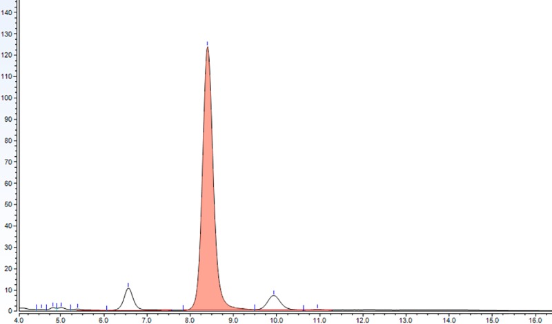
Fig 1

The cholesterol peak (coral colored) shows a characteristic RT of 8.4 minutes.

Fig 2. Representative chromatogram showing the cholesterol detection in brain tissue (coral peak).
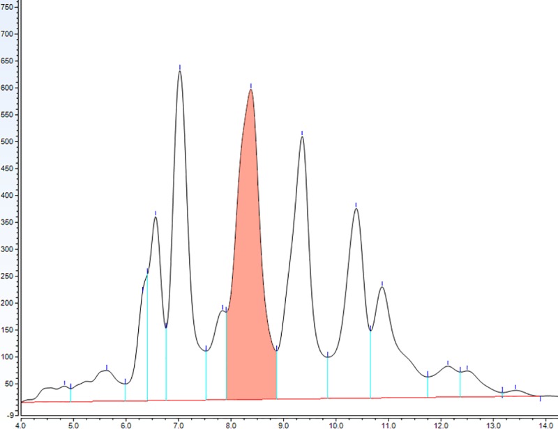
Fig 2

The dried lipids obtained after the methanol/chloroform purification were solved in 1 mL of mobile phase and 50 μL of this solution were injected for analysis.

The limit of detection (LOD) was determined as the concentration that produced a detector signal that could be clearly distinguished from the baseline (2 times larger than the baseline), being in our case 15 ng/μL. The limit of quantification (LOQ) was determined injecting decreasing concentrations of the analyte until the signal to noise ratio was 9:1. For our method the LOQ was 62.5 ng/μL.

The linearity of the method was tested using a 6-point calibration curve. At each concentration at least 3 replicates were assayed. The validation parameters of all fitted calibration curves were satisfactory. The average calibration curve used to determine the amount of brain cholesterol showed a R2 = 0.9981 (Fig 3).

Fig 3. Calibration curve used to determine the amount of cholesterol in the experimental samples.
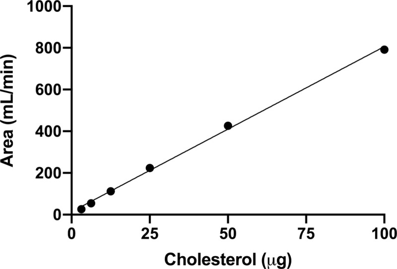
Fig 3

The linear regression showed very good correlation (y = 7.89 x + 13.47; R2 = 0.9981).

Intra- and inter-day repeatability were estimated in triplicates with cholesterol standard solutions of 62.5, 125, 250, 500 and 1000 ng/μL. All the relative standard deviations (RSD) were below 5% (Table 1), indicating that the quantification method has very good precision.

Table 1. Intra- and inter-day precision of the cholesterol determination.

The relative standard deviation (RSD) at five concentration levels of the cholesterol standard is shown.

Concentration level (ng/μL) Intra-day RSD (n = 3) Inter-day RSD (n = 3)
62.5 2.3% 3.6%
125 1.4% 2.3%
250 0.4% 2.1%
500 4.9% 2.5%
1000 4.2% 4.1%

Accuracy was determined by spiking brain samples with known amounts of cholesterol, and expressed as the percentage of analyte recovery, calculated as follows:

Recovery(%)=[(measuredamountendogenouscontent)/(addedamount)]×100

Our method showed a satisfactory analyte recovery of 86 ± 11% (n = 3).

In addition, to evaluate the robustness of the method we analyzed two variables that may impact its performance: the monitoring wavelength and the column temperature. The cholesterol RT stayed constant when we simultaneously monitored the signal at two different wavelengths, 205 and 208 nm. We decided to set 205 nm as the optimal wavelength because the obtained AUC was slightly higher than the AUC at 208 nm. Setting the column temperature either at 22°C or 28°C did not have any impact on the RT or AUC.

3.2. Application of the method to brain samples

After the analytical validation of the method, we measured the cholesterol levels in brains of C57BL6 mice at postnatal days 2 (P2) and 10 (P10). In this interval of the postnatal life, the amount of cholesterol increased from 3.7 ± 0.4 to 6.2 ± 0.7 μg/mg of wet tissue (p < 0.05, Student’s t-test; Fig 4). This 68% increase is in agreement with previous findings showing a higher cholesterol biosynthesis after birth in mouse and rats [6].

Fig 4. Cholesterol concentrations in whole brains of C57BL6 mice at postnatal days 2 (P2) and 10 (P10).
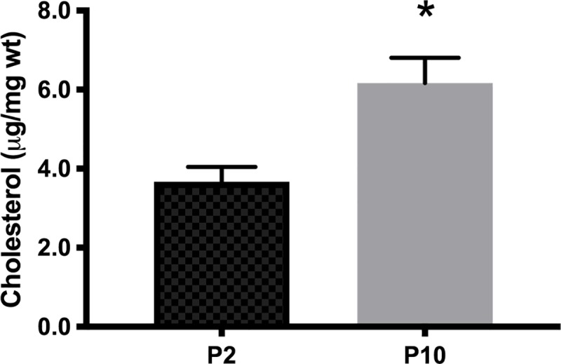
Fig 4

Data are expressed as μg of cholesterol/mg of wet tissue (μg/mg wt). Bars represent mean ± SEM (n = 3); * p < 0.05, Student’s t-test.

Discussion

In mice, the levels of brain cholesterol progressively increase from birth to 3 months of age, reaching approximately 150 micrograms per milligram of wet tissue (see Table 2). In the present work, using HPLC with UV detection, we report similar cholesterol amounts in the early postnatal mouse brain (0–10 micrograms per milligram of wet tissue). In addition, we found a significant increase in the levels of brain cholesterol in 10 days-old mice, compared to 2 days-old mice, most likely due to the higher cholesterol biosynthesis required for myelin formation in this early postnatal period.

Table 2. Comparison of brain cholesterol levels determined by different methodologies.

The reported cholesterol concentrations in early life are similar to the amounts measured by HPLC with UV detection in this work.

Report Mouse age Quantification method Measured cholesterol (μg/mg wet tissue)
Marcos J. et al. [12] 1 day GC-MS 2.5
Marcos J. et al. [12] 14 days GC-MS 6
Meljon A. et al. [13] 1 day LC-MS 2.2
Meljon A. et al. [13] 15 weeks LC-MS 16
Lu F. et al. [24] 9 days Enzymatic 8.5
Nunes V.S. et al. [25] 12 weeks GC-MS 120

The detection of brain cholesterol changes has relevance to understand the neurobiology of several disorders associated to dysfunctions in the homeostasis of this lipid [17, 18, 19, 20, 21]. Traditionally, cholesterol has been quantified in different brain preparations by the cholesterol oxidase method, coupled to colorimetric or fluorometric detection, LC/MS or GC/MS (see Table 3). Here, we describe a different way to analyze free cholesterol in brain samples from which lipids are extracted using methanol/chloroform [11]. Compared with a previously reported method [22], we found out higher signals monitoring the absorbance at 205 nm instead of 200 nm. Another difference is that we do not perform an Abell-Kendall saponification of the sample, followed by lipid extraction with petroleum ether or hexane. Instead, we use methanol and chloroform for lipid extraction, which can be manipulated and disposed in an easier way than petroleum ether or hexane. In addition, we were able to detect low amounts of cholesterol in different cell lines in culture, indicating that our method is also suitable to quantify free cholesterol in small pieces of tissue, like mouse brain structures.

Table 3. Different analytical approaches commonly used to determine cholesterol in biological samples.

The type of detection and the limit of quantification (LOQ) of each methodology are shown.

Method Detection LOQ (ng/μL)
Enzymatic/colorimetric [26] 500 nm (quinoneimine) 250
Enzymatic/fluorometric [27] 585 nm (resorufin) 0.1
HPLC-UV [present work] 205 nm (cholesterol) 62.5
LC-MS m/z < 50
GC-MS m/z < 50

Although GC or LC separation associated to MS detection remain the most sensitive methodologies for cholesterol quantification in tissues and cultured cells [23], the here reported HPLC method with UV detection represents a reliable alternative to measure cholesterol in biological samples.

Data Availability

All relevant data are within the manuscript.

Funding Statement

This work was supported by the Pontifical Catholic University of Argentina. The funder had no role in study design, data collection and analysis, decision to publish, or preparation of the manuscript.

References

  • 1.DeVries GH, Norton WT. The lipid composition of axons from bovine brain. J. Neurochem. 1974; 22(2): 259–64. 10.1111/j.1471-4159.1974.tb11588.x [DOI] [PubMed] [Google Scholar]
  • 2.Schmitt S, Castelvetri LC, Simons M. Metabolism and functions of lipids in myelin. Biochim. Biophys. Acta 2015; 1851(8): 999–1005. 10.1016/j.bbalip.2014.12.016 [DOI] [PubMed] [Google Scholar]
  • 3.Saher G, Simons M. Cholesterol and myelin biogenesis. Subcell. Biochem. 2010; 51: 489–508. 10.1007/978-90-481-8622-8_18 [DOI] [PubMed] [Google Scholar]
  • 4.Saher G, Quintes S, Nave KA. Cholesterol: a novel regulatory role in myelin formation. Neuroscientist 2011; 17(1): 79–93. 10.1177/1073858410373835 [DOI] [PubMed] [Google Scholar]
  • 5.Saher G, Stumpf SK. Cholesterol in myelin biogenesis and hypomyelinating disorders. Biochim. Biophys. Acta. 2015; 1851(8): 1083–94. 10.1016/j.bbalip.2015.02.010 [DOI] [PubMed] [Google Scholar]
  • 6.Dietschy JM, Turley SD. Thematic review series: brain lipids. Cholesterol metabolism in the central nervous system during early development and in the mature animal. J. Lipid Res. 2004; 45(8): 1375–97. 10.1194/jlr.R400004-JLR200 [DOI] [PubMed] [Google Scholar]
  • 7.Lütjohann D, Breuer O, Ahlborg G, Nennesmo I, Siden A, Diczfalusy U et al. Cholesterol homeostasis in human brain: evidence for an age-dependent flux of 24S-hydroxycholesterol from the brain into the circulation. Proc. Natl. Acad. Sci. USA 1996; 93: 9799–9804. 10.1073/pnas.93.18.9799 [DOI] [PMC free article] [PubMed] [Google Scholar]
  • 8.Norton WT, Poduslo SE. Myelination in rat brain: changes in myelin composition during brain maturation. J. Neurochem. 1973; 21(4): 759–73. 10.1111/j.1471-4159.1973.tb07520.x [DOI] [PubMed] [Google Scholar]
  • 9.Lund EG, Guileyardo JM, Russell DW. cDNAcloning of cholesterol 24-hydroxylase, a mediator of cholesterol homeostasis in the brain. Proc. Natl. Acad. Sci. USA 1999; 96: 7238–7243. 10.1073/pnas.96.13.7238 [DOI] [PMC free article] [PubMed] [Google Scholar]
  • 10.Tint GS, Yu H, Shang Q, Xu G, Patel SB. The use of the Dhcr7 knockout mouse to accurately determine the origin of fetal sterols. J. Lipid Res. 2006; 47: 1535–1541. 10.1194/jlr.M600141-JLR200 [DOI] [PMC free article] [PubMed] [Google Scholar]
  • 11.Bligh EG, Dyer WJ. A rapid method of total lipid extraction and purification. Can. J. Biochem. Physiol. 1959; 37(8): 911–7. 10.1139/o59-099 [DOI] [PubMed] [Google Scholar]
  • 12.Marcos J, Shackleton CH, Buddhikot MM, Porter FD, Watson GL. Cholesterol biosynthesis from birth to adulthood in a mouse model for 7-dehydrosterol reductase deficiency (Smith-Lemli-Opitz syndrome). Steroids 2007; 72(11–12): 802–8. 10.1016/j.steroids.2007.07.002 [DOI] [PMC free article] [PubMed] [Google Scholar]
  • 13.Meljon A, Theofilopoulos S, Shackleton CH, Watson GL, Javitt NB, Knölker HJ et al. Analysis of bioactive oxysterols in newborn mouse brain by LC/MS. J. Lipid Res. 2012; 53(11): 2469–83. 10.1194/jlr.D028233 [DOI] [PMC free article] [PubMed] [Google Scholar]
  • 14.Ghimenti S, Lomonaco T, Onor M, Murgia L, Paolicchi A, Fuoco R et al. Measurement of warfarin in the oral fluid of patients undergoing anticoagulant oral therapy. PLoS One 2011; 6(12): e28182 10.1371/journal.pone.0028182 [DOI] [PMC free article] [PubMed] [Google Scholar]
  • 15.Lomonaco T, Ghimenti S, Piga I, Onor M, Melai B, Fuoco R et al. Determination of total and unbound warfarin and warfarin alcohols in human plasma by high performance liquid chromatography with fluorescence detection. J. Chromatogr. A 2013; 1314: 54–62. 10.1016/j.chroma.2013.08.091 [DOI] [PubMed] [Google Scholar]
  • 16.Lomonaco T, Ghimenti S, Piga I, Biagini D, Onor M, Fuoco R et al. Influence of sampling on the determination of warfarin and warfarin alcohols in oral fluid. PLoS One 2014; 9(12): e114430. [DOI] [PMC free article] [PubMed] [Google Scholar]
  • 17.Martín MG, Pfrieger F, Dotti CG. Cholesterol in brain disease: sometimes determinant and frequently implicated. EMBO Rep. 2014; 15(10): 1036–52. 10.15252/embr.201439225 [DOI] [PMC free article] [PubMed] [Google Scholar]
  • 18.Arenas F, Garcia-Ruiz C, Fernandez-Checa JC. Intracellular cholesterol trafficking and impact in neurodegeneration. Front. Mol. Neurosci. 2017; 10: 382 10.3389/fnmol.2017.00382 [DOI] [PMC free article] [PubMed] [Google Scholar]
  • 19.Czuba E, Steliga A, Lietzau G, Kowiański P. Cholesterol as a modifying agent of the neurovascular unit structure and function under physiological and pathological conditions. Metab. Brain Dis. 2017; 32(4): 935–948. 10.1007/s11011-017-0015-3 [DOI] [PMC free article] [PubMed] [Google Scholar]
  • 20.Chang TY, Yamauchi Y, Hasan MT, Chang C. Cellular cholesterol homeostasis and Alzheimer's disease. J. Lipid Res. 2017; 58(12): 2239–2254. 10.1194/jlr.R075630 [DOI] [PMC free article] [PubMed] [Google Scholar]
  • 21.Valenza M, Cattaneo E. Emerging roles for cholesterol in Huntington's disease. Trends Neurosci. 2011; 34(9): 474–86. 10.1016/j.tins.2011.06.005 [DOI] [PubMed] [Google Scholar]
  • 22.Duncan IW, Culbreth PH, Burtis CA. Determination of free, total, and esterified cholesterol by high-performance liquid chromatography. J. Chromatogr. 1979; 162(3): 281–92. 10.1016/s0378-4347(00)81515-7 [DOI] [PubMed] [Google Scholar]
  • 23.Li LH, Dutkiewicz EP, Huang YC, Zhou HB, Hsu CC. Analytical methods for cholesterol quantification. J. Food Drug Anal. 2017; 27(2): 375–386. [DOI] [PMC free article] [PubMed] [Google Scholar]
  • 24.Lu F, Zhu J, Guo S, Wong BJ, Chehab FF, Ferriero DM, et al. Upregulation of cholesterol 24-hydroxylase following hypoxia-ischemia in neonatal mouse brain. Pediatr. Res. 2018; 83(6): 1218–1227. 10.1038/pr.2018.49 [DOI] [PMC free article] [PubMed] [Google Scholar]
  • 25.Nunes VS, Cazita PM, Catanozi S, Nakandakare ER, Quintão ECR. Decreased content, rate of synthesis and export of cholesterol in the brain of apoE knockout mice. J. Bioenerg. Biomembr. 2018; 50(4): 283–287. 10.1007/s10863-018-9757-9 [DOI] [PubMed] [Google Scholar]
  • 26.Allain CC, Poon LS, Chan CS, Richmond W, Fu PC. Enzymatic determination of total serum cholesterol. Clin. Chem. 1974; 20(4): 470–475. [PubMed] [Google Scholar]
  • 27.Amundson DM, Zhou M. Fluorometric method for the enzymatic determination of cholesterol. J. Biochem. Biophys. Methods 1999; 38(1): 43–52. 10.1016/s0165-022x(98)00036-0 [DOI] [PubMed] [Google Scholar]

Decision Letter 0

Tommaso Lomonaco

4 Nov 2019

PONE-D-19-26047

Analysis of cholesterol in mouse brain by HPLC with UV detection

PLOS ONE

Dear Alejandro O. Sodero,

Thank you for submitting your manuscript to PLOS ONE. After careful consideration, we feel that it has merit but does not fully meet PLOS ONE’s publication criteria as it currently stands. Therefore, we invite you to submit a revised version of the manuscript that addresses the points raised during the review process.

We would appreciate receiving your revised manuscript by 13th December. When you are ready to submit your revision, log on to https://www.editorialmanager.com/pone/ and select the 'Submissions Needing Revision' folder to locate your manuscript file.

If you would like to make changes to your financial disclosure, please include your updated statement in your cover letter.

To enhance the reproducibility of your results, we recommend that if applicable you deposit your laboratory protocols in protocols.io, where a protocol can be assigned its own identifier (DOI) such that it can be cited independently in the future. For instructions see: http://journals.plos.org/plosone/s/submission-guidelines#loc-laboratory-protocols

Please include the following items when submitting your revised manuscript:

  • A rebuttal letter that responds to each point raised by the academic editor and reviewer(s). This letter should be uploaded as separate file and labeled 'Response to Reviewers'.

  • A marked-up copy of your manuscript that highlights changes made to the original version. This file should be uploaded as separate file and labeled 'Revised Manuscript with Track Changes'.

  • An unmarked version of your revised paper without tracked changes. This file should be uploaded as separate file and labeled 'Manuscript'.

Please note while forming your response, if your article is accepted, you may have the opportunity to make the peer review history publicly available. The record will include editor decision letters (with reviews) and your responses to reviewer comments. If eligible, we will contact you to opt in or out.

We look forward to receiving your revised manuscript.

Kind regards,

Tommaso Lomonaco, Ph.D

Academic Editor

PLOS ONE

Journal Requirements:

1. When submitting your revision, we need you to address these additional requirements. Please ensure that your manuscript meets PLOS ONE's style requirements, including those for file naming. The PLOS ONE style templates can be found at http://www.journals.plos.org/plosone/s/file?id=wjVg/PLOSOne_formatting_sample_main_body.pdf and http://www.journals.plos.org/plosone/s/file?id=ba62/PLOSOne_formatting_sample_title_authors_affiliations.pdf

2. To comply with PLOS ONE submissions requirements, please provide methods of sacrifice in the Methods section of your manuscript.

Additional Editor Comments:

Dear Authors,

the submitted paper requires major revisions in order to address all the questions and suggestions proposed by the reviewers. In particular all the analytical figures of merit of the developed method must be included in the revised version of the paper.

Please, include specific quality parameters as recovery, matrix effect, inter- and intra-day recovery and precision, LOD and LOQ and stability. I suggest to include a table in order to highlight the main differences with other analytical approached already published in the literature. The authors should discuss in detail the main advantages of the proposed method. Moreover, I suggest to consider the following papers as example for the method validation and include them in the references.

DOI: 10.1371/journal.pone.0028182

DOI: 10.1016/j.chroma.2013.08.091

DOI: 10.1371/journal.pone.0114430

Best regards,

Tommaso Lomonaco

[Note: HTML markup is below. Please do not edit.]

Reviewers' comments:

Reviewer's Responses to Questions

Comments to the Author

1. Is the manuscript technically sound, and do the data support the conclusions?

The manuscript must describe a technically sound piece of scientific research with data that supports the conclusions. Experiments must have been conducted rigorously, with appropriate controls, replication, and sample sizes. The conclusions must be drawn appropriately based on the data presented.

Reviewer #1: Yes

Reviewer #2: No

**********

2. Has the statistical analysis been performed appropriately and rigorously?

Reviewer #1: Yes

Reviewer #2: No

**********

3. Have the authors made all data underlying the findings in their manuscript fully available?

The PLOS Data policy requires authors to make all data underlying the findings described in their manuscript fully available without restriction, with rare exception (please refer to the Data Availability Statement in the manuscript PDF file). The data should be provided as part of the manuscript or its supporting information, or deposited to a public repository. For example, in addition to summary statistics, the data points behind means, medians and variance measures should be available. If there are restrictions on publicly sharing data—e.g. participant privacy or use of data from a third party—those must be specified.

Reviewer #1: Yes

Reviewer #2: No

**********

4. Is the manuscript presented in an intelligible fashion and written in standard English?

PLOS ONE does not copyedit accepted manuscripts, so the language in submitted articles must be clear, correct, and unambiguous. Any typographical or grammatical errors should be corrected at revision, so please note any specific errors here.

Reviewer #1: Yes

Reviewer #2: Yes

**********

5. Review Comments to the Author

Please use the space provided to explain your answers to the questions above. You may also include additional comments for the author, including concerns about dual publication, research ethics, or publication ethics. (Please upload your review as an attachment if it exceeds 20,000 characters)

Reviewer #1: In this manuscript, the authors María Alejandra Paulazo & Alejandro Omar Sodero proposed a RP-HPLC-UV method for the analysis of cholesterol in mouse brain. The experimental design and the result are reasonable and reliable.

However, there are some points which need to be further considered.

Major comments:

1. At the end of the introduction (P4), authors need to give the other literature reports of the cholesterol determination by instrumental techniques.

2. As the authors developed and validated a RP-HPLC method for cholesterol, it is suggested to perform and include the other validation parameters like system suitability, specificity, ruggedness, robustness, etc.

3. Provide the data table(s) which includes the chromatographic and validation parameters.

4. Whether the authors cross-checked the peak purity of the cholesterol peak at RT 8.4 in Figure 2? Because as the sample run contains many peaks due to impurities in it, there are all the possibilities that the other impurities peaks may overlapped on it. It can be overruled by examining the peak purity of cholesterol (with the help of chromatographic software).

Minor comments:

1. P2, line 20; Abstract: HPLC to be expanded instead of in line 22.

5. P4, line 64-65, authors are claiming that the proposed method is in agreement with previous reports; they need to provide the literature for the previous reports.

6. P4, line 75, make +2 state of Ca/Mg superscript.

7. P5, line 96, authors claiming temperature was set at 28oC. It is not clear about the temperature of which was set at 28oC. Be clear about it. I think it is the column temperature.

8. P7, line 128, make 2 of R2 superscript.

9. P8, line 158, the sentence after the reference [18], is not correct.

10. P11, line 220, make 2 of R2 superscript.

11. Mention the name and RT on the cholesterol peak in the Figures 1 & 2.

Reviewer #2: HPLC analysis of cholesterol in biological samples had long been reported and the manuscript described a traditional method with limited analytical performance. Additionally, the validation of the method was insufficient. The presented data did not support the claim of "very good precision and accuracy". A recovery of 74% might be too low for cholesterol analysis. The specificity of the method was unknown.

**********

6. PLOS authors have the option to publish the peer review history of their article (what does this mean?). If published, this will include your full peer review and any attached files.

If you choose “no”, your identity will remain anonymous but your review may still be made public.

Do you want your identity to be public for this peer review? For information about this choice, including consent withdrawal, please see our Privacy Policy.

Reviewer #1: Yes: Manjunatha D H

Reviewer #2: No

[NOTE: If reviewer comments were submitted as an attachment file, they will be attached to this email and accessible via the submission site. Please log into your account, locate the manuscript record, and check for the action link "View Attachments". If this link does not appear, there are no attachment files to be viewed.]

While revising your submission, please upload your figure files to the Preflight Analysis and Conversion Engine (PACE) digital diagnostic tool, https://pacev2.apexcovantage.com/. PACE helps ensure that figures meet PLOS requirements. To use PACE, you must first register as a user. Registration is free. Then, login and navigate to the UPLOAD tab, where you will find detailed instructions on how to use the tool. If you encounter any issues or have any questions when using PACE, please email us at figures@plos.org. Please note that Supporting Information files do not need this step.

Attachment

Submitted filename: Comments-pLOS ONE.docx

PLoS One. 2020 Jan 24;15(1):e0228170. doi: 10.1371/journal.pone.0228170.r002

Author response to Decision Letter 0


12 Dec 2019

Dear Editor and reviewers,

We very much appreciate the criticisms and suggestions, which have led to this revised version of our manuscript entitled “Analysis of cholesterol in mouse brain by HPLC with UV detection”.

We have answered point-by-point all the major and minor comments in the document "Response to the reviewers".

Hoping that in the actual conditions our manuscript is suitable for publication,

Best regards,

Dr. Alejandro O. Sodero

Attachment

Submitted filename: Response to reviewers.docx

Decision Letter 1

Tommaso Lomonaco

31 Dec 2019

PONE-D-19-26047R1

Analysis of cholesterol in mouse brain by HPLC with UV detection

PLOS ONE

Dear Dr Alejandro O. Sodero,

Thank you for submitting your manuscript to PLOS ONE. After careful consideration, we feel that it has merit but does not fully meet PLOS ONE’s publication criteria as it currently stands. Therefore, we invite you to submit a revised version of the manuscript that addresses the points raised during the review process.

We would appreciate receiving your revised manuscript by 5th January 2020. When you are ready to submit your revision, log on to https://www.editorialmanager.com/pone/ and select the 'Submissions Needing Revision' folder to locate your manuscript file.

If you would like to make changes to your financial disclosure, please include your updated statement in your cover letter.

To enhance the reproducibility of your results, we recommend that if applicable you deposit your laboratory protocols in protocols.io, where a protocol can be assigned its own identifier (DOI) such that it can be cited independently in the future. For instructions see: http://journals.plos.org/plosone/s/submission-guidelines#loc-laboratory-protocols

Please include the following items when submitting your revised manuscript:

  • A rebuttal letter that responds to each point raised by the academic editor and reviewer(s). This letter should be uploaded as separate file and labeled 'Response to Reviewers'.

  • A marked-up copy of your manuscript that highlights changes made to the original version. This file should be uploaded as separate file and labeled 'Revised Manuscript with Track Changes'.

  • An unmarked version of your revised paper without tracked changes. This file should be uploaded as separate file and labeled 'Manuscript'.

Please note while forming your response, if your article is accepted, you may have the opportunity to make the peer review history publicly available. The record will include editor decision letters (with reviews) and your responses to reviewer comments. If eligible, we will contact you to opt in or out.

We look forward to receiving your revised manuscript.

Kind regards,

Tommaso Lomonaco, Ph.D

Academic Editor

PLOS ONE

Additional Editor Comments (if provided):

Dear Authors, the paper requires minor revision before to be accepted in PlosOne journal. The analytical figure of merits included in the new version of the paper improved the quality of the manuscript. I suggest to check all the digit number in the paper, which should be approximate according to the variability of the analytical method. Moreover, the authors should include a table in the discussion in order to compare different analytical approaches commonly used to determine cholesterol in biological samples. In addition, the following articles should be included in the references with the aim to help other readers to compare the process for method validation.

DOI: 10.1371/journal.pone.0028182

DOI: 10.1016/j.chroma.2013.08.091

DOI: 10.1371/journal.pone.0114430

Best regards,

Tommaso Lomonaco

[Note: HTML markup is below. Please do not edit.]

Reviewers' comments:

Reviewer's Responses to Questions

Comments to the Author

1. If the authors have adequately addressed your comments raised in a previous round of review and you feel that this manuscript is now acceptable for publication, you may indicate that here to bypass the “Comments to the Author” section, enter your conflict of interest statement in the “Confidential to Editor” section, and submit your "Accept" recommendation.

Reviewer #1: All comments have been addressed

**********

2. Is the manuscript technically sound, and do the data support the conclusions?

The manuscript must describe a technically sound piece of scientific research with data that supports the conclusions. Experiments must have been conducted rigorously, with appropriate controls, replication, and sample sizes. The conclusions must be drawn appropriately based on the data presented.

Reviewer #1: Yes

**********

3. Has the statistical analysis been performed appropriately and rigorously?

Reviewer #1: Yes

**********

4. Have the authors made all data underlying the findings in their manuscript fully available?

The PLOS Data policy requires authors to make all data underlying the findings described in their manuscript fully available without restriction, with rare exception (please refer to the Data Availability Statement in the manuscript PDF file). The data should be provided as part of the manuscript or its supporting information, or deposited to a public repository. For example, in addition to summary statistics, the data points behind means, medians and variance measures should be available. If there are restrictions on publicly sharing data—e.g. participant privacy or use of data from a third party—those must be specified.

Reviewer #1: Yes

**********

5. Is the manuscript presented in an intelligible fashion and written in standard English?

PLOS ONE does not copyedit accepted manuscripts, so the language in submitted articles must be clear, correct, and unambiguous. Any typographical or grammatical errors should be corrected at revision, so please note any specific errors here.

Reviewer #1: Yes

**********

6. Review Comments to the Author

Please use the space provided to explain your answers to the questions above. You may also include additional comments for the author, including concerns about dual publication, research ethics, or publication ethics. (Please upload your review as an attachment if it exceeds 20,000 characters)

Reviewer #1: (No Response)

**********

7. PLOS authors have the option to publish the peer review history of their article (what does this mean?). If published, this will include your full peer review and any attached files.

If you choose “no”, your identity will remain anonymous but your review may still be made public.

Do you want your identity to be public for this peer review? For information about this choice, including consent withdrawal, please see our Privacy Policy.

Reviewer #1: Yes: Manjunatha D H

[NOTE: If reviewer comments were submitted as an attachment file, they will be attached to this email and accessible via the submission site. Please log into your account, locate the manuscript record, and check for the action link "View Attachments". If this link does not appear, there are no attachment files to be viewed.]

While revising your submission, please upload your figure files to the Preflight Analysis and Conversion Engine (PACE) digital diagnostic tool, https://pacev2.apexcovantage.com/. PACE helps ensure that figures meet PLOS requirements. To use PACE, you must first register as a user. Registration is free. Then, login and navigate to the UPLOAD tab, where you will find detailed instructions on how to use the tool. If you encounter any issues or have any questions when using PACE, please email us at figures@plos.org. Please note that Supporting Information files do not need this step.

PLoS One. 2020 Jan 24;15(1):e0228170. doi: 10.1371/journal.pone.0228170.r004

Author response to Decision Letter 1


7 Jan 2020

Dear Editor,

It is a pleasure to submit for your consideration the revised version of our manuscript “Analysis of cholesterol in mouse brain by HPLC with UV detection”.

We have addressed your minor comments and modified the previous version accordingly.

Hoping the current version fulfills the criteria for publication.

Best regards,

Dr. Alejandro O. Sodero

Decision Letter 2

Tommaso Lomonaco

9 Jan 2020

Analysis of cholesterol in mouse brain by HPLC with UV detection

PONE-D-19-26047R2

Dear Dr. Alejandro O. Sodero,

We are pleased to inform you that your manuscript has been judged scientifically suitable for publication and will be formally accepted for publication once it complies with all outstanding technical requirements.

Within one week, you will receive an e-mail containing information on the amendments required prior to publication. When all required modifications have been addressed, you will receive a formal acceptance letter and your manuscript will proceed to our production department and be scheduled for publication.

Shortly after the formal acceptance letter is sent, an invoice for payment will follow. To ensure an efficient production and billing process, please log into Editorial Manager at https://www.editorialmanager.com/pone/, click the "Update My Information" link at the top of the page, and update your user information. If you have any billing related questions, please contact our Author Billing department directly at authorbilling@plos.org.

If your institution or institutions have a press office, please notify them about your upcoming paper to enable them to help maximize its impact. If they will be preparing press materials for this manuscript, you must inform our press team as soon as possible and no later than 48 hours after receiving the formal acceptance. Your manuscript will remain under strict press embargo until 2 pm Eastern Time on the date of publication. For more information, please contact onepress@plos.org.

With kind regards,

Tommaso Lomonaco, Ph.D

Academic Editor

PLOS ONE

Additional Editor Comments (optional):

Dear Authors, the paper is available to be published in PloSone.

Best regards.

Tommaso Lomonaco

Reviewers' comments:

Acceptance letter

Tommaso Lomonaco

15 Jan 2020

PONE-D-19-26047R2

Analysis of cholesterol in mouse brain by HPLC with UV detection

Dear Dr. Sodero:

I am pleased to inform you that your manuscript has been deemed suitable for publication in PLOS ONE. Congratulations! Your manuscript is now with our production department.

If your institution or institutions have a press office, please notify them about your upcoming paper at this point, to enable them to help maximize its impact. If they will be preparing press materials for this manuscript, please inform our press team within the next 48 hours. Your manuscript will remain under strict press embargo until 2 pm Eastern Time on the date of publication. For more information please contact onepress@plos.org.

For any other questions or concerns, please email plosone@plos.org.

Thank you for submitting your work to PLOS ONE.

With kind regards,

PLOS ONE Editorial Office Staff

on behalf of

Dr. Tommaso Lomonaco

Academic Editor

PLOS ONE

Associated Data

    This section collects any data citations, data availability statements, or supplementary materials included in this article.

    Supplementary Materials

    Attachment

    Submitted filename: Comments-pLOS ONE.docx

    Attachment

    Submitted filename: Response to reviewers.docx

    Data Availability Statement

    All relevant data are within the manuscript.


    Articles from PLoS ONE are provided here courtesy of PLOS

    RESOURCES