Abstract
Background
Methanol is the second most abundant volatile organic compound in the atmosphere, with the majority produced as a metabolic by-product during plant growth. There is a large disparity between the estimated amount of methanol produced by plants and the amount which escapes to the atmosphere. This may be due to utilisation of methanol by plant-associated methanol-consuming bacteria (methylotrophs). The use of molecular probes has previously been effective in characterising the diversity of methylotrophs within the environment. Here, we developed and applied molecular probes in combination with stable isotope probing to identify the diversity, abundance and activity of methylotrophs in bulk and in plant-associated soils.
Results
Application of probes for methanol dehydrogenase genes (mxaF, xoxF, mdh2) in bulk and plant-associated soils revealed high levels of diversity of methylotrophic bacteria within the bulk soil, including Hyphomicrobium, Methylobacterium and members of the Comamonadaceae. The community of methylotrophic bacteria captured by this sequencing approach changed following plant growth. This shift in methylotrophic diversity was corroborated by identification of the active methylotrophs present in the soils by DNA stable isotope probing using 13C-labelled methanol. Sequencing of the 16S rRNA genes and construction of metagenomes from the 13C-labelled DNA revealed members of the Methylophilaceae as highly abundant and active in all soils examined. There was greater diversity of active members of the Methylophilaceae and Comamonadaceae and of the genus Methylobacterium in plant-associated soils compared to the bulk soil. Incubating growing pea plants in a 13CO2 atmosphere revealed that several genera of methylotrophs, as well as heterotrophic genera within the Actinomycetales, assimilated plant exudates in the pea rhizosphere.
Conclusion
In this study, we show that plant growth has a major impact on both the diversity and the activity of methanol-utilising methylotrophs in the soil environment, and thus, the study contributes significantly to efforts to balance the terrestrial methanol and carbon cycle.
Video abstract
Keywords: Methanol, Rhizosphere, Stable isotope probing, Methylotroph, Methanol dehydrogenase
Introduction
The large amount of carbon released to the soil via the roots of growing plants (1–20% of total photosynthate [1]) has a profound impact on the microbial communities in soil [2]. Root exudates include organic acids, sugars, alcohols, mucilage, sloughed off cells and methanol [3, 4]. Growing and decaying plants account for the majority of methanol produced globally (149 Tg year−1), released following demethylation of pectin in the walls of restructuring plant cells [5]. In the atmosphere, methanol is the second most abundant organic gas (0.1–10 ppb) after methane (1800 ppb) [6], but there is a large disparity between the estimated amount of methanol produced and the amount entering the atmosphere. This suggests that plant-associated methylotrophic microorganisms may be responsible for oxidation of a substantial proportion of the methanol produced by plants before it can escape to the atmosphere [7].
Methanol-oxidising methylotrophs can utilise methanol as a sole source of carbon and energy and are widespread in the terrestrial environment [7]. Methylotrophs detected in soil environments in previous studies typically belong to the Proteobacteria, although others, including Verrucomicrobia, Firmicutes, Flavobacterium and Actinobacteria, have also been detected [8–11]. Previous studies have indicated that methylotrophic bacteria are enriched in the rhizosphere of certain plant species, for example, Methylobacteraceae and Hyphomicrobiaceae in the rhizosphere of Arabidopsis thaliana [12], Methylophilaceae and Comamonadaceae in the pea rhizosphere and Methylophilaceae and Methylocaldum in the wheat rhizosphere [13]. Methanol dehydrogenase genes have been detected in the rhizosphere of rice, grasses, soybeans, cereals and pea plants [14–16], methanol dehydrogenase enzymes have been detected in the rhizosphere soils of oat, wheat and A. thaliana [17], and soils in association with A. thaliana had higher rates of methanol dissimilation than non-plant-associated soils [18]. However, the reasons for changes in the abundance of methylotrophs in the soil in response to plant growth are hard to identify, since many of these methylotrophs can also use multi-carbon compounds, which could be supplied either directly from the plant or from the exudate-induced accelerated breakdown of recalcitrant soil organic matter (SOM) [19].
The oxidation of methanol to formaldehyde requires the enzyme methanol dehydrogenase. There are several methanol dehydrogenases that have been characterised in different classes of methylotrophic organisms, and the most well characterised is the canonical MxaFI [20]. This enzyme is heterotetrameric in structure, with mxaF and mxaI encoding the large and small subunits respectively [21]. The large subunit contains a pyrroloquinoline quinone (PQQ) cofactor and a calcium ion [20, 21]. The function and expression of this methanol dehydrogenase in Methylobacterium extorquens AM1 requires 25 genes [22]. More recently, a lanthanide-dependent rather than calcium-dependent methanol dehydrogenase, XoxF, has been discovered [23]. Comparison of xoxF genes in a range of methylotrophs showed that there are five distinct phylogenetic clades of xoxF, thus representing a considerable diversity of xoxF-dependent methanol dehydrogenases in bacteria [24]. The XoxF methanol dehydrogenase has a specific cytochrome CL and periplasmic solute binding protein associated with it, encoded by xoxG and xoxJ respectively [23]. Mdh2 is a recently discovered divergent PQQ-methanol dehydrogenase, thus far identified in two genera of the Burkholderiales [25]. Sequence-based analysis of Mdh2 showed that it was closely related to type I alcohol dehydrogenases rather than a highly divergent mxaF or xoxF [25].
Most cultivation-independent studies investigating the diversity of methylotrophs in the terrestrial environment have used universal 16S rRNA gene sequencing, rather than analysis of methanol dehydrogenase genes. However, there are significant issues with the use of the 16S rRNA gene to infer function, especially with methylotrophs. For example, only a few members of the genera Bacillus and Flavobacterium are methylotrophs [11, 26]. DNA-based diversity studies of methylotrophs therefore require use of a functional marker gene. Previous studies in the soil environment, using PCR primers targeting the large subunit encoding gene of the canonical methanol dehydrogenase, mxaF [8], have revealed a relatively low diversity [18, 26, 27], highlighting the necessity to incorporate the recently discovered novel MDH genes and enzymes, notably xoxF and mdh2, into analyses of methylotrophs in soil. PCR primers targeting different clades of xoxF are now available [28] but have not been extensively used in soil environments. To our knowledge, no PCR primers have been used to target the mdh2 gene in soils. Another approach for characterising a specific metabolic guild within an environment is stable isotope probing (SIP), which tracks the incorporation of specific isotope-labelled substrates into target microbes [10]. A SIP-based approach to identify microbes actively utilising plant exudates in the rhizosphere involves incubating growing plants with 13CO2 and then identifying 13C-labelled carbon in DNA extracted from the rhizosphere [29].
In this study, we aimed to examine the diversity of methanol utilisers, including those that may utilise the recently discovered MDHs XoxF and Mdh2, in the rhizosphere of two common crop plants, pea (Pisum sativum var. Avolar) and wheat (Triticum aestivum var. Paragon), and to verify that these methylotrophs used plant root exudates. Firstly, DNA-SIP with 13C-labelled methanol was used to identify methylotrophs in pea and wheat rhizosphere soil, and then 13CO2 was used to follow the flow of plant-derived carbon into rhizosphere methylotrophs.
Results and discussion
Identification of methanol utilisers in rhizosphere soil
Naturally grassed and unfertilised soil from Church Farm (a John Innes Centre site in Norfolk, UK) was used as the basis for this study. The soil from Church Farm was used to produce three experimental soils that were then analysed; pea and wheat plants were grown in containers in the laboratory, and the rhizosphere soils were collected at the reproductive stage of the life cycle of the pea and wheat plants, 4 weeks after planting, and compared with similarly treated but unplanted soil. DNA was extracted from the soils, and the microbial communities were analysed by 16S rRNA gene and methanol dehydrogenase gene PCR amplicon sequencing, using either high-throughput methods (16S rRNA gene, mxaF, xoxF1, xoxF2, and xoxF5) or clone library analysis (mdh2, xoxF3).
Sequencing of 16S rRNA genes from each habitat revealed that the most abundant phylum within the unplanted soil and the rhizosphere soils was the Proteobacteria (33%), with Hyphomicrobiaceae (13%) being the most abundant family within this phylum (Additional File 1). Actinobacteria and Planctomycetes were also abundant phyla (approximately 22 and 19% relative abundance) within these soil environments. The 16S rRNA gene sequencing identified genera that contain species previously known to oxidise and grow on methanol (extant methylotrophs) or that contain species that possess methanol dehydrogenase genes (putative methylotrophs) (Additional File 2). Thirty-four methylotrophic genera were identified in the 16S rRNA gene profile of the unplanted soil, at a combined relative abundance of 15.4%, and 35 methylotrophic genera were distributed across pea and wheat rhizosphere soils, at 15.8% and 14.4% relative abundance respectively. The most abundant confirmed methylotrophic genera (Hyphomicrobium, Methylophilus and Verrucomicrobium) and putative methylotrophic genera (Flavobacterium and Bradyrhizobium) were found in all three habitats.
Amplification of mdh2 methanol dehydrogenase genes
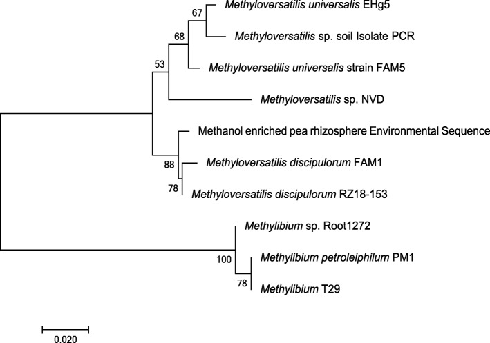
PCR primers for specific amplification of mdh2 genes were designed by aligning mdh2 gene sequences from the sequenced genomes of strains of Methylibium and Methyloversatilis as described in the “Methods” section. The specificity of the primers was assessed by performing PCR using DNA from mdh2-possessing bacteria, Methylibium sp. ROOT1272 and Methyloversatilis sp. LF1. Sequencing confirmed the identity of PCR products as mdh2 methanol dehydrogenase genes. DNA from unplanted soil and pea rhizosphere soil did not yield PCR amplicons when assayed with the mdh2 primers. However, when enriched with methanol (see the “DNA-SIP with 13C methanol” section), DNA extracted from pea rhizosphere soil yielded a PCR amplicon, and restriction fragment length polymorphism (RFLP) screening of the resultant clone library identified a single operational taxonomic unit (OTU), with a high degree of identity (96–99% nucleotide identity) with mdh2 sequences from Methyloversatilis (Fig. 1). The absence of mdh2 products in PCR assays with DNA from the soils that were not enriched with methanol, and the lack of sequence diversity in DNA from the methanol-enriched pea rhizosphere suggests that mdh2 is not abundant in this environment, although it may be more relevant to other environments, such as in freshwater systems [31], where genera that possess mdh2, including Methyloversatilis, are more abundant. Alternatively, the low number of bona fide mdh2 sequences used to design primers may have resulted in primers that are specific to a narrow group of organisms. The identification of additional mdh2-possessing organisms might enable the design of primers with broader specificity.
Fig. 1.
Phylogeny of mdh2 sequences retrieved from pea plant rhizosphere compared to mdh2 from methylotrophic bacteria. Sequenced amplicons obtained from environmental DNA and DNA from cultures of methylotrophic bacteria are labelled as “Environmental Sequence” and “Isolate PCR” respectively. Full reference gene sequences were selected from the NCBI nucleotide database. The tree was drawn in Mega7 [30] using the neighbour-joining method. Scale bar indicates 0.02 substitutions per site. Only bootstrap values ≥ 50% (based on 500 replicates) are labelled at branch points. There were a total of 164 amino acid residues in the final dataset
Diversity of mxaF and xoxF in soils
The diversity of mxaF and xoxF genes in the unplanted soil and rhizosphere samples was analysed by amplicon sequencing of PCR products generated using primers developed previously [28]. The mxaF amplicons produced from DNA extracted from the unplanted and pea rhizosphere soil were dominated (> 99%) by three OTUs affiliated with Hyphomicrobium. This genus was present at 4.5–6% relative abundance in the 16S rRNA gene profile of the unplanted soil and pea rhizosphere bacterial communities, and, of the genera predicted to contain the mxaF gene, Hyphomicrobium was the most abundant within these environments. The remainder of the mxaF sequences (< 1%) clustered with mxaF sequences from members of the family Methylophilaceae (Additional File 3). The mxaF diversity detected in the Church Farm (CF) soil shows similarities to profiles originating from topsoils of other grassland sites reported in a previous study [18]. These authors identified OTUs affiliated with Hyphomicrobium and Methylophilaceae, which were also detected here in the Church Farm soil. However, we did not detect mxaF amplicons affiliated with Methylobacterium, which were detected at high abundance in these previously characterised grassland soils from the previous study [18].
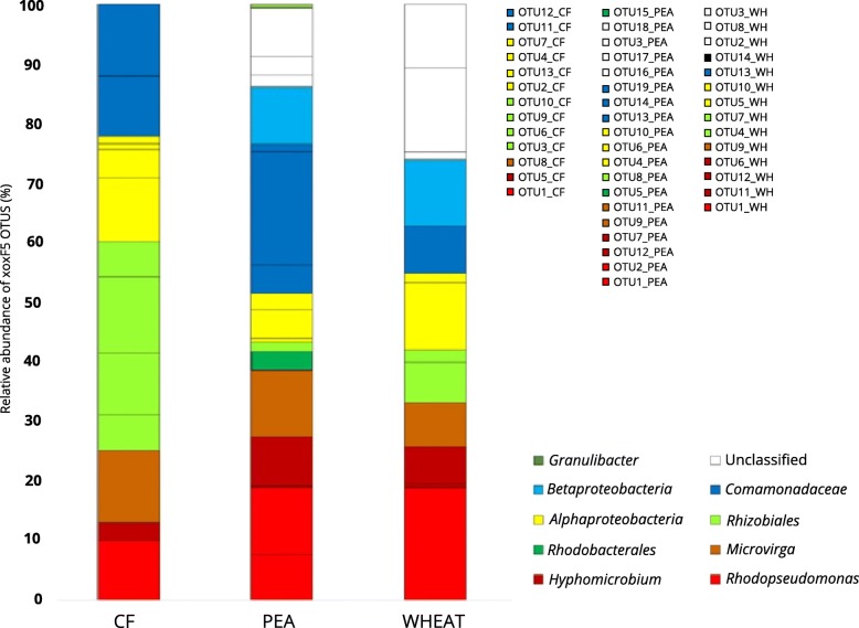
PCR assays with primers specific for the xoxF5 clade revealed that the abundant xoxF5 OTUs retrieved from the unplanted and pea and wheat rhizosphere soils had high similarity to xoxF5 from members of the Alphaproteobacteria and Betaproteobacteria (Fig. 2, Additional File 4), principally members of the genera Hyphomicrobium, Microvirga and Rhodopseudomonas. These genera were also detected in the 16S rRNA gene profiles and contain species capable of methanol oxidation [32–34]. The xoxF5 profiles of the rhizosphere environments were both enriched in OTUs with high similarity to sequences from Rhodopseudomonas, Hyphomicrobium and the Betaproteobacteria. The wheat rhizosphere also contained a greater number of divergent xoxF OTUs that could not be assigned a phylogeny. OTUs of xoxF5 representatives of members of the Comamonadaceae were detected in DNA from the pea rhizosphere at relatively high abundance (~ 25%) (Fig. 2).
Fig. 2.
Relative abundance and diversity of xoxF5 genes in soils. Relative abundance of xoxF5 amplicons generated from unplanted soil (CF), pea rhizosphere soil (PEA) and wheat rhizosphere soil (WHEAT) as revealed by amplicon sequencing. Abundance of taxonomic groups is shown at the highest level of classification for each OTU, with taxonomy of OTUs inferred from the clustering shown in the phylogenetic tree (shown in Additional File 4)
PCR primers specific to xoxF4 consistently failed to yield xoxF4 amplicons with DNA extracted from the unplanted soil, pea rhizosphere soil and wheat rhizosphere soil, indicating that this gene was either absent or below the limit of detection. Libraries of xoxF1, xoxF2 and xoxF3 amplicons were generated from DNA extracted from unplanted soil. Three of the xoxF1 OTUs (OTU1, 2 and 3) had high similarity to members of the Rhizobiales (Oharaeibacter, Methyloceanibacter and Hyphomicrobium). There was also an OTU (16%, OTU 4) that did not have high similarity to any of the xoxF1 reference sequences (Additional File 5). The xoxF3 clone library was dominated by OTU A (26/47 clones), most closely related to xoxF3 of Methylobacterium nodulans (Additional File 6). The additional diversity (OTUs D, F, G, H, I) (Additional File 6) was comprised of sequences clustering with xoxF3 genes of species of Methylosinus, a methanotroph, and Azospirillum (relative abundance 25.5% and 14.9% respectively). The most abundant xoxF2 OTU retrieved by xoxF2 amplicon sequencing was identical to a xoxF2 clone obtained after screening a small clone library (see the “Methods” section). Because this xoxF2 sequence was greater in length than xoxF2 sequences obtained using Illumina technology, it was used in further phylogenetic analysis. The xoxF2 sequence did not cluster with any of the reference sequences and showed highest similarity (84%) to a putative xoxF2 sequence found in metagenome-assembled genomes from studies investigating the microbial diversity of the Chinese and Japanese seas [35, 36] (Additional File 7). These genomes are assigned to the phylum Candidatus Entotheonella, which was not detected in the 16S rRNA gene sequence profile of the Church Farm soil.
Quantification of mxaF and xoxF5 genes in soils using qPCR
The abundance of the mxaF and xoxF5 genes in the unplanted soil and pea and wheat rhizosphere was determined using qPCR. xoxF5 was selected from the xoxF clades for quantification since xoxF5 has been proven to be a bona fide methanol dehydrogenase-encoding gene in multiple species [37–39] and sequencing of xoxF5 gene amplicons from the three different habitats identified shifts in diversity that were potentially correlated with shifts in abundance. Furthermore, the primers for xoxF3 and xoxF1 are unsuitable for qPCR analysis and their redesign was outside of the scope of this work. xoxF4 was not selected for quantification as it was not detected in the unplanted soil DNA and the primers for xoxF4 have cross-specificity for xoxF5 genes in the absence of xoxF4 [28].
Normalised to 16S rRNA gene copy number, the qPCR assays showed that within the three soil environments tested, xoxF5 genes were 36–42 times more abundant than mxaF genes (Additional File 8). However, the overall abundance of methanol dehydrogenase genes did not differ significantly between the three soil environments (two-way ANOVA, p = 0.567). Despite the prevalence of multiple xoxF5 copies in genomes, these data support the hypothesis that Xox enzymes are more abundant and have a wider distribution than Mxa-type MDHs in this type of environment [24]. This confirms the need to also investigate the diversity and distribution of xoxF genes when characterising the diversity of methylotrophic bacteria in environmental studies.
The impact of plants on soil methylotrophs as assessed by DNA-SIP
Using the same three soil treatments described above (unplanted, pea and wheat rhizosphere), the influence of plant growth on soil methylotrophs was examined to determine the differential response of these plant root-associated soil communities to addition of methanol. DNA-SIP enrichments were set up with each soil type using either 13C-labelled or 12C-unlabelled methanol. Following incorporation of 13C-label, the active methanol-assimilating taxa present in the rhizosphere of these two plant types were compared to the unplanted control soil, by amplicon sequencing of 16S rRNA genes of the 13C-labelled DNA retrieved from incubations with 13C-methanol after 6 and 17 days of incubation (T1 and T2 respectively).
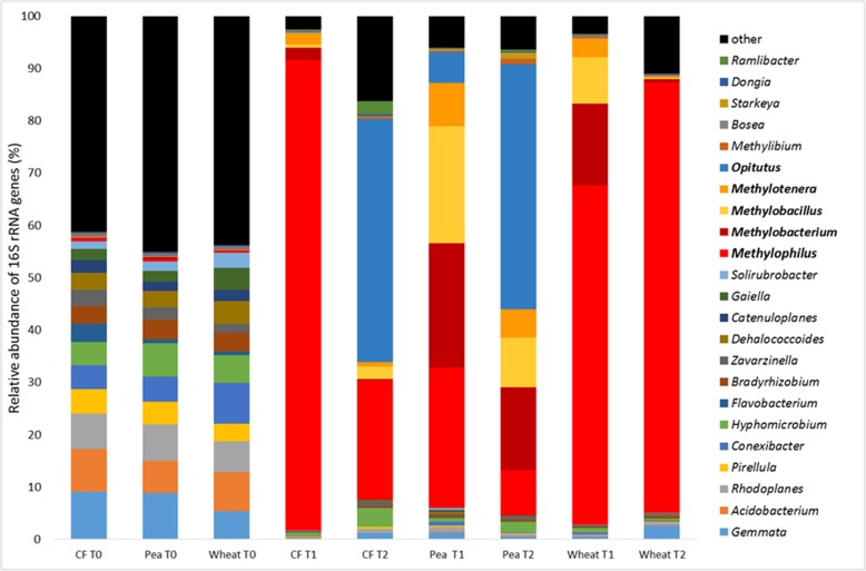
Analysis of the 16S rRNA genes, after DNA-SIP incubations of the methanol-enriched unplanted soil at T1, identified Methylophilus and Methylotenera as 13C labelled, with Methylophilus representing 90% of the 16S rRNA genes retrieved from the heavy DNA. The 13C-labelled communities of the T1 heavy fractions of the methanol-enriched pea and wheat rhizosphere soils were more diverse than that of the unplanted soil (Additional File 9). The 13C-labelled genera in the pea rhizosphere at T1 were identified as Methylophilus, Methylobacterium, Methylobacillus, Methylotenera and Opitutus (Fig. 3, Additional File 10), and the same methylotrophic genera were labelled in the wheat rhizosphere, but with a higher relative abundance of Methylophilus.
Fig. 3.
Comparison of the active methylotrophic communities of methanol-incubated unplanted and rhizosphere soils and unamended soil. Relative abundance of taxa based on 16S rRNA gene amplicons produced from DNA extracted from unplanted (CF), pea rhizosphere (Pea) and wheat rhizosphere (Wheat) soil samples at time point zero (T0) and the 13C-enriched (heavy) DNA fractions from incubations with 13C-methanol for 6 days (T1) and 17 days (T2) are shown. 12C controls are detailed in Additional File 10
In the unplanted soil incubations, the relative abundance of 16S rRNA genes of Methylobacillus, Methylocystis and Methylotenera increased between T1 and T2, but in addition to these known methylotrophs, Opitutus was detected as enriched in the heavy fraction, present at 42% relative abundance 16S rRNA genes retrieved from the heavy fraction. In the pea rhizosphere, of the genera 13C labelled at T1, only Opitutus increased in relative abundance at T2, increasing from 5 to 24%. However, 13 additional genera were labelled, notably Starkeya (1.1%). In the wheat rhizosphere, fewer genera were labelled at T2 compared with T1 and comprised of only Methylophilus and Methylotenera. The relative abundance of Methylotenera decreased tenfold to 0.34%, whereas the relative abundance of Methylophilus increased from 64 to 82%.
Additional groups of bacteria 13C labelled in the rhizosphere soil DNA-SIP experiment, albeit in low abundance, were Stigmatella (0.32% in the wheat rhizosphere and 1.19% in the pea rhizosphere) and members of the phylum Lentisphaerae (0.11% in the wheat rhizosphere). Based on the lack of MDH genes in published genome sequences and previous phenotypic characterisation of representatives of Stigmatella and Lentisphaerae [40, 41], it is possible that they were 13C labelled by cross feeding. Enrichment of Opitutus was also unexpected, as members of this genus are not known to be methanol-oxidising bacteria [42].
Metagenomes reconstructed from DNA-SIP experiments
In addition to sequencing 16S rRNA gene amplicons, the active methanol-assimilating taxa in the rhizosphere and unplanted soils were further characterised by shotgun sequencing the 13C-labelled DNA retrieved from the DNA-SIP incubations enriched with 13C-methanol at T2, resulting in a metagenome for each of the three environments. Metaphlan2 (2.0) [43] was used to analyse the taxonomic composition of the three metagenomes using the presence of taxonomically informative marker genes (the database of these marker genes can be accessed at http://huttenhower.sph.harvard.edu/metaphlan). The metagenomes were dominated by bacteria, specifically Proteobacteria. Gene sequences identified as from Eukarya, Archaea or viruses were present at below 0.1% relative abundance (Fig. 4).
Fig. 4.
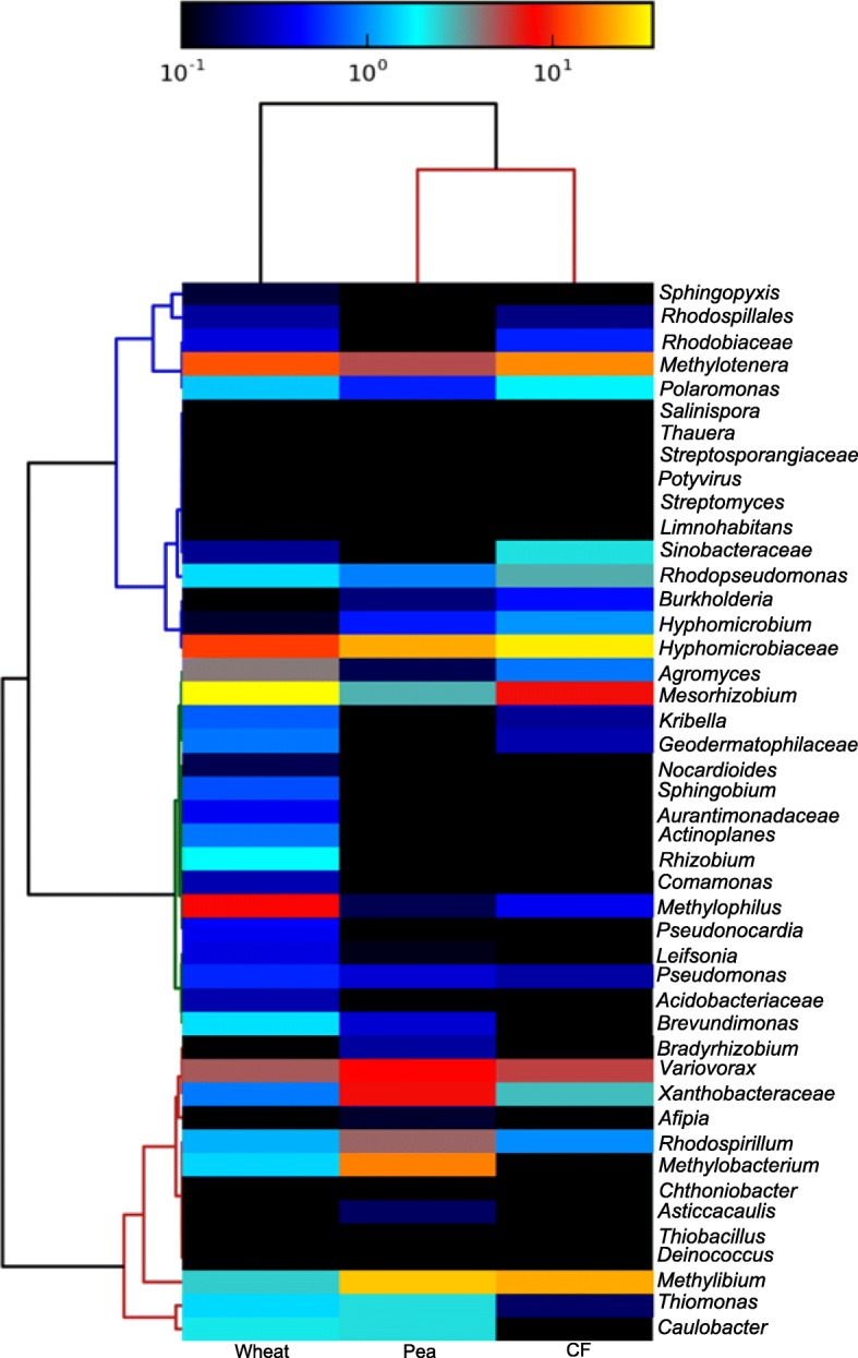
Metagenome-derived community profiles produced from the 13C-enriched (heavy) DNA fractions of 13C methanol-enriched soils. Sequences were revealed by shotgun sequencing of 13C-enriched DNA extracted from wheat rhizosphere soil (Wheat), pea plant rhizosphere soil (Pea) and unplanted soil (CF) incubated with 13C-labelled methanol for 17 days. Relative abundance of taxa is indicated by the colorimetric key assigned with a log scale, with yellow indicating abundance > 101 cells of a taxon and black representing an absent taxon
Consistent with the 16S rRNA gene profiling described above, this metagenomics approach identified members of the Methylophilaceae as highly abundant in all three environments. Furthermore, a key genus delineating the methanol-enriched plant environments from the methanol-enriched unplanted soil was Methylobacterium. This was present at 14.7% relative abundance in the pea rhizosphere and 1.5% in the wheat rhizosphere. It was not detected in the unplanted controls. Analysis of the metagenomes confirmed the differences observed between the planted and unplanted soils using 16S rRNA gene amplicon sequencing. The metagenomes revealed a higher diversity than the 16S rRNA gene amplicon approach (Additional File 9). The additional genera detected in the metagenomes that were shared between the environments include Mesorhizobium, Methylibium, Variovorax and Rhodospirillum. The metagenomes also showed more differences in community composition between the two planted soils, with Comamonas, Sphingobium, Rhizobium, Leifsonia, Mesorhizobium and Methylophilus all being present at greater abundance in DNA from the methanol-enriched wheat community whilst Variovorax, Bradyrhizobium, Afipia, Asticcacaulis and Rhodospirillum were present at greater abundance in DNA from the methanol-enriched pea rhizosphere community. In comparison with the unplanted soil, these data show that the rhizosphere soils contain multiple taxa poised to take advantage of additions of methanol. This lends weight to the hypothesis that plants support a higher diversity of bacteria, in particular methylotrophs, than are present in unplanted soil.
The metagenomes were screened for genes of interest using the blast function of BioEdit. This identified the presence of genes encoding for methanol dehydrogenases (xoxF and mxaF), enzymes involved in methylated amine utilisation (tmmD, dmmD, mauA and the N-methylglutamate pathway) and formaldehyde and formate oxidation in all three metagenomes. The screening also identified all the genes of the ribulose monophosphate and serine cycles. Genes encoding the complete pathways for assimilatory sulfate reduction, denitrification and nitrogen fixation were also detected in the three metagenomes, providing an insight into the energy and nitrogen yielding pathways active in the three soil habitats. The screening for genes of interest did not reveal a difference in the presence of these metabolic pathways between the metagenomes. To investigate differences in the abundance and diversity of methanol dehydrogenase-encoding genes in the three methanol-enriched soil habitats, the assembled and unassembled reads of the metagenomes were screened with representative sequences of each clade of methanol dehydrogenase-encoding gene (xoxF1, xoxF2, xoxF3, xoxF4, xoxF5, mxaF and mdh2). The abundance of methanol dehydrogenase genes differed between the three metagenomes (Additional Files 11, 12, 13, 14, 15, 16). mxaF and xoxF4 were present at higher abundance in the two plant habitats relative to the unplanted soil, whereas xoxF5 and xoxF3 were more abundant in the pea rhizosphere relative to the unplanted soil and wheat rhizosphere. mdh2 sequences were only detected in the methanol-enriched pea rhizosphere and unplanted soils, and these sequences showed high (> 82%) sequence identity to those of Methylibium and Methyloversatilis (Additional File 16). The diversity of the xoxF3 sequences detected in the metagenomes was greatest in the methanol-enriched pea rhizosphere soils (Additional File 16), which included sequences with high sequence identity to those possessed by members of the genera Methylobacterium and Mesorhizobium, in addition to the Comamonadaceae sequences detected in the other two enriched soil habitats (Additional File 16). xoxF2 and xoxF outgroup sequences (the Acidiphilum and Methylosinus trichosporium sequences that do not cluster with any established clade [23]) were not detected in the three metagenomes.
xoxF5 was the most abundant methanol dehydrogenase-encoding gene in the pea rhizosphere and unplanted soil but was only the second most abundant in the wheat rhizosphere, where the most abundant was xoxF4. Compared to the number of copies of mxaF and xoxF5 determined by qPCR for soils that were not enriched with methanol, these data show that following enrichment with methanol, xoxF5 was present at greater abundance than mxaF, but both genes were present at the same order of magnitude in all three soil habitats. The xoxF1, xoxF5 and mxaF sequences detected in the metagenomes arising from the methanol-enriched pea and wheat rhizospheres and unplanted soils showed similar patterns of diversity to the sequenced amplicons produced from the unenriched soil. The xoxF1 sequence profiles revealed low levels of diversity, with the detected xoxF1 sequences showing high sequence identity to those identified in genomes of strains of Hyphomicrobium and Methyloceanibacter (Additional File 12). The diversity of mxaF sequences was also low, with the mxaF sequences detected having high sequence identity to mxaF genes from Hyphomicrobium, Methylobacterium and Methylophilus in all three metagenomes (Additional File 15). The xoxF5 sequences in the three metagenomes showed high sequence identity to sequences identified in members of the Alphaproteobacteria (Rhizobium, Methylobacterium and Hyphomicrobium) and Betaproteobacteria (Methylibium and Comamonadaceae), with similar sequence diversity detected between the three soil habitats (Additional File 14). There was also similarity in the diversity of the xoxF4 sequences detected in the three methanol-enriched habitats, with high sequence identity to members of four genera within the Methylophilaceae (Methylomonas, Methylophilus, Methylobacillus and Methylovorus) (Additional File 13), but this might be an artefact of the low diversity of this clade of methanol dehydrogenase-encoding gene.
To further investigate the diversity of the methanol-enriched environments, assembled sequence data from the three metagenomes were binned into metagenome assembled genomes (MAGs). The binning produced 10 MAGs of sufficient quality (completeness score > 70% and contamination < 10%), meeting currently accepted criteria for medium- to high-quality MAGs [44]) (Additional File 17). These MAGs were also screened for genes of interest (Additional File 18). One genome (vs26) was identified as a Rubrivivax. This MAG contained a xoxF5 methanol dehydrogenase-encoding gene, genes encoding a complete tetrahydromethanopterin formaldehyde oxidation pathway [45], thiosulfate oxidation (soxABXYZ) and assimilatory sulfate reduction (cysCDHIJN), as well as an incomplete serine cycle. The genome binning also produced a MAG classified as a Methylobacterium (ss20), an abundant genus in the methanol-enriched pea and wheat rhizosphere soils. This MAG contained mxaF and xoxF5 methanol dehydrogenase genes, and genes encoding the complete N-methyl glutamate pathway for methylamine utilisation [46], an incomplete serine cycle and one of each of the four forms of formate dehydrogenase [47–49]. However, we cannot rule out the possibility that the incomplete serine cycle of these MAGs may be an artefact resulting from imperfect sequence assembly or binning (predicted MAG completeness 97% and 72% respectively).
Of the remaining MAGs, three were members of the order Methylophilales, highly enriched in all of the soil environments supplemented with methanol. The methanol dehydrogenase gene of the MAG-designated Methylotenera ss03 was of note, as the xoxF3 did not cluster with those of other Methylophilaceae (i.e. Methylobacillus flagellatus), but instead with Variovorax paradoxus strain S110 and sequences from the Alphaproteobacteria (Additional File 19), suggesting that the diversity of this clade of methanol dehydrogenase within the Methylophilaceae is greater than previously detected. The MAG-designated Methylophilales ss01 and ss29 were of interest as they showed high levels of similarity to the strains Methylobacillus sp. strain MM3 (97% average nucleotide identity (ANI)) and Methylovorus sp. strain MM2 (99% ANI) respectively, both of which were previously isolated from the same environment [50]. Despite the fact that enrichment and isolation techniques may not capture all representatives of the bona fide natural community, this suggests that both strains may have been active members of the methanol-oxidising community of this soil. In addition to methanol dehydrogenase genes, these MAGs contain formate dehydrogenases and partially complete ribulose monophosphate cycles.
Further analysis of the MAGs Archaea vs43, Bdellovibrio vs70, Deltaproteobacteria ss68 and Verrucomicrobia vs53 and ss71 showed that none of them contained a methanol dehydrogenase-encoding gene or genes encoding formaldehyde utilisation pathways. However, the MAGs Verrucomicrobia ss101 and Deltaproteobacteria ss68 both contain copies of a formate dehydrogenase (fdh4)-encoding gene and the genome ss68 possessed genes encoding dimethylamine and trimethylamine dehydrogenases, implying that these strains of bacteria may be able to utilise some C1 compounds as carbon and/or nitrogen sources. The enrichment of these taxa with 13C could therefore be explained by the utilisation of exuded C1 compounds (i.e. formate), utilisation of other exuded organic compounds, the fixation of 13C-labelled CO2 produced by the methanol oxidising methylotrophs (in the case of vs43, which contains the gene for ribulose-1,5-bisphosphatecarboxylase/oxygenase) or, in the case of Bdellovibrio (vs70), predation on the methanol oxidising methylotrophs [51].
Phylogenetic analysis of the exudate-utilising community of the pea rhizosphere
In a third series of experiments, DNA-SIP was also utilised to investigate whether methylotrophic bacteria were utilising carbon exuded from the roots of plants. Pea plants were incubated in a 13CO2 or 12CO2 (control) atmosphere for 12 days to allow sufficient 13C label to be incorporated into plant biomass and then for 13C-labelled plant exudate released from roots to be assimilated by microbes in the rhizosphere. Two CO2 concentrations were used, 350 and 1000 ppmv, reflecting environmental and elevated levels. Three hundred fifty parts per million volume was selected as the concentration of CO2 to supply to the environmental test group, as CO2 was also released by the soil, and this ensured the concentration did not exceed environmental levels (420 ppm).
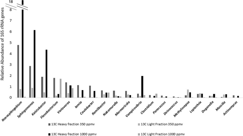
The bacteria in the pea rhizosphere that were active utilisers of plant exudates were identified by sequencing of 16S rRNA gene amplicons generated from heavy and light DNA fractions retrieved from rhizosphere and unplanted (control) soils. Analysis of 16S rRNA gene sequences retrieved from heavy fractions of DNA obtained in DNA-SIP experiments with pea plants incubated with 13CO2 indicated that labelling of bacteria of the genera Novosphingobium (4.8% relative abundance after enrichment with 350 ppmv 13CO2 and 17.7% relative abundance after enrichment with 1000 ppmv), Kaistobacter (1.9% at 350 ppmv and 4.4% at 1000 ppmv), Sphingomonas (2.9% at 350 ppmv and 6.2% at 1000 ppmv), Paracoccus (0.2% at 350 ppmv), Variovorax (1.4% at 350 ppmv), Flavobacterium (1.8% at 350 ppmv) and Ramlibacter (0.% at 350 ppmv), Methylocapsa (0.5% at 1000 ppmv) and Leptothrix (0.4% at 1000 ppmv) occurred (Fig. 5). With the exception of Novosphingobium and Kaistobacter, these genera contain species that are either methylotrophs or whose genomes contain xoxF genes [11, 52–54]. The addition of an elevated concentration of carbon dioxide to growing grasses and sedges has previously been shown to impact on the rhizosphere community [55]. The observation that growing pea plants with different levels of CO2 might favour the growth on root exudates of different groups of methylotrophs is interesting and warrants further investigation in the future.
Fig. 5.
Relative abundance of exudate-utilising genera in the rhizosphere of pea plants. Relative abundance of taxa identified as 13C labelled in the 13C test group from the heavy fractions and light fractions of DNA extracted from the rhizospheres of pea plants incubated with 13C-carbon dioxide at ambient (350 ppm) and elevated (1000 ppm) concentrations for 12 days. Genera that were also 13C labelled in the unplanted test group were excluded and are detailed in Additional Files 26 and 27. The data show the mean of duplicate incubations
Variovorax, Ramlibacter and Leptothrix genera within the Comamonadaceae were 13C labelled in the rhizospheres of pea plants incubated with ambient and elevated 13CO2. Strains from these genera have previously been found in rhizosphere environments [56–59]. The family Comamonadaceae contains bacteria that are metabolically versatile and can use a broad range of carbon substrates, enhance the cycling of sulfur in soil and suppress fungal pathogens [57, 60]. Genera within this family include Variovorax and Delftia, which contain species known to grow on methanol [58, 61]. An examination of all available genomes of members of the Comamonadaceae revealed that representatives of 28 out of 34 genera contain xoxF genes. With the recent discovery of the role of lanthanides in methylotrophy, there is a clear need to retest representatives of the Comamonadaceae for their ability to grow on methanol in medium supplemented with these rare earth elements.
Labelling of members of the Sphingomonadaceae and Actinobacteria in our DNA-SIP experiments is consistent with previous reports with other plant species [62–64], and the utilisation of exudates by members of the Sphingomonadaceae has been observed in stable isotope probing experiments and the rhizosphere of rice plants [65]. Actinobacteria are proposed to play a role as plant growth promoting bacteria through the production of antimicrobial or antifungal agents, plant hormones and siderophores [87 and references therein]. In 13CO2 rhizosphere SIP studies with oil seed rape, wheat, maize and Medicago truncatula, Actinobacteria were also observed to use root exudates [2, 64]. The 13C labelling of members of the Actinobacteria and Sphingomonadaceae in stable isotope probing studies implies they incorporated carbon exuded by the plant, suggesting that they were enriched in the rhizospheres of different plant species because of direct utilisation of plant exudates rather than cross feeding.
Comparing the 16S rRNA gene and methanol dehydrogenase gene amplicons produced from DNA extracted from the unenriched unplanted soil and rhizosphere soils to the taxonomic profiling of the labelled communities from soils that were enriched with either 13C-methanol or 13CO2 identified that a small portion of diversity was common between these different test groups (e.g. Variovorax, Additional Files 20, 21, 22). However, beta diversity analysis (Additional File 23) clearly shows that the communities in the methanol-enriched samples cluster separately from the CO2-enriched samples. This difference is largely due to the high levels of enrichment of members of the Methylophilaceae in the soils amended with methanol and their absence in the exudate utilising population of the pea rhizosphere.
Conclusions
Growing plants are a major source of methanol, and the microbial community phyllosphere of multiple plant species has been shown to include methylotrophs that are highly abundant [66–68]. However, there have been few studies attempting to characterise the impact of plant growth on the diversity and activity of methylotrophs in the rhizosphere and whether they are using carbon directly from the plant. In this study, methylotrophs were shown to be abundant in the unplanted soil, pea rhizosphere soil and wheat rhizosphere soil, and their diversity was influenced by pea and wheat plants. The use of xoxF and mxaF as functional gene markers revealed a greater diversity of methylotrophs in the rhizosphere than previously observed by 16S rRNA gene sequencing, with xoxF shown to be the more relevant indicator of methylotrophy in these soil environments and also an order of magnitude more abundant than mxaF. However, mdh2 currently is of limited utility in characterising the diversity of methylotrophs in soils.
Metagenome sequencing of 13C-labelled DNA extracted from pea and wheat rhizosphere soils and unplanted soils enriched with methanol confirmed that the growth of pea and wheat plants influenced methylotrophy. Interestingly, both plant associated environments showed the same shift in the microbial community profile, revealing a greater diversity of members of the Methylophilaceae and Methylobacterium, a cosmopolitan genus possessing plant growth-promoting traits and commonly associated with plants [30, 69, 70]. 13CO2 labelling of growing pea plants also confirmed that methylotrophs present in the pea rhizosphere were actively utilising carbon exudate from the plant. Comparing the methylotrophic genera detected in the pea rhizosphere, by sequencing of methanol dehydrogenase genes from the unplanted soil, from the exudate-utilising population from the 13CO2 SIP experiment and from soils enriched with 13C methanol, revealed that, using these approaches, only a minority of the diversity was shared. The differential enrichment of methylotrophs between the two SIP experiments indicates that there could be selection for some genera of methylotrophs in response to the higher concentrations of methanol (e.g. Methylophilaceae) whilst others can utilise methanol at a wider range of concentrations (e.g. Comamonadaceae).
Plants can also influence the availability of micronutrients, soil structure, pH and redox potential [62], and these factors could play a role in the recruitment of methylotrophs to the rhizosphere. Furthermore, the specific growth stages of the plants used in this series of experiments and their stress state might affect the amount and nature of exudate released from the roots [19, 71]. Both factors might impact on the activity of methylotrophs in the soil. Additional studies are therefore needed to define the exact relationship between methylotrophs and the rhizosphere.
Methods
Chemicals and reagents
Analytical grade reagents used were from Sigma-Aldrich (MO, USA), Melford Laboratories (Ipswich, UK) or Fisher Scientific (Loughborough, UK). Molecular biology grade reagents were from Thermo Fisher (MA, USA), Promega UK (Southampton, UK), Qiagen (Germany) and Roche (Switzerland). Gases were supplied by BOC (UK). 13CO2 and 13C-labelled methanol were supplied by Cambridge Isotope Laboratories (MA, USA). All ultracentrifuge work involved using tubes, rotors and ultracentrifuges from Beckman Coulter (CA, USA).
The experimental workflow for the research described in this study is detailed in Additional File 24.
Collection, processing and storage of soil
Soil was collected in April 2015 from a naturally grassed and unfertilised part of John Innes Centre Church Farm (Norfolk, UK) (52.6276 N, 1.1786 E). The top 10 cm was removed from a 1 m2 section and then soil to 20 cm depth was removed, air dried and sieved through 10 mm and 5 mm sieves which removed stones, roots and other detritus. This soil (designated “bulk soil”) was then used in all experiments.
Extraction of nucleic acids from soil
DNA was extracted from soil samples using a cetyltrimethyl ammonium bromide (CTAB)-based method [72] and quantified using Qubit fluorometric quantitation (Thermo Fisher).
Germination and growth of plants
Paragon wheat seeds (Triticum aestivum var. Paragon) were sterilised by washing the seeds in 5% (v/v) sodium hypochlorite solution for 1 min. Pea seeds (Pisum sativum var. Avolar) were sterilised by washing the seeds in 95% (v/v) ethanol for 1 min, washing with sterile H2O and soaking in 2% (w/v) sodium hypochlorite for 5 min. Pea and wheat seeds were then washed in sterile H2O and placed in a petri dish on filter paper disks moistened with sterile H2O [13]. The seeds were left in the dark for 3 days to germinate and manually inspected for fungal contamination before planting in 10 cm × 10 cm pots in bulk soil and growing at 22 °C under long day regimes (16:8 h) in plant growth rooms. Pots with unplanted soil were incubated alongside the growing plants as unplanted controls. Plants were harvested after 4 weeks of growth. Excess soil was removed by shaking the roots three times. Soil that remained attached to the roots after shaking was defined as rhizosphere soil. Rhizosphere soil was removed by transferring the roots to Falcon tubes, submerging in phosphate buffered saline (PBS) and vortexing for 30 s. Tubes were centrifuged at 3200×g for 15 min to pellet soil. Root material was removed and the supernatant discarded. DNA was extracted from three separate 0.5 g aliquots of soil and subsequently pooled to produce a composite sample.
DNA-SIP with 13C methanol
Two grams of aliquots of rhizosphere soil, collected as described above, and unplanted soil were dispensed into 120-ml serum vials. Forty millilitres of sterile H2O was added to reduce the heterogeneity of the soil sample within the serum vials and to facilitate substrate distribution. Vials were then supplemented with 13C methanol or 12C methanol to a concentration of 250 μM and sealed. Each test group was performed in triplicate. The serum vials were incubated at 30 °C without light in a shaking incubator (120 rpm). The concentration of methanol in the headspace of the serum vials was measured using gas chromatography (GC) on an Agilent 7820A instrument, using a flame ionisation detector, a Porapak Q column (6 ft × 1/8″ ×2.1 mm) and helium carrier gas (injector temperature, 300 °C; detector temperature, 300 °C; oven temperature, 115 °C). After depletion of methanol, samples were resupplied with methanol to the same concentration. Serum vials were opened every second day and flushed with air to prevent the development of anaerobic conditions and to avoid the build-up of 13CO2 within vials. After 6 days, when methanol oxidation stopped, samples were taken from enrichments for DNA extraction (time point 1). Dilute nitrate mineral salts medium [73] (1 ml) was supplied to the serum vials on day 7 to establish whether the enrichments were nutrient limited and the incubations were continued. Methanol consumption resumed. After 17 days, a total of 200 μmol of 13C had been consumed in incubations and soil was collected for DNA extraction (time point 2). DNA was extracted from all soil samples, and caesium chloride density gradient centrifugation was used to separate the 13C- and 12C-labelled DNA from 1–3 μg of DNA from each test group according to established protocols [74]. During fractionation of CsCl gradients, twelve samples were collected and the density of CsCl in each fraction estimated by measuring the refractive index (Reichert AR200). DNA was then recovered by precipitation [74].
DNA stable isotope probing with 13CO2
Pea plants were grown in 10 cm × 10 cm pots in bulk soil under long day growth conditions (16 h:8 h) for 16 days. After 16 days, eight pea plants and eight unplanted soil controls were transferred to acrylic tubes (approx. 3.8 L volume, 400 mm height × 110 mm internal diameter). All plants and unplanted soil controls were transferred to medium day light conditions (12 h:12 h). The acrylic tubes were flushed with carbon dioxide depleted air, sealed with plastic lids and injected with either 13CO2 or 12CO2 to a final concentration of either 350 ppmv or 1000 ppmv, with each experimental condition in duplicate. Carbon dioxide was measured by GC on an Agilent 7890A instrument equipped with a nickel catalyst, using a flame ionisation detector, an HP plot/Q (30 m × 0.530 mm, 40 μM film) and helium carrier gas (injector temperature, 250 °C; detector temperature, 300 °C; oven temperature, 50 °C). Carbon dioxide was replenished to the target concentration every 20 min during the light period. After 12 days, plants were harvested and DNA extracted from the rhizosphere soil. DNA (4 μg) for each sample was processed via ultracentrifugation and fractionation as described above.
Criteria for confirming 13C labelling of DNA from target microbes
Specific criteria were applied when analysing DNA sequence data from both SIP experiments to establish which taxa of bacteria were labelled. In order to be included in the analysis, the relative abundance of a specific taxon in the 13C-heavy DNA fraction had to be greater than 0.1%. The criteria that needed to be fulfilled for a particular taxon to be considered 13C labelled were as follows: (1) the relative abundance in the 13C-heavy fraction should be higher than in the 12C-heavy fraction (13CH > 12CH) and (2) the taxon should be enriched in the heavy fraction of 13C incubations (i.e. the relative abundance in the heavy fraction should be greater, by a specific factor, than in the light fraction (13CH > k × 13CL), but this should not be the case for 12C incubations (12CH ≤ 12CL). The factor k was chosen as k = 10 for the methanol SIP experiment, due to the substrate based stable isotope approach used and the probability of cross feeding, but k = 2 in the 13CO2-labelling SIP experiment, due to the more transient nature of the 13C labelling and the lower input of 13C. Any enrichment of autotrophic bacteria could be accounted for and observed by incubating unplanted soil controls in a 13CO2 atmosphere. Any taxon identified as 13C labelled in the unplanted test group was excluded from the list of taxa identified as 13C labelled in the rhizosphere test group (Additional Files 25 and 26).
Polymerase chain reaction (PCR) and quantitative PCR assays
Amplification of products by PCR was performed in 25 μL reaction volumes using a BIORAD Tetrad 2 thermal cycler. The reaction mixture was 1× Master Mix (PCR Biosystems, UK), 0.4 μM forward primer and 0.4 μM reverse primer. PCR primers and amplification protocols used to screen for 16S rRNA, mxaF, xoxF1-5 and mdh2 genes are detailed in Additional File 27. PCR products were purified using NucleoSpin Gel and PCR clean-up columns (Macherey-Nagel, Germany) according to the manufacturer’s instructions. The copy number of 16S rRNA, mxaF and xoxF5 genes in DNA and cDNA samples was estimated using quantitative PCR (qPCR) (Applied Biosystems Step one plus real-time PCR system, Thermo Fisher, MA, USA). The reaction mixture was BioLine Sensifast Hi Rox master mix, 0.4 μM each primer and with the addition of bovine serum albumin (0.2 μg). Standards were prepared using xoxF5 and mxaF PCR products amplified from DNA of Methylocella silvestris BL2, diluted to a copy number of 108 to 101 per microlitre. Three biological replicates from each environment were tested, each with three technical replicates. The efficiency of the amplification was 98% for mxaF and 83% for xoxF5. A two-way ANOVA test was performed using the R package dplyr to test for significant differences between the test groups.
Design of PCR primers to amplify mdh2
PCR primers were designed to amplify mdh2 genes from DNA isolated from soils. The primers were based on conserved regions identified by aligning five mdh2 sequences (Methylibium petroleiphilum PM1 AAEM01000000, Methyloversatilis universalis strain FAM5 EU548062, Methyloversatilis universalis EHg5 JN808865, Methyloversatilis discipulorum strain RZ18-153 EU548066, Methyloversatilis discipulorum strain FAM1 EU548063.1) using the MUSCLE algorithm in MEGA7 [75] and screening the alignment for a conserved region of 18-20 nucleotides, allowing for a maximum of three non-conserved bases. Specificity of the mdh2 primers was tested by performing PCR using DNA extraction from two strains of methylotrophic bacteria, Methylibium sp. ROOT1272 (NZ_LMDY00000000) and Methyloversatilis sp. soil isolate (MK795690), as positive controls and DNA from strains of bacteria that do not possess mdh2 as negative controls (Methylobacillus sp. MM3 (NZ_LXTQ00000000), Methylovorus sp. MM2 (NZ_LXUF00000000), Hyphomicrobium sp. MMN) (MK795690). DNA extracted from unplanted soil, pea rhizosphere soil and methanol-enriched pea rhizosphere soil was used as template to generate PCR amplicons of mdh2. Mdh2-specific PCR products (~ 500 bp) were cloned using the Promega pGEM-T Easy vector system according to the manufacturer’s instructions. Cloned PCR products were amplified using M13 primers (Additional File 27), and 20 clones from each library were screened by restriction fragment length polymorphism (RFLP). PCR products were digested using the restriction enzymes RsaI and AluI. RFLP profiles were analysed by gel electrophoresis using 2% (w/v) agarose gels, and representative mdh2 genes from different soil DNA samples were sequenced.
Sequencing of 16S rRNA and MDH genes
16S rRNA gene amplicons were sequenced using Roche 454 (3000 reads) and Illumina MiSeq (20,000 reads) technology by Molecular Research LP (Shallowater, TX, USA). 16S rRNA gene amplicons produced using the 454 and Illumina platforms were processed by Molecular Research LP through their proprietary pipeline. Sequences were depleted of barcodes and primers then short sequences < 200 bp were removed, together with sequences with ambiguous base calls and sequences with homopolymer runs exceeding 6 bp. Sequences were then denoised, and operational taxonomic units (OTUs) were defined as clustering at 97% similarity, following removal of singleton sequences and chimaeras [76–81]. Final OTUs were taxonomically classified using BLASTn against a curated database derived from GreenGenes, RDPII and NCBI (www.ncbi.nlm.nih.gov, http://rdp.cme.msu.edu) [82]. Beta diversity analysis was performed using the bioinformatics platform Qiime [83] to identify similarities between different the communities profiled by the sequencing of 16S rRNA gene amplicons. Weighted and unweighted UniFrac analysis was performed on 16S rRNA gene amplicons produced from the unenriched, unplanted soils and pea and wheat rhizosphere soils in addition to the heavy and light fractions produced from the DNA-SIP experiments performed with 13C-methanol and 13CO2.
Reads of functional (methanol dehydrogenase) genes sequenced using the 454 platform were analysed using a modified version of a published protocol [28]. SFF files were processed using Mothur [84] to convert the raw files into flowgrams, which were then translated to nucleotide sequences. USEARCH [85] was used for identification and removal of chimeric sequences. Sequences were clustered into OTUs using USEARCH [78], using similarity values of 80%. OTUs were aligned using the MUSCLE algorithm against a database containing representative sequences from different clades of PQQ dehydrogenase. OTUs that clustered with each clade were re-aligned at the amino acid level using a database of sequences specific to that clade (Table 1). Phylogenetic trees were produced in MEGA7 [75] using the neighbour joining algorithm with bootstrap values of 500. 454 sequencing of the xoxF3 amplicon produced data that was not of sufficient quality, and therefore, a clone library of 100 clones was made from the xoxF3 PCR amplicon obtained from DNA extracted from unplanted soil. This clone library was constructed and screened via RFLP as described above.
Table 1.
Number of OTUs in sequenced mxaF and xoxF amplicons produced from DNA extracted from soil
| Gene | Soil environment | Sequencing platform | Number of OTUs |
|---|---|---|---|
| xoxF1 | Unplanted | Roche 454 | 4 |
| xoxF2 | Unplanted | Sanger | 1 |
| xoxF3 | Unplanted | Sanger | 6 |
| xoxF5 | Unplanted | Roche 454 | 13 |
| xoxF5 | Pea rhizosphere | Roche 454 | 19 |
| xoxF5 | Wheat rhizosphere | Roche 454 | 14 |
| mxaF | Unplanted | Roche 454 | 4 |
| mxaF | Pea rhizosphere | Roche 454 | 4 |
Amplicons were produced from DNA samples from unplanted soil, pea rhizosphere soil and wheat rhizosphere soil and analysed by either 454 amplicon sequencing or Sanger sequencing. OTUs were produced using an 80% identity clustering threshold
Metagenome sequencing and analysis
DNA from the 13C-heavy fractions of the methanol SIP experiment was pooled, quantified and sequenced by the Centre for Genomic Research at the University of Liverpool. Sequencing was performed using paired-end sequencing (2 × 150 bp) on an Illumina HiSeq 4000. Short sequences and sequences of poor quality were excluded from the files using the program Trimmomatic (0.36) [86], and the quality of the metagenomes was assessed using QUAST, including the MetaQUAST expansion (5.0.0) (Table 2) [87].
Table 2.
Statistics of metagenomes assembled from the heavy fraction of methanol-enriched rhizosphere and unplanted soils
| Metagenome | |||
|---|---|---|---|
| Pea | Unplanted | Wheat | |
| # contigs (≥ 0 bp) | 1151414 | 1251579 | 981758 |
| # contigs (≥ 1000 bp) | 195697 | 192658 | 106074 |
| Total length (≥ 0 bp) | 934363676 | 922084398 | 616537133 |
| Total length (≥ 1000 bp) | 456772046 | 392066999 | 186491273 |
| Total length | 717825918 | 682211092 | 415542434 |
| GC (%) | 63.9 | 64.58 | 65.92 |
| N501 | 1397 | 1168 | 916 |
| L502 | 112046 | 145393 | 128331 |
The quality of metagenomes were analysed using the program QUAST. The metagenomes were produced by shotgun sequencing of 13C-labelled DNA extracted from methanol-enriched wheat rhizosphere soil (wheat), pea plant rhizosphere soil (pea) and unplanted soil (CF) incubated from 13C-labelled methanol for 17 days
1N50 is the length for which the collection of all contigs of that length or longer covers at least half an assembly
2L50 is the number of contigs equal to or longer than N50
Trimmed reads were analysed using Metaphlan 2 (2.0) [43]. Reads were assembled using Megahit (1.1.2) [88] and annotated using myRast (35) [89]. The metagenomes were screened for genes of interest using protein sequences of confirmed function using tblastn in BioEdit [90]. For quantification of the methanol dehydrogenase genes, the assembled and unassembled reads were converted to blast databases and screened using the blastn algorithm and representative sequences for each clade of methanol dehydrogenase-encoding gene (xoxF1, xoxF2, xoxF3, xoxF4, xoxF5, mxaF, mdh2). Appropriate stringencies for the BLAST searches were determined by selecting the lowest e value that did not yield sequences of the incorrect clade (Additional File 11). Abundance values were normalised to gene length or gene length and read number for the assembled and unassembled metagenome reads respectively. The percentage of bacteria represented in the unassembled read data that possess a methanol dehydrogenase was calculated from the abundance of the methanol dehydrogenase genes divided by the abundance of the housekeeping gene recA. The sequences with the highest sequence identity to the identified methanol dehydrogenase sequences from the assembled metagenomes were identified using the blastn algorithm against the NCBI database (Additional File 16).
Contigs were binned into metagenome-assembled genomes (MAGs) using MetaBAT (2.21.1) with the “superspecific” and “veryspecific” algorithms [91]. The completeness, contamination and heterogeneity of these MAGs were assessed using the program CheckM (1.0.13) [92]. The MAGs of sufficient quality binned from the metagenomes (completeness score above 70% and contamination below 10%, meeting currently accepted criteria for medium to high quality MAGs [44]) were also screened for genes of interest using protein sequences of confirmed function using tblastn in BioEdit [90]. Average nucleotide identity (ANI) calculations were performed to assess the similarity of the MAGs Methylophilales ss01 and ss29 to the genomes Methylovorus sp. MM2 (NZ_LXUF00000000) and Methylobacillus sp. MM3 (NZ_LXTQ00000000) [84].
Screening of the genomes of members of the Comamonadaceae for xoxF genes
Three hundred fifteen genomes of members of the Comamonadaceae were downloaded from NCBI GenBank. Genomes were screened for the presence of methanol dehydrogenase-encoding genes using local Blast searches (tblastn), using the xoxF5 sequence of Variovorax paradoxus S110 (NZ_ARNA00000000.1) as query in BioEdit [83].
Supplementary information
Additional file 1. 16S rRNA gene profiles of bacteria in unplanted, pea rhizosphere and wheat rhizosphere soils.
Additional file 2. Relative abundance (%) of methylotrophic genera in 16S rRNA gene profile of soils.
Additional file 3. Phylogeny of mxaF sequences retrieved from pea plant rhizosphere and unplanted soil.
Additional file 4. Phylogeny of xoxF5 sequences retrieved from pea plant rhizosphere, wheat plant rhizosphere and unplanted soil.
Additional file 5. Phylogeny and relative abundance of xoxF1 sequences retrieved from soil.
Additional file 6. Phylogeny and relative abundance of xoxF3 sequences retrieved from soil.
Additional file 7. Phylogeny of a xoxF2 sequence retrieved from soil.
Additional file 8. Relative abundance of methanol dehydrogenase encoding genes in unplanted and rhizosphere soils.
Additional file 9. Diversity indices of the communities in the heavy fraction of methanolenriched rhizosphere and unplanted soils.
Additional file 10. Relative abundance of 16s rRNA gene.
Additional file 11. Abundance of unique methanol dehydrogenase encoding genes sequences identified in the metagenomes asssembled from methanol-enriched soils.
Additional file 12. Diversity of xoxF1 gene sequences retrieved from the heavy fractions of soils enriched with 13C methanol.
Additional file 13. Diversity of xoxF4 gene sequences retrieved from the heavy fractions of soils enriched with 13C methanol.
Additional file 14. Diversity of xoxF5 gene sequences retrieved from the heavy fractions of soils enriched with 13C methanol.
Additional file 15. Diversity of mxaF gene sequences retrieved from the heavy fractions of soils enriched with 13C methanol.
Additional file 16. BLAST scoring details of unique mdh2 sequences identified in metagenomes produced from methanol enriched soils.
Additional file 17. Details of metagenomes assembled genomes (MAGS) binned from metagenomes assembled from methanol-enriched soils.
Additional file 18. Taxonomy of MAGS and the identification of funtional genes involved in C1, nitrogen and sulfur cycling.
Additional file 19. Phylogeny of a xoxF3 sequence retrieved from metagenome assembled genome Methylotenera ss03.
Additional file 20. Putative and confirmed methylotrophs identified in the pea rhizosphere using multiple approaches.
Additional file 21. Putative and confirmed methylotrophs identified in the wheat rhizosphere using multiple approaches.
Additional file 22. Putative and confirmed methylotrophs identified in the unplanted soil using multiple approaches.
Additional file 23. NMDS plot showing the unweighted unifrac analysis of 16S rRNA gene amplicons produced from DNA extracted from unenriched soils, soils enriched with methanol and rhizosphere soils supplemented with 1000 ppm and 350 ppm carbon dioxide.
Additional file 24. Workflow schematic of the DNA-SIP with methanol, the DNA SIP with carbon dioxide and the sequencing and quantification of the methanol dehydrogenase genes and 16S rRNA gene from the soil habitats.
Additional file 25. Relative abundance of genera detected as labelled in 16S rRNA gene profiles of the 350 ppmv supplied test groups.
Additional file 26. Relative abundance of genera detected as labelled in 16S rRNA gene profiles of the 1000 ppmv supplied test groups.
Additional file 27. PCR primers used in this study.
Acknowledgements
Not applicable
Authors’ contributions
JCM, ATC and MCM designed the series of experiments. MCM and JP collected the samples. MCM performed the experiments. JP and MCM performed the bioinformatics. All authors contributed to the development of the manuscript. All authors read and approved the final manuscript.
Funding
This work was supported by the Norwich Research Park BBSRC Doctoral Training Program, the Earth and Life Systems Alliance (ELSA) at the University of East Anglia, NERC Independent Research Fellowship NE/L010771/1 and Leverhulme Early Career Fellowship ECF-2016-626.
Availability of data and materials
Amplicon sequence data generated in this study were deposited to sequence read archives (SRA) under project numbers PRJNA533080, PRJNA533036 and PRJNA533035. Metagenome data was deposited to the SRA under project number PRJNA533040. MAGs were uploaded to the SRA under project number PRJNA533039. Clone library sequence data were deposited to NCBI GenBank under accession numbers MN207223-MN207231.
Ethics approval and consent to participate
Not applicable
Consent for publication
Not applicable
Competing interests
The authors declare that they have no competing interests
Footnotes
Publisher’s Note
Springer Nature remains neutral with regard to jurisdictional claims in published maps and institutional affiliations.
Contributor Information
Michael C. Macey, Email: Michael.macey@open.ac.uk
Jennifer Pratscher, Email: j.pratscher@hw.ac.uk.
Andrew T. Crombie, Email: a.crombie@uea.ac.uk
J. Colin Murrell, Email: j.c.murrell@uea.ac.uk.
Supplementary information
Supplementary information accompanies this paper at 10.1186/s40168-020-00801-4.
References
- 1.Marschner B. Mineral nutrition in higher plants. 2. London: Academic Press; 1995. [Google Scholar]
- 2.Haichar FEZ, Marol C, Berge O, Rangel-Castro JI, Prosser JI, Balesdent J, et al. Plant host habitat and root exudates shape soil bacterial community structure. ISME J. 2008;2:1221–1230. doi: 10.1038/ismej.2008.80. [DOI] [PubMed] [Google Scholar]
- 3.Dennis PG, Miller AJ, Hirsch PR. Are root exudates more important than other sources of rhizodeposits in structuring rhizosphere bacterial communities? FEMS Microbiol Ecol. 2010;72:313–327. doi: 10.1111/j.1574-6941.2010.00860.x. [DOI] [PubMed] [Google Scholar]
- 4.Steeghs M, Bais HP, De Gouw J, Goldan P, Kuster W, Northway M, et al. Proton-transfer-reaction mass spectrometry as a new tool for real time analysis of root-secreted volatile organic compounds in Arabidopsis 1. Plant Physiol. 2004;135:47–58. doi: 10.1104/pp.104.038703. [DOI] [PMC free article] [PubMed] [Google Scholar]
- 5.Galbally IE, Kirstine W. The production of methanol by flowering plants and the global cycle of methanol. J Atmos Chem. 2002;43:195–229. doi: 10.1023/A:1020684815474. [DOI] [Google Scholar]
- 6.Oikawa PY, Giebel BM, Sternberg LDSLO, Li L, Timko MP, Swart PK, et al. Leaf and root pectin methylesterase activity and 13C/12C stable isotopic ratio measurements of methanol emissions give insight into methanol production in Lycopersicon esculentum. New Phytol. 2011;191:1031–1040. doi: 10.1111/j.1469-8137.2011.03770.x. [DOI] [PubMed] [Google Scholar]
- 7.Kolb S. Aerobic methanol-oxidizing bacteria in soil. FEMS Microbiol Lett. 2009;300:1–10. doi: 10.1111/j.1574-6968.2009.01681.x. [DOI] [PubMed] [Google Scholar]
- 8.Mcdonald IR, Murrell JC. The methanol dehydrogenase structural gene mxaF and its use as a functional gene probe for methanotrophs and methylotrophs. Appl Environ Microbiol. 1997;63:3218–3224. doi: 10.1128/AEM.63.8.3218-3224.1997. [DOI] [PMC free article] [PubMed] [Google Scholar]
- 9.Neufeld JD, Chen Y, Dumont MG, Murrell JC. Marine methylotrophs revealed by stable-isotope probing, multiple displacement amplification and metagenomics. Environ Microbiol. 2008;10:1526–1535. doi: 10.1111/j.1462-2920.2008.01568.x. [DOI] [PubMed] [Google Scholar]
- 10.Radajewski S, Ineson P, Parekh NR, Murrell JC. Stable-isotope probing as a tool in microbial ecology. Nature. 2000;403:646–649. doi: 10.1038/35001054. [DOI] [PubMed] [Google Scholar]
- 11.Madhaiyan M, Poonguzhali S, Lee J-S, Lee KC, Sundaram S. Flavobacterium glycines sp. nov., a facultative methylotroph isolated from the rhizosphere of soybean. Int J Syst Evol Microbiol. 2010;60:2187–2192. doi: 10.1099/ijs.0.014019-0. [DOI] [PubMed] [Google Scholar]
- 12.Lundberg DS, Lebeis SL, Paredes SH, Yourstone S, Gehring J, Malfatti S, et al. Defining the core Arabidopsis thaliana root microbiome. Nature. 2012;488:86–90. doi: 10.1038/nature11237. [DOI] [PMC free article] [PubMed] [Google Scholar]
- 13.Turner TR, Ramakrishnan K, Walshaw J, Heavens D, Alston M, Swarbreck D, et al. Comparative metatranscriptomics reveals kingdom level changes in the rhizosphere microbiome of plants. ISME J. 2013;7:2248–2258. doi: 10.1038/ismej.2013.119. [DOI] [PMC free article] [PubMed] [Google Scholar]
- 14.Knief C, Delmotte N, Chaffron S, Stark M, Innerebner G, Wassmann R, et al. Metaproteogenomic analysis of microbial communities in the phyllosphere and rhizosphere of rice. ISME J. 2012;6:1378–1390. doi: 10.1038/ismej.2011.192. [DOI] [PMC free article] [PubMed] [Google Scholar]
- 15.Tsurumaru H. Metagenomic analysis of the bacterial community associated with the taproot of sugar beet. Microbes Environ. 2015;30:63–69. doi: 10.1264/jsme2.ME14109. [DOI] [PMC free article] [PubMed] [Google Scholar]
- 16.Butterfield CN, Li Z, Andeer PF, Spaulding S, Thomas BC, Singh A, et al. Proteogenomic analyses indicate bacterial methylotrophy and archaeal heterotrophy are prevalent below the grass root zone. PeerJ. 2016:1–28. [DOI] [PMC free article] [PubMed]
- 17.Li Z, Yao Q, Guo X, Crits-Christoph A, Mayes MA, Hervey WJ, IV, et al. Genome-resolved proteomic stable isotope probing of soil microbial communities using 13CO2 and 13C-methanol. Front Microbiol. 2019;10:2706. doi: 10.3389/fmicb.2019.02706. [DOI] [PMC free article] [PubMed] [Google Scholar]
- 18.Stacheter A, Noll M, Lee CK, Selzer M, Glowik B, Ebertsch L, et al. Methanol oxidation by temperate soils and environmental determinants of associated methylotrophs. ISME J. 2013;7:1051–1064. doi: 10.1038/ismej.2012.167. [DOI] [PMC free article] [PubMed] [Google Scholar]
- 19.Kuzyakov Y, Domanski G. Carbon input by plants into the soil. Rev J Plant Nutr Soil Sci. 2000;163:421–431. doi: 10.1002/1522-2624(200008)163:4<421::AID-JPLN421>3.0.CO;2-R. [DOI] [Google Scholar]
- 20.Morris CJ, Biville F, Turlin E, Lee E, Ellermann K, Fan WH, et al. Isolation, phenotypic characterization, and complementation analysis of mutants of Methylobacterium extorquens AM1 unable to synthesize pyrroloquinoline quinone and sequences of pqqD, pqqG, and pqqC. J Bacteriol. 1994;176:1746–1755. doi: 10.1128/JB.176.6.1746-1755.1994. [DOI] [PMC free article] [PubMed] [Google Scholar]
- 21.Anthony C, Williams P. The structure and mechanism of methanol dehydrogenase. Biochim Biophys Acta. 1647;2003:18–23. doi: 10.1016/s1570-9639(03)00042-6. [DOI] [PubMed] [Google Scholar]
- 22.Zhang M. Promoters and transcripts for genes involved in methanol oxidation in Methylobacterium extorquens AM1. Microbiology. 2003;149:1033–1040. doi: 10.1099/mic.0.26105-0. [DOI] [PubMed] [Google Scholar]
- 23.Keltjens JT, Pol A, Reimann J. PQQ-dependent methanol dehydrogenases: rare-earth elements make a difference. Appl Microbiol Biotechnol. 2014;98:6163–6183. doi: 10.1007/s00253-014-5766-8. [DOI] [PubMed] [Google Scholar]
- 24.Chistoserdova L. Modularity of methylotrophy, revisited. Environ Microbiol. 2011;13:2603–2622. doi: 10.1111/j.1462-2920.2011.02464.x. [DOI] [PubMed] [Google Scholar]
- 25.Kalyuzhnaya MG, Hristova KR, Lidstrom ME, Chistoserdova L. Characterization of a novel methanol dehydrogenase in representatives of Burkholderiales: implications for environmental detection of methylotrophy and evidence for convergent evolution. J Bacteriol. 2008;190:3817–3823. doi: 10.1128/JB.00180-08. [DOI] [PMC free article] [PubMed] [Google Scholar]
- 26.Morawe M, Hoeke H, Wissenbach DK, Lentendu G, Wubet T, Kröber E, et al. Acidotolerant bacteria and fungi as a sink of methanol-derived carbon in a deciduous forest soil. Front Microbiol. 2017;8:1–18. doi: 10.3389/fmicb.2017.01361. [DOI] [PMC free article] [PubMed] [Google Scholar]
- 27.Horz H-P, Tchawa Y, Liesack W. Detection of methanotroph diversity on roots of submerged rice plants by molecular retrieval of pmoA, mmoX, mxaF, and 16S rRNA and ribosomal DNA, including pmoA-based terminal restriction fragment length polymorphism profiling. Appl Environ Microbiol. 2001;67:67–72. doi: 10.1128/AEM.67.9.4177-4185.2001. [DOI] [PMC free article] [PubMed] [Google Scholar]
- 28.Taubert M, Grob C, Howat AM, Burns OJ, Dixon JL, Chen Y, et al. XoxF encoding an alternative methanol dehydrogenase is widespread in coastal marine environments. Environ Microbiol. 2015;17:3937–3948. doi: 10.1111/1462-2920.12896. [DOI] [PubMed] [Google Scholar]
- 29.Griffiths RI, Manefield M, Ostle N, McNamara N, O’Donnell AG, Bailey MJ, et al. 13CO2 pulse labelling of plants in tandem with stable isotope probing: methodological considerations for examining microbial function in the rhizosphere. J Microbiol Methods. 2004;58:119–129. doi: 10.1016/j.mimet.2004.03.011. [DOI] [PubMed] [Google Scholar]
- 30.Iguchi H, Yurimoto H, Sakai Y. Interactions of methylotrophs with plants and other heterotrophic bacteria. Microorganisms. 2015;3:137–151. doi: 10.3390/microorganisms3020137. [DOI] [PMC free article] [PubMed] [Google Scholar]
- 31.Kalyuzhnaya MG, De Marco P, Bowerman S, Pacheco CC, Lara JC, Lidstrom ME, et al. Methyloversatilis universalis gen. nov., sp. nov., a novel taxon within the Betaproteobacteria represented by three methylotrophic isolates. Int J Syst Evol Microbiol. 2006;56:2517–2522. doi: 10.1099/ijs.0.64422-0. [DOI] [PubMed] [Google Scholar]
- 32.Ciencias D, Universitaria C. Culturable facultative methylotrophic bacteria from the cactus Neobuxbaumia macrocephala possess the locus xoxF and consume methanol in the presence of Ce3+ and Ca2+ Microbes Environ. 2017;32:244–251. doi: 10.1264/jsme2.ME17070. [DOI] [PMC free article] [PubMed] [Google Scholar]
- 33.Quayle JR, Pfennig N. Utilization of methanol by Rhodospirillaceae. Arch Microbiol. 1975;102:193–198. doi: 10.1007/BF00428368. [DOI] [PubMed] [Google Scholar]
- 34.Moore R L. The Biology of Hyphomicrobium and other Prosthecate, Budding Bacteria. Annual Review of Microbiology. 1981;35(1):567–594. doi: 10.1146/annurev.mi.35.100181.003031. [DOI] [PubMed] [Google Scholar]
- 35.Sennett SH, Pomponi SA, Willenz P, Mccarthy PJ. Identification of the bacterial symbiont Entotheonella sp. in the mesohyl of the marine sponge Discodermia sp. ISME J. 2008;4206:335–339. doi: 10.1038/ismej.2007.91. [DOI] [PubMed] [Google Scholar]
- 36.Wilson MC, Mori T, Rückert C, Uria AR, Helf MJ, Takada K, et al. An environmental bacterial taxon with a large and distinct metabolic repertoire. Nature. 2014;506:58–62. doi: 10.1038/nature12959. [DOI] [PubMed] [Google Scholar]
- 37.Dziewit L, Czarnecki J, Prochwicz E, Wibberg D, Schulter A, Puhler A, et al. Genome-guided insight into the methylotrophy of Paracoccus aminophilus JCM 7686. Front Microbiol. 2015;6:1–13. doi: 10.3389/fmicb.2015.00852. [DOI] [PMC free article] [PubMed] [Google Scholar]
- 38.Fitriyanto NA, Fushimi M, Matsunaga M, Pertiwiningrum A, Iwama T, Kawai K. Molecular structure and gene analysis of Ce3+-induced methanol dehydrogenase of Bradyrhizobium sp. MAFF211645. J Biosci Bioeng. 2011;111:613–617. doi: 10.1016/j.jbiosc.2011.01.015. [DOI] [PubMed] [Google Scholar]
- 39.Huang J, Yu Z, Chistoserdova L. Lanthanide-dependent methanol dehydrogenases of XoxF4 and XoxF5 clades are differentially distributed among methylotrophic bacteria and they reveal different biochemical properties. Front Microbiol. 2018;9:1–13. doi: 10.3389/fmicb.2018.00001. [DOI] [PMC free article] [PubMed] [Google Scholar]
- 40.Sutherland IW. Polysaccharides produced by Cystobactev, Archangium, Sorangium and Stigmatella species. J Gen Appl Microbiol. 1978:1–5.
- 41.Cho JC, Vergin KL, Morris RM, Giovannoni SJ. Lentisphaera araneosa gen. nov., sp. nov, a transparent exopolymer producing marine bacterium, and the description of a novel bacterial phylum, Lentisphaerae. Environ Microbiol. 2004;6:611–621. doi: 10.1111/j.1462-2920.2004.00614.x. [DOI] [PubMed] [Google Scholar]
- 42.Chin KJ, Liesack W, Janssen PH. Opitutus terrae gen. nov., sp. nov., to accommodate novel strains of the division “Verrucomicrobia” isolated from rice paddy soil. Int J Syst Evol Microbiol. 2001;51:1965–1968. doi: 10.1099/00207713-51-6-1965. [DOI] [PubMed] [Google Scholar]
- 43.Segata N, Waldron L, Ballarini A, Narasimhan V, Jousson O, Huttenhower C. Metagenomic microbial community profiling using unique clade- specific marker genes. Nat Methods. 2012:1–7. [DOI] [PMC free article] [PubMed]
- 44.Bowers RM, Kyrpides NC, Stepanauskas R, Harmon-smith M, Doud D, Jarett J, et al. Minimum information about a single amplified genome (MISAG) and a metagenome-assembled genome (MIMAG) of bacteria and archaea. Nat Biotechnol. 2017;35:725–731. doi: 10.1038/nbt.3893. [DOI] [PMC free article] [PubMed] [Google Scholar]
- 45.Marx CJ, Chistoserdova L, Lidstrom ME. Formaldehyde-detoxifying role of the tetrahydromethanopterin-linked pathway in Methylobacterium extorquens AM1. J Bacteriol. 2003;185:7160–7168. doi: 10.1128/JB.185.23.7160-7168.2003. [DOI] [PMC free article] [PubMed] [Google Scholar]
- 46.Chen Y, McAleer KL, Colin MJ. Monomethylamine as a nitrogen source for a nonmethylotrophic bacterium, Agrobacterium tumefaciens. Appl Environ Microbiol. 2010;76:4102–4104. doi: 10.1128/AEM.00469-10. [DOI] [PMC free article] [PubMed] [Google Scholar]
- 47.Chistoserdova L., Crowther G. J., Vorholt J. A., Skovran E., Portais J.-C., Lidstrom M. E. Identification of a Fourth Formate Dehydrogenase in Methylobacterium extorquens AM1 and Confirmation of the Essential Role of Formate Oxidation in Methylotrophy. Journal of Bacteriology. 2007;189(24):9076–9081. doi: 10.1128/JB.01229-07. [DOI] [PMC free article] [PubMed] [Google Scholar]
- 48.Chistoserdova L, Laukel M, Portais J, Vorholt JA, Lidstrom ME. Multiple formate dehydrogenase enzymes in the facultative methylotroph Methylobacterium extorquens AM1 are dispensable for growth on methanol. J Bacteriol. 2004;186:22–28. doi: 10.1128/JB.186.1.22-28.2004. [DOI] [PMC free article] [PubMed] [Google Scholar]
- 49.Laukel M, Chistoserdova L, Lidstrom ME, Vorholt JA. The tungsten-containing formate dehydrogenase from Methylobacterium extorquens AM1: purification and properties. Eur J Biochem. 2003;333:325–333. doi: 10.1046/j.1432-1033.2003.03391.x. [DOI] [PubMed] [Google Scholar]
- 50.Macey MC, Pratscher J, Crombie A, Murrell JC. Draft genome sequences of obligate methylotrophs Methylovorus sp. strain MM2 and Methylobacillus sp. strain MM3, isolated from grassland soil. Microbiol Resour Announc. 2018;7:1–2. doi: 10.1128/MRA.00824-18. [DOI] [PMC free article] [PubMed] [Google Scholar]
- 51.Feng S, Tan CH, Constancias F, Kohli GS, Cohen Y, Rice SA. Predation by Bdellovibrio bacteriovorus significantly reduces viability and alters the microbial community composition of activated sludge flocs and granules. FEMS Microbiol Ecol. 2017. p. 1–12. [DOI] [PubMed]
- 52.Boden R, Thomas E, Savani P, Kelly DP, Wood AP. Novel methylotrophic bacteria isolated from the River Thames (London, UK) Environ Microbiol. 2008;10:3225–3236. doi: 10.1111/j.1462-2920.2008.01711.x. [DOI] [PubMed] [Google Scholar]
- 53.Harms N, van Spanning RJM. C1 metabolism in Paracoccus denitrificans: genetics of Paracoccus denitrificans. J Bioenerg Biomembr. 1991;23:187–210. doi: 10.1007/BF00762217. [DOI] [PubMed] [Google Scholar]
- 54.Anesti V, McDonald IR, Ramaswamy M, Wade WG, Kelly DP, Wood AP. Isolation and molecular detection of methylotrophic bacteria occurring in the human mouth. Environ Microbiol. 2005;7:1227–1238. doi: 10.1111/j.1462-2920.2005.00805.x. [DOI] [PubMed] [Google Scholar]
- 55.Drigo B, Pijl AS, Duyts H, Kielak AM, Gamper HA, Houtekamer MJ. Shifting carbon flow from roots into associated microbial communities in response to elevated atmospheric CO2. Proc Natl Acad Sci. 2010;107:10938–10942. doi: 10.1073/pnas.0912421107. [DOI] [PMC free article] [PubMed] [Google Scholar]
- 56.Noar JD, Buckley DH. Ideonella azotifigens sp. nov., an aerobic diazotroph of the Betaproteobacteria isolated from grass rhizosphere soil, and emended description of the genus Ideonella. Int J Syst Evol Microbiol. 2009;59:1941–1946. doi: 10.1099/ijs.0.003368-0. [DOI] [PubMed] [Google Scholar]
- 57.Schmalenberger A, Hodge S, Bryant A, Hawkesford M, Singh B, Kertesz M. The role of Variovorax and other Comamonadaceae in sulfur transformations by microbial wheat fertilization regimes. FEMS Microbiol Ecol. 2007. p. 1–38. [DOI] [PubMed]
- 58.Agafonova NV, Doronina NV, Kaparullina EN, Fedorov DN, Gafarov AB, Sazonova OI, et al. A novel Delftia plant symbiont capable of autotrophic methylotrophy. Microbiology. 2017;86:96–105. doi: 10.1134/S0026261717010039. [DOI] [PubMed] [Google Scholar]
- 59.Kalyuzhnaya MG, Lapidus A, Ivanova N, Copeland AC, McHardy AC, Szeto E, et al. High-resolution metagenomics targets specific functional types in complex microbial communities. Nat Biotechnol. 2008;26:1029–1034. doi: 10.1038/nbt.1488. [DOI] [PubMed] [Google Scholar]
- 60.Durán Paloma, Thiergart Thorsten, Garrido-Oter Ruben, Agler Matthew, Kemen Eric, Schulze-Lefert Paul, Hacquard Stéphane. Microbial Interkingdom Interactions in Roots Promote Arabidopsis Survival. Cell. 2018;175(4):973-983.e14. doi: 10.1016/j.cell.2018.10.020. [DOI] [PMC free article] [PubMed] [Google Scholar]
- 61.Satola B, Wübbeler JH, Steinbüchel A. Metabolic characteristics of the species Variovorax paradoxus. Appl Microbiol Biotechnol. 2013;97:541–560. doi: 10.1007/s00253-012-4585-z. [DOI] [PubMed] [Google Scholar]
- 62.Videira SS, De Araujo JLS, Da Silva RL, Baldani VLD, Baldani JI. Occurrence and diversity of nitrogen-fixing Sphingomonas bacteria associated with rice plants grown in Brazil. FEMS Microbiol Lett. 2009;293:11–19. doi: 10.1111/j.1574-6968.2008.01475.x. [DOI] [PubMed] [Google Scholar]
- 63.Gremion F, Chatzinotas A, Harms H. Comparative 16S rDNA and 16S rRNA sequence analysis indicates that Actinobacteria might be a dominant part of the metabolically active bacteria in heavy metal-contaminated bulk and rhizosphere soil. Environ Microbiol. 2003;5:896–907. doi: 10.1046/j.1462-2920.2003.00484.x. [DOI] [PubMed] [Google Scholar]
- 64.Ai C, Liang G, Sun J, Wang X, He P, Zhou W, et al. Reduced dependence of rhizosphere microbiome on plant-derived carbon in 32-year long-term inorganic and organic fertilized soils. Soil Biol Biochem . Elsevier Ltd; 2015;80:70–78.
- 65.Hernández M, Dumont MG, Yuan Q, Conrad R. Different bacterial populations associated with the roots and rhizosphere of rice incorporate plant-derived carbon. Appl Environ Microbiol. 2015;81:2244–2253. doi: 10.1128/AEM.03209-14. [DOI] [PMC free article] [PubMed] [Google Scholar]
- 66.Delmotte N, Knief C, Chaffron S, Innerebner G, Roschitzki B, Schlapbach R, et al. Community proteogenomics reveals insights into the physiology of phyllosphere bacteria. Proc Natl Acad Sci. 2009;106:16428–16433. doi: 10.1073/pnas.0905240106. [DOI] [PMC free article] [PubMed] [Google Scholar]
- 67.Knief C, Frances L, Cantet F, Vorholt JA. Cultivation-independent characterization of Methylobacterium populations in the plant phyllosphere by automated ribosomal intergenic spacer analysis. Appl Environ Microbiol. 2008;74:2218–2228. doi: 10.1128/AEM.02532-07. [DOI] [PMC free article] [PubMed] [Google Scholar]
- 68.Remus-emsermann MNP, Lücker S, Müller DB, Potthoff E, Daims H, Vorholt JA. Spatial distribution analyses of natural phyllosphere-colonizing bacteria on Arabidopsis thaliana revealed by fluorescence in situ hybridization. Environ Microbiol. 2014;16:2329–2340. doi: 10.1111/1462-2920.12482. [DOI] [PubMed] [Google Scholar]
- 69.Abanda-Nkpwatt D, Müsch M, Tschiersch J, Boettner M, Schwab W. Molecular interaction between Methylobacterium extorquens and seedlings: growth promotion, methanol consumption, and localization of the methanol emission site. J Exp Bot. 2006;57:4025–4032. doi: 10.1093/jxb/erl173. [DOI] [PubMed] [Google Scholar]
- 70.Mora M, Perras A, Alekhova TA, Wink L, Krause R, Aleksandrova A, et al. Resilient microorganisms in dust samples of the International Space Station — survival of the adaptation specialists. Microbiome. 2016. p. 1–21. [DOI] [PMC free article] [PubMed]
- 71.Chaparro Jacqueline M, Badri Dayakar V, Vivanco Jorge M. Rhizosphere microbiome assemblage is affected by plant development. The ISME Journal. 2013;8(4):790–803. doi: 10.1038/ismej.2013.196. [DOI] [PMC free article] [PubMed] [Google Scholar]
- 72.Griffiths RI, Whiteley AS, Donnell AGO, Bailey MJ. Rapid method for coextraction of DNA and RNA from natural environments for analysis of ribosomal DNA- and rRNA-based microbial community composition. Appl Environ Microbiol. 2000;66:5488–5491. doi: 10.1128/AEM.66.12.5488-5491.2000. [DOI] [PMC free article] [PubMed] [Google Scholar]
- 73.Dunfield PF, Khmelenina VN, Suzina NE, Trotsenko YA, Dedysh SN. Methylocella silvestris sp. nov., a novel methanotroph isolated from an acidic forest cambisol. Int J Syst Evol Microbiol. 2003;53:1231–1239. doi: 10.1099/ijs.0.02481-0. [DOI] [PubMed] [Google Scholar]
- 74.Neufeld JD, Vohra J, Dumont MG, Lueders T, Manefield M, Friedrich MW, et al. DNA stable-isotope probing. Nat Protoc. 2007;2:860–866. doi: 10.1038/nprot.2007.109. [DOI] [PubMed] [Google Scholar]
- 75.Kumar S, Stecher G, Tamura K. MEGA7: Molecular Evolutionary Genetics Analysis Version 7.0 for bigger datasets. Mol Biol Evol. 2016;33:1870–1874. doi: 10.1093/molbev/msw054. [DOI] [PMC free article] [PubMed] [Google Scholar]
- 76.Dowd SE, Callaway TR, Wolcott RD, Sun Y, McKeehan T, Hagevoort RG, et al. Evaluation of the bacterial diversity in the feces of cattle using 16S rDNA bacterial tag-encoded FLX amplicon pyrosequencing (bTEFAP) BMC Microbiol. 2008;8:1–8. doi: 10.1186/1471-2180-8-1. [DOI] [PMC free article] [PubMed] [Google Scholar]
- 77.Dowd SE, Sun Y, Wolcott RD, Domingo A, Carroll JA. Bacterial tag-encoded FLX amplicon pyrosequencing (bTEFAP) for microbiome studies: bacterial diversity in the Ileum of newly weaned salmonella-infected pigs. Foodborne Pathog Dis. 2008;5:459–472. doi: 10.1089/fpd.2008.0107. [DOI] [PubMed] [Google Scholar]
- 78.Edgar RC. Search and clustering orders of magnitude faster than BLAST. Bioinformatics. 2010;26:2460–2461. doi: 10.1093/bioinformatics/btq461. [DOI] [PubMed] [Google Scholar]
- 79.Eren A. Murat, Zozaya Marcela, Taylor Christopher M., Dowd Scot E., Martin David H., Ferris Michael J. Exploring the Diversity of Gardnerella vaginalis in the Genitourinary Tract Microbiota of Monogamous Couples Through Subtle Nucleotide Variation. PLoS ONE. 2011;6(10):e26732. doi: 10.1371/journal.pone.0026732. [DOI] [PMC free article] [PubMed] [Google Scholar]
- 80.Swanson KS, Dowd SE, Suchodolski JS, Middelbos IS, Vester BM, Barry KA, et al. Phylogenetic and gene-centric metagenomics of the canine intestinal microbiome reveals similarities with humans and mice. ISME J. 2011;5:639–649. doi: 10.1038/ismej.2010.162. [DOI] [PMC free article] [PubMed] [Google Scholar]
- 81.Capone KA, Dowd SE, Stamatas GN, Nikolovski J. Diversity of the human skin microbiome early in life. J Invest Dermatol. 2011;131:2026–2032. doi: 10.1038/jid.2011.168. [DOI] [PMC free article] [PubMed] [Google Scholar]
- 82.DeSantis TZ, Hugenholtz P, Larsen N, Rojas M, Brodie EL, Keller K, et al. Greengenes, a chimera-checked 16S rRNA gene database and workbench compatible with ARB. Appl Environ Microbiol. 2006;72:5069–5072. doi: 10.1128/AEM.03006-05. [DOI] [PMC free article] [PubMed] [Google Scholar]
- 83.Caporaso JG, Kuczynski J, Stombaugh J, Bittinger K, Bushman FD, Costello EK, et al. Correspondence QIIME allows analysis of high- throughput community sequencing data intensity normalization improves color calling in SOLiD sequencing. Nat Methods. 2010;7:335–336. doi: 10.1038/nmeth.f.303. [DOI] [PMC free article] [PubMed] [Google Scholar]
- 84.Schloss PD, Westcott SL, Ryabin T, Hall JR, Hartmann M, Hollister EB, et al. Introducing mothur: open-source, platform-independent, community-supported software for describing and comparing microbial communities. Nucleic Acids Res. 2009;75:7537–7541. doi: 10.1128/AEM.01541-09. [DOI] [PMC free article] [PubMed] [Google Scholar]
- 85.Edgar RC, Haas BJ, Clemente JC, Quince C, Knight R. UCHIME improves sensitivity and speed of chimera detection. Bioinformatics. 2011;27:2194–2200. doi: 10.1093/bioinformatics/btr381. [DOI] [PMC free article] [PubMed] [Google Scholar]
- 86.Bolger AM, Lohse M, Usadel B. Trimmomatic: a flexible trimmer for Illumina sequence data. Bioinformatics. 2014;30:2114–2120. doi: 10.1093/bioinformatics/btu170. [DOI] [PMC free article] [PubMed] [Google Scholar]
- 87.Gurevich A, Saveliev V, Vyahhi N, Tesler G. QUAST: quality assessment tool for genome assemblies. Bioinformatics. 2013;29:1072–1075. doi: 10.1093/bioinformatics/btt086. [DOI] [PMC free article] [PubMed] [Google Scholar]
- 88.Li D, Liu C, Luo R, Sadakane K, Lam T. Sequence analysis MEGAHIT: an ultra-fast single-node solution for large and complex metagenomics assembly via succinct de Bruijn graph. Bioinformatics. 2015;31:1674–1676. doi: 10.1093/bioinformatics/btv033. [DOI] [PubMed] [Google Scholar]
- 89.da Rocha UN, van Overbeek L, van Elsas JD. Exploration of hitherto-uncultured bacteria from the rhizosphere. FEMS Microbiol Ecol. 2009;69:313–328. doi: 10.1111/j.1574-6941.2009.00702.x. [DOI] [PubMed] [Google Scholar]
- 90.Kang DD, Froula J, Egan R, Wang Z. MetaBAT, an efficient tool for accurately reconstructing single genomes from complex microbial communities. PeerJ. 2015;3:e1165. doi: 10.7717/peerj.1165. [DOI] [PMC free article] [PubMed] [Google Scholar]
- 91.Parks DH, Imelfort M, Skennerton CT, Hugenholtz P, Tyson GW. CheckM: assessing the quality of microbial genomes recovered from isolates, single cells, and metagenomes. Genome Res. 2015;25:1043–1055. doi: 10.1101/gr.186072.114. [DOI] [PMC free article] [PubMed] [Google Scholar]
- 92.Hall AT. BioEdit: an important software for molecular biology. GERF Bull Biosci. 2011;2:60–61. [Google Scholar]
Associated Data
This section collects any data citations, data availability statements, or supplementary materials included in this article.
Supplementary Materials
Additional file 1. 16S rRNA gene profiles of bacteria in unplanted, pea rhizosphere and wheat rhizosphere soils.
Additional file 2. Relative abundance (%) of methylotrophic genera in 16S rRNA gene profile of soils.
Additional file 3. Phylogeny of mxaF sequences retrieved from pea plant rhizosphere and unplanted soil.
Additional file 4. Phylogeny of xoxF5 sequences retrieved from pea plant rhizosphere, wheat plant rhizosphere and unplanted soil.
Additional file 5. Phylogeny and relative abundance of xoxF1 sequences retrieved from soil.
Additional file 6. Phylogeny and relative abundance of xoxF3 sequences retrieved from soil.
Additional file 7. Phylogeny of a xoxF2 sequence retrieved from soil.
Additional file 8. Relative abundance of methanol dehydrogenase encoding genes in unplanted and rhizosphere soils.
Additional file 9. Diversity indices of the communities in the heavy fraction of methanolenriched rhizosphere and unplanted soils.
Additional file 10. Relative abundance of 16s rRNA gene.
Additional file 11. Abundance of unique methanol dehydrogenase encoding genes sequences identified in the metagenomes asssembled from methanol-enriched soils.
Additional file 12. Diversity of xoxF1 gene sequences retrieved from the heavy fractions of soils enriched with 13C methanol.
Additional file 13. Diversity of xoxF4 gene sequences retrieved from the heavy fractions of soils enriched with 13C methanol.
Additional file 14. Diversity of xoxF5 gene sequences retrieved from the heavy fractions of soils enriched with 13C methanol.
Additional file 15. Diversity of mxaF gene sequences retrieved from the heavy fractions of soils enriched with 13C methanol.
Additional file 16. BLAST scoring details of unique mdh2 sequences identified in metagenomes produced from methanol enriched soils.
Additional file 17. Details of metagenomes assembled genomes (MAGS) binned from metagenomes assembled from methanol-enriched soils.
Additional file 18. Taxonomy of MAGS and the identification of funtional genes involved in C1, nitrogen and sulfur cycling.
Additional file 19. Phylogeny of a xoxF3 sequence retrieved from metagenome assembled genome Methylotenera ss03.
Additional file 20. Putative and confirmed methylotrophs identified in the pea rhizosphere using multiple approaches.
Additional file 21. Putative and confirmed methylotrophs identified in the wheat rhizosphere using multiple approaches.
Additional file 22. Putative and confirmed methylotrophs identified in the unplanted soil using multiple approaches.
Additional file 23. NMDS plot showing the unweighted unifrac analysis of 16S rRNA gene amplicons produced from DNA extracted from unenriched soils, soils enriched with methanol and rhizosphere soils supplemented with 1000 ppm and 350 ppm carbon dioxide.
Additional file 24. Workflow schematic of the DNA-SIP with methanol, the DNA SIP with carbon dioxide and the sequencing and quantification of the methanol dehydrogenase genes and 16S rRNA gene from the soil habitats.
Additional file 25. Relative abundance of genera detected as labelled in 16S rRNA gene profiles of the 350 ppmv supplied test groups.
Additional file 26. Relative abundance of genera detected as labelled in 16S rRNA gene profiles of the 1000 ppmv supplied test groups.
Additional file 27. PCR primers used in this study.
Data Availability Statement
Amplicon sequence data generated in this study were deposited to sequence read archives (SRA) under project numbers PRJNA533080, PRJNA533036 and PRJNA533035. Metagenome data was deposited to the SRA under project number PRJNA533040. MAGs were uploaded to the SRA under project number PRJNA533039. Clone library sequence data were deposited to NCBI GenBank under accession numbers MN207223-MN207231.


