Abstract
Objectives The focus of this triple-blind randomized study was to evaluate the mechanical properties, antibacterial effect, and in vivo biocompatibility of glass ionomer cements (GICs) modified with ethanolic extracts of propolis (EEP).
Materials and Methods For biocompatibility tests, 135 male Wistar rats were used and divided into nine groups: Group C (control, polyethylene), Groups M, M10, M25, M50 (Meron; conventional, and modified with 10%, 25%, 50% EEP, respectively), Groups KC, KC10, KC25, KC50 (Ketac Cem; conventional, and modified with 10%, 25%, 50% EEP, respectively). The tissues were analyzed under an optical microscope for different cellular events in different time intervals. Shear bond strength test (SBST) on cementation of metal matrices ( n = 10, per group), adhesive remnant index (ARI) in bovine incisors ( n = 10, per group), and antibacterial properties by the agar diffusion test ( n = 15, per group) were analyzed.
Statistical Analysis Data were analyzed by Kruskal–Wallis test followed by Dunn, and one-way analysis of variance test followed by Tukey’s test ( p < 0.5).
Results Morphological evaluation demonstrated intense inflammatory infiltrate in Groups M10 and KC10 in the time intervals of 7 ( p = 0.001) and 15 ( p = 0.006) days. Multinucleated giant cells were shown to be more present in Group M1, with statistical difference from Control and KC50 Groups in the time interval of 7 days ( p = 0.033). The SBST showed no statistical significance among the groups ( p > 0.05). Antibacterial property showed a statistically significant difference between Meron and Meron 50%-EEP Groups, and between Ketac and Ketac 50%-EPP Groups ( p = 0.001).
Conclusions The intensity of histological changes resulting from the cements was shown to be inversely proportional to the concentration of propolis added; Ketac 50%-EPP was the concentration that had the most favorable biocompatibility results. Addition of EEP to GIC did not negatively change the SBST and ARI. Antibacterial property demonstrated a concentration-dependent effect.
Keywords : propolis, histocompatibility, orthodontics, glass ionomer cements
Introduction
Glass ionomer cement (GIC) has commonly been used for cementation of orthodontic bands and prosthetic structures, because of its anticariogenic action, fluoride release, and ability to bond to teeth and metal. 1 2 3 4 However, biofilm accumulation around these bands and structures, particularly in the cervical areas, is a potential risk factor for caries and periodontal diseases. 5
Studies 5 6 7 8 9 have demonstrated that propolis has antibacterial, 5 7 antifungal, 9 antiviral, 8 antitumoral 6 activity, and the potential for use as coadjuvant in the treatment of caries and periodontal problems. 10 11 The antimicrobial effect of propolis characteristically concerns the mechanism of action of flavanone-pinocembrin, flavonoid galangin, and caffeic acid, surely based on the inhibition of bacterial RNA polymerase. 12
Researches 13 14 15 have demonstrated the feasibility of using propolis in the field of dentistry. However, there is a lack of studies on the biocompatibility of propolis-modified GICs, an important factor for the safety of new biomaterials. 2 16 17 18 19 Furthermore, the GIC must have satisfactory physical–mechanical properties and capacity to resist masticatory functions. 5 20 However, GIC has some clinical disadvantages, such as solubility and sensitivity to dehydration during the gelification process, low wear resistance, and deficient antibacterial action 21 therefore, researchers have added substances to GIC with the purpose of improving its inherent properties. 5 10 22
The GICs modified by ethanolic extract of propolis (EEP) may have cytotoxic effects when in contact with the adjacent tissues, 2 10 23 in addition to the potential to change their mechanical properties. 24 In this sense, the focus of the present study was to evaluate the biocompatibility in vivo, mechanical properties and antibacterial effect of GICs modified with EEP in different concentrations.
Materials and Methods
Ethanolic EEP
The pure yellow propolis for use in this test was produced by bees ( Apis mellifera ligustica ) and was collected in João Pessoa, Brazil. Initially the propolis samples were frozen at 220°C. Afterward, the samples were ground (ZM 200, Retsch, Haan, Germany) for the purpose of obtaining a particle size of approximately 0.250 mm to increase the surface area and homogenize the sample for the process of extraction. Subsequently, the 2 g portions of samples in sterile volumetric flasks were weighed under aseptic conditions. Separately, each 2 g portion of the propolis sample was dissolved in 20 mL of 80% ethanol (vol/vol), using a mixer Shaker (MA 420, Marconi, SP, Brazil) under constant agitation, at ambient temperature, for a period of 24 hours. Next, supernatant particles were removed from the EEP through a filter and the suspension was separated by centrifugation at 8800 rpm (SIGMA 2–16 KL, Osterode am Harz, Germany) for a period of 30 minutes to produce the EEP. The samples were stored in tubes covered with aluminum foil and kept in a light-free place, at a temperature of 5°C until they were used, to prevent degradation of the material.
Animal Model and Experimental Groups
One hundred and thirty-five male Wistar rats (250–350 g) were used in this research that was previously approved by AREC/CSTR-UFCG/No.152017. The sample size calculation was based on a pilot study. For a standard deviation of 2.23 and a minimal intergroup difference of 5 to detect the inflammatory infiltrate, a sample of 5 animals was required to provide a statistical power of 80% with an α of 0.05.
Two GICs were used for cementation according to standard recommendations and contained 10% tartaric acid, Meron (Voco, Cuxhaven, Germany, lot: 1123187) and Ketac Cem (3M/ESPE, Seefeld, Germany, lot: 1322600597). Another three solutions of yellow EEP, which contained 10, 25, or 50% of propolis in 80% alcohol, were also used to manipulate the powder of the cements tested, in a proportion of one drop of liquid (10% tartaric acid) to one drop of yellow propolis solution, using the same dosing nozzle. This portion of EEP was afterward spatulated together with the cement powder to obtain a solid material. 5
The rats were randomly divided into nine test groups ( n = 15 for each group): Groups M, M10, M25, M50 (Meron; conventional, and modified with 10, 25, 50% EEP, respectively); Groups KC, KC10, KC25, KC50 (Ketac Cem; conventional, and modified with 10, 25, 50% EEP, respectively). Next, the animals received intraperitoneal anesthesia at a dose of 50 mg/kg (thiopental sodium, Cristália, São Paulo, Brazil). An area of hair measuring 3x4 cm was removed in the dorsal region. For antisepsis of the operative field, 4% chlorhexidine gluconate was used. 16 19
Two sagittal incisions 8 mm long were made with N.15 scalpel blade on the back of the animal. Afterward, a blunt tipped scissor (Duflex, Juiz de Fora, Brazil) was used to lateralize the subcutaneous tissue, forming two 18-mm deep tunnels. 23 Each animal received two implants (1 mm × 5 mm) of nontoxic polyethylene tubes (Scalp Vein 19G; Medix, Paraná, Brazil). The tubes were previously autoclaved at 120°C for 20 minutes and served as a vehicle for inoculation of the test materials.
The cements were manipulated in compliance with the manufacturer's powder/liquid ratio, by using disposable paper blocks and plastic spatulas, previously autoclaved at 120°C and 20 minutes. The GIC was inserted into the tube with Centrix syringe (Connecticut, United States) supported on a glass slide used as a base and a small glass slide was placed on top of the tube to flatten the material.
After this, the tubes were implanted and the animal tissues sutured with 4.0 suture needle (Ethicon, São Paulo, Brazil). An injection of 0.2 mL pentabiotic (Wyeth, New York, United States) and 0.3mL/100 g sodium dipyrone (Novalgina, Sao Paulo, Brazil) was administered. During the experiment, the animals remained in individual cages with rations and water ad libitum. After the time intervals of 7, 15, and 30 days, sedation procedures were performed to obtain excisional biopsies of the implant circumference along with a safety margin. Then the animals were sacrificed by the cervical dislocation technique.
Biocompatibility
The tissue was fixed in 4% formaldehyde for 24 hours. The samples were embedded in paraffin to obtain 6 µm-thick serial sections and then stained with hematoxylin and eosin. Cellular events were evaluated using an optical microscope (Olympus, Hamburg, Germany) at 100 to 400× magnifications. Double-blind examination was performed by two researchers previously calibrated for the study (Kappa = 0.90).
The presence of tissue events related to inflammatory infiltrate, edema, tissue necrosis, granulation reaction, mutinucleated giant cell reaction, ovoid and/or fusiform young fibroblasts, and collagen fiber deposition were rated by scores from 1 to 4, as described in other studies. 14 17 23 For each sample of the study, five sections representative of the histological condition of the tissue adjacent to the implanted materials were analyzed. 16 19 25
Shear Bond Strength Test and Adhesive Remnant Index
Eighty bovine incisors were used for the shear bond strength test (SBST). These were stored in a 0.1% thymol solution until the time they were used for the experiment. Cylindrical matrices were fabricated from PVC tubes (25 × 20 mm); the teeth were embedded in the matrices with acrylic resin ( VipiFlash, Pirassununga, Brazil), in a vertical position so that only their crowns were exposed. 26
The vestibular surface of the teeth was positioned perpendicular to matrix using a 90 degree glass set square. After polymerizing the resin, the vestibular surface of the teeth was polished with a rubber cup (KG-Sorensen, São Paulo, Brazil) associated with pumice stone (S.S. White, Juiz de Fora, Brazil) at low speed for 10 seconds, washed and dried for the same length of time, and equally divided among the different groups. 26
Eighty metal matrices ( n = 10, per group) for orthodontic bands (Morelli, SP, Brazil), measuring 4 mm high × 5 mm wide, were cut and metal brackets (Morelli, Sorocaba, SP, Brazil) were welded to them. The GICs were manipulated and each matrix was cemented in the center of the crown surface. After 5 minutes of initial setting time, the samples were stored at 37°C in relative humidity for 24 hours. 27
The SBST was performed in an Emic test machine with a load cell of 10 kg (DL-200, Curitiba, Brazil) applied by means of a chisel-shaped tip at a constant speed of 1 mm/min. Data were generated in Kgf, transformed into N and divided by the bracket base area to obtain the final results in MPa.
Afterward, the vestibular surface of the teeth was evaluated under a stereoscopic lens (Carl-Zeiss, Göttingen, Germany) with 8X magnification to evaluate the adhesive remnant index (ARI), based on the 0 to 3 scores described by Artun and Bergland. 28
Antibacterial Effect
The antibacterial activity of the GICs was evaluated by the agar diffusion test, for which 120 specimens were used ( n = 15, per group). The materials were inserted into polyethylene molds (6 x 3 mm), left at 25°C for 5 minutes with the mold surfaces covered with a glass plate, and then stored at 37°C in 100% humidity for 60 minutes. The samples were individually stored in 2 mL of deionized water and stored for time intervals of 24 hours, 30 days and 90 days, with daily changes of water.
The Streptococcus mutans bacterial strains (ATCC-25175) used were cultivated from the brain heart infusion (BHI) (DIFCO, New Jersey, United States). The dilution of 10 containing 1.2 × 10CFU/mL was used, which was determined by means of serial dilution in 0.85% saline solution. After incubation at 37°C for 48 hours, the bacterial strain was spread on BHI agar plates and remained there at ambient temperature for 30 minutes. Subsequently, four samples (control, 10%, 25%, 50% of EEP) of the same GIC were placed on each agar plate in full contact between the samples and medium. After this, the samples were incubated at 37°C for 48 hours in microaerophilic situation, and areas of inhibition zones were measured with a digital pachymeter (Mitutoyo, Tokyo, Japan) in two planes, horizontal and vertical, in the time intervals of 24 hours, 30 days and 90 days.
Statistical Analysis
This was a randomized and triple-blind study, in which each material was directed to groups labeled with roman numerals, so that the examiner and statistician were not aware of the materials evaluated (GraphPad Prism 5.0; San Diego, California, United States). Initially, data distribution was analyzed by means of the Kolmogorov–Smirnov test. The results of the cellular events did not present normal distribution; therefore, they were submitted to the Kruskal–Wallis and Dunn tests ( p < 0.05). For SBST and ARI, one-way analysis of variance and Tukey tests were used ( p < 0.05). For antibacterial properties, Kruskal–Wallis and Friedman, followed by Dunn ( p < 0.05) tests, were used.
Results
Morphological Study
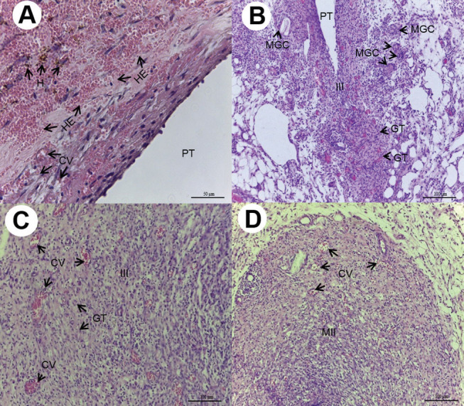
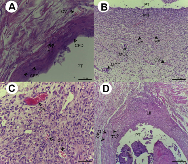
Within 7 days, an intense inflammatory infiltrate was demonstrated, singularly in Groups Meron 10%-EEP and 25%-EEP, and Groups Ketac 10%-EEP and 25%-EEP, with significant difference between the Control Group and Groups Meron 10%-EEP and Ketac 10%-EEP in the time intervals of 7 ( p = 0.001) ( Fig. 1A D ) and 15 ( p = 0.006) days ( Fig. 2A D ). In addition, a persistent chronic inflammatory infiltrate was observed in the time interval of 30 days, with significant difference between Groups C and Ketac 10%-EEP ( p = 0.010). The intensity of the inflammatory infiltrate was shown to be inversely proportional to the experimental time intervals ( Table 1 ).
Fig. 1.

( A ) Seven days after implantation, Group C: cavity surrounded by light inflammatory infiltrate, congested vessels (CV), hemorrhagic exudate (HE) with brownish hemosiderin pigments (H). (HE, 200× magnification, scale:50 µm). Area of polyethylene tube (PT) implant. ( B ) Seven days after implantation, Group M10: presence of intense inflammatory infiltrate (III), granulation tissue (GT), and multinucleated giant cells (MGC). (HE, 100×magnification, scale:100 µm). Area of PT implant. ( C ) Seven days after implantation, Group KC10: intense inflammatory infiltrate (III), CV and granulation tissue reaction (GT) (HE,100× magnification, scale:100 µm). ( D ) Seven days after implantation, Group KC50: moderate inflammatory infiltrate (MII), vascularization with numerous diminutive blood vessels, of which the majority were CV. (HE, 100× magnification, scale:100 µm).
Fig. 2.

( A ) Fifteen days after implantation, Group C: presence of congested vessels (CV), young fibroblasts (YF), and collagen fibers disposed (CFD) in parallel bundles involving the cavity ( HE, 400×magnification, scale: 25 µm). Area of polyethylene tube (PT) implant. ( B ) Fifteen days after implantation, Group M10: cavity surrounded by moderate inflammatory infiltrate (IIM), CV, presence of ovoid and fusiform YF, and multinucleated giant cells (MGC) (HE,100×magnification, scale:100µm). Area of PT implant. ( C ) Fifteen days after implantation, Group KC10; moderate inflammatory infiltrate with intense vascularization with CV of various sizes (HE, 400× magnification, scale: 25 µm). ( D ) Fifteen days after implantation, Group KC50: deposition of delicate CFD in parallel bundles, ovoid and fusiform YF, and light inflammatory infiltrate (LII) (HE, 100× magnification, scale:100 µm). Area of PT implant. HE, hematoxylin and eosin.
Table 1. Mean of the scores attributed to the GICs, with 7, 15, and 30 days, for the seven tissue conditions a .
| Condition Time/Days |
Groups | p- Value* | ||||||||||
|---|---|---|---|---|---|---|---|---|---|---|---|---|
| M | M10 | M25 | M50 | KC | KC10 | KC25 | KC50 | C | ||||
| Inflammatory infiltrate | ||||||||||||
|
a
For each sample of the study, five representative sections of the histological condition of the tissue were analyzed, when all five sections of the tissue showed the same histological condition. Scores: 1, absent (5.00); 2, scarce (10.00); 3, moderate (15.00); and 4, intense (20.00).
* p -Value indicates nonparametric Kruskal–Wallis test, followed by Dunn’s multiple comparisons test. A, B Means followed by the same single letter do not express statistically significant difference ( p > 0.05). AB Means followed by different letters express statistically significant difference ( p < 0.05). | ||||||||||||
| 7 | 13.75 AB | 20.00 A | 18.75 AB | 16.25 AB | 15.00 AB | 20.00 A | 18.75 AB | 15.00 AB | 10.00 B | 0.001 | ||
| 15 | 11.25 AB | 16.25 A | 13.75 AB | 11.25 AB | 12.50 AB | 16.25 A | 13.75 AB | 10.00 AB | 7.50 B | 0.006 | ||
| 30 | 10.00 AB | 12.50 AB | 12.50 AB | 10.00 AB | 10.00 AB | 13.75 A | 11.25 AB | 10.00 AB | 6.25 B | 0.010 | ||
| Edema | ||||||||||||
| 7 | 5.00 | 6.25 | 5.00 | 5.00 | 6.25 | 7.50 | 7.50 | 5.00 | 5.00 | 0.231 | ||
| 15 | 5.00 | 5.00 | 5.00 | 5.00 | 5.00 | 5.00 | 5.00 | 5.00 | 5.00 | 1.000 | ||
| 30 | 5.00 | 5.00 | 5.00 | 5.00 | 5.00 | 5.00 | 5.00 | 5.00 | 5.00 | 1.000 | ||
| Necrosis | ||||||||||||
| 7 | 5.00 | 5.00 | 5.00 | 5.00 | 5.00 | 6.25 | 5.00 | 5.00 | 5.00 | 0.433 | ||
| 15 | 5.00 | 5.00 | 5.00 | 5.00 | 5.00 | 5.00 | 5.00 | 5.00 | 5.00 | 1.000 | ||
| 30 | 5.00 | 5.00 | 5.00 | 5.00 | 5.00 | 5.00 | 5.00 | 5.00 | 5.00 | 1.000 | ||
| Granulation tissue | ||||||||||||
| 7 | 13.75 AB | 18.75 A | 17.50 AB | 15.00 AB | 13.75 AB | 18.75 A | 17.50 AB | 13.75 AB | 10.00 B | 0.003 | ||
| 15 | 8.75 AB | 15.00 A | 12.50 AB | 10.00 AB | 8.75 AB | 13.75 AB | 12.50 AB | 10.00 AB | 7.50 B | 0.004 | ||
| 30 | 7.50 | 11.25 | 10.00 | 10.00 | 7.50 | 10.00 | 10.00 | 10.00 | 7.50 | 0.074 | ||
| Multinucleated giant cells | ||||||||||||
| 7 | 7.50 AB | 12.50 A | 8.75 AB | 7.50 AB | 7.50 AB | 8.75 AB | 8.75 AB | 5.00 B | 5.00 B | 0.033 | ||
| 15 | 6.25 | 7.50 | 6.25 | 6.25 | 6.25 | 8.75 | 8.75 | 5.00 | 5.00 | 0.205 | ||
| 30 | 5.00 | 7.50 | 6.25 | 6.25 | 5.00 | 6.25 | 6.25 | 5.00 | 5.00 | 0.536 | ||
| Young fibroblasts | ||||||||||||
| 7 | 13.75 | 11.25 | 11.25 | 12.50 | 12.50 | 13.75 | 13.75 | 15.00 | 15.00 | 0.215 | ||
| 15 | 15.00 | 15.00 | 15.00 | 16.25 | 15.00 | 13.75 | 15.00 | 15.00 | 16.25 | 0.387 | ||
| 30 | 11.25 AB | 13.75 AB | 15.00 A | 15.00 A | 11.25 AB | 15.00 A | 15.00 A | 15.00 A | 10.00 B | 0.003 | ||
| Collagen | ||||||||||||
| 7 | 12.50 AB | 8.75 A | 10.00 AB | 11.25 AB | 11.25 AB | 8.75 A | 10.00 AB | 11.25 AB | 15.00 B | 0.019 | ||
| 15 | 16.25 AB | 10.00 A | 16.25 AB | 17.50 AB | 16.25 AB | 15.00 AB | 17.50 AB | 18.75 B | 18.75 B | 0.034 | ||
| 30 | 18.75 | 16.25 | 17.50 | 18.75 | 20.00 | 18.75 | 20.00 | 20.00 | 20.00 | 0.130 | ||
Circulatory alterations such edema and tissue necrosis were not very expressive and showed no significant difference among the groups evaluated ( p > 0.05). The granulation reaction was shown to be densely present in Groups Meron 10%-EEP and Ketac 10%-EEP with statistical difference compared with the Control ( p = 0.003) at 7 days. There was also significant difference between Groups Meron 10%-EEP and Control ( p = 0.004) at 15 days. Multinucleated giant cell reactions were shown to be more present in Group Meron 10%-EEP, with significant difference from Groups Control and Ketac 50%-EEP at 7 days ( p = 0.033).
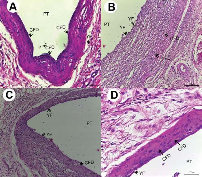
The quantities of young fibroblasts and collagen fibers increased during the course of the experimental time intervals. In the tissue repair events, Groups Meron 25%-EEP and 50%-EEP, and Groups Ketac of 10%-EEP, 25%-EEP, and 50%-EEP showed a larger quantity of young fibroblasts compared with the Group Control ( p = 0.003) at 30 days ( Fig. 3A D ). A smaller quantity of collagen fibers was observed in Groups Meron 10%-EEP and Ketac 10%-EEP compared with the Group Control ( p = 0.019) at 7 days, and smaller in Group Meron 10%-EEP when compared with the Groups Control and Ketac 50%-EEP ( p = 0.034) at 15 days.
Fig. 3.

( A ) Thirty days after implantation, Group C: thick layer of collagen fibers disposed (CFD) in parallel bundles in the midst of scarce young fibroblasts (YF) (HE, 200× magnification, scale: 50 µm). Area of polyethylene tube (PT) implant. ( B ) Thirty days after implantation, Group M10: cavity surrounded by deposition of CFD in parallel bundles and YF (HE, 100× magnification, scale:100 µm). Area of PT implant. ( C ) Thirty days after implantation, Group KC10: deposition of CFD in the midst of ovoid and fusiform YF (HE,100× magnification, scale:100µm). Area of PT implant. ( D ) 30 days after implantation, Group KC50: layer of CFD in the midst of dispersed ovoid and fusiform YF (HE, 400×magnification, scale:25 µm). Area of PT implant.
SBST and ARI Tests
The SBST showed an increase that was directly proportional to the increase in propolis concentration in the cements; there was no statistically significant difference between the Groups of Meron GICs and the Groups of Ketac Cem ( p < 0.05). In the comparison between the different concentrations for the same type of GIC, there was no statistical difference between the Groups of Meron GICs ( p = 0.170) and between the Groups Ketac Cem GICs ( p = 0.087) ( Table 2 ).
Table 2. Mean and SD of the SBST values of the different groups, expressed in Mpa.
| Groups # | n | Meron | Ketac Cem | p -Value* |
|---|---|---|---|---|
| Mean (SD) | ||||
| Abbreviations: ANOVA, analysis of variance; SBST, shear bond strength test; SD, standard deviation. # Control (10% tartaric acid solution), 10% (addition of propolis at 10%), 25% (addition of propolis at 25%), 50% (addition of propolis at 50%). *ANOVA one-way and Tukey ( p < 0.05). | ||||
| Control | 10 | 0.28 (0.11) | 0.23 (0.08) | 0.287 |
| 10% | 10 | 0.31 (0.13) | 0.28 (0.08) | 0.595 |
| 25% | 10 | 0.37 (0.17) | 0.36 (0.16) | 0.940 |
| 50% | 10 | 0.42 (0.14) | 0.36 (0.14) | 0.379 |
| p -Value * | — | 0.170 | 0.087 | — |
The ARI demonstrated that over half of the remnant cement or all of the remnant cement remained on the tooth surface after removing the specimen. The ARI showed no significant difference between the Groups of Meron GICs ( p = 0.684) and between the Groups Ketac Cem GICs ( p = 0.053) after the addition of the EEP.
Antibacterial Effect
The antibacterial effected demonstrated an increase that was directly proportional to the increase in propolis concentration in the cements; there were significant differences between Groups Meron-Control and Meron 50%-EEP, and between Groups Ketac-Control and Ketac 50%-EEP, irrespective of the time interval evaluated ( p = 0.001). The antibacterial effect was inversely proportional to the time of exposure for the same concentration; there was significant difference between the times intervals 1 and 90 days, irrespective of the propolis concentration in the cement evaluated ( p < 0.05) ( Table 3 ).
Table 3. Mean and SD, comparing the different groups of GICs according to the evaluation time intervals for the measurement of inhibition zone diameters.
| Period/days | Groups | |||||||||
|---|---|---|---|---|---|---|---|---|---|---|
| M | M10 | M25 | M50 | p- Value # | KC | KC10 | KC25 | KC50 | p -Value # | |
| Abbreviations: GIC, glass ionomer cement; SD, standard deviation. A, B Means followed by the same single letter do not express statistically significant difference ( p >0.05). AB Means followed by different letters express statistically significant difference ( p < 0.05). # p = Friedman's nonparametric test, followed by the Dunn multiple test (in-line, upper case). * p = Kruskal–Wallis non-parametric test, followed by Dunn's multiple-comparison test (column, lowercase). | ||||||||||
| 1 | 6.2 (0.5) Aa | 7.2 (1.0) Aa | 9.1(0.6) Aba | 15.3 (1.0) Ba | 0.001 | 6.6 (0.4) Aa | 8.7 (0.6) ABa | 10.9 (0.8) ABa | 16.8 (0.7) Ba | 0.001 |
| 30 | 0.0 (0.0) Ab | 6.8 (0.7) ABa | 7.1(0.6) ABab | 13.0 (0.8) Bab | 0.001 | 0.0 (0.0) Ab | 6.9 (1.1) ABab | 7.7 (0.9) ABab | 14.9 (0.6) Bab | 0.001 |
| 90 | 0.0 (0.0) Ab | 5.3 (0.2) ABb | 5.7 (0.6) ABb | 11.9 (1.0) Bb | 0.001 | 0.0 (0.0) Ab | 5.8 (0.9) ABb | 5.9 (1.2) ABb | 12.8 (1.3) Bb | 0.001 |
| p- Value* | 0.001 | 0.008 | 0.001 | 0.006 | – | 0.001 | 0.010 | 0.003 | 0.003 | – |
Discussion
The requisites of a GIC must include the capacity to resist occlusal forces, and the SBST test is one of the most frequently used mechanical tests for simulating an orthodontic clinical situation. 27 In the present study, this test demonstrated an increase that was directly proportional to that of the propolis concentration in the cements, but without significant difference when compared with the controls, corroborating the findings of other studies. 5 9
The EEP has aromatic fatty acids and phenolic compounds in its molecule, and it has been demonstrated that the polyphenols have structures favorable to improvement in the mechanical properties of the GIC, due to their high level of activity. 29 These polyphenols have shown a chelation reaction between the phenolic groups of hydroxyl and carboxyl of GIC, 30 providing a larger quantity of poly-salt linking and cross linking, thus increasing the structural complexity of the GIC with EEP, 31 32 which were aligned to better performance of the tested GICs. This suggested that the addition of the EEP did not interfere negatively in the clinical performance of the GIC.
The results found in this study for the SBST corroborated those of the ARI results, in which it was shown that over half or all of the remnant adhesive remained on the tooth surface after removal of the specimens, irrespective of the addition of the EEP, proving that the addition of the EEP did not diminish the cohesive strength of the cement, or the bond of the GIC to the dental structure. 26
In the present study, the antibacterial effect against S. mutans was shown to be concentration-dependent; this may have been correlated to the rate of elution of the antibacterial agent from the GIC, 14 in which synergism appeared to have occurred between the metal particles and the EEP. Thus, higher concentrations of EEP could diminish the participation of S. mutans in the formation of dental biofilm. 12 14
The antibacterial effect was also shown to be inversely proportional to the time of exposure to the same concentration. The EEP added to the GIC was shown to be capable of inhibiting the adhesion of S. mutans to the cement surface. 14 33 These findings corroborated those of other studies that showed that the addition of EEPs to GIC particularly in the concentrations of 25 and 50% was capable of inhibiting the growth of S. mutans 5 12 and demonstrated significant inhibition haloes compared with those of the control. 12 The antiadherence activities of the GIC with EEP may be linked to the changes in the hydrophobic bond of this association 34 furthermore, authors 14 have reported that this association did not interfere negatively in the release of fluoride by these materials.
Tissue biocompatibility investigated by quali-quantitative analysis was based on the intensity of the tissue aggression caused by the cements and its respective healing process. In the present study, initial intense inflammatory infiltrate was demonstrated by both cements with the addition of 10 and 25% of EEP in the time intervals of 7 and 15 days. The intensity of the inflammatory infiltrate was shown to be inversely proportional to the time of evaluation. The low concentration of the EEP in the Meron and Ketac Groups with 10%-EEP, allied to the presence of alcohol in its composition, which functions as a solvent or vehicle for the propolis, 35 is suggested to have generated a low potential for rapid tissue healing.
The EEP in higher concentrations demonstrated better anti-inflammatory and healing effects on live tissues. 5 36 In this sense, the Groups that contained a concentration of 50% propolis in this study showed a lower inflammatory potential, which suggested that higher concentrations of EEP would be capable of diminishing the potentially aggressive effect of alcohol on the tissues, thereby heightening the effect of anti-inflammatory agents such as the flavonoids and caffeic acid present in the composition of propolis. 15 37
Events of edema and tissue necrosis were not expressive and showed no significant difference among the experimental groups. Granulation tissue was shown to be densely present in the Groups with 10%-EEP in the time interval of 7 days and persisted significantly only in Group Meron with 10%-EEP in the time interval of 15 days. Multinucleated giant cells were shown to be more present in Group M10 at 7 days; this suggested a greater potential of the components in the composition of Meron, associated with the alcohol present in the10%-EEP which, as a response, demonstrated the presence of giant cells with the purpose of isolating the foreign body. 19
The quantities of young fibroblasts and collagen fibers increased during the course of the experimental time intervals. Meron with 10%-EEP showed a lower quantity of young fibroblasts in the time interval of 30 days. A smaller quantity of collagen fibers was also observed for this group in the time intervals of 7 and 15 days; this also occurred in the Group Ketac with 10%-EEP in the time interval of 7 days. The cements with the higher concentrations of EEP showed a larger number of collagen fibers over the course of the experiment, which allowed a faster process of healing/collagenization; this suggested a direct concentration-dependent relationship of propolis with the process of tissue healing.
Previous researches 13 38 have shown that propolis may be toxic to dental pulp fibroblasts in concentrations lower than those used in this experiment 13 and could interfere in cell viability. 15 38 In the present study, the results demonstrated a compatibility within parameters considered safe, which we attributed to the fact that the EEP was not used in its liquid form, or in paste, but rather as an intrinsic part of the polymerized GIC and was being slowly released into the live tissues.
Conclusions
Biocompatibility
The histocompatibility analysis showed that the intensity of histological changes in the cements was shown to be inversely proportional to the concentration of propolis added.
The cement Ketac with 50%-EEP was the one that demonstrated the smallest inflammatory process and fastest tissue repair.
Mechanical Testing
The addition of EEP to GIC did not negatively change the SBST and ARI.
Antibacterial Effect
Antibacterial property demonstrated a concentration-dependent effect, and incorporation of 50%-EEP into GICs has been shown to be an encouraging method for achieving an antibacterial GIC in dentistry.
Acknowledgments
The authors thank the Coordenação de Aperfeiçoamento de Pessoal de Nível Superior - Brasil (CAPES).
Footnotes
Conflict of Interest None declared.
References
- 1.Enan E T, Hammad S M. Microleakage under orthodontic bands cemented with nano-hydroxyapatite-modified glass ionomer. Angle Orthod. 2013;83(06):981–986. doi: 10.2319/022013-147.1. [DOI] [PMC free article] [PubMed] [Google Scholar]
- 2.Santos R L, Moura M de F, Carvalho F G, Guênes G M, Alves P M, Pithon M M. Histological analysis of biocompatibility of ionomer cements with an acid-base reaction. Braz Oral Res. 2014;28(01):1–7. doi: 10.1590/S1806-83242014.50000003. [DOI] [PubMed] [Google Scholar]
- 3.Ebaya M M, Ali A I, Mahmoud S H. Evaluation of marginal adaptation and microleakage of three glass ionomer-based Class V restorations: in vitro study. Eur J Dent. 2019;13(04):599–606. doi: 10.1055/s-0039-3401435. [DOI] [PMC free article] [PubMed] [Google Scholar]
- 4.Moheet I A, Luddin N, Rahman I A, Kannan T P, Nik Abd Ghani N R, Masudi S M. Modifications of glass ionomer cement powder by addition of recently fabricated nano-fillers and their effect on the properties: a review. Eur J Dent. 2019;13(03):470–477. doi: 10.1055/s-0039-1693524. [DOI] [PMC free article] [PubMed] [Google Scholar]
- 5.Hatunoğlu E, Oztürk F, Bilenler T, Aksakallı S, Simşek N. Antibacterial and mechanical properties of propolis added to glass ionomer cement. Angle Orthod. 2014;84(02):368–373. doi: 10.2319/020413-101.1. [DOI] [PMC free article] [PubMed] [Google Scholar]
- 6.Akao Y, Maruyama H, Matsumoto K et al. Cell growth inhibitory effect of cinnamic acid derivatives from propolis on human tumor cell lines. Biol Pharm Bull. 2003;26(07):1057–1059. doi: 10.1248/bpb.26.1057. [DOI] [PubMed] [Google Scholar]
- 7.Jafarzadeh Kashi T S, Kasra Kermanshahi R, Erfan M, Vahid Dastjerdi E, Rezaei Y, Tabatabaei F S. Evaluating the in-vitro antibacterial effect of Iranian propolis on oral microorganisms. Iran J Pharm Res. 2011;10(02):363–368. [PMC free article] [PubMed] [Google Scholar]
- 8.Schnitzler P, Neuner A, Nolkemper S et al. Antiviral activity and mode of action of propolis extracts and selected compounds. Phytother Res. 2010;24 01:S20–S28. doi: 10.1002/ptr.2868. [DOI] [PubMed] [Google Scholar]
- 9.Silici S, Koç N A, Ayangil D, Cankaya S. Antifungal activities of propolis collected by different races of honeybees against yeasts isolated from patients with superficial mycoses. J Pharmacol Sci. 2005;99(01):39–44. doi: 10.1254/jphs.fpe05002x. [DOI] [PubMed] [Google Scholar]
- 10.Esmeraldo M R, Carvalho M G, Carvalho R A, Lima R deF, Costa E M. Inflammatory effect of green propolis on dental pulp in rats. Braz Oral Res. 2013;27(05):417–422. doi: 10.1590/S1806-83242013005000022. [DOI] [PubMed] [Google Scholar]
- 11.Ferreira F B, Torres S A, Rosa O P et al. Antimicrobial effect of propolis and other substances against selected endodontic pathogens. Oral Surg Oral Med Oral Pathol Oral Radiol Endod. 2007;104(05):709–716. doi: 10.1016/j.tripleo.2007.05.019. [DOI] [PubMed] [Google Scholar]
- 12.Topcuoglu N, Ozan F, Ozyurt M, Kulekci G. In vitro antibacterial effects of glass-ionomer cement containing ethanolic extract of propolis on Streptococcus mutans. Eur J Dent. 2012;6(04):428–433. [PMC free article] [PubMed] [Google Scholar]
- 13.Al-Shaher A, Wallace J, Agarwal S, Bretz W, Baugh D. Effect of propolis on human fibroblasts from the pulp and periodontal ligament. J Endod. 2004;30(05):359–361. doi: 10.1097/00004770-200405000-00012. [DOI] [PubMed] [Google Scholar]
- 14.Elgamily H, Ghallab O, El-Sayed H, Nasr M. Antibacterial potency and fluoride release of a glass ionomer restorative material containing different concentrations of natural and chemical products: an in-vitro comparative study. J Clin Exp Dent. 2018;10(04):e312–e320. doi: 10.4317/jced.54606. [DOI] [PMC free article] [PubMed] [Google Scholar]
- 15.Sabir A, Sumidarti A. Interleukin-6 expression on inflamed rat dental pulp tissue after capped with Trigona sp. propolis from south Sulawesi, Indonesia. Saudi J Biol Sci. 2017;24(05):1034–1037. doi: 10.1016/j.sjbs.2016.12.019. [DOI] [PMC free article] [PubMed] [Google Scholar]
- 16.dos Santos R L, de Sampaio G A, de Carvalho F G, Pithon M M, Guênes G M, Alves P M. Influence of degree of conversion on the biocompatibility of different composites in vivo. J Adhes Dent. 2014;16(01):15–20. doi: 10.3290/j.jad.a29704. [DOI] [PubMed] [Google Scholar]
- 17.Eliades T. Orthodontic materials research and applications: part 2. Current status and projected future developments in materials and biocompatibility. Am J Orthod Dentofacial Orthop. 2007;131(02):253–262. doi: 10.1016/j.ajodo.2005.12.029. [DOI] [PubMed] [Google Scholar]
- 18.Gonçalves T S, de Menezes L M, Ribeiro L G, Lindholz C G, Medina-Silva R. Differences of cytotoxicity of orthodontic bands assessed by survival tests in Saccharomyces cerevisiae. BioMed Res Int. 2014;2014:143283. doi: 10.1155/2014/143283. [DOI] [PMC free article] [PubMed] [Google Scholar]
- 19.Lacerda-Santos R, Sampaio G A, Moura M de F et al. Effect of different concentrations of chlorhexidine in glass-ionomer cements on in vivo biocompatibility. J Adhes Dent. 2016;18(04):325–330. doi: 10.3290/j.jad.a36512. [DOI] [PubMed] [Google Scholar]
- 20.Farret M M, Lima E M, Mota E G et al. Assessment of the mechanical properties of glass ionomer cements for orthodontic cementation. Dental Press J Orthod. 2012;17(06):154–159. [Google Scholar]
- 21.Wilson A D. Developments in glass-ionomer cements. Int J Prosthodont. 1989;2(05):438–446. [PubMed] [Google Scholar]
- 22.Henn S, Nedel F, de Carvalho R V et al. Characterization of an antimicrobial dental resin adhesive containing zinc methacrylate. J Mater Sci Mater Med. 2011;22(08):1797–1802. doi: 10.1007/s10856-011-4364-x. [DOI] [PubMed] [Google Scholar]
- 23.Seidenari S, Giusti F, Pepe P, Mantovani L. Contact sensitization in 1094 children undergoing patch testing over a 7-year period. Pediatr Dermatol. 2005;22(01):1–5. doi: 10.1111/j.1525-1470.2005.22100.x. [DOI] [PubMed] [Google Scholar]
- 24.Duailibe S A, Gonçalves A G, Ahid F J. Effect of a propolis extract on Streptococcus mutans counts in vivo. J Appl Oral Sci. 2007;15(05):420–423. doi: 10.1590/S1678-77572007000500009. [DOI] [PMC free article] [PubMed] [Google Scholar]
- 25.Lacerda-Santos R, Roberto B MS, de Siqueira Nunes B, Carvalho F G, Dos Santos A, Dantas A FM. Histological analysis of biocompatibility of different surgical adhesives in subcutaneous tissue. Microsc Res Tech. 2019;82(07):1184–1190. doi: 10.1002/jemt.23267. [DOI] [PubMed] [Google Scholar]
- 26.Pithon M M, dos Santos R L, Oliveira M V et al. Evaluation of the shear bond strength of two composites bonded to conditioned surface with self-etching primer. Dental Press J Orthod. 2011;16(02):94–99. [Google Scholar]
- 27.Farret M M, de Lima E M, Mota E G, Oshima H M, Barth V, de Oliveira S D. Can we add chlorhexidine into glass ionomer cements for band cementation? Angle Orthod. 2011;81(03):496–502. doi: 10.2319/090310-518.1. [DOI] [PMC free article] [PubMed] [Google Scholar]
- 28.Artun J, Bergland S. Clinical trials with crystal growth conditioning as an alternative to acid-etch enamel pretreatment. Am J Orthod. 1984;85(04):333–340. doi: 10.1016/0002-9416(84)90190-8. [DOI] [PubMed] [Google Scholar]
- 29.Tipoe G L, Leung T M, Hung M W, Fung M L. Green tea polyphenols as an anti-oxidant and anti-inflammatory agent for cardiovascular protection. Cardiovasc Hematol Disord Drug Targets. 2007;7(02):135–144. doi: 10.2174/187152907780830905. [DOI] [PubMed] [Google Scholar]
- 30.Lo C Y, Hsiao W T, Chen X Y. Efficiency of trapping methylglyoxal by phenols and phenolic acids. J Food Sci. 2011;76(03):H90–H96. doi: 10.1111/j.1750-3841.2011.02067.x. [DOI] [PubMed] [Google Scholar]
- 31.Hu J, Du X, Huang C, Fu D, Ouyang X, Wang Y. Antibacterial and physical properties of EGCG-containing glass ionomer cements. J Dent. 2013;41(10):927–934. doi: 10.1016/j.jdent.2013.07.014. [DOI] [PubMed] [Google Scholar]
- 32.Zanata R L, Magalhães A C, Lauris J R, Atta M T, Wang L, Navarro M F. Microhardness and chemical analysis of high-viscous glass-ionomer cement after 10 years of clinical service as ART restorations. J Dent. 2011;39(12):834–840. doi: 10.1016/j.jdent.2011.09.003. [DOI] [PubMed] [Google Scholar]
- 33.Türkün L S, Türkün M, Ertuğrul F, Ateş M, Brugger S. Long-term antibacterial effects and physical properties of a chlorhexidine-containing glass ionomer cement. J Esthet Restor Dent. 2008;20(01):29–44, discussion 45. doi: 10.1111/j.1708-8240.2008.00146.x. [DOI] [PubMed] [Google Scholar]
- 34.Razak F A, Rahim Z H. The anti-adherence effect of Piper betle and Psidium guajava extracts on the adhesion of early settlers in dental plaque to saliva-coated glass surfaces. J Oral Sci. 2003;45(04):201–206. doi: 10.2334/josnusd.45.201. [DOI] [PubMed] [Google Scholar]
- 35.Troca V B, Fernandes K B, Terrile A E, Marcucci M C, Andrade F B, Wang L. Effect of green propolis addition to physical mechanical properties of glass ionomer cements. J Appl Oral Sci. 2011;19(02):100–105. doi: 10.1590/S1678-77572011000200004. [DOI] [PMC free article] [PubMed] [Google Scholar]
- 36.Wagh V D. Propolis: a wonder bees product and its pharmacological potentials. Adv Pharmacol Sci. 2013;2013:308249. doi: 10.1155/2013/308249. [DOI] [PMC free article] [PubMed] [Google Scholar]
- 37.Meto A, Meto A, Bimbari B, Shytaj K, Özcan M. Anti-inflammatory and regenerative effects of Albanian propolis in experimental vital amputations. Eur J Prosthodont Restor Dent. 2016;24(03):145–151. doi: 10.1922/EJPRD_1492Meto07. [DOI] [PubMed] [Google Scholar]
- 38.Zare Jahromi M, Ranjbarian P, Shiravi S. Cytotoxicity evaluation of Iranian propolis and calcium hydroxide on dental pulp fibroblasts. J Dent Res Dent Clin Dent Prospect. 2014;8(03):130–133. doi: 10.5681/joddd.2014.024. [DOI] [PMC free article] [PubMed] [Google Scholar]


