


ABSTRACT
Background
Studies on human intraocular tuberculosis (IOTB) are extremely challenging. For understanding the pathogenesis of IOTB, it is important to investigate the mycobacterial transcriptional changes in ocular environment.
Methods
Mice were challenged intravenously with Mycobacterium tuberculosis H37Rv and at 45 days post-infection, experimental IOTB was confirmed based on bacteriological and molecular assays. M. tuberculosis transcriptome was analyzed in the infected eyes using microarray technology. The identified M. tuberculosis signature genes were further validated and investigated in human IOTB samples using real-time polymerase chain reaction.
Results
Following intravenous challenge with M. tuberculosis, 45% (5/12) mice showed bacilli in the eyes with positivity for M. tuberculosis ribonucleic acid in 100% (12/12), thus confirming the paucibacillary nature of IOTB similar to human IOTB. M. tuberculosis transcriptome in these infected eyes showed significant upregulation of 12 M. tuberculosis genes and five of these transcripts (Rv0962c, Rv0984, Rv2612c, Rv0974c and Rv0971c) were also identified in human clinically confirmed cases of IOTB.
Conclusions
Differentially expressed mycobacterial genes identified in an intravenously challenged paucibacillary mouse IOTB model and presence of these transcripts in human IOTB samples highlight the possible role of these genes for survival of M. tuberculosis in the ocular environment, thus contributing to pathogenesis of IOTB.
Keywords: intraocular tuberculosis, mouse model, Mycobacterium tuberculosis H37Rv, transcriptome, microarray, human vitreous sample
M. tuberculosis transcriptional signatures identified in the eyes of infected animals and validated in human intraocular tuberculosis (IOTB) vitreous samples explain the molecular pathogenesis of IOTB and may aid in development of diagnostics for IOTB.
BACKGROUND
Tuberculosis (TB) in 21st century is still one of the most puzzling forms of infectious disease and the disease complexity has further been augmented due to emergence of resistant strains and the ability of Mycobacterium tuberculosis (M. tuberculosis) to cause disease in any part of the body, embodied as extrapulmonary manifestations (Pai et al. 2016). One of the extrapulmonary forms of the disease is intraocular tuberculosis (IOTB), tuberculosis of eye. The history of IOTB goes back to the 19th century (Helm and Holland 1993); however, the actual epidemiology of IOTB is still not very clear because there is no defined gold standard test (Gupta et al. 2015a) or diagnostic criteria (Gupta, Gupta and Rao 2007). Intraocular tuberculosis has also been reported in human immuno-deficiency virus (HIV)-infected individuals with or without signs of pulmonary infection (Bukulmez et al. 2009; Mehta et al. 2013). The prevalence of IOTB is higher in endemic countries such as India (∼10%) (Gupta, Gupta and Rao 2007) but nonetheless the disease has also been recognized in immunocompromised patients of nonendemic countries such as the UK (Cutrufello et al. 2010; Manousaridis et al. 2013).
Intraocular tuberculosis is a clinically specified granulomatous infection of the eye involving any part of the uveal tract (Gupta, Gupta and Rao 2007; Yeh et al. 2012) and presents as TB uveitis (Gupta et al. 2015b). Unavailability of adequate human samples from the infected eye and failure in detecting the presence of live bacteria in such an immune-privileged site make it challenging to understand the pathogenesis of IOTB (Vasconcelos-Santos, Zierhut and Rao 2009; Yeh et al. 2012; Basu et al. 2015). Given the present challenges in studying human IOTB, a well-defined animal model is required (Basu et al. 2015). Although, in animals, a guinea pig model for IOTB has previously been reported (Rao et al. 2009), however, how M. tuberculosis uniquely behaves and adapts in such an immune-privilege-site remains to be understood. The mouse model may be preferable as it is considered as a robust host to study various ocular infectious diseases (Zhang, Abdel-Razek and Wang 2017), pulmonary (Talaat et al. 2004) and extrapulmonary TB (Be et al. 2008), and could be utilized in studying the mycobacterial gene expression at the site of infection (Talaat et al. 2004). Such an information is imperative in understanding the pathogenesis (Ward et al. 2010) and how bacteria colonize, invade, interact with or disrupt the host environment to cause disease (Talaat et al. 2004) or survive in host environment (Jain et al. 2006).
Therefore, the current study was based on our previous study where we established a paucibacillary condition of IOTB in mouse model as depicted in humans (Abhishek, Gupta and Verma 2014). For further gaining the insight in the pathogenesis (Ward et al. 2010) of IOTB, gene expression profile of mycobacteria in the ocular environment of mice intravenously (i.v.) challenged with M. tuberculosis was observed. A pilot experiment was also carried out to confirm the in vivo presence of identified M. tuberculosis signatures in the vitreous samples of human IOTB.
MATERIALS AND METHODS
Animals and M. tuberculosis strain
The animal studies were performed after the Institute Animal Ethics Committee (IAEC) of Postgraduate Institute of Medical Education and Research (PGIMER), Chandigarh, India approval (#56/IAEC/300). C57BL/6 mice (4–5 weeks, 20–25 g) were obtained from Institute of Microbial Technology, Chandigarh, India. Mice were housed in cages contained within a negative pressure animal isolator at Central Animal House, PGIMER, Chandigarh with 12 hr day-light cycle, controlled temperature (24°C ± 1°C), humidity (45–55%) and were fed on standard pellet diet (Hindustan Lever Ltd., Mumbai) and water ad libitum, throughout the study. M. tuberculosis H37Rv was grown and maintained as described previously (Mir, Verma and Sharma 2014).
Human samples were obtained from ocular samples repository for ophthalmology research cleared by the Institute Ethics Committee (IEC) of PGIMER, Chandigarh, India approval (#NK/3323/study/186). A written consent was obtained from all participants before sample collection.
Mouse model of intraocular tuberculosis
Twelve mice were infected with M. tuberculosis via i.v. route (105bacilli/animal) and were sacrificed at 45th day post infection (dpi) under anesthesia as approved by IAEC, PGIMER, Chandigarh (#56/IAEC/300). Tissues (eye, lungs and spleen) from these mice were further used for colony forming unit (CFU) enumeration. The homogenized whole eyeball was also used for M. tuberculosis ribonucleic acid (RNA) presence and M. tuberculosis whole genome microarray study.
Colony forming unit enumeration
Mice eyes, lungs and spleen were aseptically removed and homogenized using a glass homogenizer separately in Phosphate Buffer Saline (PBS) containing 0.05% (v/v) Tween-80 (Sigma-Aldrich, Saint Louis, USA) (PBS-T) under sterile conditions. Each eyeball was homogenized in 500 µl of PBS-T and 200 µl of each eye homogenate was plated neat onto 7H11 agar plates with 10% Oleic Acid, Dextrose and Catalase (OADC) (BD BBL Middlebrook, Thermo Fisher Scientific, Ottawa, Ontario) for CFU enumeration. Similarly, lungs and spleen were homogenized, and plated as described above. The plates were incubated at 37°C for 3–4 weeks and colonies were counted to determine the CFU load in each organ.
M. tuberculosis RNA isolation and whole genome microarray of M. tuberculosis from mouse eye
The mouse eyeballs were examined to identify M. tuberculosis transcriptional signatures. A total of four eyes (left/right) from four animals were utilized for the microarray experiments using reference RNA isolated from M. tuberculosis culture (log-phase). Total RNA was isolated using RNAzol (MRC, Cincinnati, Ohio), and MICROBEnrich Kit (Ambion, Texas, USA) was used to deplete eukaryotic RNA followed by treatment with Turbo DNase (Ambion, Texas, USA) to remove contaminating DNA. The RNA quality and quantity were determined using Agilent 2100 Bioanalyzer. 30 ng of purified RNA from each experimental sample and reference RNA from M. tuberculosis were amplified using MessageAmp II-Bacteria RNA Amplification Kit (Ambion, Texas, USA). Complementary deoxyribonucleic acid (cDNA) was prepared from amplified RNA (aRNA) using Superscript III Reverse Transcriptase (RT) (Invitrogen, California, USA) and fluorescently labeled with Cy3 and Cy5 (reference and experimental, respectively, and vice versa) using BioPrime DNA Labeling System (Invitrogen, California, USA). Each Cy5- and Cy3-labeled cDNA (one experimental sample; one reference) was applied to M. tuberculosis whole genome array slides comprising of 70-mer oligonucleotides representing all annotated open reading frames (ORF) in the M. tuberculosis H37Rv genome (Center for Applied Genomics, PHRI, Rutgers, Newark, NJ) followed by hybridization overnight at 50°C. The slides were extensively washed, scanned and data was processed with GenePix 4000B scanner and GenePix Pro 7 software, respectively.
Real-time quantitative reverse transcription PCR
The microarray results were validated by observing the mRNA expression of four genes, in the unamplified total RNA isolated without using MICROB Enrich Kit (Ambion, Texas, USA) from the eyes of infected mice by Real-Time Quantitative Reverse Transcription Polymerase Chain Reaction (RT-qRT-PCR). Primer designing (Table S1, Supporting Information), cDNA synthesis (Thermo Scientific Revert Aid First Strand cDNA synthesis kit, USA) and mRNA expression of target genes were performed and relative gene expression was calculated as described previously (Sharma et al. 2017) using 16s ribosomal RNA for normalization.
Identification of M. tuberculosis genes in human IOTB samples
The M. tuberculosis genes identified in the eyes of mouse model of IOTB were studied in the vitreous samples of human IOTB as potential M. tuberculosis signatures in IOTB disease (Abhishek et al. 2018). Total RNA was isolated from six clinically confirmed cases of human IOTB, and DNase-treated RNA was utilized for cDNA synthesis. Real-time PCR was carried out to detect the presence or absence of these genes. Initially, Rv16s was used for M. tuberculosis RNA detection in these six samples and only positive samples were further subjected for real-time PCR for selected mycobacterial genes. Genes with early C t (cycle threshold) value in RT + (reverse transcriptase) samples (in comparison to its RT −) were defined to be positive due to RNA (R +), whereas, early C t value in RT − (in comparison to its RT +) samples were assigned to be positive based on DNA (D +).
Statistical analysis
For microarray data, the scanned chips were normalized by the print-tip Lowess method (Dudoit et al. 2002), and the Cy5/Cy3 (or Cy3/Cy5 for dye flip) intensity ratios were determined for each M. tuberculosis gene. The data obtained was used to perform Significance Analysis of Microarrays (SAM) to determine log ratios (Tusher, Tibshirani and Chu 2001) and fold changes and >1.5-fold difference (up or down) was considered differentially expressed (Fisher, Plikaytis and Shinnick 2002; Rohde, Abramovitch and Russell 2007). RT-qRT-PCR data were analyzed using unpaired two-tailed Student's t-tests (GraphPad Prism software 7.0) and were significant if P value < 0.05.
RESULTS
Mouse model of intraocular tuberculosis
The 12 mice challenged intravenously with M. tuberculosis showed bacteriological positivity in 45% mice (5/12) eyes (Table 1), while the eyes of the remaining 7 mice showed no bacteriological positivity despite considerable bacterial load in lung and spleen in the range of 106 CFUs in each organ (data not shown). Further, real-time PCR of Rv3131 gene indicated the presence of M. tuberculosis RNA in the eye lysate of all the animals (Table 1). Thus, these results indicated that i.v. challenge of C57BL/6 mice with M. tuberculosis resulted in the establishment of an IOTB model depicting the paucibacillary condition with evidence of M. tuberculosis RNA in the eyes of all the animals (12/12) and bacteriological positivity in only 45% of infected animals.
Table 1.
Mouse model of intraocular tuberculosis.
| Model (n) | Mouse identity | CFU in eye | RT-PCR (Rv3131) |
|---|---|---|---|
| 105M. tuberculosis i.v., sacrificed 45th dpi, (n=12) | M1 | ND | + |
| M2 | ND | + | |
| M3 | ND | + | |
| M4 | ND | + | |
| M5 | 3 | + | |
| M6 | ND | + | |
| M7 | 95 | + | |
| M8 | 5 | + | |
| M9 | 48 | + | |
| M10 | ND | + | |
| M11 | ND | + | |
| M12 | 3 | + |
Note: CFU enumeration and RT-PCR of intravenously challenged mice at 105 dose of M. tuberculosis infection. Mice were sacrificed 45th day post infection. Values are presented as CFU per mouse in one eye using 200 µl of whole eye lysate. CFU: colony forming unit; RT-PCR: real-time polymerase chain reaction; i.v.: intravenous; dpi: day post infection; ND: not detectable; +: positive; M1–M12: mouse identity numbers.
M. tuberculosis transcriptional signatures in the mouse model of intraocular tuberculosis
To delineate the M. tuberculosis gene expression profile associated with IOTB, M. tuberculosis whole genome transcriptome study was carried out by microarray in the eyes from mice challenged intravenously with M. tuberculosis. A total of eight arrays were analyzed (four biological replicates and four technical replicates) to infer the M. tuberculosis genes involved in IOTB. Overall,12 M. tuberculosis genes were significantly upregulated, and 3 were significantly downregulated in the mouse eyes (Fig. 1A and B). The significantly upregulated M. tuberculosis genes were Rv0962c, Rv0984, Rv2612c, Rv0974c, Rv2411c, Rv2811, Rv1744c,ORF00110, Rv0996, Rv1765c, Rv0971c and Rv0772. The significantly downregulated M. tuberculosis genes were ORFD0214, ORFD0122 and ORFD0123 (Fig. 1A and B; Table S2, Supporting Information). The nomenclature ‘ORF’ denotes a predicted gene that had not yet been annotated at the time of microarray chip design.
Figure 1.

M. tuberculosis transcriptional signatures in the mouse model of intraocular tuberculosis. (A) Heatmap view of upregulated and downregulated M. tuberculosis genes. Each row represents a specific gene, whereas each column represents each sample. The last column depicts the average. Red signifies upregulated and green signifies downregulated genes (≥1.5-fold change). Data represents fold change from eight hybridization slides (four biological replicates and four technical replicates). (B) Histogram shows the fold change (average + S.D.) of statistically differentially expressed genes depicted in (A) in comparison to reference RNA (in vitro logarithmically grown inoculum)
Validation of M. tuberculosis transcriptional signatures
The expression of top three upregulated genes (Rv0962c, Rv2612c and Rv0984) was further confirmed by RT-qRT-PCR for validating the microarray results. Additionally, Rv0986, which was not significantly upregulated (1.004-fold change) by microarray, was also evaluated through RT-qRT-PCR. Rv0962c, Rv2612c and Rv0984 showed significant upregulation (P < 0.05) with an average log2 fold change of 7.40, 4.13 and 3.47, respectively, as compared to the same genes in the reference M. tuberculosis (Fig. 2). In case of Rv0986, RT-qRT-PCR analysis showed no significant upregulation (0.83 log2 fold change) in concordance to microarray data.
Figure 2.
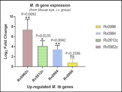
Validation of M. tuberculosis transcriptional signatures. RT-qRT-PCR analysis of the upregulated genes on microarray analysis of M. tuberculosis in the eyes of i.v. challenged mouse model of IOTB, 45th dpi. Expression is compared to the in vitro logarithmically grown inoculum and transcripts were normalized against 16s transcripts. Shown is the log2 fold change in gene expression ± S.D. P-values ≤0.05 are statistically significant. NS, not significant; M. tb, Mycobacterium tuberculosis.
M. tuberculosis signatures in human intraocular tuberculosis
Five M. tuberculosis genes (Rv0962c, Rv0984, Rv2612c, Rv0974c and Rv0971c) observed to be upregulated in the eyes of mouse IOTB were also identified to be present in human IOTB samples. M. tuberculosis signatures in term of RNA/DNA were diverse and Rv0962c, Rv0984, Rv2612c, Rv0974c and Rv0971c were identified in 3/4 human IOTB samples (Table 2). For all the genes, Ct values obtained were between 28 and 33.
Table 2.
Identification of M. tuberculosis genes in human IOTB samples.
| Rv16s | Rv0962c | Rv0984 | Rv2612c | Rv0974c | Rv0971c | |
|---|---|---|---|---|---|---|
| H-IOTB-1 | − | ND | ND | ND | ND | ND |
| H-IOTB-2 | + | D+ | R+ | D+ | D+ | D+ |
| H-IOTB-3 | + | D+ | R+ | D+ | − | D+ |
| H-IOTB-4 | − | ND | ND | ND | ND | ND |
| H-IOTB-5 | + | R+ | R+ | D+ | R+ | D+ |
| H-IOTB-6 | + | − | − | − | R+ | − |
Note: Six confirmed human IOTB samples were subjected to study the M. tuberculosis upregulated genes identified as upregulated in mouse IOTB model. cDNA was synthesized from RNA isolated from these samples and was subjected to RT-PCR to study the presence or absence of these genes. Genes which showed C t value in RT + samples were defined to be selected on the basis of RNA (R +), while early C t values in RT − samples (in comparison to RT +) were defined to be selected on the basis of DNA (D +). Only Rv16s positive samples were studied for these signature genes. cDNA: complementary deoxyribonucleic acid; RT: reverse transcriptase; RT-PCR: real-time polymerase chain reaction; H-IOTB-1–6: sample identity numbers; ND: not done; +: positive; −: negative.
DISCUSSION
The pathogenesis of IOTB was studied by defining the M. tuberculosis transcripts in ocular environment of mouse model of IOTB. Some of these M. tuberculosis signatures were also identified in in vivo human IOTB condition. In the present study, for experimental IOTB, mice were challenged intravenously (Abhishek, Gupta and Verma 2014) as M. tuberculosis disseminates to extrapulmonary sites having high blood flow (Barrios-Payán et al. 2012) and intravenous route of infection has commonly been used for the establishment of experimental models of extra pulmonary TB (Be et al. 2008). Mice sacrificed 45th dpi showed paucibacillary condition with high bacterial load in lung and spleen (data not shown) and low or no CFU count in eyes, despite positivity for M. tuberculosis RNA in all the animals (Table 1). We observed that only 45% (5/12 animals) of the i.v. challenged mice got microbiologically confirmed M. tuberculosis infection in their eyes. This observation is supported by a recent report on zebrafish model of ocular TB in which M. marinum bacterial load was obtained only in 20% of the zebrafish infected by caudal vein injection (Ramakrishnan, Basu and Takaki 2018). In another study on Balb/c mouse model of keratitis, viable bacilli were obtained in the eyes of one out of four infected animals (Moore et al. 2009). Earlier in humans also, examination of the removed eyes of IOTB have shown the paucibacillary nature of the disease (Wroblewski et al. 2011; Basu et al. 2012), along with or without the evidence of pulmonary infection (Bukulmez et al. 2009; Mehta et al. 2013). Rao et al. also observed bacteriological evidence of ocular infection in only two of the six aerosol-infected guinea pigs and positive IS6110 q-RT-PCR for mycobacterial DNA in four animals (Rao et al. 2009).
Altogether, considering the positivity for M. tuberculosis RNA in the eyes of all (100%) the mice of M. tuberculosis infected group (Table 1), mycobacterial transcriptional signatures in the ocular samples were investigated using four biological and four technical replicates. Due to paucibacillary condition of M. tuberculosis in eye, we utilized the whole eye for microarray experiments. Differentially expressed M. tuberculosis genes in IOTB could play a potential role in the establishment of disease and identification of such genes may aid in understanding the pathogenesis of IOTB (Ward et al. 2010). Considering the limitation of technology to measure the bacterial gene expression from such a low number of bacilli in a mixture of host tissue, yielding low amounts of mycobacterial RNA, it was necessary to optimize the small accessible amount of M. tuberculosis RNA to generate measurable data from gene-expression analysis (Talaat et al. 2004; Be et al. 2008). Thus, the prokaryotic RNA was amplified from 30 and 100 ng to final amounts of 3 μg aRNA, adequate for microarray analysis. Among the 12 upregulated genes (+1.5-fold change) observed with 100 ng input were also observed with 30 ng input RNA (data not shown). Further confirmation of the top three upregulated genes in unamplified RNA using qRT-PCR validated the microarray results (Fig. 2). Several studies have utilized similar approach wherein mycobacteria gene expression in infected mouse lungs has been compared with in vitro grown M. tuberculosis culture (Talaat et al. 2004, 2007) and a confirmatory validation performed through RT-PCR (Ward et al. 2010). These studies used the mycobacterial genome-directed primers (GDPs) to ensure the complete priming of mycobacterium genome even in the presence of contaminating host transcripts (Talaat, Hunter and Johnston 2000). In present study also, microarray slides with probe specific for mycobacterial genome (Table S3, Supporting Information) were used and each microarray slide had several oligonucleotides (70-mer oligonucleotides) which were run as negative controls with each set to avoid any false positive results. Although, in the present study, RNA purification was carried out by MICROB Enrich kit which is supposed to remove both the mRNA and rRNA of host cells from the mixture of RNA preparation, however it is quite possible that purified RNA will still be containing host RNA species. Thus, considering the possibility of contaminating host RNA, it is reasonable to expect that using the equivalent amount of RNA isolated from infected mouse eye and M. tuberculosis culture for comparative microarray may not produce dependable data for gene expression profile. To rule out this possibility, a spiking experiment was performed wherein different amount of mycobacterial RNA (from in vitro grown M. tuberculosis culture) was spiked with different amount of eukaryotic RNA isolated from uninfected mouse eye (Supplementary Method, Supporting Information) to make the total concentration to 100 ng. Real-time PCR (qRT-PCR) was carried out using the equal amount (100 ng) of pure and spiked M. tuberculosis RNA for two genes (Rv0962 and Rv0984) observed to be upregulated in infected eyes in vivo as compared to in vitro grown M. tuberculosis based on microarray data. Interestingly it was observed that spiking with eukaryotic RNA did not result in any change in delta Ct values for Rv0962 gene with slight change in delta Ct for Rv0984 (Figure S1, Supporting Information) thus indicating that presence of contaminating eukaryotic RNA does not affect the mycobacterial gene expression even if there is change in the effective concentration of mycobacterial RNA. All the 4000 genes were detected through transcriptome analysis and significantly change in expression of genes with a cutoff of ±1.5-fold was used in our study as per earlier reports (Fisher, Plikaytis and Shinnick 2002; Rohde, Abramovitch and Russell 2007). Although, several studies are available on the mouse model of tuberculosis showing bacterial load in various organs such as lungs (Talaat et al. 2004) and brain (Be et al. 2008) but there are only two reports where mycobacterial infection has been shown in the eyes of Zebrafish (Ramakrishnan, Basu and Takaki 2018) and guinea pigs (Rao et al. 2009) and none of these studies have shown the mycobacterial gene expression in the ocular environment. On the other hand, there are reports indicating mycobacterial gene expression in the lungs and brain of the mice challenged intravenously with M. tuberculosis as done in the present study as well as by other routes including intranasal and aerosol (Talaat et al. 2004, 2007; Dutta et al. 2014). A study on the expression of mycobacterial genes in the lungs of Balb/c mice infected by intranasal route reported that nearly 20 genes were belonging to a contiguous region of 34.1 kb of the M. tuberculosis genome sequence and this region was denoted as in vivo-expressed genomic island (iVEGI) (Talaat et al. 2004). The genes belonging to iVEGI region have been suggested to play an important role in the virulence due to their presence only in pathogenic mycobacteria (Talaat et al. 2004).In the present study, amongst the up regulated genes in the eyes of infected mice, five M. tuberculosis genes (Rv0962c, Rv0984, Rv0974c, Rv0996 and Rv0971c) also belong to iVEGI and some of these genes have also been reported to be upregulated in the human blood brain barrier model of M. tuberculosis infection, a microenvironment quite distinct from lung (Jain et al. 2006).These five genes encode for cell wall and cell processes (Rv0962c and Rv0996), intermediary metabolism and respiration (Rv0984) and lipid metabolism (Rv0974c and Rv0971c) (Cole et al. 1998; Lew et al. 2011). Interestingly, among these genes Rv0971c is also found to be highly upregulated (9.1-fold upregulation) in sputum of smear positive pulmonary TB patients (Sharma et al. 2017), thus, suggesting the possibility of these genes to be also present in other forms of TB. Though the role of proteins encoded by most of these genes in the establishment of IOTB in an immune privileged site needs to be further explored, but some of these genes have been reported to be expressed at protein level also from M. tuberculosis infected guinea pig lungs (Kruh et al. 2010) at different days post infection thus suggesting the selective expression of M. tuberculosis genes/proteins depending on the stage of infection (Ward et al. 2010). Additionally, seven upregulated genes not belonging to iVEGI were from the functional categories of cell wall and cell processes (Rv1744c), intermediary metabolism and respiration (Rv0772), conserved hypotheticals (Rv2811, Rv2411c and Rv1765c) and lipid metabolism (Rv2612c) (Cole et al. 1998; Lew et al. 2011). The Rv2612c gene has been reported to be an essential gene for in vitro growth and also for the adaptation of M. tuberculosis to the host environment (Talaat et al. 2004; Albesa-Jové et al. 2016). However, there are several genes that have been reported to have altered expression in the lungs of infected animals without any significant change in the eyes of infected animal in the present study. Talaat et al. (2004) reported 40 genes upregulated in Balb/c mice lung (Talaat et al. 2004) that have not been observed in the current IOTB (mouse eye environment) model as well as in CNS-TB (mouse brain environment) models (Be et al. 2008) thus suggesting different expression profiles of M. tuberculosis in these immune-privileged organs (eye and brain). These organs are separated from circulation by complex barriers (endothelial) and gates (epithelial) which facilitate immune tolerance at these sites for selective regulation (Shechter, London and Schwartz 2013). However, certain mycobacterial genes e.g. Rv0311, Rv0805, Rv0931c and MT3280 not observed in the ocular tissue in the present study were identified in CNS model of TB and suggested to have roles in invasion of and/or survival in the CNS, with no role in lung tissue (Be et al. 2008). Thus, these studies clearly demonstrate that M. tuberculosis express different genes in differing microenvironments such as brain, lung and ocular tissues and there may be some M. tuberculosis genes specifically involved in the invasion of and survival in particular environment. Further studies are needed to specifically delineate the role of proteins encoded by genes specifically expressed in the eyes of infected mice in the present study and have not been earlier reported in other tissues of M. tuberculosis infected animals. Further, to understand the importance of these genes in human IOTB, a pilot study was carried out in six confirmed IOTB patients for detecting the mycobacterial signatures (Table 2) identified in mouse model and the results obtained not only indicated the in vivo presence of bacteria in human IOTB disease but also suggested the potential of these targets for the development of nucleic acid amplification test (NAAT) for the diagnosis of IOTB. However, further studies need to be carried out with large number of ocular samples from confirmed/presumed and non-TB uveitis cases.
CONCLUSIONS
The study lays the foundation for understanding the molecular pathogenesis of IOTB. The mycobacterial genes (Rv2612c, Rv0772, Rv0962c, Rv0984, Rv0996, Rv0971c, Rv1744c, Rv2811, Rv2411c, Rv0974c and Rv1765c) identified in the current mouse model of IOTB could be utilized not only to understand the molecular mechanism but also for the development of diagnostics/therapeutics for human IOTB.
DECLARATIONS
Ethics approval and consent to participate
The animal studies were performed after the Institute Animal Ethics Committee (IAEC) of Postgraduate Institute of Medical Education and Research, Chandigarh, India approval (#56/IAEC/300).
Human samples were obtained from ocular samples repository for ophthalmology research cleared by the Institute Ethics Committee (IEC) of Postgraduate Institute of Medical Education and Research, Chandigarh, India approval (#NK/3323/study/186).
Consent for publication
Not applicable.
Availability of data and materials
Any additional data will be provided on demand from editors and reviewers.
Supplementary Material
ACKNOWLEDGEMENTS
Dr Javaid Ahmed Sheikh, PhD, Dept. of Biochemistry, PGIMER for introducing to animal experimentation; Dr Richa Sharma, PhD, Dept. of Biochemistry, PGIMER for helping during animal work.
Author Contributions: Conception and design: IV, SA and AG; acquisition of data: SA, MBR, VG and NS; analysis and interpretation: SA, MBR, SL and IV; drafting of manuscript: SA and IV; revising this manuscript: IV, SA, AC, SS, AG, MBR and SL. IV had full access to all of the data in the study and takes responsibility for the integrity of the data and the accuracy of the data analysis.
FUNDING
This work was supported by the Department of Biotechnology, Ministry of Science & Technology, Government of India [Grant #BT/01/CEIB/11/I/02 to Indu Verma] and the National Institute of Health, Fogarty International Centre, USA [Grant #D43 TW009588 to Suman Laal].
Conflict of interest. None declared.
REFERENCES
- Abhishek S, Gupta A, Verma I. Experimental mouse model of tuberculous uveitis. BMC Infect Dis. 2014;14:O20. [Google Scholar]
- Abhishek S, Saikia UN, Gupta Aet al.. Transcriptional profile of Mycobacterium tuberculosis in an in vitro model of intraocular tuberculosis. Front Cell Infect Microbiol. 2018;8:1–20. [DOI] [PMC free article] [PubMed] [Google Scholar]
- Albesa-Jové D, Svetlíková Z, Tersa Met al.. Structural basis for selective recognition of acyl chains by the membrane-associated acyltransferase PatA. Nat Commun. 2016;7:10906. [DOI] [PMC free article] [PubMed] [Google Scholar]
- Barrios-Payán J, Saqui-Salces M, Jeyanathan Met al.. Extrapulmonary locations of mycobacterium tuberculosis DNA during latent infection. J Infect Dis. 2012;206:1194–205. [DOI] [PubMed] [Google Scholar]
- Basu S, Mittal R, Balne PKet al.. Intraretinal tuberculosis. Ophthalmology. 2012;119:2192–3.e2. [DOI] [PubMed] [Google Scholar]
- Basu S, Wakefield D, Biswas Jet al.. Pathogenesis and pathology of intraocular tuberculosis. Ocul Immunol Inflamm. 2015;23: 353–7. [DOI] [PubMed] [Google Scholar]
- Be NA, Lamichhane G, Grosset Jet al.. Murine model to study the invasion and survival of mycobacterium tuberculosis in the central nervous system. J Infect Dis. 2008;198:1520–8. [DOI] [PubMed] [Google Scholar]
- Bukulmez A, Koken R, Melek Het al.. Presumed ocular tuberculosis in an immunocompetent eight-year-old boy. Singap Med J. 2009;50:120–3. [PubMed] [Google Scholar]
- Cole ST, Brosch R, Parkhill Jet al.. Deciphering the biology of Mycobacterium tuberculosis from the complete genome sequence. Nature. 1998;393:537–44. [DOI] [PubMed] [Google Scholar]
- Cutrufello NJ, Karakousis PC, Fishler Jet al.. Intraocular tuberculosis. Ocul Immunol Inflamm. 2010;18:281–91. [DOI] [PubMed] [Google Scholar]
- Dudoit S, Yang YH, Callow MJet al.. Statistical methods for identifying differentially expressed genes in replicated cDNA microarray experiments. Stat Sinica. 2002;12:111–39. [Google Scholar]
- Dutta NK, Bandyopadhyay N, Veeramani Bet al.. Systems biology-based identification of mycobacterium tuberculosis persistence genes in mouse lungs. MBio. 2014;5:e01066–13. [DOI] [PMC free article] [PubMed] [Google Scholar]
- Fisher MA, Plikaytis BB, Shinnick TM. Microarray analysis of the Mycobacterium tuberculosis transcriptional response to the acidic conditions found in phagosomes. J Bacteriol. 2002;184:4025–32. [DOI] [PMC free article] [PubMed] [Google Scholar]
- Gupta A, Sharma A, Bansal Ret al.. Classification of intraocular tuberculosis. Ocul Immunol Inflamm. 2015a;23:7–13. [DOI] [PubMed] [Google Scholar]
- Gupta V, Gupta A, Rao NA. Intraocular tuberculosis—an update. Surv Ophthalmol. 2007;52:561–87. [DOI] [PubMed] [Google Scholar]
- Gupta V, Shoughy SS, Mahajan Set al.. Clinics of ocular tuberculosis. Ocul Immunol Inflamm. 2015b;23:14–24. [DOI] [PubMed] [Google Scholar]
- Helm CJ, Holland GN. Ocular tuberculosis. Surv Ophthalmol. 1993;38:229–56. [DOI] [PubMed] [Google Scholar]
- Jain SK, Paul-Satyaseela M, Lamichhane Get al.. Mycobacterium tuberculosis invasion and traversal across an in vitro human blood-brain barrier as a pathogenic mechanism for central nervous system tuberculosis. J Infect Dis. 2006;193:1287–95. [DOI] [PubMed] [Google Scholar]
- Kruh NA, Troudt J, Izzo Aet al.. Portrait of a pathogen: the Mycobacterium tuberculosis proteome in vivo. PLoS One. 2010;5:e13938. [DOI] [PMC free article] [PubMed] [Google Scholar]
- Lew JM, Kapopoulou A, Jones LMet al.. TubercuList—10 years after. Tuberculosis. 2011;91:1–7. [DOI] [PubMed] [Google Scholar]
- Manousaridis K, Ong E, Stenton Cet al.. Clinical presentation, treatment, and outcomes in presumed intraocular tuberculosis: experience from Newcastle upon Tyne, UK. Eye. 2013;27:480–6. [DOI] [PMC free article] [PubMed] [Google Scholar]
- Mehta S, Mansoor H, Khan Set al.. Ocular inflammatory disease and ocular tuberculosis in a cohort of patients co-infected with HIV and multidrug-resistant tuberculosis in Mumbai, India: a cross-sectional study. BMC Infect Dis. 2013;13:225. [DOI] [PMC free article] [PubMed] [Google Scholar]
- Mir SA, Verma I, Sharma S. Immunotherapeutic potential of recombinant ESAT-6 protein in mouse model of experimental tuberculosis. Immunol Lett. 2014;158:88–94. [DOI] [PubMed] [Google Scholar]
- Moore QC, McCormick CC, Norcross EWet al.. Development of a streptococcus pneumoniae keratitis model in mice. Ophthalmic Res. 2009;42:141–6. [DOI] [PMC free article] [PubMed] [Google Scholar]
- Pai M, Behr MA, Dowdy Det al.. Tuberculosis. Nat Rev Dis Prim. 2016;2:16076. [DOI] [PubMed] [Google Scholar]
- Ramakrishnan L, Basu S, Takaki K. A zebrafish model for ocular tuberculosis. PLoS One. 2018;13:e0194982. [DOI] [PMC free article] [PubMed] [Google Scholar]
- Rao NA, Albini TA, Kumaradas Met al.. Experimental ocular tuberculosis in guinea pigs. Arch Ophthalmol. 2009;127:1162–6. [DOI] [PMC free article] [PubMed] [Google Scholar]
- Rohde KH, Abramovitch RB, Russell DG. Mycobacterium tuberculosis invasion of macrophages: linking bacterial gene expression to environmental cues. Cell Host Microbe. 2007;2:352–64. [DOI] [PubMed] [Google Scholar]
- Sharma S, Ryndak MB, Aggarwal ANet al.. Transcriptome analysis of mycobacteria in sputum samples of pulmonary tuberculosis patients. PLoS One. 2017;12:e0173508. [DOI] [PMC free article] [PubMed] [Google Scholar]
- Shechter R, London A, Schwartz M. Orchestrated leukocyte recruitment to immune-privileged sites: absolute barriers versus educational gates. Nat Rev Immunol. 2013;13:206–18. [DOI] [PubMed] [Google Scholar]
- Talaat AM, Hunter P, Johnston SA. Genome-directed primers for selective labeling of bacterial transcripts for DNA microarray analysis. Nat Biotechnol. 2000;18:679–82. [DOI] [PubMed] [Google Scholar]
- Talaat AM, Lyons R, Howard STet al.. The temporal expression profile of Mycobacterium tuberculosis infection in mice. Proc Natl Acad Sci USA. 2004;101:4602–7. [DOI] [PMC free article] [PubMed] [Google Scholar]
- Talaat AM, Ward SK, Wu CWet al.. Mycobacterial bacilli are metabolically active during chronic tuberculosis in murine lungs: insights from genome-wide transcriptional profiling. J Bacteriol. 2007;189:4265–74. [DOI] [PMC free article] [PubMed] [Google Scholar]
- Tusher VG, Tibshirani R, Chu G. Significance analysis of microarrays applied to the ionizing radiation response. Proc Natl Acad Sci USA. 2001;98:5116–21. [DOI] [PMC free article] [PubMed] [Google Scholar]
- Vasconcelos-Santos DV, Zierhut M, Rao NA. Strengths and weaknesses of diagnostic tools for tuberculous uveitis. Ocul Immunol Inflamm. 2009;17:351–5. [DOI] [PMC free article] [PubMed] [Google Scholar]
- Ward SK, Abomoelak B, Marcus SAet al.. Transcriptional profiling of Mycobacterium tuberculosis during infection: lessons learned. Front Microbiol. 2010;1:1–9. [DOI] [PMC free article] [PubMed] [Google Scholar]
- Wroblewski KJ, Hidayat AA, Neafie RCet al.. Ocular tuberculosis: a clinicopathologic and molecular study. Ophthalmology. 2011;118:772–7. [DOI] [PubMed] [Google Scholar]
- Yeh S, Sen HN, Colyer Met al.. Update on ocular tuberculosis. Curr Opin Ophthalmol. 2012;23:551–6. [DOI] [PubMed] [Google Scholar]
- Zhang Z, Abdel-Razek O, Wang G. A mouse model for ocular surface staphylococcus aureus infection. Current Protocols in Mouse Biology. Hoboken, NJ, USA: John Wiley & Sons, Inc, 2017, 55–63. [DOI] [PMC free article] [PubMed] [Google Scholar]
Associated Data
This section collects any data citations, data availability statements, or supplementary materials included in this article.
Supplementary Materials
Data Availability Statement
Any additional data will be provided on demand from editors and reviewers.