Abstract
Significant controversy surrounds current recommendations for breast cancer screening. This has resulted in wide variation among national organizations in breast cancer screening guidelines. With the expanding field of breast imaging techniques, risk assessment and genetic testing, it has become clear that the recommendations for breast cancer screening need to be individualized in order to maximize the benefit and minimize harms of screening.
Introduction
The purpose of breast cancer screening is to detect a malignancy at its earliest stage with the intent to decrease disease-related mortality. Randomized breast cancer screening trials have demonstrated a 15% reduction in breast cancer mortality with the routine use of screening mammography in women aged 40 to 74 years,1 with the same 15% reduction expected from screening in women aged 39 to 69 years described by the U.S. Preventive Services Task Force in both 2009 and 2016.2–4 A meta-analysis of 8 randomized controlled trials showed screening mammography to have a 24% mortality reduction.5 Due to the well documented risks of screening mammography, including false positives, false negatives, and over-diagnosis, both the age at which to start screening and the interval in which imaging should be performed has been questioned. At the same time, supplemental screening modalities have been introduced to improve breast cancer detection, without clear guidelines on which patients should be screened with these additional tests. This has led to a tremendous variation in recommendations, leaving patients, primary care providers (PCP) and ob/gyn specialists in a state of confusion.
Risk Assessment
In order to have an informed discussion with our patients regarding appropriate breast cancer screening, it is essential to assess their lifetime risk of breast cancer development. Known risk factors for breast cancer include a family history of breast cancer, a personal history of mantle irradiation between the ages of 10 and 30,6–7 personal history of atypical hyperplasia or locular carcinoma in situ, early menarche (<12), late menopause (>55), nulliparity or age at first childbirth >30, hormone replacement therapy with combination estrogen and progesterone, high alcohol intake (>1 drink/day), obesity,8–9 and dense breast tissue on mammography.10
Ideally, a risk assessment should be performed on every woman starting between the ages of 25 and 30 years by a health care professional. In otherwise healthy women, this would generally be done during a yearly physical with her PCP or well-woman exam with her gynecologist. As there can be changes to family history as well as personal health information from year to year, this risk assessment and subsequent screening recommendations should be updated annually. The general population risk of breast cancer development is estimated to be 12%. If a patient’s lifetime risk of breast cancer is calculated to be >20%, she would be considered at high risk.
Multiple computer-based models are readily available to assess risk of breast cancer development based on many of the above factors. While there are other similar validated risk assessment programs, there is prospective data suggesting that the current Tyrer-Cuzick model is the most consistently accurate.11–12 It includes personal patient information (BMI, age at menarche, age at menopause, parity, history of previous breast biopsies, breast density) in addition to family history of malignancies and family history of genetic predispositions to malignancy. It can be downloaded at http://www.ems-trials.org/riskevaluator/. It should be noted that all risk assessment models have significant limitations in all minority populations.
In patients with a significant family history of malignancy or a family history of a genetic predisposition to malignancy, her eligibility for genetic testing should be considered. The criteria for genetic testing have expanded greatly and can be found at https://www2.tri-kobe.org/nccn/guideline/gynecological/english/genetic_familial.pdf. A genetic predisposition is identified in up to 10% of breast cancers diagnosed in the United States,11 and 50% of these will be either BRCA1 or BRCA2.12 These two mutations confer the highest lifetime risk of breast cancer development, at 50–85% and 45% respectively.
Breast Density
Breast density refers to the amount of glandular tissue that is seen on mammography. Breast density is divided into 4 categories: almost entirely fatty, scattered fibroglandular densities, heterogeneously dense and extremely dense. Having increased breast density (heterogeneously or extremely dense) is an independent risk factor for breast cancer development and decreases the sensitivity of screening mammograms.13 Reporting the category of breast density is now a required component of mammography reports in most states. While nearly 50% of women over age 40 are found to have dense breast tissue on mammogram, this notification of level of breast density should be taken as an opportunity to open a line of communication between the patient and her PCP regarding breast awareness. Combining breast density found on mammogram with the patient’s other personal risk factors and family history will allow an appropriate risk assessment to be performed and subsequent screening recommendations established.
Breast Imaging Modalities for Screening
Digital mammography is considered the gold standard for breast cancer screening. This has an estimated sensitivity of 78% and specificity of 99%.16 The sensitivity and specificity decreases, however, with increasing breast density to 70% and 91%, respectively, in women with heterogeneously dense or extremely dense breasts.17 Digital breast tomosynthesis (DBT), more commonly known as 3-D mammography, permits individual planes of the breast to be visualized while simultaneously reducing the impact of overlapping or superimposed tissue.18 The addition of DBT has been shown to increase cancer detection rates in women with breast density described as scattered fibroglandular densities or heterogeneously dense, but not in women with almost entirely fatty breasts or extremely dense breasts. DBT was found to decrease recall rates in women in all breast density categories.19 While there was initially concern over the amount of radiation exposure associated with DBT, the most current units have begun reconstructing synthetic 2D images from the tomosynthesis image dataset to reduce the radiation dose by 50%, ultimately matching the dose delivered by standard 2D digital mammography alone.20
Breast MRI, as a screening tool in higher risk women, has a higher sensitivity than either mammography or ultrasound.21 Although its specificity is quite variable, its implementation in high risk patients increases the cancer detection rate significantly and more patients are diagnosed with smaller tumors than with mammography or ultrasound.22
Whole breast ultrasound has been shown to increase the incremental cancer detection rates in women at higher risk. Prospective data has demonstrated an additional 4.2 detected cancers per 1,000 women deemed to be at a higher than average risk for breast cancer who underwent supplemental physician-performed screening. Screening with automated whole breast ultrasound in women of all risk categories results in an additional 1.9 cancers detected per 1,000 women.23 While breast MRI is more sensitive than either mammography or ultrasound in women at higher risk, whole breast ultrasound could be used in those settings where breast MRI can not be performed (i.e. cost prohibitive, anxiety/claustrophobia, pregnancy, renal failure, gadolinium allergy).
Women at Average Risk of Breast Cancer
While there are multiple competing guidelines for breast cancer screening, updated reports from the Cancer Intervention and Surveillance Modeling Network (CISNET) models comparing the differing recommendations have demonstrated the greatest mortality reduction is achieved with annual screening mammograms starting at age 40.24 For this reason, the American College of Radiology, the Society of Breast Imaging, the American Society of Breast Surgeons and the National Comprehensive Cancer Network agree that women with an average risk of breast cancer should begin yearly screening mammograms at age 40 and continue annual imaging until life expectancy is less than 10 years.
Women at Intermediate (15–19%) Risk of Breast Cancer
Women at an intermediate risk of breast cancer development who are found to have heterogeneously dense or extremely dense breast tissue on mammogram may also benefit from supplemental screening with yearly whole breast ultrasound. Supplemental ultrasound screening has been demonstrated to lead to incremental cancer detection increases of 3–4 per 1,000 women. However, adding ultrasound after a negative mammogram was shown to increase the false-positive rate and benign biopsies, resulting in a decreased screening positive predictive value.25 An informed discussion regarding the expected benefit (increased cancer detection rate) as well as the potential risks (higher false positive rate) of ultrasound should be held between the patient and her PCP or other health care provider prior to initiation of supplemental imaging.
Women at Higher-than-Average Risk of Breast Cancer
Women with a greater than 20% lifetime risk of breast cancer are deemed to be at higher-than-average risk. As outlined above, this risk can be estimated either based on a genetic predisposition to malignancy, a history of chest irradiation between ages 10 and 30 and/or computer models according to personal and family history. According to the NCCN guidelines,26 recommendations from the American College of Radiology27 and the American Society of Breast Surgeons Position Statement28 released in April 2019, women deemed to be at higher risk should begin yearly mammography (3D modality preferred) no sooner than age 30 and also consider supplemental screening with yearly breast MRI no sooner than age 25.
Final Thoughts
There are risks and benefits to breast cancer screening, just as there are with any other screening modality or intervention. In order to determine the appropriate imaging strategy, including both the type of imaging to perform as well as the interval in which to have it performed, each patient should undergo a risk assessment. Breast cancer risk assessment offers health care providers the opportunity to identify healthy women who are at a higher than average risk of breast cancer development and individualize their screening regimen accordingly. This is also an excellent platform to encourage lifestyle changes to decrease their risk of breast cancer and improve their overall health.
Footnotes
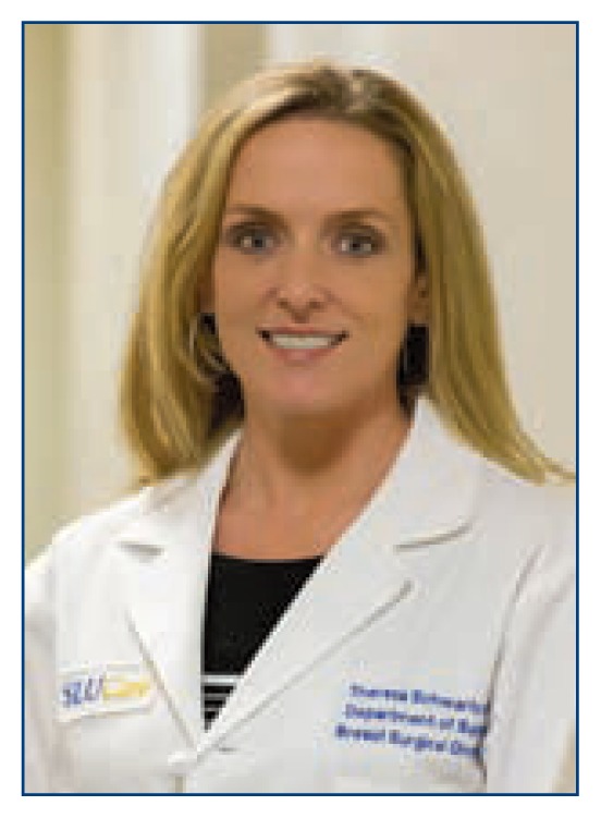
Kaitlin Farrell, MD, is Assistant Professor of Surgery, Debbie Lee Bennett, MD, is Associate Professor of Radiology, and Theresa L. Schwartz, MD, MS, FACS, (above), is Associate Professor of Surgery, all at the Saint Louis University School of Medicine, St. Louis, Missouri.
Contact: theresa.schwartz@health.slu.edu
Disclosure
None reported.
References
- 1.Berry DA, et al. Cancer interventions and surveillance modeling network (CISNET) Collaborators. Effect of screening and adjuvant therapy on mortality from breast cancer. N Engl J Med. 2005;353(17):1784–92. doi: 10.1056/NEJMoa050518. [DOI] [PubMed] [Google Scholar]
- 2.U.S. Preventive Services Task Force. Screening for breast cancer: U.S. Preventive Services Task Force Recommendation Statement. Ann Intern Med. 2009;151:716–26. doi: 10.7326/0003-4819-151-10-200911170-00008. [DOI] [PubMed] [Google Scholar]
- 3.Nelson HD, et al. Screening for breast cancer: an update for the U.S. Preventive Services Task Force. Ann Intern Med. 2009;151:738–47. doi: 10.1059/0003-4819-151-10-200911170-00009. [DOI] [PMC free article] [PubMed] [Google Scholar]
- 4.Nelson HD, et al. Effectiveness of breast cancer screening: systematic review and meta-analysis to update the 2009 U.S. Preventive Services Task Force Recommendation. Ann Intern Med. 2016;164:244–55. doi: 10.7326/M15-0969. [DOI] [PubMed] [Google Scholar]
- 5.Duffy SW, et al. The mammographic screening trials: commentary on the recent work by Olsen and Gotzsche. Cancer J Clin. 2002;52(2):68–71. doi: 10.3322/canjclin.52.2.68. [DOI] [PubMed] [Google Scholar]
- 6.Bhatia S, et al. High risk of subsequent neoplasms continues with extended follow-up of childhood Hodgkins Disease: report from the late effects study group. JCO. 2003;21(23):4386–94. doi: 10.1200/JCO.2003.11.059. [DOI] [PubMed] [Google Scholar]
- 7.Kenney LB, et al. Breast cancer after childhood cancer: a report from the childhood cancer survivor study. Ann Intern Med. 2004;141(8):590–7. doi: 10.7326/0003-4819-141-8-200410190-00006. [DOI] [PubMed] [Google Scholar]
- 8.Singletary SE. Rating the risk factors for breast cancer. Ann Surg. 2003;237(4):474–82. doi: 10.1097/01.SLA.0000059969.64262.87. [DOI] [PMC free article] [PubMed] [Google Scholar]
- 9.McDonald JA, et al. Alcohol intake and breast cancer risk: weighing the overall evidence. Curr Breast Cancer Rep. 2013;5(3) doi: 10.1007/s12609-013-0114-z.. [DOI] [PMC free article] [PubMed] [Google Scholar]
- 10.Boyd NF, et al. Mammographic density and the risk and detection of breast cancer. N Engl J Med. 2007;356:227–36. doi: 10.1056/NEJMoa062790. [DOI] [PubMed] [Google Scholar]
- 11.Amir E, et al. Evaluation of breast cancer risk assessment packages in the family history evaluation and screening programme. J Med Genet. 2003;40(11):807–14. doi: 10.1136/jmg.40.11.807. [DOI] [PMC free article] [PubMed] [Google Scholar]
- 12.Terry MB, et al. 10-year performance of four models of breast cancer risk: a validation study. Lancet Oncol. 2019;20(4):504–27. doi: 10.1016/S1470-2045(18)30902-1. [DOI] [PubMed] [Google Scholar]
- 13.The Cancer Genome Atlas Network. Comprehensive molecular portraits of human breast tumours. Nature. 2012;490:61–70. doi: 10.1038/nature11412. [DOI] [PMC free article] [PubMed] [Google Scholar]
- 14.Castera L, et al. Next-generation sequencing for the diagnosis of hereditary breast and ovarian cancer using genomic capture targeting multiple candidate genes. Eur J Hum Genetc. 2014;22:1305–13. doi: 10.1038/ejhg.2014.16. [DOI] [PMC free article] [PubMed] [Google Scholar]
- 15.Boyd NF, et al. Mammographic density and breast cancer risk: current understanding and future prospects. Breast Cancer Res. 2011;13(6):223. doi: 10.1186/bcr2942. [DOI] [PMC free article] [PubMed] [Google Scholar]
- 16.Kolb TM, et al. Comparison of the performance of screening mammography, physical examination and breast us and evaluation of factors that influence them: an analysis of 27,825 patient evaluations. Radiology. 2002;225(1):165–75. doi: 10.1148/radiol.2251011667. [DOI] [PubMed] [Google Scholar]
- 17.Pisano ED, et al. Diagnostic performance of digital versus film mammography for breast-cancer screening. N Engl J Med. 2005;353:1773–83. doi: 10.1056/NEJMoa052911. [DOI] [PubMed] [Google Scholar]
- 18.Niklason LT, et al. Digital tomosynthesis in breast imaging. Radiology. 1997;205(2):399–406. doi: 10.1148/radiology.205.2.9356620. [DOI] [PubMed] [Google Scholar]
- 19.Rafferty EA, et al. Breast cancer screening using tomosynthesis and digital mammography in dense and nondense breasts. JAMA. 2016;315(16):1784–86. doi: 10.1001/jama.2016.1708. [DOI] [PubMed] [Google Scholar]
- 20.Simon K, et al. Accuracy of synthetic 2D mammography compared with conventional 2D digital mammography obtained with 3D tomosynthesis. AJR. 2019;212(6):1406–11. doi: 10.2214/AJR.18.20520. [DOI] [PubMed] [Google Scholar]
- 21.Kuhl CK, et al. Mammography, breast ultrasound, and magnetic resonance imaging for surveillance of women at high familial risk for breast cancer. J Clin Oncol. 2005;23(33):8469–76. doi: 10.1200/JCO.2004.00.4960. [DOI] [PubMed] [Google Scholar]
- 22.Saadatmand S, et al. MRI versus mammography for breast cancer screening in women with familial risk (FaMRIsc): a multicentre, randomised, controlled trial. Lancet Oncol. 2019;20(8):1136–47. doi: 10.1016/S1470-2045(19)30275-X. [DOI] [PubMed] [Google Scholar]
- 23.Brem RF, et al. Assessing improvement in detection of breast cancer with three-dimensional automated breast ultrasound in women with dense breast tissue: the SomoInsight study. Radiology. 2014;17 doi: 10.1148/radiol.14132832. [DOI] [PubMed] [Google Scholar]
- 24.Arleo EK, et al. Comparison of recommendations for screening mammography using CISNET models. Cancer. 2017;123(19):3673–80. doi: 10.1002/cncr.30842. [DOI] [PubMed] [Google Scholar]
- 25.Berg WA, et al. Combined screening with ultrasound and mammography vs mammography alone in women at elevated risk of breast cancer. JAMA. 2008;299(18):2151–63. doi: 10.1001/jama.299.18.2151. [DOI] [PMC free article] [PubMed] [Google Scholar]
- 26.National Comprehensive Cancer Network. Breast Cancer Screening and Diagnosis Version 1.2019. https://www.nccn.org/professionals/physician_gls/pdf/breast-screening.pdf.
- 27.Monticciolo DL, et al. Breast cancer screening in women at higher-than-average risk: recommendations from the ACR. J Am Coll Radiol. 2018;15(3 Pt A):408–14. doi: 10.1016/j.jacr.2017.11.034. [DOI] [PubMed] [Google Scholar]
- 28.American Society of Breast Surgeons Position Statement on Screening Mammography. Apr, 2019. https://www.breastsurgeons.org/docs/statements/Position-Statement-on-Screening-Mammography.pdf.


