Abstract
Enrichment of omega-3 fatty acids (ɷ-3 FAs) in natural oils is important to realize their health benefits. Lipases are promising catalysts to perform this enrichment, however, fatty acid specificity of lipases is poor. We attempted to improve the fatty acid selectivity of a lipase from Geobacillus thermoleovorans (GTL) by two approaches. In a semi-rational approach, amino acid positions critical for binding were identified by docking the substrate to the GTL and best substitutes at these positions were identified by site saturation mutagenesis followed by screening to obtain a variant of GTL (CM-GTL). In the second approach based on rational design, a variant of GTL was designed (DM-GTL) wherein the active site was narrowed by incorporating two heavier amino acids in the lining of acyl-binding pocket to hinder access to bulky ɷ-3 FAs. The affinities DM-GTL with designed substrates were evaluated in silico. Both, CM-GTL and DM-GTL have shown excellent ability to discriminate against the ɷ-3 FAs during hydrolysis of oils. Engineering the binding pocket of an enzyme of a complex substrate, such as a triglyceride, by incorporating the information on substrate structure and computationally derived binding modes, has resulted in designing two efficient lipase variants with improved substrate selectivity.
Introduction
Health benefits of ɷ-3 fatty acids (FAs) as food supplements have been documented extensively [1–3]. ɷ-3 FAs help in managing cardiovascular disease, stroke, obesity, arteriosclerosis, hypertriglyceridemia, inflammation and autoimmune diseases [4–10]. Their role in neuronal development is also well established [11]. American Heart Association recommends that patients with coronary heart diseases should consume one gram of ɷ-3 FAs daily. FDA has approved the use of ɷ-3 FAs concentrate as a drug against cardiovascular disease and hypertriglyceridemia. Many plant and fish sources, including Flax, Tuna, and Anchovy contain substantial amounts of ɷ-3 FAs (~30 mole %). cis-5,8,11,15,17-eicosapentaenoic acid (EPA) and cis-4,7,10,13,16,19-docosahexaenoic acid (DHA) are the major bioactive ɷ-3 FAs. To be used in pharmaceutical applications, the content of ɷ-3 FAs in oils should be more than 60 mole %. Studies revealed that health benefits depend on the omega-3/omega-6 ratio in oils and not merely on the omega-3 content [12,13].
One solution to the above problem is in concentrating ɷ-3 FAs in their natural sources. Various chemical methods such as urea complexation, low temperature crystallization, distillation, supercritical fluid extraction, and chromatographic separation have been developed in order to concentrate ɷ-3 FAs [14–18]. However, each of these processes has issues on either economy of the process, or on the assurance of product quality [19]. Lipase-mediated selective hydrolysis can provide a safer and cheaper alternative for concentrating ɷ-3 FAs. For the process to become effective, it requires a lipase with excellent fatty acid selectivity. Efforts to isolate a fatty acid-selective lipase or to improve the natural lipases for fatty acid selectivity have failed to provide fruitful results [20,21]. In this work, we took a lipase from Geobacillus thermoleaverans (GTL) as a model protein and engineered it to enhance its ɷ-3 FAs selectivity using semi-rational and rational approaches.
Geobacillus thermoleaverans is a thermo-alkalophilic bacterium. The lipase (GTL) is a 43kDa lipase with a lid and is active on oil/water interfaces. Lipases homologous to GTL show maximum activity between pH 9 and 10 and in the temperature range of 55 to 60°C [22]. These GTL homologous lipases bind to one calcium and one zinc atom which enhance their stability [23]. Their melting temperature (Tm)is close to 75°C [24]. Excellent activity and stability make these lipases suitable for industrial applications. The large area of contact with the substrate makes these lipases a promising choice for altering the fatty acid selectivity. However, so far the fatty acid specificity of these lipases in hydrolysis has not been evaluated. We have evaluated the fatty acid selectivity of GTL and attempted to improve its fatty acid selectivity by protein engineering. GTL showed higher preference towards saturated fatty acids compared to hydrolysis of ɷ-3 FAs while hydrolysing anchovy oil. Therefore, we propose to enhance this discrimination further leading to accumulation of ɷ-3 FAs in the glyceride portion of the fish oil hydrolysate.
Both rational and semi-rational approaches in protein engineering have been successful in altering substrate selectivity of enzymes [25–27]. Amino acids near the enzyme active site, which interact with the substrate during catalysis, play a major role in determining activity and fatty acid selectivity [28]. In the semi-rational approach, we have identified the amino acids that interact with the substrates during hydrolysis by covalent docking of two representative substrates with the lipase using AutoDock Vina [29]. We excluded the functionally important amino acids and subjected amino acids which frequently interact with the substrate to site saturation mutagenesis (SSM). The best mutations were combined to test their substrate selectivity. In the rational approach, we exploited the structural difference between ɷ-3 FAs and non-ɷ-3 FAs. We hypothesized that by narrowing the fatty acid-binding channel in the active site of GTL, the binding of bulkier ɷ-3 FAs would be restricted and excluded/discriminated during hydrolysis. The lipase variants thus generated with two amino acid substitutions were tested for fatty acid selectivity in anchovy oil. Both the mutant GTLs showed significant alteration in fatty acid selectivity and were able to concentrate ɷ-3 FAs in glyceride portion better than the GTL.
Materials
Bleached anchovy oil was obtained from Ocean Nutrition Canada, Canada. Gum arabic, glycerol tributyrate, para-nitrophenyl butyrate (pNPB), hydrogen chloride-methanol solution, butylated hydroxytoluene (BHT) and methyl nonadecanoate were purchased from Sigma Aldrich, India. Silica gel TLC plates were purchased from MERCK, India. All the solvent and other chemicals used were of analytical grade or better. Geobacillus thermoleovorans strain (Acc. No. 4219) was purchased from MTCC, Chandigarh, India.
Methods
Cloning and expression of GTL
GTL gene was amplified from the genomic DNA of Geobacillus thermoleovorans. Signal peptide of GTL was removed and the resulting gene was cloned into pET21d between Nco I and Hind III [30]. Mutations, identified by methods described below, were incorporated into the GTL gene by overlap extension PCR method [31]. PCR mix was prepared by adding 20 ng pET21d plasmid containing GTL gene, 5 μl of 5 pM primer each to 25 μl Phusion master mix (2X) and diluting to 50 μl with MilliQ water. PCR was carried out for 30 cycles. Each PCR cycle consists of three steps: 1) denaturation at 98°C for 10 seconds 2) annealing at 55°C for 30 seconds 3) extension at 72°Cfor 30seconds/kb. In addition, 30 seconds of denaturation at 98°C at the beginning and 10 minutes of extension at 72°C at the end of PCR was carried out. First, fragments were generated by amplifying the first half of the gene with T7 forward primer and a designed primer. Similarly, the second half of the sequence was generated by a designed primer and T7 reverse primer by PCR. The complete mutated gene was obtained by overlap extension PCR using the fragments obtained in the previous step [31]. For Site Saturation Mutagenesis (SSM), MNN/NNK degenerate codon sets were used [32]. SSM was performed for each of the positions identified by docking (vide infra). Sequences of the primers used in this study are provided in the (S1 Table). All methods employed are as per the standard molecular biology protocols.
Modelling protein structures
All the protein structures were modelled by homology modelling using Modeller 9.14. [33]. PyMod 1.0 and PyMod 2.0, Pymol plug-ins were helpful in running modeller as well as Ramachandran plot analysis [34,35]. Open lid conformation of lipase L1 from Geobacillus thermocatenulatus (PDB ID 2w22, 95% sequence identity) was used as template [36]. Open lid conformation are essential for modelling the substrate binding with a lipase. The sequence of the protein to be modelled was aligned with that of the template with the help of SALIGN module provided with MODELLER package. For modelling each protein structure, 10 structures were generated and the structure with the best DOPE score was selected [37]. Heteroatoms and water molecules were removed and additional energy minimization was avoided as these led to poor models that allowed more amino acids in disallowed region of the Ramachandran plot. The generated model was validated by Ramachandran plot analysis using PyMod as well as analysis of other geometry parameters (bonds, angles, rotamers, etc.) using MolProbity [38]. Ramachandran plots of all the modelled proteins are given in S1 Fig.
Semi-rational approach to design GTL for fatty acid selectivity
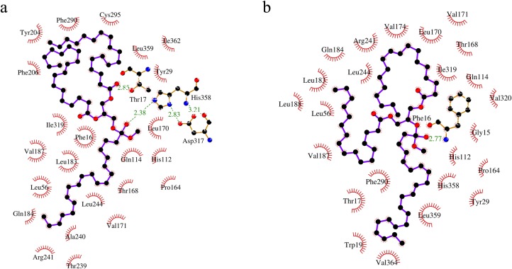
Amino acids of an enzyme that interact with the substrate play a critical role in enzyme substrate selectivity. In the semi-rational approach, we have identified the amino acids of GTL that interact with the substrate, i.e., triglyceride, by covalent docking of two representative substrates with GTL. Representative substrates were chosen based on the published information on fatty acid composition and distribution of fatty acids among the three carbon positions on the glycerol backbone [39]. Substrate 1 has EPA at sn-1, octadecanoic acid at sn-2 and 9-octadecenoic acid at sn-3 of a glycerol molecule and substrate 2 has octadecanoic acid at sn-1, DHA at sn-2 and 9-octadecenoic acid at sn-3 of a glycerol molecule. AutoDock Vina was used for docking calculation [29]. AutoDock Tools and Pymol were used for the preparation of input files as well as data analysis [40,41]. Covalent docking was performed by the flexible side chain method using a hydrogen atom as a pseudo ligand [42]. Carbonyl carbon of the fatty acid undergoing hydrolysis was covalently attached to γ-oxygen of the active serine (S113) by Pymol. This structure was then processed by AutoDock Tool (MGL software package, version 1.5.6) where AutoDock atom type and charges were added. Enzyme without S113 side chain was saved as the rigid receptor and S113 side chain covalently attached to triglyceride was saved as flexible residue. Kollman charges were added to the rigid part of the receptor, while Gasteiger charges were computed for the flexible side chain (triglyceride substrate covalently linked to S113) [43,44]. All the bonds, except double bonds and terminal bonds, were kept rotatable. Position and dimensions of grid box were such that it covers the entire active site and allows complete stretching of the flexible side chains. Grid spacing was kept at 0.375 Å. The 10 best binding modes for each substrate were analyzed to find the amino acids, which interact with the substrate during hydrolysis. Amino acids that were frequently observed (at least 15 out of 20 modes) to be interacting with the substrates were considered to be important for substrate selectivity (Fig 1 and Table 1). However, many of the substrate-interacting amino acids play an important role in the activity of the enzyme and hence these positions were excluded and the remaining positions are considered for site saturation mutagenesis.
Fig 1. Amino acids interacting with triglyceride during hydrolysis as observed by covalent docking.
(a) Representative Substrate 1 has EPA at sn-1 position and (b) Representative Substrate 2 has DHA at sn-2 position. Other positions are occupied by octadecanoic acid and 9-octadecenoic acid. Docking was performed using AutoDock Vina and the figure was generated using Ligplot [45].
Table 1. Interaction of substrate with GTL.
| Frequency of interactions | Amino acid positions |
|---|---|
| 20 | 16, 17, 29, 112, 113, 114, 168, 241, 244, 319, 358 |
| 19 | 56, 183 |
| 18 | 171 |
| 17 | 170, 359 |
Amino acid positions which interact with the substrate during hydrolysis of triglyceride as predicted by covalent docking using AutoDock Vina.
Mutant library preparation
Amplicons of SSM, generated by the method described above, were cloned into pET 22b and transformed into E. coli BL21 DE3 expression system. Individual colonies were inoculated into a well of 96-well plate containing 200 μl of LBA (LB broth containing1mM ampicillin) and grown overnight at 37°C and at 180 rpm. Supernatants were removed after cold centrifugation at 1700 RCF for 5 min. E. coli cells in each well were resuspended in 100 μl of LBA. 100 μl of 60% sterile glycerol was added to each well and the plate was stored at -20°C.
Screening the mutant library
To identify positive mutants, a two-stage medium-throughput screening method was followed. The first stage is a qualitative assay, which involves placing the culture supernatants containing mutant lipase in wells made in a tributyrin-agar plate. Lipase in the supernatants hydrolyzes tributyrin breaking the gum arabic-tributyrin emulsion, which leads to the formation of transparent halo in a milky white background of tributyrin-agar plate [46].
Tributyrin-agar plates were prepared by mixing 7.2 ml of 0.5 M glycine (pH 9.5), 1.44 ml of 2.5 M NaCl, 0.72 ml of 0.5 M CaCl2 and 1.5 g gum arabic till gum arabic dissolved. To this mixture, glycerol-tributyrate (1.5 ml) was added and the solution was sonicated on ice to form a milky white emulsion. 0.4 g of agarose was dissolved in 40 ml of water by boiling and cooled to 40–50°C. The above emulsion was mixed with agarose and poured into a 150 mm diameter Petri plate. After the agar solidified, holes were made in 96 well plate format using gel punching apparatus.
Stage 1: Halo formation assay
20 μl inoculum from each well of the library was re-cultured in a corresponding labelled 96-well plate containing 200 μl LBA in each well overnight as described above. Cells were pelleted by cold centrifugation at 1700 RCF. 5 μl of the supernatant was placed in each well of the tributyrin-agar plate and incubated at 37°C overnight for halo formation. This step was intended to screen out the inactive mutants. The mutants, whose halo size is less than one-third of the halo size of the wild type GTL on the same plate, are not considered for the next screen.
Stage 2: Differential activity assay
In the second stage, the active mutants identified in the first stage were tested for their substrate selectivity by comparing their activity against two oils. Anchovy oil (rich in ɷ-3 FAs) and coconut oil (lacks ɷ-3 FAs) were chosen for this purpose. Using a pH indicator, phenol red, the activity of mutants was estimated in a 96 well plate. Buffer containing 25 mM glycine, 25 mM CaCl2, 1% gum arabic and 1% oil at pH 9.5 was emulsified by sonication. Phenol red (pH indicator) was added to a final concentration of 0.04 mg/ml. 200 μl of the above mixture was dispensed into each well of the 96-well plate and 20 μl of culture supernatant (obtained in the same manner as in Stage 1) was added and incubated in an incubator shaker at 37°C and 180 RPM. Pictures of the plates were taken at various time points to record the colour change. Based on the extent of colour change brought about by the mutants in comparison with GTL, the mutants were scored on a scale of 1 to 5, where a score of 1 indicates the least active mutant and 5, the most active mutant. The difference in scores obtained by a given mutant, with coconut oil and anchovy oil as substrates, was considered to evaluate the fatty acid selectivity.
Rational approach to design GTL for fatty acid selectivity
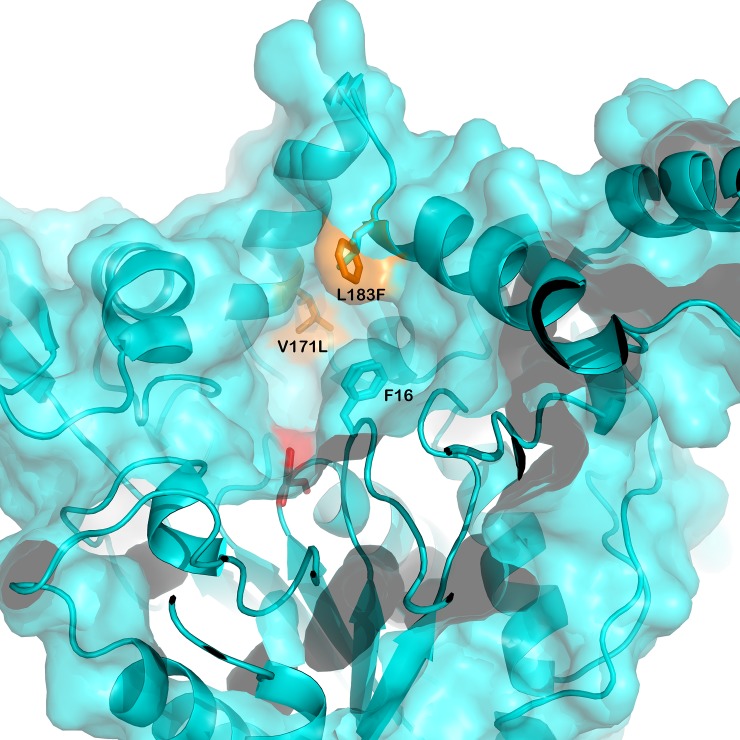
ɷ-3 FAs are structurally very similar to saturated and monounsaturated fatty acids. Lack of polar atoms and highly flexible nature of the acyl chains reduces the structural diversity that is necessary for lipases to discriminate between the different types of fatty acids. Therefore, most of the natural lipases possess little fatty acid selectivity. The only structural difference between ɷ-3 FAs and non-ɷ-3 FAs is their bulkiness. ɷ-3 FAs, being cis-polyunsaturated, are bulkier, due to double bonds, compared to their saturated and monounsaturated counterparts. Few lipases discriminate between ɷ-3 FAs and non-ɷ-3 FAs to some extent based on their bulkiness [47]. Bulkier ɷ-3 FAs are poorly accommodated in the active site due to steric hindrance and consequently poorly hydrolyzed [48,49]. It is known that out of the 4 pockets (namely HA, HB, HH and oxyanion hole) in the active site of Geobacillus thermocatenulatus lipase, a close homolog of GTL, the HA pocket is ideally oriented with respect to catalytic Ser113 and it is lined by F16, S57, V171, V174, L183, V187, L188, L208, W211, L213, L244. It was observed that the fatty acid bound in the HA is subjected to hydrolysis and, henceforth, is referred to as acyl pocket (also referred to acyl binding site) [36]. In other lipases, it is known that selectivity of the lipase towards different substrates depends on the interaction of substrates with the acyl pockets [50,51]. A bent in the middle provides the characteristic “L” shape to the HA. This bent, which is also the narrowest part, was surrounded by amino acid F16, V171, and L183 (Fig 2). Excluding F16 which has a critical role in lid opening, V171 and L183 were replaced by the next bulkier amino acids i.e. leucine and phenylalanine, respectively. The GTL variant thus produced is referred to as DM-GTL.
Fig 2. Mutation designed to narrow down the acyl pocket opening.
Modelled structures of GTL and DM-GTL are superimposed (cyan), mutations (yellow) and active serine (red) are shown.
In silico fatty acid selectivity of GTL and DM-GTL
In silico fatty acid selectivities of GTL and DM-GTL were estimated by calculating their affinities towards the representative substrates 1 and 2, while carboxyl carbon of different fatty acids was bound to γ-oxygen of S113, as employed in semi-rational approach. However, covalent docking using the flexible side chain method does not provide interaction energy between receptor and covalently attached substrate. To obtain the interaction energies, a two-step method was used. In the first step, we calculated the best substrate binding mode. Only conformations, where the fatty acid which is undergoing hydrolysis (covalently attached to S113), occupies the acyl pocket were considered relevant. In the next step, we saved the coordinates (except Cα of 113SER) of the best binding modes of the flexible side chain and treated it as a ligand to calculate binding energy, without performing any further search (by activating option–score only).
Overexpression and purification of lipases
Lipase genes, cloned into pET21D vector for cytoplasmic expression were transformed into E. coli BL21 DE3 expression system. Cells were cultured in LBA till OD600 reached 0.6. Protein overexpression was induced by adding 1mM IPTG. After 3 hours of expression, cells were harvested by centrifuging at 4000 RCF. The cells were lysed by lysozyme treatment followed by sonication. Cell debris was removed by cold centrifugation at 10000 RCF. Lipases were purified by ethylene glycol gradient (0–80%) on a HIC (Hydrophobic Interaction Chromatography) column with phenyl Sepharose as the stationary phase. The purity of GTL was confirmed by SDS-PAGE electrophoresis and the protein content was estimated by Lowry’s method using albumin standards.
pNPB hydrolysis
4.2 mg of pNPB was dissolved in 1 ml of acetonitrile (20 mM). 20 μl of the above solution was diluted with 50 mM sodium phosphate at pH 7.5 to 1 ml. The reaction was started by adding 0.1 μg of the enzyme and the absorbance change was measured at 405 nm for two minutes to calculate the rate of hydrolysis.
Hydrolysis of anchovy oil
Substrate for lipase was prepared by mixing 10 mM glycine, 10 mM CaCl2, 5% gum arabic and 5% anchovy oil in water and the pH was adjusted to 9.5, and the mixture was emulsified by sonication. Hydrolysis was initiated by adding 100 μg purified lipase at 37°C. Maintaining pH as well as monitoring reaction rates were carried out by titrating the released fatty acids against 1 M NaOH by a pH stat (Ω Metrohm 718 STAT Tritino). Samples were drawn at regular intervals. Free fatty acids were separated from glycerides by solvent extraction procedure. In this procedure, the mixture of free fatty acid and glyceride was mixed with 30% ethanolic KOH (0.5M) where free fatty acids get solubilised as potassium salt and insoluble glycerides were extracted twice with hexane. Free fatty acids were regenerated by lowering the pH below 2 with HCl [39]. The purity of separated fatty acids, as well as glycerides, was tested on silica gel TLC using hexane: diethyl ether: acetic acid (60:40:1, v/v) as mobile phase.
Methylation and fatty acid analysis by GC-MS
Methylation of fatty acids from both the fractions was carried out by acid catalysis using hydrogen chloride–methanol as methylating reagent as described earlier [39], with one modification i.e., a commercial mixture of hydrogen chloride–methanol was used instead of preparing it by adding acetyl chloride in methanol. 1 μl of hexane containing fatty acid methyl esters (FAMEs) was analyzed by GC-MS (Agilent 6890). HP-5MS (30 mt X 250 μm X 0.25 μm) column was used for separation of FAMEs. Helium gas was used as carrier gas at a rate of 40 cm/sec. Injector temperature was kept at 250°C. The oven temperature was kept initially at 140°C for 2 min, increased to 290°C at a rate of 6°C/min and kept at 290°C for 3 min. The fatty acids were quantitated by using known quantities of an external fatty acid standard. Methyl nonadecanoate was used as an internal standard as described earlier [39]. Selectivity for a given fatty acid (Fax) was calculated by using the following equation:
Thermal unfolding of GTL mutants
Thermal melting of GTL, CM-GTL, and DM-GTL was measured by recording the molar ellipticity at 222nm. Protein (0.1 mg/ml) in sodium phosphate buffer (10 mM, pH 7.5) was taken in a 1cm path length cuvette. The temperature was increased from 50°C to 90°C at a rate of 1°C/min using JASCO peltier type temperature controller and the ellipticity was measured using JASCO J-815 spectropolarimeter. Percentage unfolded fraction of protein was calculated using the following equation:
Results
Amino acid positions chosen for SSM
In the semi-rational approach, to identify the amino acids that interact with substrate during hydrolysis, covalent docking of representative substrates with GTL was carried out. Fig 1 shows the amino acid interacting with representative substrates in the best mode of interaction. Best ten interaction modes for each substrate were considered. Amino acids that interact with substrate in at least 75% (15 out of total 20 modes) of the cases were considered as important for substrate selectivity (Table 1). However, many of the substrate interacting amino acids play an important role in the activity of the enzyme and hence these positions were excluded. The following amino acid positions were excluded from any substitutions for the stated reasons: F16 and T17 have a critical role in structural rearrangement during lid opening; S113 is the active residue involved in hydrolysis; H112 stabilizes active serine (S113) in the closed conformation; Q114 and T168 form hydrogen bonds with substrates; R241 forms salt bridge with D178 which stabilizes open lid conformation. After the exclusion of these amino acid positions, amino acid positions 170, 171, 244, 319, 358 and 359 were chosen for SSM (S2 Fig).
Screening active mutants
In the first stage of screening, SSM was performed at six positions and the positive (active) substitutions were identified initially by qualitative halo formation assay as described in methods. For each amino acid position at least 150 colonies were picked to ensure the inclusion of all possible amino acid substitutions at each position [32]. Mutation at some positions had little impact on activity, while at other positions the substitutions resulted in mostly inactive enzyme (Fig 3). Out of the 960 individual mutants, generated by SSM at six positions, 210 colonies showed lipolytic activity and were subjected to a second screen.
Fig 3. Halo formation by mutants of GTL on agar-tributyrin plate.
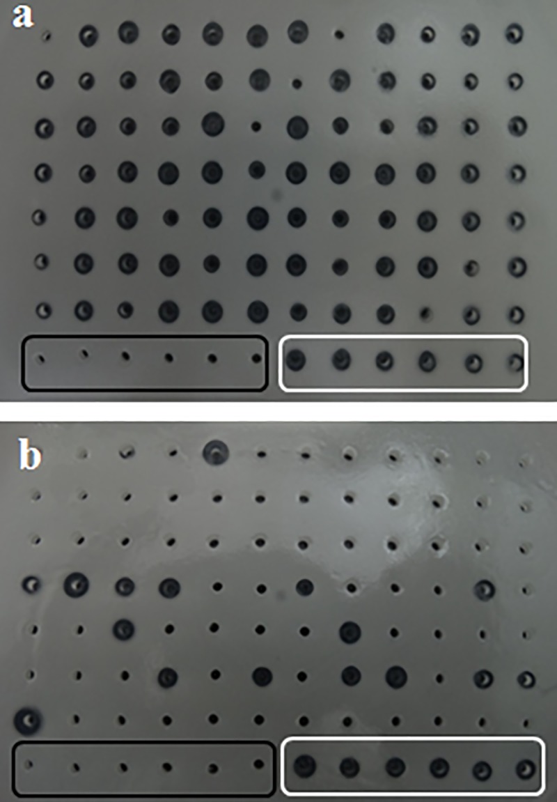
Wells within black rectangle are negative controls (without any lipase) and wells within white rectangle are positive controls (contain GTL). (a) Most of the mutants at position 244 are active, (b) while mutants at position 359 are mostly inactive.
Screening for differential activity of the mutants of GTL
In the second stage of our screening, fatty acid selectivities of the 210 active mutants were evaluated by comparing their activities on two oils with different fatty acid compositions. The oils used were coconut oil, which lacks ɷ-3 FAs, and anchovy oil, which is rich in ɷ-3 FAs. The activity was measured in a medium-throughput way, by recording the time required for a decrease in pH due to the release of fatty acids. Changes in pH were observed by the pH indicator dye, phenol red, which changes colour from red (above pH 7) to yellow (below pH 7). Fig 4 shows a representative plate that demonstrates the colour change due to the hydrolysis of anchovy oil by GTL mutants.
Fig 4. Change of pH due to hydrolysis of triglyceride by mutants of GTL indicated by phenol red.
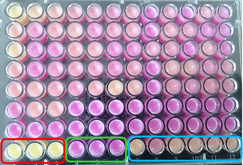
Wells in red rectangle are positive controls (pH <6, without lipase), in green rectangle are negative controls (pH 9.5, without any lipase) and in blue rectangle are with GTL. Acidic samples are yellow in colour (due to activity of lipase) and alkaline samples are purple in colour.
We hypothesized that the difference in activity in two oils by the mutants could be because of fatty acid selectivity. Since we already know that GTL preferentially hydrolyzes saturated fatty acids, mutants that showed comparatively more activity with coconut oil were considered positive and the corresponding lipase genes were sequenced. Out of the 210 mutant colonies screened, 28 colonies showed improved hydrolysis of coconut oil over anchovy oil compared to that of the GTL. Out of the 28 mutants sequenced, in 6 mutant sequences the base substitution did not result in amino acid substitution. The remaining 22 sequences were found to contain 13 different mutations. These were found at positions 170, 171 and 359 (Fig 5). At position 170, leucine was replaced by glycine in four lipase mutants and by tryptophan in two mutants. More diverse mutations were observed at position 171, where valine was replaced by arginines in three mutants, by proline in two, by tryptophan in two mutants and by alanine, phenylalanine, lysine, and threonine in one mutant each. Mutations at position 359 include the replacement of leucine by cysteine in two mutants and by alanine, lysine, and threonine in one mutant each. The most frequent mutations at each of these 3 positions are L170G, V171R, and L359C. A lipase variant with these mutations was generated by site directed mutagenesis (named as CM-GTL). CM-GTL was cloned into the expression vector, over-expressed and purified. The CM-GTL fatty acid selectivity was investigated along with the mutants generated by rational approaches as described below.
Fig 5. Mutations at different positions which showed improved differential activity during screening.
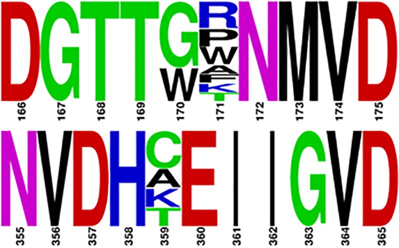
Occurrences of GTL amino acids at the positions of mutation are ignored for better presentation. The figure and the colour code is generated by WebLogo [52].
In silico fatty acid selectivity of GTL and DM-GTL
As described in the methods section on designing a lipase variant using the rational approach, a double mutant, named as DM-GTL, was generated with the following amino acid substitutions V171L and L183F. The positions were identified after modelling the binding of substrate to various pockets in acyl chain binding in GTL. Table 2 summarizes the binding energies of both the triglycerides when each of the three fatty acids of the glycerides is in a configuration that is suitable for hydrolysis. It is clear that DM-GTL has less affinity towards EPA and DHA hydrolysis and more affinity for saturated fatty acids compared to that of GTL. DM-GTL showed a mixed effect on mono unsaturated fatty acids. The mutations were incorporated in GTL and the resulting mutant was tested for its fatty acid selectivity.
Table 2. Affinities of two designed substrates with GTL and DM-GTL.
| Triglyceride model | Fatty acid undergoing hydrolysis | Affinity of GTL (kJ/mol) | Affinity of DM-GTL (kJ/mol) | Difference in affinities |
|---|---|---|---|---|
| 1-EPA-2-oleoyl-3-stearolyglycerol | EPA | -5.27 | -4.64 | 0.63 |
| Oleic acid | -5.42 | -5.12 | 0.3 | |
| Stearic acid | -5.11 | -5.86 | -0.75 | |
| 1- Oleoyl-2-DHA-3-stearolyglycerol | Oleic acid | -5.44 | -5.67 | -0.23 |
| DHA | -5.41 | -5.08 | 0.33 | |
| Stearic acid | -5.25 | -5.77 | -0.52 |
Affinities of GTL and DM-GTL towards hydrolysis of different fatty acids. Affinity of a fatty acid is calculated to be the binding energy between the representative substrate and enzyme when that fatty acid is undergoing hydrolysis.
Fatty acid selectivity of GTL DM-GTL and CM-GTL
The rate of hydrolysis of anchovy oil by GTL, CM- and DM-GTL was monitored by pH-Stat. During hydrolysis, aliquots of the reaction were taken at various time points for processing to separate the fatty acids from the unhydrolyzed portions. Free fatty acids were methylated and the derived FAMEs were quantified using GC-MS. The data presented in Fig 6 shows the selectivity of the lipases at various extents of hydrolysis. Value of selectivity can have a minimum value of -1 which means complete discrimination (no hydrolysis) of that fatty acid. Selectivity value of zero indicates no discrimination or preference for the given fatty acid and positive selectivity value indicates a preference for that fatty acid during hydrolysis. All three lipases preferentially hydrolyzed saturated fatty acids at the beginning of the reaction and the selectivity parameter plateaued with an increase in hydrolysis. Both CM-GTL and DM-GTL showed an improvement in selectivity in fatty acid hydrolysis compared to the GTL. The hydrolysis of individual fatty acids was estimated by GC- MS. The selectivity for EPA during hydrolysis was unaltered between GTL and CM-GTL while the selectivity for DHA changed from -0.33 for GTL to -0.75 for CM-GTL (Fig 6B). This decrease in selectivity (increase in discrimination) in hydrolysis by CM-GTL was, although to a lesser extent, retained until the end of the reaction time. On the other hand, DM-GTL showed decrease in selectivity for both EPA (from -0.5 to -0.83) and DHA (-0.33 to -0.95). Also, DM-GTL retained this selectivity for a longer duration (Fig 6C). However, the ability to discriminate between different fatty acids during hydrolysis was compromised after the extent of hydrolysis crossed 30 percent. To obtain a quantitative comparison among the variants, discrimination (magnitude of selectivity) of GTL, CM- and DM-GTL towards EPA and DHA was compared after 20 percent hydrolysis (S3 Fig). The data shows excellent discrimination by DM-GTL for both EPA and DHA, while the discrimination of CM-GTL increased only for DHA.
Fig 6. Fatty acid selectivity of GTL and mutants.
Fatty acid selectivity of (a) GTL, (b) CM-GTL and (c) DM-GTL at various extents of hydrolysis of anchovy oil. Saturated fatty acids (Open square), mono-unsaturated (Dark circle), EPA (Open triangle) and DHA (Dark Triangle). Selectivity value 0 indicates neutral hydrolysis, 1 indicates total preference and -1 indicates total discrimination (no hydrolysis).
All three lipases hydrolyzed saturated and mono unsaturated fatty acids more efficiently than ɷ-3 FAs which indicated that the ɷ-3 FAs are accumulated in the glyceride fraction. To evaluate their ability to concentrate ɷ-3 FAs, we compared EPA and DHA content of glyceride layer after 20 percent hydrolysis by GTL and DM-GTL, which shows that both the enzymes enriched ɷ-3 FAs (S4 Fig). While GTL showed enrichment of EPA only, DM-GTL showed significantly better enrichment in both EPA and DHA.
Thermal stability of GTL mutants
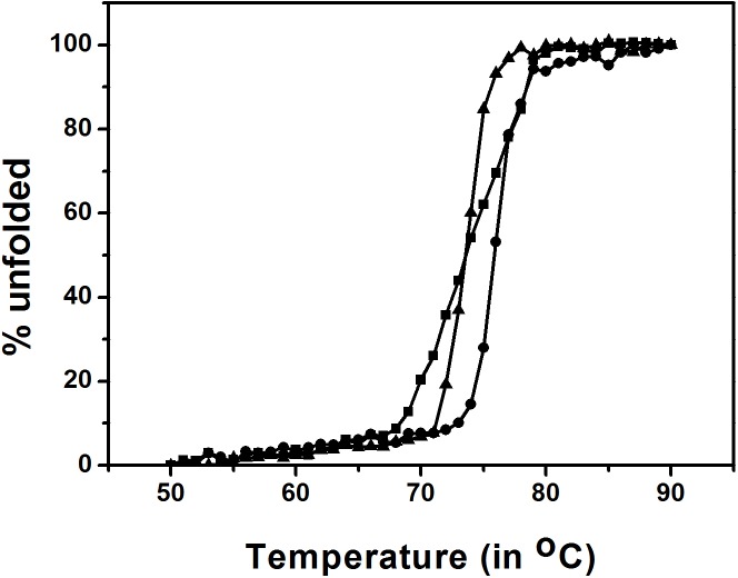
Sourced from a thermophile, GTL is highly thermostable. To assess the influence of the newly incorporated mutations on thermal stability, GTL mutants were characterized. The thermal unfolding of GTL, CM-GTL, and DM-GTL was observed by changes in ellipticity at 222 nm [53]. Fig 7 shows the unfolding of the three lipases upon heating. The mid-point of melting (Tm) transition for GTL was estimated to be 76.5°C. The Tm of the mutants (CM-GTL and DM-GTL) was lower than that of GTL by 3°C, suggesting that the mutations have partially destabilised the native protein. The thermal transition profile of DM-GTL, like GTL, exhibits a sharp transition, indicating cooperativity, whereas the width of the thermal transition of CM-GTL was broader, indicating less cooperativity during the unfolding of CM-GTL.
Fig 7. Thermal unfolding of GTL and its mutants.
Fraction unfolded was calculated from the ellipticity at 222 nm. GTL is represented by circle, DM-GTL by triangle and CM-GTL by square.
Activity of GTL and its mutants
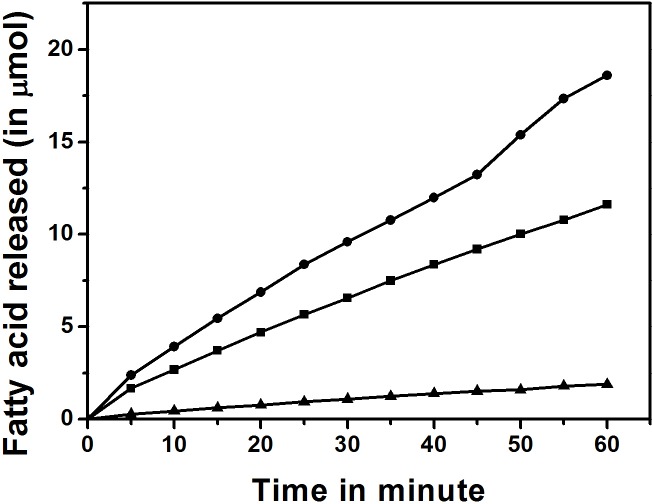
Mutation at the active site can affect the activity of an enzyme. Since all the mutations incorporated are in the active site of the lipase, we investigated the activity of lipases on water soluble choromogenic substrate, pNPB, using a spectrophotometer and on water insoluble anchovy oil by pH Stat method. Activity (unit = micromoles pNPB hydrolysed /min/mg of protein) of GTL, DM-GTL, and CM-GTL was 161.5, 210.6 and 16.8 units respectively with pNPB as substrate. Compared to GTL, the activity of DM-GTL improved significantly whereas the activity of CM-GTL was severely compromised. With anchovy oil, the observed activities (unit = micromoles of OH added /min/mg of protein) estimated by pH Stat method, were 312.5, 187.5 and 37.5 units with GTL, DM-GTL and CM-GTL, respectively (Fig 8). The data suggests that the incorporated mutations reduced the activity of DM-GTL for anchovy oil but not for pNPB. CM-GTL activity was significantly reduced compared to GTL with both the substrates.
Fig 8. Activity of GTL and mutants with anchovy oil as substrate.
Activity monitored by a pH Stat method. GTL (circle), DM-GTL (square) and CM-GTL (triangle).
Discussion
Among all the methods employed in concentrating ɷ-3 FAs in triglyceride form, selective hydrolysis of non-ɷ-3 FAs by lipases is considered to produce a cleaner product and at a less environmental load. In addition, ɷ-3 FAs concentrated in the form of triglycerides were shown to be biologically more valuable than free ɷ-3 FAs [39,54–56]. For better yields and improved economics of the process, substrate selectivity of the lipase is the key factor for ɷ-3 FAs concentration. Since most of the known lipases have low fatty acid selectivity, it is essential to enhance their selectivity before they can be successfully used to concentrate ɷ-3 FAs. Several strategies, including substrate imprinting, enzyme immobilization, enzyme entrapment, reaction engineering, solvent engineering, protein engineering, have been used to enhance fatty acid selectivity of lipases with only a marginal success [57–62]. Though challenging, protein engineering is the most effective as well as most economical method to alter substrate preferences of enzymes.
Protein engineering has been successfully used to alter substrate selectivity of many enzymes including several lipases [63–65]. Candida rugosa lipase has been engineered for selectivity of fatty acid chain length by blocking the active site tunnel [66]. Candida antarctica lipase B has been successfully engineered for enantio selectivity based on the enzyme-substrate interaction [65]. In this study, decreased volume of stereo-selective pocket correlates with increased stereo-selectivity. Enzyme selectivity also depends on the active site flexibility, wherein a less flexible site is more selective [67]. All these results are in line with our rational approach in designing the DM-GTL. The DM-GTL constructed based on the narrowing of the acyl binding pocket in the active site of GTL showed significantly improved fatty acid selectivity till 20 percent hydrolysis of anchovy oil and enriched ɷ-3 FAs in the glyceride fraction.
Selective hydrolysis of saturated fatty acids leads to an increase in the concentration of ɷ-3 FAs in the glyceride layer. At 20% hydrolysis concentration of ɷ-3 FAs increased from less than 30 mole% to over 40 mole%. Similar levels of enrichment, with best lipases, have been observed after 50 to 80% hydrolysis [68]. In the studies where higher efficiencies in concentrating ɷ-3 FAs were observed, these efficiencies were shown to be achieved by repeated hydrolysis [69], trans-esterification with ɷ-3 FAs [70], or combining lipase catalysis with other methods [71,72]. Taiwo et al. have achieved up to 80% ɷ-3 FAs concentration in fish oil hydrolysate [73]. However, the reported concentration is the concentration of ɷ-3 FAs as mono-glyceride. The actual concentration in glyceride mixture will be many folds less as mono-glyceride constitutes a very small fraction of fish oil hydrolysate. Also, unlike other lipases which concentrate only one out of many ɷ-3 FAs, DM-GTL is effective in concentrating both EPA and DHA [72,74]. The selectivity of the lipase can be further improved by either narrowing the end of the acyl pocket, which might lead to improved distinction between ɷ-3 FAs and other unsaturated fatty acids or by narrowing the other acyl chain pockets, which may lead to distinction between ɷ-3 FAs and non-ɷ-3 FAs in alternate binding modes. Also ɷ-3 FAs are concentrated as DG and MG instead of TG. The obtained glyceride can be trans-esterified to obtained ɷ-3 FAs concentrate as TG [70].
The enhancement in discrimination against ɷ-3 FAs observed in this study by narrowing the binding channel requires structural proof to rule out other contributions of the mutations in altering the specificity. To confirm the effect of substitutions on the acyl binding pocket, we attempted a wide set of conditions to crystallize the DM-GTL. In some conditions, we obtained very small crystals that were not suitable for diffraction.
Semi-rational approaches also delivered several success stories [75,76]. CM-GTL, constructed based on the SSM on the substrate contact sites of the active site amino acids in GTL, also showed improved selectivity, however, the improvement was not as significant as that of DM-GTL. At 20% hydrolysis, we did not expect significant improvement in the enrichment of ɷ-3 FAs with CM-GTL compared to GTL, hence the product profile was not evaluated. All the mutations observed in the screening were concentrated into three positions, out of which two were in the acyl pocket. This observation is in line with the previous works wherein the substrate selectivity were altered by incorporating mutations in the acyl pocket [77,78]. Mutations most frequently observed upon screening at each position were combined to enhance fatty acid selectivity. Examining all the combinations of mutations is not practical. Therefore, we limit our study to one mutant with the most frequent substitutions, although the epistatic effect could be either synergistic or antagonistic.
We have also evaluated the effects of mutations on the activity and the stability of the lipase. Both CM- and DM-GTL are less active on anchovy oil compared to GTL. The activity of DM-GTL on anchovy oil was 65% that of GTL and the activity of CM-GTL was 10% that of GTL activity. This result is as per our expectation as the mutations were incorporated to disfavour ɷ-3 fatty acids which constitute 30 percent of anchovy oil. DM-GTL hydrolyzed pNPB better than GTL. This could be because a greater fraction of DM-GTL was in the lid open conformation in the aqueous phase. The poor activity of CM-GTL on both the substrates could be because of the proximity of the L359C mutation to the active site than other mutations. The incorporation of mutations in the GTL have reduced the thermostability of the GTL to some extent; however, this decrease in stability is marginal and therefore does not hinder their industrial applications.
GTL as well as the CM- and DM-GTL mutants lose their selectivity with the progress of hydrolysis, which is probably due to buildup of the products in the form of mono-, diglycerides, and free fatty acids. Similar phenomena have been observed for other lipases as well [79,80]. The continuously altering composition profile of the lipase “substrate” and consequent changes in the nature of the substrate emulsions lead to further loss of selectivity. The presence of alternative substrate binding mode in other lipases is also being speculated as a possible reason for the loss of selectivity [81]. If present, these can become a prominent mode of binding in the later part of the hydrolysis and may compromise the selectivity.
Altering fatty acid selectivity of lipases during hydrolysis of natural oil poses several formidable challenges. Highly flexible structure and lack of polar atoms in fatty acids make it difficult for any enzyme to select one fatty acid over the other. Natural oil sources of ɷ-3 FAs, such as anchovy, Tuna, etc. usually contains several species of triglycerides with varying fatty acid composition and also with variations in their positional distribution [82]. Hydrolysis of a triglyceride molecule may lead to six different possible products depending on which bond is cleaved. Some of the products (di and mono glyceride) also simultaneously can act as substrates confounding the kinetics. Also, fatty acid selectivities of many lipases depend on the overall substrate structures [83]. Compositional complexities of natural oils further increase the difficulty in arriving at a designing strategy. Lipases without a lid, mostly do not show fatty acid selectivity. In these lipases, the area of contact between the active site and their substrates was not extensive enough to distinguish between different fatty acids. Lipases with lid, such as GTL, require interfacial activation. This requires emulsifying reagent and involves major structural rearrangement near the active site. Both the emulsifying reagent and the structural rearrangement affect substrate selectivity and are difficult to model [84].
Conclusion
We have altered the ɷ-3 FAs selectivity of GTL by both rational and semi-rational approaches. Lipase engineered by the rational approach (DM-GTL) showed almost complete discrimination against ɷ-3 FAs at the beginning of the hydrolysis. With the progress in hydrolysis, selectivity starts decreasing. Further improvement of selectivity on the designed mutants might open the door for economical enrichment of ɷ-3 FAs. While mutations lining acyl pocket are obvious to alter substrate selectivity of GTL, amino acid at other positions can also contribute to it. The complexity of the substrate along with poor knowledge about alternate substrate binding modes are the challenges to overcome for rational designing of lipases.
Supporting information
Ramachandran plot of modelled GTL (a) and DM-GTL (b) in lid open conformation. Triangle represents glycine and square represents proline. Other amino acids are represented by circle. Red colour indicates most favoured region, deep yellow indicates additional allowed region and light yellow indicates generously allowed region.
(PDF)
Amino acid positions chosen for SSM were shown in white, while amino acids interacting with substrate, but not included for SSM, are shown in magenta and active serine (S113) is shown in red. Triglyceride molecule shown in green represents substrate as shown in Fig 1A and the molecule shown in yellow represents substrate as shown in Fig 1B.
(PDF)
EPA (Light grey) and DHA (Dark).
(PDF)
(PDF)
Primers used in SSM (top) and for SDM (bottom). Mutations are shown in bold.
(PDF)
Mutants showing improved difference in activity (>2) are sequenced and the mutations were identified.
(PDF)
Acknowledgments
The authors thank Drs. Colin Barrow and T Akanbi for gracious inputs during the initial stages of the work.
Data Availability
All relevant data are within the manuscript and its Supporting Information files.
Funding Statement
We acknowledge Indo-Australian Biotechnology Fund (http://dbtindia.gov.in/latest-announcement/call-indo-australian-biotechnology-fund-round-12-2019-2020; GAP373) for the financial support and TRM acknowledges the research fellowship received from Council for Scientific and Industrial Research (https://csirhrdg.res.in). Funders didn't play any role in the study design, data collection and analysis, decision to publish, or preparation of the manuscript.
References
- 1.Ruxton C, Reed SC, Simpson M, Millington K (2004) The health benefits of omega‐3 polyunsaturated fatty acids: a review of the evidence. Journal of Human Nutrition and Dietetics 17: 449–459. 10.1111/j.1365-277X.2004.00552.x [DOI] [PubMed] [Google Scholar]
- 2.Swanson D, Block R, Mousa SA (2012) Omega-3 fatty acids EPA and DHA: health benefits throughout life. Advances in Nutrition: An International Review Journal 3: 1–7. [DOI] [PMC free article] [PubMed] [Google Scholar]
- 3.Ruxton C, Reed S, Simpson M, Millington K (2007) The health benefits of omega-3 polyunsaturated fatty acids: a review of the evidence. Journal of human nutrition and dietetics: the official journal of the British Dietetic Association 20: 275–285. [DOI] [PubMed] [Google Scholar]
- 4.Lorente-Cebrián S, Costa AG, Navas-Carretero S, Zabala M, Martínez JA, et al. (2013) Role of omega-3 fatty acids in obesity, metabolic syndrome, and cardiovascular diseases: a review of the evidence. Journal of physiology and biochemistry 69: 633–651. 10.1007/s13105-013-0265-4 [DOI] [PubMed] [Google Scholar]
- 5.Massaro M, Scoditti E, Carluccio MA, De CR (2008) Basic mechanisms behind the effects of n-3 fatty acids on cardiovascular disease. Prostaglandins LeukotEssentFatty Acids 79: 109–115. [DOI] [PubMed] [Google Scholar]
- 6.Kris-Etherton PM, Harris WS, Appel LJ, Committee N (2002) Fish consumption, fish oil, omega-3 fatty acids, and cardiovascular disease. circulation 106: 2747–2757. 10.1161/01.cir.0000038493.65177.94 [DOI] [PubMed] [Google Scholar]
- 7.Iso H, Rexrode KM, Stampfer MJ, Manson JE, Colditz GA, et al. (2001) Intake of fish and omega-3 fatty acids and risk of stroke in women. Jama 285: 304–312. 10.1001/jama.285.3.304 [DOI] [PubMed] [Google Scholar]
- 8.Simopoulos AP (2002) Omega-3 fatty acids in inflammation and autoimmune diseases. J AmCollNutr 21: 495–505. [DOI] [PubMed] [Google Scholar]
- 9.Harris WS, Connor WE, Inkeles SB, Illingworth DR (1984) Dietary omega-3 fatty acids prevent carbohydrate-induced hypertriglyceridemia. Metabolism 33: 1016–1019. 10.1016/0026-0495(84)90230-0 [DOI] [PubMed] [Google Scholar]
- 10.Harris WS, Miller M, Tighe AP, Davidson MH, Schaefer EJ (2008) Omega-3 fatty acids and coronary heart disease risk: clinical and mechanistic perspectives. Atherosclerosis 197: 12–24. 10.1016/j.atherosclerosis.2007.11.008 [DOI] [PubMed] [Google Scholar]
- 11.Innis SM (2007) Dietary (n-3) fatty acids and brain development. J Nutr 137: 855–859. 10.1093/jn/137.4.855 [DOI] [PubMed] [Google Scholar]
- 12.Harris WS (2006) The omega-6/omega-3 ratio and cardiovascular disease risk: uses and abuses. CurrAtherosclerRep 8: 453–459. [DOI] [PubMed] [Google Scholar]
- 13.Simopoulos AP (2011) Evolutionary aspects of diet: the omega-6/omega-3 ratio and the brain. Mol Neurobiol 44: 203–215. 10.1007/s12035-010-8162-0 [DOI] [PubMed] [Google Scholar]
- 14.Ratnayake W, Olsson B, Matthews D, Ackman R (1988) Preparation of Omega‐3 PUFA Concentrates from Fish Oils via Urea Complexation. Lipid/Fett 90: 381–386. [Google Scholar]
- 15.Haraldsson G (1984) Separation of saturated/unsaturated fatty acids. Journal of the American Oil Chemists’ Society 61: 219–222. [Google Scholar]
- 16.Wijesundera R, Ratnayake W, Ackman R (1989) Eicosapentaenoic acid geometrical isomer artifacts in heated fish oil esters. Journal of the American Oil Chemists’ Society 66: 1822–1830. [Google Scholar]
- 17.Nilsson WB, Gauglitz EJ, Hudson JK (1989) Supercritical fluid fractionation of fish oil esters using incremental pressure programming and a temperature gradient. Journal of the American Oil Chemists Society 66: 1596–1600. [Google Scholar]
- 18.Beebe JM, Brown PR, Turcotte JG (1988) Preparative-scale high-performance liquid chromatography of omega-3 polyunsaturated fatty acid esters derived from fish oil. Journal of Chromatography A 459: 369–378. [DOI] [PubMed] [Google Scholar]
- 19.Rubio-Rodríguez N, Beltrán S, Jaime I, Sara M, Sanz MT, et al. (2010) Production of omega-3 polyunsaturated fatty acid concentrates: a review. Innovative Food Science & Emerging Technologies 11: 1–12. [Google Scholar]
- 20.Kralovec JA, Zhang S, Zhang W, Barrow CJ (2012) A review of the progress in enzymatic concentration and microencapsulation of omega-3 rich oil from fish and microbial sources. Food Chemistry 131: 639–644. [Google Scholar]
- 21.Gupta A, Singh D, Byreddy AR, Thyagarajan T, Sonkar SP, et al. (2016) Exploring omega‐3 fatty acids, enzymes and biodiesel producing thraustochytrids from Australian and Indian marine biodiversity. Biotechnology journal 11: 345–355. 10.1002/biot.201500279 [DOI] [PubMed] [Google Scholar]
- 22.Quintana-Castro R, Díaz P, Valerio-Alfaro G, García HS, Oliart-Ros R (2009) Gene cloning, expression, and characterization of the Geobacillus thermoleovorans CCR11 thermoalkaliphilic lipase. Molecular biotechnology 42: 75–83. 10.1007/s12033-008-9136-6 [DOI] [PubMed] [Google Scholar]
- 23.Choi W-C, Kim MH, Ro H-S, Ryu SR, Oh T-K, et al. (2005) Zinc in lipase L1 from Geobacillus stearothermophilus L1 and structural implications on thermal stability. FEBS letters 579: 3461–3466. 10.1016/j.febslet.2005.05.016 [DOI] [PubMed] [Google Scholar]
- 24.Soliman NA, Knoll M, Abdel-Fattah YR, Schmid RD, Lange S (2007) Molecular cloning and characterization of thermostable esterase and lipase from Geobacillus thermoleovorans YN isolated from desert soil in Egypt. Process Biochemistry 42: 1090–1100. [Google Scholar]
- 25.Chen C-KJ, Shokhireva TK, Berry RE, Zhang H, Walker FA (2008) The effect of mutation of F87 on the properties of CYP102A1-CYP4C7 chimeras: altered regiospecificity and substrate selectivity. JBIC Journal of Biological Inorganic Chemistry 13: 813–824. 10.1007/s00775-008-0368-5 [DOI] [PubMed] [Google Scholar]
- 26.Nilsson LO, Gustafsson A, Mannervik B (2000) Redesign of substrate-selectivity determining modules of glutathione transferase A1–1 installs high catalytic efficiency with toxic alkenal products of lipid peroxidation. Proceedings of the National Academy of Sciences 97: 9408–9412. [DOI] [PMC free article] [PubMed] [Google Scholar]
- 27.May O, Nguyen PT, Arnold FH (2000) Inverting enantioselectivity by directed evolution of hydantoinase for improved production of L-methionine. Nature biotechnology 18: 317 10.1038/73773 [DOI] [PubMed] [Google Scholar]
- 28.Morley KL, Kazlauskas RJ (2005) Improving enzyme properties: when are closer mutations better? Trends in biotechnology 23: 231–237. 10.1016/j.tibtech.2005.03.005 [DOI] [PubMed] [Google Scholar]
- 29.Trott O, Olson AJ (2010) AutoDock Vina: improving the speed and accuracy of docking with a new scoring function, efficient optimization, and multithreading. Journal of computational chemistry 31: 455–461. 10.1002/jcc.21334 [DOI] [PMC free article] [PubMed] [Google Scholar]
- 30.Abdel-Fattah YR, Gaballa AA (2008) Identification and over-expression of a thermostable lipase from Geobacillus thermoleovorans Toshki in Escherichia coli. Microbiological research 163: 13–20. 10.1016/j.micres.2006.02.004 [DOI] [PubMed] [Google Scholar]
- 31.Ho SN, Hunt HD, Horton RM, Pullen JK, Pease LR (1989) Site-directed mutagenesis by overlap extension using the polymerase chain reaction. Gene 77: 51–59. 10.1016/0378-1119(89)90358-2 [DOI] [PubMed] [Google Scholar]
- 32.Kretz KA, Richardson TH, Gray KA, Robertson DE, Tan X, et al. (2004) Gene site saturation mutagenesis: a comprehensive mutagenesis approach. Methods in enzymology: Elsevier. pp. 3–11. [DOI] [PubMed] [Google Scholar]
- 33.Fiser A, Šali A (2003) Modeller: generation and refinement of homology-based protein structure models. Methods in enzymology 374: 461–491. 10.1016/S0076-6879(03)74020-8 [DOI] [PubMed] [Google Scholar]
- 34.Bramucci E, Paiardini A, Bossa F, Pascarella S (2012) PyMod: sequence similarity searches, multiple sequence-structure alignments, and homology modeling within PyMOL. BMC bioinformatics 13: S2. [DOI] [PMC free article] [PubMed] [Google Scholar]
- 35.Janson G, Zhang C, Prado MG, Paiardini A (2016) PyMod 2.0: improvements in protein sequence-structure analysis and homology modeling within PyMOL. Bioinformatics 33: 444–446. [DOI] [PubMed] [Google Scholar]
- 36.Carrasco-López C, Godoy C, de las Rivas B, Fernández-Lorente G, Palomo JM, et al. (2009) Activation of bacterial thermoalkalophilic lipases is spurred by dramatic structural rearrangements. Journal of Biological Chemistry 284: 4365–4372. 10.1074/jbc.M808268200 [DOI] [PubMed] [Google Scholar]
- 37.My Shen, Sali A(2006) Statistical potential for assessment and prediction of protein structures. Protein science 15: 2507–2524. 10.1110/ps.062416606 [DOI] [PMC free article] [PubMed] [Google Scholar]
- 38.Davis IW, Leaver-Fay A, Chen VB, Block JN, Kapral GJ, et al. (2007) MolProbity: all-atom contacts and structure validation for proteins and nucleic acids. Nucleic acids research 35: W375–W383. 10.1093/nar/gkm216 [DOI] [PMC free article] [PubMed] [Google Scholar]
- 39.Moharana TR, Byreddy AR, Puri M, Barrow C, Rao NM (2016) Selective enrichment of omega-3 fatty acids in oils by phospholipase A1. PloS one 11: e0151370 10.1371/journal.pone.0151370 [DOI] [PMC free article] [PubMed] [Google Scholar]
- 40.Morris GM, Huey R, Lindstrom W, Sanner MF, Belew RK, et al. (2009) AutoDock4 and AutoDockTools4: Automated docking with selective receptor flexibility. Journal of computational chemistry 30: 2785–2791. 10.1002/jcc.21256 [DOI] [PMC free article] [PubMed] [Google Scholar]
- 41.DeLano WL (2002) Pymol: An open-source molecular graphics tool. CCP4 Newsletter On Protein Crystallography 40: 82–92. [Google Scholar]
- 42.Bianco G, Forli S, Goodsell DS, Olson AJ (2016) Covalent docking using autodock: Two‐point attractor and flexible side chain methods. Protein Science 25: 295–301. 10.1002/pro.2733 [DOI] [PMC free article] [PubMed] [Google Scholar]
- 43.Besler BH, Merz KM, Kollman PA (1990) Atomic charges derived from semiempirical methods. Journal of Computational Chemistry 11: 431–439. [Google Scholar]
- 44.Gasteiger J, Marsili M (1980) Iterative partial equalization of orbital electronegativity—a rapid access to atomic charges. Tetrahedron 36: 3219–3228. [Google Scholar]
- 45.Wallace AC, Laskowski RA, Thornton JM (1995) LIGPLOT: a program to generate schematic diagrams of protein-ligand interactions. Protein engineering, design and selection 8: 127–134. [DOI] [PubMed] [Google Scholar]
- 46.Lawrence R, Fryer T, Reiter B (1967) Rapid method for the quantitative estimation of microbial lipases. Nature 213: 1264. [Google Scholar]
- 47.Halldorsson A, Kristinsson B, Haraldsson GG (2004) Lipase selectivity toward fatty acids commonly found in fish oil. European Journal of Lipid Science and Technology 106: 79–87. [Google Scholar]
- 48.Bottino NR, Vandenburg GA, Reiser R (1967) Resistance of certain long-chain polyunsaturated fatty acids of marine oils to pancreatic lipase hydrolysis. Lipids 2: 489–493. 10.1007/BF02533177 [DOI] [PubMed] [Google Scholar]
- 49.Wanasundara UN, Shahidi F (1998) Lipase-assisted concentration of n-3 polyunsaturated fatty acids in acylglycerols from marine oils. Journal of the American Oil Chemists' Society 75: 945–951. [Google Scholar]
- 50.Pleiss J, Fischer M, Schmid RD (1998) Anatomy of lipase binding sites: the scissile fatty acid binding site. Chemistry and physics of lipids 93: 67–80. 10.1016/s0009-3084(98)00030-9 [DOI] [PubMed] [Google Scholar]
- 51.Yang J, Koga Y, Nakano H, Yamane T (2002) Modifying the chain-length selectivity of the lipase from Burkholderia cepacia KWI-56 through in vitro combinatorial mutagenesis in the substrate-binding site. Protein Engineering 15: 147–152. 10.1093/protein/15.2.147 [DOI] [PubMed] [Google Scholar]
- 52.Crooks GE, Hon G, Chandonia J-M, Brenner SE (2004) WebLogo: a sequence logo generator. Genome research 14: 1188–1190. 10.1101/gr.849004 [DOI] [PMC free article] [PubMed] [Google Scholar]
- 53.Kelly SM, Price NC (1997) The application of circular dichroism to studies of protein folding and unfolding. Biochimica Et Biophysica Acta-Protein Structure and Molecular Enzymology 1338: 161–185. [DOI] [PubMed] [Google Scholar]
- 54.Schuchardt JP, Hahn A (2013) Bioavailability of long-chain omega-3 fatty acids. Prostaglandins, Leukotrienes and Essential Fatty Acids (PLEFA) 89: 1–8. [DOI] [PubMed] [Google Scholar]
- 55.Lawson LD, Hughes BG (1988) Human absorption of fish oil fatty acids as triacylglycerols, free acids, or ethyl esters. Biochemical and biophysical research communications 152: 328–335. 10.1016/s0006-291x(88)80718-6 [DOI] [PubMed] [Google Scholar]
- 56.Lyberg AM, Adlercreutz P (2008) Lipase‐catalysed enrichment of DHA and EPA in acylglycerols resulting from squid oil ethanolysis. European journal of lipid science and technology 110: 317–324. [Google Scholar]
- 57.Fishman A, Cogan U (2003) Bio-imprinting of lipases with fatty acids. Journal of Molecular Catalysis B: Enzymatic 22: 193–202. [Google Scholar]
- 58.Lee CH, Parkin KL (2001) Effect of water activity and immobilization on fatty acid selectivity for esterification reactions mediated by lipases. Biotechnology and bioengineering 75: 219–227. 10.1002/bit.10009 [DOI] [PubMed] [Google Scholar]
- 59.Stamatis H, Xenakis A, Provelegiou M, Kolisis FN (1993) Esterification reactions catalyzed by lipases in microemulsions: The role of enzyme localization in relation to its selectivity. Biotechnology and bioengineering 42: 103–110. 10.1002/bit.260420114 [DOI] [PubMed] [Google Scholar]
- 60.Wescott CR, Klibanov AM (1994) The solvent dependence of enzyme specificity. Biochimica et Biophysica Acta (BBA)-Protein Structure and Molecular Enzymology 1206: 1–9. [DOI] [PubMed] [Google Scholar]
- 61.Phillips RS (1996) Temperature modulation of the stereochemistry of enzymatic catalysis: prospects for exploitation. Trends in biotechnology 14: 13–16. [Google Scholar]
- 62.Brundiek HB, Evitt AS, Kourist R, Bornscheuer UT (2012) Creation of a lipase highly selective for trans fatty acids by protein engineering. Angewandte Chemie International Edition 51: 412–414. 10.1002/anie.201106126 [DOI] [PubMed] [Google Scholar]
- 63.Varadarajan N, Gam J, Olsen MJ, Georgiou G, Iverson BL (2005) Engineering of protease variants exhibiting high catalytic activity and exquisite substrate selectivity. Proceedings of the National Academy of Sciences of the United States of America 102: 6855–6860. 10.1073/pnas.0500063102 [DOI] [PMC free article] [PubMed] [Google Scholar]
- 64.Wenz C, Selent U, Wende W, Jeltsch A, Wolfes H, et al. (1994) Protein engineering of the restriction endonuclease EcoRV: replacement of an amino acid residue in the DNA binding site leads to an altered selectivity towards unmodified and modified substrates. Biochimica et Biophysica Acta (BBA)-Gene Structure and Expression 1219: 73–80. [DOI] [PubMed] [Google Scholar]
- 65.Rotticci D, Rotticci‐Mulder JC, Denman S, Norin T, Hult K (2001) Improved enantioselectivity of a lipase by rational protein engineering. ChemBioChem 2: 766–770. [DOI] [PubMed] [Google Scholar]
- 66.Schmitt J, Brocca S, Schmid RD, Pleiss J (2002) Blocking the tunnel: engineering of Candida rugosa lipase mutants with short chain length specificity. Protein engineering 15: 595–601. 10.1093/protein/15.7.595 [DOI] [PubMed] [Google Scholar]
- 67.Hvorecny KL, Bahl CD, Kitamura S, Lee KSS, Hammock BD, et al. (2017) Active-site flexibility and substrate specificity in a bacterial virulence factor: Crystallographic snapshots of an epoxide hydrolase. Structure 25: 697–707. e694. 10.1016/j.str.2017.03.002 [DOI] [PMC free article] [PubMed] [Google Scholar]
- 68.Kahveci D, Falkeborg M, Gregersen S, Xu X (2010) Upgrading of farmed salmon oil through lipase-catalyzed hydrolysis. The Open Biotechnology Journal 4. [Google Scholar]
- 69.Kahveci D, Xu X (2011) Repeated hydrolysis process is effective for enrichment of omega 3 polyunsaturated fatty acids in salmon oil by Candida rugosa lipase. Food Chemistry 129: 1552–1558. [Google Scholar]
- 70.Akanbi TO, Barrow CJ (2015) Lipase-catalysed incorporation of EPA into emu oil: formation and characterisation of new structured lipids. Journal of functional foods 19: 801–809. [Google Scholar]
- 71.Solaesa ÁG, Sanz MT, Falkeborg M, Beltrán S, Guo Z (2016) Production and concentration of monoacylglycerols rich in omega-3 polyunsaturated fatty acids by enzymatic glycerolysis and molecular distillation. Food chemistry 190: 960–967. 10.1016/j.foodchem.2015.06.061 [DOI] [PubMed] [Google Scholar]
- 72.Kojima Y, Sakuradani E, Shimizu S (2006) Different specificity of two types of Pseudomonas lipases for C20 fatty acids with a Δ5 unsaturated double bond and their application for selective concentration of fatty acids. Journal of bioscience and bioengineering 101: 496–500. 10.1263/jbb.101.496 [DOI] [PubMed] [Google Scholar]
- 73.Akanbi TO, Barrow CJ (2017) Candida antarctica lipase A effectively concentrates DHA from fish and thraustochytrid oils. Food chemistry 229: 509–516. 10.1016/j.foodchem.2017.02.099 [DOI] [PubMed] [Google Scholar]
- 74.Yadwad V, Ward O, Noronha L (1991) Application of lipase to concentrate the docosahexaenoic acid (DHA) fraction of fish oil. Biotechnology and bioengineering 38: 956–959. 10.1002/bit.260380818 [DOI] [PubMed] [Google Scholar]
- 75.Hoffmann G, Bönsch K, Greiner-Stöffele T, Ballschmiter M (2011) Changing the substrate specificity of P450cam towards diphenylmethane by semi-rational enzyme engineering. Protein Engineering, Design & Selection 24: 439–446. [DOI] [PubMed] [Google Scholar]
- 76.Liang L, Zhang J, Lin Z (2007) Altering coenzyme specificity of Pichia stipitis xylose reductase by the semi-rational approach CASTing. Microbial Cell Factories 6: 36 10.1186/1475-2859-6-36 [DOI] [PMC free article] [PubMed] [Google Scholar]
- 77.Klein RR, King G, Moreau RA, Haas MJ (1997) Altered acyl chain length specificity of Rhizopus delemar lipase through mutagenesis and molecular modeling. Lipids 32: 123–130. 10.1007/s11745-997-0016-1 [DOI] [PubMed] [Google Scholar]
- 78.Wu Q, Soni P, Reetz MT (2013) Laboratory evolution of enantiocomplementary Candida antarctica lipase B mutants with broad substrate scope. J Am Chem Soc 135: 1872–1881. 10.1021/ja310455t [DOI] [PubMed] [Google Scholar]
- 79.Moore SR, McNeill GP (1996) Production of triglycerides enriched in long-chain n-3 polyunsaturated fatty acids from fish oil. Journal of the American Oil Chemists' Society 73: 1409–1414. [Google Scholar]
- 80.Akanbi TO, Barrow CJ, Byrne N (2012) Increased hydrolysis by Thermomyces lanuginosus lipase for omega-3 fatty acids in the presence of a protic ionic liquid. Catalysis Science & Technology 2: 1839–1841. [Google Scholar]
- 81.Berglund P, Vallikivi I, Fransson L, Dannacher H, Holmquist M, et al. (1999) Switched enantiopreference of Humicola lipase for 2-phenoxyalkanoic acid ester homologs can be rationalized by different substrate binding modes. Tetrahedron: asymmetry 10: 4191–4202. [Google Scholar]
- 82.Buchgraber M, Ulberth F, Emons H, Anklam E (2004) Triacylglycerol profiling by using chromatographic techniques. European Journal of Lipid Science and Technology 106: 621–648. [Google Scholar]
- 83.Lyberg A-M, Adlercreutz P (2008) Lipase specificity towards eicosapentaenoic acid and docosahexaenoic acid depends on substrate structure. Biochimica et Biophysica Acta (BBA)-Proteins and Proteomics 1784: 343–350. [DOI] [PubMed] [Google Scholar]
- 84.Nik AM, Wright AJ, Corredig M (2011) Impact of interfacial composition on emulsion digestion and rate of lipid hydrolysis using different in vitro digestion models. Colloids and Surfaces B: Biointerfaces 83: 321–330. 10.1016/j.colsurfb.2010.12.001 [DOI] [PubMed] [Google Scholar]
Associated Data
This section collects any data citations, data availability statements, or supplementary materials included in this article.
Supplementary Materials
Ramachandran plot of modelled GTL (a) and DM-GTL (b) in lid open conformation. Triangle represents glycine and square represents proline. Other amino acids are represented by circle. Red colour indicates most favoured region, deep yellow indicates additional allowed region and light yellow indicates generously allowed region.
(PDF)
Amino acid positions chosen for SSM were shown in white, while amino acids interacting with substrate, but not included for SSM, are shown in magenta and active serine (S113) is shown in red. Triglyceride molecule shown in green represents substrate as shown in Fig 1A and the molecule shown in yellow represents substrate as shown in Fig 1B.
(PDF)
EPA (Light grey) and DHA (Dark).
(PDF)
(PDF)
Primers used in SSM (top) and for SDM (bottom). Mutations are shown in bold.
(PDF)
Mutants showing improved difference in activity (>2) are sequenced and the mutations were identified.
(PDF)
Data Availability Statement
All relevant data are within the manuscript and its Supporting Information files.


