Abstract
Enhancers are cis-acting DNA segments with the ability to increase target gene expression. They show high sensitivity to DNase and contain specific DNA elements in an open chromatin state that allows the binding of transcription factors (TFs). While numerous enhancers are annotated in the maize genome, few have been characterized genetically. KERNEL ROW NUMBER4 (KRN4), an intergenic quantitative trait locus for kernel row number, is assumed to be a cis-regulatory element of UNBRANCHED3 (UB3), a key inflorescence gene. However, the mechanism by which KRN4 controls UB3 expression remains unclear. Here, we found that KRN4 exhibits an open chromatin state, harboring sequences that showed high enhancer activity toward the 35S and UB3 promoters. KRN4 is bound by UB2-centered transcription complexes and interacts with the UB3 promoter by three duplex interactions to affect UB3 expression. Sequence variation at KRN4 enhances ub2 and ub3 mutant ear fasciation. Therefore, we suggest that KRN4 functions as a distal enhancer of the UB3 promoter via chromatin interactions and recruitment of UB2-centered transcription complexes for the fine-tuning of UB3 expression in meristems of ear inflorescences. These results provide evidence that an intergenic region helps to finely tune gene expression, providing a new perspective on the genetic control of quantitative traits.
Author summary
With the completion of increasing numbers of plant genome sequences and continuous accumulation of multiomics data, numerous regulatory elements are annotated in those intergenic regions containing open chromatin. Enhancers are cis-acting DNA elements with the ability to increase target gene expression. They show high sensitivity to DNase and contain specific DNA elements in an open chromatin state that allows the binding of transcription factors. KERNEL ROW NUMBER4 (KRN4) is an intergenic region located downstream of a key inflorescence gene UNBRANCHED3 (UB3). However, the mechanism by which KRN4 regulates UB3 expression remains unknown. Here, we showed the genetic interactions between KRN4 and UB3 as well as UNBRANCHED2 (UB2) in controlling inflorescence architecture, enhancer activity of KRN4 toward UB3 promoters, and KRN4- recruited UB2-centered transcription complex for UB3 transcription. These results provide evidence that an intergenic region helps to finely tune gene expression and quantitative traits.
Introduction
A large proportion of the plant genome consists of intergenic regions. In the maize (Zea mays L.) genome, intergenic regions account for ~85% of the genome [1,2]. In the 1960s, these regions were referred to as “junk DNA,” as they were thought to lack biological function [3]. With the sequencing of increasing numbers of plant genomes, the functional characterization of these intergenic regions remains a great challenge [4]. Through the comprehensive use of next-generation sequencing and joint analysis of multiomics data, this mystery is now being solved [5]. For instance, intergenic regions containing open chromatin occupied by DNaseI hypersensitive sites, micrococcal nuclease (MNase) sites, and histone modifications have been identified at the genome-wide level in Arabidopsis, rice and maize [6–8]. These relatively open chromatin structures are characteristic of functionally important regions including recombination breakpoints, enhancers and other possible remote elements of genes that are involved in important biological processes. For example, MNase-hypersensitive regions account for less than 1% of the maize genome but explain approximately 40% of heritable phenotypic variance [8].
A few intergenic regions have been characterized through cloning genes for complex traits. In rice, a 21 kb genomic region, ~5 kb upstream of qSW5/GW5 is responsible for its expression and for quantitative variation in grain width and weight [9]. In maize, an 853 bp tandem repeat sequence located 100 kb upstream of BOOSTER1 (B1) modulates anthocyanin biosynthesis by regulating B1 expression [10,11], and a miniature inverted-repeat transposable element insertion in a 70 kb upstream noncoding regulatory element of ZmRap2.7 regulates ZmRAP2.7 expression by affecting its epigenetic modification, leading to early flowering time [12,13]. TEOSINTE BRANCHED1 (TB1) is a key domestication gene whose expression level is associated with shoot branching [14]. Variations in the expression of TB1 alleles are caused by the insertion of two transposable elements (TEs), Hopscotch and Tourist, in a region ~60 kb upstream of TB1. The presence of the proximal Hopscotch element causes increased TB1 expression, whereas the distal region containing Tourist represses its expression [15]. Thus, intergenic regions function in the regulation of the transcription of target genes through complex genetic interaction networks involving epigenetic modification [16,17] and play diverse roles in regulating plant architecture, metabolism, growth, development, and domestication.
Noncoding DNA sequences that regulate gene expression can act in two ways: in cis or in trans. Most intergenic regions in plant genomes consist of TEs, TE-like sequences and other repeats, which are frequently transcribed into intergenic noncoding RNAs. These noncoding RNAs act as trans-acting factors to regulate the transcription, translation or DNA modification of their targets [11,18]. The cis-regulation of target genes is achieved via complex interactions among cis-regulatory elements (CREs) that are a key class of noncoding DNA sequences and act to regulate the transcription of a neighboring gene. The interactions among CREs mainly include promoter-promoter, promoter-enhancer and enhancer-enhancer interactions, in a highly ordered three dimensional (3D) chromatin conformation. CREs in noncoding regions are located up- or downstream of their target genes by up to several Mb and function cooperatively with transcription factors (TFs) in an orientation-independent manner [19–21]. A total of 10,044 intergenic enhancers were predicted to be present in Arabidopsis based on open chromatin signatures [22]. Thousands of intergenic regions have been identified in maize as enhancers through integrating data on DNA methylation, chromatin accessibility and H3K9ac enrichment [23–25].
KRN4, an intergenic quantitative trait locus (QTL) for kernel row number, has been mapped to a region ~60 kb downstream of UNBRANCHED3 (UB3), which negatively regulates maize kernel row number [26,27]. The presence of different KRN4 alleles in near isogenic lines (NILs) results in different levels of UB3 expression [27]. However, how KRN4 regulates the expression of UB3 long-distance remains unknown. In this study, we evaluated the genetic and molecular effect of KRN4, and found that UB3 expression and inflorescence development is finely tuned by an UNBRANCHED2 centered transcriptional complex and the distal enhancer KRN4.
Results
Allelic variation of KRN4 affects UB3 expression and kernel row number
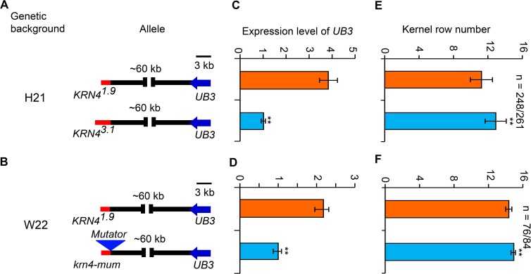
UB3, a member of the SQUAMOSA promoter-binding protein (SBP)-box family of genes, negatively regulates kernel row number and tassel branch number by controlling initiation of reproductive lateral primordia [26]. The KRN4 locus was found responsible for kernel row number and tightly associated with UB3 expression level [27].To confirm the role of KRN4 in maize inflorescences, we analyzed UB3 expression in two sets of NILs (Fig 1A–1D). In the H21 genetic background, the expression level of UB3 linked with the KRN43.1 allele was ~3-fold lower than that of UB3 linked with the KRN41.9 allele (p = 0.002), and the kernel row number of the KRN43.1 -UB3 genotype was approximately two rows greater than that of the KRN41.9 -UB3 genotype (p = 5.7 ×10⁻⁵) (Fig 1C and 1E). In the W22 genetic background with the KRN41.9 allele, a krn4-mum allele was created by a Mutator insertion into the site 55 bp from the 3ʹ-end of KRN4 (S1A Fig). The UB3 expression in krn4-mum individuals was lower than that in KRN41.9 homozygotes (p = 0.003), and the kernel row number of krn4-mum (14.93±0.18) individuals was significantly higher than that of KRN41.9 individuals (14.36±0.36) (p = 0.03) (Figs 1D and 1F and S1A and S1B). These results demonstrate that KRN4 functions as a long-distant regulator of UB3 expression and thus kernel row number.
Fig 1. Genetic effects of KRN4 alleles in two genetic backgrounds.
(A) Schematic diagrams of KRN4 alleles (KRN43.1 and KRN41.9) in the H21 genetic background. (B) Schematic diagrams of KRN4 alleles (krn4-mum and KRN41.9) in the W22 genetic background. The red line stands for KRN4 locus; the triangle indicates the inserted Mutator element; bar = 3 kb. (C, D) UB3 expression levels in 2–3 mm ears of near-isogenic lines in the H21 (C) and W22 (D) genetic backgrounds using quantitative PCR, with three biological replicates in each case. The values show the fold changes. (E, F) Kernel row number of near-isogenic lines in the H21 (E) and W22 (F) genetic backgrounds. Values are shown as the mean ± s.d. Comparsion with the line with KRN41.9, expression level of UB3 was significantly lower but kernel row number was significantly higher in the line with KRN3.1 or krn-mum. The P-value was calculated by one-way ANOVA. **, p<0.01. n: number of individual ears.
UB3 regulates inflorescence architecture
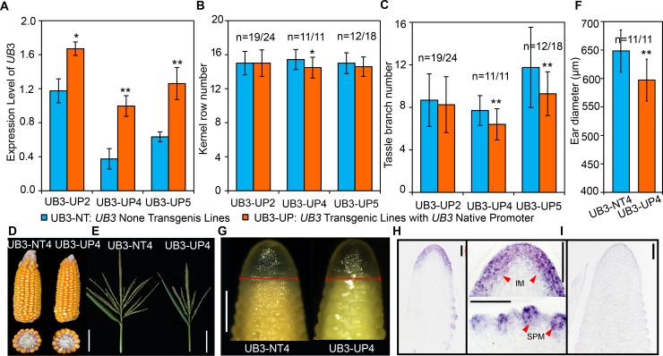
Because overexpression of UB3 strongly represses maize callus regeneration [28], we generated maize lines showing moderate UB3 expression by introducing the UB3 native promoter-driven UB3 coding sequence-yellow fluorescent protein (YFP) fusion construct into the inbred line ZZC01. From the three transgenic lines obtained (transgenic lines with the UB3 Native Promoter were referred to as UB3-UP2, UB3-UP4, UB3-UP5), UB3 expression and inflorescence architecture traits were determined. In addition to kernel row number, we determined tassel branch number (TBN). We found that in 2–3 mm ears, UB3 expression in the transgenic lines was significantly higher than that in the respective non-transgenic lines (UB3-NT2, UB3-NT4, UB3-NT5) (Fig 2A). The transgenic line UB3-UP4 produced less kernel row number (14.48 ± 1.22) and TBN (6.43 ± 1.47) than the nontransgenic line UB3-NT4 (15.43 ± 1.18 for kernel row number and 7.70 ±1.40 for TBN) (Fig 2B–2D). Similarly, reduced kernel row number, TBN and tassel length (TL) were observed in UB3-UP2 and UB3-UP5 compared with UB3-NT2 and UB3-NT5, respectively (Figs 2B–2D and S2A), indicating negative regulations of inflorescence branches and length by UB3. Consistent with the results regarding kernel row number, the diameter of the ear inflorescence meristem (IM) was significantly smaller in UB3-UP4 (597.2 ± 36.9 μm) than in UB3-NT4 (648.4 ± 30.4 μm) (Fig 2F and 2G). Furthermore, to determine the expression pattern of UB3 transcripts in the immature ear, we did mRNA in situ hybridization. UB3 mRNAs were enriched in the peripheral zone of the ear IM and the spikelet pair meristems (SPMs), but not in the center of the ear IM (Fig 2H and 2I), similar to the results using a UB3 antibody for immunolocalization [26]. The size of the IM is thought to be positively correlated with kernel row number [29]; therefore, we propose that UB3 regulates branching in inflorescences by modulating the size of IMs.
Fig 2. Inflorescence architecture of UB3 transgenic lines.
(A) Expression levels of UB3 in 2–3 mm ears of the three independent transgenic lines and non-transgenic lines, three biological replicates. UB3-UP: UB3 transgenic lines containing constructs driven by UB3 native promoter: UB3-UP2, UB3-UP4, UB3-UP5. UB3-NT: UB3 nontransgenic lines: UB3-NT2, UB3-NT4, UB3-NT5. Values are shown as the mean ± s.e. *, p<0.05; **, p<0.01. (B, C) Phenotypes of inflorescence traits including kernel row number (B), and the tassel branch number (TBN, C) in the UB3 transgenic and nontransgenic lines. (D-E) Architecture of inflorescences in UB3-NT4 and UB3-UP4. Bar = 6 cm. (F, G) Inflorescence meristems (F) and diameters (G) of 2–3 mm ears in UB3-NT4 (n = 9) and UB3-UP4 (n = 11). Bar = 500 μm in (G). (H, I) mRNA in situ hybridization of UB3 in the inflorescence meristem and spikelet pair meristems (H) using anti-sense probes. The arrow refers to the signal region. UB3 sense probes as negative controls (I), bar = 100 μm. In (B, C, F) values are shown as the mean ± s.d; n, number of individual ears. The significant differences were estimated using the one-way ANOVA. *, p<0.05; **, p<0.01.
KRN4 genetically interacts with UB2
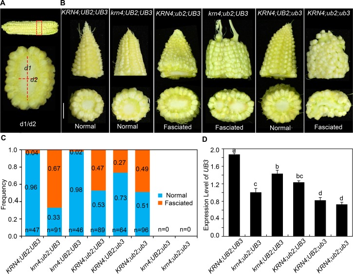
UB2, a paralog of UB3, also encoding SBP-box transcription factors, plays a redundant role with UB3 in controlling the initiation of reproductive axillary meristems. The ub2;ub3 double mutant produces fasciated ears with an increased kernel row number compared to the ub3 and the ub2 single mutant [26]. To identify the effect of KRN4 on the ear architecture of the ub2 and ub3 mutant, krn4-mum (referred to as krn4) was crossed to the ub2;ub3 double mutant and then self-pollinated to segregate single mutants, double mutants and triple mutants. However, due to the tight linkage between UB3 and KRN4, we could not detect the krn4;ub3 and the krn4;ub2;ub3 genotypes in a segregating population with 577 individuals. To identify the phenotype of the ear tip, we defined fasciated ears on the basis of the ratio of the widest diameter (d1) to the narrowest diameter (d2) at the tip of the ear, as illustrated in Fig 3A. If d1/d2 ≥1.2, the ear considered to be fasciated. The results showed that the ears of the krn4 mutant resembled those of the KRN4;UB2;UB3, exhibiting a cone-like tip, and a 15.5 ± 1.0 kernel row number on average, which did not significantly differ from the kernel row number of wild type (15.0 ± 1.2), only 2% of krn4 ears showed fasciation (Fig 3B and 3C). Notably, approximately 27% of ub3 ears and 47% of ub2 ears showed fasciation. The proportion of fasciated ears was obviously increased in the double mutants: 49% for ub2;ub3, and 67% for krn4;ub2 (Fig 3B and 3C), and the fasciation of the krn4;ub2 ears was more severe than that in the ub2 mutant (Fig 3B). These results indicated that krn4 increases the penetrance and expressivity of fasciated ears in the krn4;ub2 double mutants, showing genetic interaction between krn4 and ub2. Furthermore, in 2–3 mm ears, UB3 expression was decreased in both krn4 and ub2 mutants relative to that in wild type (Fig 3D). Compared with the expression of UB3 in ub2, that in krn4;ub2 was lower, showing a coordinated role of both KRN4 and UB2 in the regulation of UB3 transcription (Fig 3D). Additionally, variations at KRN4 locus strongly influenced UB3 expression as shown in Fig 1, and ub2 enhanced defective phenotypes of ub3 ears as shown in Fig 3B. Thus, we suggest the increased proportion of fasciated ears in krn4;ub2 is associated with the significant inhibition of UB3 transcription.
Fig 3. Genetic interactions between krn4, ub2 and ub3.
(A) Definition of the fasciated ears. Both d1 and d2 refer to as the widest and narrowest diameters of the ear tip, respectively. (B) Phenotype of ears in the wild type, single and double mutants of krn4, ub2 and ub3. Bar = 1 cm. (C) Frequency of fasciated ears in diverse genotypes. n = the number of measured ears. (D) Expression level of UB3 in 2–3 mm ears of diverse genotypes determined using quantitative PCR, three biological replicates each. Different letter at top of each column indicates a significant difference at P < 0.05 determined by Tukey HSD test.
UB2 is one of the key factors mediating the KRN4-UB3 interaction
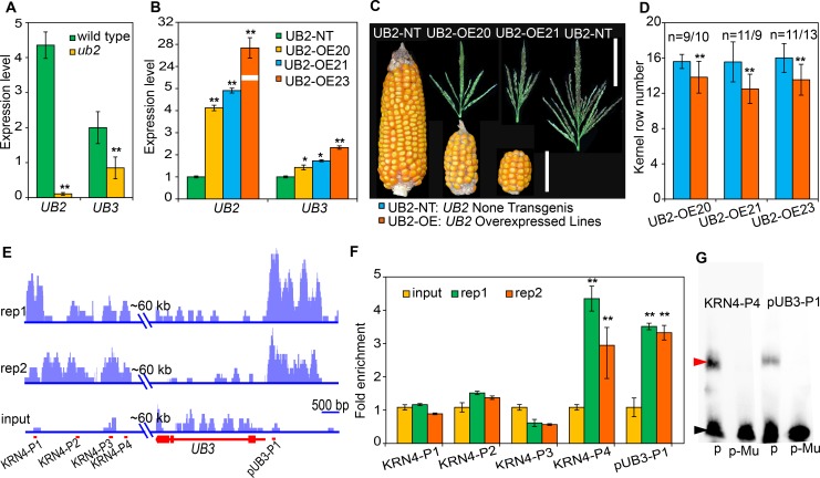
To explore the molecular mechanism of the coordinated roles of both KRN4 and UB2, we generated three transgenic lines (UB2-OE20, UB2-OE21 and UB2-OE23) harboring pCaMV35S-driven UB2-YFP constructs. In the ub2 mutant, UB3 transcription level was 2-fold lower than that in wild type (Fig 4A). In all three transgenic lines, UB2 transcription levels exhibited >8-fold increases, and UB3 transcription levels were also increased by 1–2-fold relative to the respective non-transgenic lines (UB2-NTs) (Fig 4B). Additionally, the UB2-overexpressing lines showed significantly reduced kernel row number, TBN and TL relative to the respective UB2-NT lines (Figs 4C and 4D and S2B–S2D). These results suggest that UB2 might be one of the regulators of UB3 expression.
Fig 4. Expression levels of UB2 and UB3 and target genes bound by UB2.
(A, B) Expression levels of UB2 and UB3 in 3–5 mm ears of wild type and ub2 (A), and three independent UB2-overexpressing lines and a nontransgenic line (B). Gene expression levels were analyzed by quantitative PCR with three biological replicates. (C) Ears and tassels of UB2-overexpressing lines (UB2-OEs) and the nontransgenic line (UB2-NT). Bars = 6 cm for the ears and 10 cm for the tassels. (D) Kernel row number of UB2-overexpressing lines and the non-transgenic lines. n, the number of individual ears. The blue and orange columns represent UB2 non-transgenic and overexpressed lines, respectively. (E) UB2-bound regions in KRN4 and UB3 revealed by chromatin immunoprecipitation sequencing (ChIP-seq) with two biological replicates. Bar = 500 bp. (F) Quantitative PCR verification of UB2-bound regions with two biological replicates. The maize tubulin gene (AC195340.3) was used as the internal control. (G) Electrophoretic mobility shift assay of UB2-GST fusion protein binding to the KRN4-P4 and pUB3-P1 segments. p, KRN4-P4 or pUB3-P1 probes; p-Mu, mutated probes of KRN4-P4 or pUB3-P1, in which GTAC motif was substituted by GCAC. The upper arrow points to bound probes; the lower arrow points to free probes. The values in (A, B, D and F) are means ± s.d.; *, p<0.05; **, p<0.01, which were estimated by the one-way ANOVA.
To test this hypothesis, we performed chromatin immunoprecipitation sequencing (ChIP-seq) to determine the UB2-occupied targets in the ear inflorescence of UB2-OE20 in which UB2-YFP fusion proteins were detected (S2E and 2F Fig). High-confidence peaks corresponding to UB2-bound DNA regions were found in the KRN4 region and UB3 promoter (Fig 4E), indicating the direct binding of UB2 to these two locations in vivo, and this binding was confirmed using ChIP-qPCR, which detected >3-fold enrichment in the KRN4-P4 and the UB3-P1 over the input control in vitro (Fig 4F), indicating that both UB3 and KRN4 are strong candidates for direct targets of UB2.
To further investigate the direct binding of UB2 to KRN4-P4 and UB3-P1, we carefully analyzed the DNA sequences of these fragments and found that they both contain a known plant SQUAMOSA PROMOTER BINDING PROTEIN-LIKE (SPL) specific binding motif (GTAC motif) [30], and then performed an electrophoretic mobility shift assay. We found that UB2 bound to KRN4-P4 and UB3-P1 in vitro (Fig 4G), and detected significantly increased LUC activity in cells coexpressing the p35S-UB2 effector with the KRN4-pUB3-mp35S-luc reporter (p = 3.44 × 10⁻⁸) compared to cells coexpressing the p35S-UB2 effector with the mp35S-luc reporter (S3 Fig). These results indicated that UB2 can directly bind to both KRN4 and the UB3 promoter to positively regulate UB3 expression.
KRN4 acts as an enhancer spatially interacting with the UB3 promoter
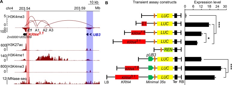
The 3D configuration of the genome is crucial for the dynamic regulation of gene expression [31]. Chromatin interaction analysis by paired-end tag sequencing (ChIA-PET) is a robust method for capturing genome-wide chromatin interactions [32]. Open chromatin and active enhancers are modified with H3K4me1, H3K4me3 and H3K27ac marks [33,34]. Recently, spatial interactions of the KRN4 and UB3 promoter have been detected in apical meristems and seedling leaves [24,25]. These data showed that the spatial interaction anchors identified by H3K4me3 and H3K27ac antibodies occupied three high-confidence remote sites (A1, A2, A3), which were located approximately 33.1 kb—49.9 kb downstream from UB3 (Fig 5A, data from [24]). In particular, KRN4 directly interacted with A1 to form one duplex interaction; A1 with A3, and A2 with the UB3 promoter also displayed two duplex interactions (Fig 5A, data from [24]), showing that the KRN4-UB3 interaction is mediated by complex interactions among the three duplex interactions. In addition, a strong MNase-hypersensitive signal detected in the KRN4 region (Fig 5A, data from [8]) indicated an open chromatin state of KRN4. ChIA-PET revealed the close proximity between UB3 and KRN4 in 3D chromatin configuration, but the spatial proximity between KRN4 and UB3 might be indirect due to interconnections among three duplex interactions and the uncertain surrounding regions (S4 Fig). The results, cross-validated by data generated in different laboratories [8,24,25], provided strong direct evidence of the KRN4-UB3 spatial interaction.
Fig 5. KRN4 spatially interacts with UB3 promoter and acted as an enhancer.
(A) Chromatin interaction analysis between KRN4 and UB3 by paired-end tag sequencing (ChIA-PET). Top panel shows interaction loops inferred from H3K4me3 occupancy and the locations of KRN43.1 (light red column), A1, A2, A3 and UB3 (light blue column). Bottom panels show histone modifications (H3K27ac, H3K4me1, H3K4me3, respectively) and chromatin accessibility (MNase). (B) Transient expression assays showing the effects of KRN43.1, KRN41.9 and the E1 segment on gene expression. LB and RB, left and right boundary of the constructs. Orange box, 91 bp minimal 35S; green box, promoter of UB3 (pUB3); yellow box, luciferase ORF (LUC); light green box, Renilla luciferase ORF (REN); black box, nopaline synthase terminator (Ter); red boxes, KRN4 alleles or 84 bp E1 element. Transient expression assays were all performed in maize leaf protoplasts. For each transient assay, 4–6 biological replicates and two technical replicates were performed. The value is presented as the mean (LUC/REN) ± s.e. ** P < 0.01, * P < 0.05.
To detect the effect of the spatial KRN4-UB3 interaction on gene expression, we separately ligated two KRN4 alleles, KRN43.1 and KRN41.9, to the minimal 35S promoter (mp35S) at a spacing of 2.8 kb (Fig 5B), and then ligated the fusion promoter with the firefly luciferase (LUC) gene. The transient assays were performed in maize protoplasts to test the effects of KRN4 alleles on LUC expression using Renilla luciferase (REN) as an internal control. Compared to mp35S-driven LUC activity, KRN41.9-mp35S-driven and KRN43.1-mp35S-driven LUC activities were 12-fold (p = 3.4×10⁻⁴) and 8-fold (p = 9.3×10⁻⁵) higher, respectively, demonstrating that LUC expression was increased by the presence of both KRN4 alleles, and the enhancement effect of KRN41.9 was greater than that of KRN43.1 (Fig 5B). These findings agree with the UB3 expression levels detected in both sets of NILs (Fig 1), suggesting that KRN4 acts as an enhancer to promote UB3 expression and that the two KRN4 alleles show differences in enhancer activity. Importantly, both KRN4 alleles also increased the expression of UB3 promoter (pUB3)-driven LUC, and enhancer activity of KRN41.9 was stronger than that of KRN43.1 (Fig 5B).
KRN4 exhibits enhancer activity by recruiting the OBF1 and OBF4 proteins
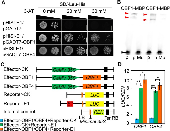
Early studies on enhancers established that sequences with repeated GTGG motifs (where G may be replaced by A) act as enhancers [35]. Within the KRN41.9 region, we detected an 84 bp fragment harboring five putative enhancer elements (one copy of ATGG, GTAG and GTGA and two copies of GTGG), which was designated E1 (S5 Fig). The presence of E1 significantly increased the expression of mp35S-driven LUC, which was much higher than that driven by KRN41.9 and KRN43.1 (Fig 5C). In addition, the 84 bp E1 region contained an ACGT motif (S5 Fig), which is a specific binding site for basic leucine zipper domain proteins (bZIPs) [36]. We tested the binding of two known maize OCS-binding factors, OBF1 and OBF4 [37,38] to E1 in vitro using yeast one-hybrid assays, and revealed both OBF1 and OBF4 bound to E1 (Fig 6A), their binding was further cross-validated by the presence of OBF1-MBP and OBF4-MBP-DNA interacting bands in the EMSAs (Fig 6B).
Fig 6. Enhancer effects of KRN4 and the binding of OBF1 and OBF4 to the E1 segment.
(A, B) Yeast one-hybrid and electrophoretic mobility shift assays revealing the binding of OBF1 and OBF4 to the E1 segment in vitro. The upper arrows point to bound probes, and the lower arrow points to free probes. Bar = 0.3 cm. p, E1 probes; p-Mu, mutated E1 probes, in which CACGT motif was substituted by ACATG; (C, D) Transient expression assays showing that the binding of OBF1 and OBF4 to E1 promotes gene expression. (C) Schematic illustration of the effector and reporter constructs. (D) Luciferase activity detected via transient expression analysis. LB and RB, left and right boundary of the constructs. Orange box, 91 bp minimal 35S; yellow box, luciferase ORF (LUC); light green box, Renilla luciferase ORF (REN); black box, nopaline synthase terminator (Ter); red boxes, 84 bp E1 element. Transient expression assays were all performed in maize leaf protoplasts. For each transient assay, 4–6 biological replicates and two technical replicates were performed. The value is presented as the mean (LUC/REN) ± s.e. ** P < 0.01, * P < 0.05.
To further evaluate the effects of OBF1 and OBF4 on the expression of the E1-driven downstream genes, we performed transient assays with effector vectors (CaMV 35S-OBF1 and CaMV 35S-OBF4) and reporter vectors (mp35S-luc and E1-mp35S-luc) (Fig 6C); the pRL-null vectors harboring mp35S-Renlia luciferase (REN) (mp35S-REN) was used as the internal control. Coexpression of p35S-OBF1 or p35S-OBF4 with E1-mp35S-luc and mp35S-REN resulted in a 10- or 9- fold increases in LUC activity, respectively, compared to the control, indicating that both OBF1 and OBF4 target the E1 to increase LUC expression (Fig 6D).
UB2, UB3, OBF1, and OBF4 are coexpressed in meristems of the ear inflorescence
Because two enhancer-binding proteins (OBF1 and OBF4) were found to bind to KRN4 and UB2 also binds to KRN4 and the UB3 promoter, we reasoned that these three proteins might be recruited by KRN4 and form a transcriptional complex to fine-tune UB3 expression. Thus we analyzed the expression patterns of UB2, OBF1, and OBF4 in developing ears. The expression domains of UB2, UB3, OBF1 and OBF4 in the ear inflorescence partially overlapped. The UB2, UB3, OBF1 and OBF4 transcripts were enriched in the peripheral zone of the IM and SPM primordia, while UB2 specifically expressed in the basal domain of SPMs (Figs 2H–2I and 7A–7G). The similar spatiotemporal expression patterns of these genes suggest that OBF1, OBF4 and UB2 form a complex and coregulate UB3 expression.
Fig 7. mRNA expression patterns of KRN4 binding proteins.
(A-G), mRNA in situ hybridization of UB2 (A-C), OBF1 (D, E) and OBF4 (F, G) in 2–3 mm B73 ears using anti-sense probes, (C) UB2 sense probes as a negative control. The arrows refer to the signal regions. (H, I) Protein–protein interaction identified by a luciferase complementation image assay. (H) Diagram of the experimental design on a single tobacco leaf, including one treatment (upper left) and three control groups. (I) Interaction signals of UB2-OBF1, UB2–OBF4 and OBF1–OBF4. NLUC and CLUC represent N-terminal and C-terminal domains of Firefly Luciferase, respectively. NP and CP, NLUC-fused protein and CLUC-fused protein detected in the luciferase complementation image assay, respectively.
Furthermore, we fused the coding sequences of OBF1, OBF4 and UB2 to the C- and N-terminal domains of LUC (CLUC and NLUC, respectively) and used these fusion constructs for luciferase complementation image assays. Cotransfection with OBF1-CLUC with UB2-NLUC produced weak LUC activity, whereas cotransfection of OBF4-CLUC with OBF1-NLUC or UB2-NLUC produced strong LUC activity (Fig 7H and 7I). By contrast, individual infiltration of the three vectors with the corresponding empty vector failed to produce visible signals, demonstrating a physical interaction between the three proteins. Taken together, the overlapping expression pattern of these four genes and the interactions between OBF1, OBF4 and UB2 proteins suggest the existence of an OBF1, OBF4 and UB2 protein complex in ear inflorescences.
Discussion
An understanding of the genetic basis of maize inflorescence development has resulted from analysis of a large number of developmental mutants. Among these genes, three SPL genes (UB2, UB3, TSH4) affect inflorescences architecture [26]. Notably, UB3 is thought to be tightly linked to QTLs for inflorescence traits such as kernel row number and TBN according to analyses in association mapping and linkage mapping populations, and UB3 genetically interacts with KRN4 [26,27]. However, it has been unclear how KRN4 regulates UB3 expression in meristems of inflorescences. Here, we analyzed the genetic and molecular effects of KRN4 with UB2 and UB3, to reveal the mechanism whereby KRN4 fine-tunes UB3 expression and, thus, inflorescence development.
A large number of functionally important elements (promoters, enhancers, insulators) and transcribed RNAs are being annotated in intergenic regions of the maize genome [1,2]. In a few well-studied cases, intergenic regions have been shown to regulate gene expression and associate with phenotypic variation, such as B1 [11,12], TB1 [14], Hydroxyproline-rich glycoprotein [39], Pericarp Color1 [40], VGT1 [12] and Benzoxazinless1 (BX1) [41]. KRN4 is also a functional intergenic region based on several lines of evidence. First, KRN4 was mapped to a 3.1 kb intergenic region. Sequence alignment failed to reveal RNA transcripts that were mapped to KRN4 with high confidence, while a Harbinger-like fragment has been mapped to a 1.2 kb insertion/deletion, which causes two allelic variants in NILs [27], suggesting that KRN4 likely functions as a cis-acting element in controlling inflorescence branching. Second, sequence variations in KRN4 including the 1.2 kb insertion/deletion in the H21 background and a Mutator insertion/deletion in the W22 background cause changes in UB3 expression and quantitative variation of inflorescence branches (kernel row number and tassel branch number). Additionally, genetic interactions between UB2 and UB3 [26] and between KRN4 and UB3 [27] were found to be involved in the regulation of inflorescence branching. In this study, the double mutant krn4/ub2 showed an approximately 20-fold higher frequency of fasciated ears than single krn4 and ub2 mutants, showing strong genetic interaction between KRN4 and UB2 in the regulation of inflorescence branching. Similarly, a genetic interaction between KRN4 and UB2 involved in the regulation of UB3 expression was also found. Finally the KRN4–UB3 genomic intervals are open chromatin regions, as revealed in the maize MNase HS map [8], and allelic variation of KRN4 changed UB3 expression, indicating that the intergenic KRN4 sequence with open chromatin state achieves its cis-activity through spatially interacting with the UB3 promoter. Two parents used in KRN4 mapping, H21 which has KRN41.9 allele and H21NX531 which has KRN43.1 allele, show significantly difference in kernel row number and UB3 expression level [27]. In this study, we found that KRN41.9 in H21 exhibits a stronger enhancer activity than KRN43.1 in regulating UB3 expression level that is negatively correlated with kernel row number. The result will help to understand the KRN4 effects on UB3 expression level and kernel row number in the original mapping population. This finding also provides an additional case of variation in an intergenic region that fine-tunes gene expression, providing a new perspective on the genetic control of a quantitative trait.
The intergenic QTL KRN4 has key features often found in enhancers. Intergenic regions usually exert their functions as cis-regulatory DNA elements (enhancers or insulators) or trans-regulatory factors. Enhancers were initially described as short DNA fragments with the ability to positively drive target gene expression independent of distance and orientation, high sensitivity to DNase, and the presence of specific DNA elements in an open chromatin state that allows the binding of transcription factors (TFs) and transcription coactivators [42,43,44]. In this study, we found that KRN4 harbored an 84 bp fragment containing five putative enhancer elements. Moreover, the 84 bp fragment and two KRN4 alleles showed strong transcriptional activation activity toward the promoters of 35S and UB3. Indeed, three TFs including an UB2 and two known enhancer-binding proteins (OBF1 and OBF4), were shown to interact with each other and bind KRN4. In particular, UB2 was able to bind KRN4 and the UB3 promoter, and double mutants ub2;ub3, ub2;krn4 showed a higher level of fasciated ears than ub3 or krn4 single mutants, suggesting that UB2 is a key mediator of the long-distant regulation of the UB3 promoter by KRN4. One of the most accepted models for long-distance regulatory interactions is the looping model, which hypothesizes that interactions between enhancers–promoters are in physical contact, forming an intervening chromatin loop [45,46]. The chromatin interactions provide close proximity among distant elements. Two recent studies have detected the enriched peaks on KRN4 locus by H3K4me3, H3K27ac and RNA Pol II [24,25]. The KRN4–UB3 promoter interaction was mediated by complex interactions among duplexes, while the intervening sequences might be looped outward. Additionally, the KRN4 region is highly sensitive to MNase, indicating that KRN4 is in an open chromatin state, which is one of the signatures of an enhancer. These results demonstrated that intergenic KRN4, in cooperation with UB2, OBF1 and OBF4, functions as an enhancer of the UB3 promoter via interconnections among duplex chromatin interactions (S6 Fig).
Since the discovery of enhancer-promoter interactions, the mechanism of their actions is a major interest of the scientific community. The DNA looping and the binding of larger protein complexes explain how enhancers communicate with the targeted promoters over long distances. With the advance of genomics, chromatin immunoprecipitation (ChIP), chromosome conformation capture (3C) and 3C-derived methods combined with sequencing have identified numerous enhancer-like elements in humans and plant species, and have revealed the super-enhancers that are large clusters of highly active enhancers regulating cell type-specific and phenotype-related genes [47,48]. In recent years, a conceptual framework for a phase separation model has been proposed to elucidate transcription control of super enhancers [49]. This model suggests that a high density of proteins and nucleic acids kept in close spatial proximity form phase-separated droplets in order to maintain a compartmentalized and concentrated state for cell type-specific transcriptional regulatory processes [48–50]. Two such super enhancers, BRD4 and MED1 in human cells, control the expression of key genes controlling cell fate and disease occurrence [50]. Whether the transcription factors UB2, OBF1, OBF4 and others enriched in the KRN4-UB3 chromatin interaction interval form a phase-separated droplet remains to be determined.
Materials and methods
Plant materials and phenotypic analysis
The maize krn4-mum mutant (mu1023400), a Mutator (Mu)-insertion mutant, was provided by the Maize Genetics Cooperation Stock Center at the University of Illinois, Champaign-Urbana. The Mu is inserted in a 55 bp downstream of the 3'-end of KRN4. The Mu insertion site was detected by PCR with KRN4-specific and TIR6 primers (S2 Table). Heterozygous individuals (+/krn4-mum) were selected and crossed to wild-type individuals two times, then self-crossed to segregate the homozygous krn4-mum mutants and the wild-type individuals that were evaluated in the field in two environments: summer 2016 in Wuhan (30°N, 114°E) and spring 2017 in Sanya (18.34°N, 109.62°E), China. Approximately 100 individuals per genotype were evaluated in each environment. One-way ANOVA (analysis of variance) was used to estimate the significance of the phenotypic difference between the mutants and wild-type plants.
For assessment the genetic interactions among KRN4, UB2 and UB3, krn4-mum (referred to as krn4) was crossed with the ub2-mum1 ub3-mum1 double mutant (referred to as ub2/ub3) in the W22 background as described by Chuck et al. (2014), followed by self-crossing to obtain the segregating F2 population. A total of 577 individuals were phenotyped in the spring 2018 in Wuhan (30°N, 114°E) using gene-specific primers (S2 Table). For phenotyping, the widest diameter (d1) and the narrowest diameter (d2) at the tip of the ears were measured 20 days after pollination (DAP) (as shown in Fig 3A), and d1/d2 was calculated to define ear fasciation. If d1/d2> = 1.2, the ear was considered fasciated. The frequency of ear fasciation was calculated from the number of fasciated ears to the total number of measured ears of a given genotype.
Expression analysis
Total RNA was isolated from the tissues of ~2 mm and 5 mm ears of various maize plants (KRN43.1and KRN41.9 in the H21 background, krn4-mum and W22 plants, ub2::mum and wild-type plants, UB2 and UB3 transgenic plants) using TRIzol reagent (Life Technologies, Invitrogen, Carlsbad, CA, USA) according to the manufacturer’s instructions. cDNA was synthesized from mRNA using an EasyScript One-step gDNA-removal and cDNA-Synthesis Supermix Kit (Transgene, Beijing, China) according to the manufacturer’s instructions. RT-PCR was conducted using gene-specific primers (S3 Table) and SYBR Green PCR Master Mix (KAPA, Beijing, China). The maize beta-actin (NM_001155179) gene was used as an internal control. All assays were performed with three biological and three technical replicates.
In situ hybridization
Immature B73 ears (2–3 mm) were fixed in a 4% PFA solution (4g of paraformaldehyde (Sigma-Aldrich) dissolved in 100 mL of 1× PBS, pH 6.5–7), embedded in paraplast plus (Sigma-Aldrich), and sectioned to a thickness of 8 μm. To construct sense and antisense RNA probes for UB2, UB3, OBF1 and OBF4, probe fragments were amplified and cloned into pSPT18 (Roche), digested with HindIII and EcoRI, transcribed using SP6 and T7 RNA polymerase (Roche) in vitro, and labeled with digoxigenin-UTP (Roche). RNA hybridization, immunologic detection and signal capture of the hybridized probes were performed as described previously [51]. The in situ primers for UB2, UB3, OBF1 and OBF4 are listed in S3 Table.
Dual-luciferase transient assay in maize protoplasts
To construct reporter plasmids, KRN43.1, KRN41.9 and the E1 segment were separately cloned into the site between BamHI and SalI, which is located approximately 2.8 kb from multiple cloning site (MCS) region, in the PGL3-basic vector (Promega, Madison, WI, USA) with Minimal 35S (mp35S)-driven Luciferase (Luc). The UB3 promoter (pUB3) was also ligated to mp35S in the MCS region. To construct effector plasmids, the CDSs of UB2, OBF1 and OBF4 were individually cloned into the pRTL2 vector with an enhanced CaMV35S promoter (p35S). The pRL-null vector (Promega) with mp35S-driven Renilla Luciferase (REN) was used as the internal control. The primer pairs used for vector construction are listed in S2 Table. Seedlings of maize inbred line B73 were grown in the dark at 28°C for 13–15 days, and etiolated leaves were harvested for protoplast isolation. Transformation was performed as described previously [52], and dual-luciferase detection was conducted according to the manual of the Dual-LuciferaseReporter Assay System E1960 (Promega). For each assay, four to six biological replicates and two technological replicates were performed.
Yeast one-hybrid assay
The full-length CDS of OBF1 and OBF4 were cloned into the pGADT7 vector (Clontech, Beijing, China) to generate pGADT7–OBF1 and pGADT7–OBF4, respectively. The E1 segment was cloned into the pHISi-1 vector (Clontech, Beijing, China) to generate pHISi–E1. The pHisi-E1 vector was linearized with XhoI and transferred into the yeast strain YM4271, followed by incubation for 3 to 5 days at 30°C. A positive pHISi-E1 clone was used to test the proper concentration of 3-amino-1, 2, 4-triazole (3-AT) in the SD/-His medium for background expression. Selection medium containing 20 mm 3-AT was used to screen for enhancer binding proteins. pGADT7, pGADT7–OBF1 and pGADT7–OBF4 were transferred into YM4271 yeast cells containing the pHisi-E1 recombinant plasmid and grown in SD/-Leu-His supplemented with 20 mm 3-AT. After a 3 to 5–day incubation at 30°C, positive clones confirmed by PCR-sequencing were diluted 10–100 fold and spotted onto SD/-Leu-His plates containing 0, 20 and 30 mm 3-AT. The primers used in the Y1H assay are listed in S2 Table.
Electrophoretic mobility shift assay
To construct prokaryotic expression plasmids, the full-length CDS of OBF1, OBF4 and UB2 were cloned into pMAL-c2x (N–MBP) and PGEX-4T-1 (N–GST), respectively. The expression of the OBF1–MBP, OBF4–MBP and UB2-GST fusion protein was induced in Trans BL21 cells and Transetta (DE3) E. coli cells (Transgene, Beijing, China) with 0.2 mM isopropyl-1-thio-D-galactopyranoside at 16°C for 14 h. The OBF1–MBP and OBF4–MBP fusion proteins were purified using amylose resin (E8021S, NEB, USA), and the UB2-GST fusion proteins was purified using Glutathione Sepharose 4 EF (GE Healthcare Life Sciences China, Beijing, China), and the proteins were then quantified using the protein assay reagent (Sangon Biotech, Shanghai, China) following the manufacturer’s protocol and detected by 12% SDS-PAGE. Single-stranded DNA probes were synthesized with a 5'-end biotin label (S1 Table); probe dilution, renaturation and binding reactions for the EMSA were performed as described previously [28]. The purified proteins (20–40 ng fusion protein per reaction) were incubated with labeled probes (final concentration 10 fM) in binding reaction mixture (10 mM Tris, 50 mM KCL, 1 mM dithiothreitol, 5 mM MgCl2, 2.5% glycerol, poly (dI-dC), 0.05% NP-40; pH 7.5) at room temperature for 20 min. The reaction products were combined with protein loading buffer (25 mM Tris–HCL [pH 7.5], 0.04% bromophenol blue, 80% glycerol), then loaded onto 6% nondenaturing PAGE gels and subjected to electrophoresis (100 V) at 4°C for 1–1.5 h in 0.5× TBE buffer (45 mM Tris, 45 mM boric acid, 2 mM EDTA, pH 8.3). After transfer to a membrane (100 V at 4°C for 1 h in 0.5× TBE buffer) and UV crosslinking (120 mJ/cm2, UV-light crosslinking instrument with 254 nm bulbs, 45–60 s exposure), shifts on the nylon membrane were detected using an LightShiftChemiluminescent EMSA Kit and Chemiluminescent Nucleic Acid Detection Module following the manufacturer’s protocols (Thermo Scientific, Waltham, MA, USA).
Chromatin immunoprecipitation and data analysis
The pCaMV35S::UB2-YFP construct was transformed into the maize inbred line ZZC01 Three transgenic overexpression lines, UB2–OE20, UB2–OE21 and UB2–OE23, were produced by CHINASEED (China National Seed Group Co. Ltd., Beijing, China). The transgenic and corresponding nontransgenic plants were grown in the field. Target gene expression was verified by fluorescence detection and immunoblot analysis (S2 Fig). Immature ears (5 mm long) were collected from UB2-OE20, dissected and cross-linked in 30 of mL 1× PBS buffer (137 mM NaCl, 2.7 mM KCl, 10 mM Na2HPO4, 2 mM KH2PO4) containing 1% formaldehyde for 15 min under a vacuum. Cross-linking was terminated by adding 0.15 M glycine, followed by 5 min of incubation under vacuum. The samples were washed three times with distilled water, dried with paper towels and quickly frozen in liquid nitrogen. Chromatin was extracted with extraction buffer 1 (EB1, 0.4 M sucrose, 10 mM Tris-HCl pH 8.0, 10 mM MgCl2, 5 mM mercaptoethanol and Plant Protease Inhibitor Cocktail). The sample was centrifuged for 20 min at 1000 g at 4°C, and the pellet was washed five times with 5 mL EB2 (0.25 M sucrose, 10 mM Tris-HCl, pH 8.0, 10 mM MgCl2, 1% Triton X-100, 5 mM -mercaptoethanol and plant protease inhibitor cocktail) and once with 5mL EB3 (1.7 M sucrose, 10 mM Tris-HCl, pH 8.0, 2 mM MgCl2, 0.15% Triton X-100, 5 mM -mercaptoethanol and a plant protease inhibitor cocktail), followed by sonication in 300 μL of sonication buffer (50 mM Tris-HCl, pH 8.0, 10 mM EDTA, 1% SDS and plant protease inhibitor cocktail) using a Diagenode Bioruptor sonicator (Diagenode SA, Seraing, Belgium) for 15 cycles (30 s ON/30 s OFF). The sonicated DNA fragments were 200–700 bp long. DiaMag protein A-coated magnetic beads (C03010020, Diagnode SA, Seraing, Belgium) were incubated with anti-green fluorescent protein (A11122, Thermo Fisher Scientific, Waltham, MA, USA), and the sonicated chromatin was immunoprecipitated with antibody-bead complexes. DNA elution, decrosslinking and isolation were performed using an IPure Kit v2 according to the manufacturer’s instructions (C01010150, Diagenode SA, Seraing, Belgium). A small quantity of sonicated chromatin that was not incubated with the antibody was used as the total input DNA control. DNA libraries were constructed using an Ovation Low Input DR Kit (NuGEN Technologies) and sequenced on the Illumina HiSeq 2500 platform, including two IP and two input libraries.
The FastQC program was used to assess high-quality clean reads [53], which were aligned to the maize reference genome (AGPv2) using Hisat2 v.2.0.5 [54]. Picard Mark Duplicates (v.2.9.0) and MACS (v.1.4.2) were used to remove PCR duplicates and for peak calling, respectively [55]. The p-value of the enriched peaks was p < 1e-05 in each ChIP-seq library compared to the input DNA. Integrative Genomics Viewer was used to visualize the ChIP tracks of the UB2-YFP fusion protein binding sites [56].
ChIP–qPCR
ChIP-qPCR was performed with specific primers within targeted peaks located at KRN4 and the UB3 promoter region. The maize tubulin gene (AC195340.3) was used as the internal control. Two biological replicates and three technological replicates were performed. The primers used for quantitative ChIP–PCR analysis are listed in S3 Table. The abundance of a target was normalized to genomic regions with nonspecific binding genomic regions, and the fold enrichment was compared to the input sample. P-values were obtained via Student’s t-test.
Firefly luciferase complementation imaging
To analyze protein-protein interactions, the full-length CDS of UB2, OBF1 and OBF4 were cloned into JW771 (NLUC) and JW772 (CLUC) to produce recombinant the UB2–NLUC, OBF1–NLUC, OBF4–NLUC, UB2–CLUC, OBF1–CLUC and OBF4–CLUC vectors, respectively [57]. Young but fully expanded Nicotiana benthamiana leaves (8–10 weeks old) were infiltrated with Agrobacterium tumefaciens GV3101 cells that had been transfected with the above constructs. At four different areas on a leaf, one sample and three negative control combinations were infiltrated. After 48 h of growth under a 16 h-light/8 h-dark cycle, the abaxial epidermis of each leaf was coated with 1 mM luciferin (Promega). LUC signals were captured using the Tanon-5200 imaging system (Tanon, Shanghai, China). The experiments were repeated three to four times to obtain consistent results.
Supporting information
(A) Schematic diagram of the genomic locations of KRN4 and UB3 and the Mutator insertion site. Bar = 500 bp. Red line shows KRN4 locus.
(B) Ears of wild type and krn4-mum. A represent ear of krn4-mum having 16 kernel rows (right) and a represent ear of wild type having 14 kernel rows (left) are shown. Bar = 4 cm.
(TIF)
(A) Tassel length of the three independent UB3 transgenic lines (UB3-UP) and non-transgenic lines (UB3-NT).
(B-D) Tassel branch and tassel length of UB2 transgenic plants (UB2-OE) and non- transgenic plants (UB2-NT).
(E) Immunoblot analysis using YFP antibody. Proteins were extracted from the ears of UB2-OE20 and UB2-OE21 transgenic plants.
(F) UB2-YFP fusion protein signals observed in 2 mm ears by confocal microscopy. Strong signals indicate that UB2-YFP fusion protein was expressed in the ears of the transgenic lines. n, number of individual tassel. Values are means ± SD, P-value was calculated by Student’s t-test. *, p<0.05; **, p<0.01.
(TIF)
(A) Effector and reporter constructs.
(B) Relative luciferase activity determined by transient expression analysis of KRN4-UB3 promoter-driven LUC in maize protoplasts co-infiltrated with UB2 effector. For each transient assay, 4–6 biological replicates and two technical replicates were performed. The value is presented as mean (LUC/REN) ± SD. ** P < 0.01; * P < 0.05.
(TIF)
(TIF)
Sequences highlighted in red are enhancer-like elements. The sequence highlighted in green is the bZIP binding motif.
(TIF)
The two long-distant elements (KRN4, UB3 promoter) kept in close proximity to each other by chromatin interaction. Three interacting transcription factors UB2, OBF1 and OBF4 bind to specific cis-elements (GTAC motif or enhancer elements) harbored in UB3 promoter and KRN4 to promote the transcription of UB3 which negatively controls the initiation of reproductive axillary meristems and in turn the ear inflorescence branching.
(TIF)
(DOCX)
(DOCX)
(DOCX)
Data Availability
All relevant data are within the manuscript and its Supporting Information files.
Funding Statement
This work was supported by the National Natural Science Foundation of China (91935305, 31701431) and the National Key Research and Development Program of China (2016YFD0100404). The funders had no role in study design, data collection and analysis, decision to publish, or preparation of the manuscript.
References
- 1.Schnable PS, Ware D, Fulton RS, Stein JC, Wei F, Pasternak S, et al. The B73 maize genome: complexity, diversity, and dynamics. Science. 2009; 326(5956):1112–5. 10.1126/science.1178534 [DOI] [PubMed] [Google Scholar]
- 2.Springer NM, Anderson SN, Andorf CM, Ahern KR, Bai F, Barad O, et al. The maize W22 genome provides a foundation for functional genomics and transposon biology. Nat Genet. 2018; 50(9):1282–1288. 10.1038/s41588-018-0158-0 [DOI] [PubMed] [Google Scholar]
- 3.Michael T.P., VanBuren R. Progress, challenges and the future of crop genomes. Curr Opin Plant Biol. 2015; 24:71–81. 10.1016/j.pbi.2015.02.002 [DOI] [PubMed] [Google Scholar]
- 4.Castillo-Davis C.I. The evolution of noncoding DNA: how much junk, how much func? Trends Genet. 2005; 21(10):533–6. 10.1016/j.tig.2005.08.001 [DOI] [PubMed] [Google Scholar]
- 5.Alexander R.P., Fang G., Rozowsky J., Snyder M., Gerstein M.B. Annotating non-coding regions of the genome. Nat Rev Genet. 11,559–571 (2010). 10.1038/nrg2814 [DOI] [PubMed] [Google Scholar]
- 6.Zhang X., Bernatavichute Y.V., Cokus S., Pellegrini M., Jacobsen S.E. Genome-wide analysis of mono-, di- and trimethylation of histone H3 lysine 4 in Arabidopsis thaliana. Genome Biol. 2009; 10(6):R62 10.1186/gb-2009-10-6-r62 [DOI] [PMC free article] [PubMed] [Google Scholar]
- 7.Du Z, Li H, Wei Q, Zhao X, Wang C, Zhu Q, et al. Genome-wide analysis of histone modifications: H3K4me2, H3K4me3, H3K9ac, and H3K27ac in Oryza sativa L. Japonica. Mol Plant. 2013; 6(5):1463–72. 10.1093/mp/sst018 [DOI] [PMC free article] [PubMed] [Google Scholar]
- 8.Rodgers-Melnick E., Vera D.L., Bass H.W., Buckler E.S. Open chromatin reveals the functional maize genome. Proc Natl Acad Sci USA 2016; 113(22):E3177–84. 10.1073/pnas.1525244113 [DOI] [PMC free article] [PubMed] [Google Scholar]
- 9.Liu J, Chen J, Zheng X, Wu F, Lin Q, Heng Y, et al. GW5 acts in the brassinosteroid signalling pathway to regulate grain width and weight in rice. Nat Plants. 2017; 3:17043 10.1038/nplants.2017.43 [DOI] [PubMed] [Google Scholar]
- 10.Stam M., Belele C., Dorweiler J.E., Chandler V.L. Differential chromatin structure within a tandem array 100 kb upstream of the maize b1 locus is associated with paramutation. Genes Dev. 2002; 16(15):1906–18. 10.1101/gad.1006702 [DOI] [PMC free article] [PubMed] [Google Scholar]
- 11.Arteaga-Vazquez M.A., Chandler V.L. Paramutation in maize: RNA mediated trans-generational gene silencing. Curr Opin Genet Dev. 2010; 20(2):156–63. 10.1016/j.gde.2010.01.008 [DOI] [PMC free article] [PubMed] [Google Scholar]
- 12.Castelletti S., Tuberosa R., Pindo M., Salvi S. A MITE transposon insertion is associated with differential methylation at the maize flowering time QTL Vgt1. G3 (Bethesda). 2014; 4(5):805–12. [DOI] [PMC free article] [PubMed] [Google Scholar]
- 13.Salvi S, Sponza G, Morgante M, Tomes D, Niu X, Fengler KA, et al. Conserved noncoding genomic sequences associated with a flowering-time quantitative trait locus in maize. Proc Natl Acad Sci USA. 2007; 104(27):11376–81. 10.1073/pnas.0704145104 [DOI] [PMC free article] [PubMed] [Google Scholar]
- 14.Clark R.M., Wagler T.N., Quijada P., Doebley J. A distant upstream enhancer at the maize domestication gene tb1 has pleiotropic effects on plant and inflorescent architecture. Nat Genet. 2006; 38(5):594–7. 10.1038/ng1784 [DOI] [PubMed] [Google Scholar]
- 15.Studer A., Zhao Q., Ross-Ibarra J., Doebley J. Identification of a functional transposon insertion in the maize domestication gene tb1. Nat Genet. 2011; 43(11):1160–3. 10.1038/ng.942 [DOI] [PMC free article] [PubMed] [Google Scholar]
- 16.Bird C.P., Stranger B.E., Dermitzakis E.T. Functional variation and evolution of non-coding DNA. Curr Opin Genet Dev. 2006; 16(6):559–64. 10.1016/j.gde.2006.10.003 [DOI] [PubMed] [Google Scholar]
- 17.Wei J.W., Huang K., Yang C., Kang C.S. Non-coding RNAs as regulators in epigenetics. Oncol Rep. 2017; 37(1):3–9. 10.3892/or.2016.5236 [DOI] [PubMed] [Google Scholar]
- 18.McCue A.D., Nuthikattu S., Slotkin R.K. Genome-wide identification of genes regulated in trans by transposable element small interfering RNAs. RNA Biol. 2013; 10(8):1379–95. 10.4161/rna.25555 [DOI] [PMC free article] [PubMed] [Google Scholar]
- 19.Denker A., de Laat W. A long-distance chromatin affair. Cell. 2015; 162(5):942–3. 10.1016/j.cell.2015.08.022 [DOI] [PubMed] [Google Scholar]
- 20.Li G, Ruan X, Auerbach RK, Sandhu KS, Zheng M, Wang P, et al. Extensive promoter-centered chromatin interactions provide a topological basis for transcription regulation. Cell. 2012; 148(1–2):84–98. 10.1016/j.cell.2011.12.014 [DOI] [PMC free article] [PubMed] [Google Scholar]
- 21.Weber B., Zicola J., Oka R., Stam M. Plant enhancers: A call for discovery. Trends Plant Sci. 2016; 21(11):974–987. 10.1016/j.tplants.2016.07.013 [DOI] [PubMed] [Google Scholar]
- 22.Zhu B., Zhang W., Zhang T., Liu B., Jiang J. Genome-wide prediction and validation of intergenic enhancers in Arabidopsis using open chromatin signatures. Plant Cell. 2015; 27(9):2415–26. 10.1105/tpc.15.00537 [DOI] [PMC free article] [PubMed] [Google Scholar]
- 23.Oka R, Zicola J, Weber B, Anderson SN, Hodgman C, Gent JI, et al. Genome-wide mapping of transcriptional enhancer candidates using DNA and chromatin features in maize. Genome Biol. 2017; 18(1):137 10.1186/s13059-017-1273-4 [DOI] [PMC free article] [PubMed] [Google Scholar]
- 24.Li E, Liu H, Huang L, Zhang X, Dong X, Song W, et al. Long-range interactions between proximal and distal regulatory regions in maize. Nat Commun. 2019; 10(1):2633 10.1038/s41467-019-10603-4 [DOI] [PMC free article] [PubMed] [Google Scholar]
- 25.Peng Y, Xiong D, Zhao L, Ouyang W, Wang S, Sun J, et al. Chromatin interaction maps reveal genetic regulation for quantitative traits in maize. Nat Commun. 2019; 10(1):2632 10.1038/s41467-019-10602-5 [DOI] [PMC free article] [PubMed] [Google Scholar]
- 26.Chuck G.S., Brown P.J., Meeley R., Hake S. Maize SBP-box transcription factors unbranched2 and unbranched3 affect yield traits by regulating the rate of lateral primordia initiation. Proc Natl Acad Sci USA. 2014; 111(52):18775–80. 10.1073/pnas.1407401112 [DOI] [PMC free article] [PubMed] [Google Scholar]
- 27.Liu L, Du Y, Shen X, Li M, Sun W, Huang J, et al. KRN4 controls quantitative variation in maize kernel row number. PLoS Genet. 2015; 1(11):e1005670. [DOI] [PMC free article] [PubMed] [Google Scholar]
- 28.Du Y, Liu L, Li M, Fang S, Shen X, Chu, et al. UNBRANCHED3 regulates branching by modulating cytokinin biosynthesis and signaling in maize and rice. New Phytol. 2017; 214(2):721–733. 10.1111/nph.14391 [DOI] [PubMed] [Google Scholar]
- 29.Bommert P, Nagasawa NS, Jackson D. Quantitative variation in maize kernel row number is controlled by the FASCIATED EAR2 locus. Nat Genet. 2013; 45(3):334–7. 10.1038/ng.2534 [DOI] [PubMed] [Google Scholar]
- 30.Birkenbihl R.P., Jach G., Saedler H., Huijser P. Functional dissection of the plant-specific SBP-domain: overlap of the DNA-binding and nuclear localization domains. J Mol Biol. 2005; 352(3):585–96. 10.1016/j.jmb.2005.07.013 [DOI] [PubMed] [Google Scholar]
- 31.Rowley MJ, Nichols MH, Lyu X, Ando-Kuri M, Rivera ISM, Hermetz K, et al. Evolutionarily conserved principles predict 3D chromatin organization. Mol Cell. 2017; 67(5):837–852.e7. 10.1016/j.molcel.2017.07.022 [DOI] [PMC free article] [PubMed] [Google Scholar]
- 32.Li X, Luo OJ, Wang P, Zheng M, Wang D, Piecuch E, et al. Long-read ChIA-PET for base-pair-resolution mapping of haplotype-specific chromatin interactions. Nat Protoc. 2017; 12(5):899–915. 10.1038/nprot.2017.012 [DOI] [PMC free article] [PubMed] [Google Scholar]
- 33.Creyghton MP, Cheng AW, Welstead GG, Kooistra T, Carey BW, Steine EJ, et al. Histone H3K27ac separates active from poised enhancers and predicts developmental state. Proc Natl Acad Sci USA 2010; 107(50):21931–6. 10.1073/pnas.1016071107 [DOI] [PMC free article] [PubMed] [Google Scholar]
- 34.Pekowska A, Benoukraf T, Zacarias-Cabeza J, Belhocine M, Koch F, Holota H, et al. H3K4 tri-methylation provides an epigenetic signature of active enhancers. EMBO J. 2011; 30(20):4198–210. 10.1038/emboj.2011.295 [DOI] [PMC free article] [PubMed] [Google Scholar]
- 35.McPherson C.E., Shim E.Y., Friedman D.S., Zaret K.S. An active tissue-specific enhancer and bound transcription factors existing in a precisely positioned nucleosomal array. Cell. 1993; 75(2):387–98. 10.1016/0092-8674(93)80079-t [DOI] [PubMed] [Google Scholar]
- 36.Foley RC, Grossman C, Ellis JG, Llewellyn DJ, Dennis ES, Peacock WJ, et al. Isolation of a maize bZIP protein subfamily: candidates for the ocs-element transcription factor. Plant J. 1993; 3(5):669–79. [PubMed] [Google Scholar]
- 37.Singh K, Dennis ES, Ellis JG, Llewellyn DJ, Tokuhisa JG, Wahleithner JA, et al. OCSBF-1, a maize ocs enhancer binding factor: isolation and expression during development. Plant Cell. 1990; 2(9):891–90. 10.1105/tpc.2.9.891 [DOI] [PMC free article] [PubMed] [Google Scholar]
- 38.Zhang B., Foley R.C., Singh K.B. Isolation and characterization of two related Arabidopsis ocs-element bZIP binding proteins. Plant J. 1993; 4(4):711–6. 10.1046/j.1365-313x.1993.04040711.x [DOI] [PubMed] [Google Scholar]
- 39.Hood K.R., Baasiri R.A., Fritz S.E., Hood E.E. Biochemical and tissue print analyses of hydroxyproline-rich glycoproteins in cell walls of sporophytic maize tissues. Plant Physiol. 1991; 96(4):1214–9. 10.1104/pp.96.4.1214 [DOI] [PMC free article] [PubMed] [Google Scholar]
- 40.Sidorenko L., Li X., Tagliani L., Bowen B., Peterson T. Characterization of the regulatory elements of the maize P-rr gene by transient expression assays. Plant Mol Biol. 1999; 39(1):11–9. 10.1023/a:1006172815663 [DOI] [PubMed] [Google Scholar]
- 41.Zheng L, McMullen MD, Bauer E, Schön CC, Gierl A, Frey M, et al. Prolonged expression of the BX1 signature enzyme is associated with a recombination hotspot in the benzoxazinoid gene cluster in Zea mays. J Exp Bot. 2015; 66(13):3917–30. 10.1093/jxb/erv192 [DOI] [PMC free article] [PubMed] [Google Scholar]
- 42.Wallace JG, Bradbury PJ, Zhang N, Gibon Y, Stitt M4, Buckler ES, et al. Association mapping across numerous traits reveals patterns of functional variation in maize. PLoS Genet. 2014; 10(12):e1004845 10.1371/journal.pgen.1004845 [DOI] [PMC free article] [PubMed] [Google Scholar]
- 43.Bulger M., Groudine M. Functional and mechanistic diversity of distal transcription enhancers. Cell. 2011; 144(3):327–39. 10.1016/j.cell.2011.01.024 [DOI] [PMC free article] [PubMed] [Google Scholar]
- 44.Ong C.T., Corces V.G. Enhancer function: new insights into the regulation of tissue-specific gene expression. Nat Rev Genet. 2011; 12(4):283–93. 10.1038/nrg2957 [DOI] [PMC free article] [PubMed] [Google Scholar]
- 45.Krivega I., Dean A. Enhancer and promoter interactions-long distance calls. Curr Opin Genet Dev. 2012; 22(2):79–85. 10.1016/j.gde.2011.11.001 [DOI] [PMC free article] [PubMed] [Google Scholar]
- 46.Vernimmen D., Bickmore W.A. The hierarchy of transcriptional activation: from enhancer to promoter. Trends Genet. 2015; 31(12):696–708. 10.1016/j.tig.2015.10.004 [DOI] [PubMed] [Google Scholar]
- 47.Hnisz D, Abraham BJ, Lee TI, Lau A, Saint-André V, Sigova AA, et al. Super-enhancers in the control of cell identity and disease. Cell. 2013; 155(4):934–47. 10.1016/j.cell.2013.09.053 [DOI] [PMC free article] [PubMed] [Google Scholar]
- 48.Hnisz D., Shrinivas K., Young R.A., Chakraborty A.K., Sharp P.A. A phase separation model for transcriptional control. Cell. 2017; 169(1):13–23. 10.1016/j.cell.2017.02.007 [DOI] [PMC free article] [PubMed] [Google Scholar]
- 49.Boeynaems S, Alberti S, Fawzi NL, Mittag T, Polymenidou M, Rousseau F, et al. Protein phase separation: A new phase in cell biology. Trends Cell Biol. 2018; 28(6):420–435. 10.1016/j.tcb.2018.02.004 [DOI] [PMC free article] [PubMed] [Google Scholar]
- 50.Sabari BR, Dall'Agnese A, Boija A, Klein IA, Coffey EL, Shrinivas K, et al. Coactivator condensation at super-enhancers links phase separation and gene control. Science. 2018; 361(6400). [DOI] [PMC free article] [PubMed] [Google Scholar]
- 51.Jackson, D. P. In-situ hybridization in plants. pp. 1991; 163–174.
- 52.Yoo S.D., Cho Y.H., Sheen J. Arabidopsis mesophyll protoplasts: a versatile cell system for transient gene expression analysis. Nat Protoc. 2007; 2(7):1565–72. 10.1038/nprot.2007.199 [DOI] [PubMed] [Google Scholar]
- 53.Wingett S.W., Andrews S. FastQ Screen: A tool for multi-genome mapping and quality control. Version 2. F1000Res. 2018; 7:1338 10.12688/f1000research.15931.2 [DOI] [PMC free article] [PubMed] [Google Scholar]
- 54.Kim D., Langmead B., Salzberg S.L. HISAT: a fast spliced aligner with low 716 memory requirements. Nat Methods. 2015; 12(4):357–60. 10.1038/nmeth.3317 [DOI] [PMC free article] [PubMed] [Google Scholar]
- 55.Zhang Y, Liu T, Meyer CA, Eeckhoute J, Johnson DS, Bernstein BE, et al. Model-based analysis of ChIP-Seq (MACS). Genome Biol. 2008; 9(9):R137 10.1186/gb-2008-9-9-r137 [DOI] [PMC free article] [PubMed] [Google Scholar]
- 56.Thorvaldsdóttir H., Robinson J.T., Mesirov J.P. Integrative Genomics Viewer (IGV): high-performance genomics data visualization and exploration. Brief Bioinform. 2013; 14(2):178–92. 10.1093/bib/bbs017 [DOI] [PMC free article] [PubMed] [Google Scholar]
- 57.Chen H, Zou Y, Shang Y, Lin H, Wang Y, Cai R, et al. Firefly luciferase complementation imaging assay for protein-protein interactions in plants. Plant Physiol. 2008; 146(2):368–76. 10.1104/pp.107.111740 [DOI] [PMC free article] [PubMed] [Google Scholar]


