Abstract
Background: The use of EOS technology provides information about scoliosis and sagittal balance as well as pelvic parameters. Given that there are few studies about the normal range of kyphosis and lordosis angles in individuals through EOS imaging in Iran, the current study aims to evaluate the normal range of thoracic kyphosis and lumbar lordosis angles using EOS imaging.
Material and methods: This cross-sectional descriptive-analytical study was conducted on adult males and females with low back pain who were referred to the Radiology Department of Shahid Sadoughi Hospital, Iran, for spinal imaging during 2016-2018. Kyphosis and lordosis angles were measured by EOS imaging. Information including sex and age were extracted from medical records.
Results: In the current study, a total of 403 patients, of which 214 (53.1%) women and 189 (46.9%) men, were classified into four age groups (18-30, 31-40, 41-50 and 51-60 years old). The mean angle of lordosis and kyphosis was 32.42±6.29 and 43.55±6.44, respectively. The mean angle of lordosis in women was greater than men in all age groups. Comparison of kyphosis angle in men and women aged over 40 showed that men had greater values than women. Moreover, there was a significant relation between kyphosis and lordosis in men (r=0.286, p.0.001) and women (r=0.519, p.0.001), respectively. In men, no significant correlation was seen between age and lordosis (p=0.842) and kyphosis (p=0.459). In women, age was not notably associated with either lordosis (r=0.087, p=203) or kyphosis (r=0.010, p=0.123).
Conclusion: Assessment of the angle of kyphosis and lordosis can be used to detect early spinal and pelvic anomalies. It is also used for standardization of the spinal column after fracture of the vertebrae or congenital and pathologic defects. Moreover, individuals’ age did not affect the angle of kyphosis and lordosis. In addition, the mean angle of lordosis was sex-dependent.
Keywords:EOS, kyphosis, lordosis.
INTRODUCTION
The structure of the spinal column should be rigid to protect the trunk and extremities, support the spinal cord, but also pliable to allow the movement of the head and trunk in different directions (1). Combined features of spine mobility and rigidity can lead to numerous problems (1) with unpleasant effects on the psychological, social, and physiological function (2, 3). Kyphosis is an ordinary spinal deformity in Western societies (3) and Iranian population. Neuro-muscular disorders, congenital anomalies and positional difficulties play an important role in the development of kyphosis (3). Kyphosis causes shortening of the thoracic muscles and weakness of the respiratory muscles and affects the pulmonary system by reducing the volume of the thorax and lungs (3). Moreover, hyperkyphosis is associated with an increased risk of falls, physical function impairments (4-6), fractures (7, 8), and decreased quality of life. If kyphosis is progressive (9), ongoing assessment is necessary to control the thoracic curve and evaluate treatment outcomes.
Moreover, lordosis as the normal inward lordotic curvature of the lumbar in the human spine causes many problems including back pain (10) and imbalance of standing position (11). These abnormal curvatures lead to a variety of illnesses, including lumbar disc herniation, spondylolysis and spondylolisthesis. Therefore, for early diagnosis of these abnormalities, knowing the natural range of these angles is necessary. In addition, kyphosis and lordosis should have a normal angle to act properly.
Evaluation of spinal curves is possible via standing radiographs as the most commonly used method (12). However, this method is not considered an ideal technique due to its high cost and exposure to ionizing radiation (13). The use of EOS technology is a good analysis of overall sagittal balance. This technique using a low-dose X-ray system allows a 3D modeling of the spine in terms of 2-dimensional X-rays acquired in an upright position (14). It prepares information about scoliosis and sagittal balance and pelvic parameters (14). The EOS software performs 3D modeling of the bone envelope in terms of anatomic references.
Given that sagittal curve values varied in heterogeneous populations according to different investigators (15) and only few studies have evaluated the value of standard angles of kyphosis and lordosis using an EOS device, the aim of this study was to evaluate the value of standard angles of kyphosis and lordosis by EOS imaging.
METHOD
This cross-sectional descriptive-analytic study was approved by the Ethics Committee of Shahid Sadoughi University of Medical Sciences, Iran. Adult males and females with low back pain, who had been referred to the Radiology Department of Shahid Sadoughi Hospital, Yazd, were selected during 2016-2018.
Individuals with scoliosis, spondylolisthesis, lordosis and pathologic kyphosis, bone tumors, fracture, previous history of surgery or pelvic pathology, spinal column deformity, infection and congenital malformation, as well as patients aged under 18 and over 60 were excluded from the study.
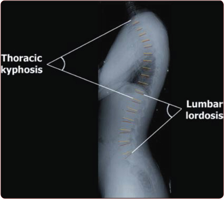
The X-ray spinal images used in the experiments were acquired using a EOS medical imaging system. In this technique, patients are in a weight bearing position with arms folded at 45° to reduce superimposition on the spine. The procedure provides 2D X-ray spinal images in the anterior- posterior view (AP view) in grayscale format. In the current clinical diagnosis, the standard curvature estimation method for assessing the curvature quantitatively is done by measuring the Cobb angle. The lumbar angle (the superior endplate of L1 and the inferior endplate of L5) and kyphosis angle (the superior endplate of T1 and the inferior end-plate of T12) was the main outcome measure in the current study.
Inclusion criteria were as follows: 1) students at Tabriz Dental School; 2) willingness to participate in the study and signed written consent; 3) attendance of final year 4 courses; 4) students’ age range 24–30 years old.
Statistical analysis
Data were entered to SPSACRAL SLOPE version 21. Linear regression and Pearson correlation coefficient was used for data analysis. A P-value <0.05 was considered statistically significant.
RESULTS
The present study was conducted with the aim of examining the normal range of kyphosis and lordosis angles in 403 individuals, of which 214 (53.1%) were women and 189 (46.9%) men. Subjects were classified into four age groups: 18-31, 31-40, 41-50 and 51-60 years old.
Table 1 shows the mean and range of lordosis and kyphosis angle.
Table 2 shows the mean angle of lordosis according to patients’ age group.
Figure 1 shows how to evaluate patients’ thoracic kyphosis and lumbar lordosis.
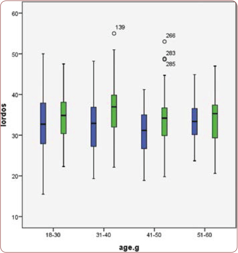
Figure 2 shows the mean angle of lordosis by age group in men and women selected for the current study.
As shown in Table 2 and Figure 2, the mean angle of lordosis in women was greater than men in all age groups.
Table 3 shows the mean angle of kyphosis by age group in men and women.
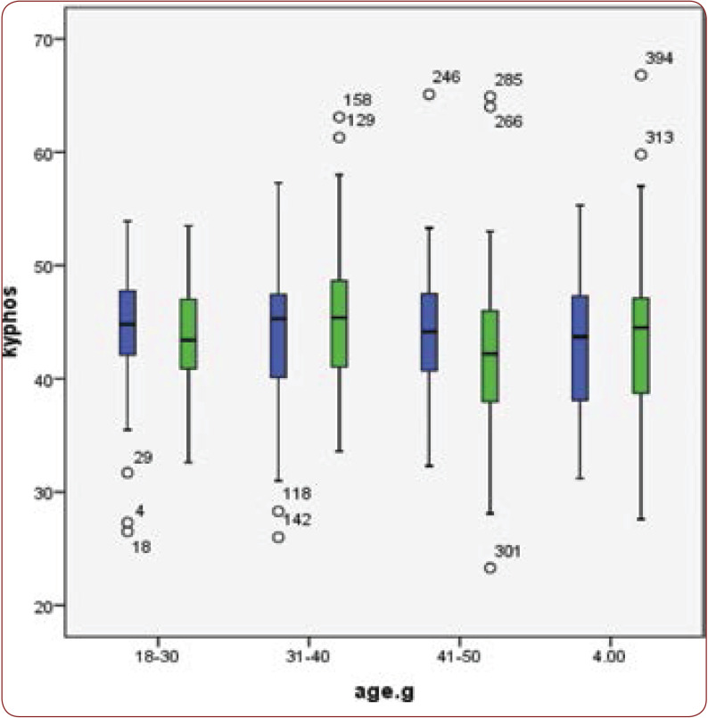
Figure 3 shows the mean angle of kyphosis by age group in normal men and women.
Comparison of kyphosis angle in participants aged over 40 showed that this angle was higher among men than in female study population.
Also, according to our findings, there was a significant relation between kyphosis and lordosis in men (r=0.286, p≤0.001) and women (r=0.519, p≤0.001), respectively.
In men, no significant relation was seen between age and lordosis (r=0.015, p=0.842) and kyphosis (r=0.054, p=0.459); in women, age was not notably correlated with lordosis (r=0.087, p=203) and kyphosis (r=0.010, p=0.123).
DISCUSSION
IIf kyphosis and lordosis are not quickly detected and prevented, irreparable complications will occur. In order to detect these abnormalities, we must know the normal range of these angles; therefore, we measured kyphosis and lordosis angles in normal people aged 18-60 years.
Our findings showed that the mean value of kyphosis and lordosis angles were 43.55±6.44 and 32.42±6.29, respectively. Hosseinfar et al. evaluated the lumbar and kyphotic curve in students of Zahedan University (16); their findings showed that the mean angle of kyphosis and lordosis was 23.7±7.8 and 25±11.4, respectively. Nodehi-Moghadam et al. evaluated the kyphosis and lordosis angles in old and young persons; they found that the value of kyphosis and lordosis angles in old individuals was 36.19±8.97 and 27.93±8.11, respectively (17). It seems that the difference between studies was due to age range, angle assessment method, sample size and body mass index.
In the current study, the kyphosis angle was greater than that of lordosis. Moreover, the normal ranges for kyphosis and lordosis angles were calculated by age groups in men and women. Kyphosis angle was directly related to the lordosis angle in both genders. Moreover, Jean-Luc Clément et al. reported a significant linear regression between thoracic kyphosis and lordosis (18), which was consistent with our study.
In our study, participants’ age was not significantly correlated with angles of kyphosis and lordosis. Moghadam et al. reported similar results (17). Gelb et al. evaluated sagittal spinal alignment in middle-aged and eldrely volunteers; their findings showed that the mean angle of lumbar lordosis (T12-S1) was 64±10°, which was greater than that measured by us. It seems that factors including race, classification of age groups and type of imaging device may be the cause of differences between two studies (19). Giglio et al. reported that kyphotic curves increased from 25° at seven years old to 38° at 19 years old, while lordotic curves increased from 22° at five years old to 32° at 20 years old (15).
Korovessis et al. (20) performed segmental analysis of the sagittal plane alignment of the normal thoracic, and lumbosacral spines; ninety-nine (38 men, 61 women) volunteers with a mean age of 52.7±15 years old were included in their prospective study, and results showed that kyphosis was significantly related to age, which was inconsistent to the findings of our study.
Jackson et al. evaluated radiographic analysis of sagittal plane alignment in patients with back pain and reported that there was no significant relation between segmental lumbar lordosis and age (21), while Zuluaga et al. believed that the size of lumbar lordosis at young age was 55° and it decreased to 42° with increasing age (22). Walter et al. evaluated the degree of kyphosis in the absence of vertebral fracture and reported that the following distribution of thoracic kyphotic angle by age range: 27.8±10 in individuals aged 18-35, 30.9±9.8 in those aged 36-50, 36±9.7 in the group of 58-65 years and 42±13 in subjects aged over 65. The mean kyphosis in these groups (young, middle-aged and elderly groups) was lower than the average of people studied by us (23).
It seems that these differences between studies are due to the angle assessment method, race, sample size, age group classification and estimation of angles in different sections.
In our study, the mean kyphosis angle in older women (over 40) was less than older men. In contrast, Fon et al. evaluated the correlation between the degree of kyphosis and patients’ sex and observed that the rate of this increase was higher in females than males (24). Katzman et al. have also reported that kyphosis angle was beginning to increase more rapidly in women than men after the age of 40 (25). It seems that this difference is due to osteoporosis and reduction of spinal height, as well as weakness of the spinal cord protection ligaments due to the lack of estrogen secretion in older women (23). Korovessis et al. performed a segmental analysis of the sagittal plane alignment of the normal thoracic and lumbosacral spines on 99 (38 men, 61 women) volunteers with a mean age of 52.7±15; the authors found that thoracic kyphosis was not related to sex (20). Factors including race and lifestyle may be the cause of these differences in studies.
In addition, the mean lordosis angle was greater in women than men. Orahilly et al. (26) evaluated the mean lordosis angle according to subjects’ sex and reported that women had a higher average lordosis angle than men, which was consistent with our study. But Giglio et al. (15) assessed the angle of kyphosis and lordosis during the growth process and reported no significant difference between the angle of lumbar lordosis according to sex, which was inconsistent with our findings. It seems that the difference between the two studies may be mainly explained by the sample size, so that we considered a greater sample size compared to Giglio’s study (15).
CONCLUSION
The assessment of kyphotic and lordotic angles can be used to detect early spinal and pelvic anomalies. It is also useful for standardization of the spinal column after fracture of the vertebrae or congenital and pathologic defects. Our findings clearly showed that individuals’ age did not affect the angle of kyphosis and lordosis, and the mean angle of lordosis was sex-dependent.
Conflicts of interest: none declared.
Financial support: none declared.
TABLE 1.
The mean and range of the lordosis and kyphosis angle
TABLE 2.
The mean angle of lordosis by age group in men and women
TABLE 3.
The mean angle of kyphosis by age group in men and women
FIGURE 1.
Evaluation of thoracic kyphosis and lumbar lordosis
FIGURE 2.
The mean angle of lordosis by age group in normal men and women
FIGURE 3.
The mean angle of kyphosis by age group in normal men and women
Contributor Information
Seyed Mohammad Jalil ABRISHAM, Department of Orthopedics, Shahid Sadoughi University of Medical Sciences, Yazd, Iran.
Mohammad Reza Sobhan ARDEKANI, Department of Orthopedics, Shahid Sadoughi University of Medical Sciences, Yazd, Iran.
Mohammad Ali Babaee MZARCH, Department of Orthopedics, Shahid Sadoughi University of Medical Sciences, Yazd, Iran.
References
- 1.Devereaux M. Anatomy and Examination of the Spine. Neurol Clin. 2007;25:331–351. doi: 10.1016/j.ncl.2007.02.003. [DOI] [PubMed] [Google Scholar]
- 2.Mirbagheri S-S, Rasa A-A, Farmani F, et al. Evaluating Kyphosis and Lordosis in Students by Using a Flexible Ruler and Their Relationship with Severity and Frequency of Thoracic and Lumbar Pain. Asian Spine J. 2015;3:416–422. doi: 10.4184/asj.2015.9.3.416. [DOI] [PMC free article] [PubMed] [Google Scholar]
- 3.Nitzschke E, Hildenbrand M. Epidemiology of kyphosis in school children. Z Orthop Ihre Grenzgeb. 1990;128:477–481. doi: 10.1055/s-2008-1039600. [DOI] [PubMed] [Google Scholar]
- 4.Katzman WB, Vittinghoff E, Kado DM. Age-related hyperkyphosis, independent of spinal osteoporosis, is associated with impaired mobility in older community-dwelling women. Osteoporos Int. 2011;22:85–90. doi: 10.1007/s00198-010-1265-7. [DOI] [PMC free article] [PubMed] [Google Scholar]
- 5.Eum R, Leveille SG, Kiely DK, et al. Is kyphosis related to mobility, balance, and disability? Am J Phys Med Rehabil. 2013;92:980–989. doi: 10.1097/PHM.0b013e31829233ee. [DOI] [PMC free article] [PubMed] [Google Scholar]
- 6.Katzman WB, Harrison SL, Fink HA. Physical function in older men with hyperkyphosis. J Gerontol A Biol Sci Med Sci. 2015;5:635–640. doi: 10.1093/gerona/glu213. [DOI] [PMC free article] [PubMed] [Google Scholar]
- 7.Huang MH, Barrett-Connor E, Greendale GA, Kado DM. Hyperkyphotic posture and risk of future osteoporotic fractures: the Rancho Bernardo study. J Bone Miner Res. 2006;21:419–423. doi: 10.1359/JBMR.051201. [DOI] [PMC free article] [PubMed] [Google Scholar]
- 8.Kado DM, Miller-Martinez D, Lui LY, et al. Hyperkyphosis, kyphosis progression, and risk of non-spine fractures in older community dwelling women: the study of osteoporotic fractures (SOF). J Bone Miner Res. 2014;29:2210–2216. doi: 10.1002/jbmr.2251. [DOI] [PMC free article] [PubMed] [Google Scholar]
- 9.Moe JH, Lonstein JE. Moe’s textbook of scoliosis and other spinal deformities.Philadelphia: W.B. Saunders. 1996.
- 10.Libby D. Acute respiratory failure in scoliosis or kyphosis. Am J Med. 1982;4:532–538. doi: 10.1016/0002-9343(82)90332-1. [DOI] [PubMed] [Google Scholar]
- 11.Kendall FP, McCreary EK, Provance PG, Rodgers MM, Romani WA. Muscles: testing and function with posture and pain. Baltimore: Lippincott Williams & Wilkins. 2005.
- 12.Van Blommestein. Reliability of Measuring Thoracic Kyphosis Angle, Lumbar Lordosis Angle and Straight Leg Raise with an Inclinometer. The Open Spine Journal. 2012;10:10–15. [Google Scholar]
- 13.D’Osualdo F, Schierano S, Lannis M. Validation of clinical measurement of kyphosis with a simple instrument, the arcometer. Spine. 1997;22:408–413. doi: 10.1097/00007632-199702150-00011. [DOI] [PubMed] [Google Scholar]
- 14.Rehm J. 3D-modeling of the spine using EOS imaging system: Inter-reader reproducibility and reliability. PLoS ONE. 2017;2:e0171258. doi: 10.1371/journal.pone.0171258. [DOI] [PMC free article] [PubMed] [Google Scholar]
- 15.Giglio ÆC. Development and evaluation of thoracic kyphosis and lumbar lordosis during growth. J Child Orthop. 2007;1:187–193. doi: 10.1007/s11832-007-0033-5. [DOI] [PMC free article] [PubMed] [Google Scholar]
- 16.Hosseinfar M. The relationship between lumbar and thoratic curves with body mass index and low back pain in students of Zahedan University. J Med Sci. 2007;6:984–990. [Google Scholar]
- 17.Nodehi-Moghadam A, Taghipour M, Goghatin Alibazi R, Baharlouei H. The comparison of spinal curves and hip and ankle range of motions between old and young persons. Med J Islam Repub Iran. 2014;28:74. [PMC free article] [PubMed] [Google Scholar]
- 18.Jean-Luc Clément. Relationship between thoracic hypokyphosis, lumbar lordosis and sagittal pelvic parameters in adolescent idiopathic scoliosis. Eur Spine J. 2013;11:2414–2420. doi: 10.1007/s00586-013-2852-z. [DOI] [PMC free article] [PubMed] [Google Scholar]
- 19.Gelb DE, Lenke LG, Bridwell KH, et al. An analysis of sagittal spinal alignment in 100 asymptomatic middle and older aged volunteers. Spine (Phila Pa 1976) . 1995;12:1351–1358. [PubMed] [Google Scholar]
- 20.Korovessis PG, Stamatakis MV, Baikousis AG. Reciprocal angulation of vertebral bodies in the sagittal plane in an asymptomatic Greek population. Spine(Phila Pa 1976) 1998;6:700–704. doi: 10.1097/00007632-199803150-00010. [DOI] [PubMed] [Google Scholar]
- 21.Jackson RP, MCManus AC. Radiographic analysis of sagittal plane alignment and balance in standing volunteers and patients with Low back pain matched for age, sex, size. Spine. 1994;14:1611–1618. doi: 10.1097/00007632-199407001-00010. [DOI] [PubMed] [Google Scholar]
- 22.Zuluaga M, et al. Sports physiotherapy, 1st ed, Melbourne Churchill Livingstone. 1995. pp. 485–458.
- 23.Bartynski WS, Heller MT, Grahovac SZ, et al. Severe thoracic kyphosis in the older patient in the absence of vertebral fracture: association of extreme curve with age. AJNR Am J Neuroradiol. 2005;8:2077–2085. [PMC free article] [PubMed] [Google Scholar]
- 24.Fon GT, Pitt MJ, Thies AC, Jr. Thoracic kyphosis: range in normal subjects. AJR Am J Roentgenol. 1980;5:979–983. doi: 10.2214/ajr.134.5.979. [DOI] [PubMed] [Google Scholar]
- 25.Katzman W. Age-Related Hyperkyphosis: Its Causes, Consequences, and Management. J Orthop Sports Phys Ther. 2010;6:352–360. doi: 10.2519/jospt.2010.3099. [DOI] [PMC free article] [PubMed] [Google Scholar]
- 26.Orahalli. The human vertebral column at the end of the embryonic period proper. J Anat. 1980;131:565–575. [PMC free article] [PubMed] [Google Scholar]


