Introduction
An 84-year-old man with a past medical history of hypertension was brought to the emergency department in respiratory distress after being found at home with oxygen saturation of 40%. The patient reported fever, shortness of breath, cough, and abdominal pain for 2 weeks leading up to presentation. On examination, he was found to have a nonreactive, pinpoint left pupil and new onset atrial fibrillation with rapid ventricular response. Laboratory analysis revealed lymphopenia of 0.29 x 10³/uL, procalcitonin of 0.25 ng/mL, elevated D-dimer of 21.6 mcg/mL, and elevated troponin T of 35 ng/mL. Reverse transcription polymerase chain reaction (RTPCR) COVID-19 testing was positive. As his respiratory status deteriorated, the patient was expeditiously intubated.
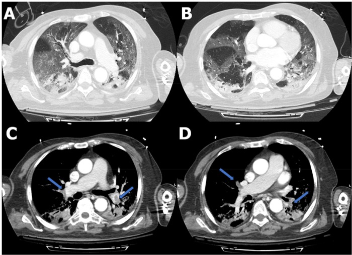
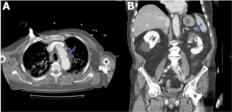
CT of the chest performed on the night of admission revealed diffuse ground-glass opacities and bibasilar consolidations compatible with severe COVID-19 pneumonia, as described in a recent consensus statement (1). In addition, chest CT demonstrated bilateral lobar pulmonary emboli (Fig 1). The patient was started on a low-molecular-weight heparin infusion. CT of the abdomen and pelvis, performed concurrently with the chest CT, demonstrated a new left renal infarct (Fig 2).
Figure 1.
Axial contrast-enhanced CT of the chest. A, B, Diffuse bilateral ground-glass opacities and small bibasilar consolidations compatible with COVID-19 pneumonia. C, D, Filling defects consistent with pulmonary emboli within the right upper lobe, right middle lobe, right lower lobe, and left lower lobe pulmonary arteries (arrows).
Figure 2.
A, Axial contrast-enhanced CT of the chest demonstrates a filling defect in the aortic arch (arrow) consistent with thrombus. B, Coronal contrast-enhanced CT of the abdomen and pelvis demonstrates a wedge-shaped low-attenuation region in the superior pole of the left kidney (arrow) consistent with renal infarct.
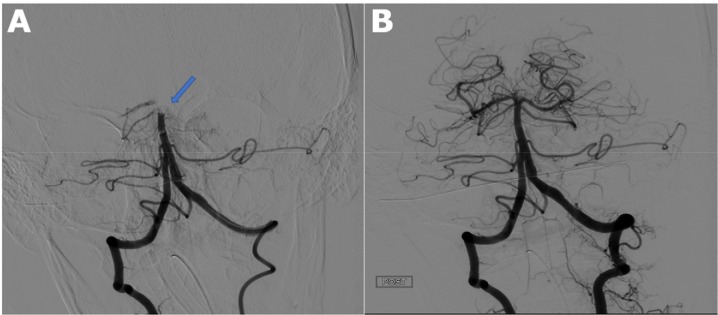
Given the patient’s unequal pupils, CT of the head and CT angiography of the head and neck were obtained on the night of admission, revealing occlusion of the distal basilar artery, which extended into the proximal posterior cerebral arteries (Fig 3). There was a small thrombus in the aortic arch (Fig 2). The patient was taken emergently for catheter angiography, followed by mechanical thrombectomy with subsequent restoration of blood flow (Fig 3). Despite all treatment measures, the patient rapidly decompensated and succumbed to his illness the next day.
Figure 3.
Catheter directed cerebral angiography. A, Pre-thrombectomy angiogram demonstrates an occluded distal basilar artery (arrow). B, Postthrombectomy angiogram demonstrates successful restoration of the posterior circulation.
This case illustrates severe coagulopathy in a patient with COVID-19 pneumonia. Possible explanations of his multifocal thromboembolic disease involving the pulmonary, cerebral, and renal circulations include coagulopathy due to COVID-19 versus cardioembolic etiology in the setting of atrial fibrillation. Given that the patient had no known hypercoagulable conditions and no prior history of atrial fibrillation, the latter etiology was felt to be less likely.
There are emerging global reports of coagulopathy in the setting of COVID-19, including pulmonary emboli, cerebral infarcts, and limb ischemia (2-4). A recent publication identified antiphospholipid antibodies in a COVID-19 patient with significant coagulopathy (5). It has also been proposed that coagulopathy may portend a poor prognosis in COVID-19 patients and may require early intervention (6-7). Our case supports the growing body of data by demonstrating a poor outcome in a patient with multifocal thromboembolic disease in the setting of COVID-19.
References
- 1.Simpson S, Kay FU, Abbara S. et al. Radiological Society of North America Expert Consensus Statement on Reporting Chest CT Findings Related to COVID-19. Endorsed by the Society of Thoracic Radiology, the American College of Radiology, and RSNA. Radiology: Cardiothoracic Imaging 2020; 2 (2). 25 March 2020. 10.1148/ryct.2020200152 [DOI] [PMC free article] [PubMed] [Google Scholar]
- 2.Danzi GB, Loffi M, Galeazzi G. and Gherbesi E. Acute pulmonary embolism and COVID-19 pneumonia: a random association? Eur Heart J 2020; 30 March 2020. 10.1093/eurheartj/ehaa254. [DOI] [PMC free article] [PubMed] [Google Scholar]
- 3.Zhang Y, Cao W, Xiao M. et al. Clinical and coagulation characteristics of 7 patients with critical COVID-2019 pneumonia and acro-ischemia. Chinese Journal of Hematology 2020; 41. DOI: 10.3760 / cma.j.issn.0253-2727.2020.0006. [DOI] [PubMed] [Google Scholar]
- 4.Mei H. and Hu Y. Characteristics, causes, diagnosis and treatment of coagulation dysfunction in patients with COVID-19. Chinese Journal of Hematology 2020; 41. DOI: 10.3760 / cma.j.issn.0253-2727.2020.0002. [DOI] [PMC free article] [PubMed] [Google Scholar]
- 5.Zhang Y, Xiao M, Zhang S. et al. Coagulopathy and antiphospholipid antibodies in patients with Covid-19. N Engl J Med 2020. 8 April 2020. DOI: 10.1056/NEJMc2007575. [DOI] [PMC free article] [PubMed] [Google Scholar]
- 6.Tang N, Bai H, Chen X, Gong J, Li D. and Sun Z. Anticoagulant treatment is associated with decreased mortality in severe coronavirus disease 2019 patients with coagulopathy. J Thromb Haemost 2020; 27 Mar 2020. DOI: 10.1111/jth.14817. [DOI] [PMC free article] [PubMed] [Google Scholar]
- 7.Tang N, Li D, Wang X. and Sun Z. Abnormal coagulation parameters are associated with poor prognosis in patients with novel coronavirus pneumonia. J Thromb Haemost 2020; 18(4):844-847. DOI: 10.1111/jth.14768. [DOI] [PMC free article] [PubMed] [Google Scholar]


