Abstract
Despite decades of research, diabetic retinopathy remains the leading cause of blindness in working age adults. Treatments for early phases for the disease remain elusive. One pathway that appears to regulate neuronal, vascular, and inflammatory components of diabetic retinopathy is the cyclic adenosine 3′, 5′-monophosphate (cAMP) pathway. In this review, we discuss the current literature on cAMP actions on the retina, with a focus on neurovascular changes commonly associated with preproliferative diabetic retinopathy models.
Introduction
Diabetic retinopathy is the leading cause of blindness in working age adults. Although a large number of therapeutics have been tested in animal models, none has led to a successful treatment for non-proliferative diabetic retinopathy. Anti-VEGF is effective for some cases of proliferative diabetic retinopathy and macular edema [1,2]. Reactive oxygen species, increased inflammatory factors, imbalanced growth factors, dyslipidemia, and dysfunctional insulin signal transduction have all been suggested to be involved in the pathogenesis of diabetic retinopathy [3-7]. In addition, altered cAMP signaling can regulate many of these same pathways, leading to retinal damage.
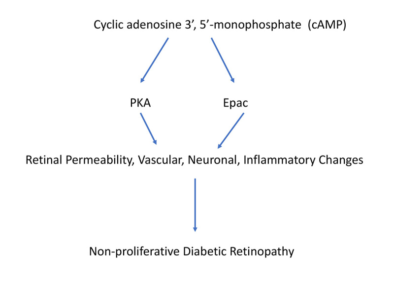
Cyclic adenosine 3′, 5′-monophosphate (cAMP) is a G-protein mediated signaling pathway that can regulate several downstream pathways, including those related to gluconeogenesis, muscle contraction, a large number of transcription factors, as well as many others [8]. Once activated, cAMP can lead to activation of protein kinase A (PKA), exchange protein activated by cAMP (Epac), and popeye domain containing proteins (Popdc) [9-11], as well as ion channels. Although cAMP can regulate a plethora of signaling cascades, its role in diabetic retinopathy has been less well studied. In diabetic retinopathy, changes in permeability, neurons, and vasculature, and inflammatory proteins have been recorded in animal models of non-proliferative diabetic retinopathy (Figure 1) [12-14].
Figure 1.

Schematic of cAMP signaling.
cAMP signaling in permeability changes in the diabetic retina
Original studies from bovine retinal cells showed that beta-adrenergic receptors increase cAMP, leading to decreased permeability [15]. More recent studies on bovine retinal endothelial cells found cAMP was key in the maintenance of the retinal barrier, and that TNFα decreased intracellular cAMP levels [16]. We developed a novel beta-adrenergic receptor agonist, Compound 49b, and reported that Compound 49b regulated key barrier proteins, occludin and zonula occludens 1 (ZO-1), in retinal endothelial cells grown in high glucose [17]. In that study, we used Epac1 siRNA to demonstrate that Compound 49b required Epac1 to maintain the barrier [17]. To further investigate the role of Epac1 in retinal permeability in diabetes, we made diabetic Epac1 floxed and cdh5Cre-Epac1 mice to eliminate Epac1 in endothelial cells and used fluorescein angiography and Evan’s blue studies to demonstrate that Epac1 is key to reduced retinal permeability in the diabetic retina [18]. Similarly, work by Ramos et al. demonstrated that Epac1 activation of Rap1 reverses cytokine-induced increases in permeability in bovine retinal endothelial cells [19]. The work with Epac1 mice and bovine retinal endothelial cells agrees with a review article by Wilson and Ye, suggesting that Epac1 and Rap1 regulate retinal permeability [20]. Less has been done to focus on PKA in retinal permeability in diabetes. However, studies in healthy mice showed that PKA can phosphorylate connexin 36 to reduce retinal permeability in amacrine cells [21]. There remains a need to further understand the specific mechanisms by which Epac1 and PKA can regulate permeability in the diabetic retina. Taken together, data suggest that cAMP signaling is key to barrier maintenance.
Role of cAMP signaling in vascular damage in diabetic retinopathy
Few groups have explored the role of cAMP signaling in the diabetic retinal vasculature. We showed that Compound 49b, which likely increased Epac1 and PKA, protects against the formation of degenerate capillaries in the diabetic retina [22]. We also recently reported that Epac1 endothelial cell knockout (KO) mice have increased numbers of degenerate capillaries when exposed to diabetes [18] and ischemia/reperfusion (I/R) at 10 days post-ischemia [23]. An additional study of retinal pericytes in culture confirmed that PKA is key to retinal pericyte contractility [24]. In contrast to work in the diabetic mouse models, a recent study showed that Epac1 inhibition reduces retinal angiogenesis in the oxygen-induced retinopathy model [25]. Other studies in cancer models also showed that Epac1 promotes angiogenesis through various signaling cascades [26,27], suggesting that Epac1 may have different actions on the vasculature depending on the cellular milieu. Thus, more studies on the role of Epac1 in the diabetic retinal vasculature are needed.
cAMP signaling actions on neuronal changes in the diabetic retina
Retinal ischemia can produce neuronal changes similar to diabetes [28], including reduced retinal thickness and loss of cell numbers in the ganglion cell layer. Using the I/R model with measurements at 2 days post-ischemia, studies have shown that increasing cAMP signaling can increase retinal ganglion cell (RGC) regeneration following damage [29]. Similarly, we found that Epac1 prevents the loss of retinal thickness and cell numbers in diabetic mice [18] and in the I/R model using endothelial cell–specific Epac1 KO mice [23]. These findings agree with those of a study that used the I/R model showing that Epac2 protects against neuronal damage in the retina [30]. In contrast, another group using whole animal Epac1 KO mice found that Epac1 promotes retinal neurodegeneration [31]. These authors performed their measurements at 24 h post-ischemia and measured apoptosis, not retinal thickness. Few studies have investigated PKA actions on neuronal changes in the diabetic retina. Therefore, additional work is needed to clarify cAMP actions on retinal neuronal changes in response to diabetes.
Actions of cAMP signaling in inflammatory markers in the diabetic retina
Work in healthy rodents showed that norepinephrine and cAMP are key to regulation of a large number of night and day genes, including a large number of inflammatory genes [32]. Work in Epac2 knockout mice exposed to I/R showed increased glial fibrillary acidic protein (GFAP) in the retina [30]. Increased GFAP signaling is often associated with increased inflammatory mediators in the retina [33]. Linking cAMP signaling to diabetic retinopathy, one group showed that Epac1 regulates O-GlyNAcylation in mice on a high-fat diet [34]. These changes included reduced Mas signaling and measurements of mitochondrial superoxide dismutase [35]. In previous work, we showed that Compound 49b reduces TNFα levels in retinal endothelial cells exposed to high glucose, which is associated with decreased retinal damage in response to diabetes [22]. Recently, we reported that Epac1 decreases inflammatory mediators in human primary retinal endothelial cells exposed to high glucose and in the diabetic retina [18,36]. We also showed that PKA directly regulates inflammatory mediators, in the absence of Epac1 in human retinal endothelial cells grown in high glucose [37]. Taken together, the data strongly suggest that cAMP signaling can reduce inflammatory mediators in multiple retinal cell types, which protects the retina against stressors.
Conclusions
Despite knowledge of the actions of the cAMP pathway on a large number of signaling cascades, much less work has been conducted to investigate actions of cAMP and its downstream mediators, PKA and Epac, on the diabetic retina. Most studies have shown that cAMP signaling is protective to the diabetic retina, reducing neuronal, vascular, permeability, and inflammatory changes; however, other studies showed detrimental effects. Additional work is needed to determine the cellular mechanisms by which cAMP can act on the retina, as well as optimizing cAMP-based therapies for systemic delivery.
References
- 1.Stitt AW, Curtis TM, Chen M, Medina RJ, McKay GJ, Jenkins A, Gardiner TA, Lyons TJ, Hammes HP, Simo R, Lois N. The progress in understanding and treatment of diabetic retinopathy. Prog Retin Eye Res. 2016;51:156–86. doi: 10.1016/j.preteyeres.2015.08.001. [DOI] [PubMed] [Google Scholar]
- 2.Gross JG, Glassman AR, Liu D, Sun JK, Antoszyk AN, Baker CW, Bressler NM, Elman MJ, Ferris FL, 3rd, Gardner TW, Jampol LM, Martin DF, Melia M, Stockdale CR, Beck RW.and Diabetic Retinopathy Clinical Research N. Five-Year Outcomes of Panretinal Photocoagulation vs Intravitreous Ranibizumab for Proliferative Diabetic Retinopathy: A Randomized Clinical TrialJAMA Ophthalmol 20181361138–1148. [DOI] [PMC free article] [PubMed] [Google Scholar]
- 3.Zheng L, Du Y, Miller C, Gubitosi-Klug RA, Kern TS, Ball S, Berkowitz BA. Critical role of inducible nitric oxide synthase in degeneration of retinal capillaries in mice with streptozotocin-induced diabetes. Diabetologia. 2007;50:1987–96. doi: 10.1007/s00125-007-0734-9. [DOI] [PubMed] [Google Scholar]
- 4.Tang J, Kern TS. Inflammation in diabetic retinopathy. Prog Retin Eye Res. 2011;30:343–58. doi: 10.1016/j.preteyeres.2011.05.002. [DOI] [PMC free article] [PubMed] [Google Scholar]
- 5.Kowluru RA. Diabetes-induced elevations in retinal oxidative stress, protein kinase C and nitric oxide are interrelated. Acta Diabetol. 2001;38:179–85. doi: 10.1007/s592-001-8076-6. [DOI] [PubMed] [Google Scholar]
- 6.Jarajapu YP, Cai J, Yan Y, Li Calzi S, Kielczewski JL, Hu P, Shaw LC, Firth SM, Chan-Ling T, Boulton ME, Baxter RC, Grant MB. Protection of blood retinal barrier and systemic vasculature by insulin-like growth factor binding protein-3. PLoS One. 2012;7:e39398. doi: 10.1371/journal.pone.0039398. [DOI] [PMC free article] [PubMed] [Google Scholar]
- 7.Kielczewski JL, Li Calzi S, Shaw LC, Cai J, Qi X, Ruan Q, Wu L, Liu L, Hu P, Chan-Ling T, Mames RN, Firth S, Baxter RC, Turowski P, Busik JV, Boulton ME, Grant MB. Free insulin-like growth factor binding protein-3 (IGFBP-3) reduces retinal vascular permeability in association with a reduction of acid sphingomyelinase (ASMase). Invest Ophthalmol Vis Sci. 2011;52:8278–86. doi: 10.1167/iovs.11-8167. [DOI] [PMC free article] [PubMed] [Google Scholar]
- 8.Alberts B, Bray D, Lewis J, Raff M, Roberts K, Watson JD. Molecular biology of the cell. 2 ed. 1989, New York: Garland. 1–1219. [Google Scholar]
- 9.Gloerich M, Bos JL. Epac: defining a new mechanism for cAMP action. Annu Rev Pharmacol Toxicol. 2010;50:355–75. doi: 10.1146/annurev.pharmtox.010909.105714. [DOI] [PubMed] [Google Scholar]
- 10.Simrick S, Schindler RF, Poon KL, Brand T. Popeye domain-containing proteins and stress-mediated modulation of cardiac pacemaking. Trends Cardiovasc Med. 2013;23:257–63. doi: 10.1016/j.tcm.2013.02.002. [DOI] [PMC free article] [PubMed] [Google Scholar]
- 11.Meinkoth JL, Alberts AS, Went W, Fantozzi D, Taylor SS, Hagiwara M, Montminy M, Feramisco JR. Signal transduction through the cAMP-dependent protein kinase. Mol Cell Biochem. 1993;127–128:179–86. doi: 10.1007/BF01076769. [DOI] [PubMed] [Google Scholar]
- 12.Joussen AM, Poulaki V, Le ML, Koizumi K, Esser C, Janicki H, Schraermeyer U, Kociok N, Fauser S, Kirchhof B, Kern TS, Adamis AP. A central role for inflammation in the pathogenesis of diabetic retinopathy. FASEB J. 2004;18:1450–2. doi: 10.1096/fj.03-1476fje. [DOI] [PubMed] [Google Scholar]
- 13.Antonetti DA, Barber AJ, Bronson SK, Freeman WM, Gardner TW, Jefferson LS, Kester M, Kimball SR, Krady JK, LaNoue KF, Norbury CC, Quinn PG, Sandirasegarane L, Simpson IA. Diabetic retinopathy: seeing beyond glucose-induced microvascular disease. Diabetes. 2006;55:2401–11. doi: 10.2337/db05-1635. [DOI] [PubMed] [Google Scholar]
- 14.Robinson R, Barathi VA, Chaurasia SS, Wong TY, Kern TS. Update on animal models of diabetic retinopathy: from molecular approaches to mice and higher mammals. Dis Model Mech. 2012;5:444–56. doi: 10.1242/dmm.009597. [DOI] [PMC free article] [PubMed] [Google Scholar]
- 15.Zink S, Rosen P, Lemoine H. Micro- and macrovascular endothelial cells in beta-adrenergic regulation of transendothelial permeability. Am J Physiol. 1995;269:C1209–18. doi: 10.1152/ajpcell.1995.269.5.C1209. [DOI] [PubMed] [Google Scholar]
- 16.van der Wijk AE, Vogels IMC, van Noorden CJF, Klaassen I, Schlingemann RO. TNFalpha-Induced Disruption of the Blood-Retinal Barrier In Vitro Is Regulated by Intracellular 3′,5′-Cyclic Adenosine Monophosphate Levels. Invest Ophthalmol Vis Sci. 2017;58:3496–505. doi: 10.1167/iovs.16-21091. [DOI] [PubMed] [Google Scholar]
- 17.Jiang Y, Liu L, Steinle JJ. Compound 49b Regulates ZO-1 and Occludin Levels in Human Retinal Endothelial Cells and in Mouse Retinal Vasculature. Invest Ophthalmol Vis Sci. 2017;58:185–9. doi: 10.1167/iovs.16-20412. [DOI] [PMC free article] [PubMed] [Google Scholar]
- 18.Liu L, Jiang Y, Steinle JJ. Epac1 and Glycyrrhizin Both Inhibit HMGB1 Levels to Reduce Diabetes-Induced Neuronal and Vascular Damage in the Mouse Retina. J Clin Med. 2019;783:8. doi: 10.3390/jcm8060772. [DOI] [PMC free article] [PubMed] [Google Scholar]
- 19.Ramos CJ, Lin C, Liu X, Antonetti DA. The EPAC-Rap1 pathway prevents and reverses cytokine-induced retinal vascular permeability. J Biol Chem. 2018;293:717–30. doi: 10.1074/jbc.M117.815381. [DOI] [PMC free article] [PubMed] [Google Scholar]
- 20.Wilson CW, Ye W. Regulation of vascular endothelial junction stability and remodeling through Rap1-Rasip1 signaling. Cell Adh Mig. 2014;8:76–83. doi: 10.4161/cam.28115. [DOI] [PMC free article] [PubMed] [Google Scholar]
- 21.Urschel S, Hoher T, Schubert T, Alev C, Sohl G, Worsdorfer P, Asahara T, Dermietzel R, Weiler R, Willecke K. Protein kinase A-mediated phosphorylation of connexin36 in mouse retina results in decreased gap junctional communication between AII amacrine cells. J Biol Chem. 2006;281:33163–71. doi: 10.1074/jbc.M606396200. [DOI] [PubMed] [Google Scholar]
- 22.Zhang Q, Guy K, Pagadala J, Jiang Y, Walker RJ, Liu L, Soderland C, Kern TS, Ferry R, Jr, He H, Yates CR, Miller DD, Steinle JJ. Compound 49b Prevents Diabetes-Induced Apoptosis through Increased IGFBP-3 Levels. Invest Ophthalmol Vis Sci. 2012;53:3004–13. doi: 10.1167/iovs.11-8779. [DOI] [PMC free article] [PubMed] [Google Scholar]
- 23.Liu L, Jiang Y, Steinle JJ. Epac1 protects the retina against ischemia/reperfusion-induced neuronal and vascular damage. PLoS One. 2018;13:e0204346. doi: 10.1371/journal.pone.0204346. [DOI] [PMC free article] [PubMed] [Google Scholar]
- 24.Markhotina N, Liu GJ, Martin DK. Contractility of retinal pericytes grown on silicone elastomer substrates is through a protein kinase A-mediated intracellular pathway in response to vasoactive peptides. IET Nanobiotechnol. 2007;1:44–51. doi: 10.1049/iet-nbt:20060019. [DOI] [PubMed] [Google Scholar]
- 25.Liu H, Mei FC, Yang W, Wang H, Wong E, Cai J, Toth E, Luo P, Li YM, Zhang W, Cheng X. Epac1 inhibition ameliorates pathological angiogenesis through coordinated activation of Notch and suppression of VEGF signaling. Sci Adv. 2020;6:xxx. doi: 10.1126/sciadv.aay3566. [DOI] [PMC free article] [PubMed] [Google Scholar]
- 26.Kumar N, Prasad P, Jash E, Jayasundar S, Singh I, Alam N, Murmu N, Somashekhar SP, Goldman A, Sehrawat S. cAMP regulated EPAC1 supports microvascular density, angiogenic and metastatic properties in a model of triple negative breast cancer. Carcinogenesis. 2018;39:1245–53. doi: 10.1093/carcin/bgy090. [DOI] [PMC free article] [PubMed] [Google Scholar]
- 27.Fang Y, Olah ME. Cyclic AMP-dependent, protein kinase A-independent activation of extracellular signal-regulated kinase 1/2 following adenosine receptor stimulation in human umbilical vein endothelial cells: role of exchange protein activated by cAMP 1 (Epac1). J Pharmacol Exp Ther. 2007;322:1189–200. doi: 10.1124/jpet.107.119933. [DOI] [PubMed] [Google Scholar]
- 28.Abcouwer SF, Lin CM, Shanmugam S, Muthusamy A, Barber AJ, Antonetti DA. Minocycline prevents retinal inflammation and vascular permeability following ischemia-reperfusion injury. J Neuroinflammation. 2013;163:149. doi: 10.1186/1742-2094-10-149. [DOI] [PMC free article] [PubMed] [Google Scholar]
- 29.Kurimoto T, Yin Y, Omura K, Gilbert HY, Kim D, Cen LP, Moko L, Kugler S, Benowitz LI. Long-distance axon regeneration in the mature optic nerve: contributions of oncomodulin, cAMP, and pten gene deletion. J Neurosci. 2010;30:15654–63. doi: 10.1523/JNEUROSCI.4340-10.2010. [DOI] [PMC free article] [PubMed] [Google Scholar]
- 30.Liu J, Yeung PK, Cheng L, Lo AC, Chung SS, Chung SK. Epac2-deficiency leads to more severe retinal swelling, glial reactivity and oxidative stress in transient middle cerebral artery occlusion induced ischemic retinopathy. Sci China Life Sci. 2015;58:521–30. doi: 10.1007/s11427-015-4860-1. [DOI] [PubMed] [Google Scholar]
- 31.Liu W, Ha Y, Xia F, Zhu S, Li Y, Shi S, Mei FC, Merkley K, Vizzeri G, Motamedi M, Cheng X, Liu H, Zhang W. Neuronal Epac1 mediates retinal neurodegeneration in mouse models of ocular hypertension. J Exp Med. 2020;163:217. doi: 10.1084/jem.20190930. [DOI] [PMC free article] [PubMed] [Google Scholar]
- 32.Bailey MJ, Coon SL, Carter DA, Humphries A, Kim JS, Shi Q, Gaildrat P, Morin F, Ganguly S, Hogenesch JB, Weller JL, Rath MF, Moller M, Baler R, Sugden D, Rangel ZG, Munson PJ, Klein DC. Night/day changes in pineal expression of >600 genes: central role of adrenergic/cAMP signaling. J Biol Chem. 2009;284:7606–22. doi: 10.1074/jbc.M808394200. [DOI] [PMC free article] [PubMed] [Google Scholar]
- 33.Feit-Leichman RA, Kinouchi R, Takeda M, Fan Z, Mohr S, Kern TS, Chen DF. Vascular damage in a mouse model of diabetic retinopathy: relation to neuronal and glial changes. Invest Ophthalmol Vis Sci. 2005;46:4281–7. doi: 10.1167/iovs.04-1361. [DOI] [PubMed] [Google Scholar]
- 34.Dierschke SK, Toro AL, Barber AJ, Arnold AC, Dennis MD. Angiotensin-(1–7) Attenuates Protein O-GlcNAcylation in the Retina by EPAC/Rap1-Dependent Inhibition of O-GlcNAc Transferase. Invest Ophthalmol Vis Sci. 2020;61:24–35. doi: 10.1167/iovs.61.2.24. [DOI] [PMC free article] [PubMed] [Google Scholar]
- 35.Simoes e Silva AC, Silveira KD, Ferreira AJ, Teixeira MM. ACE2, angiotensin-(1–7) and Mas receptor axis in inflammation and fibrosis. Br J Pharmacol. 2013;169:477–92. doi: 10.1111/bph.12159. [DOI] [PMC free article] [PubMed] [Google Scholar]
- 36.Liu L, Jiang Y, Chahine A, Curtiss E, Steinle JJ. Epac1 agonist decreased inflammatory proteins in retinal endothelial cells, and loss of Epac1 increased inflammatory proteins in the retinal vasculature of mice. Mol Vis. 2017;23:1–7. [PMC free article] [PubMed] [Google Scholar]
- 37.Liu L, Patel P, Steinle JJ. PKA regulates HMGB1 through activation of IGFBP-3 and SIRT1 in human retinal endothelial cells cultured in high glucose. Inflamm Res. 2018;67:1013–9. doi: 10.1007/s00011-018-1196-x. [DOI] [PMC free article] [PubMed] [Google Scholar]


