Abstract
Achilles tendinopathy is a painful overuse injury that is extremely common in athletes, especially those who participate in running and jumping sports. In addition to pain, Achilles tendinopathy is accompanied by alterations in the tendon's structure and mechanical properties, altered lower extremity function, and fear of movement. Cumulatively, these impairments limit sport participation and performance. A thorough evaluation and comprehensive treatment plan, centered on progressive tendon loading, is required to ensure full recovery of tendon health and to minimize the risk of reinjury. In this review, we will provide an update on the evidence-based evaluation, outcome assessment, treatment, and return-to-sport planning for Achilles tendinopathy. Furthermore, we will provide the strength of evidence for these recommendations using the Strength of Recommendation Taxonomy system.
Keywords: tendinitis, rehabilitation, tendon, exercise therapy, loading
Achilles tendinopathy (ie, Achilles tendinitis) is a painful overuse injury of the Achilles tendon.1–4 This injury is rampant among athletes, especially those involved in running and jumping sports.1–3 Among elite track and field athletes, 43% reported having either current or prior symptoms of Achilles tendinopathy, with the highest prevalence (83%) in middle-distance runners.3 Two-thirds of the athletes in this study also noted that the tendon pain negatively affected their performance.3 However, Achilles tendinopathy is not purely an athletic injury, given that 65% of injuries diagnosed in a general practice setting are not sport related.4 Despite our improved understanding of the injury, it remains a devastating, albeit slowly progressing, injury. Symptomatic athletes often continue sport participation, although performance is often affected and the injury will worsen if ignored.3,5 Full recovery can take a year or longer and reinjury is common, especially when the return to sport is rushed.5,6 If the initial symptoms of soreness and stiffness are recognized and addressed early, instead of being ignored, injury severity can be reduced, with a smaller effect on sport performance and a shorter time to full recovery.6 In this “Current Clinical Concepts” review, we will present the most current evidence-based recommendations for diagnosis, outcome assessment, treatment, and return-to-sport planning. The Strength of Recommendation (SOR) Taxonomy will be used to grade the strength of evidence (Table 1).7 Furthermore, we will propose a holistic approach to evaluation and care, with suggestions on how to prevent the devastating consequences of this injury by recognizing its early signs and using a return-to-sport program to minimize injury recurrence and reinjury.
Table 1.
The Strength of Recommendation Taxonomy7
| Strength of Recommendation |
Definition |
| A | Recommendation based on consistent and good-quality patient-oriented evidence |
| B | Recommendation based on inconsistent or limited-quality patient-oriented evidence |
| C | Recommendation based on consensus, usual practice, opinion, disease-oriented evidence, or case series for studies of diagnosis, treatment, prevention, or screening |
ACHILLES TENDINOPATHY
Achilles tendinopathy is a clinical diagnosis when the patient presents with a combination of localized pain, swelling of the Achilles tendon, and loss of function.8 Achilles tendon injuries can be separated into insertional tendinopathy (20%–25% of the injuries), midportion tendinopathy (55%–65%), and proximal musculotendinous junction (9%–25%) injuries, according to the location of pain.8 However, patients may present with symptoms at the insertion and midportion concurrently, and approximately 30% have bilateral pain.6
Tendinopathy is described as either degeneration or failed healing due to continuous overload without appropriate recovery.9 The use of the term tendinitis is discouraged because it implies inflammatory activity, which may or may not be present in the injured tendon.10 In addition, inflammation cannot routinely be assessed clinically.9,10 At the tissue level, tendinopathy is characterized by localized or diffuse increases in thickness (tendinosis), loss of normal collagen architecture, an increased amount of proteoglycans, and general breakdown of tissue organization.9,10 These structural changes in the tendon result in increased cross-sectional area, reduced tendon stiffness, and altered viscoelastic properties in both symptomatic and asymptomatic tendons.9 Moreover, in insertional Achilles tendinopathy, the tendon change (tendinosis) is often accompanied by additional conditions, such as enlargement of the retrocalcaneal bursa, intratendinous calcifications, and bone defects.11
Injury Mechanism
The Achilles tendon is mechanoresponsive, meaning it will adapt to the loading demands placed on the tissue.9,12 The exact cause of tendinopathy varies; however, the most common cause in athletes is excessive loading with inadequate recovery time between training sessions.13 Among athletes who developed Achilles tendinopathy, 60% to 80% described a sudden change or increase in training intensity or duration (ie, training-load error).8,14 However, not all cases are sport related, and increases in work or daily activity can contribute to excessive loading.4 At the Achilles tendon insertion, compressive forces on the Achilles tendon and calcaneus from footwear or activities that place the ankle in dorsiflexion (eg, uphill running) or an anatomical abnormality (eg, Haglund deformity) may contribute to the development of pain.15 SOR: A
Risk Factors
The risk for developing Achilles tendinopathy is considered multifactorial, with intrinsic or extrinsic risk factors that relate to causing either decreased load tolerance of the tendon or movement patterns that overload the tendon.16 Decreased plantar-flexor strength, deficits in hip neuromuscular control, abnormal ankle dorsiflexion and subtalar-joint range of motion, increased foot pronation, and increased body weight are intrinsic risk factors that can be addressed during treatment.16 Systemic disease, genetic variants, and a family history of tendinopathy have also been identified as intrinsic risk factors.17 The use of fluoroquinolone antibiotics has been linked to both tendinopathy and tendon rupture, with the onset occurring approximately 8 days after the start of treatment.9,16–18 Limited evidence has associated footwear, sport participation, surface, training-load errors (such as a sudden increase in training duration, mileage, or intensity; a decrease in recovery time; or resumption of full activity after a break), and environmental conditions with the development of Achilles tendinopathy.16–18 SOR: A
Symptoms
Pain and reduced function are the primary symptoms of Achilles tendinopathy.19
Athletes often describe a gradual onset of symptoms that include stiffness in the morning or after prolonged sitting, pain with palpation, pain with activity (running or jumping), and deficits in strength or performance. Pain related to activity may vary according to severity. An early symptom is pain at the start of activity that subsides soon into a single training bout.3 It is not uncommon for an athlete to experience reduced athletic performance (slower running time or decreased jump performance) before pain during the performance.3 Athletes who ignore the minor symptoms may progress to experiencing pain during and after activity and further reduced performance.5,13 It is important to note that patients with Achilles tendinopathy often have no pain in the absence of loading.10 SOR: A
Signs and Diagnostic Tests
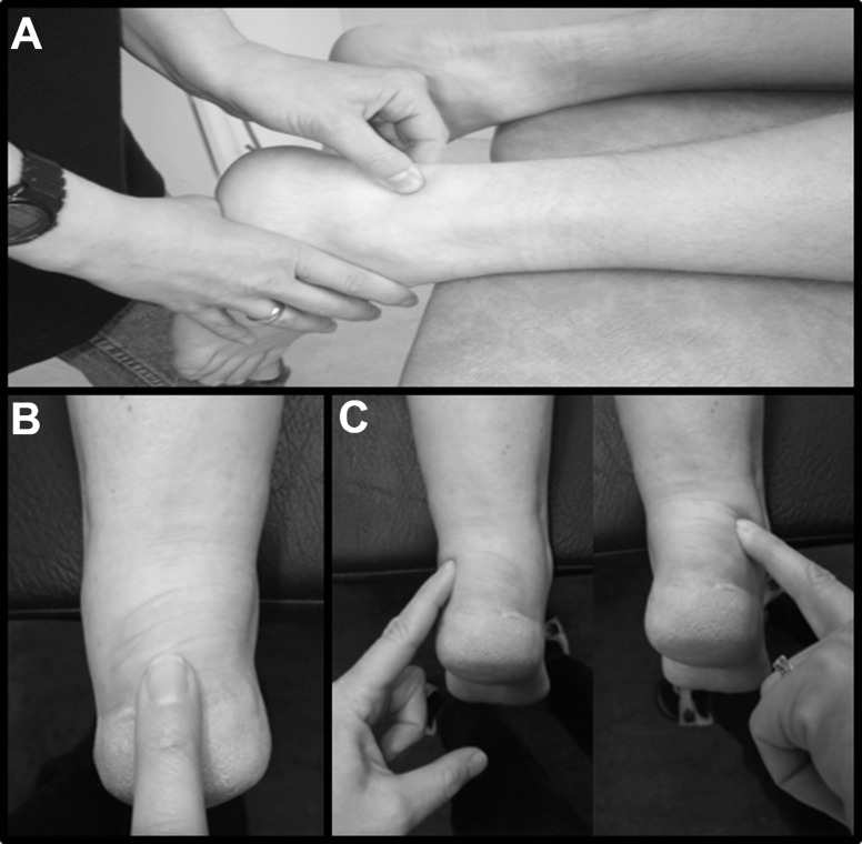
Pain on palpation of the tendon and the subjective report of pain in the midportion (2–6 cm above the calcaneal insertion) are reliable and valid tests for diagnosing midportion Achilles tendinopathy (palpation: sensitivity = 84%, specificity = 73%; self-report: sensitivity = 78%, specificity = 77%).20 The location of pain on palpation is useful for distinguishing between an insertional or midportion injury and for the differential diagnosis (Figure 1). For example, if palpation causes a greater degree of pain anterior to the tendon than in the tendon itself, then posterior ankle impingement or os trigonum syndrome might be a more likely diagnosis. Other differential diagnoses to consider in patients with posterior ankle pain are acute Achilles tendon rupture, accessory soleus muscle, sural nerve irritation, fat-pad irritation, and systemic inflammatory disease.16
Figure 1.
Palpation of the A, midportion; B, insertion of the Achilles tendon; and C, the medial and lateral fat pad and bursa.
The arc sign and the Royal London Hospital test are additional diagnostic tests used for confirming midportion Achilles tendinopathy.16,19,21 For the arc sign, the tendon is first palpated to identify any thickened nodules. If thickening is present, the area is lightly pinched while the patient actively dorsiflexes and plantar flexes the ankle. The test is considered positive if the evaluator feels a thickened nodule moving (sensitivity = 25%, specificity = 100%).19–21 For the Royal London Hospital test, the tendon is pinched to identify the most symptomatic location with the foot at rest. The patient then actively dorsiflexes the foot and the examiner once again pinches the previously identified location. Reduced pain with palpation when the ankle is dorsiflexed indicates a positive test (sensitivity = 51%, specificity = 93%).19–21 SOR: A
Assessing Response to Treatment
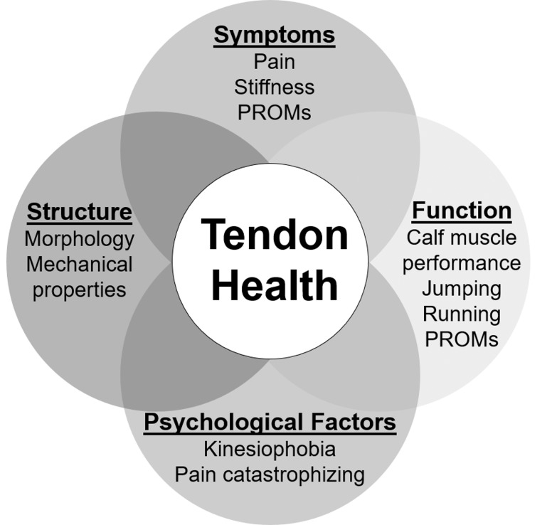
Recovery from Achilles tendinopathy can take a year or more.6 To ensure continued progress with treatment, it is important to use reliable and valid outcome measures.22 The severity of Achilles tendinopathy historically has been based on patient-reported pain and symptoms.13 However, patients also have impaired muscle performance, decreased lower extremity function, altered tendon structure, and a heightened fear of movement, or kinesiophobia.6,22,23 A consensus statement regarding core outcome domains for tendinopathy was recently developed.22 In this statement, the recommended core outcome domains for tendinopathy were the patient's rating of the overall condition, participation, pain on loading or activity and over a specified period of time, function, psychological factors, physical function capacity, disability, and quality of life.22 The degree of deficit and symptoms in each domain vary among individuals with Achilles tendinopathy. Evaluating each aspect of tendon health is vital to evaluating progress, discussing the patient's goals and expectations, and making return-to-sport decisions for each athlete. Moreover, structural change can occur without symptoms and may even precede symptoms.24 Other patients may have a number of symptoms and functional deficits but minimal structural change. In addition, full symptomatic recovery does not ensure full recovery of function.5 Therefore, using only symptom resolution as the guide for recovery without ensuring recovery of all the domains of tendon health may be a primary reason for the high reinjury rates (27%–44%) for Achilles tendinopathy.25,26 Therefore, we advocate the use of a battery of tests and outcome measures that considers all domains (symptoms, function, structure, and psychological factors) of tendon health to evaluate recovery in patients with Achilles tendinopathy (Figure 2).
Figure 2.
Domains of tendon health. Abbreviation: PROMs, patient-reported outcome measures.
OUTCOME MEASURES
Symptom severity and pain, as well as their effect on participation and activity, are often assessed using pain scales and patient-reported outcome measures and questionnaires.27 Pain on palpation and pain with activity, such as hopping, is evaluated not only for diagnosis, as described previously, but is also useful for assessing change with treatment.6,20,27 Patients rate their pain during palpation and activity on a numeric scale where 0 = no pain and 10 = worst pain imaginable.20,27 SOR: A
Patient-Reported Outcome Measures
The Victorian Institute of Sport Assessment–Achilles questionnaire (VISA-A) was designed to evaluate pain and symptom severity with activity in patients with Achilles tendinopathy.28 The VISA-A contains 8 questions, and scores range from 0 to 100, with 100 indicating no symptoms and full participation in physical activity. The VISA-A score is valid and reliable (test-retest r = 0.93–0.98), responsive to change, and has been cross-culturally adapted for numerous languages.27,28 The minimal clinically important difference has been reported27 to be 6.5 to 16 points. Most clinical studies use a change of 10 points as reflecting meaningful improvement for the patient.27 SOR: A
The Foot and Ankle Outcome Score (FAOS) is a questionnaire arranged in 5 subscales: Pain, Symptoms, Activities of Daily Living, Sport and Recreation Function, and Foot- and Ankle-Related Quality of Life.29 Scores range from 0 to 100, with 100 indicating no symptoms or impact of a foot and ankle injury.30 The FAOS was developed for patients with ankle injuries and has excellent reliability (test-retest r = 0.96, 0.89, 0.85, 0.92, and 0.92 for each subscale, respectively).29 The FAOS has demonstrated responsiveness in patients with Achilles tendinopathy.16,30 SOR: B
The Lower Extremity Functional Scale and the Foot and Ankle Ability Measure (FAAM) have also been recommended for assessing the effect of Achilles tendinopathy on activity and participation.16,31 The FAAM evaluates changes in functional ability using 2 subscales: Activities of Daily Living and Sports. The FAAM is valid and reliable (test-retest r = 0.87–0.89) for Achilles tendinopathy. The minimal detectable change (MDC) and minimal clinically important difference values are 5.7 and 8 points and 12.3 and 9 points, respectively, for the Activities of Daily Living and Sports subscales.31 SOR: A
Functional Impairments
Range of Motion
Decreased dorsiflexion range of motion has been identified as a risk factor for Achilles tendinopathy.16 Therefore, evaluation of ankle-dorsiflexion range of motion in patients with Achilles tendinopathy is recommended. Range of motion can be assessed with the patient non–weight bearing using goniometry or weight bearing using the ankle-lunge test.32 The SEM and MDC scores for the ankle-lunge test are 1.4° and 3.8°, respectively, with excellent intertester reliability (test-retest r = 0.89–0.99, intraclass correlation coefficient [ICC] = 0.97).32 In addition, because limitations in plantar flexion, inversion, or eversion range of motion may alter loading of the Achilles tendon, thereby predisposing athletes to injury, goniometric evaluation may be helpful. SOR: B
Heel-Rise Test for Calf-Muscle Endurance
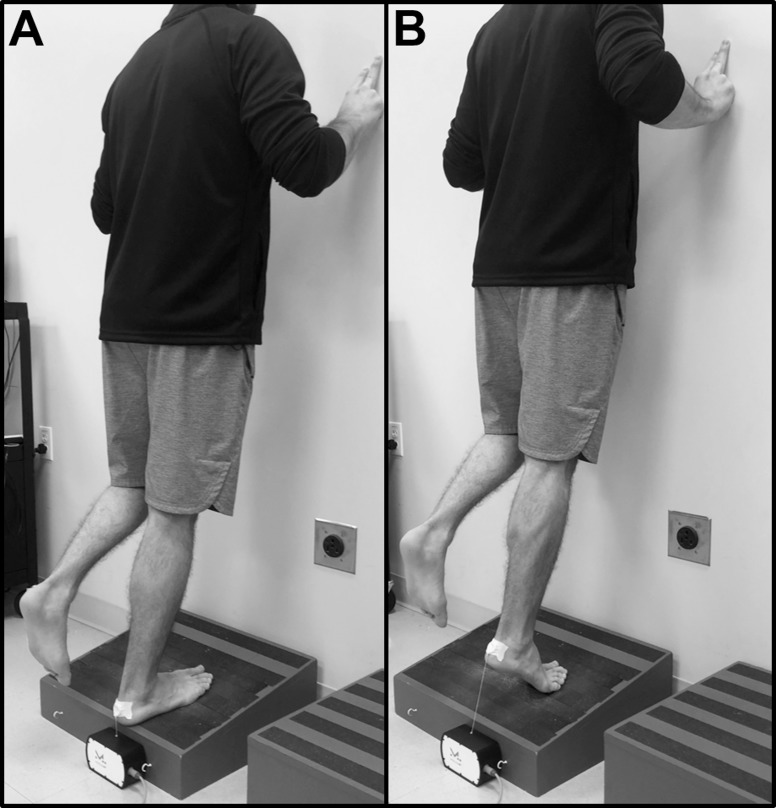
The heel-rise test is a measure of calf-muscle endurance and is used to detect changes with treatment over time (test-retest reliability: ICC = 0.78–0.84).33,34 It is important to standardize the starting position; in the literature, the starting position is either 0° or 10° of dorsiflexion (Figure 3). The patient is instructed to rise as high as possible on the heel each time, keeping the knee straight, and continue until fatigued. To maintain balance, the patient is allowed to place 2 fingertips per hand on the wall. A metronome should be used to ensure consistent rhythm, with a frequency of 30 heel rises per minute.33 The number of repetitions, maximum heel-rise height, and total amount of work performed are recorded.33 The limb symmetry index (injured limb/uninjured limb × 100) can be calculated to compare the degree of functional deficit between limbs. SOR: A
Figure 3.
A, Starting position, and B, elevated position for the heel-rise endurance test.
Calf-Muscle Strength
Addressing strength deficits is important because reduced plantar-flexor strength is a modifiable risk factor for Achilles tendinopathy.16 Traditional methods such as dynamometry can be used to measure isometric or dynamic maximal strength.35 Isokinetic dynamometry has been used to assess ankle plantar-flexor and dorsiflexor strength both concentrically and eccentrically at various angular velocities (ICC = 0.37–0.95).35 Others35 have measured isometric strength using a dynamometer, usually with the ankle at 0° of dorsiflexion (ICC = 0.90). SOR: A
Jumping Ability
Single-legged hopping and the drop-countermovement jump can be used to measure function as well as the degree of pain with jumping.33 Single-legged hopping involves a series of continuous rhythmic jumps, similar to jumping rope. Contact time and flight time may be used to calculate a plyometric quotient (flight time/contact time; test-retest reliability for plyometric quotient: ICC = 0.83–0.94).33 The drop-countermovement jump is performed on a single leg by jumping from a box or elevated surface, landing, and immediately jumping up for maximum height (test-retest reliability for jump height: ICC = 0.88–0.92).33 The level of pain during the jumping tests is also assessed on the numeric pain-rating scale.6 SOR: A
Key Points
Achilles tendinopathy is a clinical diagnosis based on localized tendon pain and swelling and pain with activities.
Exercise that provides mechanical loading of the Achilles tendon is the treatment with the highest level of evidence.
Treatment should focus on activity modification and progressive tendon-loading exercises.
Full recovery of symptoms does not ensure full recovery of function or tendon structure.
The best prevention is to recognize early “minor” symptoms and treat with load control.
Tendon Structure
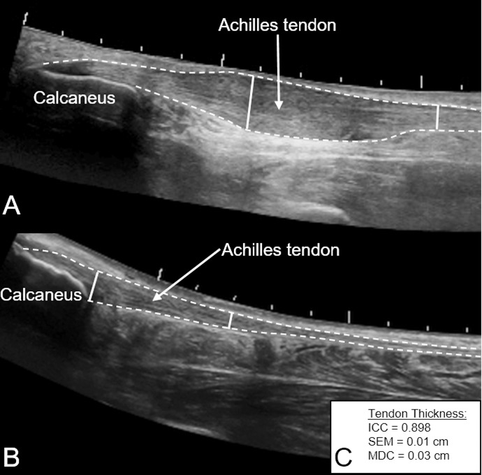
Ultrasound imaging (Figure 4) can be used to assess tendon morphology (tendon length, thickness, and cross-sectional area) and the presence of pathologic conditions (tendinosis, intratendinous calcification, bony deformity, bursitis, paratenonitis, and neovascularization).11,37 Morphologic changes can exist in asymptomatic tendons but are associated with an increased risk in developing symptoms, symptom severity, and prognosis with treatment.23,24 Measures of tendon thickness (ICC = 0.898, SEM = 0.02 cm, MDC = 0.03 cm) can be compared with the contralateral, uninjured limb or an unaffected region of the tendon in the injured limb to provide an index of tendon thickening.23,37 SOR: A
Figure 4.
Extended field-of-view ultrasound images of A, midportion, and B, insertional Achilles tendinopathy with measures of tendon thickness at the symptomatic region and an unaffected region of the tendon (solid line). C, Psychometric properties of tendon thickness measures.36 Abbreviations: ICC, intraclass correlation coefficient; MDC, minimal detectable change; SEM, standard error of measure.
Shear-wave elastography (SWE) is a form of ultrasound imaging capable of measuring the mechanical properties of tendons that is increasingly being used in sports medicine to track patients' progression or recovery after tendon injuries. A valid and reliable technique (ICC = 0.70–0.86), SWE can detect changes in mechanical properties that are associated with tendon healing.38,39 In addition, SWE measures have been closely correlated (r = .80) with symptoms.40 SOR: B
Psychological Factors
The chronic and debilitative nature of Achilles tendinopathy can lead to anxiety, depression, and poor overall quality of life.41 Athletes whose sense of identity is tied to their sport or performance are particularly susceptible. Yet the presence of negative psychological factors may not be readily apparent. Athletes may develop fear of pain with movement, or kinesiophobia, which can affect treatment participation and the perception of injury severity.42 The Tampa Scale of Kinesiophobia is a 17-item questionnaire designed to assess kinesiophobia severity.42 The Tampa Scale of Kinesiophobia has excellent internal consistency (Cronbach α = 0.84) and construct validity (average r = .44 for all items). Scores range from 0 to 68, with higher scores indicating a greater degree of kinesiophobia.42 SOR: C
TREATMENT
The treatment with the highest level of evidence for Achilles tendinopathy is exercise rehabilitation (Table 2).6,16,33 The purpose of exercise is to provide mechanical load to the tendon to promote remodeling, decrease pain, and improve calf-muscle endurance and strength and lower leg function.6,43 The optimal loading factors have not been established; however, the tendon appears to respond more favorably to higher loads at longer durations than to lower loads at shorter durations.12 Also, an upper limit likely exists beyond which the load can be detrimental.12 Achilles tendon–loading programs historically have consisted of eccentric muscle contractions, but protocols involving isolated concentric or a combination of concentric and eccentric contractions have all been used with positive results.6,44 More recently, isometric exercise as the initial treatment for tendinopathy has been proposed as superior; however, no current evidence supports it as better than any other type of exercise for Achilles tendinopathy.45 Therefore, whether the tendon's response is differentiated on the basis of the type of muscle contraction has been questioned.12,46 To improve tendon strength and size, exercise with higher loads and slower contractions seems to be of the greatest benefit. Nonetheless, the load on the tendon is also increased by increasing the speed of movement; therefore, the tolerance to fast speeds should also be addressed during the later phases of rehabilitation. In summary, any type of muscle contraction can be used to mechanically load the tendon. The choice of exercise should be progressive and based on the individual patient's response.
Table 2.
A Comprehensive Achilles Tendon-Loading Protocol7
| Symptom-management and load-reduction phase: wk 1 to 2 (or longer if needed) | |
| Patient status | |
| Pain and difficulty with all activities, difficulty performing 10 one-legged heel rises | |
| Loading intensity | |
| Progress loading up to 100% body weight with slow, controlled motion. If needed, begin with aquatic therapy, body-weight support, or isometric plantar flexion. | |
| Goals | |
| Start to exercise and understand nature of the injury and how to use the pain-monitoring model | |
| Treatment program: Perform exercise once a day | |
| Pain-monitoring model information and advice on exercise activity | |
| Circulation exercise (moving foot up or down) | |
| Two-legged heel rises standing on the floor (3 × 10 to 15 repetitions) | |
| One-legged heel rises standing on the floor (3 × 10 repetitions) | |
| Eccentric heel rises standing on the floor (3 × 10 repetitions) | |
| Sitting heel rises (3 × 10 repetitions) | |
| Recovery phase: wk 2 to 5 (or longer if needed) | |
| If pain at the distal insertion of the tendon, continue standing on the floor | |
| Patient status | |
| Pain with exercise, morning stiffness, pain when performing heel rises | |
| Loading intensity | |
| The load on the Achilles tendon is increased by increasing the speed of movement and by adding external resistance. External resistance is introduced once the patient can complete the body-weight treatment program without difficulty. | |
| Goals | |
| Start strengthening | |
| Treatment program: Perform exercises once a day | |
| Two-legged heel rises standing on edge of a step (3 × 15 repetitions) | |
| One-legged heel rises standing on edge of a step (3 × 15 repetitions) | |
| Eccentric heel rises standing on edge of a step (3 × 15 repetitions) | |
| Sitting heel rises (3 × 15 repetitions) | |
| Quick-rebounding heel rises (3 × 20 repetitions) | |
| Rebuilding phase: wk 3 to 12 (or longer if needed) | |
| If pain at the distal insertion of the tendon, continue standing on the floor | |
| Patient status | |
| Tolerates the recovery phase exercise program well, no pain at the distal tendon insertion, possibly decreased or increased morning stiffness | |
| Loading intensity | |
| Continue to progress external resistance and initiate plyometric exercises according to patient tolerance. | |
| Goals | |
| Heavier strength training, increase or start running or jumping activity | |
| Treatment program: Perform exercises every day and with heavier load 2 to 3x/wk | |
| One-legged heel rises standing on edge of step with added weight (3 × 15 repetitions) | |
| Eccentric heel rises standing on edge of step with added weight (3 × 15 repetitions) | |
| Sitting heel rises (3 × 15 repetitions) | |
| Quick-rebounding heel rises (3 × 20 repetitions) | |
| Plyometrics training (sport specific) | |
| Return-to-sport phase: 3 to 6 mo (or longer if needed) | |
| If pain at the distal insertion of the tendon, continue standing on the floor | |
| Patient status | |
| Minimal symptoms, some but not daily morning stiffness, can participate in sports without difficulty | |
| Loading intensity | |
| Progress from previous phase to include sport-specific loading speed and movement patterns on high-intensity days. | |
| Goals | |
| Maintenance exercise, no symptoms | |
| Treatment program: Perform exercises 2 to 3x/wk | |
| One-legged heel rises standing on edge of step with added weight (3 × 15 repetitions) | |
| Eccentric heel rises standing on edge of step with added weight (3 × 15 repetitions) | |
| Quick-rebounding heel rises (3 × 20 repetitions) | |
Exercise Rehabilitation
The rehabilitation of an athlete with Achilles tendinopathy can be divided into 4 phases: (1) symptom management and load reduction, (2) recovery, (3) rebuilding, and (4) return to sport (Table 3). Exercise to promote tendon recovery is initiated immediately in the symptom-management and load-reduction phase, whereas sport-specific loading is generally introduced in the later phases. Complete rest from all activities during the early phases of treatment may not be necessary if rest is adjusted on the basis of the symptoms.6
Table 3.
Classification of Achilles Tendon-Loading Activities and Suggested Recovery Days40
| Characteristic |
Classification of Activities |
||
| Light |
Medium |
High |
|
| Numeric pain-rating score (range = 0–10) | |||
| During activity | 1–2 | 2–3 | 4–5 |
| After activity (immediately or next day) | 1–2 | 3–4 | 5–6 |
| Rating of perceived tendon exertion (range = 0–10)48 | 0–1 | 2–4 | 5–10 |
| Recovery needed between activities, d | 0 (can be performed daily) | 2 | 3 |
| Activity examples for a runner | Walking for 70 min | Jogging on flat surface for 30 min | Running at 85% of preinjury speed for 20 min |
Symptom-Management and Load-Reduction Phase
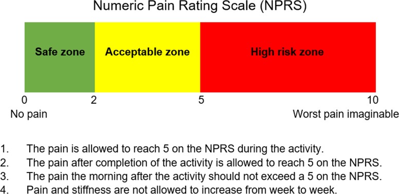
The purpose of this phase is to stop the negative cycle of overloading and continued injury progression (Table 3). Taking a thorough history of the injury progression is important for understanding the injury, symptoms, and tissue irritability, as well as how to address load reduction. Because complete rest and tendon unloading can be detrimental and prolong recovery, it is helpful to use the level of symptoms and pain as a guide when determining the appropriate level of activity.12 Patients vary in their pain perception and tolerance, but avoiding moderate and severe pain and worsening symptoms over time is a priority when considering the activity level. The pain-monitoring model (Figure 5) has been used successfully as a guide for tendon loading in patients with Achilles tendinopathy.6 This model is a useful tool that helps clinicians and patients determine how exercise programs and load management should progress. Alternative activities (such as deep-water running and biking) that impart lower loads on the Achilles tendon may be used to maintain cardiovascular fitness.
Figure 5.
The pain-monitoring model. Reprinted with permission from Silbernagel KG, Crossley KM. A proposed return-to-sport program for patients with midportion Achilles tendinopathy: rationale and implementation. J Orthop Sports Phys Ther. 2015;45(11):876–886.
Patient education is vital for successful outcomes and compliance. The patient needs a thorough understanding of the purpose of the exercises, the use of the pain-monitoring model, and the expected prognosis. Load management, especially in athletes, needs to be discussed with all stakeholders (player, coaches, trainers, managers, and medical staff) because many considerations come into play in decision making.
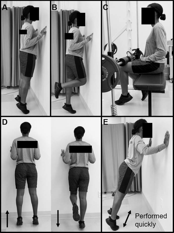
Throughout the course of treatment, the specific exercises used for Achilles tendon loading are various types of heel rises (Figure 6). In the early phase, it is beneficial to start on a level surface and ensure that the patient can control both the concentric and eccentric movements of the heel rise. If the patient has difficulty performing a heel rise, loading may be performed using isometric contractions or resistance bands. For those with insertional tendinopathy, limiting the degree of dorsiflexion is helpful to prevent compression between the tendon and the calcaneus. The degree of dorsiflexion can be limited by placing a lift under the heel (such as a 0.5- to 1-in [1.27- to 2.54-cm] plank) when performing the heel rises. The patient's degree of symptoms and irritability guides the progression through the phases of rehabilitation. A training diary in which the patient documents the exercises performed, other activities, and pain rating (in the morning and with activity) is beneficial for both the patient and clinician to assess progress.
Figure 6.
Commonly used Achilles tendon-loading exercises. A, Bilateral heel rise. B, Unilateral heel rise. C, Seated heel rise. D, Eccentric heel rise. E, Rebounding heel rise.
Recovery Phase
The aim of the recovery phase is to regain the strength of the calf muscles and improve the Achilles tendon's tolerance to load (Table 3). Exercises are performed daily. As symptoms subside and calf-muscle function improves, the exercises are progressed by increasing the number of repetitions, range of motion, and speed of movement. Heel-rise exercises are progressed to standing on an incline or with the heel off a step for those with midportion symptoms. The addition of quick-rebounding heel rises is meant to increase the Achilles tendon's tolerance to fast loads in preparation for plyometric exercises. A body-weight–supported treadmill or deep-water running during rehabilitation is an excellent way to resume running while controlling the load on the tendon. In addition, it is important to address any other lower extremity impairments detected during the evaluation and potential risk factors (such as deficits in ankle or foot strength and range of motion as well as hip neuromuscular control) at this time. Improving the strength of the ankle and foot muscles, especially the intrinsic foot muscles, will help the push-off phase during running and jumping, whereas ensuring good knee and hip strength and function will help ensure good lower extremity biomechanics.
Rebuilding Phase
The purpose of this phase is to transition to heavier strength training of the calf muscles, increase or start running and jumping activities, and further promote tendon recovery (Table 3). At this stage, external loads are added during the heel-rise exercises. This can be done by performing the exercises with a weighted vest or backpack or while in a leg press or Smith machine. Exercises performed at home are normally started with 10 lb (4.5 kg); when 3 sets of 15 repetitions are performed easily, the weight is increased. An alternative is to perform heavier heel rises at a gym 3 times per week and then lighter heel rises at home daily. It is often important to remind the athlete during this stage that despite having recovered calf-muscle strength and endurance, the heel-rise exercises should be continued to promote tendon healing and recovery. Plyometric training starts with bilateral jumps and progresses to unilateral jumps. Preparation for the return to participation starts with initiating specific sport activities. This phase generally overlaps with the return-to-sport phase. A good rule of thumb is that before an athlete is allowed to return to running and jumping, pain during daily walking should be minimal (1–2 of 10 on the numeric pain-rating scale). Pain level during single-legged hopping can also be used as a return-to-sport criterion.6 A patient who has a pain level above 2 (of 10) when performing 20 hops will struggle to start running while remaining within the pain-monitoring guidelines.
Return-to-Sport Phase
The goal of this phase is to return the athlete to full sport participation and performance while Achilles tendon and lower extremity functions continue to improve. Based on our research and clinical experience, we developed a return-to-sport program for athletes with Achilles tendinopathy.43 The goal of the program is to facilitate the decision-making process in returning an athlete with Achilles tendinopathy to full sport participation while minimizing the risk of recurrence. Because the athlete might not have symptoms from the Achilles tendon injury during sport participation at the latter stages of rehabilitation, he or she might be tempted to return prematurely. The evaluation of symptoms, stiffness, pain, and swelling after training, especially the following day, is crucial before determining the appropriate increase in training intensity. During this stage, the pain-monitoring model (Figure 5) is a useful guide for determining the appropriate activity level. Various factors must be considered when planning an athlete's return to sport after Achilles tendinopathy. The most obvious aspect, which is also addressed most often in the literature, is the level of pain with physical activity. However, recovery of tendon tissue, strength, range of motion, and function, as well as the demands of the specific sport, should be part of the decision-making process. Even in the absence of symptoms, impairments and functional deficits may persist. Therefore, it is crucial to ensure that an athlete has full recovery of function, as measured with the heel-rise and jumping tests, along with symptomatic recovery. Also beneficial are sport-specific functional tests to ensure symmetry between limbs and for comparison with preinjury data if available.
Our return-to-sport program is based on the premise that the tendon tissue requires a longer recovery from heavy loading activities (36–72 hours), whereas lighter activities can be performed more often.47 The intensity, duration, and frequency of Achilles tendon loading are considered when progressively increasing the demand during the return-to-sport phase. Sport activities are then classified at a light, medium, or high level based on the athlete's ratings of pain (during and after activity) and perceived Achilles tendon exertion using the Borg scale (Table 4).48 The classification of sport activity is determined by the clinician, athlete, and coach. Light-level activities can be performed daily, medium-level activities need to be followed by 2 recovery days, and high-level activities need to be followed by 3 days of recovery. A training schedule that combines low- to high-level activities and ensures proper recovery time is then planned for approximately 3 weeks. In our experience, allowing high-level activities ensures both the proper training dose to improve the athlete's capacity and the appropriate recovery time. Activities are generally reclassified every 3 to 4 weeks, and new high-level activities are added to progress toward a full return to sport and performance. The athlete is required to document pain and symptoms daily and grade the perceived exertion on the Achilles tendon after each activity in a training diary that is reviewed with the clinician and coach. We propose that diligently following this return-to-sport program will help prevent a premature return to sport and reinjury and result in a successful return to sport.25,26
Table 4.
| Intervention |
Evidence |
Strength of Recommendation8 |
| Exercise16 | Strongest evidence to support effectiveness in reducing symptoms and improving function | A |
| Any mechanical loading (eccentric or slow concentric and eccentric) is effective | ||
| Exercise should be completed at least 2x/wk | ||
| Orthotics2,16 | No recommendation can be made | B |
| No differences between custom and generic orthotics | ||
| May reduce strain on Achilles | ||
| No effect in reducing pain | ||
| Low-level laser therapy47 | Conflicting evidence | B |
| May be beneficial as adjunct to exercise | ||
| Taping16 | May alter foot posture and reduce strain on the Achilles tendon; however, limited evidence has shown effect in reducing pain | C |
| Iontophoresis16 | Effective in reducing pain and symptoms of acute midportion tendinopathy | B |
| Instrument-assisted soft tissue mobilization16,50 | Effective if ankle-joint range of motion is decreased | C |
| Extracorporeal shockwave therapy2,16 | Effective for pain relief and promoting tendon healing in short term | B |
| When combined with eccentric loading, more beneficial than eccentric loading alone | ||
| Patient outcomes vary widely | ||
| Injection therapies16,50 | No evidence to support use of injections alone or in combination with exercise therapy | A |
ADDITIONAL INTERVENTIONS
Numerous interventions have been suggested to promote recovery from Achilles tendinopathy, either in conjunction with exercise therapy or in isolation. However, progressive tendon loading remains the cornerstone of Achilles tendinopathy treatment. A summary of proposed interventions and SOR grades is provided in Table 2.
PREVENTION
To effectively prevent Achilles tendinopathy and the debilitating symptoms that affect sport participation, we must address perpetual overloading at an early stage. The current difficulty with early detection and prevention is that the cardinal sign for Achilles tendinopathy is pain in the Achilles tendon that limits sport participation. However, pain with activity is generally preceded by weeks or months of morning stiffness and minor pain, often ignored by both the athlete and the clinician as long as the athlete can participate in sport.13,18 Some athletes have described a period of decreased performance (such as increased running times) before developing symptoms, and this needs to be taken seriously.3 Therefore, monitoring training load, sport participation, and recovery sessions is beneficial for detecting sudden changes in training loads that increase the risk for Achilles tendinopathy. Also, the athlete's performance should be tested periodically to identify any decline that could indicate a change in Achilles tendon health. Changes in tendon structure, such as tendinosis, can occur before the athlete has any symptoms and are a risk factor for developing Achilles tendinopathy.24 A structural change seen on ultrasound imaging does not mean the athlete will develop symptoms, but the clinician can use the information in combination with measures of training load and performance to determine whether an athlete needs to be removed from practice and pursue alternative training.
We therefore propose that the best prevention is to recognize the early “minor” symptoms and treat these with load control (adjusting training loads) instead of ignoring them or only addressing the symptoms. The earlier the injury is detected, the shorter the expected time for full recovery.
SUMMARY
Achilles tendinopathy is a debilitating injury that affects multiple domains of tendon health and physical function. Therefore, it is critical to perform a comprehensive evaluation that includes reliable and valid outcome measures of each affected domain. Treatment should be centered on activity modification and progressive tendon-loading exercises to promote tendon remodeling and restoration of calf-muscle function. Furthermore, these interventions should be paired with patient education to establish a realistic timeline for return to sport and the role of pain in guiding activity and rest. To ensure full recovery of tendon health and minimize the risk of reinjury, additional impairments and risk factors might need to be addressed. In rare cases, more invasive interventions may need to be considered when an athlete has failed rehabilitation based on the principles described herein. However, these interventions should be viewed as a last resort.
ACKNOWLEDGMENTS
This study was supported by the National Institute of Arthritis and Musculoskeletal and Skin Diseases of the National Institutes of Health under Award No. R01AR072034 (K.G. Silbernagel and S. Hanlon). The work was supported in part by a Florence P. Kendall and a Promotion of Doctoral Studies I scholarship from the Foundation for Physical Therapy (A. Sprague).
REFERENCES
- 1.Sobhani S, Dekker R, Postema K, Dijkstra PU. Epidemiology of ankle and foot overuse injuries in sports: a systematic review. Scand J Med Sci Sports. 2013;23(6):669–686. doi: 10.1111/j.1600-0838.2012.01509.x. [DOI] [PubMed] [Google Scholar]
- 2.Magnussen RA, Dunn WR, Thomson AB. Nonoperative treatment of midportion Achilles tendinopathy: a systematic review. Clin J Sport Med. 2009;19(1):54–64. doi: 10.1097/JSM.0b013e31818ef090. [DOI] [PubMed] [Google Scholar]
- 3.Janssen I, van der Worp H, Hensing S, Zwerver J. Investigating Achilles and patellar tendinopathy prevalence in elite athletics. Res Sports Med. 2018;26(1):1–12. doi: 10.1080/15438627.2017.1393748. [DOI] [PubMed] [Google Scholar]
- 4.de Jonge S, van den Berg C, de Vos RJ, et al. Incidence of midportion Achilles tendinopathy in the general population. Br J Sports Med. 2011;45(13):1026–1028. doi: 10.1136/bjsports-2011-090342. [DOI] [PubMed] [Google Scholar]
- 5.Silbernagel KG, Thomeé R, Eriksson BI, Karlsson J. Full symptomatic recovery does not ensure full recovery of muscle-tendon function in patients with Achilles tendinopathy. Br J Sports Med. 2007;41(4):276–280. doi: 10.1136/bjsm.2006.033464. [DOI] [PMC free article] [PubMed] [Google Scholar]
- 6.Silbernagel KG, Thomeé R, Eriksson BI, Karlsson J. Continued sports activity, using a pain-monitoring model, during rehabilitation in patients with Achilles tendinopathy: a randomized controlled study. Am J Sports Med. 2007;35(6):897–906. doi: 10.1177/0363546506298279. [DOI] [PubMed] [Google Scholar]
- 7.Ebell MH, Siwek J, Weiss BD, et al. Strength of Recommendation Taxonomy (SORT): a patient-centered approach to grading evidence in the medical literature. Am Fam Physician. 2004;69(3):548–556. [PubMed] [Google Scholar]
- 8.Kvist M. Achilles tendon injuries in athletes. Ann Chir Gynaecol. 1991;80(2):188–201. [PubMed] [Google Scholar]
- 9.Scott A, Backman LJ, Speed C. Tendinopathy: update on pathophysiology. J Orthop Sports Phys Ther. 2015;45(11):833–841. doi: 10.2519/jospt.2015.5884. [DOI] [PubMed] [Google Scholar]
- 10.Scott A, Squier K, Alfredson H, et al. ICON 2019: International Scientific Tendinopathy Symposium consensus: clinical terminology [published online August 9, 2019]. Br J Sports Med. doi: 10.1136/bjsports-2019-100885. [DOI] [PubMed]
- 11.Zellers JA, Bley BC, Pohlig RT, Alghamdi NH, Silbernagel KG. Frequency of pathology on diagnostic ultrasound and relationship to patient demographics in individuals with insertional Achilles tendinopathy. Int J Sports Phys Ther. 2019;14(5):761–769. [PMC free article] [PubMed] [Google Scholar]
- 12.Magnusson P, Kjaer M. The impact of loading, unloading, ageing, and injury on the human tendon. J Physiol. 2019;597(5):1283–1298. doi: 10.1113/JP275450. [DOI] [PMC free article] [PubMed] [Google Scholar]
- 13.Leadbetter WB. Cell-matrix response in tendon injury. Clin Sports Med. 1992;11(3):533–578. [PubMed] [Google Scholar]
- 14.Järvinen TA, Kannus P, Maffulli N, Khan KM. Achilles tendon disorders: etiology and epidemiology. Foot Ankle Clin. 2005;10(2):255–266. doi: 10.1016/j.fcl.2005.01.013. [DOI] [PubMed] [Google Scholar]
- 15.van Dijk CN, van Sterkenburg MN, Wiegerinck JI, Karlsson J, Maffulli N. Terminology for Achilles tendon related disorders. Knee Surg Sports Traumatol Arthrosc. 2011;19(5):835–841. doi: 10.1007/s00167-010-1374-z. [DOI] [PMC free article] [PubMed] [Google Scholar]
- 16.Martin RL, Chimenti R, Cuddeford T, et al. Achilles pain, stiffness, and muscle power deficits: midportion achilles tendinopathy revision 2018. J Orthop Sports Phys Ther. 2018;48(5):A1–A38. doi: 10.2519/jospt.2018.0302. [DOI] [PubMed] [Google Scholar]
- 17.Magnan B, Bondi M, Pierantoni S, Samaila E. The pathogenesis of Achilles tendinopathy: a systematic review. Foot Ankle Surg. 2014;20(3):154–159. doi: 10.1016/j.fas.2014.02.010. [DOI] [PubMed] [Google Scholar]
- 18.van der Vlist AC, Breda SJ, Oei EHG, Verhaar JAN, de Vos RJ. Clinical risk factors for Achilles tendinopathy: a systematic review. Br J Sports Med. 2019;53(21):1352–1361. doi: 10.1136/bjsports-2018-099991. [DOI] [PMC free article] [PubMed] [Google Scholar]
- 19.Maffulli N, Kenward MG, Testa V, Capasso G, Regine R, King JB. Clinical diagnosis of Achilles tendinopathy with tendinosis. Clin J Sport Med. 2003;13(1):11–15. doi: 10.1097/00042752-200301000-00003. [DOI] [PubMed] [Google Scholar]
- 20.Hutchison AM, Evans R, Bodger O, et al. What is the best clinical test for Achilles tendinopathy? Foot Ankle Surg. 2013;19(2):112–117. doi: 10.1016/j.fas.2012.12.006. [DOI] [PubMed] [Google Scholar]
- 21.Maffulli N, Oliva F, Loppini M, Aicale R, Spiezia F, King JB. The Royal London Hospital test for the clinical diagnosis of patellar tendinopathy. Muscles Ligaments Tendons J. 2017;7(2):315–322. doi: 10.11138/mltj/2017.7.2.315. [DOI] [PMC free article] [PubMed] [Google Scholar]
- 22.Vicenzino B, de Vos RJ, Alfredson H, et al. ICON 2019: International Scientific Tendinopathy Symposium consensus: there are nine core health-related domains for tendinopathy (CORE DOMAINS): a Delphi Study of healthcare professionals and patients [published online November 4, 2019]. Br J Sports Med. doi: 10.1136/bjsports-2019-100894. [DOI] [PubMed]
- 23.Corrigan P, Cortes DH, Pontiggia L, Silbernagel KG. The degree of tendinosis is related to symptom severity and physical activity levels in patients with midportion Achilles tendinopathy. Int J Sports Phys Ther. 2018;13(2):196–207. [PMC free article] [PubMed] [Google Scholar]
- 24.McAuliffe S, McCreesh K, Culloty F, Purtill H, O'Sullivan K. Can ultrasound imaging predict the development of Achilles and patellar tendinopathy? A systematic review and meta-analysis. Br J Sports Med. 2016;50(24):1516–1523. doi: 10.1136/bjsports-2016-096288. [DOI] [PubMed] [Google Scholar]
- 25.Gajhede-Knudsen M, Ekstrand J, Magnusson H, Maffulli N. Recurrence of Achilles tendon injuries in elite male football players is more common after early return to play: an 11-year follow-up of the UEFA Champions League injury study. Br J Sports Med. 2013;47(12):763–768. doi: 10.1136/bjsports-2013-092271. [DOI] [PubMed] [Google Scholar]
- 26.Hagglund M, Walden M, Ekstrand J. Lower reinjury rate with a coach-controlled rehabilitation program in amateur male soccer: a randomized controlled trial. Am J Sports Med. 2007;35(9):1433–1442. doi: 10.1177/0363546507300063. [DOI] [PubMed] [Google Scholar]
- 27.Murphy M, Rio E, Debenham J, Docking S, Travers M, Gibson W. Evaluating the progress of mid-portion Achilles tendinopathy during rehabilitation: a review of outcome measures for muscle structure and function, tendon structure, and neural and pain associated mechanisms. Int J Sports Phys Ther. 2018;13(3):537–551. [PMC free article] [PubMed] [Google Scholar]
- 28.Robinson JM, Cook JL, Purdam C, et al. The VISA-A questionnaire: a valid and reliable index of the clinical severity of Achilles tendinopathy. Br J Sports Med. 2001;35(5):335–341. doi: 10.1136/bjsm.35.5.335. [DOI] [PMC free article] [PubMed] [Google Scholar]
- 29.Golightly YM, DeVellis RF, Nelson AE, et al. Psychometric properties of the foot and ankle outcome score in a community-based study of adults with and without osteoarthritis. Arthritis Care Res (Hoboken) 2014;66(3):395–403. doi: 10.1002/acr.22162. [DOI] [PMC free article] [PubMed] [Google Scholar]
- 30.Roos EM, Brandsson S, Karlsson J. Validation of the foot and ankle outcome score for ankle ligament reconstruction. Foot Ankle Int. 2001;22(10):788–794. doi: 10.1177/107110070102201004. [DOI] [PubMed] [Google Scholar]
- 31.Martin RRL, Irrgang JJ, Burdett RG, Conti SF, Van Swearingen JM. Evidence of validity for the Foot and Ankle Ability Measure (FAAM) Foot Ankle Int. 2005;26(11):968–983. doi: 10.1177/107110070502601113. [DOI] [PubMed] [Google Scholar]
- 32.Bennell KL, Talbot RC, Wajswelner H, Techovanich W, Kelly DH, Hall AJ. Intra-rater and inter-rater reliability of a weight-bearing lunge measure of ankle dorsiflexion. Aust J Physiother. 1998;44(3):175–180. doi: 10.1016/s0004-9514(14)60377-9. [DOI] [PubMed] [Google Scholar]
- 33.Silbernagel KG, Gustavsson A, Thomeé R, Karlsson J. Evaluation of lower leg function in patients with Achilles tendinopathy. Knee Surg Sports Traumatol Arthrosc. 2006;14(11):1207–1217. doi: 10.1007/s00167-006-0150-6. [DOI] [PubMed] [Google Scholar]
- 34.Byrne C, Keene DJ, Lamb SE, Willett K. Intrarater reliability and agreement of linear encoder derived heel-rise endurance test outcome measures in healthy adults. J Electromyogr Kinesiol. 2017;36:34–39. doi: 10.1016/j.jelekin.2017.07.004. [DOI] [PubMed] [Google Scholar]
- 35.Möller M, Lind K, Styf J, Karlsson J. The reliability of isokinetic testing of the ankle joint and a heel-raise test for endurance. Knee Surg Sports Traumatol Arthrosc. 2005;13(1):60–71. doi: 10.1007/s00167-003-0441-0. [DOI] [PubMed] [Google Scholar]
- 36.Webber SC, Porter MM. Reliability of ankle isometric, isotonic, and isokinetic strength and power testing in older women. Phys Ther. 2010;90(8):1165–1175. doi: 10.2522/ptj.20090394. [DOI] [PubMed] [Google Scholar]
- 37.Silbernagel KG, Shelley K, Powell S, Varrecchia S. Extended field of view ultrasound imaging to evaluate Achilles tendon length and thickness: a reliability and validity study. Muscles Ligaments Tendons J. 2016;6(1):104–110. doi: 10.11138/mltj/2016.6.1.104. [DOI] [PMC free article] [PubMed] [Google Scholar]
- 38.Corrigan P, Zellers JA, Balascio P, Silbernagel KG, Cortes DH. Quantification of mechanical properties in healthy Achilles tendon using continuous shear wave elastography: a reliability and validation study. Ultrasound Med Biol. 2019;45(7):1574–1585. doi: 10.1016/j.ultrasmedbio.2019.03.015. [DOI] [PMC free article] [PubMed] [Google Scholar]
- 39.Cortes DH, Suydam SM, Silbernagel KG, Buchanan TS, Elliott DM. Continuous shear wave elastography: a new method to measure viscoelastic properties of tendons in vivo. Ultrasound Med Biol. 2015;41(6):1518–1529. doi: 10.1016/j.ultrasmedbio.2015.02.001. [DOI] [PMC free article] [PubMed] [Google Scholar]
- 40.Dirrichs T, Quack V, Gatz M, et al. Shear wave elastography (SWE) for monitoring of treatment of tendinopathies: a double-blinded, longitudinal clinical study. Acad Radiol. 2017;25(3):265–272. doi: 10.1016/j.acra.2017.09.011. [DOI] [PubMed] [Google Scholar]
- 41.McAuliffe S, Synott A, Casey H, McCreesh K, Purtill H, O'Sullivan K. Beyond the tendon: experiences and perceptions of people with persistent Achilles tendinopathy. Musculoskelet Sci Pract. 2017;29:108–114. doi: 10.1016/j.msksp.2017.03.009. [DOI] [PubMed] [Google Scholar]
- 42.French DJ, France CR, Vigneau F, French JA, Evans RT. Fear of movement/(re)injury in chronic pain: a psychometric assessment of the original English version of the Tampa Scale for Kinesiophobia (TSK) Pain. 2007;127(1–2):42–51. doi: 10.1016/j.pain.2006.07.016. [DOI] [PubMed] [Google Scholar]
- 43.Silbernagel KG, Crossley KM. A proposed return-to-sport program for patients with midportion Achilles tendinopathy: rationale and implementation. J Orthop Sports Phys Ther. 2015;45(11):876–886. doi: 10.2519/jospt.2015.5885. [DOI] [PubMed] [Google Scholar]
- 44.Beyer R, Kongsgaard M. Hougs Kjær B, Øhlenschlæger T, Kjær M, Magnusson SP. Heavy slow resistance versus eccentric training as treatment for Achilles tendinopathy: a randomized controlled trial. Am J Sports Med. 2015;43(7):1704–1711. doi: 10.1177/0363546515584760. [DOI] [PubMed] [Google Scholar]
- 45.O'Neill S, Radia J, Bird K, et al. Acute sensory and motor response to 45-s heavy isometric holds for the plantar flexors in patients with Achilles tendinopathy. Knee Surg Sports Traumatol Arthrosc. 2018;27(9):2765–2773. doi: 10.1007/s00167-018-5050-z. [DOI] [PMC free article] [PubMed] [Google Scholar]
- 46.Couppé C, Svensson RB, Silbernagel KG, Langberg H, Magnusson SP. Eccentric or concentric exercises for the treatment of tendinopathies? J Orthop Sports Phys Ther. 2015;45(11):853–863. doi: 10.2519/jospt.2015.5910. [DOI] [PubMed] [Google Scholar]
- 47.Magnusson SP, Langberg H, Kjaer M. The pathogenesis of tendinopathy: balancing the response to loading. Nat Rev Rheumatol. 2010;6(5):262–268. doi: 10.1038/nrrheum.2010.43. [DOI] [PubMed] [Google Scholar]
- 48.Borg G. Borg's Perceived Exertion and Pain Scales. Champaign, IL: Human Kinetics; 1998. [Google Scholar]
- 49.Tumilty S, Mani R, Baxter GD. Photobiomodulation and eccentric exercise for Achilles tendinopathy: a randomized controlled trial. Lasers Med Sci. 2016;31(1):127–135. doi: 10.1007/s10103-015-1840-4. [DOI] [PubMed] [Google Scholar]
- 50.Kearney RS, Parsons N, Metcalfe D, Costa ML. Injection therapies for Achilles tendinopathy. Cochrane Database Syst Rev. 2015. (5):CD010960. [DOI] [PMC free article] [PubMed]


