Abstract
Polyphosphate (polyP) is a food additive with antimicrobial activity. Here we evaluated the effects of sodium tripolyphosphate (polyP3, Na5P3O10) on four major oral bacterial species, in both single- and mixed-culture. PolyP3 inhibited three opportunistic pathogenic species: Fusobacterium nucleatum, Prevotella intermedia, and Porphyromonas gingivalis. On the contrary, a commensal bacterium Streptococcus gordonii was relatively less susceptible to polyP3 than the pathogens. When all bacterial species were co-cultured, polyP3 (≥ 0.09%) significantly reduced their total growth and biofilm formation, among which the three pathogenic bacteria were selectively inhibited. Collectively, polyP3 may be an alternative antibacterial agent to control oral pathogenic bacteria.
Key words: polyphosphate, antibacterial, oral bacteria, commensal, pathogenic
Introduction
Periodontitis is a polymicrobial biofilm-mediated disease of the oral cavity and is a chronic inflammatory condition of the periodontium, leading to the damage of structural tooth support, resorption of bone, and eventually tooth loss (Schaudinn et al. 2009). The microbial etiology of periodontitis is defined by oral biofilm, also called dental plaque, in which resides an interdependent microbial community containing numerous bacterial species (Sundus et al. 2016). A typical dental biofilm begins with the formation of the salivary acquired pellicle, which is a result of the selective adsorption of salivary components, such as statherin, histatin, acidic proline-rich proteins, albumin, mucins, and α-amylase onto the tooth surface (Kreth et al. 2009; Willems et al. 2016). Early colonizers then directly attach to various molecules of the acquired pellicle via the selective adhesin-receptor binding and form an initial dental plaque (Kreth et al. 2009). Representative bacteria that constitute the initial biofilm are streptococci, which are mostly commensal/non-periodontopathogenic bacteria and makeup over 80% of the early biofilm inhabitants (Kreth et al. 2009), quickly followed by Actinomyces, Gemella, Neisseria, and Veillonella. As plaque matures, the composition of the biofilm changes. With the introduction and an increasing number of anaerobic bacteria such as Porphyromonas, Fusobacterium, Prevotella, Veillonella, and Capnocytophaga, the diversity of the species constituting the plaque increases (Willems et al. 2016). Several experimental studies suggested that periodontal diseases are generally associated with Gram-negative anaerobic bacteria, such as Aggregatibacter actinomycetemcomitans, Tannerella forsythia, Prevotella spp., Fusobacterium spp. and Porphyromonas gingivalis (Torkko and Asikainen 1993; Tanner and Izard 2006; Han 2015; How et al. 2016; Suprith et al. 2018). However, it is now widely accepted that not any one of these bacterial species alone, but a concerted interaction of these members can cause the destructive events involved in the periodontal disease progression (Liu et al. 2012; Hajishengallis 2015; Khan et al. 2015; Patini et al. 2018).
Polyphosphate (polyP) is an inorganic polymer composed of three to several hundred orthophosphate residues linked by phosphoanhydride bonds (Kornberg et al. 1999). The type of polyP varies depending on the length of the phosphate chain constituting it, for example, polyP3, polyP5, polyP15, polyP45, and so on, even up to polyP chains of many hundreds of phosphate residues. The Food and Drug Administration (FDA) has listed sodium pyro-, tri-, and hexametaphosphates as the Generally Recognized as Safe (GRAS) food additives. PolyP is often added to dairy and meat products for water binding, ion exchange, emulsification, and antioxidation (Ellinger 1972). In addition, due to the antimicrobial effects against various Gram-positive bacteria and fungi (Knabel et al. 1991), thereby suppressing food spoilage, polyP has drawn the attention of food industry.
Recently, it was reported that some Gram-negative anaerobic periodontal pathogens were highly sensitive to polyP. The minimum inhibitory concentrations (MICs) of polyP against P. gingivalis W83 and P. intermedia ATCC 49046 ranged from 0.06 to 0.075%, which were much lower than those previously reported for Gram-positive bacteria (Moon et al. 2011; Jang et al. 2016). Bacterial species rarely inhabit infection sites alone instead reside in diverse, multispecies communities (Stacy et al. 2016). Hence, to adequately assess the potential of polyP as a controlling agent against oral pathogenic bacteria, it is necessary to ascertain the effect of polyP not only on an individual microbial species but also on the consortium of mixed-species. In the present study, we investigated the effects of sodium tripolyphosphate (polyP3, Na5P3O10), listed as a GRAS, on four major oral bacterial species, including commensal and pathogenic bacteria. We also evaluated the antimicrobial effect of polyP3 against the mixed-species consortium consisting of the four bacterial species.
Experimental
Materials and Methods
Bacterial strains and culture condition. We used S. gordonii G9B, F. nucleatum ATCC 23726, P. gingivalis ATCC 53977 (previously designated A7A1-28), and P. intermedia ATCC 49046. Each bacterial strain was grown either in Brucella agar (Becton, Dickinson and Company, Sparks, MD, USA) supplemented with 5% sheep blood, 5 µg/ml hemin (Sigma Chemical Co., St. Louis, MO, USA) and 1 μg/ml vitamin K1 (Sigma), or in Brucella broth (Becton, Dickinson and Company) supplemented with 5 µg/ml hemin and 1 μg/ml vitamin K1 (B-HK). The bacterial culture was incubated at 37°C under anaerobic conditions (85% N2, 10% H2 and 5% CO2).
MIC determination and individual bacterial growth. PolyP with chain length 3 (polyP3, Na5P3O10, Sigma) was dissolved in distilled water to 10% (wt/vol) and sterilized using a 0.22-µm filter. The MIC of polyP3 against the individual bacterial species was determined by agar dilution method according to CLSI guidelines (CLSI 2007). Briefly, optical density at 600 nm (OD600) of each bacterial suspension was adjusted, then the suspension was inoculated at a cell density of approximately 105 to 106 cells/spot on Brucella blood agar plates containing polyP3 (final concentrations of 0.015–0.2%). The number of the inoculated bacterial cells was confirmed by serial dilution and colony forming unit (CFU) count. All inoculated plates were incubated at 37°C for 72 h. The MIC was defined as the lowest concentration that inhibited the bacterial growth on the plate. To evaluate the effect of polyP3 on the growth of the bacteria at a high inoculum density, each bacterial strain cultured to exponential phase was adjusted to approximately 108 cells/ml in B-HK, then exposed to polyP3 at various concentrations. The bacterial growth was measured by reading OD600 after 24 hours.
Total growth and biofilm formation of mixed bacterial culture. We also investigated the effect of polyP3 on the total growth and biofilm formation of the four bacterial species in a mixed culture. In preliminary studies, we observed that a mixed-species biofilm with relatively uniform distribution of four strains can be developed by inoculating S. gordonii, F. nucleatum, P. gingivalis and P. intermedia cells at a ratio of 1 : 1 : 100 : 100. We estimated the number of individual bacterial cells by measuring and adjusting of OD600 of each culture, then mixed so that the cell number ratio was as above. The mixed bacterial suspension was dispensed in triplicate into wells (500 μl/well) of a 24-well plate containing B-HK (500 μl) supplemented with polyP3 at various concentrations. The final bacterial suspension was approximately 108 to 109 cells/ml. Two identical 24-well plates were prepared for each of three independent experiments and cultured for 72 h under the same environment as the culture condition for single bacterial species, without agitation and media replenishment. To assess the total growth (planktonic and biofilm bacterial growth), the biofilm bacterial cells were dispersed and mixed thoroughly with planktonic bacterial cells, and OD600 of the suspension was measured (Moon et al. 2013, 2015). The suspension was diluted 2 to 10 times, and OD600 of the diluted suspension was also measured. The amount of biofilm formed by the mixed bacterial species was measured using the other plate. The planktonic bacterial cells and the spent media were removed, and the biofilm formed in the wells was washed twice with physiological saline, then stained with 0.1% crystal violet for 10 min. The plate was washed three times with physiological saline and air dried. Then, 500 µl of 95% ethanol was added to release the crystal violet from the biofilm, and the OD600 was recorded.
Scanning electron microscopy (SEM). Mixed species biofilms were developed as described above, then washed, dried, fixed in ethanol and dried again, as described previously (Jang et al. 2016). The biofilm samples were coated with gold using a sputter-coater (IB-3, Eiko, Tokyo, Japan) and then observed at 10 kV under a scanning electron microscope (Model S-4700; Hitachi High Technologies America. Inc., Pleasanton, CA, USA).
Results and Discussion
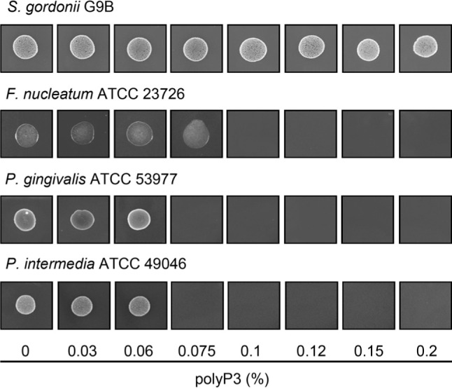
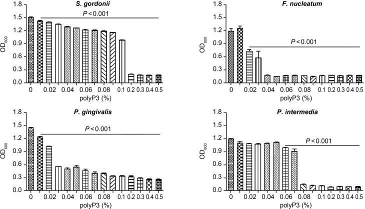
PolyP3 effectively inhibits the major oral pathogenic bacteria. It has been reported that polyP has antibacterial activity against various Gram-positive bacteria (such as Staphylococcus aureus, Bacillus cereus, Listeria monocytogenes, and mutans streptococci) at the concentrations between 0.1 and 0.5%, although it varies depending on the bacterial strain, inoculum density and culture medium (Post et al. 1963; Shibata and Morioka 1982; Zaika and Kim 1993; Lee et al. 1994). In the present study, the growth of a Gram-positive bacterium S. gordonii was not inhibited by polyP3 (up to 0.2%) on an agar plate (Fig. 1). Meanwhile, in liquid medium, the bacterial growth was slightly but significantly reduced by polyP3 in the range of 0.01–0.1%, and almost completely inhibited at concentrations ≥ 0.2% (Fig. 2). Compared to S. gordonii, the three pathogenic bacteria: F. nucleatum ATCC 23726, P. gingivalis ATCC 53977, and P. intermedia ATCC49046 were more susceptible to polyP3. As shown in Fig. 1, the MICs of polyP3 against the bacteria were 0.1, 0.075 and 0.075 %, respectively, as determined by the agar dilution method. Notably, despite the bacterial inoculum concentration approximately 1000-fold higher than that used for the MIC determination, polyP3 still exerted a strong antibacterial activity in liquid medium (BH-K) (Fig. 2). The difference between the antibacterial effects of polyP3 observed in the agar and broth dilution methods is likely due to the fact that the MIC determined by the agar dilution method was the concentration at which the bacterial growth was completely inhibited bacteriostatically and/or bactericidally. MIC determined by agar dilution method is not affected by whether an antibacterial agent is bactericidal or bacteriostatic. In contrast, if the bacterial cells were killed by polyP3 in the liquid medium, they may have been lysed, lowering the OD600 values. It is also possible that the liquid medium enhanced the contact of polyP3 with individual bacterial cells, resulting in an excellent antimicrobial effect of polyP3 in the liquid medium.
Fig. 1.

MIC determination of polyP3 by the agar dilution method. The bacterial cells were spot-inoculated (approximately 105 to 106 cells/spot) onto Brucella blood agar plates containing polyP3 at various concentrations and incubated at 37°C for 3 days anaerobically. The MIC was defined as the lowest concentration that inhibited the bacterial growth on the plate. The results for P. intermedia ATCC 49046 are the same as those reported in our previous study (Jang et al. 2016).
Fig. 2.
Effect of polyP3 on the growth of the bacteria at a high inoculum density. Each bacterial strain cultured to exponential phase was adjusted to approximately 108 cells/ml in B-HK, then exposed to polyP3 at various concentrations. The bacterial growth was measured by reading optical density at 600 nm (OD600) after the 24-hour incubation. Data are means ± SDs from two independent experiments performed in triplicate. One-way ANOVA with Tukey’s post-hoc tests p < 0.001, versus control (0% polyP3).
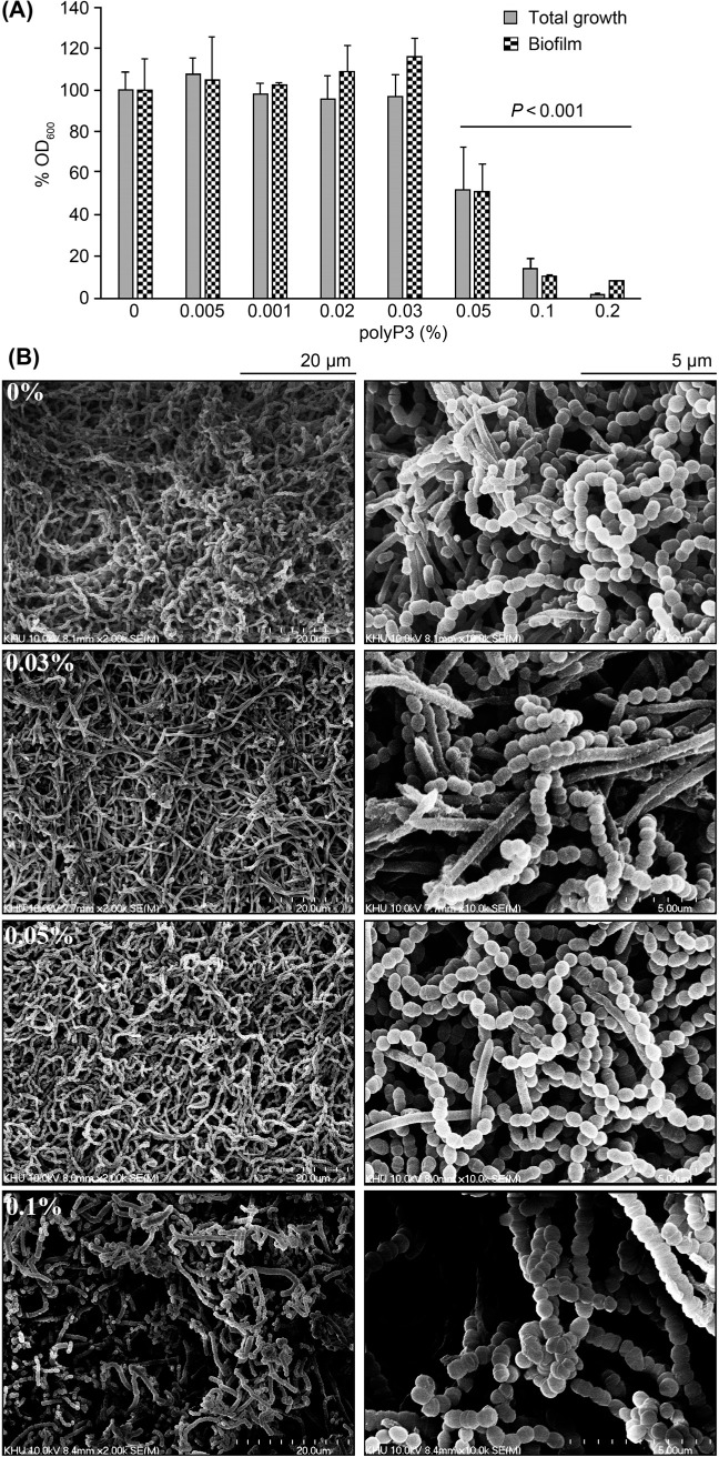
PolyP3 selectively inhibits the major oral pathogenic bacteria in the mixed-culture. When the four bacterial species were co-cultured, the total growth, as well as the biofilm formation, was enhanced, probably due to their symbiotic relationship. As shown in Fig. 3A, the total growth and biofilm formation of the mixed species were significantly inhibited by polyP3 (≥ 0.05%). In the morphological analysis by SEM (Fig. 3B), the control biofilm (not exposed to polyP3) showed a multilayered structure consisting of spherical-shaped S. gordonii, spindle-shaped F. nucleatum, along with rod-shaped bacterial cells presumed to be P. gingivalis and P. intermedia. On the other hand, in the biofilms exposed to polyP3 (≥ 0.05%), spindle- and rod-shaped cells decreased. The biofilm exposed to 0.1% polyP3 was a simple structure composed of only streptococci, indicating that polyP3 still exerts stronger antimicrobial effects against the three Gram-negative pathogenic bacteria than S. gordonii in the mixed culture.
Fig. 3.
Effect of polyP3 on the growth and biofilm formation of the four bacterial species in a mixed culture. (A) To assess the total growth (planktonic and biofilm bacterial growth), the biofilm bacterial cells were dispersed, mixed thoroughly with planktonic bacterial cells, and OD600 of the suspension was measured. The suspension was diluted 2 to 10 times, and OD600 of the diluted suspension was also measured. The amount of biofilm formed by the mixed bacterial species was quantitated by crystal violet staining. Readings were expressed as mean %OD600 (%OD600 = OD600 ∕ mean OD600 of control × 100). One-way ANOVA with Tukey’s post-hoc tests p < 0.001, versus control (0% polyP3). (B) SEM images of the biofilms at a magnification of 20 000 × (left) and 100 000 × (right).
There are a vast number of U.S. patents for the use of polyP3 in oral care preparations. Although STPP has been included in oral care products for stain removal and brightening of the enamel, antibacterial effect of polyP3 apparently has not drawn the attention of the dentistry academy and the oral health industry. Here, we are inclined to emphasize the powerful potential of polyP3 with antibacterial activity, which is required for an ideal oral hygiene product. PolyP3 can be an effective antibacterial agent to control the major oral pathogenic bacteria P. intermedia, P. gingivalis, and F. nucleatum. The ability of polyP3 to selectively inhibit pathogens and maintain the proportion of commensal bacteria may be effective in preventing oral diseases caused by these pathogenic bacteria.
Acknowledgements
This research was supported by the National Research Foundation of Korea (NRF) funded by the Ministry of Science & ICT (NRF-2016R1D1A1B03932450 and NRF-2018R1A2B6002173).
Footnotes
Conflict of interest
The authors do not report any financial or personal connections with other persons or organizations, which might negatively affect the contents of this publication and/or claim authorship rights to this publication.
ORCID
Ji-Hoi Moon 0000-0003-0286-5297
Literature
- CLSI.. Methods for antimicrobial susceptibility testing of anaerobic bacteria approved standard. 7th ed Document M11-A7 Wayne (PA): Clinical and Laboratory Standards Institute; 2007. [PubMed] [Google Scholar]
- Ellinger RH.. Phosphates as food ingredients. Cleveland (OH): CRC Press; 1972. p. 3–14. [Google Scholar]
- Hajishengallis G.. Periodontitis: from microbial immune subversion to systemic inflammation. Nat Rev Immunol. 2015. Jan;15(1): 30–44. doi: 10.1038/nri3785 Medline [DOI] [PMC free article] [PubMed] [Google Scholar]
- Han YW.. Fusobacterium nucleatum: a commensal-turned pathogen. Curr Opin Microbiol. 2015. Feb;23:141–147. doi: 10.1016/j.mib.2014.11.013 Medline [DOI] [PMC free article] [PubMed] [Google Scholar]
- How KY, Song KP, Chan KG.. Porphyromonas gingivalis: an overview of periodontopathic pathogen below the gum line. Front Microbiol. 2016. Feb 09;7:53. doi: 10.3389/fmicb.2016.00053 Medline [DOI] [PMC free article] [PubMed] [Google Scholar]
- Jang EY, Kim M, Noh MH, Moon JH, Lee JY.. In vitro effects of polyphosphate against Prevotella intermedia in planktonic phase and biofilm. Antimicrob Agents Chemother. 2016. Feb;60(2):818–826. doi: 10.1128/AAC.01861-15 Medline [DOI] [PMC free article] [PubMed] [Google Scholar]
- Khan SA, Kong EF, Meiller TF, Jabra-Rizk MA.. Periodontal diseases: bug induced, host promoted. PLoS Pathog. 2015. Jul 30; 11(7):e1004952. doi: 10.1371/journal.ppat.1004952 Medline [DOI] [PMC free article] [PubMed] [Google Scholar]
- Knabel SJ, Walker HW, Hartman PA.. Inhibition of Aspergillus flavus and selected gram-positive bacteria by chelation of essential metal cations by polyphosphates. J Food Prot. 1991. May;54(5):360–365. doi: 10.4315/0362-028X-54.5.360 [DOI] [PubMed] [Google Scholar]
- Kornberg A, Rao NN, Ault-Riché D.. Inorganic polyphosphate: a molecule of many functions. Annu Rev Biochem. 1999. Jun;68(1): 89–125. doi: 10.1146/annurev.biochem.68.1.89 Medline [DOI] [PubMed] [Google Scholar]
- Kreth J, Merritt J, Qi F.. Bacterial and host interactions of oral streptococci. DNA Cell Biol. 2009. Aug;28(8):397–403. doi: 10.1089/dna.2009.0868 Medline [DOI] [PMC free article] [PubMed] [Google Scholar]
- Lee RM, Hartman PA, Olson DG, Williams FD.. Bactericidal and bacteriolytic effects of selected food-grade phosphates, using Staphylococcus aureus as a model system. J Food Prot. 1994. Apr; 57(4):276–283. doi: 10.4315/0362-028X-57.4.276 [DOI] [PubMed] [Google Scholar]
- Liu B, Faller LL, Klitgord N, Mazumdar V, Ghodsi M, Sommer DD, Gibbons TR, Treangen TJ, Chang YC, Li S, et al.. Deep sequencing of the oral microbiome reveals signatures of periodontal disease. PLoS One. 2012. Jun 4;7(6):e37919. doi: 10.1371/journal.pone.0037919 Medline [DOI] [PMC free article] [PubMed] [Google Scholar]
- Moon JH, Jang EY, Shim KS, Lee JY.. In vitro effects of N-acetyl cysteine alone and in combination with antibiotics on Prevotella intermedia. J Microbiol. 2015. May;53(5):321–329. doi: 10.1007/s12275-015-4500-2 Medline [DOI] [PubMed] [Google Scholar]
- Moon JH, Kim C, Lee HS, Kim SW, Lee JY.. Antibacterial and antibiofilm effects of iron chelators against Prevotella intermedia. J Med Microbiol. 2013. Sep 01;62(Pt_9):1307–1316. doi: 10.1099/jmm.0.053553-0 Medline [DOI] [PubMed] [Google Scholar]
- Moon JH, Park JH, Lee JY.. Antibacterial action of polyphosphate on Porphyromonas gingivalis. Antimicrob Agents Chemother. 2011. Feb;55(2):806–812. doi: 10.1128/AAC.01014-10 Medline [DOI] [PMC free article] [PubMed] [Google Scholar]
- Patini R, Staderini E, Lajolo C, Lopetuso L, Mohammed H, Rimondini L, Rocchetti V, Franceschi F, Cordaro M, Gallenzi P.. Relationship between oral microbiota and periodontal disease: a systematic review. Eur Rev Med Pharmacol Sci. 2018. Sep;22(18): 5775–5788. Medline [DOI] [PubMed] [Google Scholar]
- Post FJ, Krishnamurty GB, Flanagan MD.. Influence of sodium hexametaphosphate on selected bacteria. Appl Microbiol. 1963. Sep; 11: 430–435. Medline [DOI] [PMC free article] [PubMed] [Google Scholar]
- Schaudinn C, Gorur A, Keller D, Sedghizadeh PP, Costerton JW.. Periodontitis: an archetypical biofilm disease. J Am Dent Assoc. 2009. Aug;140(8):978–986. doi: 10.14219/jada.archive.2009.0307 Medline [DOI] [PubMed] [Google Scholar]
- Shibata H, Morioka T.. Antibacterial action of condensed phosphates on the bacterium Streptococcus mutans and experimental caries in the hamster. Arch Oral Biol. 1982;27(10):809–816. doi: 10.1016/0003-9969(82)90034-6 Medline [DOI] [PubMed] [Google Scholar]
- Stacy A, Fleming D, Lamont RJ, Rumbaugh KP, Whiteley M.. A commensal bacterium promotes virulence of an opportunistic pathogen via cross-respiration. MBio. 2016. Jul 06;7(3):e00782-16. doi: 10.1128/mBio.00782-16 Medline [DOI] [PMC free article] [PubMed] [Google Scholar]
- Sundus H, Mukhtar H, Nawaz A.. Industrial applications and production sources of serine alkaline proteases: A review. J Bacteriol Mycol Open Acces. 2016;3:191–194. [Google Scholar]
- Suprith S, Setty S, Bhat K, Thakur S.. Serotypes of Aggregatibacter actinomycetemcomitans in relation to periodontal status and assessment of leukotoxin in periodontal disease: A clinico-micro biological study. J Indian Soc Periodontol. 2018;22(3):201–208. doi: 10.4103/jisp.jisp_36_18 Medline [DOI] [PMC free article] [PubMed] [Google Scholar]
- Tanner ACR, Izard J.. Tannerella forsythia, a periodontal pathogen entering the genomic era. Periodontol 2000. 2006. Oct;42(1):88–113. doi: 10.1111/j.1600-0757.2006.00184.x Medline [DOI] [PubMed] [Google Scholar]
- Torkko H, Asikainen S.. Occurrence of Porphyromonas gingivalis with Prevotella intermedia in periodontal samples. FEMS Immunol Med Microbiol. 1993. Mar;6(2-3):195–198. doi: 10.1111/j.1574-695X.1993.tb00325.x Medline [DOI] [PubMed] [Google Scholar]
- Willems HME, Xu Z, Peters BM.. Polymicrobial biofilm studies: from basic science to biofilm control. Curr Oral Health Rep. 2016. Mar;3(1):36–44. doi: 10.1007/s40496-016-0078-y Medline [DOI] [PMC free article] [PubMed] [Google Scholar]
- Zaika LL, Kim AH.. Effect of sodium polyphosphates on growth of Listeria monocytogenes. J Food Prot. 1993. Jul;56(7):577–580. doi: 10.4315/0362-028X-56.7.577 [DOI] [PubMed] [Google Scholar]


