PLOS Computational Biology logoLink to PLOS Computational Biology
. 2020 Jun 8;16(6):e1007927. doi: 10.1371/journal.pcbi.1007927

Measurable health effects associated with the daylight saving time shift

Hanxin Zhang 1,2, Torsten Dahlén 3, Atif Khan 2, Gustaf Edgren 3,4, Andrey Rzhetsky 1,2,5,*
Editor: Lars Juhl Jensen6
PMCID: PMC7302868  PMID: 32511231

Abstract

The transition to daylight saving time (DST) is beneficial for energy conservation but at the same time it has been reported to increase the risk of cerebrovascular and cardiovascular problems. Here, we evaluate the effect of the DST shift on a whole spectrum of diseases—an analysis we hope will be helpful in weighing the risks and benefits of DST shifts. Our study relied on a population-based, cross-sectional analysis of the IBM Watson Health MarketScan insurance claim dataset, which incorporates over 150 million unique patients in the US, and the Swedish national inpatient register, which incorporates more than nine million unique Swedes. For hundreds of sex- and age-specific diseases, we assessed effects of the DST shifts forward and backward by one hour in spring and autumn by comparing the observed and expected diagnosis rates after DST shift exposure. We found four prominent, elevated risk clusters, including cardiovascular diseases (such as heart attacks), injuries, mental and behavioral disorders, and immune-related diseases such as noninfective enteritis and colitis to be significantly associated with DST shifts in the United States and Sweden. While the majority of disease risk elevations are modest (a few percent), a considerable number of diseases exhibit an approximately ten percent relative risk increase. We estimate that each spring DST shift is associated with negative health effects–with 150,000 incidences in the US, and 880,000 globally. We also identify for the first time a collection of diseases with relative risks that appear to decrease immediately after the spring DST shift, enriched with infections and immune system-related maladies. These diseases’ decreasing relative risks might be driven by the documented boosting effect of a short-term stress (such as that experienced around the spring DST shift) on the immune system.

Author summary

Over a quarter of the world population is subjected to the daylight saving time (DST) shift twice a year, which disrupts both human work and rest schedules and possibly the body’s biological clock. Several clinical studies have reported an increased risk of cerebrovascular and cardiovascular problems with DST shifts but little is known about other potential health effects. The DST shift represents a natural exposure experiment which allows us the unique opportunity of linking health outcomes to an external, state-wide event in the US and Sweden. We performed a comprehensive, phenome-wide screening of the putative health effects of the DST shift by analyzing two independent, country-scale health datasets, and found both adverse and protective associations with DST shifts in several clusters of conditions. We successfully verified previously reported associations, such as heart diseases and injuries, and identified new signals–for example, immune-related conditions. We suggest that the ramifications of daylight-saving time shifts should be acknowledged and further tested.

Introduction

The idea of introducing daylight saving time (DST) was attractive at the time of candles and gas lamps, as it allowed workers to use sunlight a bit longer during working hours as well as saving employers’ energy for lighting. Much has changed since then. Today, only a small fraction of electricity expenditure actually corresponds to producing light after sunset (in the US, it is about six percent in the residential sector and eight percent in the industrial sector) [1]. Yet, over a quarter of the world population is subjected to the DST shift twice a year, which disrupts both human work and rest schedules and possibly their circadian clock rhythms [2]. DST shifts have been shown to have a measurable effect on electric power consumption, although not necessarily in the intended direction [3,4]. Previous studies have demonstrated that the spring DST shift causes noticeable alterations in human behavior in terms of waking-up time and self-reported alertness, [5] a significant increase in fatal traffic accidents (up to 30 percent on the day of commencing DST), [6] a short-term rise in workplace injuries (5.7 percent after the spring DST shift as employees sleep 40 minutes less on average), [7] and elevated rates of acute myocardial infarction (up by about 3.9 percent) [8]. The study described in [6] and [9] reported conflicting results regarding whether the DST shifts are associated with accident incidence. The study described in [10] found increased mental health- and behavioral health-oriented emergency department visits in certain seasons, but did not obtain conclusive results on whether they could be linked to the DST shifts.

Remarkable progress has been made in the past decade towards understanding the neurology of sleep-wake cycles and circadian rhythms, and how they affect our behavior [11]. Despite these advances, significant gaps remain in our knowledge of how changes in the social clock (DST shifts) interact with the body’s biological clock and impact human health. Recent studies have urged for investigations into the clinical implications of DST shifts on human health [12,13].

The DST shift represents a natural exposure experiment which allows us the unique opportunity of linking health outcomes to an external, state-wide event in the US and Sweden. Earlier analyses of DST shift effects typically examined a single medical condition per study, often with conflicting or inconclusive results [6,9,10]. In addition, these studies often relied on small, disease-specific datasets with thousands of observations from a single country or a single hospital, making it impossible to run phenome-wide screening. In the present study, we used the electronic health records (EHRs) of hundreds of millions of people across two countries, for the purpose of: (1) examining the temporal disease risk dynamics in relationship to DST shifts, and; (2) identifying those population strata which manifest health changes linked to DST-related schedule disruption.

Methods

Data and Other Materials

Our study accessed EHRs from two countries: In the US, through the IBM Watson Health MarketScan dataset, [14] and in Sweden, through the Swedish national inpatient register [15]. The version of the MarketScan dataset we used in this study incorporated health information about more than 150 million unique patients, observed during the time interval between 2003 and mid-2014. Individuals followed asynchronous enrollment and disenrollment on insurance policies, leading to variance in their “visibility” durations and endpoints in the data. The mean follow-up time for the patient in the US MarketScan database was 154 weeks. The Swedish register described more than nine million unique Swedes, nearly all observed continuously from 1968 to 2011, except in cases of death or emigration.

In both datasets, disease diagnoses were represented with codes defined by the World Health Organization (WHO) International Classification of Diseases (ICD) taxonomies, versions 9 and 10 for the US, and versions 8, 9, and 10 for Sweden, along with a day-level temporal label recording the date of diagnosis in the US data or the discharge date in the Swedish data. To evaluate the risk of DST shifts across the whole spectrum of diseases, we grouped ICD codes into 263 condition classes under 31 biological systems using the WHO ICD-10 guidelines (S1 Table) [16]. The grouping is hierarchical and exhaustive, so neighboring codes fall in the same or similar condition classes, and no ICD code is left uncategorized.

Statistical Analyses

Taking advantage of this ICD code grouping, we summarized the daily incidences of each class of conditions for females and males in the following age groups: 0–20 years old (or, alternatively, in the larger US dataset, separately 0–10, and 11–20), 21–40, 41–60, and over 60. Building on a previous study’s methodology [8], we quantified the relative risk (RR) involved with shifting to and from DST by comparing the diagnosis rate for each day during the week following a DST shift to the linear expectation, which is the average diagnosis rate of the same week day, two weeks before and two weeks after the day of interest (please refer to S1 Appendix Fig A for additional clarification). The diagnosis rate is the expected proportion of patients diagnosed with a given disease on the day of interest out of all enrollees at a given time point. We estimated week-level RR values by collapsing incidences reported during the whole week following a DST shift and comparing it to the average of corresponding week-level diagnoses rates two weeks before and after the time point. We corrected the possible effect of holidays (see the S1 Appendix Section 1.5 for a full list of holidays considered) and different day lengths (23 hours at the spring DST shift and 25 hours at the autumn). We obtained all the RR estimates in a Bayesian framework and shrunk them towards one. The shrinkage represented our prior belief that we expected the DST shifts to show no health effects for most conditions. The Bayesian procedure pooled information about multiple diseases–with a hierarchical prior distribution imposed over RR estimates. This shrinkage technique resolved the multiple comparisons problem and also yielded statistically efficient estimates [17]. To provide our readers with a frame of reference, we supplemented all of our Bayesian analyses with their frequentist counterparts (see S1 Appendix Section 1.6). The results of our Bayesian and frequentist analyses were very similar after a false coverage rate (FCR) adjustment of frequentist confidence intervals (see S1 Appendix Section 1.7), [18] suggesting a practical equivalence of FCR-corrected frequentist and hierarchical Bayesian (with shrinkage prior) frameworks.

Bayesian and frequentist methods should produce compatible but not identical results. Each method comes with its own advantages and disadvantages. The most obvious difference between them is associated with specification of a prior distribution over parameter values. In our Bayesian analysis, we assumed that most of the estimated RR values would be close to one (shrinkage assumption, implemented as a prior distribution strongly pulling estimates towards the central mean). This assumption forces the results to be conservative (which eliminates weaker signals) and removes the need to correct the results for multiple tests. The frequentist analysis is agnostic in regard to likely distribution of priors. Some statisticians argue that this type of analysis is less subjective and easier to interpret. The frequentist analyses require an explicit correction for multiple statistical tests and are likely to produce estimates with larger absolute deviations from one.

To control for possible false positive discoveries, we designed a few negative control experiments. Because DST was not adopted in Sweden until 1980, we compared the RRs of time transitions before 1980 (at some “pseudo-DST” shift points, i.e., when time shift would have happened, see S1 Appendix Section 1.8 for details) and after 1980 at real DST shift points. For the US data, we analyzed all patients residing in states not observing DST as a negative control. Furthermore, we introduced another negative control by repeating the RR estimation procedure at “pseudo-DST” shift dates, which were set to 28 days after each real DST shift in the spring and 28 days before each real DST shift in the autumn. The latter negative control resulted in the most statistically powerful test among the three, because it covered the largest population comparable in size to the groups being tested for association.

Because we ran all our analyses in parallel in both Bayesian and frequentist frameworks, we decided to present Bayesian results in the main text, highlighting the differences between the two approaches when relevant. The decision to analyze inpatient data separately was driven by the consideration that patients who were hospitalized (“inpatient”) may have been subject to fewer of the social and environmental confounders that drive spurious associations. Inpatient admissions are typically associated with a set of health problems distinct from outpatient visits, with more severe conditions, such as acute heart attacks, most commonly treated in hospital inpatient settings.

Results

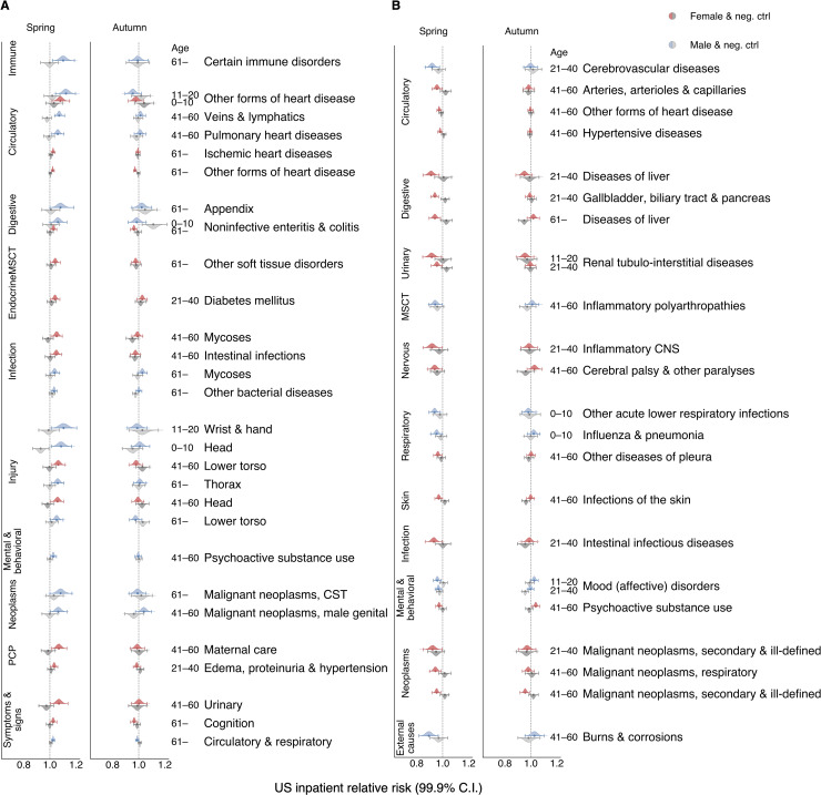
In the US inpatient cohort, we detected a significant risk elevation in a number of disease and condition groups (see S1 Appendix 2.1 and the summary table); for example, complications related to pregnancy, childbirth, and puerperium (PCP), as well as injuries, symptoms, and signs across various systems, and circulatory diseases (Fig 1A). We observed stand-out increases in the RR for some injuries, immune disorders, heart diseases, and possibly in related conditions such as renal failure (urinary system-related, shown in S2 Table but not Fig 1A as it is not among the top 30 for the effect size) and circulatory/cognition symptoms and signs (Fig 1A). Signals of relative risk change presented in Fig 1 were automatically selected according to their effect size and for their significance as shown in a comparison between the experimental and negative control tests (see S1 Appendix Section 2.1). For the whole spectrum of conditions considered in this study, we present the RR changes with DST shifts in S2 Table. The results of experiments with the negative controls are shown in S3 Table. We conducted similar analyses with the frequentist approach and found results consistent with those using the Bayesian framework. In some cases, we even noticed larger estimated effect sizes after adjusting for FCR (S4 Table and S5 Table).

Fig 1. Daylight saving time (DST) shifts appear to affect the relative risk (RR) of numerous diseases spanning several human biological systems.

Fig 1

Color-coded violin plots and error bars represent RR estimates’ posterior density distributions and credible interval (CI) boundaries, respectively. We adjusted the credible intervals (CI) for multiple tests with a Bayesian shrinkage procedure that ensured 99.9 and 99 percent significance levels for US and Swedish data, respectively. Gray-colored violin plots and error bars indicate analogous RR distribution results computed for the negative control (pseudo-DST shift dates, the same populations as for the real DST shift dates). The pseudo-DST shift date was selected at 28 days after the real DST shift in spring and 28 days before the real one in autumn. For the Swedish tests, negative controls were performed on data before 1980 when DST was not observed in Sweden. We selected all depicted signals automatically (see S1 Appendix Section 2.1), as the significant RR largest in effect size that were: (1) significantly greater than one in the spring DST shift analyses (colored), and: (2) not significantly greater than one in the negative control (gray) analyses, or vice versa for decreased signals. We also excluded too broadly defined, ambiguous clinical and laboratory findings, examinations, and health services from the figures (they are retained in the Supplementary Tables). (A) The top 30 conditions exhibiting the largest increasing RRs (effect sizes) for the results of the US inpatient analyses. The results suggest risk expansion in diseases involving the immune, circulatory, and digestive systems, the musculoskeletal system and connective tissue (MSCT), the endocrine systems, some infections and injuries, mental and behavioral disorders, neoplasms, problems with pregnancy, childbirth, and the puerperium (PCP), and symptoms and signs across various systems. (B) All disease conditions with significantly decreased RR in inpatient data after the spring DST shift (minus ambiguous procedures).

To the best of our knowledge, we are the first to report the DST-related RRs of disorders involving the digestive system (such as noninfective enteritis and colitis), which rose three percent after the spring DST shift in females over 60 and six percent in males under ten.

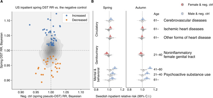
We also observed, for the first time, that the RR for a number of diseases appears to reduce after the spring DST shift (see Fig 1B). Such diseases include a set of infectious and inflammatory diseases. In the Bayesian analysis, the effect sizes of the negative RR changes tend to be smaller than those for positive RR changes. The gray contour in Fig 2A shows the joint distribution estimation of the spring DST’s RR versus the negative control’s RR in inpatients, spotlighting those increased and decreased RR change signals (see S1 Appendix Section 2.1 for more details).

Fig 2.

Fig 2

(A) Spring RR estimates versus the negative control results in the US inpatient population. The gray contour represents the empirical estimation of the joint distribution for all RR estimates. The blue and orange markers accent the increased and decreased signals selected by an impartial procedure based on effect size and significance (see S1 Appendix Section 2.1) (B) All conditions showing significant change around the DST shift analyses in the Swedish data after 1980. None of their corresponding negative controls were significantly different from one. As in the US data, we observed an increased RR in ischemic and other forms of heart disease in the senior population and mental and behavioral disorders due to psychoactive substance use in middle-age males. The RR for cerebrovascular diseases in senior inpatients increased in Sweden in the week following the spring DST shift, confirming the increase (with no statistical significance) in US inpatients. By contrast, in the US all-patient population, cerebrovascular diseases actually decreased significantly (S10 Table and S12 Table).

In the smaller Swedish dataset, as expected, we found fewer significant RR change signals. Under a less conservative significance level (99 percent versus 99.9 percent in the US analyses), we were able to reproduce the RR change signals for a subset of cardiovascular diseases in the elderly population (Fig 2B, S6 Table, and S7 Table). The RRs of some heart and cerebrovascular diseases go up in the spring, when the day length shrinks by one hour, but not in the autumn. As would be expected for a real effect, the RR for circulatory diseases increased after 1980 in the spring DST shift but not in the autumn one. Corresponding frequentist results are shown in S8 Table and S9 Table. Interestingly, the RR of psychoactive substance use increases as much as nine percent with the spring DST shift and 12 percent with the autumn DST shift but only among males age 20 or above population (Fig 2B and S6 Table and S7 Table).

Discussion

Our analyses reproduced the major past literature’s findings, such as an elevation in ischemic heart disease rates in males and females older than 60, [8,1921] and a rise in accidents [6,22] and injuries [7]. We also discovered novel significant RR change signals, such as a DST-shift-associated increase in mental and behavioral disorders due to the aforementioned elevated psychoactive substance use in the male adult population. The strongest effect size was observed among males between the ages 41–60 and the signal was consistent in both the US and Sweden. A large body of studies have shown that circadian disruption increases the risk of substance abuse [2326], with some studies providing in-depth mechanistic details [27]. Because psychoactive substance users generally have very disrupted diurnal rhythms [28], it seems plausible that further acute disruption due to DST shifts may lead to abnormal clock function, resulting in increased vulnerability for substance abuse.

The findings in the US “all-patients” dataset (S1 Appendix Fig B, S10S13 Tables) resolves the inconclusive results of a previous study focusing only on emergency admissions [10]. To the best of our knowledge, never reported before, immune-related disorders tend to become more common than expected in the first week following each spring DST shift. The largest effect sizes is observed for the following conditions: an approximate ten percent increase in the RR for some cardiovascular and heart diseases in inpatients under 20, injuries at various locations and ages (in the frequentist framework, RR estimates increased by 30 percent), and some immune disorders in senior males. The absolute risk posed by the DST shift is discussed in Section 2.5 of the S1 Appendix. The comparison of Bayesian and frequentist analyses indicates that there is not much data in support of the estimates computed for a subset of diseases, and that the prior has a strong influence on the estimate.

Our analysis identified several population strata that appeared responsive to the time and schedule disruption in terms of changes in disease RR: (1) very young (0–10, or 11–20) and older (41–60 and over 60) patients with (probably preexisting) chronic diseases, for example, acute myocardial infarction, behavioral and emotional disorders, and stress-related immune disorders such as inflammatory bowel diseases and noninfective enteritis; (2) children (0–10) and teenagers/young adults (11–20) who were prone to accidents resulting in injuries of the head, wrist, and hand; and (3) older adults (41–60 and older than 60) who were more likely to injure the lower torso or thorax. Note that while MarketScan represents the United States fairly evenly, geographically, it excludes the uninsured population.

In a DST shift effect analysis, one should keep in mind that there are two distinct phenomena in play: (1) the natural variation of day length, the amount of light over the year, and the daily sunlight intensity cycle affects the human circadian clock, behavior, and numerous diseases [2945], and; (2) the disruption of individuals' daily schedule due to the DST shift. Our analyses were designed to target the effects associated with the latter, adjusting for the former with negative controls. Furthermore, it is not always possible to distinguish between increased diseased incidence (for a truly new diagnosis) and increased care (for an established ongoing diagnosis). For a subset of patients under observation in the US dataset since their birth, we were able to carefully identify the first disease diagnosis and validate the RR change signal for noninfective enteritis and colitis after the spring DST shift (see S1 Appendix Sections 1.10 and 2.4). We performed an analogous “first-diagnosis” analysis for older US patients in the all-patients dataset, which resulted in only one statistically significant result: an increased rate of “other forms of heart disease” in females over 60 (see S1 Appendix Sections 1.10 and 2.4).

We have considered using a more complicated model, such as a mixed-effect regression model, which would allow us to include some chronic diseases as confounding factors to obtain better estimates of disease relative risks. We did not attempt to control for any particular disease for two reasons: (1) introducing control diseases would cause a combinatorial explosion in the model space, and; (2) controlling for specific diseases would exponentially increase the number of necessary statistical tests. Both issues would increase analysis complexity, make interpretation more difficult, and decrease our ability to detect RR change signals. Therefore, we decided against using this approach.

What mechanism could plausibly explain the reduced RR observed for some of the infectious and immune diseases during the spring DST shift? The spring DST shift might act as a short-term stressor, as one hour is subtracted from a normal night’s schedule. A transient, mild stress was shown to enhance the immune system (as opposed to long-term stress, which has the opposite, suppressive action), possibly accounting for the reduced RR for urinary, skin, and other infections. As F.S. Dhabhar put it, a “short-term stress can enhance the acquisition and/or expression of immune-protective (wound healing, vaccination, anti-infectious agent, anti-tumor) or immuno-pathological (pro-inflammatory, autoimmune) responses.” [44,45] Of course, this is but one possible explanation and possible alternative explanations could be suggested.

Limitations

This study suggests that even a one-hour change of the clock may impact population health significantly, but a number of caveats accompany this assertion.

First, it is important to keep in mind that diseases are not truly independent of each other; one illness can facilitate the development of another, and environmental insults may also lead to the exacerbation of a (pre-existing) chronic disease. Therefore, our analysis cannot distinguish between “driver” and “passenger” diseases. Note that the low p-values and narrow credible/confidence intervals are driven by the large data set. This does not necessarily mean that the data set is free of bias, and any bias will propagate and can very well drive associations.

Second, while the US dataset records the actual diagnoses dates (rather than “billing dates”), chronic diseases, such as hypertension and diabetes, are likely to have been developing for a long time before the actual diagnoses were made and recorded. It is possible that environmental insult (mild stress) acts as a trigger to worsen an already pre-existing conditions, so that patients are forced to see a physician (which results in diagnoses entered in records). The Swedish national registry records the “discharge dates” instead of the actual dates of diagnosis. In most cases, neither the date of diagnosis as in the US data nor the discharge date is expected to be the same as the actual time of disease attack. But these dates still more accurately reflect the time of attack than the “billing dates” do. We tried to smooth out the discrepancy between the dates by adding up all codes of interest in the following week of the daylight saving time change, and estimated the week-level RR based on it.

Third, it is possible that disease coding errors could influence our RR estimates. A simple way to spot miscoding is to look for sex-specific codes assigned to the wrong genders. For instance, the data shows males having pregnancy, ovarian cancer, and females having prostate cancer. There are two scenarios to explain the origins of such errors: Either the sex was recorded wrongly or the code itself is inaccurate. The former scenario would not affect our analyses substantially because we anticipated for symmetric coding errors in both sexes that offset against each other. On the other hand, if a diagnosis itself is miscoded, it may influence the RR estimation and tests. We estimated that the miscoding rate at the MarketScan data is around 0.52 percent, (see S1 Appendix Section 1.12. Data Limitations, for details of this estimation). The error rate is positive but is small in comparison to the observed DST shift effect sizes. Importantly, simple disease coding errors are unlikely to be in any way related to DST shifts, and would only bias RR estimates towards the null model.

Fourth, the major difficulties in our analysis were associated with changing coding standards (especially in Sweden, which went from ICD8 to ICD9, then to ICD10) and insufficient data sample despite the fact that datasets spanned whole countries. In particular, we did not have sufficient data to analyze every condition in every age/sex group. We alleviated the coding problem using two synergistic measures: (1) careful ICD version mapping, and; (2) considering relatively large constellations of diseases instead of following very specific conditions (for example, we analyzed “ischemic heart diseases,” including acute myocardial infarction, instead of specifically “acute myocardial infarction”). We felt that these categories of larger disease groups were robust enough to withstand changes in medical practice and diagnosis criteria. The larger disease group we used in our analysis also helped with increasing disease-specific patient sample size, as the collective incidence of a group of diseases is the sum of incidences of individual diseases in the group.

Supporting information

S1 Appendix. Complete details of materials and methods.

(PDF)

S1 Table. A mapping from ICD-10 codes to conditions and diseases.

(CSV)

S2 Table. Week-level RR estimates of the US inpatient analysis, via the Bayesian method.

(CSV)

S3 Table. Week-level RR estimates of the US inpatient analysis on pseudo-DST shift dates as a negative control, via the Bayesian method.

(CSV)

S4 Table. Week-level RR estimates of the US inpatient analysis, via the frequentist method.

(CSV)

S5 Table. Week-level RR estimates of the US inpatient analysis on pseudo-DST shift dates as a negative control, via the frequentist method.

(CSV)

S6 Table. Week-level RR estimates of the Swedish inpatient analysis since 1980, via the Bayesian method.

(CSV)

S7 Table. Week-level RR estimates of the Swedish inpatient analysis before 1980 as a negative control, via the Bayesian method.

(CSV)

S8 Table. Week-level RR estimates of the Swedish inpatient analysis since 1980, via the frequentist method.

(CSV)

S9 Table. Week-level RR estimates of the Swedish inpatient analysis before 1980 as a negative control, via the frequentist method.

(CSV)

S10 Table. Week-level RR estimates of the US all-patient analysis, via the Bayesian method.

(CSV)

S11 Table. Week-level RR estimates of the US all-patient analysis on pseudo-DST shift dates as a negative control, via the Bayesian method.

(CSV)

S12 Table. Week-level RR estimates of the US all-patient analysis, via the frequentist method.

(CSV)

S13 Table. Week-level RR estimates of the US all-patient analysis on pseudo-DST shift dates as a negative control, via the frequentist method.

(CSV)

S14 Table. A mapping from ICD-9-CM to ICD-8.

(CSV)

S15 Table. A mapping from the US modification of ICD-8, 9, 10 to conditions.

(CSV)

S16 Table. A mapping from the Swedish modification of ICD-8, 9, 10 to conditions.

(CSV)

S17 Table. A count summary of some female-specific diseases in the US data set.

(CSV)

S18 Table. A count summary of some male-specific diseases in the US data set.

(CSV)

S19 Table. A summary of conditions with increased RRs (Bayesian, US all-patient).

(CSV)

S20 Table. A summary of conditions with increased RRs (frequentist, US all-patient).

(CSV)

S21 Table. A summary of conditions with increased RRs (Bayesian, US inpatient).

(CSV)

S22 Table. A summary of conditions with increased RRs (frequentist, US inpatient).

(CSV)

S23 Table. A summary of conditions with decreased RRs (Bayesian, US all-patient).

(CSV)

S24 Table. A summary of conditions with decreased RRs (frequentist, US all-patient).

(CSV)

S25 Table. A summary of conditions with decreased RRs (Bayesian, US inpatient).

(CSV)

S26 Table. A summary of conditions with decreased RRs (frequentist, US inpatient).

(CSV)

S27 Table. Estimation of cost associated with the DST shift (Bayesian, US all-patient).

(CSV)

S28 Table. Estimation of cost associated with the DST shift (frequentist, US all-patient).

(CSV)

S29 Table. Estimation of cost associated with the DST shift (Bayesian, US inpatient).

(CSV)

S30 Table. Estimation of cost associated with the DST shift (Frequentist, US inpatient).

(CSV)

Acknowledgments

We are grateful to E. Gannon, R. Melamed, and M. Rzhetsky, for comments on earlier versions of this manuscript.

Data Availability

All data and code underlying our study and necessary to reproduce our results are available on Github: https://github.com/hanxinzhang/dst/tree/master/data.

Funding Statement

This work was funded by the DARPA Big Mechanism program under ARO contract W911NF1410333; by National Institutes of Health grants R01HL122712, 1P50MH094267, and U01HL108634-01; and by a gift from Liz and Kent Dauten. The funders had no role in study design, data collection and analysis, decision to publish, or preparation of the manuscript.

References

  • 1.US Energy Information Administration. How much electricity is used for lighting in the United States?—FAQ—U.S. Energy Information Administration (EIA) In: www.eia.gov. 2016. [Google Scholar]
  • 2.Kantermann T, Juda M, Merrow M, Roenneberg T. The Human Circadian Clock’s Seasonal Adjustment Is Disrupted by Daylight Saving Time. Curr Biol. Elsevier BV; 2007;17: 1996–2000. 10.1016/j.cub.2007.10.025 [DOI] [PubMed] [Google Scholar]
  • 3.Rivers N. Does Daylight Savings Time Save Energy? Evidence from Ontario. SSRN Electron J. Elsevier BV; 2016; 10.2139/ssrn.2772048 [DOI] [Google Scholar]
  • 4.Kotchen M, Grant L. Does Daylight Saving Time Save Energy? Evidence from a Natural Experiment in Indiana [Internet]. National Bureau of Economic Research; 2008. 10.3386/w14429 [DOI] [Google Scholar]
  • 5.Mink TH, Folkard S. Adjusting to the changes to and from Daylight Saving Time. Nature. Springer Science and Business Media LLC; 1976;261: 688–689. 10.1038/261688a0 [DOI] [PubMed] [Google Scholar]
  • 6.Prats-Uribe A, Tobías A, Prieto-Alhambra D. Excess Risk of Fatal Road Traffic Accidents on the Day of Daylight Saving Time Change. Epidemiology. Ovid Technologies (Wolters Kluwer Health); 2018;29: e44–e45. 10.1097/ede.0000000000000865 [DOI] [PubMed] [Google Scholar]
  • 7.Barnes CM, Wagner DT. Changing to daylight saving time cuts into sleep and increases workplace injuries. Journal of Applied Psychology. American Psychological Association (APA); 200994: 1305–1317. 10.1037/a0015320 [DOI] [PubMed] [Google Scholar]
  • 8.Janszky I, Ahnve S, Ljung R, Mukamal KJ, Gautam S, Wallentin L, et al. Daylight saving time shifts and incidence of acute myocardial infarction–Swedish Register of Information and Knowledge About Swedish Heart Intensive Care Admissions (RIKS-HIA). Sleep Med. Elsevier BV; 2012;13: 237–242. 10.1016/j.sleep.2011.07.019 [DOI] [PubMed] [Google Scholar]
  • 9.Lahti TA, Haukka J, Lönnqvist J, Partonen T. Daylight saving time transitions and hospital treatments due to accidents or manic episodes. BMC Public Health. Springer Nature; 2008;8 10.1186/1471-2458-8-74 [DOI] [PMC free article] [PubMed] [Google Scholar]
  • 10.Heboyan V, Stevens S, McCall W V. Effects of seasonality and daylight savings time on emergency department visits for mental health disorders. Am J Emerg Med. Elsevier BV; 2019;37: 1476–1481. 10.1016/j.ajem.2018.10.056 [DOI] [PubMed] [Google Scholar]
  • 11.Fuller PM, Gooley JJ, Saper CB. Neurobiology of the sleep-wake cycle: Sleep architecture, circadian regulation, and regulatory feedback. Journal of Biological Rhythms. 2006. 10.1177/0748730406294627 [DOI] [PubMed] [Google Scholar]
  • 12.Roenneberg T, Winnebeck EC, Klerman EB. Daylight saving time and artificial time zones—A battle between biological and social times. Frontiers in Physiology. 2019. 10.3389/fphys.2019.00944 [DOI] [PMC free article] [PubMed] [Google Scholar]
  • 13.Malow BA, Veatch OJ, Bagai K. Are Daylight Saving Time Changes Bad for the Brain? JAMA Neurology. 2019. 10.1001/jamaneurol.2019.3780 [DOI] [PubMed] [Google Scholar]
  • 14.IBM MarketScan Research Databases [Internet]. 2018. Available: https://www.ibm.com/downloads/cas/4QD5ADRL
  • 15.Ludvigsson JF, Andersson E, Ekbom A, Feychting M, Kim J-L, Reuterwall C, et al. External review and validation of the Swedish national inpatient register. BMC Public Health. Springer Science and Business Media LLC; 2011;11 10.1186/1471-2458-11-11 [DOI] [PMC free article] [PubMed] [Google Scholar]
  • 16.World Health Organization. International statistical classification of diseases and related health problems, 10th revision (ICD-10). World Heal Organ. 2016; [Google Scholar]
  • 17.Gelman A, Hill J, Yajima M. Why We (Usually) Don’t Have to Worry About Multiple Comparisons. J Res Educ Eff. Informa UK Limited; 2012;5: 189–211. 10.1080/19345747.2011.618213 [DOI] [Google Scholar]
  • 18.Benjamini Y, Yekutieli D. False Discovery Rate–Adjusted Multiple Confidence Intervals for Selected Parameters. J Am Stat Assoc. Informa UK Limited; 2005;100: 71–81. 10.1198/016214504000001907 [DOI] [Google Scholar]
  • 19.Janszky I, Ljung R. Shifts to and from daylight saving time and incidence of myocardial infarction. New England Journal of Medicine. 2008. 10.1056/NEJMc0807104 [DOI] [PubMed] [Google Scholar]
  • 20.Manfredini R, Fabbian F, De Giorgi A, Zucchi B, Cappadona R, Signani F, et al. Daylight saving time and myocardial infarction: Should we be worried? A review of the evidence. Eur Rev Med Pharmacol Sci. 2018; 10.26355/eurrev_201802_14306 [DOI] [PubMed] [Google Scholar]
  • 21.Meira e Cruz M, Miyazawa M, Manfredini R, Cardinali D, Madrid JA, Reiter R, et al. Impact of Daylight Saving Time on circadian timing system: An expert statement. European Journal of Internal Medicine. 2019. 10.1016/j.ejim.2019.01.001 [DOI] [PubMed] [Google Scholar]
  • 22.Robb D, Barnes T. Accident rates and the impact of daylight saving time transitions. Accid Anal Prev. 2018; 10.1016/j.aap.2017.11.029 [DOI] [PubMed] [Google Scholar]
  • 23.Menet JS, Rosbash M. When brain clocks lose track of time: Cause or consequence of neuropsychiatric disorders. Current Opinion in Neurobiology. 2011. 10.1016/j.conb.2011.06.008 [DOI] [PMC free article] [PubMed] [Google Scholar]
  • 24.Hasler BP, Soehner AM, Clark DB. Circadian rhythms and risk for substance use disorders in adolescence. Current Opinion in Psychiatry. 2014. 10.1097/YCO.0000000000000107 [DOI] [PMC free article] [PubMed] [Google Scholar]
  • 25.Webb IC. Circadian Rhythms and Substance Abuse: Chronobiological Considerations for the Treatment of Addiction. Current Psychiatry Reports. 2017. 10.1007/s11920-017-0764-z [DOI] [PubMed] [Google Scholar]
  • 26.Hasler BP, Smith LJ, Cousins JC, Bootzin RR. Circadian rhythms, sleep, and substance abuse. Sleep Medicine Reviews. 2012. 10.1016/j.smrv.2011.03.004 [DOI] [PMC free article] [PubMed] [Google Scholar]
  • 27.Logan RW, Williams WP, McClung CA. Circadian rhythms and addiction: Mechanistic insights and future directions. Behav Neurosci. 2014; 10.1037/a0036268 [DOI] [PMC free article] [PubMed] [Google Scholar]
  • 28.Falcón E, McClung CA. A role for the circadian genes in drug addiction. Neuropharmacology. 2009. 10.1016/j.neuropharm.2008.06.054 [DOI] [PMC free article] [PubMed] [Google Scholar]
  • 29.Martino TA, Young ME. Influence of the Cardiomyocyte Circadian Clock on Cardiac Physiology and Pathophysiology. J Biol Rhythms. SAGE Publications; 2015;30: 183–205. 10.1177/0748730415575246 [DOI] [PubMed] [Google Scholar]
  • 30.Fletcher E, Kanki M, Morgan J, Delbridge L, Ray D, Fuller P, et al. Cardiomyocyte Transcription Is Controlled By Combined Mr And Circadian Clock Signalling. J Endocr Soc. The Endocrine Society; 2019;3 10.1210/js.2019-or04-4 [DOI] [PubMed] [Google Scholar]
  • 31.Sulli G, Lam MTY, Panda S. Interplay between Circadian Clock and Cancer: New Frontiers for Cancer Treatment. Trends in Cancer. Elsevier BV; 2019;5: 475–494. 10.1016/j.trecan.2019.07.002 [DOI] [PMC free article] [PubMed] [Google Scholar]
  • 32.Zhanfeng N, Hechun X, Zhijun Z, Hongyu X, Zhou F. Regulation of Circadian Clock Genes on Sleep Disorders in Traumatic Brain Injury Patients. World Neurosurg. Elsevier BV; 2019;130: e475–e486. 10.1016/j.wneu.2019.06.122 [DOI] [PubMed] [Google Scholar]
  • 33.Olaoye OA, Masten SH, Mohandas R, Gumz ML. Circadian Clock Genes in Diabetic Kidney Disease (DKD). Curr Diab Rep. Springer Science and Business Media LLC; 2019;19 10.1007/s11892-019-1138-1 [DOI] [PMC free article] [PubMed] [Google Scholar]
  • 34.Shi D, Chen J, Wang J, Yao J, Huang Y, Zhang G, et al. Circadian Clock Genes in the Metabolism of Non-alcoholic Fatty Liver Disease. Front Physiol. Frontiers Media SA; 2019;10 10.3389/fphys.2019.00423 [DOI] [PMC free article] [PubMed] [Google Scholar]
  • 35.Faragó A, Zsindely N, Bodai L. Mutant huntingtin disturbs circadian clock gene expression and sleep patterns in Drosophila. Sci Rep. Springer Science and Business Media LLC; 2019;9 10.1038/s41598-019-43612-w [DOI] [PMC free article] [PubMed] [Google Scholar]
  • 36.Chang WH, Lai AG. Timing gone awry: distinct tumour suppressive and oncogenic roles of the circadian clock and crosstalk with hypoxia signalling in diverse malignancies [Internet]. Cold Spring Harbor Laboratory; 2019. 10.1101/556878 [DOI] [PMC free article] [PubMed] [Google Scholar]
  • 37.Kaneshiro K, Yoshida K, Morii K, Oketani Y, Uchida K, Yaekura A, et al. Expressions of circadian clock genes represent disease activities of RA patients treated with biological DMARDs. Mod Rheumatol. Informa UK Limited; 2019; 1–8. 10.1080/14397595.2019.1602242 [DOI] [PubMed] [Google Scholar]
  • 38.Maury E. Off the Clock: From Circadian Disruption to Metabolic Disease. Int J Mol Sci. MDPI AG; 2019;20: 1597 10.3390/ijms20071597 [DOI] [PMC free article] [PubMed] [Google Scholar]
  • 39.Mukherji A, Bailey SM, Staels B, Baumert TF. The circadian clock and liver function in health and disease. J Hepatol. Elsevier BV; 2019;71: 200–211. 10.1016/j.jhep.2019.03.020 [DOI] [PMC free article] [PubMed] [Google Scholar]
  • 40.Crnko S, Du Pré BC, Sluijter JPG, Van Laake LW. Circadian rhythms and the molecular clock in cardiovascular biology and disease. Nat Rev Cardiol. Springer Science and Business Media LLC; 2019;16: 437–447. 10.1038/s41569-019-0167-4 [DOI] [PubMed] [Google Scholar]
  • 41.Davis K, Roden LC, Leaner VD, van der Watt PJ. The tumour suppressing role of the circadian clock. IUBMB Life. Wiley; 2019;71: 771–780. 10.1002/iub.2005 [DOI] [PubMed] [Google Scholar]
  • 42.Li H, Song S, Wang Y, Huang C, Zhang F, Liu J, et al. Low-Grade Inflammation Aggravates Rotenone Neurotoxicity and Disrupts Circadian Clock Gene Expression in Rats. Neurotox Res. Springer Science and Business Media LLC; 2018;35: 421–431. 10.1007/s12640-018-9968-1 [DOI] [PMC free article] [PubMed] [Google Scholar]
  • 43.Kim P, Oster H, Lehnert H, Schmid SM, Salamat N, Barclay JL, et al. Coupling the Circadian Clock to Homeostasis: The Role of Period in Timing Physiology. Endocr Rev. The Endocrine Society; 2018;40: 66–95. 10.1210/er.2018-00049 [DOI] [PubMed] [Google Scholar]
  • 44.Dhabhar FS. Effects of stress on immune function: the good, the bad, and the beautiful. Immunol Res. Springer Science and Business Media LLC; 2014;58: 193–210. 10.1007/s12026-014-8517-0 [DOI] [PubMed] [Google Scholar]
  • 45.Dhabhar FS, Saul AN, Daugherty C, Holmes TH, Bouley DM, Oberyszyn TM. Short-term stress enhances cellular immunity and increases early resistance to squamous cell carcinoma. Brain Behav Immun. Elsevier BV; 2010;24: 127–137. 10.1016/j.bbi.2009.09.004 [DOI] [PMC free article] [PubMed] [Google Scholar]
PLoS Comput Biol. doi: 10.1371/journal.pcbi.1007927.r001

Decision Letter 0

Lars Juhl Jensen, Virginia E Pitzer

12 Feb 2020

Dear Dr. Rzhetsky,

Thank you very much for submitting your manuscript "Measurable Health Effects Associated with the Daylight Saving Time Shift" for consideration at PLOS Computational Biology.

As with all papers reviewed by the journal, your manuscript was reviewed by members of the editorial board and by several independent reviewers. In light of the reviews (below this email), we would like to invite the resubmission of a revised version that takes into account the reviewers' comments.

The reviewers were overall quite positive about the manuscript, but made many concrete requests for clarifications. Two of the reviewers also recommended correcting for additional potential confounding factors, which should be possible to do given the data used.

We cannot make any decision about publication until we have seen the revised manuscript and your response to the reviewers' comments. Your revised manuscript is also likely to be sent to reviewers for further evaluation.

When you are ready to resubmit, please upload the following:

[1] A letter containing a detailed list of your responses to the review comments and a description of the changes you have made in the manuscript. Please note while forming your response, if your article is accepted, you may have the opportunity to make the peer review history publicly available. The record will include editor decision letters (with reviews) and your responses to reviewer comments. If eligible, we will contact you to opt in or out.

[2] Two versions of the revised manuscript: one with either highlights or tracked changes denoting where the text has been changed; the other a clean version (uploaded as the manuscript file).

Important additional instructions are given below your reviewer comments.

Please prepare and submit your revised manuscript within 60 days. If you anticipate any delay, please let us know the expected resubmission date by replying to this email. Please note that revised manuscripts received after the 60-day due date may require evaluation and peer review similar to newly submitted manuscripts.

Thank you again for your submission. We hope that our editorial process has been constructive so far, and we welcome your feedback at any time. Please don't hesitate to contact us if you have any questions or comments.

Sincerely,

Lars Juhl Jensen

Associate Editor

PLOS Computational Biology

Virginia Pitzer

Deputy Editor

PLOS Computational Biology

***********************

Reviewer's Responses to Questions

Comments to the Authors:

Please note here if the review is uploaded as an attachment.

Reviewer #1: The paper presents a large-scale analysis of how daylight-saving time (DST) affects people’s health for a wide range of diseases using EMR data. They use US market scan with around 120 million patients and the Swedish national patient registry with 9 million patients. They use Bayesian inference to estimate the effect size and include a frequentist approach for reference. They reproduce diseases earlier shown to be affected by DST and come with novel results, like pregnancy related diagnoses and mental diseases. They provide literature references to substantiate these and show that they reproduce in both the US and Swedish data.

The paper presents original research. Although DST time shift have been researched before, no one have done it this systematically for as many different diseases as here. The statistical method seems to be a standard Bayesian approach without any novelties in it. With a minor exception it seems rigorous and sound. The main novelties are that they include a wide range of diseases in one study and use a larger population than used in earlier studies of DST effect on health. For example, they state that the DST effect on pregnancy diagnoses have never been studied before. The article has relevance both for the scientific community and for decision makers who are arguing over stopping DST. It contains conclusions only possible due to the large data set. It is my impression that the paper should be published with minor revision. However, I have a number of questions, which I would like the authors to address.

Regarding the calculation of relative risk: The sentence about which days are compared to which (week after DST to +/- two weeks) can be complicated to understand, and I suggest putting in a figure reference to fig S2.

As the authors mention as a weakness, there might be other causes that drive the difference in disease rate than DST. However, the authors have chosen not to control for other than age, and gender (and race/ethnicity?). Given the data there it is possible to control for pre-existing diseases/diagnoses in the patients, even in large-scale analysis. Or to repeat a selection of the significant results in a mores strictly controlled manner. I would like the authors comments on why they did not choose to include control for any other diseases.

A very crucial detail in the author’s work is that they made negative controls. In this way, they demonstrate that it is the DST having an effect on the results and that it is not reproducible in a random week. However, I find it highly questionable to use data from prior to 1980 as negative controls in Sweden. These numbers represent a different era of medicine than the newest data and coding practices might have changed significantly. I see their validity to demonstrate that the trend starts with introduction of DST, but not as comparison to the general numbers.

The data contains coding in both ICD versions 8, 9, and 10. There are very big differences going from version 9 to 10. Have the authors investigated if the coding frequency change abruptly because of the change? And have they verified in any way that this does not affect their results.

The author’s mention inconclusive results from reference 6, 12, and 13 (p 5, l 60). What are the results? They later write that these the inconclusive results from reference 13 are resolved (p. 10 l 179-180), but does not clearly state which results were resolved and how.

Did the authors have any way of distinguishing between acute and planned/follow-up visits? It seems likely that the acute visits are more affected by the DST than visits planned from before. E.g. for the injuries mentioned p. 10 l 193-194, where planned visits are probably not related to the DST. Could this change the result of the analysis?

The results show a change of childbirth RR (p 8 l 146). If the number of childbirth changes, so does the number of complications related to childbirths. Could this not alone explain the other maternal codes (p 8 l 144)? I.e. does the rate of complications increase more than the observed increase in birthrate?

In page 10 l 90 the authors mention that chronic diseases were probably preexisting. How was this investigated? There is no documentation for this statement, which should be easy to produce (i.e. number of cases where the chronic disease was seen before). Was such analysis carried out for other diseases?

In supplementary section 2.4 (p 15), the significant first-time signal is dismissed as “likely to be due to seasonal convexity or concavity”. I assume that the numbers are calculated with the same +/- 14 days comparison and that this handles the seasonal convexity or concavity. How come that the main analysis does not have the same problem?

It is not clear what the goal of the geographic location comparison is, if it can a priori be dismissed because many other covariates could influence the signal.

The authors present three sets of results with Bayesian, “half Bayesian” and frequentist methods. All the comparison is in the supplementary methods. Can they provide more information on the advantages / disadvantages for these methods in the main section?

I unfortunately do not know a lot about Bayesian statistics, and therefore I am not able to assess the choice of models. In the interest of the uninformed reader, I would ask that the authors explain their method in a way more readable to people with less knowledge about Bayesian statistics. I give examples of what I would like to be clearer here.

It is not clear at all how the actual data comes in to the model. In frequentist statistics, you estimate the parameters by maximizing the likelihood function. The authors give a formula to calculate the diagnosis rate on the time point of interest from the RR (5). But is the RR not the value you want to estimate and p0 a value you can calculate from the data? It would help understanding if the authors made more clear what loss function is being minimized / maximized to estimates the parameters.

In the interest of understanding the flow of the model, I don't understand how the x* (6a-6c) affects the model. They are not mentioned in the formulas for estimating the RR, but depends on the p* values.

The authors correct for the number of enrolled patients in the US data. As far as I understand they compare data points within +/- 2 week from the DST shift. Is the change in enrollment really so large that you have to correct for difference over a few weeks? Could they give a number on how much it changes on average? Or is it to correct for the yearly difference? In the latter case, it can be argued that the Swedish population also has changed during the study period.

Regarding the Bayesian correction for multiple testing, I don't understand how it works. I would suggest that the authors give more background to explain this. As I understand, the method forces the RR values towards the mean (i.e. 1 or neutral value?) when there is insufficient data. This makes a lot of sense. However, it is not clear how this shrinking is affected by the number of RR values that are estimated. And it is not clear how you can set the different thresholds for the US and Swedish data. Also, I find that the sentence regarding the shrinking in the main method-overview section should be extended to explain.

I assume that all the author’s choices of priors are well thought off and perhaps even standard choices. It would help if they cite some method papers that informs the reader about these choices.

The need to create the corrected RR value is well argued. However, I do not understand how the expected RR ( E[RR] hat ) is calculated and how this solves the problem. Is the expected RR the distribution's mean across all RR estimates that naturally have the temporal change in RR factored in?

Also, there are no comment on choice of parameters for the optimization. Are there any non-standard choices that might be relevant for readers with knowledge about such models?

The typesetting of Supplementary Materials is off: There are several unintended space, some of the letters also almost overlap, and the line spacing is also varying much.

Examples

Suppl Materials page 3, section 1.1 Mappings

All ICD-10 codes are categorized i n this grouping, so i t i s also exhaustive.

Suppl Materials page 4 btw formula (4) and (4a)

"mean and l et i nformation flow [...] comparisons problem i s alleviated

Reviewer #2: The study by Zhang et al presents a phenome-wide study of the effects of daylight savings time and disease incidence. The study is interesting, and it is encouring to see results for both men and women. Some of the results piques my interest, although I would like to see a more thorough dissemination of the results. I was also disappointed to not see a more through comparison of the two cohorts in the main text. Surely the advantage of working with multiple cohorts must be to replicate the findings and/or perform a meta analysis. I also have some concerns with the study design, statistical analysis, and terminology throughout the article. I find it very positive that the authors have provided all code and data in a Github Repo. I also see that the authors actually report some absolute risk differences. I think this would be much more intuitive, compared to the ratios used throughout the article.

INTRODUCTION

The introduction reads nicely, and provides a timely summary of the subject. One minor thing is I believe the appropriate terminology for this kind of analysis is "natural experiment" and not "large-scale intervention experiment". The authors note that previou studies typically focus on a small number of diseases. There is actually a perfectly good reason for this, which I will get back to.

METHODS

The description of the two data sources seem incomplete. Surely not only the Swedish inpatient registry was used. This would not contain information on death/emigration.

L72: "visibility" is typically called "under observation" in epidemiological jargon. Likewise, subjects are "censored" when they are not under observation. It would be very benefecial to provide average/mean follow-up time for each individual.

L72: In all fairness, how are these patients garantued to be unique? Are they linked through a social security number similar to Sweden?

L75-76: unclear why individuals that are dead or or have emigrated are excluded. This is very atypical. Please provide clarification.

From the Data description it is not clear which date is used for the patients. The admission or discharge date?

L85: It is not clear at all how the incidence is calculated. If incidence is calculated as #cases / population at risk, the age ranges are far too wide in my opinion. Also, are cases from the previous year considered in the next year? Or are they completely excluded from the calculation? This can also be seen in the Results section, where women aged 41-60 have an increased incidence of maternal care codes. The vast majority of this group of women will not be at risk to even be pregnant. This goes back to the point as to why previous studies typically deal with a limited number of conditions: defining the population at risk is not trivial, and it is something the authors need to consider in more detail for each condition. Just using the full population will severely deflate and provide nonsense incidence estimates.

L95-98: I would really like to see a correction for year as well.

L98-101: The specification of the Bayesian model is poorly explained. Please provide more detail. Additionally, I am not sure what kind of pooling the authors are using. The main text suggestes a complete pooling, but the Supplementary Material looks like a no pooling scheme to me. The multiple comparison problem is not necessarily solved by just specifying priors. If, however, the authors had used a partial pooling scheme across diagnoses, this would indeed solve the multiple comparison problem. See e.g. the 8 schools example by Gelman and Gelman's Bayesian Data Analysis, 3rd ed page 101. Given that the authors work in the ICD-10 space it should be possible to make use of that heirarchy in a model with partial pooling across diagnoses.

How was the convergence of the Bayesian model assessed? From the github repository, it seems that the authors used PyMC3. PyMC3, to my knowledge, uses the HMC-NUTS that provides a lot of diagnostics. Please also add a citation to PyMC3 in the Main Text.

L100: The use of a conservative prior is good, but the prior specifications in the Supplementary Material need a lot more justification. Also, the use is non-informative priors is generally not recommended. The authors should use priors that put more emphasis on the null value if that is the prior belief.

L113: by "not observing", do the authors mean that these states do not have DST?

L121: when was the decision made to analyse inpatient only? And in which data set?

L124: I disagree with the notion of urgent problems in inpatient care. What I believe the authors mean is that there are a different kind of diagnosis associated with each encounter. E.g. pneumonia, sexually transmitted diseases, etc may be handled in outpatient or GP care. Likewise, routine surgeries represent a fairly low risk and non-acute inpatient group.

RESULTS

L128-129: Could the author write how many, or refer to a table? just refering to "a number" seems very imprecise.

L131: What are stand-out increases? Do the author mean relative large effect sizes?

L134-135: "Signal" - would that be the change in relative risk? What is "automatically detected according to their effect size"?

L140: This is unclear. Did the point estimate of the frequentist confidence interval increase after FCR correction? Or do the authors mean that the frequentist point estimate is greater (in absolute terms), compared to the Bayesian analysis? The latter is expected due to the shrinkage, and should not come as a surprise to the authors.

L142-143: I am not convinced about this result due to the lack of a proper at-risk group. The group at-risk should, in this case, be pregnant women, and not just all women.

L159: Is it to be understood that the only signal that replicates are the cardiovascular diseases under a less stringent treshold? This is very disappointing, and in my opinion this strongly indicates that there is a bias in the data set that confers these increases that is unrelated to DST. A population of 9 million individuals is not a small data set, and it should have plenty of power to detect differences in incidence for single diseases.

In general, it would be good to see the all results simultaneously to see if there are trends within disease areas, using e.g. the ICD-10 chapter structure.

DISCUSSION

L179-180: not clear what exactly is meant here. What is the previous result, and why was it inconclusive?

L184: was this result presented previous? A _ten_ percent increase in cardiovascular and heart disease in a population of children and teenagers seems very extreme. How many children actually had a cardiovascular disease? An absolute risk would give more insight into the actual consequence.

L184-185: I am not a big fan of presenting both Bayesian and Frequest estimates jointly. The Bayesian model that the authors have specified naturally shrinks the estimates towards the null, so it is no surprise that point estimate is smaller than the frequentist procedure. This also indicates, strongly, that there is not a lot of data in support of the values, and that the prior has a strong influence on the estimate.

L189-190: Could these groups very well be the individuals that are covered by an insurance plan? I would imagine children and teenagers are covered by their parents insurance plan.

L195: What does "emergency care" refer to here? Were they admitted to the ER?

L198-199: without any previous studies exploring this, this is pure unfounded speculation and should be stated as such.

L202: This is not garantueed. Insured individuals have a completely different socioeconomic profile versus the uninsured, and extrapolating between the two requires information on the uninsured. This is a general concern when using insurance data, namely that it is highly biased towards specific socioeconomic profiles. This could be explored in the Swedish data set, as there should be registries containing information regarding employment for these dates.

L210-215: I am surprised to see this analysis done in the US data set, and not the Swedish data set, as this would cover more age groups since there is complete follow-up since 1977. I.e. it would be possible to follow an individual for up-to 37years.

L216-220: Or, alternatively, in some years there has been pandemics around the DST. This would not be the case if the authors had adjusted for calendar year.

L231-232: This is false. The low p-values / narrow intervals are driven by the large data set. This does not mean that the data set is free of bias, and any bias will propagate and can very well drive associations. This is ultimately the cause as to why replication and meta analysis are so vital, and it would improve the article immensely if the authors did this.

L240: Do the Swedish data set actually record the exact diagnosis date, and not just the dates of the inpatient stay?

L246: That is an interesting point. How was the sex of each patient actually identified? Could an alternative explanation not also be that some people change sex?

L252: If one is to compare the miscoding rate to the observed effects, it would be better to estimate the miscoding rate per diagnosis. Giving the overall rate does not provide a lot of information, since some diagnosis are far more frequent than others.

Reviewer #3: In this manuscript the authors demonstrate measurable health effects that are associated with the Daylight Saving Time Shift.

Using two databases, USA claims and Sweden's inpatient register, the authors found known (in literature) associations in heart diseases and injuries and also identified some novel associations related to maternal problems related to pregnancy, mental and behavioral disorders, immune-related conditions. This is a very innovative use of patient data to provide data and numbers to the debate whether DST shifts are healthy or unhealthy for populations. These associations are quite interesting and with further study can uncover additional relevant health effects associated to DST changes. Making this work highly relevant in the field.

The study design is quite clear and the methodology is exhaustively detailed within the manuscript and the abundant supplemental materials, code is available for replication. However, replicability is still not directly possible as the underlying data sources are not available (commercial and protected data) and the pre-processing of said data sources is not fully detailed or available either. The literature review is quite sufficient highlighting relevant papers to the posed problem and the methods used. While the main manuscript details are good, the real meat of this article is on the supplemental materials. The use of positive and well defined negative controls is rigorous and adequate for this type of study.

One issue that is not mentioned is that sometimes claims and EHR data is anonimzed in a way that patient timelines are date-shifted. This would greatly impact any results that have to do with temporality of patient diagnoses. While I don't believe that Marketscan is time shifted, I believe is something that should be addressed.

The signals found on the Swedish data are quite few, and it is unclear that if the relaxation they did (99.9 vs 99 CI) actually allowed them to find more (table S19 shows the Swedish signals found this way?), Finding 7 significant associations leaves plenty of the the ones found in the USA data unmatched an non evaluated(?). This makes a case to potentially use a third data source for sanity.

On potential improvement of this work is the user of a standardized common data model, such as OMOP or pcornet. This would also allow the same study to the executed at other places with data converted to the common data model and the associations could be easily validated, without the need to share any data. This would greatly enhance the impact of the contribution and provide a clear way of validating the findings.

Minor details:

Supplemental Materials (S1 Appendix) figure 1 is extremely hard to read, with the confidence intervals not clearly showing.

**********

Have all data underlying the figures and results presented in the manuscript been provided?

Large-scale datasets should be made available via a public repository as described in the PLOS Computational Biology data availability policy, and numerical data that underlies graphs or summary statistics should be provided in spreadsheet form as supporting information.

Reviewer #1: Yes

Reviewer #2: Yes

Reviewer #3: No: Data is not available, It is understandable as one is a commercial dataset and the other is patient data. However, aggregated counts can be provided to verify the statistical claims made in the study. Confidence interval calculations are provided, but can't be verified.

**********

PLOS authors have the option to publish the peer review history of their article (what does this mean?). If published, this will include your full peer review and any attached files.

If you choose “no”, your identity will remain anonymous but your review may still be made public.

Do you want your identity to be public for this peer review? For information about this choice, including consent withdrawal, please see our Privacy Policy.

Reviewer #1: No

Reviewer #2: No

Reviewer #3: No

Figure Files:

While revising your submission, please upload your figure files to the Preflight Analysis and Conversion Engine (PACE) digital diagnostic tool, https://pacev2.apexcovantage.com. PACE helps ensure that figures meet PLOS requirements. To use PACE, you must first register as a user. Then, login and navigate to the UPLOAD tab, where you will find detailed instructions on how to use the tool. If you encounter any issues or have any questions when using PACE, please email us at figures@plos.org.

Data Requirements:

Please note that, as a condition of publication, PLOS' data policy requires that you make available all data used to draw the conclusions outlined in your manuscript. Data must be deposited in an appropriate repository, included within the body of the manuscript, or uploaded as supporting information. This includes all numerical values that were used to generate graphs, histograms etc.. For an example in PLOS Biology see here: http://www.plosbiology.org/article/info%3Adoi%2F10.1371%2Fjournal.pbio.1001908#s5.

Reproducibility:

To enhance the reproducibility of your results, PLOS recommends that you deposit laboratory protocols in protocols.io, where a protocol can be assigned its own identifier (DOI) such that it can be cited independently in the future. For instructions, please see http://journals.plos.org/compbiol/s/submission-guidelines#loc-materials-and-methods

PLoS Comput Biol. doi: 10.1371/journal.pcbi.1007927.r003

Decision Letter 1

Lars Juhl Jensen, Virginia E Pitzer

29 Apr 2020

Dear Dr. Rzhetsky,

Thank you very much for submitting your manuscript "Measurable Health Effects Associated with the Daylight Saving Time Shift" for consideration at PLOS Computational Biology. As with all papers reviewed by the journal, your manuscript was reviewed by members of the editorial board and by several independent reviewers. The reviewers appreciated the attention to an important topic. Based on the reviews, we are likely to accept this manuscript for publication, providing that you modify the manuscript according to the review recommendations.

As is evident from the reports, all reviewers where happy with the revised version of the manuscript and only two of them have minor points left. I am thus sending the manuscript back to you for minor revision, simply to give you the opportunity further strengthen the manuscript by taking this last round of input can be taken into account.

Please prepare and submit your revised manuscript within 30 days. If you anticipate any delay, please let us know the expected resubmission date by replying to this email. 

When you are ready to resubmit, please upload the following:

[1] A letter containing a detailed list of your responses to all review comments, and a description of the changes you have made in the manuscript. Please note while forming your response, if your article is accepted, you may have the opportunity to make the peer review history publicly available. The record will include editor decision letters (with reviews) and your responses to reviewer comments. If eligible, we will contact you to opt in or out

[2] Two versions of the revised manuscript: one with either highlights or tracked changes denoting where the text has been changed; the other a clean version (uploaded as the manuscript file).

Important additional instructions are given below your reviewer comments.

Thank you again for your submission to our journal. We hope that our editorial process has been constructive so far, and we welcome your feedback at any time. Please don't hesitate to contact us if you have any questions or comments.

Sincerely,

Lars Juhl Jensen

Associate Editor

PLOS Computational Biology

Virginia Pitzer

Deputy Editor

PLOS Computational Biology

***********************

A link appears below if there are any accompanying review attachments. If you believe any reviews to be missing, please contact ploscompbiol@plos.org immediately:

[LINK]

Reviewer's Responses to Questions

Comments to the Authors:

Please note here if the review is uploaded as an attachment.

Reviewer #1: The authors have addressed all my mentioned concerns except one very minor issue:

They cited the prior choice recommendation in the answer to my and another reviewer. However, they chose not to include this. I would encourage the authors to cite it.

https://github.com/stan-dev/stan/wiki/Prior-Choice-Recommendations

I trust that this can be fixed without further input from my side.

Reviewer #2: I would like to commend the authors on doing a good job on clarifying uncertainties, which has led to an improvement of the manuscript. I have a few remaining comments,

The mean follow-up time for the US MarketScan database is 154, or just shy of 3 years. I think this would be valuable to add to the Methods section, as this would heavily reflect the denonimator.

Regarding the date of diagnosis, I am not sure I follow. In a 2011 description of the Swedish Inpatient Registry, the variables listed are "Admission date" and "Discharge date" (https://www.ncbi.nlm.nih.gov/pmc/articles/PMC3142234/). Typically, the date is validated on a per-disease basis, and for spinal cord injuray there was only a 62% agreement. There is no "Date of diagnosis", as far as I can see. Could the authors please clarify this? Aditionally, could the authors also comments on how an inaccuracy of the diagnosis date would reflect their results?

I am still not sure I understand the heirarchical model that the authors employ. If all RR estimates are are sampled simultaneously within the same framework, does that mean that all estimates come from a gamma distribution with mean mu? If so, is this not problematic and can lead to hyper-shrinkage? Normally this would not be an issue, but in a model as this that pools together so many different outcomes (injuries, heart disease, pregnancy), is it really expected that they all have the same mu?

I also find it odd that the authors still highlight complications in maternal care despite using a seriously flawed denominator. I think the authors would see this if they counted the number of women > 45 that are pregnant in both cohorts. If the authors are serious about this, they should validate it using a carefully crafted at-risk group.

Reviewer #3: Thanks for addressing my concerns and sharing the incidence numbers they found on the Marketscan data.

**********

Have all data underlying the figures and results presented in the manuscript been provided?

Large-scale datasets should be made available via a public repository as described in the PLOS Computational Biology data availability policy, and numerical data that underlies graphs or summary statistics should be provided in spreadsheet form as supporting information.

Reviewer #1: Yes

Reviewer #2: Yes

Reviewer #3: Yes

**********

PLOS authors have the option to publish the peer review history of their article (what does this mean?). If published, this will include your full peer review and any attached files.

If you choose “no”, your identity will remain anonymous but your review may still be made public.

Do you want your identity to be public for this peer review? For information about this choice, including consent withdrawal, please see our Privacy Policy.

Reviewer #1: No

Reviewer #2: No

Reviewer #3: No

Figure Files:

While revising your submission, please upload your figure files to the Preflight Analysis and Conversion Engine (PACE) digital diagnostic tool, https://pacev2.apexcovantage.com. PACE helps ensure that figures meet PLOS requirements. To use PACE, you must first register as a user. Then, login and navigate to the UPLOAD tab, where you will find detailed instructions on how to use the tool. If you encounter any issues or have any questions when using PACE, please email us at figures@plos.org.

Data Requirements:

Please note that, as a condition of publication, PLOS' data policy requires that you make available all data used to draw the conclusions outlined in your manuscript. Data must be deposited in an appropriate repository, included within the body of the manuscript, or uploaded as supporting information. This includes all numerical values that were used to generate graphs, histograms etc.. For an example in PLOS Biology see here: http://www.plosbiology.org/article/info%3Adoi%2F10.1371%2Fjournal.pbio.1001908#s5.

Reproducibility:

To enhance the reproducibility of your results, PLOS recommends that you deposit laboratory protocols in protocols.io, where a protocol can be assigned its own identifier (DOI) such that it can be cited independently in the future. For instructions see http://journals.plos.org/ploscompbiol/s/submission-guidelines#loc-materials-and-methods

PLoS Comput Biol. doi: 10.1371/journal.pcbi.1007927.r005

Decision Letter 2

Lars Juhl Jensen, Virginia E Pitzer

6 May 2020

Dear Dr. Rzhetsky,

We are pleased to inform you that your manuscript 'Measurable Health Effects Associated with the Daylight Saving Time Shift' has been provisionally accepted for publication in PLOS Computational Biology.

Before your manuscript can be formally accepted you will need to complete some formatting changes, which you will receive in a follow up email. A member of our team will be in touch with a set of requests.

Please note that your manuscript will not be scheduled for publication until you have made the required changes, so a swift response is appreciated.

IMPORTANT: The editorial review process is now complete. PLOS will only permit corrections to spelling, formatting or significant scientific errors from this point onwards. Requests for major changes, or any which affect the scientific understanding of your work, will cause delays to the publication date of your manuscript.

Should you, your institution's press office or the journal office choose to press release your paper, you will automatically be opted out of early publication. We ask that you notify us now if you or your institution is planning to press release the article. All press must be co-ordinated with PLOS.

Thank you again for supporting Open Access publishing; we are looking forward to publishing your work in PLOS Computational Biology. 

Best regards,

Lars Juhl Jensen

Associate Editor

PLOS Computational Biology

Virginia Pitzer

Deputy Editor

PLOS Computational Biology

***********************************************************

PLoS Comput Biol. doi: 10.1371/journal.pcbi.1007927.r006

Acceptance letter

Lars Juhl Jensen, Virginia E Pitzer

1 Jun 2020

PCOMPBIOL-D-20-00022R2

Measurable Health Effects Associated with the Daylight Saving Time Shift

Dear Dr Rzhetsky,

I am pleased to inform you that your manuscript has been formally accepted for publication in PLOS Computational Biology. Your manuscript is now with our production department and you will be notified of the publication date in due course.

The corresponding author will soon be receiving a typeset proof for review, to ensure errors have not been introduced during production. Please review the PDF proof of your manuscript carefully, as this is the last chance to correct any errors. Please note that major changes, or those which affect the scientific understanding of the work, will likely cause delays to the publication date of your manuscript.

Soon after your final files are uploaded, unless you have opted out, the early version of your manuscript will be published online. The date of the early version will be your article's publication date. The final article will be published to the same URL, and all versions of the paper will be accessible to readers.

Thank you again for supporting PLOS Computational Biology and open-access publishing. We are looking forward to publishing your work!

With kind regards,

Laura Mallard

PLOS Computational Biology | Carlyle House, Carlyle Road, Cambridge CB4 3DN | United Kingdom ploscompbiol@plos.org | Phone +44 (0) 1223-442824 | ploscompbiol.org | @PLOSCompBiol

Associated Data

    This section collects any data citations, data availability statements, or supplementary materials included in this article.

    Supplementary Materials

    S1 Appendix. Complete details of materials and methods.

    (PDF)

    S1 Table. A mapping from ICD-10 codes to conditions and diseases.

    (CSV)

    S2 Table. Week-level RR estimates of the US inpatient analysis, via the Bayesian method.

    (CSV)

    S3 Table. Week-level RR estimates of the US inpatient analysis on pseudo-DST shift dates as a negative control, via the Bayesian method.

    (CSV)

    S4 Table. Week-level RR estimates of the US inpatient analysis, via the frequentist method.

    (CSV)

    S5 Table. Week-level RR estimates of the US inpatient analysis on pseudo-DST shift dates as a negative control, via the frequentist method.

    (CSV)

    S6 Table. Week-level RR estimates of the Swedish inpatient analysis since 1980, via the Bayesian method.

    (CSV)

    S7 Table. Week-level RR estimates of the Swedish inpatient analysis before 1980 as a negative control, via the Bayesian method.

    (CSV)

    S8 Table. Week-level RR estimates of the Swedish inpatient analysis since 1980, via the frequentist method.

    (CSV)

    S9 Table. Week-level RR estimates of the Swedish inpatient analysis before 1980 as a negative control, via the frequentist method.

    (CSV)

    S10 Table. Week-level RR estimates of the US all-patient analysis, via the Bayesian method.

    (CSV)

    S11 Table. Week-level RR estimates of the US all-patient analysis on pseudo-DST shift dates as a negative control, via the Bayesian method.

    (CSV)

    S12 Table. Week-level RR estimates of the US all-patient analysis, via the frequentist method.

    (CSV)

    S13 Table. Week-level RR estimates of the US all-patient analysis on pseudo-DST shift dates as a negative control, via the frequentist method.

    (CSV)

    S14 Table. A mapping from ICD-9-CM to ICD-8.

    (CSV)

    S15 Table. A mapping from the US modification of ICD-8, 9, 10 to conditions.

    (CSV)

    S16 Table. A mapping from the Swedish modification of ICD-8, 9, 10 to conditions.

    (CSV)

    S17 Table. A count summary of some female-specific diseases in the US data set.

    (CSV)

    S18 Table. A count summary of some male-specific diseases in the US data set.

    (CSV)

    S19 Table. A summary of conditions with increased RRs (Bayesian, US all-patient).

    (CSV)

    S20 Table. A summary of conditions with increased RRs (frequentist, US all-patient).

    (CSV)

    S21 Table. A summary of conditions with increased RRs (Bayesian, US inpatient).

    (CSV)

    S22 Table. A summary of conditions with increased RRs (frequentist, US inpatient).

    (CSV)

    S23 Table. A summary of conditions with decreased RRs (Bayesian, US all-patient).

    (CSV)

    S24 Table. A summary of conditions with decreased RRs (frequentist, US all-patient).

    (CSV)

    S25 Table. A summary of conditions with decreased RRs (Bayesian, US inpatient).

    (CSV)

    S26 Table. A summary of conditions with decreased RRs (frequentist, US inpatient).

    (CSV)

    S27 Table. Estimation of cost associated with the DST shift (Bayesian, US all-patient).

    (CSV)

    S28 Table. Estimation of cost associated with the DST shift (frequentist, US all-patient).

    (CSV)

    S29 Table. Estimation of cost associated with the DST shift (Bayesian, US inpatient).

    (CSV)

    S30 Table. Estimation of cost associated with the DST shift (Frequentist, US inpatient).

    (CSV)

    Attachment

    Submitted filename: reply_to_reviewers_V6.docx

    Attachment

    Submitted filename: response to reviewers_DST_AR.docx

    Data Availability Statement

    All data and code underlying our study and necessary to reproduce our results are available on Github: https://github.com/hanxinzhang/dst/tree/master/data.


    Articles from PLoS Computational Biology are provided here courtesy of PLOS

    RESOURCES