

To the Editor:
In a randomized trial involving 42 patients, we assessed deep brain stimulation targeting the fornix to improve cognition in patients with Alzheimer’s disease (ClinicalTrials.gov number, NCT01608061; the protocol for the trial is available with the published article1). Although the primary trial outcome of improvement on cognitive scales was negative, we encountered a phenomenon of flashback-like cognitive experiences that occurred during initial programming of the stimulator (before randomization) in some of the patients in the trial, which may inform the understanding of human memory.
For deep brain stimulation of the fornix in these patients we used the Medtronic lead model 3387, which contains four contacts spanning 10.5 mm, arranged linearly. Stimulation was administered 2 weeks after implantation. Each contact was stimulated, and the voltage was gradually increased from 0 to 10 V or until the onset of side effects. A total of 20 patients (48%) spontaneously reported having vivid experiences of ostensible previous events in their lives. For example, a patient described an entire experience of being inebriated while drinking a margarita at a resort in Aruba. Another patient experienced a sense of satiety after recalling the taste of eating sardines on his front porch 20 years earlier. Details of the experiences in all 42 patients in the trial are provided in the Supplementary Appendix, available with the full text of this letter at NEJM.org.
A total of 85 unique memory recollections were described by these 20 patients. A total of 12 patients had 29 memory experiences with elaborate spatiotemporal details. Six patients reported having ancillary experiences involving emotions, smells, or ambient temperatures. The experiences became more robust as stimulation voltage increased. For example, a patient’s recollection at 7-V stimulation evolved from a generic notion of “helping a guy find something on his property” to, at 10-V stimulation, remembering “that this event occurred at night around Halloween” (Fig. 1). With increasing stimulation voltages, in many patients these reports were accompanied by very uncomfortable sensory or autonomic effects that precluded stimulation with higher voltages.
Figure 1. Increase in Detail and Clarity of Memory Flashbacks with Increases in Stimulation Voltage.
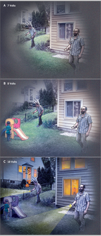
Memory experiences became more robust as the voltage of deep brain stimulation increased. For example, one patient’s recollection at 7-V stimulation evolved from a generic notion of “helping a guy find something on his property” to, at 8-V stimulation, remembering “helping the man’s son,” to at 10-V stimulation, also recalling “that this event occurred at night around Halloween.”
The occurrence of memory flashbacks did not correlate at the patient or group level with the trial outcomes of changes on cognitive testing, total brain volume, or hippocampal volume. There was no relationship at the group level between the development of memory flashbacks and the side of hippocampal stimulation.
These flashbacks are consistent with the iconic reports by Penfield and Perot and others of complex “experiential phenomena” with cortical electrical stimulation.2,3 In our trial, the regions that were stimulated included the fornix, anterior commissure, subcallosum (subcallosal region of the corpus callosum), and precommissural archicortex (inner and inferior portion of the subcallosal area). The fornix is part of a circuit that processes episodic memory (autobiographic events) and semantic memory (ideas and concepts). The subcallosal area is involved in memories with emotional and spatial properties. Brain maps that have been obtained with the use of diffusion tensor imaging in patients with Alzheimer’s disease have shown lower numbers of tracts in the fornix and greater numbers of subcallosal tracts than were observed in controls without Alzheimer’s disease.4 On the basis of localization data from tractographic studies (three-dimensional modeling of nerve tracts) involving our patients, we found that most memory flashbacks (87%) were associated with the dorsal brain contacts that probably stimulated both the fornix and the subcallosal area.
The memory flashbacks from brain stimulation in our trial may provide information regarding the neuroanatomical substrates and pathways of memory encoding and retrieval. Future studies of brain stimulation could further correlate stimulated recollections with results on tractography and functional imaging.
Supplementary Material
THIS WEEK’S LETTERS.
783 Fornix-Region Deep Brain Stimulation–Induced Memory Flashbacks in Alzheimer’s Disease
785 Early Neuromuscular Blockade in the Acute Respiratory Distress Syndrome
788 Ibrutinib and Venetoclax for First-Line Treatment of CLL
Acknowledgments
Supported by a grant (R01AG042165) from the National Institute on Aging and by the Federal Economic Development Agency for Southern Ontario, Medtronic, and Functional Neuromodulation.
Footnotes
Disclosure forms provided by the authors are available with the full text of this article at NEJM.org.
Contributor Information
Wissam Deeb, University of Florida, Gainesville, FL
Bryan Salvato, Florida State University, Tallahassee, FL
Leonardo Almeida, University of Florida, Gainesville, FL
Kelly D. Foote, University of Florida, Gainesville, FL
Robert Amaral, Douglas Mental Health University Research Institute, Montreal, QC, Canada
Jurgen Germann, Douglas Mental Health University Research Institute, Montreal, QC, Canada
Paul B. Rosenberg, Johns Hopkins University, Baltimore, MD
David F. Tang-Wai, University of Toronto, Toronto, ON, Canada
David A. Wolk, University of Pennsylvania, Philadelphia, PA
Anna D. Burke, Barrow Neurological Institute, Phoenix, AZ
Stephen Salloway, Alpert Medical School of Brown University, Providence, RI
Marwan N. Sabbagh, Cleveland Clinic Lou Ruvo Center for Brain Health, Las Vegas, NV
M. Mallar Chakravarty, Douglas Mental Health University Research Institute, Montreal, QC, Canada
Gwenn S. Smith, Johns Hopkins University School of Medicine, Baltimore, MD
Constantine G. Lyketsos, Johns Hopkins University, Baltimore, MD
Andres M. Lozano, University of Toronto, Toronto, ON, Canada
Michael S. Okun, University of Florida, Gainesville, FL
References
- 1.Lozano AM, Fosdick L, Chakravarty MM, et al. A phase II study of fornix deep brain stimulation in mild Alzheimer’s disease. J Alzheimers Dis 2016;54:777–87. [DOI] [PMC free article] [PubMed] [Google Scholar]
- 2.Penfield W, Perot P. The brain’s record of auditory and visual experience: a final summary and discussion. Brain 1963;86:595–696. [DOI] [PubMed] [Google Scholar]
- 3.Curot J, Busigny T, Valton L, et al. Memory scrutinized through electrical brain stimulation: a review of 80 years of experiential phenomena. Neurosci Biobehav Rev 2017;78:161–77. [DOI] [PubMed] [Google Scholar]
- 4.Kuchtova B, Wurst Z, Mrzilkova J, et al. Compensatory shift of subcallosal area and paraterminal gyrus white matter parameters on DTI in patients with Alzheimer disease. Curr Alzheimer Res 2018;15:590–9. [DOI] [PubMed] [Google Scholar]
Associated Data
This section collects any data citations, data availability statements, or supplementary materials included in this article.