Family studies have indicated that heritability explains 50-60% of the venous thromboembolism (VTE) events,1 and in recent years, several single nucleotide polymorphisms (SNP) have been found to influence the VTE risk.2 Glycoprotein VI (GP6) rs1613662, also known as T13254C, is an A/G single nucleotide variation in amino acid 219, which results in a serine to proline substitution affecting the glycoprotein VI (GPVI) receptor for collagen.3 Platelets carrying the minor allele (G-allele/Pro219) at GP6 rs1613662 express fewer GPVI receptors,4 which leads to attenuated platelet adhesion and activation.5 Previous observational studies in selected populations have consistently demonstrated that carriers of the A-allele at GP6 have a 15% higher risk of VTE than non-carriers, and inversely, that G-allele carriers have a 20% lower VTE risk.6,7
Cancer is associated with a highly increased risk of VTE,8 and the complex interactions between inherited and acquired risk factors on VTE risk (e.g. genetic alterations and platelet count) are more compound and often different in malignancy.9,10 The impact of GP6 rs1613662 on the risk of VTE has only been investigated in case-control studies, and the combined effect of GP6 rs1613662 and cancer on VTE risk has not been previously investigated. We therefore aimed (i) to investigate the association between GP6 rs1613662 and VTE risk in the general population and stratified by cancer status, and (ii) to explore the combined effects of GP6 rs1613662 and active cancer on the risk of VTE, using a large case-cohort recruited from the general population.
Cases with symptomatic, incident VTE (n=1,493) and a subcohort (n=13,072) were recruited from the fourth survey of the Tromsø Study and the second survey of the Nord-Trøndelag Health Study (HUNT), conducted in 1994-1995 and 1995-1997, respectively. Detailed descriptions of both studies have been published previously.11,12 The Regional Committee of Medical Health Research Ethics approved the studies, and all participants gave their informed written consent to participate. VTE events were identified by broad searches at the hospitals providing health care for the two regions and thoroughly validated by review of medical records, as previously described in detail.13,14 We excluded participants not registered as inhabitants of Tromsø or Nord-Trøndelag at study inclusion (n=3), subjects with a cancer diagnosis prior to inclusion (n=573) or with missing values for GP6 rs1613662 (n=7). Eventually, the case-cohort consisted of 13,982 participants: 1,395 VTE cases and 12,587 subjects in the subcohort. GP6 rs1613662 was genotyped with the Sequenom platform as previously described else-where.10 The HUNT study performed genotyping using the Illumina HumanCore Exome array. Information regarding date of cancer diagnosis, primary site of malignancy (ICD-7-codes 140-205) and cancer stage during the entire follow-up, was obtained by linkage to the Cancer Registry of Norway.
Statistical analyses were performed using STATA version 15.0 (Stata Corporation LP, College Station, TX, USA). Cancer was entered as a time-varying covariate, and the data was split in relation to the date of cancer diagnosis. A VTE were considered cancer-related if it occurred within six months prior to and up to 2 years after the date of cancer diagnosis. Subjects with cancer who were alive and VTE-free at the end of the active cancer period were censored from this date onwards. Age-, sex- and body mass index (BMI)-adjusted hazard ratios (HR) with 95% confidence intervals (CI) for VTE were calculated according to variants at GP6 rs1613662 subjects with and without cancer. Subjects homozygous for the major allele at GP6 (i.e. AA) were used as the reference group in the categorized analyses. The proportional hazard assumption was confirmed by the use of Schoeneld’s global test. The Fine–Gray model15 was applied in a sensitivity analysis to account for mortality as a competing event, and subdistribution hazard ratios (SHR) were estimated. Mortality rates and HR for death by categories of GP6 alleles were estimated for disease free subjects, subjects with cancer only, subjects with VTE only, and subjects with cancer-related VTE. The distribution of cancer sites and cancer stages were displayed across GP6 variants (i.e. AA, AG and GG).
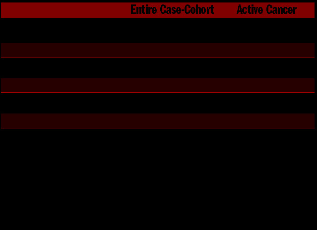
Baseline characteristics in the entire cohort and in subjects with active cancer are presented in Table 1. The allele frequency of GP6 was 0.17 in both the entire case-cohort and in the active cancer group, which is similar to the frequency reported in reference populations.2,3
Table 1.
Baseline characteristics of study participants.

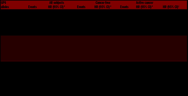
Of the 1395 VTE events, 819 (58.7%) were deep vein thrombosis (DVT) and 576 (41.3 %) were pulmonary embolism (PE). The HR for VTE, DVT and PE adjusted for age, sex and BMI by categories of GP6 alleles and cancer-status are presented in Table 2. The HR for incident VTE was 21% (HR 0.79, 95% CI: 0.70-0.89) lower in subjects with ≥1 G-alleles at GP6, when compared to subjects homozygous for the A-allele. The VTE risk was essentially similar for heterozygous (AG) and homozygous (GG) subjects compared to those homozygous for the major allele (AA). The number of G-alleles appeared to have a differential effect on the risk of DVT and PE. However, these findings should be interpreted with caution, as the number homozygous carriers in each category was low. The mortality rates for the entire case-cohort did not differ across the GP6 variants, and coherently, the risk estimates from the competing risk by death model were similar to those obtained with the traditional Cox regression model (data not shown).
Table 2.
Hazard ratios with 95% confidence intervals for venous thromboembolism, deep vein thrombosis and pulmonary embolism by categories of GP6 rs1613662 alleles and cancer.
In cancer-free subjects, the risk of incident VTE decreased with the number of minor alleles, and subjects homozygous for the GP6 allele (GG) had 34% decreased risk of incident VTE (HR 0.66, 95% CI: 0.43–1.01) compared to subjects homozygous for the major allele (A allele) at GP6.
There were 1,536 patients with active cancer of which 233 (15.2%) experienced a VTE. Among cancer patients, subjects heterozygous for the minor GP6 allele (AG) exhibited decreased VTE risk (HR 0.82, 95% CI: 0.60–1.11), which was of similar magnitude to that found in subjects without cancer. In contrast, cancer patients homozygous for the G-allele had a higher risk of VTE, DVT and PE, when compared to cancer patients homozygous for the A-allele (AA). In cancer patients, the risk of PE was particularly high in homozygous carriers of the G-allele (HR 1.96, 95% CI: 0.78–4.94).
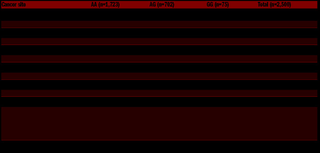
The G-alleles were found to be associated with more prothrombotic cancers (i.e. lung, colorectal, hematological cancer and lymphomas) and presented with more severe stages of cancer (i.e. distant metastasis), when compared to subjects heterozygous or homozygous for the A-allele (Table 3). To test whether the association between homozygosity for the G-allele and increased risk of cancer-related VTE could be explained by variant-dependent differences in cancer types, cancer stages, and mortality rates, additional adjustments of age, sex, BMI, cancer types and stages as well as the Fine-Gray model were applied (Table 2). When cancer types and stages were included in the adjusted model, the risk estimates were moderately attenuated. Moreover, the risk estimates for cancer-related VTE in homozygous carriers of the G-allele remained essentially unchanged when competing risk by death was taken into account (data not shown).
Table 3.
The distribution of cancer types and stages across genotypes at GP6 rs1613662 alleles (AA, AG, GG).
Our findings support the notion that platelet function is involved in the pathogenesis of VTE. The mechanism(s) underlying the combined, but apparent opposite effect of homozygosity at the G-allele of GP6 rs1613662 and cancer on VTE risk remains elusive. The inverse effect may be explained by a differential impact of the G-allele at GP6 rs1613662 on the cancer type, cancer stages and mortality rates. However, variants at the GP6 rs1613662 may also have differential impact on platelet reactivity under various conditions.
In contrast to cancer-free subjects where the VTE risk decreased with the number of G-alleles, cancer patients homozygous for the G-allele at GP6 rs1613662 displayed an increased VTE risk. Our findings imply that measures of platelet function may have differential impact on the VTE risk in cancer-free subjects and cancer patients. Elevated platelet counts are frequently observed in cancer patients, are associated with decreased survival,16 and have been shown to predict future cancer-related VTE events.9 In contrast, no association between platelet count and the VTE risk has been observed in general populations.9 In addition, mean platelet volume (MPV), a marker of platelet reactivity, has shown differential association with VTE risk in subjects without and with cancer. Whereas high MPV is associated with an increased risk of VTE in the general population,17 it is associated with a lower VTE risk and improved survival in cancer patients.18
The main strengths of our study are the prospective design and the validation of VTE events and cancer diagnoses. The high attendance rate and wide age distribution in the parent cohorts reduces the chance of selection bias in the subcohort. Some limitations merit consideration. Unfortunately, cancer treatment modality was not available and restricted the possibility to evaluate the relationship between treatment related factors and genetics. Moreover, our study had limited statistical power, particularly in some subgroups. This resulted in wide CI, and our risk estimates should therefore be interpreted with caution.
In conclusion, the GP6 rs1613662 G-allele displayed a protective effect on VTE risk in cancer-free subjects, while an increased risk of VTE was observed in cancer patients homozygous for the G-allele. Our findings support a role of platelet reactivity in the pathogenesis of VTE, which may differ according to cancer status.
Footnotes
Information on authorship, contributions, and financial & other disclosures was provided by the authors and is available with the online version of this article at www.haematologica.org.
References
- 1.Larsen TB, Sorensen HT, Skytthe A, Johnsen SP, Vaupel JW, Christensen K. Major genetic susceptibility for venous thromboembolism in men: a study of Danish twins. Epidemiology. 2003; 14(3):328-332. [PubMed] [Google Scholar]
- 2.Morange PE, Suchon P, Tregouet DA. Genetics of venous thrombosis: update in 2015. Thromb Haemost. 2015;114(5):910-919. [DOI] [PubMed] [Google Scholar]
- 3.Watkins NA, O'Connor MN, Rankin A, et al. Definition of novel GP6 polymorphisms and major difference in haplotype frequencies between populations by a combination of in-depth exon resequencing and genotyping with tag single nucleotide polymorphisms. J Thromb Haemost. 2006;4(6):1197-1205. [DOI] [PubMed] [Google Scholar]
- 4.Yee DL, Bray PF. Clinical and functional consequences of platelet membrane glycoprotein polymorphisms. Semin Thromb Hemost. 2004;30(5):591-600. [DOI] [PubMed] [Google Scholar]
- 5.Joutsi-Korhonen L, Smethurst PA, Rankin A, et al. The low-frequency allele of the platelet collagen signaling receptor glycoprotein VI is associated with reduced functional responses and expression. Blood. 2003;101(11):4372-4379. [DOI] [PubMed] [Google Scholar]
- 6.Tregouet DA, Heath S, Saut N, et al. Common susceptibility alleles are unlikely to contribute as strongly as the FV and ABO loci to VTE risk: results from a GWAS approach. Blood. 2009;113(21):5298-5303. [DOI] [PubMed] [Google Scholar]
- 7.Austin H, De Staercke C, Lally C, Bezemer ID, Rosendaal FR, Hooper WC. New gene variants associated with venous thrombosis: a replication study in White and Black Americans. J Thromb Haemost. 2011;9(3):489-495. [DOI] [PMC free article] [PubMed] [Google Scholar]
- 8.Blom JW, Doggen CJ, Osanto S, Rosendaal FR. Malignancies, prothrombotic mutations, and the risk of venous thrombosis. JAMA. 2005;293(6):715-722. [DOI] [PubMed] [Google Scholar]
- 9.Jensvoll H, Blix K, Braekkan SK, Hansen JB. Platelet count measured prior to cancer development is a risk factor for future symptomatic venous thromboembolism: the Tromso Study. PLoS One. 2014; 9(3):e92011. [DOI] [PMC free article] [PubMed] [Google Scholar]
- 10.Gran OV, Smith EN, Braekkan SK, et al. Joint effects of cancer and variants in the Factor 5 gene on the risk of venous thromboembolism. Haematologica. 2016;101(9):1046-1053. [DOI] [PMC free article] [PubMed] [Google Scholar]
- 11.Jacobsen BK, Eggen AE, Mathiesen EB, Wilsgaard T, Njolstad I. Cohort profile: the Tromso Study. Int J Epidemiol. 2012;41(4):961-967. [DOI] [PMC free article] [PubMed] [Google Scholar]
- 12.Holmen J, Midthjell K, Krüger Ø, et al. The Nord-Trøndelag Health Study 1995–97 (HUNT 2): Objectives, contents, methods and participation. Norsk Epidemiologi. 2003;13(1):19-32. [Google Scholar]
- 13.Braekkan SK, Mathiesen EB, Njolstad I, Wilsgaard T, Stormer J, Hansen JB. Family history of myocardial infarction is an independent risk factor for venous thromboembolism: the Tromso study. J Thromb Haemost. 2008;6(11):1851-1857. [DOI] [PubMed] [Google Scholar]
- 14.Naess IA, Christiansen SC, Romundstad P, Cannegieter SC, Rosendaal FR, Hammerstrom J. Incidence and mortality of venous thrombosis: a population-based study. J Thromb Haemost. 2007; 5(4):692-699. [DOI] [PubMed] [Google Scholar]
- 15.Fine JP, Gray RJ. A Proportional hazards model for the subdistribution of a competing risk. J Am Stat Assoc. 1999;94(446):496-509. [Google Scholar]
- 16.Ploquin A, Olmos D, Lacombe D, et al. Prediction of early death among patients enrolled in phase I trials: development and validation of a new model based on platelet count and albumin. Br J Cancer. 2012;107(7):1025-1030. [DOI] [PMC free article] [PubMed] [Google Scholar]
- 17.Braekkan SK, Mathiesen EB, Njolstad I, Wilsgaard T, Stormer J, Hansen JB. Mean platelet volume is a risk factor for venous thromboembolism: the Tromso Study, Tromso, Norway. J Thromb Haemost. 2010;8(1):157-162. [DOI] [PubMed] [Google Scholar]
- 18.Ferroni P, Guadagni F, Riondino S, et al. Evaluation of mean platelet volume as a predictive marker for cancer-associated venous thromboembolism during chemotherapy. Haematologica. 2014; 99(10):1638-1644. [DOI] [PMC free article] [PubMed] [Google Scholar]


