PLOS ONE logoLink to PLOS ONE
. 2020 Jul 1;15(7):e0235143. doi: 10.1371/journal.pone.0235143

Mass spectrometry-based absolute quantification of amyloid proteins in pathology tissue specimens: Merits and limitations

Makiko Ogawa 1,2,3,#, Yukako Shintani-Domoto 1,#, Yoshiki Nagashima 4,5, Koji L Ode 4, Aya Sato 6, Yoshihiro Shimizu 6, Kenichi Ohashi 7, Michael H A Roehrl 2,3, Tetsuo Ushiku 1, Hiroki R Ueda 4,8, Masashi Fukayama 1,9,*
Editor: Harald Mischak10
PMCID: PMC7329117  PMID: 32609750

Abstract

To clarify the significance of quantitative analyses of amyloid proteins in clinical practice and in research relating to systemic amyloidoses, we applied mass spectrometry–based quantification by isotope-labeled cell-free products (MS-QBIC) to formalin-fixed, paraffin-embedded (FFPE) tissues. The technique was applied to amyloid tissues collected by laser microdissection of Congo red-stained lesions of FFPE specimens. Twelve of 13 amyloid precursor proteins were successfully quantified, including serum amyloid A (SAA), transthyretin (TTR), immunoglobulin kappa light chain (IGK), immunoglobulin lambda light chain (IGL), beta-2-microglobulin (B2M), apolipoprotein (Apo) A1, Apo A4, Apo E, lysozyme, Apo A2, gelsolin, and fibrinogen alpha chain; leukocyte cell–derived chemotaxin-2 was not detected. The quantification of SAA, TTR, IGK, IGL, and B2M confirmed the responsible proteins, even when the immunohistochemical results were not decisive. Considerable amounts of Apo A1, Apo A4, and Apo E were deposited in parallel amounts with the responsible proteins. Quantification of amyloid protein by MS-QBIC is feasible and useful for the classification of and research on systemic amyloidoses.

Introduction

Amyloidosis comprises a large group of diseases in which misfolding of extracellular proteins plays a fundamental role. Dynamic processes, occurring in parallel with or as an alternative to physiologic folding, generate insoluble and toxic protein aggregates that are deposited in tissues in β-sheet fibrils. More than 30 different proteins are known to cause amyloidosis [1]. The various forms of amyloidosis require different treatment approaches, including high-dose melphalan with stem cell transplantation in AL amyloidosis, and liver transplantation or use of transthyretin (TTR)-tetramer stabilizer in hereditary TTR amyloidosis [2, 3]. Treatments for systemic amyloidoses have advanced over the years, and precise typing is key for adequate treatment.

The diagnosis of amyloidosis requires identification of amyloid in tissue specimens. Congo red staining in conjunction with polarized light microscopy is the primary procedure for histological confirmation of amyloid deposits, which is followed by subtype classification. The approach for the subtyping of amyloidosis has evolved over the last 30 years, moving from histochemistry to more specific immunohistochemistry of the major precursor proteins, such as serum amyloid A (SAA), transthyretin (TTR), immunoglobulin kappa light chain (IGK), immunoglobulin lambda light chain (IGL), and beta-2-microglobulin (B2M). However, immunohistochemistry has an inherent weakness that often results in nonspecific staining that can lead to false positive results. The assessment of the immunostaining intensity of amyloid proteins is subjective or at most semiquantitative. More recently, liquid chromatography (LC)–tandem mass spectrometry (MS/MS) has been introduced for direct identification of the amyloid-forming protein in biopsy specimens in several special institutions throughout the world [4]. The LC-MS/MS method frequently detects multiple amyloid proteins (such as apolipoproteins), even in the presence of a single major dominant amyloid protein. This occurrence sometimes makes it difficult to determine the amyloid subtype [57]. These additional proteins may act as cofactors in amyloid deposition, but detailed quantitative analysis of these proteins has not yet been undertaken.

In the present study, we developed MS-based absolute quantification of amyloid proteins for the subtyping of systemic amyloidoses in formalin-fixed, paraffin-embedded (FFPE) tissue sections. We adopted the technique, MS-based quantification by isotope-labeled cell-free products (MS-QBIC), which was reported by Narumi et al [8]. The method was originally developed to quantify subtle variations in mouse circadian clock proteins. In the MS-QBIC method, isotope-labeled peptides are prepared for the internal control of LC-MS analysis by using a cell-free synthesis system. Therefore, we expected that the application of MS-QBIC to the diagnosis of amyloidosis would be feasible if we could design appropriate peptide sequences for reference quantification. Furthermore, quantification of the deposited amyloid proteins may be useful to understand possible interactive processes in the maturation of amyloid deposits in the tissue.

The present study is an attempt to answer the following three questions: (1) whether the MS-QBIC method worked for FFPE specimens; (2) whether the method was feasible (by comparing the quantitative data obtained by the MS-QBIC process with immunohistochemical data, which are used routinely in the subclassification of amyloidosis); and (3) how the deposited amounts of major amyloid proteins were compared with other amyloid proteins. Thus, we aimed to clarify the merits and limitations of a quantitative analysis of amyloid proteins in both clinical practice and in research on systemic amyloidosis.

Materials and methods

Patients and tissue samples

Specimens from 30 patients with systemic amyloidosis were retrieved from the files of autopsy records of the Department of Pathology of the University of Tokyo from January 1999 through December 2016. The diagnosis of systemic amyloidosis was based on the presence of amyloid in FFPE tissue sections from more than two organs by Congo red staining. Patients’ medical records were reviewed for the presence of a monoclonal gammopathy using immunofixation of serum and urine as well as a serum-free light-chain assay.

Amyloid deposition was evaluated using Congo red-stained tissue sections. The staining was confirmed in representative sections under a fluorescein isothiocyanate (FITC) filter on a BZ-X710 all-in-one fluorescence microscope (Keyence Ltd., Osaka, Japan). We selected two specimens from each patient for the study: one from the cardiac wall of the left ventricle (n = 30) and one each from another organ (n = 28). Only myocardial tissue was available for examination in two cases, since the amounts of amyloid were too small for the subsequent MS analyses. The amount of amyloid deposition was scored in five grades from minimal (grade 1) to severe (grade 5) in myocardial tissue sections (S1 Fig).

For the application of the MS-QBIC method to the absolute quantification of amyloid protein, target peptides need to be selected as an isotope-labeled internal control of for the MS analysis. The target peptides were determined by the rules as described below with reference to previous LC-MS analyses, which examined the specimens derived from patients with systemic amyloidosis and related multiple myeloma, and from those with local amyloidosis including diabetes mellitus and Alzheimer’s disease (S1 Table). The materials were chosen with reference to the files of autopsy or biopsy records of the Department of Pathology of the University of Tokyo from January 1994 through December 2015.

The study was approved by Research Ethics Committee, Graduate School of Medicine, The University of Tokyo (No. 10461-4-(5)). Informed consent for the use of patient specimens for the research was obtained from the responsible parties.

Immunohistochemistry

For the identification of amyloid protein, antibodies against four amyloid precursor proteins (SAA, TTR, IGK, B2M) were used: anti-SAA antibody (mouse monoclonal, mc1; Dako Denmark A/S, Glostrup, Denmark, dilution 1:1000), anti-TTR antibody (rabbit monoclonal, EPR3219; Abcam plc, Cambridge, UK, 1:200), anti-IGK antibody (rabbit monoclonal, H16-E; Abnova Corp., Taipei, Taiwan, 1:1000), and anti-B2M antibody (rabbit polyclonal; Dako, 1:1000) [916]. For IGL, anti-lambda (118–134) antiserum was provided by Prof. Yoshinobu Hoshii (Department of Laboratory Science, Yamaguchi University Graduate School of Medicine, Ube, Japan, 1:800) [13, 17]. Immunostaining was performed using the BenchMark ULTRA immunohistochemistry/in situ hybridization system (Roche Diagnostics, Basel, Switzerland). For the immunostaining of IGK and IGL, pretreatment was performed in formic acid for 5 min. In this study, the results of immunostaining were evaluated on a scale of 0 to 3+ by the immunostaining intensity and stained area as follows: 0 = negative, 1+ = weakly and focally positive, 2+ = intermediate in both intensity and area, and 3+ = strongly and diffusely positive.

Construction of MS-QBIC-based methods for the quantification of amyloid proteins

Absolute quantification of amyloid proteins was performed according to the MS-QBIC workflow [8] (S2 Fig). MS-QBIC based quantification consists of following processes: selection of target peptide candidates, synthesis of the MS-QBIC peptides, quantification of the MS-QBIC peptides, confirmation of signal linearity and determination of the Lower Limit of quantification (LLOQ).

Selection of target peptide candidates of amyloid proteins

Thirteen amyloid precursor proteins were selected as target proteins on the basis of the guidelines on the management of amyloidosis, 2010 [18] for evaluation: apolipoprotein A-1 (Apo A1), apolipoprotein A-2 (Apo A2), apolipoprotein A-4 (Apo A4), apolipoprotein E (Apo E), gelsolin, fibrinogen alpha chain (FGA), lysozyme, and leukocyte cell–derived chemotaxin-2 (LECT2), in addition to SAA, TTR, IGK, IGL, and B2M. To select the target peptides for MS-QBIC based quantification, several tissue samples (S1 Table) were analyzed by a data-dependent MS/MS analysis.

All the samples were digested to recover the peptides according to the phase-transfer surfactant (PTS) method [8, 19] with several modifications. In brief, sample was dissolved in PTS buffer [8, 19] and heated at 95°C for 3 h to reverse the formalin-crosslinks. The sample was reduced with 100 mM dithiothreitol (FUJIFILM Wako Pure Chemical Corp., Tokyo, Japan) at room temperature for 30 min, and then alkylated with 1 M iodoacetamide (Sigma-Aldrich Co., St. Louis, MO, USA) at room temperature for 30 min. Next, the sample was digested by adding 1 μg of lysyl endopeptidase (Lys-C) (FUJIFILM Wako Pure Chemical Corp.). After incubating the samples at 37°C for 3 h, 1 μg trypsin (Roche) was added and the mixture was further incubated at 37°C overnight. The detergents were removed by ethyl acetate/trifluoroacetic acid (TFA) solution according to the PTS method. The digested samples were then desalted using a self-prepared C18 stage tip [20]. The peptides were solubilized in 2% acetonitrile and 0.1% TFA and loaded to the LC-MS system to be separated by a gradient using mobile phases A (0.1% formic acid/H2O) and B (0.1% formic acid and 100% acetonitrile) at a flow rate of 300 nL/min (4% to 36% B in 20 min, 36% to 95% B in 1 min, 95% B for 5 min, 95% to 4% B in 1 min and 4% B for 18 min) with a home-made capillary column (length of 200 mm and inner diameter of 100 μm) packed with 3μm C18 resin (L-column2, Chemicals Evaluation and Research Institute, Japan). The eluted peptides were electrosprayed (1.8–2.3 kV) and introduced into the Q-Exactive MS instrument (Thermo Fisher Scientific, Tokyo, Japan) in positive ion mode with data-dependent MS/MS. The obtained raw data was subjected to database search (UniProt, reviewed human database as of May 7th 2014) with the Sequest HT algorithm implemented in Proteome Discoverer 1.4 (Thermo Fisher Scientific). The parameters for database searches were as follows. Peptide cleavage was set to trypsin. Missed cleavage was allowed up to 2 and minimum and maximum peptide length was 6–144 amino acids. The mass tolerances were set to 10 ppm for precursor ions and 0.02 Da for fragment ions. For modification conditions, carbamidomethylation at cysteine was set as fixed modification and oxidation at methionine was set as variable modification. A significance threshold of P<0.05 was applied.

The identified peptides derived from the amyloid precursor proteins were further selected according to the following rules: (i) the amino acid sequences were unique to the target protein, (ii) each peptide contained a lysine or arginine residue only at its C-terminus, (iii) peptides containing cysteine were permitted because we used a protocol to convert cysteine almost completely to its alkylated form by reduction and alkylation [8], (iv) peptides with natural variants reported in several publications (searched via UniProtKB, https://www.uniprot.org/) were excluded (references are listed in the S1 Appendix) except for TTR, SAA and immunoglobulin light chains because excluding all the reported variants for these peptides limits the number of available peptide sequences, (v) target peptides of immunoglobulin light chains (IGK and IGL) were selected via the amino acid residues in constant lesions, which were the antibody-binding sites, as detected via immunohistochemistry [13, 14, 17, 2123]. For Apo AII and LECT2, the data-dependent MS/MS analysis only identified fewer than four kinds of peptides; so because we aimed to select at least four peptides per one amyloid precursor protein for the MS-QBIC method, peptide sequences that matched the above-mentioned criteria (i)-(v) were selected manually. The maximum length of manually selected peptides was set as25 amino acids. The selected target peptides are summarized in S2 Table.

Synthesis of MS-QBIC peptides

MS-QBIC peptides were prepared using the PURE system, which is a reconstituted cell-free protein synthesis system as described by Shimizu et al. [24]. Using the MS-QBIC vector [8] as a template, PCR was performed for the amplification of the templates of MS-QBIC peptide synthesis with a T7 promoter primer (5'-GGGCCTAATACGACTCACTATAG-3') as a forward primer and appropriate reverse primers (S2 Table). PCR mixtures were directly added to yield 25 μL of PURE system reaction mixtures containing 13C6 15N4 L-arginine and 13C6 L-lysine (Thermo Fisher Scientific) as substitutes for non-labeled L-arginine and L-lysine, respectively. The mixtures were incubated at 37°C for 30 min, and then the synthesized peptides were purified using anti-FLAG M2 magnetic beads (Sigma-Aldrich Co., St. Louis, MO, USA), according to the manufacturer’s instructions. The recovered peptides were then stored at −80°C until used.

Quantification of MS-QBIC peptides

The concentration of the MS-QBIC peptide (stable isotope-labeled peptide) was first determined by comparison with the ion peak intensity of commercially available bovine serum albumin (BSA) as follows. First, weighed BSA powder (≥98.0% purity, Sigma-Aldrich Co., St. Louis, MO, USA) was dissolved in PTS buffer. The MS-QBIC product was then mixed with BSA and the mixture was subjected to trypsin digestion according to the PTS method as described above except 100 mM tris (2-carboxyethyl) phosphine Hydrochloride (Nacalai Tesque Inc., Kyoto, Japan) was used instead of 100 mM dithiothreitol. The pre-heating procedure was not applied to digest the non-fixed BSA/MS-QBIC samples. To reduce the non-specific binding of peptides to sample tubes, all the processes were carried out in the presence of 1 pmol/μL of trypsin-digested α-enolase (Sigma-Aldrich Co., St. Louis, MO, USA) prepared according to the PTS method. The signal linearity of the quantification tag (a peptide sequence derived from BSA: S2 Fig) was confirmed by analyzing different amounts of BSA (S3A Fig), although the stable-isotope based quantification does not heavily depend on the linearity of the signal with an increase in the peptide amount [25].

For the quantification of the tag sequence and all MS-QBIC-based quantification, it was carried out by selected reaction monitoring (SRM) analysis using a TSQ Quantiva triple-stage quadrupole mass spectrometer (Thermo Fisher Scientific). The following parameters were selected: positive mode, scan width of 0.002 m/z, Q1 and Q3 resolutions of 0.7 full width of half maximum (FWHM), cycle time of 3 s, and gas pressure of 1.8 Torr. The spectrometer was equipped with an UltiMate 3000 RSLCnano nano-high performance liquid chromatography (HPLC) system (Thermo Fisher Scientific), and a PepMap HPLC trap column (C18, 5 μm, 100 Å; Thermo Fisher Scientific) for loading samples. Analytical samples were solubilized in 2% acetonitrile and 0.1% TFA, and separated by reversed-phase chromatography using a PepMap rapid separation liquid chromatography (RSLC) EASY-Spray column (C18, 3 μm, 100 Å, 75 μm × 15 cm; Thermo Fisher Scientific) using mobile phases A (0.1% formic acid/H2O) and B (0.1% formic acid and 100% acetonitrile) at a flow rate of 300 nL/min (4% B for 5 min, 4% to 36% B in 20 min, 36% to 95% B in 1 min, 95% B for 5 min, 95% to 4% B in 1 min and 4% B for 13 min). The eluted material was directly electro-sprayed into the MS. SRM transitions of the target peptides were optimized using Pinpoint software, version 1.3 (Thermo Fisher Scientific) (S2 Table). The Quan Browser of the Xcalibur data system, version 2.2 (Thermo Fisher Scientific) was used for data processing. The calibration curve of the BSA-derived quantification tag (S3A Fig) shows linear increase of signal intensity according to the increase of injected BSA; thus the amount of each MS-QBIC peptide was estimated to be proportional to the quantified heavy (MS-QBIC product) to light (weighed BSA) ratio. To determine the stock concentration of the MS-QBIC peptide, 0.2 μL of stock solution containing 100 fmol of digested BSA was analyzed. All the quantifications were within the range of 0.1–10 heavy (quantification tag derived from MS-QBIC peptide) to light (the tag derived from digested BSA) ratios (S3B Fig).

Confirmation of signal linearity for each MS-QBIC product

The signal linearity for each MS-QBIC product was then evaluated by analyzing different amounts of MS-QBIC peptides (S2 Appendix). For the linear regression evaluation, we excluded MS-QBIC peptides that failed to have the linear regression line that could be fitted to ≥4 consecutive amounts with R2>0.95 and a %difference for each amount <30. The MS-QBIC peptides were further investigated to determine if the peptides could be detected robustly in the presence of peptides recovered from tissue samples. Several test tissue samples were prepared and mixed with MS-QBIC peptides according to the method described in the next section. A few MS-QBIC peptides that could not be detected robustly in the presence of tissue samples were excluded for the absolute quantification steps.

Lower limit of quantification

Next, based on the liner regression analysis, we defined the Lower Limit of Quantification (LLOQ) by determining the minimum amount of each MS-QBIC peptide for which the intensity was within a 30% difference of the linear regression line obtained in the each calibration plot; if all analyzed amounts were within the 30% difference, the minimum amount used to produce the liner regression line was set as the LLOQ. The values of the LLOQ are shown in S3 Table.

Laser Capture Microdissection (LMD) and enzymatic digestion

A 10 μm FFPE section of each tissue sample was stained with Congo red. Positively stained areas were dissected using the PALM MicroBeam LMD system (Carl Zeiss Meditec AG, Tokyo, Japan). The samples were collected using PTS buffer as reported previously [8, 19]. An area of 0.07 to 0.27 mm2 was dissected from each sample, with one exception (0.07 mm2). Three samples were collected from each section, and a total of 174 amyloid samples (3 samples × 2 tissues × 28 patients and 3 samples × 1 tissue × 2 patients) were analyzed individually.

Enzymatic digestion of the 174 amyloid samples and MS-QBIC peptides was performed basically according to the PTS protocol as previously described above. The samples collected in the PTS buffer were heated at 95°C for 3 h to reverse formalin-crosslinks, prior to the reducing step with 100 mM dithiothreitol. A 0.0007–0.0027 mm3 volume of collected amyloid sample may contain 0.7–2.7 μg of protein, assuming that the amount of protein is approximately 10% of the wet weight of the tissue sample. Thus, the addition of 1 μg of Lys-C/trypsin into each sample should be sufficient to digest all the amyloid proteins. The digested samples were desalted using a self-prepared C18 stage tip and dissolved in water containing 2% acetonitrile and 0.1% TFA. The MS-QBIC peptide mixture was added to the sample at this stage and then the sample was subjected to the MS analysis.

Mass spectrometry for the MS-QBIC based absolute quantification

A TSQ Quantiva triple-stage quadrupole mass spectrometer (Thermo Fisher Scientific) was used for SRM analysis as described above. The target peptide derived from the amyloid sample was quantified by comparing the intensity of the nonlabeled target peptide in the sample with that of the MS-QBIC peptide of known concentration, which had been added to the sample prior to the measurement.

At least two or more target peptides were quantified per amyloid precursor protein. The quantitative value of each amyloid precursor protein was presented as the quantified value (pmol) of the protein divided by the volume (mm3) that was resected from each sample via LMD. We used the average of the quantitative value of the amyloid protein for three samples of the same specimen for further analyses. S3 Table summarizes the raw quantification values. Approximately 80% of the quantified amount was calculated within the range of 0.1–10 heavy (MS-QBIC peptide) to light (endogenous amyloid peptide) ratios, indicating that in most cases, the concentrations of the spiked peptide and the quantification targets were well balanced. The remaining ~20% of quantification values were calculated within the range of 0.01–100 heavy to light ratios.

The chromatogram of precursor ion and the spectrum of fragments of ions for a representative peptide for all the quantified amyloid proteins (excluding LECT2 that we could not detect in this study) are presented in S3 Appendix. The chromatograms of the endogenous and isotope-labeled peptides indicated that the two co-eluted peaks (endogenous and MS-QBIC peptides) were clearly detected and almost no-overlapping with other peaks from the background occurred. This peak shape provided information regarding the purity of the MS-QBIC peptides, indicating that possible contaminants or unexpected sub-products of the MS-QBIC peptides made a negligible contribution to the quantification of selective ion monitoring using the triple quadrupole mass spectrometer. The raw data from the SRM analysis were deposited to the Peptide Atlas SRM Experiment Library (PASSEL) (http://www.peptideatlas.org/PASS/PASS01558; see S3 Table for the relationship between raw file names and quantified results).

Amyloid precursor protein in nonamyloid samples

To evaluate the quantity of amyloid precursor protein in the background tissue (nonamyloid sample), their relative amounts to amyloid deposition were determined by comparing nonamyloid tissue samples with those of amyloid deposition. Nonamyloid and amyloid samples were differently labeled with light CH2O and heavy CD2O isotopes, respectively, through dimethyl labeling with formaldehyde [26].

The stromal components of 11 specimens devoid of amyloid (derived from seven patients with conditions unrelated to amyloidosis) were micro-dissected using LMD. Each microdissection contained an area of 2.0–2.8 mm2 in specimens without amyloidosis, and three samples from each specimen were analyzed. The samples of amyloid deposition were similarly obtained from 10 amyloidosis specimens (three samples per specimen). A total of 33 nonamyloid samples and 30 amyloid samples were analyzed. Enzymatic digestion of amyloid samples and nonamyloid samples was performed by the PTS method as described above. The samples collected in the PTS buffer were heated at 95 °C for 3 h to reverse formalin-crosslinks, prior to the reducing step with 100 mM dithiothreitol. The trypsin-digested samples were desalted by a C18 stage tip as described above, and before the elution from the tip, dimethyl-labeling was applied to peptides trapped on the tip [27]. CD2O and NaBH3CN (heavy label) were added to the amyloid samples, while CH2O and NaBH3CN (light label) were added to the nonamyloid samples. After elution from the C18 stage tips, all 30 amyloid samples were mixed as a reference. The mixture of the amyloid samples was dispensed to the 33 nonamyloid samples.

The samples were analyzed by the Q-Exactive MS instrument as described in the “Selection of target peptide candidates of amyloid proteins” section, except for the gradient conditions of mobile phases A and B (4%–36% B in 55 min, 36%–95% B in 1min, 95%B for 5min, 95%–4% B in 1 min and 4% B for 8 min). The Sequest HT/Proteome Discoverer 1.4 parameters for the database searches were same as described above, except that dimethylation (heavy- and light-labeled form) at the peptide N-terminus and at lysine was set as a fixed modification. The heavy to light ratios of identified labeled peptides were quantified by Proteome Discoverer 1.4 (Thermo Fisher Scientific).

Statistical analysis

Statistical analysis was performed using JMP software, version 5.0.1J (SAS Institute, Inc., Cary, NC, USA). To assess correlations between the four main causative amyloid proteins (SAA, TTR, IGK, and IGL) and Apo A1, Apo A4, Apo E, and lysozyme, we calculated the Spearman’s rank correlation coefficient.

Results

Immunohistochemical subclassification of systemic amyloidosis

Clinical information and involved organs used in the study are presented in Table 1. These cases are ordered considering the results of immunohistochemistry (Figs 1 and 2). In 24 of the 30 patients, amyloidosis was subclassified by clinical and pathological examination at autopsy. Seven patients had AA amyloidosis. Of seven patients with ATTR/TTR amyloidosis, two patients had hereditary disease, and five patients had acquired disease. Six patients had associated multiple myeloma, and three patients were diagnosed as primary or systemic amyloidosis. One patient had dialysis-associated amyloidosis.

Table 1. Demographic features of patients with systemic amyloidosis.

Case No. Age (y) M/F Amyloid subtype Clinical diagnosis at autopsy Main organ deposit used Deposition grade in cardiac tissue
1 59 F AA Rheumatoid arthritis Adrenal gland 2
2 78 F AA Granulomatosis with polyangiitis Stomach 1
3 71 F AA Systemic lupus erythematosus, Sjögren syndrome Submandibular gland 5
4 48 M AA Dilated cardiomyopathy Thyroid 3
5 64 M AA Rheumatoid arthritis Kidney 5
6 49 F AA Rheumatoid arthritis Thyroid 5
7 75 F AA Diffuse large B-cell lymphoma, Sjögren syndrome Tongue 1
8 77 F ATTR S/O: Senile systemic amyloidosis Esophagus 5
9 65 M ATTR Familial amyloidosis Bladder 5
10 75 M ATTR Familial amyloidosis Thyroid 3
11 76 M ATTR Alkaptonuria Tongue 3
12 85 M ATTR Hepatocellular carcinoma, S/O: Senile systemic amyloidosis Not available 2
13 85 M ATTR S/O: Senile systemic amyloidosis Not available 2
14 84 F ATTR S/O: Senile systemic amyloidosis Stomach 3
15 77 M ND Infective cerebral aneurysm, Systemic amyloidosis Esophagus 3
16 67 M ND Hypertrophic cardiomyopathy, Systemic amyloidosis Thyroid 2
17 68 M ND Multiple myeloma Rectum 2
18 54 M ALκ Multiple myeloma Cecum 5
19 72 F ALκ Multiple myeloma Diaphragm 1
20 67 F ALκ Multiple myeloma Stomach 3
21 63 M ALκ Multiple myeloma Spinal dura 1
22 76 M ND Lymphoplasmacytic lymphoma, Diffuse large B-cell lymphoma Lymph node 1
23 82 M ND Systemic amyloidosis Thyroid 3
24 74 F ALλ Multiple myeloma Tongue 3
25 57 M ALλ Multiple myeloma Kidney 2
26 72 F ALλ Primary amyloidosis (serum IgA-λ, BJP-λ) Spleen 3
27 69 M ND S/O: Primary amyloidosis (BJP+) Kidney 5
28 60 M ALλ S/O: Primary amyloidosis (BJP+) Tongue 5
29 77 M ALλ Systemic amyloidosis Tongue 4
30 62 M AB2M S/O: Dialysis related amyloidosis Prostate 2

Amyloid proteins were determined by immunohistochemistry. AA, Amyloid A amyloidosis; ALκ, amyloid light chain (kappa) amyloidosis; ALλ, amyloid light chain (lambda) amyloidosis; ATTR, transthyretin amyloidosis; AB2M, beta-2-microglobulin amyloidosis; DRA, dialysis-related amyloidosis; ND, not determined; S/O, suspected of.

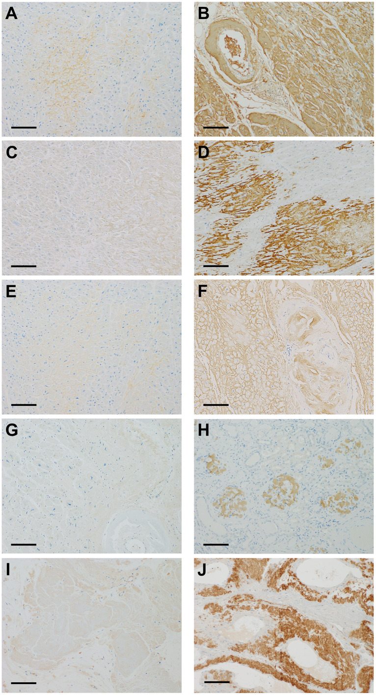
Fig 1. Immunohistochemical staining of five major amyloid precursor proteins.

Fig 1

Immunostaining using 3,3′-diaminobenzidine. A and B: immunohistochemistry of serum amyloid A (SAA), showing weakly positive (1+) (A), and strongly positive (3+) (B). C and D: immunohistochemistry of transthyretin (TTR), showing weakly positive (1+) (C), and strongly positive (3+) (D). E and F: immunohistochemistry of immunoglobulin kappa light chain (IGK), showing weakly positive (1+) (E), and strongly positive (3+) (F). G and H: immunohistochemistry of immunoglobulin lambda light chain (IGL), showing weakly positive (1+) (G), and strongly positive (3+) (H). I and J: immunohistochemistry of beta-2-microglobulin (B2M), showing weakly positive (1+) (I), and strongly positive (3+) (J). Scale bars, 200μm.

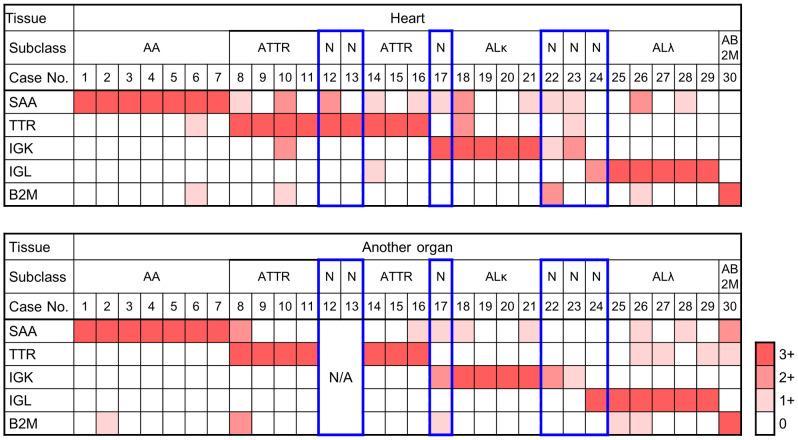
Fig 2. Subclassification of amyloidosis by immunohistochemistry of major amyloid proteins.

Fig 2

The Immunohistochemical results of serum amyloid A (SAA), transthyretin (TTR), immunoglobulin kappa light chain (IGK), immunoglobulin lambda light chain (IGL), and beta-2-macroglobulin (B2M) were evaluated with a four-tiered scales (0 to 3+). The scales are presented in white, and light, intermediate, and dark orange. Note the boxes delineated in red show three sets of cases that did not meet the criteria for subclassification: (1) only one section available (#12 and #13), (2) no 3+ immunostaining (#22 and #23), and (3) 3+ and 2+ immunostaining in each pair (#17 and #24). AA, AA amyloidosis; AB2M, beta-2-macroglobulin amyloidosis; ALκ, amyloid light chain (kappa) amyloidosis; ALλ, amyloid light chain (lambda) amyloidosis; ATTR, ATTR amyloidosis; N, subclassification could not be determined at autopsy; N/A, tissues not available.

Representative images of the immunohistochemical analysis using the panel antibodies are presented in Fig 1. Strong homogeneous immunostaining (3+) of at least one protein was observed in the entire amyloid deposits in 27 of 30 sections of the cardiac wall and in 25 of 28 sections of other organs (Fig 2). Positive results of 3+ were concordant in cardiac and other organs in 24 patients, and there were no instances showing contradictory results in the paired tissue sections. Relatively weak staining (2+) was accompanied by 3+ staining in four sections from the cardiac wall (#10, 12, 18, and 26) and two sections from the other organs (#8 and 30). Subclassification into five major types of amyloidosis was possible in 24 patients on the basis of 3+ immunostaining in the paired tissue sections, as shown in Table 1: AA amyloidosis, seven patients (#1–7); ATTR amyloidosis, seven patients (#8-#11 and #14-#16); ALκ amyloidosis, four patients (#18-#21); ALλ amyloidosis, five patients (#25-#29); and B2M amyloidosis, one patient (#30).

There were three sets of specimens that did not meet the above criteria for the following reasons: (1) there was only one section available (#12 and #13), (2) there was no 3+ immunostaining (#22 and #23), and (3) 3+ and 2+ immunostaining in each pair (#17 and #24).

Quantification of amyloid proteins in LMD samples of Congo red–positive deposits

A total of 98 peptides were selected as the target peptides for 13 proteins (SAA, TTR, IGK, IGL, B2M, Apo A1, Apo A2, Apo A4, Apo E, gelsolin, FGA, LECT2, and lysozyme). Fifty-three peptides for 12 proteins were selected to obtain the absolute abundances of amyloid proteins in LMD-dissected samples (Table 2). There were no peptides identified for quantification of LECT 2. The reasons for exclusion of peptides were as follows. (1) Two synthesized MS-QBIC peptides could not be detected by MS (potentially because of the low efficiency of the peptide synthesis by the PURE system or a low efficiency of ionization). (2) Twenty-six MS-QBIC peptides were excluded because of the low quality of the linear regression lines. (3) Two MS-QBIC peptides were also excluded for the absolute quantification because these peptides were not quantified robustly when mixed with peptides derived from LMD samples. (4) Fifteen peptides had signals that could not be detected or separated from the adjacent signals in the LMD sample.

Table 2. MS-QBIC peptides selected for the absolute quantification of amyloid proteins in LMD-dissected samples.

Protein(quantified/examined peptides) Quantified peptide sequence Non-quantified peptide sequence
SAA (2/4) EANYIGSDK GNYDAAK (2)
DPNHFRPAGLPEK AYSDMR (2)
TTR (2/7) GSPAINVAVHVFR ALGISPFHEHAEVVFTANDSGPR (2)
AADDTWEPFASGK YTIAALLSPYSYSTTAVVTNPK (2)
TSESGELHGLTTEEEFVEGIYK (2)
VEIDTK (2)
CPLMVK (2)
IGK (3/5) TVAAPSVFIFPPSDEQLK DSTYSLSSTLTLSK (2)
SGTASVVCLLNNFYPR VYACEVTHQGLSSPVTK (2)
VDNALQSGNSQESVTEQDSK
IGL (3/5) AAPSVTLFPPSSEELQANK ATLVCLISDFYPGAVTVAWK (2)
AGVETTTPSK SYSCQVTHEGSTVEK (2)
YAASSYLSLTPEQWK
B2M (3/6) VNHVTLSQPK SNFLNCYVSGFHPSDIEVDLLK (2)
VEHSDLSFSK DWSFYLLYYTEFTPTEK (2)
IQVYSR DEYACR (2)
Apo A1 (7/9) VSFLSALEEYTK AELQEGAR (2)
QGLLPVLESFK LAEYHAK (2)
DLATVYVDVLK
EQLGPVTQEFWDNLEK
AKPALEDLR
LLDNWDSVTSTFSK
DYVSQFEGSALGK
Apo A2 (2/4) EQLTPLIK EPCVESLVSQYFQTVTDYGK (4)
SPELQAEAK LLAATVLLLTICSLEGALVR (1)
Apo A4 (11/12) SELTQQLNALFQDK LNHQLEGLTFQMK (2)
LAPLAEDVR
LVPFATELHER
LEPYADQLR
LLPHANEVSQK
LTPYADEFK
VNSFFSTFK
SLAPYAQDTQEK
ALVQQMEQLR
IDQNVEELK
IDQTVEELR
Apo E (5/6) AATVGSLAGQPLQER ALMDETMK (4)
LGPLVEQGR
FWDYLR
AQAWGER
QQTEWQSGQR
Gelsolin (7/12) EVQGFESATFLGYFK HVVPNEVVVQR (4)
TGAQELLR FDLVPVPTNLYGDFFTGDAYVILK (2)
QTQVSVLPEGGETPLFK AQPVQVAEGSEPDGFWEALGGK (4)
AGALNSNDAFVLK VPVDPATYGQFYGGDSYIILYNYR (2)
AVEVLPK VPEARPNSMVVEHPEFLK (2)
TPITVVK
EPGLQIWR
FGA (6/17) GLIDEVNQDFTNR DSHSLTTNIMEILR (2)
QLEQVIAK MELERPGGNEITR (2)
NSLFEYQK HPDEAAFFDTASTGK (4)
GGSTSYGTGSETESPR VQHIQLLQK (4)
AQLVDMK ESSSHHPGIAEFPSR (4)
GSESGIFTNTK MKPVPDLVPGNFK (4)
QHLPLIK (4)
LEVDIDIK (2)
ALTDMPQMR (2)
TVIGPDGHK (4)
VSEDLR (3)
LECT2 (0/5) LGTLLPLQK (4)
SSNEIR (4)
NAINNGVR (3)
HGCGQYSAQR (2)
MFYIKPIK (1)
Lysozyme (2/6) STDYGIFQINSR TPGAVNACHLSCSALLQDNIADAVACAK (4)
AWVAWR GISLANWMCLAK (2)
LGMDGYR (4)
WESGYNTR (4)

(1) The synthesized MS-QBIC peptide cannot be detected by mass spectrometers, potentially due to the low efficiency of the peptide synthesis by the PURE system or low efficiency of ionization. (2) These MS-QBIC peptides were not used for the absolute quantification because of the low quality of signal linearity evaluation. (3) These MS-QBIC peptides were not used for the absolute quantification because the peptides were not detected robustly when they were mixed with LMD samples. (4) These peptides derived from the LMD samples could not be separated from other adjacent signals or could not be detected in the samples. Apo A1, apolipoprotein A-1; Apo A2, apolipoprotein A-2; Apo A4, apolipoprotein A-4; Apo E, apolipoprotein E; B2M, beta-2-microglobulin; FGA, fibrinogen alpha chain; IGK, immunoglobulin kappa light chain; IGL, immunoglobulin lambda light chain; LRCT2, leukocyte cell-derived chemotaxin-2; MS, mass spectrometry; MS-QBIC, mass spectrometry–quantification by isotope-labeled cell-free products; SAA, serum amyloid A; TTR, transthyretin.

Three samples from each specimen were subjected to analysis of 12 proteins. The average of the quantitative value of the peptides for each protein was used for further analyses, and the results of the quantification are shown in Table 3. There were three groups of amyloid proteins in the present study: Group 1, amyloid proteins with immunohistochemical analyses of deposition (SAA, TTR, IGK, IGL, B2M); Group 2, certain amounts of proteins detected without evidence of specific deposition (Apo A1, Apo A4, Apo E, and lysozyme); and Group 3, extremely low amounts detected (Apo A2, gelsolin, and FGA).

Table 3. Quantitative values of the amyloid proteins.

Amyloid protein Range
(pmol/mm3)
Median
(pmol/mm3)
Mean ± SD
(pmol/mm3)
Immuno-histochemistry
SAA 0.000–268.439 0.770 33.577 ± 69.440 yes
TTR 0.000–367.852 0.854 63.902 ± 112.086 yes
IGK 0.000–128.572 3.857 17.539 ± 31.021 yes
IGL 0.000–108.994 3.274 10.138 ± 18.590 yes
B2M 0.307–77.763 0.638 2.973 ± 12.185 yes
Apo A1 0.000–21.918 2.731 3.951 ± 4.186 no
Apo A2 0.000–0.967 0.000 0.074 ± 0.224 no
Apo A4 0.523–36.247 5.933 7.103 ± 6.602 no
Apo E 1.543–46.008 9.637 13.074 ± 9.767 no
Gelsolin 0.098–1.766 0.595 0.644 ± 0.374 no
FGA 0.000–4.968 1.221 1.385 ± 1.210 no
Lysozyme 2.104–23.802 4.477 4.950 ± 2.962 no

Apo A1, apolipoprotein A-1; Apo A2, apolipoprotein A-2; Apo A4, apolipoprotein A-4; Apo E, apolipoprotein E; B2M, beta-2-microglobulin; FGA, fibrinogen alpha chain; IGK, immunoglobulin kappa light chain; IGL, immunoglobulin lambda light chain; LRCT2, leukocyte cell-derived chemotaxin-2, SAA, serum amyloid A; TTR, transthyretin

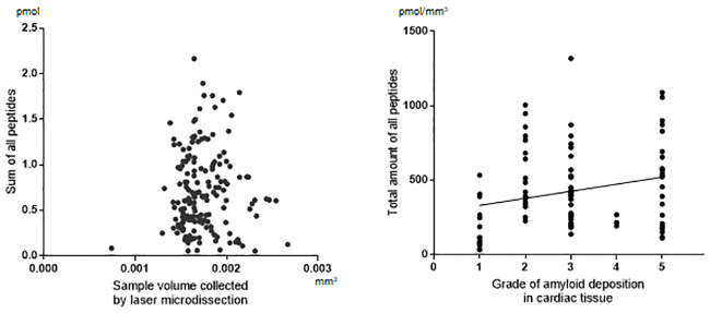
For the effect of LMD procedures on the recovery of amyloid proteins from the tissue, there was no significant correlation between the total amount of peptides in MS-quantification and the area collected by LMD (Fig 3A). The amyloid deposition grades showed positive correlation with increased total peptides, although the correlation was relatively weak (R = 0.2707) (Fig 3B).

Fig 3. Recovery of amyloid proteins from tissue specimens.

Fig 3

A. Effect of tissue collection by laser microdissection. Scatter plot of the total amounts of peptides (pmol) in mass spectrometry (MS) quantification and the area collected by laser microdissection. B. Effect of the amount of tissue amyloid. Distribution plot of the density of amyloid protein peptides (pmol/mm3) by the deposition grades of the cardiac tissues. Spearman’s rank correlation, R = 0.2707, P = 0.0099.

Type-specific amyloid protein deposition

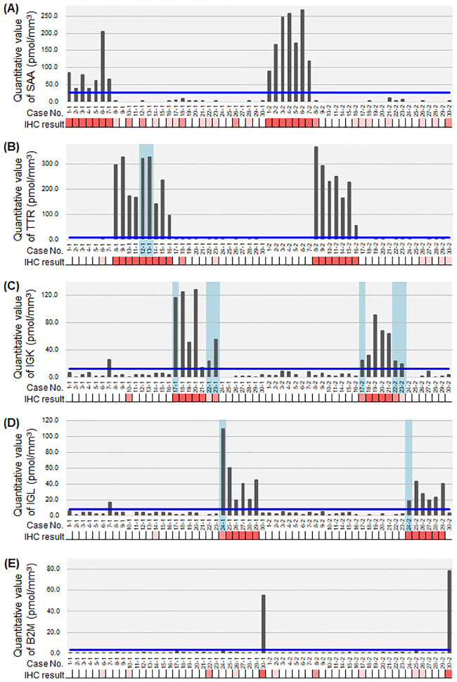
When each protein amount was compared with the immunohistochemical results (Fig 4), all samples in the 3+ grade of immunohistochemistry showed higher amounts of the amyloid protein. In patients #12 and #13, in whom the immunohistochemical results were confirmed in only one section, the quantification provided further evidence of the diagnosis. In patients #17, #22, #23, and #24, the quantification confirmed the suspected diagnosis, even if immunohistochemical results lacked enough evidence. There were two samples that showed higher than the lowest value in the corresponding subgroups, IGK and IGL in #7–1. The clinical diagnosis in this patient was Sjögren syndrome complicated by lymphoma. The results suggest a small amount of light chain deposition in this specific case.

Fig 4. Mass spectrometry quantification of major amyloid proteins with corresponding immunohistochemical scores.

Fig 4

Mass spectrometry (MS) quantification of serum amyloid A (SAA) (A), transthyretin (TTR) (B), immunoglobulin kappa light chain (IGK) (C), immunoglobulin lambda light chain (IGL) (D), and beta-2-microglobulin (B2M) (E). Immunohistochemical results of each corresponding protein are presented as the scales: 0, white; 1+, light orange; 2+, intermediate orange; and 3+, dark pink. The cases with an undetermined subclass are highlighted in a light blue-colored box. The blue line represents the proposed cut-off value for determination of subclass as shown in Table 4.

For setting the cut-off value for MS-quantification in the present study, we used a simulation as shown in Table 4. Based on the protein amounts in the immunohistochemically determined groups, the cut-off value for MS-quantification was at first set as the mean+2 SD of each protein. However, in SAA, TTR, and B2M, the difference between the minimum amount in each corresponding group was much higher than in the maximum of the other groups. Therefore, in these cases, the cut-off values were arbitrarily set as the maximum +6 SD.

Table 4. Proposed cut-off values for determination of the major amyloid subclasses.

Protein Corresponding subgroup
(pmol/mm3)
Other subgroups
(pmol/mm3)
Cut-off value
Mean ± SD Range min-max Mean ± SD Max (#sample)
SAA AA subgroup (n = 14/7 cases) n = 34 max+6SD
134.976 ± 80.031 38.054–268.439 1.188 ± 2.466 11.484 (#21–2) 26.280
TTR ATTR subgroup (n = 14/7case) n = 34 max+6SD
216.195 ± 85.117 56.514–367.852 0.538 ± 0.898 3.499 (#27–2) 8.887
IGK ALk subgroup (n = 8/4cases) n = 40 mean+2SD
71.898 ± 38.314 14.751–128.572 4.214 ± 4.371 26.404 (#7–1) 12.956
IGL ALl subgroup (n = 10/5cases) n = 38 mean+2SD
33.771 ± 13.212 19.037–60.228 2.912 ± 2.694 16.742 (#7–1) 8.300
B2M AB2M subgroup (n = 2) n = 46 max+6SD
- 55.141–77.763 0.641 ± 0.303 1.837 (#25–2) 3.655

Analysis was performed using the data of immunohistochemically determined subgroups (each corresponding amyloid subgroup vs. other subgroups)

B2M, beta-2-microglobulin; IGK, immunoglobulin kappa light chain; IGL, immunoglobulin lambda light chain; SAA, serum amyloid A; TTR, transthyretin

Non-type-specific protein deposition

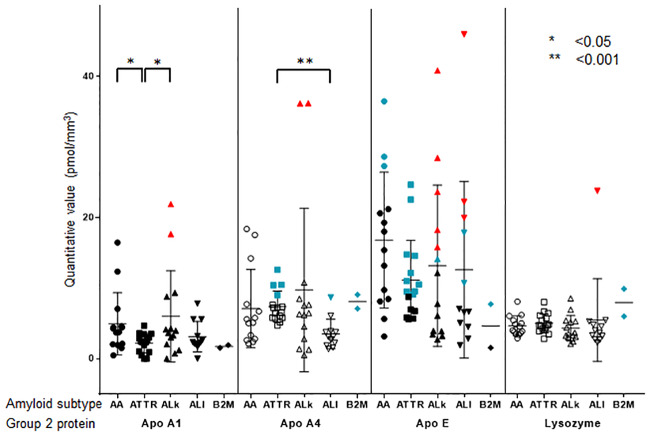
Apo A1, Apo A4, Apo E, and lysozyme were also detected in all 58 specimens from 30 patients (Fig 5). Their absolute amounts were less than the minimum amounts of SAA and TTR. However, two samples each showed an excess amount of Apo A1 and Apo A4 relative to the minimum of ALκ. Many samples showed an excess amount of Apo E relative to the minimum of ALκ and ALλ. One sample showed an excess amount of lysozyme relative to the minimum of ALλ.

Fig 5. Mass spectrometry quantification of Group 2 proteins in each of five major amyloid subtypes.

Fig 5

Bee swarm plot of the amount of Group 2 proteins (apolipoprotein A-1, Apo A1; apolipoprotein A-4, Apo A4; apolipoprotein E, Apo E; and lysozyme) by five major amyloid subtypes (serum amyloid A, SAA, circle; transthyretin, TTR, rectangle; immunoglobulin kappa light chain, IGK, triangle; immunoglobulin lambda light chain, IGL, inverted triangle; and beta-2-microglobulin, B2M, rhombus). The samples with the values higher than the arbitrarily set cut-off value for each corresponding protein, are labeled in blue-green. The samples with values higher than the minimum value for each corresponding protein, are labeled in red. Statistical differences in the protein amounts are presented when significant. MS, mass spectrometry.

It is possible that the deposition of these proteins has some role in amyloid deposition; for example, as a facilitating cofactor. To test this assumption, we conducted additional analyses. First, to evaluate quantitative correlations between the four main type-specific amyloid proteins (SAA, TTR, IGK, and IGL) and Apo A1, Apo A4, Apo E, and lysozyme, we calculated the Spearman’s rank correlation coefficient (Table 5), demonstrating that positive correlations were present except for lysozyme.

Table 5. Spearman’s rank correlation coefficient for Group 2 amyloid proteins with Group 1 amyloid proteins in major amyloid subgroups.

Subgroup / Protein Apo A1 Apo A4 Apo E Lysozyme
r p r p r p r p
AA SAA 0.352 NS 0.414 <0.01 0.367 NS 0.122 NS
ATTR TTR 0.450 <0.01 0.372 <0.01 0.493 <0.001 -0.117 NS
ALk IGK 0.575 <0.001 0.508 <0.01 0.694 <0.0001 0.073 NS
ALλ IGL 0.082 NS 0.661 <0.0001 0.121 NS 0.265 NS

B2M, beta-2-microglobulin; IGK, immunoglobulin kappa light chain; IGL, immunoglobulin lambda light chain; SAA, serum amyloid A; TTR, transthyretin

Second, the amounts of these proteins in nonamyloid tissues were measured relative to those in amyloid deposition (Table 6). The mean+2SD for these ratios was less than 0.5, except for lysozyme.

Table 6. Relative ratios of Group 2 amyloid proteins found in nonamyloid tissue.

Protein Ratio Mean ± SD
Apo A1 0.137 ± 0.139
Apo A4 0.099 ± 0.171
Apo E 0.013 ± 0.015
Lysozyme 0.321 ± 0.465

Apo A1, apolipoprotein A-1; Apo A2, apolipoprotein A-2; Apo A4, apolipoprotein A-4; Apo E, apolipoprotein E.

Discussion

The MS-QBIC method enables high-throughput preparation of internal standards by using a reconstituted cell-free protein synthesis system and thereby facilitates multiplexed quantification of absolute amounts of target proteins with high sensitivity. The present study is the first application of this method in the field of pathology. We chose the classification of systemic amyloidosis as the subject of this application because it is sometimes difficult to determine by immunohistochemistry and even by label free LC-MS/MS analysis.

Immunohistochemistry is the most common method used in pathology laboratories for subtyping amyloidosis. In the present study, using the panel of five antibodies against five major amyloid proteins, we encountered three kinds of difficulty: (1) there was only one section available, (2) there was no 3+ immunostaining, and (3) there was 3+ and 2+ immunostaining in each pair. Furthermore, there were many specimens that showed 2+ immunostaining in addition to 3+ immunostaining relative to other proteins. Absolute quantification of amyloid protein by MS-QBIC demonstrated strict correspondence between 3+ immunostaining and protein amounts. We observed false positive 2+ immunostaining in patients with AA (six of six specimens) and ATTR (one of one specimen), while a true positive was observed in patients with ALκ (three of four specimens) and ALλ (one of one specimen). With these results, we could strengthen the immunohistochemical results even in single section and clarify the significance of 2+ immunostaining for each antibody. The absolute quantification of amyloid protein could resolve all three problems of immunohistochemistry described above. Thus, the combination of more than 2+ immunostaining and absolute quantification of IGK and IGL could be the criteria to determine both subtypes.

There is a question as to whether Apo A1, Apo A4, Apo E, and lysozyme are nonspecifically deposited in the amyloid. The present study demonstrated differences between the three apolipoproteins and lysozyme. There was quantitative correlation between apolipoproteins (Apo A1, A4, and E) and each type-specific protein in the amyloid, but not in lysozyme. In nonamyloid background tissues, the apolipoproteins deposited were less than 0.5 (at most) compared with those in the amyloid deposits. Apo E and Apo A4 sometimes coexist with the main causative amyloid proteins [57, 28]. Amyloid deposits contain many kinds of biological molecules, such as apolipoprotein, serum amyloid P, and glycoprotein [29]. These molecules can constitute a scaffold, facilitating the initial phases of fibril nucleation, and they have a targeting role in the localization of amyloid deposits in the tissue. Apo E, Apo A4, serum amyloid P, and vitronectin are associated with amyloid fibrils and are involved in amyloid formation. Apo E is a common constituent of amyloid deposits in Alzheimer’s disease. The major effect of Apo E isoforms occurs via the effect on Aβ aggregation and clearance, influencing the onset of Aβ deposition [28]. The Apo A4 fragment is strongly fibrillogenic in vitro and enhances fibril formation from wild-type TTR [5]. The apolipoproteins may facilitate the genesis of amyloid, especially in the cases of Apo A4 with ATTR and ALλ, and Apo E with ALκ. However, a characteristic of the lysozyme protein is a changeable conformation [30]. Enrichment of this protein may be passive and unrelated to the genesis of amyloid.

The amounts of Apo E and lysozyme proteins frequently exceeded the minimal amounts of IGK and IGL in ALκ and ALλ amyloidosis, respectively. It is possible that amyloidogenic immunoglobulin light chains, working as an initiator, precipitate other proteins in greater amounts than the initiator. Alternatively, these facts indicate a limitation of the method of absolute quantification in the present study. Many mutations in immunoglobulin light chains, either in the constant or the variable region, might cause complex conformation changes, resulting in difficulty in obtaining enough amounts of peptides from amyloid deposition of IGK and IGL.

In the present study, there was one patient with Sjögren syndrome and lymphoma, with deposition of SAA, IGK, and IGL but negative immunohistochemical results. The heavy mutation burden in immunoglobulin light chains might have prevented detection by immunohistochemistry. Alternatively, accumulated SAA protein covered the epitopes of immunoglobulin molecules. Although the LS-MS/MS method improves detection sensitivity by improving data matching techniques, some peptides with mutations might also escape detection in LC-MS/MS analysis. In these circumstances, absolute quantification will identify such a case and contribute to the understanding of the complicated processes involved in amyloidosis.

Absolute quantification is potentially useful to compare the various effects of different types of amyloid in the diseased tissue. However, there are limitations in the present study. The effect of formalin fixation might not be uniform at each amino acid residue in a single protein, which may affect the accuracy of quantification given by a limited number of peptide candidates for one amyloid protein. Second, the amount of amyloid deposits was evaluated in units of pmol/mm3 with the volume of amyloid as the denominator. This evaluation ignores the spatial distribution of amyloid proteins within the deposit area. However, the physical properties of each amyloid protein might be different if, for example, high concentrations of a particular type of amyloid protein are deposited in a particular region of the deposit area.

In conclusion, we successfully applied the MS-QBIC method to the absolute quantification of amyloid deposits in systemic amyloidosis. The quantitative data clarified the significance of immunohistochemical results and provided basis for the interpretation of amyloidosis classification by immunohistochemical panel analysis. Furthermore, the quantification by MS analysis disclosed the significance of apolipoproteins, which are components of amyloid aggregates. Absolute quantification of amyloid protein by MS-QBIC is a feasible and useful complement for the classification of and research into systemic amyloidosis.

Supporting information

S1 Appendix. References for the determination of peptide sequences.

(PDF)

S2 Appendix. Signal linearity of each MS-QBIC peptide measured by mass spectrometry.

(PDF)

S3 Appendix. Chromatogram of precursor ions and spectrum of fragment ions for a representative peptide of the quantified amyloid proteins.

Case ID (e.g., P1) and raw file ID (e.g., A003) correspond to the ID numbers shown in S3 Table. (A) and (M) serum amyloid A (SAA). (B) and (N) transthyretin (TTR), (C) and (O) immunoglobulin kappa light chain (IGK), (D) and (P) immunoglobulin lambda light chain (IGL), (E) and (Q) beta-2-microglobulin (B2M), (F) and (R) apolipoprotein (Apo) A1, (G) and (S) Apo A2, (H) and (T) Apo A4, (I) and (U) Apo E, (J) and (V) gelsolin, (K) and (W) fibrinogen alpha chain (FGA), (L) and (X) lysozyme, (A-L) specimens are derived from the heart, and (M-X) from other organs.

(PDF)

S1 Fig. Amyloid deposition by Congo red staining.

The amount of Congo red-positive deposition was scored in five grades 1–5. from minimal to severe (A, C, E). The staining is well visualized under the FITC filter of a BZ-X710 all-in-one fluorescent microscope (B, D, F). Grade 2 (A and B), Grade 3 (C and D), and Grade 5 (E and F). Scale bars, 200μm.

(PDF)

S2 Fig. Workflow of quantification of amyloid proteins by Mass Spectrometry–based Quantification By Isotope-labeled Cell-free products (MSQBIC).

The workflow has two parts: the synthesis and quantification of the MS-QBIC peptides (left side) and the quantification of target peptides using the MS-QBIC peptide as reference (right side).

(PDF)

S3 Fig. Supporting information for the estimation of the concentration of MS-QBIC peptides.

(A) Signal linearity of the quantification tag measured by mass spectrometry. (B) Heavy to light ratios of quantification tag signals used to estimate the concentrations of MS-QBIC peptides.

(PDF)

S1 Table. Specimens used for data-dependent MS/MS analysis.

(DOCX)

S2 Table. List of MS-QBIC target peptides, primer sequences for the production of MS-QBIC peptides, and SRM method for the quantification.

(XLSX)

S3 Table. List of information used for the absolute quantification of amyloid proteins in the tissue samples, including the intensities and heavy/light ratios of MS-QBIC- and tissue-derived peptide signals.

(XLSX)

Acknowledgments

We thank Andrea Baird, MD, from Edanz Group (https://en-author-services.edanzgroup.com/) for editing a draft of this manuscript.

Data Availability

All relevant data are within the paper and its Supporting Information files. The raw data of SRM analysis were deposited to the PeptideAtlas SRM Experiment Library (PASSEL) (http://www.peptideatlas.org/PASS/PASS01558).

Funding Statement

This work was supported by The Japan Society for the Promotion of Science (Grant No. 17K08717 to Y.S-D.). The funder had no role in study design, data collection and analysis, decision to publish, or preparation of the manuscript. H.R.U conducted a collaborative research project with Thermo Fisher Scientific Inc. Y.N. is an employee of Thermo Fisher Scientific, Inc. The company provided support in the form of salary for Y.N., and technical advice on the setup of mass spectrometers. However, the company did not have any additional role in the study design, data collection and analysis, decision to publish, or preparation of the manuscript. M.H.R. acknowledges NCI R21 CA231109, NCI R21 CA251992, the Farmer Foundation, a Cycle for Survival Equinox Innovation grant, and partial funding through the MSKCC NIH/NCI Cancer Center Support Grant P30 CA008748. None of these companies had any influence in support, design, execution, data analysis, or any other aspect of this study. The specific roles of these authors are articulated in the ‘author contributions’ section.

References

  • 1.Sipe JD, Benson MD, Buxbaum JN, Ikeda S, Merlini G, Saraiva MJ, et al. Nomenclature 2014: Amyloid fibril proteins and clinical classification of the amyloidosis. Amyloid. 2014;21(4):221–4. 10.3109/13506129.2014.964858 [DOI] [PubMed] [Google Scholar]
  • 2.Kastritis E, Dimopoulos MA. Recent advances in the management of AL Amyloidosis. Br J Haematol. 2016;172(2):170–86. 10.1111/bjh.13805 [DOI] [PubMed] [Google Scholar]
  • 3.Ando Y, Coelho T, Berk JL, Cruz MW, Ericzon BG, Ikeda S, et al. Guideline of transthyretin-related hereditary amyloidosis for clinicians. Orphanet J Rare Dis. 2013;8:31 10.1186/1750-1172-8-31 [DOI] [PMC free article] [PubMed] [Google Scholar]
  • 4.Mollee P, Renaut P, Gottlieb D, Goodman H. How to diagnose amyloidosis. Intern Med J. 2014;44(1):7–17. 10.1111/imj.12288 [DOI] [PubMed] [Google Scholar]
  • 5.Bergstrom J, Murphy C, Eulitz M, Weiss DT, Westermark GT, Solomon A, et al. Codeposition of apolipoprotein A-IV and transthyretin in senile systemic (ATTR) amyloidosis. Biochem Biophys Res Commun. 2001;285(4):903–8. 10.1006/bbrc.2001.5260 [DOI] [PubMed] [Google Scholar]
  • 6.Vrana JA, Theis JD, Dasari S, Mereuta OM, Dispenzieri A, Zeldenrust SR, et al. Clinical diagnosis and typing of systemic amyloidosis in subcutaneous fat aspirates by mass spectrometry-based proteomics. Haematologica. 2014;99(7):1239–47. 10.3324/haematol.2013.102764 [DOI] [PMC free article] [PubMed] [Google Scholar]
  • 7.Aoki M, Kang D, Katayama A, Kuwahara N, Nagasaka S, Endo Y, et al. Optimal conditions and the advantages of using laser microdissection and liquid chromatography tandem mass spectrometry for diagnosing renal amyloidosis. Clin Exp Nephrol. 2018;22(4):871–80. 10.1007/s10157-018-1533-y [DOI] [PubMed] [Google Scholar]
  • 8.Narumi R, Shimizu Y, Ukai-Tadenuma M, Ode KL, Kanda GN, Shinohara Y, et al. Mass spectrometry-based absolute quantification reveals rhythmic variation of mouse circadian clock proteins. Proc Natl Acad Sci USA. 2016;113(24):E3461–7. 10.1073/pnas.1603799113 [DOI] [PMC free article] [PubMed] [Google Scholar]
  • 9.Kieninger B, Eriksson M, Kandolf R, Schnabel PA, Schonland S, Kristen AV, et al. Amyloid in endomyocardial biopsies. Virchows Arch. 2010;456(5):523–32. 10.1007/s00428-010-0909-5 [DOI] [PubMed] [Google Scholar]
  • 10.Eriksson M, Schonland S, Yumlu S, Hegenbart U, von Hutten H, Gioeva Z, et al. Hereditary apolipoprotein AI-associated amyloidosis in surgical pathology specimens: identification of three novel mutations in the APOA1 gene. J Mol Diagn. 2009;11(13):257–62. [DOI] [PMC free article] [PubMed] [Google Scholar]
  • 11.Matsutani H, Hoshii Y, Setoguchi M, Kawano H, Gondo T, Takahashi M, et al. Vascular amyloid of unknown origin and senile transthyretin amyloid in the lung and gastrointestinal tract of old age: histological and immunohistochemical studies. Pathol Int. 2001;51(5):326–32. 10.1046/j.1440-1827.2001.01213.x [DOI] [PubMed] [Google Scholar]
  • 12.Gilbertson JA, Theis JD, Vrana JA, Lachmann H, Wechalekar A, Whelan C, et al. A comparison of immunohistochemistry and mass spectrometry for determining the amyloid fibril protein from formalin-fixed biopsy tissue. J Clin Pathol. 2015;68(4):314–7. 10.1136/jclinpath-2014-202722 [DOI] [PubMed] [Google Scholar]
  • 13.Hoshii Y, Kiyama M, Cui D, Kawano H, Ishihara T. Immunohistochemical study of immunoglobulin light chain amyloidosis with antibodies to the immunoglobulin light chain variable region. Pathol Int. 2006;56:324–30. 10.1111/j.1440-1827.2006.01953.x [DOI] [PubMed] [Google Scholar]
  • 14.Hoshii Y, Nanbara H, Cui D, Takahashi M, Ikeda E. Immunohistochemical examination of Akappa amyloidosis with antibody against adjacent portion of the carboxy terminus of immunoglobulin kappa light chain. Med Mol Morphol. 2012;45(6):124–8. [DOI] [PubMed] [Google Scholar]
  • 15.von Hutten H, Mihatsch M, Lobeck H, Rudolph B, Eriksson M, Rocken C. Prevalence and origin of amyloid in kidney biopsies. Am J Surg Pathol. 2009;33(8):1198–205. 10.1097/PAS.0b013e3181abdfa7 [DOI] [PubMed] [Google Scholar]
  • 16.Kebbel A, Rocken C. Immunohistochemical classification of amyloid in surgical pathology revisited. Am J Surg Pathol. 2006;30(6):673–83. 10.1097/00000478-200606000-00002 [DOI] [PubMed] [Google Scholar]
  • 17.Kiyama M, Hoshii Y, Cui D, Kawano H, Kanda T, Ishihara T. Immunohistochemical and immunochemical study of amyloid in liver affected by systemic Alambda amyloidosis with antibodies against three different regions of immunoglobulin lambda light chain. Pathol Int. 2007;57(6):343–50. 10.1111/j.1440-1827.2007.02106.x [DOI] [PubMed] [Google Scholar]
  • 18.Sipe JD, Benson MD, Buxbaum JN, Ikeda S, Merlini G, Saraiva MJ, et al. Amyloid fibril protein nomenclature: 2010 recommendations from the nomenclature committee of the International Society of Amyloidosis. Amyloid. 2010;17(3–4):101–4. 10.3109/13506129.2010.526812 [DOI] [PubMed] [Google Scholar]
  • 19.Masuda T, Tomita M, Ishihama Y. Phase transfer surfactant-aided trypsin digestion for membrane proteome analysis. J Proteome Res. 2008;7(2):731–40. 10.1021/pr700658q [DOI] [PubMed] [Google Scholar]
  • 20.Rappsilber J, Mann M, Ishihama Y. Protocol for micro-purification, enrichment, pre-fractionation and storage of peptides for proteomics using StageTips. Nat Protoc. 2007;2(8):1896–906. 10.1038/nprot.2007.261 [DOI] [PubMed] [Google Scholar]
  • 21.Benson MD, Liepnieks JJ, Kluve-Beckerman B. Hereditary systemic immunoglobulin light-chain amyloidosis. Blood. 2015;125(21):3281–6. 10.1182/blood-2014-12-618108 [DOI] [PubMed] [Google Scholar]
  • 22.Murphy C, Eulitz D, Weiss D, Solomon A. Light chain deamidation in ALlambda amyloid-associated protein. Amyloid. 2011;18 Suppl 1:27–8. [DOI] [PubMed] [Google Scholar]
  • 23.Dasari S, Theis JD, Vrana JA, Meureta OM, Quint PS, Muppa P, et al. Proteomic detection of immunoglobulin light chain variable region peptides from amyloidosis patient biopsies. J Proteome Res. 2015;14(4):1957–67. 10.1021/acs.jproteome.5b00015 [DOI] [PubMed] [Google Scholar]
  • 24.Shimizu Y, Inoue A, Tomari Y, Suzuki T, Yokogawa T, Nishikawa K, et al. Cell-free translation reconstituted with purified components. Nat Biotechnol. 2001;19(8):751–5. 10.1038/90802 [DOI] [PubMed] [Google Scholar]
  • 25.Ong SE, Mann M. Mass spectrometry-based proteomics turns quantitative. Nat Chem Biol. 2005;1(5):252–62. 10.1038/nchembio736 [DOI] [PubMed] [Google Scholar]
  • 26.Boersema PJ, Raijmakers R, Lemeer S, Mohammed S, Heck AJ. Multiplex peptide stable isotope dimethyl labeling for quantitative proteomics. Nat Protoc. 2009;4(4):484–494. 10.1038/nprot.2009.21 [DOI] [PubMed] [Google Scholar]
  • 27.Ode KL, Ukai H, Susaki EA, Narumi R, Matsumoto K, Hara J, et al. Knockout-Rescue Embryonic Stem Cell-Derived Mouse Reveals Circadian-Period Control by Quality and Quantity of CRY1. Mol Cell. 2017;65(1):176–90. 10.1016/j.molcel.2016.11.022 [DOI] [PubMed] [Google Scholar]
  • 28.Kim J, Basak JM, Holtzman DM. The role of apolipoprotein E in Alzheimer’s disease. Neuron. 2009;63(3):287–303. 10.1016/j.neuron.2009.06.026 [DOI] [PMC free article] [PubMed] [Google Scholar]
  • 29.Merlini G, Bellotti V: Molecular mechanisms of amyloidosis. N Engl J Med. 2003;349(6):583–96. 10.1056/NEJMra023144 [DOI] [PubMed] [Google Scholar]
  • 30.Swaminathan R, Ravi VK, Kumar S, Kumar MV, Chandra N. Lysozyme: a model protein for amyloid research. Adv Protein Chem Struct Biol. 2011;84:63–111. 10.1016/B978-0-12-386483-3.00003-3 [DOI] [PubMed] [Google Scholar]

Decision Letter 0

Harald Mischak

19 Feb 2020

PONE-D-20-02311

Mass spectrometry–based absolute quantification of amyloid proteins in pathology tissue specimens: merits and limitations

PLOS ONE

Dear Prof. Fukayama,

Thank you for submitting your manuscript to PLOS ONE. After careful consideration, we feel that it has merit but does not fully meet PLOS ONE’s publication criteria as it currently stands. Therefore, we invite you to submit a revised version of the manuscript that addresses the points raised during the review process.

We would appreciate receiving your revised manuscript by Apr 04 2020 11:59PM. When you are ready to submit your revision, log on to https://www.editorialmanager.com/pone/ and select the 'Submissions Needing Revision' folder to locate your manuscript file.

If you would like to make changes to your financial disclosure, please include your updated statement in your cover letter.

To enhance the reproducibility of your results, we recommend that if applicable you deposit your laboratory protocols in protocols.io, where a protocol can be assigned its own identifier (DOI) such that it can be cited independently in the future. For instructions see: http://journals.plos.org/plosone/s/submission-guidelines#loc-laboratory-protocols

Please include the following items when submitting your revised manuscript:

  • A rebuttal letter that responds to each point raised by the academic editor and reviewer(s). This letter should be uploaded as separate file and labeled 'Response to Reviewers'.

  • A marked-up copy of your manuscript that highlights changes made to the original version. This file should be uploaded as separate file and labeled 'Revised Manuscript with Track Changes'.

  • An unmarked version of your revised paper without tracked changes. This file should be uploaded as separate file and labeled 'Manuscript'.

Please note while forming your response, if your article is accepted, you may have the opportunity to make the peer review history publicly available. The record will include editor decision letters (with reviews) and your responses to reviewer comments. If eligible, we will contact you to opt in or out.

We look forward to receiving your revised manuscript.

Kind regards,

Harald Mischak

Academic Editor

PLOS ONE

Journal Requirements:

When submitting your revision, we need you to address these additional requirements.

1. Please ensure that your manuscript meets PLOS ONE's style requirements, including those for file naming. The PLOS ONE style templates can be found at

http://www.journals.plos.org/plosone/s/file?id=wjVg/PLOSOne_formatting_sample_main_body.pdf and http://www.journals.plos.org/plosone/s/file?id=ba62/PLOSOne_formatting_sample_title_authors_affiliations.pdf

2. Please include your tables as part of your main manuscript and remove the individual files. Please note that supplementary tables (should remain/ be uploaded) as separate "supporting information" files

3. Thank you for stating the following in the Financial Disclosure  section:

"This work was supported by The Japan Society for the Promotion of Science (Grant No. 17K08717 to Y.S-D.).

The funder had no role in study design, data collection and analysis, decision to publish, or preparation of the manuscript."

We note that one or more of the authors are employed by a commercial company: 'Thermo Fisher Scientific K.K., Yokohama, Kanagawa, Japan'.

a)    Please provide an amended Funding Statement declaring this commercial affiliation, as well as a statement regarding the Role of Funders in your study. If the funding organization did not play a role in the study design, data collection and analysis, decision to publish, or preparation of the manuscript and only provided financial support in the form of authors' salaries and/or research materials, please review your statements relating to the author contributions, and ensure you have specifically and accurately indicated the role(s) that these authors had in your study. You can update author roles in the Author Contributions section of the online submission form.

Please also include the following statement within your amended Funding Statement.

“The funder provided support in the form of salaries for authors [insert relevant initials], but did not have any additional role in the study design, data collection and analysis, decision to publish, or preparation of the manuscript. The specific roles of these authors are articulated in the ‘author contributions’ section.”

If your commercial affiliation did play a role in your study, please state and explain this role within your updated Funding Statement.

b) Please also provide an updated Competing Interests Statement declaring this commercial affiliation along with any other relevant declarations relating to employment, consultancy, patents, products in development, or marketed products, etc.  

Within your Competing Interests Statement, please confirm that this commercial affiliation does not alter your adherence to all PLOS ONE policies on sharing data and materials by including the following statement: "This does not alter our adherence to  PLOS ONE policies on sharing data and materials.” (as detailed online in our guide for authors http://journals.plos.org/plosone/s/competing-interests) . If this adherence statement is not accurate and  there are restrictions on sharing of data and/or materials, please state these. Please note that we cannot proceed with consideration of your article until this information has been declared.

Please include both an updated Funding Statement and Competing Interests Statement in your cover letter. We will change the online submission form on your behalf.

4. Please include captions for your Supporting Information files at the end of your manuscript, and update any in-text citations to match accordingly. Please see our Supporting Information guidelines for more information: http://journals.plos.org/plosone/s/supporting-information.

Additional Editor Comments (if provided):

As you can see, both authors found the paper potentially of value, but indicated multiple issues that need to be addressed. Many of these are required to ascertain validity of the results, and reproducibility. I agree with the comments, and hope that you can revise the manuscript accordingly

[Note: HTML markup is below. Please do not edit.]

Reviewers' comments:

Reviewer's Responses to Questions

Comments to the Author

1. Is the manuscript technically sound, and do the data support the conclusions?

The manuscript must describe a technically sound piece of scientific research with data that supports the conclusions. Experiments must have been conducted rigorously, with appropriate controls, replication, and sample sizes. The conclusions must be drawn appropriately based on the data presented.

Reviewer #1: Yes

Reviewer #2: Partly

**********

2. Has the statistical analysis been performed appropriately and rigorously?

Reviewer #1: Yes

Reviewer #2: Yes

**********

3. Have the authors made all data underlying the findings in their manuscript fully available?

The PLOS Data policy requires authors to make all data underlying the findings described in their manuscript fully available without restriction, with rare exception (please refer to the Data Availability Statement in the manuscript PDF file). The data should be provided as part of the manuscript or its supporting information, or deposited to a public repository. For example, in addition to summary statistics, the data points behind means, medians and variance measures should be available. If there are restrictions on publicly sharing data—e.g. participant privacy or use of data from a third party—those must be specified.

Reviewer #1: Yes

Reviewer #2: No

**********

4. Is the manuscript presented in an intelligible fashion and written in standard English?

PLOS ONE does not copyedit accepted manuscripts, so the language in submitted articles must be clear, correct, and unambiguous. Any typographical or grammatical errors should be corrected at revision, so please note any specific errors here.

Reviewer #1: Yes

Reviewer #2: Yes

**********

5. Review Comments to the Author

Please use the space provided to explain your answers to the questions above. You may also include additional comments for the author, including concerns about dual publication, research ethics, or publication ethics. (Please upload your review as an attachment if it exceeds 20,000 characters)

Reviewer #1: Dear Authors and Editor,

The authors have applied their previously published MS-QBIC to quantitate the amyloids present in FFPE tissue samples. Their skill using mass spectrometry, pathology, and microdissection is obvious. They used PTS method to extract the proteins from the FFPE tissues. PTS is most appropriate for unfixed tissue.

Formalin fixing uses formaldehyde to crosslink proteins by their lysine and other residues. The authors used stable isotope-labeled formaldehyde in this work and they ensured that the consequent imine formation was reduced using sodium cyanoborohydride. The crosslinking resulting from formalin fixing would make some proteins inaccessible to trypsin and prevent some trypsin cleavage on proteins that were accessible to trypsin. The authors used the PTS method to extract proteins from this fixed tissue. This would assist in extracting the proteins, but it does not undo the formaldehyde mediated cross linking of the formalin. Other researchers use high temperatures and free amines to reverse the fixation process and yield intact proteins.

The authors need to revise the manuscript by including an additional limitation: namely that fixation may not be uniform for each protein. This may be why the authors were able to observe some tryptic peptides and not others. It also means that discussions about quantitation of proteins makes no sense when the authors cannot ensure that the proteins they are quantitating are extracted in proportion to their prevalence.

Some specific changes are listed below.

The authors use acronyms such as PTS (page 8) before they define it as phase transfer surfactants (page 10). They should carefully check the manuscript to ensure that the acronyms are defined before they are used.

The statement: “The various forms of amyloidosis require different approaches, including high-dose…”

Should be revised to include “treatment”:

“The various forms of amyloidosis require different treatment approaches, including high-dose…”

“13C6 15N4 L-arginine and 13C6 L-lysine.” Is Lysine unlabeled with 15N?

“For the quantification, the concentration of the MS-QBIC peptide (stable isotope-labeled peptide) was first determined by comparing the ion peak intensities of commercially available BSA.” The authors should state that they used BSA digested with trypsin.

The authors need to include the calibration curves they used to relate the signal intensities of the various peptides to one another. This should be included in the Supporting Information

“Nonamyloid and amyloid samples were differently labeled with light CH2O and heavy DH2O isotopes through dimethyl labeling with formaldehyde, respectively [24]” Do the authors mean CD2O or 13CD2O instead of DH2O?

Table 2: The lines separating proteins should be thicker than those used to delineate the peptides associated with that protein to avoid confusion. For example, the lines above and below B2M and its three peptides (VNHVTLSQPK, VEHSDLSFSK, and IQVYSR) should be thicker than the line used to separate VNHVTLSQPK and VEHSDLSFSK. In addition, there should be a line to separate VEHSDLSFSK, and IQVYSR.

Figure 4: The resolution is poor. The figure is unreadable.

References:

PLoS ONE requires full page numbers. The references need to be properly formatted.

Supporting Information Figure 1 and 2 are not available.

Reviewer #2: In this manuscript, authors applied mass spectrometry based quantification by isotope labeled cell free products (MS-QBIC) to formalin fixed, paraffin embedded (FFPE) tissues. The method was applied to amyloid tissues collected by laser capture microdissection of Congo red stained lesions of FFPE specimens. Authors managed to quantify 12 out of 13 selected proteins relevant to amyloidosis such as: serum amyloid A (SAA), transthyretin (TTR), immunoglobulin kappa light chain (IGK), immunoglobulin lambda light chain (IGL), beta-2 microglobulin (B2M), apolipoprotein (Apo) A1, Apo A2, Apo A4, Apo E, lysozyme, gelsolin, and fibrinogen alpha chain. Leukocyte cell derived chemotaxin-2 was not detected. Authors claim that quantification of amyloid protein by MS-QBIC is feasible and useful for the classification and research on systemic amyloidosis. The study although interesting requires further refinement to be considered for publication in Plos One.

Major Comments

1. The authors tried to quantify their MS-QBIC standard peptides with the help of the quantification tag from BSA which was included in all of the sequences of their MS-QBIC peptides. By trypsinizing a sample that included BSA of known concentration and the MS-QBIC peptide, the quantification tag of BSA was released (unlabeled-light from BSA and labeled-heavy from MS-QBIC peptide) and by comparing the signal intensity of light/heavy the concentration of the MS-QBIC peptide was determined. This is for sure an indirect method to quantify the standard peptides (MS-QBIC) that will be used later for the quantification of the selected target proteins. This method raised a lot of concerns since many data for this quantification are missing from the manuscript. Authors present only a supplementary figure (suppl. fig 2) which just describes the general workflow followed for the quantification of the MS-QBIC peptides but does not provide any data. Authors should present all the light (quantification tag from BSA) / heavy (quantification tag from MS-QBIC) ratios, the intensity values of each tag, the different known concentrations of BSA that were utilized in this analysis. Was it observed any saturation on the signal produced by the BSA tag and if yes at which concentration? It would be strongly recommended that the authors would test 6-8 different concentrations of BSA and select the values that are in the linear range. Light/Heavy ratios should be reported as well. Each sample of the BSA different concentration should be analyzed in triplicate and the authors should provide the mean value along with the standard deviation of the measurement. This is a very important step since based on these values the authors will better define the concentration-amount of their MS-QBIC isotope labeled peptides which will be used for the quantification of the selected target proteins.

2. Authors report the selected proteins, the precursors and the fragments measured for each protein with the SRM method. However, Light (endogenous) / Heavy (labeled standards) ratios are missing from the manuscript. Intensities of light, heavy and their respective ratios should be reported for all the quantified proteins along with the estimated concentration. Were the ratios obtained in the range of 0.1-10 (concentration balanced)? If not there is a big possibility that the reported concentrations are not accurate.

3. Authors report that more than three target peptides were quantified for each amyloid protein but it is not clear in the text if for the quantification the sum of the signals of all precursor ions was used or if the quantification was performed with the precursor ion that had the maximum intensity. Please elaborate on this. If the sum of all precursor ions intensity was used, then it would be highly recommended to provide the individual values of each precursor ion for each target protein as well. Authors should try to explain in more details the quantification workflow followed.

4. Standard curves for the peptides of interest are entirely missing from the manuscript. The authors should perform standard curves for the MS-QBIC peptides utilized in the present manuscript. Without the standard curve how did they estimate the appropriate amount of MS-QBIC peptide that had to be spiked in to the sample to provide a concentration balanced ratio (Light/Heavy ratio in the range of 0.1-10). Without this information it is really difficult to estimate the appropriate amount of the heavy standard to be spiked into the sample. If the Light/Heavy ratio is not in the range of 0.1-10 then the reported values are not reliable. With the standard curve, the ideal concentration of the heavy labeled peptide to be added into the sample is estimated in order to have a more accurate result. This ideal concentration is the "concentration balanced" value where the Light/Heavy ratio is in the range of 0.1-10. In the present study authors used 3 peptides per target protein. Ideally, standard curves of all the peptides should be prepared. If not possible for all the peptides per target protein then at least for the one with the maximum signal intensity for each protein. 6-8 different concentrations of the MS-QBIC peptide should be used for the standard curve.

5. Along the same lines, authors should try to define the Lower Limit Of Detection (LLOD) and the Lower Limit Of Quantification (LLOQ) for the proteins reported in the manuscript.

6. Concentration values reported in pmol/mm2 is not very helpful. It would be better to define the pmol for each target protein per sample. The FFPE area that was utilized for the targeted proteomics analysis was identical for each sample? Are there any reference values from the literature with regards to the selected target proteins quantified? If yes, please compare the reference values with the ones obtained from this study.

7. Please provide some representative figures showing the precursor and the fragments (transitions) spectra for each one of the target proteins for all the samples analyzed. This can be submitted as supplementary information. Peak-shape, retention time, quality of the spectra and co-elution of endogenous and isotope labeled peptide transitions should be clearly shown in this figures.

8. Please provide any relative information regarding the purity of the MS-QBIC peptides utilized in the study. Was the purity somehow estimated and if yes what method was applied?

9. It would be helpful for the readers and very beneficial for the scientific community if the raw files were submitted as supplementary material or if they were deposited in an open access public repository such as PRIDE or Proteome Xchange.

Minor Comments

1. It is not very clear if the MS-QBIC (isotope labeled peptides) were spiked into the sample from the begining of the sample preparation process or if they were added after the trytpic digestion of the samples. Please specify.

2. In the section: "Target peptides of amyloid proteins" (page 8 last paragraph) please provide some more information with regards to the PD settings utilized such as: mass tolerance (for the precursor and fragment), modifications (static and/or dynamic), FDR. It is also not clear in the text if the authors performed reduction and alkylation during the sample preparation. Please elaborate on this.

3. What was the trypsin/protein ratio utilized for the tryptic digestion? How many ng of trypsin were utilized per sample?

**********

6. PLOS authors have the option to publish the peer review history of their article (what does this mean?). If published, this will include your full peer review and any attached files.

If you choose “no”, your identity will remain anonymous but your review may still be made public.

Do you want your identity to be public for this peer review? For information about this choice, including consent withdrawal, please see our Privacy Policy.

Reviewer #1: No

Reviewer #2: No

[NOTE: If reviewer comments were submitted as an attachment file, they will be attached to this email and accessible via the submission site. Please log into your account, locate the manuscript record, and check for the action link "View Attachments". If this link does not appear, there are no attachment files to be viewed.]

While revising your submission, please upload your figure files to the Preflight Analysis and Conversion Engine (PACE) digital diagnostic tool, https://pacev2.apexcovantage.com/. PACE helps ensure that figures meet PLOS requirements. To use PACE, you must first register as a user. Registration is free. Then, login and navigate to the UPLOAD tab, where you will find detailed instructions on how to use the tool. If you encounter any issues or have any questions when using PACE, please email us at figures@plos.org. Please note that Supporting Information files do not need this step.

PLoS One. 2020 Jul 1;15(7):e0235143. doi: 10.1371/journal.pone.0235143.r002

Author response to Decision Letter 0


13 May 2020

PONE-D-20-02311

Mass spectrometry–based absolute quantification of amyloid proteins in pathology tissue specimens: merits and limitations

Responses to Reviewers’ Comments

We would like to thank both reviewer for their thorough reading of our manucript. We greatly appreciate the insightful comments and useful suggestions.

Before our point-to-point responses, we would like to list the major revisions that were made in accordance with the reviewers’ suggestions.

A: Methods (analysis of MS)

1) Calibration curves of BSA: We added the calibration curves of BSA in a supporting figure (S3 A Fig.). The calibration curve shows that the signal increases almost linearly with the increase in concentration of BSA.

2) Heavy/light ratios of MS-QBIC peptides: The concentrations of the synthesized MS-QBIC peptides were determined by analyzing 100 fmol of digested BSA (light-labeled: known concentration) and co-injected BSA-tag of the MS-QBIC peptides (heavy-labeled). For the initial quantification of the synthesized MS-QBIC products, all the heavy/light ratios of the BSA-quantification tags were within the range of the calibration curve and within the range of 0.1–10 heavy (MS-QBIC peptide)/light (endogenous amyloid peptide) ratios (S3 B Fig.).

4) Signal linearity of each MS-QBIC product: The signal lineality of each MS-QBIC product was evaluated by analyzing different amounts of MS-QBIC peptides (S2 Appendix).

5) Lower Limit of Quantification (LLOQ): We have defined the LLOQ amount of each MS-QBIC peptide by determining the value of the minimum amount tested for which the intensity was within a 30% difference of the linear regression line obtained for each calibration.

6) Criteria for selecting MS-QBIC peptides for the absolute quantification: MS-QBIC peptides were excluded from the analysis when (1) the MS-QBIC peptides were not able to be synthesized, (2) the MS-QBIC peptides failed to have a linear regression line that could be fitted to ≥4 consecutive amounts with R2>0.95 and a % difference for each quantification point <30, (3) the MS-QBIC peptides could not be detected robustly after mixing with laser microdissection (LMD) samples, and (4) the peptides could not be separated from other adjacent signals in the LMD sample, or could not be detected in the samples.

B: Data presentation

1) Raw data of amyloid peptides: We have included endogenous/labeled-standard ratios for the quantification of all the peptides in a supplementary table (S3 Table). The quantitative value for each amyloid precursor protein is presented as the quantified value (pmol) of the protein divided by the volume (mm3) that was resected from each sample via LMD. We used the average of the quantitative values of three samples of the amyloid protein from the same sections for further analyses.

2) Raw data of SRM analysis: The raw data of SRM analysis were deposited to the PeptideAtlas SRM Experiment Library (PASSEL) (http://www.peptideatlas.org/passel/). (Note that we have set the password “msqbic” during the peer-review process of this manuscript.)

3) Chromatogram/spectrum: The precursor and fragment ions chromatogram/spectrum for a representative peptide of all the quantified amyloid proteins (excluding LECT2 that we could not detect in this study) are presented in S3 Appendix.

C. Results

According to the refinements in the absolute quantification described above, the data of the MS analysis were re-evaluated.

1) Approximately 80% of the quantified amount was calculated within the range of 0.1–10 heavy (MS-QBIC peptide)/light (endogenous amyloid peptide) ratios, indicating that in most cases, the concentrations of the spiked peptide and the quantification targets were well balanced. The remaining ~20% of quantification values were calculated within the range of 0.01–100 heavy/light ratios.

2) A total of 4971 quantified values were used for the absolute quantification, instead of 8177 in the original analysis. Because most of the excluded values were trace amounts, this filtration of values did not affect the original results, except for lysozyme. We excluded ‘WESGYNTR’ from the peptides of lyzsozyme because the re-evaluation indicated that the peak for this peptide was not clearly separated. This exclusion considerably lowered the estimated amounts of lysozyme, most of which were below the minimum amounts of the major amyloid proteins.

D. Figure and Tables

Tables 2–6 were revised according to the re-evaluation of data.

We added the following supporting information.

S2 Appendix. Signal linearity of each MS-QBIC peptide measured by mass spectrometry

S3 Appendix. Precursor and fragment ions chromatogram/spectrum for a representative peptide of the quantified amyloid proteins

S3 Fig. Supporting information for the estimation of the concentration of MS-QBIC peptides

S2 Table. List of MS-QBIC target peptides, primer sequences for the production of MS-QBIC peptides, and SRM method for the quantification.

S3 Table. List of information used for the absolute quantification of amyloid proteins in the tissue samples, including the intensities and heavy/light ratios of MS-QBIC- and tissue-derived peptide signals.

Point-by-point responses

The reviewers’ comments are italicized and enclosed in square brackets. Modifications to the revised manuscript are highlighted in red.

[Reviewer #1]

[The authors have applied their previously published MS-QBIC to quantitate the amyloids present in FFPE tissue samples. Their skill using mass spectrometry, pathology, and microdissection is obvious. They used PTS method to extract the proteins from the FFPE tissues. PTS is most appropriate for unfixed tissue.

Formalin fixing uses formaldehyde to crosslink proteins by their lysine and other residues. The authors used stable isotope-labeled formaldehyde in this work and they ensured that the consequent imine formation was reduced using sodium cyanoborohydride. The crosslinking resulting from formalin fixing would make some proteins inaccessible to trypsin and prevent some trypsin cleavage on proteins that were accessible to trypsin. The authors used the PTS method to extract proteins from this fixed tissue. This would assist in extracting the proteins, but it does not undo the formaldehyde mediated cross linking of the formalin. Other researchers use high temperatures and free amines to reverse the fixation process and yield intact proteins.]

As the reviewer pointed out, we actually heated FFPE samples to reverse the formaldehyde cross-links. The revised method section describes the details of the sample preparation:

(line 173)

All the samples were digested to recover the peptides according to the phase-transfer surfactant (PTS) method [8, 19] with several modifications. In brief, samples were dissolved in PTS buffer [8, 19] and heated at 95°C for 3 h to reverse formalin cross-linking.

(line 314)

Samples collected in PTS buffer were heated at 95°C for 3 h to reverse formalin cross-linking prior to the reducing step with 100 mM dithiothreitol.

(line 374)

Samples collected in PTS buffer were heated at 95°C for 3 h to reverse formalin cross-links prior to the reducing step with 100 mM dithiothreitol.

[The authors need to revise the manuscript by including an additional limitation: namely that fixation may not be uniform for each protein. This may be why the authors were able to observe some tryptic peptides and not others. It also means that discussions about quantitation of proteins makes no sense when the authors cannot ensure that the proteins they are quantitating are extracted in proportion to their prevalence.]

We appreciate the insightful comments of the reviewer. We commented on this important issue in the Discussion.

(line 678-688)

Absolute quantification is potentially useful to compare the various effects of different types of amyloid in the diseased tissue. However, there are limitations in the present study. The effect of formalin fixation might not be uniform at each amino acid residue in a single protein, which may affect the accuracy of quantification afforded by a limited number of peptide candidates for one amyloid protein. Second, the amount of amyloid deposits was evaluated in units of pmol/mm3 with the volume of amyloid as the denominator. This evaluation ignores the spatial distribution of amyloid proteins within the deposit area. However, the physical properties of each amyloid protein might be different if, for example, high concentrations of a particular type of amyloid protein are deposited in a particular region of the deposit area.

Some specific changes are listed below.

[The authors use acronyms such as PTS (page 8) before they define it as phase transfer surfactants (page 10). They should carefully check the manuscript to ensure that the acronyms are defined before they are used.]

We have carefully checked the manuscript to ensure that the acronyms are all defined at first mention. We revised our manuscript as follows:

(line172)

All the samples were digested to recover the peptides according to the phase-transfer surfactant (PTS) method.

[The statement: “The various forms of amyloidosis require different approaches, including high-dose…” Should be revised to include “treatment”: “The various forms of amyloidosis require different treatment approaches, including high-dose…”]

We revised our manuscript as follows:

(line 58)

The various forms of amyloidoses require different treatment approaches, such as high-dose melphalan with stem cell transplantation in AL amyloidosis and liver transplantation or use of transthyretin (TTR)-tetramer stabilizer in hereditary TTR amyloidosis.

[“13C6 15N4 L-arginine and 13C6 L-lysine.” Is Lysine unlabeled with 15N?]

Yes. Lysine is not labeled with 15N. (line 230)

[“For the quantification, the concentration of the MS-QBIC peptide (stable isotope-labeled peptide) was first determined by comparing the ion peak intensities of commercially available BSA.” The authors should state that they used BSA digested with trypsin.]

The undigested MS-QBIC peptide and undigested BSA were mixed and then digested with trypsin. We revised our manuscript as follows:

(line 242)

MS-QBIC product was then mixed with BSA and the mixture was subjected to trypsin digestion by following to the PTS method as described above.

[The authors need to include the calibration curves they used to relate the signal intensities of the various peptides to one another. This should be included in the Supporting Information]

We have included S3 Fig. (the calibration curves of BSA, and the MS-QBIC peptides and samples). The linearity of the MS-QBIC peptides is presented in S2 Appendix.

[“Nonamyloid and amyloid samples were differently labeled with light CH2O and heavy DH2O isotopes through dimethyl labeling with formaldehyde, respectively [24]” Do the authors mean CD2O or 13CD2O instead of DH2O?]

As the reviewer has pointed out, CH2O was incorrectly described. We revised our manuscript as follows:

(line 363)

Nonamyloid and amyloid samples were differently labeled with light CD2O and heavy 13CD2O isotopes through dimethyl labeling with formaldehyde, respectively.

[Table 2: The lines separating proteins should be thicker than those used to delineate the peptides associated with that protein to avoid confusion. For example, the lines above and below B2M and its three peptides (VNHVTLSQPK, VEHSDLSFSK, and IQVYSR) should be thicker than the line used to separate VNHVTLSQPK and VEHSDLSFSK. In addition, there should be a line to separate VEHSDLSFSK, and IQVYSR.]

We revised Table 2 in accordance with the reviewer’s comments.

[Figure 4: The resolution is poor. The figure is unreadable.]

We have revised Figure 4 with higher resolution.

[PLoS ONE requires full page numbers. The references need to be properly formatted.]

We have added “full page numbers” for the references.

[Supporting Information Figure 1 and 2 are not available.]

Supporting information has been made available according to the PLoS One rules.

[Reviewer #2]

[Major Comments]

[1. The authors tried to quantify their MS-QBIC standard peptides with the help of the quantification tag from BSA which was included in all of the sequences of their MS-QBIC peptides. By trypsinizing a sample that included BSA of known concentration and the MS-QBIC peptide, the quantification tag of BSA was released (unlabeled-light from BSA and labeled-heavy from MS-QBIC peptide) and by comparing the signal intensity of light/heavy the concentration of the MS-QBIC peptide was determined. This is for sure an indirect method to quantify the standard peptides (MS-QBIC) that will be used later for the quantification of the selected target proteins. This method raised a lot of concerns since many data for this quantification are missing from the manuscript. Authors present only a supplementary figure (suppl. fig 2) which just describes the general workflow followed for the quantification of the MS-QBIC peptides but does not provide any data.  Authors should present all the light (quantification tag from BSA) / heavy (quantification tag from MS-QBIC) ratios, the intensity values of each tag, the different known concentrations of BSA that were utilized in this analysis. Was it observed any saturation on the signal produced by the BSA tag and if yes at which concentration? It would be strongly recommended that the authors would test 6-8 different concentrations of BSA and select the values that are in the linear range. Light/Heavy ratios should be reported as well. Each sample of the BSA different concentration should be analyzed in triplicate and the authors should provide the mean value along with the standard deviation of the measurement. This is a very important step since based on these values the authors will better define the concentration-amount of their MS-QBIC isotope labeled peptides which will be used for the quantification of the selected target proteins.]

We agree with the reviewer’s concerns. We have added the calibration curves of BSA in S3 Fig. The calibration curve shows that the signal increases almost linearly with increasing concentrations of BSA. The concentrations of the synthesized MS-QBIC peptides were determined by analyzing 100 fmol of digested BSA (light-labeled: known concentration) and co-injected quantification-tags of the MS-QBIC peptides (heavy-labeled). An advantage of stable isotope-based quantification is that this method works even in the presence of appreciable levels of ion suppression (i.e., when the calibration curve is no longer linear). Nonetheless, for the initial quantification of these synthesized MS-QBIC products, all the heavy/light ratios of the BSA-quantification tags were withing the range of the calibration curve and within the range of 0.1–10 heavy (MS-QBIC peptide)/light (endogenous amyloid peptide) ratios. Therefore, we estimated the amount of MS-QBIC peptide to be proportional to the quantified heavy/light ratio. We determined the MS-QBIC peptide concentration based on a single analysis; as the reviewer has pointed out, this may limit the accuracy of our quantification values for each MS-QBIC peptide. However, we used different peptides for the quantification of a single amyloid protein, and the quantified values were overall consistent between the different peptides used to quantify the same amyloid protein. Thus, we believe that our quantification well represents the amount of deposited amyloid protein in each microdissected tissue.

To explain these processes, we have revised our manuscript as follows:

(line 232-282)

The concentration of the MS-QBIC peptide (stable isotope-labeled peptide) was first determined by comparison with the ion peak intensity of commercially available bovine serum albumin (BSA) as follows. First, weighed BSA powder (≥98.0% purity, Sigma-Aldrich) was dissolved in PTS buffer. The MS-QBIC product was then mixed with BSA and the mixture was subjected to trypsin digestion according to the PTS method as described above except 100 mM tris (2-carboxyethyl) phosphine hydrochloride (Nacalai Tesque, Kyoto, Japan) was used instead of 100 mM dithiothreitol. The pre-heating procedure was not applied to digest the non-fixed BSA/MS-QBIC samples. To reduce the non-specific binding of peptides to sample tubes, all the processes were carried out in the presence of 1 pmol/μL of trypsin-digested α-enolase (Sigma-Aldrich) prepared according to the PTS method. The signal linearity of the quantification tag (a peptide sequence derived from BSA: S2 Figure) was confirmed by analyzing different amounts of BSA (S3A Figure), although the stable isotope-based quantification does not heavily depend on the linearity of the signal with an increase in the peptide amount [25].

For the quantification of the tag sequence and all MS-QBIC-based quantification, it was carried out by selected reaction monitoring (SRM) analysis using a TSQ Quantiva triple-stage quadrupole mass spectrometer (Thermo Fisher Scientific). The following parameters were selected: positive mode, scan width of 0.002 m/z, Q1 and Q3 resolutions of 0.7 full width of half maximum (FWHM), cycle time of 3 s, and gas pressure of 1.8 Torr. The spectrometer was equipped with an UltiMate 3000 RSLCnano nano-high performance liquid chromatography (HPLC) system (Thermo Fisher Scientific), and a PepMap HPLC trap column (C18, 5 µm, 100 Å; Thermo Fisher Scientific) for loading samples. Analytical samples were solubilized in 2% acetonitrile and 0.1% TFA, and separated by reversed-phase chromatography using a PepMap rapid separation liquid chromatography (RSLC) EASY-Spray column (C18, 3 µm, 100 Å, 75 µm x 15 cm; Thermo Fisher Scientific) using mobile phases A (0.1% formic acid/H2O) and B (0.1% formic acid and 100% acetonitrile) at a flow rate of 300 nL/min (4% B for 5 min, 4% to 36% B in 20 min, 36% to 95% B in 1 min, 95% B for 5 min, 95% to 4% B in 1 min and 4% B for 13 min). The eluted material was directly electro-sprayed into the MS. SRM transitions of the target peptides were optimized using Pinpoint software, version 1.3 (Thermo Fisher Scientific) (S2 Table). The Quan Browser of the Xcalibur data system, version 2.2 (Thermo Fisher Scientific) was used for data processing. The calibration curve of the BSA-derived quantification tag (S3 A Fig) shows linear increase of signal intensity according to the increase of injected BSA; thus the amount of each MS-QBIC peptide was estimated to be proportional to the quantified heavy (MS-QBIC product) to light (weighed BSA) ratio. To determine the stock concentration of the MS-QBIC peptide, 0.2 µL of stock solution containing 100 fmol of digested BSA was analyzed. All the quantifications were within the range of 0.1-10 heavy (quantification tag derived from MS-QBIC peptide) to light (the tag derived from digested BSA) ratios (S3 B Fig).

[2. Authors report the selected proteins, the precursors and the fragments measured for each protein with the SRM method. However, Light (endogenous) / Heavy (labeled standards) ratios are missing from the manuscript. Intensities of light, heavy and their respective ratios should be reported for all the quantified proteins along with the estimated concentration. Were the ratios obtained in the range of 0.1-10 (concentration balanced)? If not there is a big possibility that the reported concentrations are not accurate.]

We have included the endogenous to labeled standard ratios for all quantifications in a supplementary table (S3 Table). As indicated, many quantifications were carried out within the range of 0.1-10. However, it is technically difficult to control the amount of spiked peptide over 10,000 quantifications, and approximately 20% of the quantification processes were carried out outside of the range of 0.1-10 (but still within a 0.01-100 ratio). In the revised manuscript, we have also defined the LLOQ (please see our response below) and excluded quantified values below the LLOQ. Therefore, again, we believe that our quantification well represents the amount of deposited amyloid protein in each microdissected tissue.

The corresponding parts of the revised manuscript are as follows:

(lines 326-343)

A TSQ Quantiva triple-stage quadrupole mass spectrometer (Thermo Fisher Scientific) was used for SRM analysis as described above. The target peptide derived from the amyloid sample was quantified by comparing the intensity of the nonlabeled target peptide in the sample with that of the MS-QBIC peptide of known concentration, which had been added to the sample prior to the measurement.

At least two or more target peptides were quantified per amyloid precursor protein. The quantitative value of each amyloid precursor protein was presented as the quantified value (pmol) of the protein divided by the volume (mm3) that was resected from each sample via LMD. We used the average of the quantitative value of the amyloid protein for three samples of the same specimen for further analyses. S3 Table summarizes the raw quantification values. Apporximately 80% of the quantified amount was calculated within the range of 0.1-1 0 heavy (MS-QBIC peptide) to light (endogenous amyloid peptide) ratios, indicating that in most cases, the concentrations of the spiked peptide and the quantification targets were well balanced. The remaining ~20% of quantification values were calculated within the range of 0.01-100 heavy to light ratios.

[3. Authors report that more than three target peptides were quantified for each amyloid protein but it is not clear in the text if for the quantification the sum of the signals of all precursor ions was used or if the quantification was performed with the precursor ion that had the maximum intensity. Please elaborate on this. If the sum of all precursor ions intensity was used, then it would be highly recommended to provide the individual values of each precursor ion for each target protein as well. Authors should try to explain in more details the quantification workflow followed.]

We appreciate the reviewer’s comment. In the present study, we used the sum of the signals (average) instead of the maximum intensity. Further evaluation will be necessary in the future to determine the best method for the quantification of different amyloid proteins. We have presented the raw data in S3 Table.

[4. Standard curves for the peptides of interest are entirely missing from the manuscript. The authors should perform standard curves for the MS-QBIC peptides utilized in the present manuscript. Without the standard curve how did they estimate the appropriate amount of MS-QBIC peptide that had to be spiked into the sample to provide a concentration balanced ratio (Light/Heavy ratio in the range of 0.1-10). Without this information it is really difficult to estimate the appropriate amount of the heavy standard to be spiked into the sample. If the Light/Heavy ratio is not in the range of 0.1-10 then the reported values are not reliable. With the standard curve, the ideal concentration of the heavy labeled peptide to be added into the sample is estimated in order to have a more accurate result. This ideal concentration is the "concentration balanced" value where the Light/Heavy ratio is in the range of 0.1-10. In the present study authors used 3 peptides per target protein. Ideally, standard curves of all the peptides should be prepared. If not possible for all the peptides per target protein then at least for the one with the maximum signal intensity for each protein. 6-8 different concentrations of the MS-QBIC peptide should be used for the standard curve.]

[5. Along the same lines, authors should try to define the Lower Limit Of Detection (LLOD) and the Lower Limit Of Quantification (LLOQ) for the proteins reported in the manuscript.]

We have included a description of our evaluation method of the accuracy of our quantifications that we believe addresses the concerns #4 and #5. We evaluated the linearity of the intensity of different concentrations of MS-QBIC peptides for all the sequences. In the revised manuscript, we have used only the MS-QBIC sequences with acceptable calibration curves, that is only those that meet the revised criteria. We have further defined the LLOQ amount of each MS-QBIC peptide by determining the value of the minimum tested amount for which the intensity was within a 30% difference of the linear regression line obtained for each calibration.

By incorporating these evaluations, we analyzed the data again, and obtained results as follows:

1) Approximately 80% of the quantified amount was calculated within the range of 0.1-10 heavy (MS-QBIC peptide) to light (endogenous amyloid peptide) ratios, indicating that in most cases, the concentrations of the spiked peptide and the quantification targets were well balanced. The remaining ~20% of quantification values were calculated within the range of 0.01-100 heavy to light ratios.

2) This filtration of values did not affect the original results, except for lysozyme. We excluded ‘WESGYNTR’ from the peptides of lyzsozyme because the re-evaluation indicated that the peak for this peptide was not clearly separated. This exclusion considerably lowered the estimated amounts of lysozyme, most of which were below the minimum amounts of the major amyloid proteins.

The corresponding parts of revised manuscript are as follows:

(lines 283-301)

Confirmation of signal linearity for each MS-QBIC product: The signal linearity for each MS-QBIC product was then evaluated by analyzing different amounts of MS-QBIC peptides (S2 Appendix). For the linear regression evaluation, we excluded MS-QBIC peptides that failed to have the linear regression line that could be fitted to ≥4 consecutive amounts with R2>0.95 and a % difference for each amount <30. The MS-QBIC peptides were further investigated to determine if the peptides could be detected robustly in the presence of peptides recovered from tissue samples. Several test tissue samples were prepared and mixed with MS-QBIC peptides according to the method described in the next section. A few MS-QBIC peptides that could not be detected robustly in the presence of tissue samples were excluded for the absolute quantification steps.

Lower Limit of Quantification: Next, based on the liner regression analysis, we defined the Lower Limit of Quantification (LLOQ) by determining the minimum amount of each MS-QBIC peptide for which the intensity was within a 30% difference of the linear regression line obtained in the each calibration plot. If all analyzed amounts were within the 30% difference, the minimum amount used to produce the linear regression line was set as the LLOQ. The values of the LLOQ are shown in S3 Table.

[6. Concentration values reported in pmol/mm2 is not very helpful. It would be better to define the pmol for each target protein per sample. The FFPE area that was utilized for the targeted proteomics analysis was identical for each sample? Are there any reference values from the literature with regards to the selected target proteins quantified? If yes, please compare the reference values with the ones obtained from this study.]

In the revised manuscript, we present the estimated concentration of each peptide in units of pmol/mm3. The supplementary table S3 includes the volume of the dissected samples (in mm3) so that readers can convert the concentrations to pmol depending on their interest.

We commented on this topic (limitations) in the Discussion.

(lines 683-688)

Second, the amount of amyloid deposits was evaluated in units of pmol/mm3 with the volume of amyloid as the denominator. This evaluation ignores the spatial distribution of amyloid proteins within the deposit area. However, the physical properties of each amyloid protein might be different if, for example, high concentrations of a particular type of amyloid protein are deposited in a particular region of the deposit area.

[7. Please provide some representative figures showing the precursor and the fragments (transitions) spectra for each one of the target proteins for all the samples analyzed. This can be submitted as supplementary information. Peak-shape, retention time, quality of the spectra and co-elution of endogenous and isotope labeled peptide transitions should be clearly shown in this figures.]

[8. Please provide any relative information regarding the purity of the MS-QBIC peptides utilized in the study. Was the purity somehow estimated and if yes what method was applied?]

In accordance with the reviewer’s advice, points #7 and #8, we have presented the precursor and fragment ions chromatograms/spectra for the representative peptides.

The peak shape provided the relevant information regarding the purity of the MS-QBIC peptides.

The revised manuscript includes this information as follows:

(lines 344-354)

The chromatogram of precursor ion and the spectrum of fragments of ions for a representative peptide for all the quantified amyloid proteins (excluding LECT2 that we could not detect in this study) are presented in S3 Appendix. The chromatograms of the endogenous and isotope-labeled peptides indicated that the two co-eluted peaks (endogenous and MS-QBIC peptides) were clearly detected and almost no overlap with other peaks from the background occurred. This peak shape provided information regarding the purity of the MS-QBIC peptides, indicating that possible contaminants or unexpected sub-products of the MS-QBIC peptides made a negligible contribution to the quantification of selective ion monitoring using the triple quadrupole mass spectrometer.

[9. It would be helpful for the readers and very beneficial for the scientific community if the raw files were submitted as supplementary material or if they were deposited in an open access public repository such as PRIDE or Proteome Xchange.]

PRIDE may not be the best place to deposit a SRM/MRM dataset without information regarding MS/MS-based protein identification. We have uploaded the raw data to the PeptideAtlas.

(line 354-357)

The raw data from the SRM analysis were deposited to the Peptide Atlas SRM Experiment Library (PASSEL; http://www.peptideatlas.org/passel/; dataset identifier: PASS01558; see S3 Table for the relationship between raw file names and quantified results).

Note that we have set the password “msqbic” during the peer-review process of this manuscript.

[Minor Comments]

[1. It is not very clear if the MS-QBIC (isotope labeled peptides) were spiked into the sample from the begining of the sample preparation process or if they were added after the trytpic digestion of the samples. Please specify.]

The MS-QBIC peptides digested by trypsin were spiked into the amyloid samples after the tryptic digestion of samples.

We have specified this point as follows:

(lines 320-323)

The digested samples were desalted using a self-prepared C18 stage tip and dissolved in water containing 2% acetonitrile and 0.1% TFA. The MS-QBIC peptide mixture was added to the sample at this stage and then the sample was subjected to the MS analysis.

[2. In the section: "Target peptides of amyloid proteins" (page 8 last paragraph) please provide some more information with regards to the PD settings utilized such as: mass tolerance (for the precursor and fragment), modifications (static and/or dynamic), FDR. It is also not clear in the text if the authors performed reduction and alkylation during the sample preparation. Please elaborate on this.]

We have revised our Methods section to fully describe the settings of Proteome Discoverer as follows:

(lines 173-204):

All the samples were digested to recover the peptides according to the phase-transfer surfactant (PTS) method [8, 19] with several modifications. In brief, the sample was dissolved in PTS buffer [8, 19] and heated at 95 °C for 3 h to reverse formalin cross-links. The sample was reduced with 100 mM dithiothreitol (FUJIFILM Wako Pure Chemical Corp., Tokyo, Japan) at room temperature for 30 min, and then alkylated with 1 M iodoacetamide (Sigma-Aldrich) at room temperature for 30 min. Next, the sample was digested by adding 1 μg of lysyl endopeptidase (Lys-C) (FUJIFILM Wako Pure Chemical Corp.). After incubating the samples at 37 °C for 3 h, 1 μg trypsin (Roche) was added and the mixture was further incubated at 37 °C overnight. The detergents were removed by ethyl acetate/trifluoroacetic acid (TFA) solution according to the PTS method. The digested samples were then desalted using a self-prepared C18 stage tip [20]. The peptides were solubilized in 2% acetonitrile and 0.1% TFA and loaded to the LC-MS system to be separated by a gradient using mobile phases A (0.1% formic acid/H2O) and B (0.1% formic acid and 100% acetonitrile) at a flow rate 300 nL/min (4% to 36% B in 20 min, 36% to 95% B in 1 min, 95% B for 5 min, 95% to 4% B in 1 min, and 4% B for 18 min) with a home-made capillary column (length of 200 mm and inner diameter of 100 μm) packed with 3μm C18 resin (L-column2, Chemicals Evaluation and Research Institute, Japan). The eluted peptides were electrosprayed (1.8-2.3 kV) and introduced into the Q-Exactive MS equipment (Thermo Fisher Scientific) in positive ion mode with data-dependent MS/MS. The obtained raw data was subjected to database search (UniProt, reviewed human database as of May 7th 2014) with the Sequest HT algorithm running on Proteome Discoverer 1.4 (Thermo Fisher Scientific). The parameters for database searches were as follows. Peptide cleavage was set to trypsin. Missed cleavage was allowed up to 2 and minimum and maximum peptide length was 6-144 amino acids. The mass tolerances were set to 10 ppm for precursor ions and 0.02 Da for fragment ions. For modification conditions, carbamidomethylation at cysteine was set as fixed modification and oxidation at methionine was set as variable modification. A significance threshold of P<0.05 was applied.

[3. What was the trypsin/protein ratio utilized for the tryptic digestion? How many ng of trypsin were utilized per sample?]

We collected 0.0007-0.0027 mm3 volumes of amyloid deposits. If the amount of protein is approximately 10% of the wet weight of the tissue sample, then the 0.0007–0.0027 mm3 samples should contain 0.7–2.7 µg of protein. We added 1 µg of trypsin into each sample, thus the amount of trypsin was assumed to be sufficient to digest all the proteins.

The revised Methods section now reads:

(lines 312-323)

Enzymatic digestion of the 174 amyloid samples and MS-QBIC peptides was performed essentially according to the PTS protocol as previously described above. Samples collected in PTS buffer were heated at 95°C for 3 h to reverse the formalin cross-links, prior to the reducing step with 100 mM dithiothreitol. A 0.0007-0.0027 mm3 volume of collected amyloid sample may contain 0.7-2.7 mg of protein, assuming that the amount of protein is approximately 10% of the wet weight of the tissue sample. Thus, the addition of 1 µg of Lys-C/trypsin into each sample should be sufficient to digest all the amyloid proteins. The digested samples were desalted using a self-prepared C18 stage tip and dissolved in water containing 2% acetonitrile and 0.1% TFA. The MS-QBIC peptide mixture was added to the sample at this stage and then the sample was subjected to the MS analysis.

Again, thank you for giving us the opportunity to strengthen our manuscript with your valuable comments and queries. We have worked hard to incorporate your feedback and hope these revisions will make this manuscript acceptable for publication.

Attachment

Submitted filename: Amyloid_Ogawa_RL.docx

Decision Letter 1

Harald Mischak

3 Jun 2020

PONE-D-20-02311R1

Mass spectrometry–based absolute quantification of amyloid proteins in pathology tissue specimens: merits and limitations

PLOS ONE

Dear Dr. Fukayama,

Thank you for submitting your manuscript to PLOS ONE. After careful consideration, we feel that it has merit but does not fully meet PLOS ONE’s publication criteria as it currently stands. Therefore, we invite you to submit a revised version of the manuscript that addresses the points raised during the review process.

Please submit your revised manuscript by Jul 18 2020 11:59PM. If you will need more time than this to complete your revisions, please reply to this message or contact the journal office at plosone@plos.org. When you're ready to submit your revision, log on to https://www.editorialmanager.com/pone/ and select the 'Submissions Needing Revision' folder to locate your manuscript file.

Please include the following items when submitting your revised manuscript:

  • A rebuttal letter that responds to each point raised by the academic editor and reviewer(s). You should upload this letter as a separate file labeled 'Response to Reviewers'.

  • A marked-up copy of your manuscript that highlights changes made to the original version. You should upload this as a separate file labeled 'Revised Manuscript with Track Changes'.

  • An unmarked version of your revised paper without tracked changes. You should upload this as a separate file labeled 'Manuscript'.

If you would like to make changes to your financial disclosure, please include your updated statement in your cover letter. Guidelines for resubmitting your figure files are available below the reviewer comments at the end of this letter.

If applicable, we recommend that you deposit your laboratory protocols in protocols.io to enhance the reproducibility of your results. Protocols.io assigns your protocol its own identifier (DOI) so that it can be cited independently in the future. For instructions see: http://journals.plos.org/plosone/s/submission-guidelines#loc-laboratory-protocols

We look forward to receiving your revised manuscript.

Kind regards,

Harald Mischak

Academic Editor

PLOS ONE

Additional Editor Comments (if provided):

The paper is in principle accceptable, but a few corrections are required, as indicated by reviewer 1. Please implement these minor changes and resubmit, so the paper can be accepted

[Note: HTML markup is below. Please do not edit.]

Reviewers' comments:

Reviewer's Responses to Questions

Comments to the Author

1. If the authors have adequately addressed your comments raised in a previous round of review and you feel that this manuscript is now acceptable for publication, you may indicate that here to bypass the “Comments to the Author” section, enter your conflict of interest statement in the “Confidential to Editor” section, and submit your "Accept" recommendation.

Reviewer #1: (No Response)

Reviewer #2: All comments have been addressed

**********

2. Is the manuscript technically sound, and do the data support the conclusions?

The manuscript must describe a technically sound piece of scientific research with data that supports the conclusions. Experiments must have been conducted rigorously, with appropriate controls, replication, and sample sizes. The conclusions must be drawn appropriately based on the data presented.

Reviewer #1: Yes

Reviewer #2: Yes

**********

3. Has the statistical analysis been performed appropriately and rigorously?

Reviewer #1: Yes

Reviewer #2: Yes

**********

4. Have the authors made all data underlying the findings in their manuscript fully available?

The PLOS Data policy requires authors to make all data underlying the findings described in their manuscript fully available without restriction, with rare exception (please refer to the Data Availability Statement in the manuscript PDF file). The data should be provided as part of the manuscript or its supporting information, or deposited to a public repository. For example, in addition to summary statistics, the data points behind means, medians and variance measures should be available. If there are restrictions on publicly sharing data—e.g. participant privacy or use of data from a third party—those must be specified.

Reviewer #1: Yes

Reviewer #2: Yes

**********

5. Is the manuscript presented in an intelligible fashion and written in standard English?

PLOS ONE does not copyedit accepted manuscripts, so the language in submitted articles must be clear, correct, and unambiguous. Any typographical or grammatical errors should be corrected at revision, so please note any specific errors here.

Reviewer #1: Yes

Reviewer #2: Yes

**********

6. Review Comments to the Author

Please use the space provided to explain your answers to the questions above. You may also include additional comments for the author, including concerns about dual publication, research ethics, or publication ethics. (Please upload your review as an attachment if it exceeds 20,000 characters)

Reviewer #1: Dear Editor and Authors,

The authors have revised the manuscript in accordance with this reviewer’s requested. In addition, they have also made other corrections not noted by the reviewer. There are some minor corrections that should be made. This reviewer does not need to see them.

Line 203, 429: The authors refer to the Q-Exactive mass spectrometer as “equipment”. The term instrument is more commonly used.

Line 410: “CD2O and heavy 13CD2O isotopes, respectively, through dimethyl labeling with” Should it be “CH2O and heavy 13CD2O isotopes, respectively, through dimethyl labeling with” Are the authors using CH2O (+0 Da), CD2O (+2 Da), and 13CD2O (+3 Da) or CH2O (+0 Da) and 13CD2O (+ 3 Da) in their analysis? Lines 424-425 suggest the latter is the Authors’ intent.

Line 412: “The interstitial tissues of 11 specimens without amyloidosis” “interstitial” seem to be the incorrect word for the sentence.

Lines 450-456: 6+7+2+5+6 =26 Should it be 26 of 30 or 24 of 30, but some patients had two forms? Compare with lines 502-507: 7+7+4+5+1=24.

Lines 606-608: An awkward sentence that should be revised: “For setting the the cut-off value for MS-quantification in the present study, we used a simulation to obtain the cut-off value as shown in Table 4.”

Lines 414, 592, 724: It is Sjögren or Sjögren’s syndrome

Reviewer #2: The authors have adequately addressed the comments raised. Revised manuscript can be now considered for publication.

**********

7. PLOS authors have the option to publish the peer review history of their article (what does this mean?). If published, this will include your full peer review and any attached files.

If you choose “no”, your identity will remain anonymous but your review may still be made public.

Do you want your identity to be public for this peer review? For information about this choice, including consent withdrawal, please see our Privacy Policy.

Reviewer #1: No

Reviewer #2: No

[NOTE: If reviewer comments were submitted as an attachment file, they will be attached to this email and accessible via the submission site. Please log into your account, locate the manuscript record, and check for the action link "View Attachments". If this link does not appear, there are no attachment files.]

While revising your submission, please upload your figure files to the Preflight Analysis and Conversion Engine (PACE) digital diagnostic tool, https://pacev2.apexcovantage.com/. PACE helps ensure that figures meet PLOS requirements. To use PACE, you must first register as a user. Registration is free. Then, login and navigate to the UPLOAD tab, where you will find detailed instructions on how to use the tool. If you encounter any issues or have any questions when using PACE, please email PLOS at figures@plos.org. Please note that Supporting Information files do not need this step.

PLoS One. 2020 Jul 1;15(7):e0235143. doi: 10.1371/journal.pone.0235143.r004

Author response to Decision Letter 1


8 Jun 2020

Responses to Reviewers’ Comments

We greatly appreciate the critical comments and useful suggestions for our submission from reviewer #1.

Point-by-point responses

The reviewer’s comments are italicized and enclosed in square brackets. Modifications in the revised manuscript are highlighted in red.

Line 203, 429: The authors refer to the Q-Exactive mass spectrometer as “equipment”. The term instrument is more commonly used.]

As the reviewer pointed out, we have corrected “equipment” to “instrument” in

Revised manuscript, Line 197, and Revised manuscript, Line 386.

[Line 410: “CD2O and heavy 13CD2O isotopes, respectively, through dimethyl labeling with” Should it be “CH2O and heavy 13CD2O isotopes, respectively, through dimethyl labeling with” Are the authors using CH2O (+0 Da), CD2O (+2 Da), and 13CD2O (+3 Da) or CH2O (+0 Da) and 13CD2O (+ 3 Da) in their analysis? Lines 424-425 suggest the latter is the Authors’ intent.

We are very sorry for our mistake, and we have corrected “CD2O” to “CH2O”:

Revised manuscript, Line 367

Nonamyloid and amyloid samples were differently labeled with light CH2O and heavy CD2O isotopes, respectively

[Line 412: “The interstitial tissues of 11 specimens without amyloidosis” “interstitial” seem to be the incorrect word for the sentence.]

As the reviewer pointed out, we changed it as follows;

Revised manuscript, Line 369

The stromal components of 11 specimens devoid of amyloid (derived from seven patients with conditions unrelated to amyloidosis) were micro-dissected using LMD.

[Lines 450-456: 6+7+2+5+6 =26 Should it be 26 of 30 or 24 of 30, but some patients had two forms? Compare with lines 502-507: 7+7+4+5+1=24.]

We are very sorry for the confusing description.

Revised manuscript, Lines 406-412:

In 24 of the 30 patients, amyloidosis was subclassified by clinical and pathological examination at autopsy, based on immunohistochemical criteria. Seven patients were AA amyloidosis. Of the seven patients with ATTR/TTR amyloidosis, two patients had hereditary disease, and five patients had acquired disease. Six patients had associated multiple myeloma, and three patients were diagnosed as primary or systemic amyloidosis. One patient had dialysis related amyloidosis.

[Lines 606-608: An awkward sentence that should be revised: “For setting the cut-off value for MS-quantification in the present study, we used a simulation to obtain the cut-off value as shown in Table 4.”]

We are very sorry for the awkward sentence. We change the sentence as follows.

Revised manuscript, Lines 408-415:

For setting the cut-off value for MS-quantification in the present study, we used a simulation as shown in Table 4.

[Lines 414, 592, 724: It is Sjögren or Sjögren’s syndrome]

We have corrected “Sjogren” and “Sjögren’s” to “Sjögren” (Revised manuscript, Table 1 and line 540).

Attachment

Submitted filename: PONE_revise2.docx

Decision Letter 2

Harald Mischak

10 Jun 2020

Mass spectrometry–based absolute quantification of amyloid proteins in pathology tissue specimens: merits and limitations

PONE-D-20-02311R2

Dear Dr. Fukayama,

We’re pleased to inform you that your manuscript has been judged scientifically suitable for publication and will be formally accepted for publication once it meets all outstanding technical requirements.

Within one week, you’ll receive an e-mail detailing the required amendments. When these have been addressed, you’ll receive a formal acceptance letter and your manuscript will be scheduled for publication.

An invoice for payment will follow shortly after the formal acceptance. To ensure an efficient process, please log into Editorial Manager at http://www.editorialmanager.com/pone/, click the 'Update My Information' link at the top of the page, and double check that your user information is up-to-date. If you have any billing related questions, please contact our Author Billing department directly at authorbilling@plos.org.

If your institution or institutions have a press office, please notify them about your upcoming paper to help maximize its impact. If they’ll be preparing press materials, please inform our press team as soon as possible -- no later than 48 hours after receiving the formal acceptance. Your manuscript will remain under strict press embargo until 2 pm Eastern Time on the date of publication. For more information, please contact onepress@plos.org.

Kind regards,

Harald Mischak

Academic Editor

PLOS ONE

Additional Editor Comments (optional):

Reviewers' comments:

Acceptance letter

Harald Mischak

19 Jun 2020

PONE-D-20-02311R2

Mass spectrometry-based absolute quantification of amyloid proteins in pathology tissue specimens: merits and limitations

Dear Dr. Fukayama:

I'm pleased to inform you that your manuscript has been deemed suitable for publication in PLOS ONE. Congratulations! Your manuscript is now with our production department.

If your institution or institutions have a press office, please let them know about your upcoming paper now to help maximize its impact. If they'll be preparing press materials, please inform our press team within the next 48 hours. Your manuscript will remain under strict press embargo until 2 pm Eastern Time on the date of publication. For more information please contact onepress@plos.org.

If we can help with anything else, please email us at plosone@plos.org.

Thank you for submitting your work to PLOS ONE and supporting open access.

Kind regards,

PLOS ONE Editorial Office Staff

on behalf of

Prof. Harald Mischak

Academic Editor

PLOS ONE

Associated Data

    This section collects any data citations, data availability statements, or supplementary materials included in this article.

    Supplementary Materials

    S1 Appendix. References for the determination of peptide sequences.

    (PDF)

    S2 Appendix. Signal linearity of each MS-QBIC peptide measured by mass spectrometry.

    (PDF)

    S3 Appendix. Chromatogram of precursor ions and spectrum of fragment ions for a representative peptide of the quantified amyloid proteins.

    Case ID (e.g., P1) and raw file ID (e.g., A003) correspond to the ID numbers shown in S3 Table. (A) and (M) serum amyloid A (SAA). (B) and (N) transthyretin (TTR), (C) and (O) immunoglobulin kappa light chain (IGK), (D) and (P) immunoglobulin lambda light chain (IGL), (E) and (Q) beta-2-microglobulin (B2M), (F) and (R) apolipoprotein (Apo) A1, (G) and (S) Apo A2, (H) and (T) Apo A4, (I) and (U) Apo E, (J) and (V) gelsolin, (K) and (W) fibrinogen alpha chain (FGA), (L) and (X) lysozyme, (A-L) specimens are derived from the heart, and (M-X) from other organs.

    (PDF)

    S1 Fig. Amyloid deposition by Congo red staining.

    The amount of Congo red-positive deposition was scored in five grades 1–5. from minimal to severe (A, C, E). The staining is well visualized under the FITC filter of a BZ-X710 all-in-one fluorescent microscope (B, D, F). Grade 2 (A and B), Grade 3 (C and D), and Grade 5 (E and F). Scale bars, 200μm.

    (PDF)

    S2 Fig. Workflow of quantification of amyloid proteins by Mass Spectrometry–based Quantification By Isotope-labeled Cell-free products (MSQBIC).

    The workflow has two parts: the synthesis and quantification of the MS-QBIC peptides (left side) and the quantification of target peptides using the MS-QBIC peptide as reference (right side).

    (PDF)

    S3 Fig. Supporting information for the estimation of the concentration of MS-QBIC peptides.

    (A) Signal linearity of the quantification tag measured by mass spectrometry. (B) Heavy to light ratios of quantification tag signals used to estimate the concentrations of MS-QBIC peptides.

    (PDF)

    S1 Table. Specimens used for data-dependent MS/MS analysis.

    (DOCX)

    S2 Table. List of MS-QBIC target peptides, primer sequences for the production of MS-QBIC peptides, and SRM method for the quantification.

    (XLSX)

    S3 Table. List of information used for the absolute quantification of amyloid proteins in the tissue samples, including the intensities and heavy/light ratios of MS-QBIC- and tissue-derived peptide signals.

    (XLSX)

    Attachment

    Submitted filename: Amyloid_Ogawa_RL.docx

    Attachment

    Submitted filename: PONE_revise2.docx

    Data Availability Statement

    All relevant data are within the paper and its Supporting Information files. The raw data of SRM analysis were deposited to the PeptideAtlas SRM Experiment Library (PASSEL) (http://www.peptideatlas.org/PASS/PASS01558).


    Articles from PLoS ONE are provided here courtesy of PLOS

    RESOURCES