Embryology and anatomy of the testis
Embryology
Development of the testis
Genetic mechanisms involved in sex determination and testicular differentiation
Sexual differentiation is the result of complex genetic and endocrine mechanisms that are closely associated with the development of both the genitourinary system and the adrenal glands. Formation of the bipotential gonad and, subsequently, of the ovaries and testes, depends on gene expression in both sex and autosomal chromosomes. Testes secrete steroid and peptidic hormones that are necessary for the development of inner and outer male genitalia. These hormonal actions are mediated by specific receptors that are transcriptional regulators. Alteration of these genetic events leads to sexual dimorphism involving the inner and outer genitalia, and can also hinder the development of other organs.1
Chromosomal gender is established at fecundation with formation of an egg with either a 46XY (male) or a 46XX (female) karyotype. Each chromosomal constitution initiates a cascade of genetic events leading to the development of female (ovaries) or male (testes) gonads (gonadal gender). Hormonal secretions from the ovaries or testes are essential for the development of external genitalia (phenotypic gender). The relationship between the individual and the environment determines the social gender.
There are multiple genes involved in the formation of the undifferentiated gonad. The two most important for the proper formation of the bipotential gonad are WT1 (Wilms’ tumor gene) and NR5A1 (Fig. 12-1 ).
Fig. 12-1.

Genetic msechanisms involved in sex determination and testicular differentiation.
WT1 contains 10 exons located on chromosome 11p13, with two alternative splicing loci in introns 5 and 9. Intron 9 splicing can lead to the inclusion or exclusion of three amino acids (KTS: lysine, threonine and serine), giving rise to KTS+ or KTS-isoforms. An adequate KTS+/KTS-balance is crucial for normal expression of the gene. Translation of this gene may generate up to 24 isoforms with several zinc-finger domains. This gene is expressed mainly in the kidneys and gonads, and mediates the transition from stroma to epithelium and morphogenetic differentiation (inhibits those genes that encode proliferative factors and activates those that enhance epithelial differentiation). WT1 gene anomalies lead to a wide variety of phenotypes; deletions are associated with minimal genitourinary alterations and predisposition to develop Wilms’ tumor.2, 3, 4 Missense heterozygous mutations give rise to Denys–Drash syndrome (complete or partial 46XY gonadal dysgenesis, renal disease of early onset with diffuse mesangial sclerosis, and Wilms’ tumor (OMIM 19408)).5 Loss of the KTS+ isoform accounts for Frasier's syndrome (46XY gonadal dysgenesis, renal disease of late onset and absence of Wilms’ tumor (OMIM 136680)).6
NR5A1 gene product is termed SF-1 (steroidogenic factor 1). The gene has seven exons in chromosome 9q33.3, and is expressed in the urogenital ridge that forms the gonads and adrenal glands. SF-1 promotes the expression of the anti-müllerian hormone (AMH) and joins elements that regulate upstream the AMH gene. SF-1 is first detected in the developing Sertoli cells of sex cords, but later is mainly localized in Leydig cells.7 A heterozygous deletion causes a female phenotype in patients with 46XY, adrenal failure during the first weeks of extrauterine life, persistence of normal müllerian structures, and gonads consisting of poorly differentiated tubules embedded in abundant connective tissue. These patients do not respond to hCG stimulation.8 In 46XX patients, ovarian development is not modified by SF-1 mutations, and they present with adrenal failure only.9
LIM-1 is another gene involved in the formation of the bipotential gonad and kidneys. It was recently identified in mice that bore homozygous deletions and presented alterations in both organs.10 FGF-9 (fibroblastic growth factor 9) has also been related to gonadal development.
Both gonosomal and autosomal genes mediate the progression of the bipotential gonad toward testicular differentiation. The signal is triggered by the SRY gene on the distal portion of the short arm of the Y chromosome (sex-determining region of the Y chromosome; Yp11.3), also called TDF (testis determining factor gene).11 This gene stimulates the differentiation of Sertoli cell precursors and germ cells, is responsible for the production of the anti-müllerian hormone,12 and regulates other genes of the downstream cascade. These are either activated or inhibited by other genes in such a way that dozens of genes are involved in testicular differentiation.13
The SRY gene contains a single exon that encodes a 204 amino acid protein whose central part (79 amino acids) encodes a DNA-binding domain termed HMG (high mobility group). Immunohistochemical studies have demonstrated expression of the SRY gene in the nuclei of both Sertoli cells and germ cells,14 suggesting that this gene acts in somatic cells of genital ridge and germ cells. SRY works with the AMH promoter gene and also regulates steroidogenic hormone expression.15 SRY mutations produce pure gonadal dysgenesis (Swyer's syndrome) or true hermaphroditism; the karyotype of patients with the male phenotype lacking Y chromosome is either 46XX SRY+ (80%) or 46XX SRY- (20%), and all have male external genitalia, testes, azoospermia and no müllerian structures. Some 46XX SRY- patients have SOX-9 duplication.16
Following discovery of the SRY gene, the knowledge about genes involved in gonadal formation advanced experimentally with knockout mice and the study of human syndromes. Now, there are numerous reported genes (including SOX-8, SOX-9, DAX-1, LHX-9, LIM-1 and DMRT-1) that encode associated transcription factors.
SOX-8 and SOX-9 (SRYY box 8 and 9 or SRY HMG-BOX gene 9) are related to autosomal genes. SOX-9 is on chromosome 17q24,3q25,1 and is expressed after SRY expression in the same cell type (the pre-Sertoli cell).17 This gene is also essential for the development of the cartilaginous extracellular matrix. In the mouse gonad, SOX-9 inhibits testicular development or Sertoli cell marker expression, and the gonad acquires an ovarian pattern.18 SOX-9 haploinsufficiency (loss of a functional allele) causes camptomelic dysplasia (a syndrome characterized by abnormal formation of cartilage) and a 46XY constitution with female phenotype,17, 19 whereas SOX-9 duplication results in 46XX patients with male phenotype.20
SOX-8 is other cofactor in AMH regulation and acts by protein–protein interaction with SF-1. Experimental models show that SOX-9 dysfunction results in replacement by SOX-8 expression via a feedback mechanism.21
DAX-1 (dosage-sensitive sex-reversal, adrenal hyperplasia, X-linked) gene is involved in the development of testes, ovaries, and adrenal glands. DAX-1, on X chromosome, is expressed during ovarian formation and inhibited by SRY during testicular formation. Duplication of the DAX-1 region in Xp21 results in 46XY gonadal dysgenesis.22, 23 Conversely, DAX-1 mutations decrease gene expression, resulting in absence of adrenal cortex and hypogonadotropic hypogonadism;10 testicular determination is normal.
Deletions in chromosomes 9p24 and 10q25 are associated with the female phenotype in 46XY individuals. Chromosome 9p deletions are also associated with facial malformations, premature closure of the frontal suture, hydronephrosis, and delayed development. Deletions of two genes (DMRT1 and DMRT2) on chromosome 9p24.3 may be found in 46XY females. Terminal deletions in chromosome 10q are associated with genital malformations, multiple phenotypic anomalies, and mental retardation.
Histological differentiation of genital ridges
In the fourth week of gestation, the urogenital ridges appear as two parallel prominences along the posterior abdominal wall. These give rise to two important pairs of structures: the genital ridges arising from the medial prominences, and the mesonephric ridges from the lateral prominences.
The genital ridges are the first primordium of the gonad and stand out as a pair of prominences about the midline. In 30–32-day embryos, each genital ridge is lateral to the aorta and medial to the mesonephric duct (Fig. 12-2 ). The celomic epithelium forming the genital ridges grows as cordlike structures to create the primary sex cords. Immediately beneath the celomic epithelium there are several mesonephric ductuli and glomeruli (Fig. 12-3 ).
Fig. 12-2.

Longitudinal section of a fetus showing the relationship of the primitive gonad, mesonephros, and metanephros.
Fig. 12-3.

Longitudinal section of the gonad showing the close relation between the gonadal blastema and mesonephric glomeruli.
The origin of the gonadal blastema results from the junction of two cell types: epithelial cells from the celomic epithelium and mesenchymal cells from the mesonephric region,26, 27 although experimental data are conflicting. One of the earliest effects of SRY expression is induction of mesonephric cell migration toward the genital ridge.28, 29 Histochemical studies revealed that an early event is also disruption of the celomic epithelium basal lamina, permitting the migration of these epithelial cells inside the gonad. If chromosomal constitution is XY, these cells give rise to Sertoli cells.30 Cells derived from the celomic epithelium are recognized by their pale cytoplasm, large size, and ovoid euchromatic nucleus. The cells of mesonephric origin are darker and have a mesenchymal pattern.
Initially, the genital ridges are devoid of germ cells. In the third week, primordial germ cells appear in the extraembryonal mesoderm lining the posterior wall of the yolk sac near the allantoic evagination. They are ovoid, measuring 12–14 μm in diameter, and are easily detected histochemically by a high content of alkaline phosphatase. The nuclei are spherical and possess one or two prominent central nucleoli. The cytoplasm contains mitochondria with tubular cristae, lysosomes, microfilaments, lipid inclusions, numerous ribosomes, and abundant glycogen granules. Attracted by chemotactic factors, the primordial germ cells migrate along the mesenchyma of the mesentery and reach the genital ridge by 32–35 days.
The seminiferous cords arise from the gonadal blastema.31, 32 Many germ cells reach the seminiferous cords, but some degenerate during migration. The seminiferous cords are delimited from the stroma by a basement membrane33 and lose their connection to the celomic epithelium, which reduces its depth to one or two cell layers only. The intercordal mesenchyma, composed chiefly of cells that migrated from the mesonephric stroma, differentiate later into myoid cells, Leydig cells, fibroblasts, and blood vessels.34
Up to the sixth week, the gonads appear similar, although the incipient testes have more numerous blood vessels, more abundant stroma,35 and a higher total DNA content, suggesting more rapid growth.
Sertoli cells arise from somatic sex cord cells. These cells differentiate at the end of the seventh week from the somatic cells in the cords, develop adherent junctions between them and a basal lamina on the other cord surface, and begin to express AMH.36
In the eighth week, Leydig cells differentiate from the intercordal gonadal blastema,37 and immunohistochemical detection of 3β-HSD is apparently the first step in this process. Leydig cell development peaks during the 18th week, and numbers subsequently decrease progressively.38
The rete testis originates from mesonephric remnants of sex cords that are in continuity with the seminiferous cords. The connection between the testis and the mesonephros becomes progressively thinner (Fig. 12-4 ). The testis has a round transversal section, and remains located between two suspensory ligaments: the cranial and the caudal, the latter of which gives rise to the gubernaculum.
Fig. 12-4.

Transverse section of a fetus showing the relationship between the fetal testis, mesonephros and metanephros.
Development of the urogenital tract
The development of the urogenital tract begins at the stage of the undifferentiated gonad, with the appearance of two different pairs of ducts: the wolffian and the müllerian.
The wolffian ducts are formed in the mesonephros in the third week of gestation, when the cranial region of the segmented intermediate mesoderm gives rise to 10 pairs of tubules (the nephric tubules) that are metamerically arranged. These tubules form the pronephros. On each side of the body, the tubules converge to form a longitudinal duct that opens in the celomic cavity. In the fourth week, the pronephros disappears and is replaced by another tubular system (derived from the intermediate mesoderm, which is not segmented) that forms the mesonephros. The medial ends of the mesonephric tubules do not open to the celomic cavity but are connected to glomeruli at one end and the wolffian duct at the other. At the end of the second month of gestation, the mesonephros is replaced by the metanephros or definitive kidney. However, in the male, the most caudal mesonephric tubules and the wolffian duct persist. The former give rise to the ductuli efferentes, and the latter forms the ductus epididymidis, the ductus deferens, the seminal vesicle, and the ejaculatory duct.
Both müllerian ducts originate from a longitudinal invagination of the celomic epithelium in the anterolateral aspect of the genital ridge. The cranial end of each duct is a funnel that opens in the celomic cavity. Each duct runs parallel and lateral to the respective wolffian duct and, as they pass caudally, the müllerian duct crosses over the wolffian duct and lies medial to it. Finally, the two müllerian ducts fuse into the uterovaginal duct. This elongates caudally up to the posterior aspect of the urogenital sinus, forming the müllerian tubercle. The wolffian ducts terminate at either side of this tubercle.
The remaining structures of the male genital system are derived from the urogenital sinus. Epithelium with endodermal origin forms the prostate, the urethra, and the bulbourethral and periurethral glands. The primitive urogenital sinus derives from the cloaca, a structure that appears at the end of the first month and which consists of a dilation of the terminal portion of the primitive posterior intestine. The cloaca is closed by the cloacal membrane. In the third week, mesenchyma proliferates in the outer aspect of the cloacal membrane to form the cloacal folds and the cloacal eminence. In the sixth week, the cloacal folds enlarge to form the genital (or urethral) tubercle. External to the genital folds, another mesenchymal thickening develops into the genital prominences or genital swellings.
In the fifth week, a septum forms, dividing the cloaca into two compartments. The anterior compartment is the primitive urogenital sinus that is covered by the urogenital membrane. The posterior compartment is the anorectal canal, covered by the anal membrane. The primitive urogenital sinus then divides into two new compartments: superior and inferior. The superior compartment is the vesicourethral canal that later forms the urinary bladder and the urethra. The inferior compartment is the definitive urogenital sinus that will develop later according to the gender.
Hormonal control
The development of the male genital system is directly influenced by the action of multiple hormones, including anti-müllerian hormone (AMH), dihydrotestosterone (derived from testosterone), and the pituitary hormones follicle-stimulating hormone (FSH) and luteinizing hormone (LH) (Fig. 12-5 ).
Fig. 12-5.

Development of the genital system during the first months of intrauterine life.
AMH (müllerian inhibitory substance; MIS),39 secreted by the Sertoli cells, is a glycoprotein polymer consisting of two identical 72 kDa subunits linked by disulfide bonds.40, 41, 42 It belongs to the TGF-β family and is synthesized as a 560 amino-acid precursor protein with proteolytic cleavage at 109 amino acids from the C terminal. Cleavage is necessary to activate the hormone. AMH is encoded by a 2.75 kb gene that comprises five exons and is located on the p13.2 region of chromosome 19.
AMH is secreted by somatic cells only in both sexes: Sertoli cells in males and granulosa cells in females. It is detected by 6–7 weeks of gonadal development (8–9 weeks of gestation), probably as soon as germ cells make contact with pre-Sertoli cells, a week before the müllerian ducts lose their responsiveness.43, 44 AMH is at high concentration in the second trimester, but drops precipitously in the third trimester.45 Levels rise again during the first year after birth, are detectable during infancy and childhood, and finally drop definitively to undetectable levels at the onset of puberty. The secreted amount of AMH is inversely correlated to the degree of Sertoli cell maturation.
AMH regulation is incompletely understood. Its expression is controlled by steroidogenic factor 1 (SF-1), also called Ad4BP,46 which is an orphan nuclear receptor that acts as a transcriptional regulator of all steroidogenic genes. AMH regulates SRY expression, which in Sertoli cells is detected immediately before AMH expression.47 During puberty, AMH is negatively regulated by androgens.48
AMH acts on the testis, genital tract, and extragenital structures. It causes involution of the ipsilateral müllerian duct. Action begins at the caudal testicular pole and progresses rapidly. In adults, remnants of the müllerian ducts include the appendix testis at the cranial end and the prostatic utricle (verumontanum) at the caudal end. AMH also stimulates development of the tunica albuginea, formed by insertion of mesenchyma between the celomic epithelium and primordial sex cords. This mesenchyma is also the origin of collagenized connective tissue, with deposition of collagen fibers in several layers that parallel the testicular surface.49 AMH also hinders the entry of spermatogonia in meiosis.50 The best-known function of AMH in the extragonadal system is the maturation of fetal lungs.51
Testosterone is synthesized by the Leydig cells. These first appear among the sex cords in the eighth week of gestation, and their number increases to 48 million per pair of testes by the 16th week,52 occupying about 50% of the testicular volume (Fig. 12-6 ). The relative number of Leydig cells decreases from the 16th to the 24th week, owing to rapid enlargement of the testis during this period. However, the absolute number of Leydig cells remains constant. From the 24th week to birth, the number of Leydig cells decreases to 18 million per pair of testes. Testosterone synthesis begins after the 56th day of gestation.
Fig. 12-6.

A 16-week-old fetal testis showing slightly convoluted seminiferous tubules and numerous Leydig cells in the testicular interstitium.
Testosterone secretion is regulated by hCG and LH concentrations. hCG peaks between weeks 11 and 17 and drops markedly thereafter; hCG-dependent testosterone is the most important determinant of genital differentiation. Wolffian duct differentiation occurs only as a response to the testosterone secreted by the ipsilateral testis. This secretion stimulates differentiation of the ductus epididymidis, ductus deferens, and seminal vesicle. Anomalies in and-rogen synthesis lead to incomplete masculinization and cryptorchidism.
Dihydrotestosterone (DHT) derives from testosterone by the action of the enzyme 5α-reductase and is responsible for differentiation of the prostate and the development of the external genitalia, male urethra, penis and scrotum. It induces fusion of the labioscrotal folds in the middle plane to form the scrotum and the middle scrotal raphe. The urethral folds become fused to form the penile urethra. The genital tubercle enlarges to form the glans penis. An ectodermal invagination of the glans tip forms the terminal portion of the urethra. The urogenital sinus gives rise to the urinary bladder, prostatic urethra, and prostate.53 The initial effects of DHT (labioscrotal fusion) occur on approximately day 70; the urethral groove is closed on about day 74; and the external genitalia are completely developed by week 20.
The actions of these hormones occur at precise moments in development. Failure in the amount or timing of secretion or in the responsiveness of target tissues causes most of the malformations found in intersex conditions.52
FSH and LH both play an important role in the last months of gestation. LH appears in the fetal circulation during the 10th week and peaks by the 18th, decreasing progressively and slowly thereafter until birth. LH chiefly regulates androgen production during the second half of fetal life. FSH is an essential mitogen for Sertoli cells that reach the highest mitotic ratio at the end of fetal life (Fig. 12-7 ).54, 55
Fig. 12-7.

Testis from a 24-week-old fetus. The seminiferous tubules contain Sertoli cells (small dark nuclei) and gonocytes (spherical cells with larger nuclei and central nucleoli). At this age, the interstitium still contains numerous Leydig cells.
Testicular descent
Testicular descent is the result of hormonal and mechanical actions that are not fully understood. Three steps are recognized: nephric displacement, transabdominal descent, and inguinal descent. In nephric displacement, the gonad detaches from the metanephros in the seventh week of gestation. Transabdominal descent occurs in the 12th week and consists of the displacement of the testis towards the deep inguinal ring. Inguinal descent occurs between the seventh month and birth.56 Clinically, the term testicular descent often refers only to this last step, in which the testis passes from the abdominal cavity to the scrotum.
Testicular descent is directed by the gubernaculum testis, a structure that appears in the sixth week as an elongate condensation of mesenchymal cells extending from the genital ridge to the presumptive inguinal region.57, 58 At this level in the abdominal wall, the gubernaculum cells persist as a simple mesenchyma while the remaining abdominal wall cells differentiate into muscle. These mesenchymal cells give rise to the inguinal canal. Thus, the testis lies on a continuous column of mesenchyma limited by the cranial testicular ligament in the upper pole and by the plica gubernaculi that joins the testis to the future scrotal region in the inferior pole. The periphery of this mesenchymal tissue is invaded by the processus vaginalis, which develops from a peritoneal pouch that grows into this mesenchyma. Once the inguinal canal and the plica gubernaculi are formed, development slows. In the seventh month the processus vaginalis undergoes active growth, the cremasteric muscle develops from the mesenchyma outside the processus vaginalis, and the distal end of the gubernaculum enlarges markedly. Gubernacular enlargement occurs from the 16th to the 24th weeks of gestation period and is caused by hyperplasia, hypertrophy, and the absorption of a great volume of water by the glycosaminoglycans of the matrix.59 The tissue is reminiscent of Wharton's jelly of the umbilical cord. By this time, the testis–epididymis complex is pear-shaped and its largest component is the gubernaculum. The testis and epididymis slide through the inguinal canal behind the gubernaculum. Simultaneously, development of the processus vaginalis is completed and the gubernaculum begins to shorten, the epididymis develops further, and the testicular blood vessels and vas deferens lengthen.60
Testicular descent is a complex process integrating several essential factors, including normal function of the hypothalamopituitary–testicular axis, normal development of abdominal musculature, gubernaculum and the processus vaginalis,61, 62 and a testis with normal endocrine function.
The critical role of normal hormonal function is supported by clinical and experimental observations: destruction of the hypophysis in laboratory animals impedes testicular descent; anencephalic fetuses usually have undescended testes; many cryptorchid patients have transitory neonatal hypogonadotropic hypogonadism; and some undescended testes descend after treatment with human chorionic gonadotropin or gonadotropin-releasing hormone. Adequate intra-abdominal pressure is another requisite.63, 64 In the prune-belly syndrome, bilateral cryptorchidism is associated with urologic malformations and absence of the abdominal wall musculature. In a variant of this syndrome, termed pseudo-prune-belly syndrome, there is a positive correlation between the development of the abdominal wall musculature and testicular descent. Development of the processus vaginalis also plays a critical role in testicular descent. This structure grows within the gubernaculum; if it is partially replaced by fibrous tissue, the testis will follow other directions in its descent and end in an ectopic location. If fibrous tissue completely replaces the gubernaculum, the processus vaginalis and cremasteric muscle fail to develop fully, and descent of the testis is mechanically blocked.62
The hormonal requirements for testicular descent are not clear.65 The most important factor in transabdominal descent is the androgen-independent peptide insulin-like factor 3 (INSF-3), a member of the relaxin–insulin family that is produced by fetal Leydig cells. This peptide stimulates gubernaculum cells to initiate gubernaculum swelling, a necessary step for the initiation of testicular descent.66 Mutations in INSL-3 gene or its receptors LGRB-8 (leucine-rich repeat-containing G protein-coupled receptor 8) or GREAT (G protein-coupled receptor affecting testicular descent) interfere with transabdominal descent and cause cryptorchidism.67, 68 AMH and androgens are also involved in the gubernaculum swelling reaction; androgens also facilitate regression of the cranial suspensory ligament.
Uncertainty exists regarding the mechanism of inguinoscrotal descent and its hormonal control. Androgens and the genitofemoral nerve are two factors strongly implicated in these processes. The role of androgens on the gubernaculum is very limited, because this structure has neither muscular cells69 nor androgen receptors at the time of testicular descent. Androgenic effects are explained by the hypothesis of the genitofemoral nerve.70 Androgens appear to act on the nucleus of the genitofemoral nerve in the spinal cord rather than directly on the gubernacula, producing masculinization of the neurons that form this nucleus71 (these neurons are much more numerous in males than in females) and secreting great amounts of calcitonin gene-related peptide (CGRP). In rats, CGRP causes rapid rhythmic contractions of the gubernaculum and it has been suggested that the gubernaculum might have embryonic cardiac muscle cells. However, it is also possible that CGRP acts on the cremasteric muscle that develops within the gubernaculum and is innervated by the genitofemoral nerve. This hypothesis is supported by the observation of neurogenic atrophy of this muscle in cryptorchid patients.72
Other factors involved in testicular descent are estrogens and epidermal growth factor (EGF). During the first trimester of gestation, mothers of cryptorchid infants have free estradiol serum concentrations that are significantly higher than those of controls.73 Experimental studies have shown that estradiol diminishes gubernacular swelling and stabilizes müllerian ducts. It has been proposed that estradiol inhibits the cell proliferation that causes gubernaculum swelling.74, 75 EGF may facilitate testicular descent throughout the placental–gonadal axis. Maternal EGF levels increase just before fetal masculinization occurs.76 The placenta has an elevated concentration of EGF receptors, and placental stimulation by EGF might stimulate hCG production, which may also stimulate fetal Leydig cells to produce androgens; hypothetically, these and/or other factors may determine testicular descent.
After birth, the gubernaculum and processus vaginalis regress. The gubernaculum is replaced by fibrous tissue that forms the scrotal ligament. The cephalic segment of the processus vaginalis atrophies after testicular descent. An exaggerated resorption of the processus vaginalis with pulling up of the testis may induce a testis that had descended normally to ascend, resulting in cryptorchidism.77
Prepubertal testis
From birth to puberty the testis is a dynamic structure, an important consideration in interpreting biopsies from children. All testicular components undergo waves of proliferation and differentiation prior to puberty.78 Three waves of germ cell proliferation occur: during the neonatal period, infancy, and puberty.79 The last gives rise to complete spermatogenesis. There also are three waves of Leydig cell proliferation (fetal, neonatal, and pubertal); the last corresponds to the pubertal wave of germ cell proliferation.
Development of the testis from birth to puberty
The testis at birth
The newborn testis has a volume of about 0.57 mL80 and is covered by a thin tunica albuginea from which the intratesticular septa arise. These divide the testis into lobules containing the seminiferous tubules and testicular interstitium (Fig. 12-8 ). The seminiferous tubules measure 60–65 μm in diameter, with no apparent lumina, and are filled with Sertoli cells and germ cells. Sertoli cells are the most abundant, with 26–28 cells per tubular cross-section (Fig. 12-9 ).81 They form a pseudostratified cellular layer and have elongated to oval nuclei with darker chromatin than that of mature Sertoli cells, as well as one or two small peripheral nucleoli. The cytoplasm contains abundant rough endoplasmic reticulum, several Golgi complexes and numerous vimentin filaments, and expresses inhibin B (Fig. 12-10 ). No specialized intercellular junctions appear between Sertoli cells, but desmosome-like junctions are present between Sertoli cells and germ cells.82
Fig. 12-8.

Longitudinal section of the testis and the epididymis from a newborn. Intratesticular septa split the testis into lobules.
Fig. 12-9.

The seminiferous tubules contain two germ cell types: gonocytes and spermatogonia. The gonocytes have large nuclei with large central nucleoli. The spermatogonia have smaller nuclei and pale cytoplasm. Several Leydig cells are seen in the interstitium.
Fig. 12-10.

Newborn testis. Both Sertoli cells and Leydig cells are intensely immunoreactive for inhibin.
Two types of germ cell are present at birth: gonocytes and spermatogonia. Gonocytes are usually located near the center of the tubules, with voluminous nuclei and large central nucleoli.82 Gonocyte migration is probably facilitated by cell adhesion molecules such as P cadherin, which is expressed by Sertoli cells of immature testes.83 Spermatogonia are mainly located on the basal lamina, and possess smaller nuclei and less cytoplasm than gonocytes; the nucleoli are peripheral and very small. At birth, most spermatogonia correspond to the adult type A (see discussion on the adult testis below) (Fig. 12-11 ).
Fig. 12-11.

Spermatogonia show wide cytoplasm and regularly outlined nuclei with eccentric nucleoli. The cytoplasm contains mitochondria joined by electron-dense bars.
The testicular interstitium contains fetal Leydig cells that resemble adult Leydig cells but lack Reinke's crystalloids (Fig. 12-12 ).84, 85 Additionally, mast cells, macrophages, and hematopoietic cell are present.86
Fig. 12-12.

Leydig cells have eccentric, round nuclei, abundant smooth endoplasmic reticulum and mitochondria, lysosomes, and stacks of rough endoplasmic reticulum cisternae.
The first wave of testicular development occurs during the neonatal period and involves germ cells and Leydig cells. These changes are caused by a significant increase in secretion of both FSH and LH during the third postnatal month.87, 88, 89 Testicular weight and volume increase. LH stimulates the Leydig cells to produce testosterone,90, 91 which stimulates the transformation of gonocytes to spermatogonia of the Ad type (Fig. 12-13 ). Afterwards, some of these divide to form Ap spermatogonia (see discussion on the adult testis below). Six months after birth, gonocytes are absent, coinciding with the loss of fetal germ cell markers (placental alkaline phosphatase and c-kit).
Fig. 12-13.

Testis from a 4-day-old infant. Gonocytes are strongly immunoreactive for c-kit.
Paraganglia are often observed in epididymides and spermatic cords from newborns. This is not surprising, as paraganglia are the main source of catecholamine before birth (Fig. 12-14 ).92
Fig. 12-14.

Newborn epididymis showing a paraganglium around the epididymal duct.
The testis in infancy
From the sixth month to approximately the second half of the third year of life, the testis is in a resting period; this quiescence is broken by the second wave of germ cell proliferation.93 The number of Ap spermatogonia increases, and B spermatogonia (derived from Ap spermatogonia) appear. In some normal testes at this age, meiotic primary spermatocytes and round spermatids are observed (Fig. 12-15 ). This spermatogenic attempt fails and many degenerate germ cells may be present.94, 95 The testis continues to produce AMH (by Sertoli cells)96 and inhibin B.97 AMH modulates the number and function of Leydig cells by regulating differentiation of their mesenchymal precursors and the expression of steroidogenic enzymes.98 Inhibin B plays a role in FSH inactivation during infancy.
Fig. 12-15.

Testis from a 4-year-old infant. The seminiferous tubules have spermatogonial proliferation and contain a central group of primary spermatocytes.
The cause of this second wave of germ cell proliferation is unknown; there is no elevation of FSH or LH serum concentrations between 6 months and 10 years of life. After the sixth year, there is a slight increase in adrenal androgens, but testicular testosterone levels increase only after the 10th year.99, 100 By the third year, most Leydig cells have degenerated: from a peak of about 18 million at birth, only 60 000 remain by the age of 6 years. At this age, testosterone levels are similar to those of girls,99 and most androgens are of adrenal origin.
The testis in childhood
At about 9 years of age, the third and definitive wave of spermatogenesis begins,101 coinciding with a significant elevation of LH. This is followed by additional increases in the level of this hormone between 13 and 15 years of age. LH induces fibroblast-like Leydig cell precursors to differentiate into mature Leydig cells.102 By the end of puberty, the population of Leydig cells per testis has risen to about 786 million.103 Leydig cells secrete androgens, which, together with the rise in FSH between 11 and 14 years of age, cause Sertoli cell maturation, germ cell development, and the appearance of tubular lumina (Fig. 12-16 ),103 increasing the size of the testes between the ages of 11.5 and 12.5 years of life.104 At 13.5 years, before the testis reaches adult size, spermatozoa are present, secondary sex characteristics are completely developed, and the epiphyses close.105
Fig. 12-16.

Testis from an 11-year-old boy. Germ cell development varies from one tubule to another. The number of spermatogonia is lower than that of the adult testes. Residual immature Sertoli cells show elongated nuclei with small nucleoli. Leydig cells are scant.
Interpretation of testicular biopsy from prepubertal testes
Testicular biopsy in children is useful for diagnosing those with ambiguous genitalia, a history of leukemia or lymphoma whose testes underwent a rapid enlargement, or precocious testicular maturation of unknown cause. In other situations, the value of testicular biopsy is less established. For example, the value of biopsy of cryptorchid testes during orchidopexy is controversial. Evaluation of biopsies of the prepubertal testis should involve the assessment of several features, including tunica albuginea thickness, mean tubular diameter, and the number of germ cells, Sertoli cells, and Leydig cells.
Tunica albuginea
The most frequent anomalies of the tunica albuginea include thin, poorly collagenized tunica albuginea with abnormal tubules typical of testicular dysgenesis (see the section on male pseudohermaphrodites with müllerian remnants, below); well-collagenized tunica albuginea containing ectopic seminiferous tubules, a frequent finding in cryptorchidism; and poorly collagenized tunica albuginea con-taining ovocytes characteristic of true hermaphroditic ovotestes.
Mean tubular diameter
The mean tubular diameter is an excellent indicator of the development of the seminiferous epithelium. In the prepubertal testis, tubular diameter depends principally on the Sertoli cells and thus indicates whether they are adequately stimulated by FSH. Tubular diameter varies throughout, being smallest in the end of the third year of life, slowly enlarging up to 9 years of age, and rapidly enlarging thereafter up to 15 years (Fig. 12-17 ).
Fig. 12-17.

Changes in mean tubular diameter (MTD), tubular fertility index (TFI), and Sertoli cell number per cross-sectioned tubule (SCN) from birth to puberty.
The most frequent abnormality in the prepubertal testis is a low mean tubular diameter. This is seen in undescended testes as well as in hypogonadotropic or hypergonadotropic hypogonadism. In the latter, the lesion results from anomalous Sertoli cell responsiveness to FSH.106
There are three levels of severity of low tubular diameter: slight tubular hypoplasia (up to 10% reduction in relation to the diameter normal for the age); marked tubular hypoplasia (from 10% to 30% reduction); and severe tubular hypoplasia (more than 30% reduction). Many testicular biopsies show malformed seminiferous tubules that vary from straight or branched tubules up to ring-shaped. These are megatubules formed by either tight spiral or bell-shaped tubules. The presence of these malformations suggests the child will be infertile in adulthood.
Diffuse increase in mean tubular diameter may be unilateral or bilateral. Unilateral increase is found in monorchidism (compensatory testicular hypertrophy) and some testes that are contralateral to cryptorchid testes. Most frequently, diffuse enlargement occurs with benign idiopathic macroorchidism or macroorchidism associated with fragile X chromosome, familial testotoxicosis, hypothyroidism, or different forms of precocious puberty. Focal increases in mean tubular diameter are usually associated with precocious maturation of the seminiferous epithelium layers, and occur at the periphery of some Sertoli cell and Leydig cell tumors.
Germ cell number
Germ cells can be counted in two ways: calculation of the number of cells per tubular cross-section, or determination of the tubular fertility index. The former counts the number of germ cells in a light microscopic field and divides this by the number of cross-sectioned tubules in the same field. In the first 6 months of postnatal life the normal testis has two germ cells per cross-sectioned tubule. This number drops to 1.5 at the end of the first year and to 0.5 at the end of the third year. The number of germ cells increases to 1.8 cells at the age of 3–4 years, which coincides with the appearance of spermatocytes in some tubules.
The tubular fertility index reflects the percentage of tubular sections containing germ cells. In newborns, 68% of tubular sections contain at least one germ cell. From birth to 3 years this decreases to 50%, followed by a progressive increase to 100% at puberty.93 If the numbers of gonocytes and spermatogonia are calculated separately, it is possible to determine when the transformation of gonocytes to spermatogonia occurs. The most accurate measure is calculation of total germ cell numbers per testis. This is more difficult because it requires morphometric assessment of intratubular volume and careful clinical measurement of the three axes of the testis.
Congenital decrease of germ cells occurs in numerous conditions, including trisomies 13, 18, and 21, some forms of primary hypogonadism such as Klinefelter's syndrome, anencephaly, many cryptorchid testes, and in patients with posterior urethral valves and severe obstruction of the urinary ducts.107 An increased number of germ cells may be seen at the periphery of germ cell tumor, gonadal–stromal tumor, and paratesticular sarcoma. At the periphery of Leydig cell tumor, seminiferous tubular cellular maturation may be complete.
Three levels of severity of germinal hypoplasia are recognized: slight (tubular fertility index >50), marked (tubular fertility index between 50 and 30), and severe (tubular fertility index <30) (Fig. 12-17). Marked and severe germinal hypoplasia is usually associated with marked or severe tubular hypoplasia, in most cases resulting from tubular dysgenesis. It also is useful to determine whether the seminiferous tubules devoid of germ cells are randomly distributed. If they are grouped, they probably belong to the same lobule or group of lobules that never will develop normally.
Other germ cells observed are multinucleate or hypertrophied spermatogonia and gonocyte-like cells; these latter may require immunohistochemical studies to exclude intratubular germ cell neoplasia.
Sertoli cell number
The number of Sertoli cells per tubular cross-section varies during childhood as a result of slow proliferation from 4 years to 12 years108 and the redistribution of Sertoli cells as the seminiferous tubules become longer and broader. The pseudostratified cellular pattern characteristic of Sertoli cells at birth changes slowly to a columnar pattern at puberty (Fig. 12-17). Testicular biopsies may reveal hypoplasia or hyperplasia of Sertoli cells; hyperplasia is usually pronounced and a sign of tubular dysgenesis, often detected during the first year of life or the beginning of puberty.109 Some biopsies reveal one or several tubular sections containing Sertoli cells with eosinophilic and granular cytoplasm that is positive to CD68 and α1-antitrypsin. These oncocytic changes are the result of lysosomal accumulation.110
Leydig cell number
Calculation of Leydig cell numbers during childhood is difficult because at this age the population is scant.102 Semi-thin sections or immunohistochemistry to detect testosterone-containing cells may be helpful.111 Selection of the appropriate denominator to express the Leydig cell population is another problem. The most frequent measures are Leydig cell number per tubular section, per unit area, or total number per testis.104
Low numbers of Leydig cell are observed in undescended testes, hypogonadotropic hypogonadism, some variants of male pseudohermaphroditism caused by a defect in the LH receptor, and in anencephalic fetuses. High numbers of Leydig cells occur in congenital Leydig cell hyperplasia,112 triploid fetuses,113 variants of precocious puberty, several syndromes such as leprechaunism and Beckwith–Wiederman syndrome, and in most male pseudohermaphroditisms.
Intertubular connective tissue
An apparent increase in loose connective tissue is found in patients with marked tubular hypoplasia; in addition, disordered thick fusiform cell bundles are seen in patients with androgen insensitivity. Other alterations include the presence of excessively developed lymphatic vessels (lymphangiectasis), focal hematopoiesis, leukemic infiltration, and the presence of cells similar to those of the adrenal cortex (tumors of the adrenogenital syndrome).
Adult testis
Anatomy
The adult testis is an egg-shaped organ that hangs in the scrotum from the spermatic cord, the retroepididymal surface, and the scrotal ligament. Mean weight in Caucasian men is 21.6 ± 0.4 g for the right testis and 20 ± 0.4 g for the left. Mean testicular diameter is 4.6 cm (range, 3.6–5.5 cm) for the longest axis and 2.6 cm (range, 2.1–3.2 cm) for the shortest.114, 115, 116, 117 Testicular volume varies from 15 to 25 mL.
Supporting structures
The tunica albuginea and interlobular septa make up the connective tissue framework of the testis. The tunica albuginea consists of three connective tissue layers and an outer surface covered by mesothelium. From the outer to the inner layers, the amount of collagen fibers decreases while the number of cells increases. The fibers and cells in the two outermost layers form planes parallel to the testicular surface; cell types include fibroblasts, myofibroblasts, and mast cells. Myofibroblasts are more numerous in the posterior portion of the testis. The thickness of the tunica albuginea increases with age from 400–450 μm in young men to more than 900 μm in elderly men.118 It acts as a semipermeable membrane that produces the fluid of the vaginal cavity. The presence of many contractile cells showing high concentrations of GMP suggests that the tunica albuginea undergoes impulses of contraction and relaxation. These cells might regulate testicular size119 and favor the transport of spermatozoa into the epididymis.120
The innermost layer, the tunica vasculosa, consists of loose connective tissue containing blood and lymphatic vessels. The interlobular septa consist of fibrous connective tissue with blood vessels supplying the testicular parenchyma. The interlobular septa divide the testis into approximately 250 pyramidal lobules with their bases at the tunica albuginea and vertices at the mediastinum testis. Each lobule contains two to four seminiferous tubules and numerous Leydig cells.121
Seminiferous tubules
Adult seminiferous tubules are 180–200 μm in diameter and 30–80 cm long. The total combined length of the seminiferous tubules is about 540 m (range, 299–981 m).122 They are highly convoluted and tightly packed within the lobules. The seminiferous tubules comprise about 80% of testicular volume. The tubular lining of germ cells and Sertoli cells is surrounded by a lamina propria (tunica propria) (Fig. 12-18 ).
Fig. 12-18.

Seminiferous tubule with complete spermatogenesis.
Sertoli cells
Sertoli cells are columnar cells that extend from the basal lamina to the tubular lumen, with 10–12 cells per cross-sectioned tubule. They are easily identified by their nuclear characteristics. The nucleus is located near the basal lamina and has a triangular shape with indented outline, pale chromatin, and a large central nucleolus (Fig. 12-19 ). Charcot–Böttcher's crystals and lipid droplets often are visible in the cytoplasm.123, 124, 125, 126
Fig. 12-19.

Germ cell development progresses from the basal lamina towards the lumen of the tubule. Each germ cell type forms a different layer in the seminiferous rubules and may be identified by its nuclei. Spermatogonia are basal cells with pale cytoplasm, round nuclei, and eccentric nucleoli. Above these cells, the Sertoli cell nuclei may be recognized by their large central nucleoli. The inner layers consist of primary spermatocytes showing the chromatin pattern characteristic of meiosis. (Semi-thin section.)
Ultrastructurally, Sertoli cells have characteristic nucleoli, plasma membranes, and cytoplasmic components. The nucleolus has a tripartite structure with a round fibrillar center, a compact granular portion, and a three-dimensional net composed of intermingled fibrillar and granular portions.127, 128, 129 The plasma membrane has two types of intercellular junction which develop at puberty: junctions between adjacent Sertoli cells, and junctions between Sertoli cells and germ cells.130 The inter-Sertoli cell junctions are tight-junction complexes. The adjacent cytoplasm has numerous actin filaments and parallel-arranged smooth endoplasmic reticula cisternae. In adjacent plasma membranes there are adhesion molecules, including connexin-43. Between the plasma membrane and the adjacent endoplasmic reticulum cisterna there are many molecules, including those required for actin filament anchorage, vinculin, zonula occludens-1, plakoglobin, and radixin. The inter-Sertoli cell junctions are the morphologic basis for the blood–testis barrier and divide the seminiferous epithelium into two compartments: the basal compartment (which contains spermatogonia and newly formed primary spermatocytes) and the adluminal compartment (which contains meiotic primary spermatocytes, secondary spermatocytes and spermatids). These junctions permit each compartment to have its own microenvironment for spermatogenic development.131, 132, 133 The Sertoli cell–germ cell junctions persist from the primary spermatocyte stage through spermatozoon release. These junctions are desmosomes and gap-type junctions. The adhesion among Sertoli cells and germ cells is mediated by N-cadherin. These junctions have also occasionally been observed between spermatogonia.134
Sertoli cell cytoplasm contains abundant smooth endoplasmic reticulum, elongated mitochondria, annulate lamellae, lysosomes, residual bodies, glycogen granules, microtubules, vimentin filaments around the nucleus (Fig. 12-20 ),135 actin filaments in both inter-Sertoli cell junctions and ectoplasmic specializations that surround germ cells,136 lipid droplets in amounts that vary with the seminiferous tubular cycle,137 Charcot–Böttcher crystals (structures several micrometers long, formed of multiple parallel laminae of protein), and scant rough endoplasmic reticulum and ribosomes.138
Fig. 12-20.

Cross-section of seminiferous tubule showing Sertoli cells that are intensely immunoreactive for vimentin.
The number of Sertoli cells decreases with age, from about 250 million per testis in young men to 125 million in men over 50 years.139, 140 There is a positive correlation between the number of Sertoli cells and daily sperm production.141 Sertoli cells are the target of FSH142, 143 and androgen action (Fig. 12-21 ).144 In adulthood, they produce testicular fluid through an active transport mechanism, and synthesize multiple products to ensure the nutrition, proliferation and maturation of germ cells, to stimulate other cells such as Leydig cells and peritubular cells,145 and to contribute to hormonal regulation (inhibin secretion) (Table 12-1 ). The transport of small molecules (<600–700 Da) such as pyruvate, lactate, and probably choline from the Sertoli cell, to germ cells occurs through gap junctions. Large or small soluble molecules are transported by proteins that are synthesized by the Sertoli cell, and include androgen-binding protein, transferrin, ceruloplasmin, sulfated glycoproteins, α2-macroglobulin, and γ-glutamyl transpeptidase.146 Activin and inhibin are Sertoli cell-secreted proteins that induce the proliferation and differentiation of germ cells. Whereas activin stimulates FSH production and, subsequently, spermatogonial proliferation, inhibin B inhibits FSH secretion, and is an important marker of spermatogenesis.147 Other Sertoli cell secretions are interleukins, mainly IL-1,148 and growth factors such as transforming growth factor-β (TGF-β), insulin growth factors 1 and 2 (IGF-1 and IGF-2), and seminiferous growth factor (SGF) or stem cell factor (SCF). Some of these growth factors, such as TGF-α, TGF-β, and IGF-1, are involved in the regulation of Leydig cell function. Other secreted substances include clusterin, the steroid 3-α-4-pregnen-20-one (3HP), and prostaglandin D synthase (Table 12-2 ).
Fig. 12-21.

Sertoli cell nuclei immunostained for androgen receptors.
Table 12-1.
Sertoli cell–Leydig cell regulatory interactions
| Paracrine factor | Origin | Receptor | Action |
|---|---|---|---|
| Androgens | Leydig cell | Sertoli cell | Regulate/maintain function and differentiation |
| Pro-opiomelanocortin peptides | Leydig cell | Sertoli cell | Decrease FSH actions |
| β-endorphin | Leydig cell | Sertoli cell | Decrease steroidogenesis |
| GnRH-like factor | Sertoli cell | Leydig cell | Decrease steroidogenesis |
| Estrogens | Sertoli cell | Leydig cell | Decrease steroidogenesis |
| TGF-α | Sertoli cell | Leydig cell | Decrease steroidogenesis |
| IL-1 | Sertoli cell | Leydig cell | Decrease steroidogenesis |
| IGF-1 | Sertoli cell | Leydig cell | Increase steroidogenesis |
Table 12-2.
Major Sertoli cell secretory products
| Products | Functions and/or characteristics |
|---|---|
| Transport-Binding Proteins | |
| Androgen-binding protein (ABP) | Androgen transport |
| Transferrin | Iron transport |
| Ceruloplasmin | Copper transport |
| Sulfated glycoprotein-1 | Sphingolipid binding |
| Regulatory Proteins | |
| Inhibin | Endocrine-paracrine agent |
| Müllerian duct inhibitory agent | Development |
| Sulfated glycoprotein-2 | Sperm coating-immunosuppressant |
| Growth Factors | |
| TGF-α | Growth stimulation |
| TGF-β | Growth inhibition |
| IGF-1 | Maintain growth/differentiation |
| IL-1 | Growth regulation |
| Metabolites | |
| Lactate-pyruvate | Energy metabolites |
| Estrogens | Steroid hormone–endocrine–paracrine |
| Proteases/inhibitors | |
| Plasminogen activator | Plasminogen activation |
| Cyclic protein-2 | Cathepsin activity |
| α2-Macroglobulin | Protease inhibitor |
| Extracellular Matrix Components | |
| Laminin | |
| Collagens I and IV | |
| Proteoglycans | |
TGF, transforming growth factor; IGF-1, insulin-like growth factor; IL-1, interleukin.
Sertoli cells are also involved in migration of differentiating germ cells towards the tubular lumen. This movement leads to a continuous remodeling of the plasma membrane and requires synthesis of several proteases, including urokinase, tissue-type plasminogen activator, cyclic protein 2, collagenase IV, other metalloproteins, and several antiproteases, such as cystatin C, tissue inhibitor of metalloproteinase type 2, and α2-macroglobulin.149 The Sertoli cell also regulates germ cell apoptosis by the production of Fas-ligand, which binds to the Fas-ligand receptor (APO-1, CD95) in germ cell plasma membranes. In addition, Sertoli cells possess receptors for several factors such as the nerve growth factor (NGF) produced by spermatocytes and young spermatids, emphasizing the complexity of the Sertoli cell–germ cell relationship. Sertoli cells also produce some steroid hormones (estradiol and testosterone) and several components of the seminiferous tubule wall, including laminin, type IV collagen, and heparin sulfate-rich proteoglycans.
Germ cells
The germ cells of the adult testis include spermatogonia, primary and secondary spermatocytes, and spermatids (Fig. 12-18).
Spermatogonia
There are two types of spermatogonia: A and B. Type A are about 12 μm in diameter, rest on the basal lamina, and are surrounded by the cytoplasm of the adjacent Sertoli cells. The nuclei of type A spermatogonia are spherical, contain several peripheral nucleoli, and have four different patterns: Ad (dark), Ap (pale), Al (long), and Ac (cloudy).150, 151 The cytoplasm of these spermatogonia contains a moderate number of ribosomes, small ovoid mitochondria joined by electron-dense bars, and Lubarsch's crystals. These are several micrometers long and are composed of numerous 8–15 nm parallel filaments intermingled with ribosome-like granules.
Ad spermatogonia are thought to be stem cells in spermatogenesis. Some of them replicate DNA and, during replication, acquire the Al pattern. Afterwards, they divide to make another Ad (maintaining the stem cell reservoir) and an Ap spermatogonium. During replication, Ap spermatogonia become Ac and then divide to form two type B spermatogonia.152, 153, 154
Type B spermatogonia are the most numerous, and their contact with the basal lamina is less extensive than that of type A. The nuclei usually are more distant from the basal lamina than those of type A spermatogonia and contain one or two large central nucleoli. The cytoplasm contains more ribosomes than type A spermatogonia and intermitochondrial bars are usually not observed. Type B spermatogonia divide to form primary spermatocytes.
Primary spermatocytes
Interphase primary spermatocytes lose contact with the basal lamina and inhabit cavities formed by the Sertoli cell cytoplasm. Their cytoplasm contains more rough endoplasmic reticulum than that of spermatogonia, and the Golgi complex is more developed.155 Meiotic primary spermatocytes are readily identified by their chromatin pattern. The leptotene spermatocyte, with filamentous chromatin, leaves the basal compartment, migrates to an intermediate compartment and then to the adluminal compartment. In the zygotene spermatocyte, chromosomes are shorter and pairing of homologous chromosomes begins. Ultrastructural studies show coarse chromatin masses in which synaptonemal complexes and sex pairs may be present. The nucleolus acquires a peculiar appearance, with segregation of the fibrillar and granular portions. Associated with the nucleolus is the round body that contains proteins but no nucleic acids.128 In the pachytene spermatocyte, homologous chromosomes are completely paired, and on electron microscopy the chromatin masses appear larger and less numerous than in the zygotene spermatocyte. In the diplotene spermatocyte, paired homologous chromosomes begin to separate and remain joined by the points of interchange (chiasmata); neither synaptonemal complexes nor sex pairs are observed. The diakinesis spermatocyte shows maximal chromosome shortening and the chiasmata begin to resolve by displacement towards the chromosomal ends. The nuclear envelope and the nucleolus disintegrate. The spermatocyte completes the other phases of the first meiotic division (metaphase, anaphase and telophase), forming two secondary spermatocytes; the first meiotic division lasts 24 days.156
Secondary spermatocytes are haploid cells, smaller than primary spermatocytes, and show coarse chromatin granules and abundant rough endoplasmic reticulum cisternae.157 These cells rapidly undergo the second meiotic division and within 8 hours give rise to two spermatids. The newly formed spermatids differ from secondary spermatocytes, having smaller nuclei with homogeneously distributed chromatin.
Spermiogenesis
The transformation of spermatids into spermatozoa is called spermiogenesis. During this process pronounced changes occur in the nucleus and cytoplasm.158 The nucleus becomes progressively darker and elongated.159 The cytoplasm develops the acrosome and flagellum,160 the mitochondria cluster around the first portion of the spermatozoon tail, and the remaining cytoplasm is phagocytosed by Sertoli cells.161, 162 By electron microscopy, there are four transient stages of spermatid development: Golgi, cap, acrosome, and maturation. These correspond to those defined by light microscopy of nuclear morphology: Sa, Sb, Sb1, Sb2, Sc, Sd1 and Sd2.163, 164 These phases may be grouped as early (or round) spermatids that comprise the stages with round nuclei (Sa and Sb), and as late (or elongated) spermatids that comprise the stages with elongated nuclei (Sc and Sd). Mature spermatids (Sd2) are the spermatozoa that are released into the tubular lumen (spermiation). All the germ cells derived from the same stem cell remain interconnected by cytoplasmic bridges that ensure synchronous maturation during the spermatogenic process.165
Cycle of the seminiferous epithelium
At first glance, the arrangement of the germ cells in the seminiferous tubules appears disorderly. However, closer study reveals that these cells are grouped into six successive associations, designated I–VI. In contrast to other mammals, in humans the volume occupied by each association is small, so that several associations may be observed in the same tubular cross-section. Stereological studies have shown that the successive associations are organized helically along the length of the seminiferous tubule.126, 165, 166, 167 Each association persists for a specific number of days (I, 4.8 days; II, 3.1 days; III, 1 day; IV, 1.2 days; V, 5 days; and VI, 0.8 days), and each successively transforms into the following one. Finally, at the end of association VI, the cycle is repeated; the spermatogenic process requires 4.6 cycles.168 Because each cycle lasts 15.9 days, the transformation of spermatogonium into spermatozoon takes 74 days (Fig. 12-22 ).
Fig. 12-22.

The six different germ cell associations of the seminiferous tubules and the sequence of spermatogenesis. Completion of spermatogenesis requires more than four cycles and lasts for approximately 74 days. Each association is indicated by Roman numerals with its corresponding duration. Ad: dark type of A spermatogonia; Ap: pale type of a spermatogonia; B: B spermatogonia; I: interphase primary spermatocyte; L: leptotene primary spermatocyte; Z: zygotene primary spermatocyte; P: pachytene primary spermatocyte; II: secondary spermatocyte (only in stage VI). Sa, Sb1, Sb2, Sc, Sd1, and Sd2 represent the progressive stages of spermatid differentiation into spermatozoa.
The succession of different associations probably depends on cyclic Sertoli cell activity. Cyclic changes in the mitochondria, rough endoplasmic reticulum, Golgi complex, lysosomes, and lipid droplets have been reported.169, 170, 171 This cyclic activity is probably regulated by germ cell signals.172 The yield of human spermatogenesis is lower than that of most mammalian species, including primates, with maximal cell degeneration occurring at the end of meiosis.173
Tunica propria
The seminiferous tubule is surrounded by a 6 μm thick lamina propria (tunica propria) consisting of a basement membrane, myofibroblasts, fibroblasts, collagen and elastic fibers, and extracellular matrix.174, 175
The basement membrane measures 100–200 nm in thickness, and displays three layers: lamina lucida (beneath the Sertoli cells), lamina densa (basal lamina), and lamina reticularis (a discontinuous layer containing fibers). The basal lamina contains laminin, type IV collagen, entactin (nidogen), and heparan sulfate.176 External to the basal lamina there are five to seven layers of flattened, elongated peritubular cells that have important secretory functions (Table 12-3 ).177 The cells forming the three to five innermost layers are myofibroblasts containing numerous actin, myosin, and desmin filaments. These cells play an important role in the rhythmic tubular contractions that propel spermatozoa toward the rete testis.178, 179 The two outermost cell layers consist of fibroblasts without desmin filaments, and with less actin and myosin than the myofibroblasts.
Table 12-3.
Major peritubular cell secretory products
| Products | Functions |
|---|---|
| P-mod-S | Paracrine regulatory agent |
| Plasminogen activator inhibitor | Inhibition of plasminogen activator activity |
| Fibronectin | Extracellular matrix component |
| Collagen I | Extracellular matrix component |
| Proteoglycans | Extracellular matrix component |
| TGF-α | Growth stimulation/EGF-like |
| TGF-β | Growth inhibition |
| IGF-1 | Maintenance growth/differentiation |
TGF, transforming growth factor; IGF-1, insulin-like growth factor.
Collagen fibers are present among the peritubular cells and are abundant between the basal lamina and the peritubular cells. Elastic fibers are located mainly at the periphery of peritubular cells. Because elastic fibers appear at puberty, their absence in adults is a sign of tubular immaturity or dysgenesis.180 The extracellular matrix contains proteoglycans and fibronectin. In addition, the tubular wall contains capillaries and Leydig cells. These are very similar to the interstitial Leydig cells and are named peritubular Leydig cells.
The most important functions of myofibroblasts are contraction of seminiferous tubules and control of Sertoli cells.181 Myofibroblasts have α and β adrenergic and muscarinic receptors.182 Contractility depends on several factors produced in the testis (endothelin-1, vasopressin, oxytocin, and TGF-β) and prostaglandins. Relaxation can be facilitated by the NO/cGMP system because myofibroblasts are also able to synthesize nitric oxide. Sertoli cell control by myofibroblasts is facilitated by the production of P-Mod-S, which activates aromatase activity, inhibin production, and the secretion of androgen-binding protein and transferrin.
Testicular interstitium
The interstitium between the seminiferous tubules contain Leydig cells, macrophages, neuron-like cells, mast cells, blood vessels, lymphatic vessels, and nerves, accounting for 12–20% of testicular volume.183
Connective tissue cells
The most numerous connective tissue cells are fibroblasts and myofibroblasts. The former are also known as interstitial dendritic cells or CD34-positive stromal cells. They display a network around the seminiferous tubules and Leydig cells, and also form the outermost layers of the tubular wall.184 This distribution begins in fetal life. Some of these cells are in contact with typical macrophages, so it has been suggested that they might be involved in immune surveillance. Myofibroblasts, in addition to their presence in the inner layer of the tubular wall, are numerous in the tunica albuginea.
Leydig cells
Leydig cells are distributed single or in clusters, and form about 3.8% of testicular volume. Most are in the testicular interstitium, although they may also be found in the tubular tunica propria, mediastinum testis, tunica albuginea, epididymis, and spermatic cord. Extratesticular Leydig cells are usually seen within or near nerve trunks.185, 186, 187
Leydig cells have spherical eccentric nuclei with one or two eccentric nucleoli and prominent nuclear lamina. The cytoplasm is abundant, eosinophilic, and contains lipid droplets and lipofuscin granules (residual bodies) (Fig. 12-23 ). Reinke's crystalloids are found only in the Leydig cells of adults and, although it was believed that these crystals were present exclusively in humans, they have also been observed in the wild bush rat. Reinke's crystalloids are up to 20 μm long and 2–3 μm wide, consisting of a complicated meshwork of 5 nm filaments with a trigonal lattice arrangement. Depending on the plane of section, three basic aspects of this lattice can be discerned. Frequently, the crystalloids display pale lines, considered to be potential planes of cleavage. The filaments are grouped into 19 nm-wide hexagons visible on cross-section. In some areas there are aggregates of electron-dense, rod-shaped structures. Some Leydig cells contain other types of paracrystalline inclusion, the most common of which consists of multiple parallel-folded laminae.188
Fig. 12-23.

Leydig cells with round nuclei, abundant smooth endoplasmic reticulum, and Reinke's crystalloids.
Leydig cells contain abundant well-developed smooth endoplasmic reticulum, pleomorphic mitochondria with tubular cristae, lysosomes, and peroxisomes. Leydig cells react with antibodies to S100 protein and neuron-specific enolase.189
Leydig cells immunoreact to LH receptors, 3-β-hydroxy-steroid dehydrogenase (3-β-HSD), relaxin-like factor,190 inhibin, and ghrelin.191 Relaxin-like factor, also known as insulin-like factor 3 (INSF-3), is a peptide that is involved in testicular descent and can be found in serum. Its concentration is a maker of the Leydig cell functional status. As occurs with testosterone, INSF-3 production is associated with that of LH.192 Leydig cells immunoreact with calretinin, a 29 kDa calcium-binding protein that has a buffering effect to avoid abnormal increases in intracellular calcium.193 Calretinin is a more sensitive marker than inhibin, albeit less specific (Fig. 12-24 ).194 Leydig cells also contain VEGF and its two receptors (Flt-1 and KDR), and endothelin and its two receptors (α and β). VEGF and endothelin are involved in paracrine and autocrine control of Leydig cells. Leydig cells near seminiferous tubules show immunoreactivity for glial fibrillar acid protein (GFAP)195 (Fig. 12-24). The demonstration of several substances that are characteristic of nerve cells, such as substance P, neurofilament triplet proteins (NF-L, NF-M and NF-H), and the ultrastructural observation of microtubules, intermediate filaments, and clear and dense core vesicles, qualifies Leydig cells for inclusion within the family of the diffuse endocrine system or paraneurons.196, 197
Fig. 12-24.

Leydig cells form small intertubular clusters that are immunostained for calretinin.
Leydig cells of the adult testis originate from fibroblastic precursor cells at puberty under LH stimulation.198 Experimental studies in rats have shown that adult Leydig cells differentiate from peritubular cells (myofibroblasts and blood capillary pericytes). Precursor Leydig cells are reminiscent of neural stem cells because they express nestin and eventually acquire properties of neurons and glial cells.199
The human testis contains about 200 million Leydig cells. This number decreases with age: the testes of 60-year-old men contain about half as many as those of 20-year-old men.202, 203 Mitotic figures are seen occasionally in normal Leydig cells.204
Leydig cells are the target cell of LH, in response to which they produce testosterone and other androgens necessary for the maintenance of spermatogenesis and many structures of the male genital tract, as well as other tissues such as bone, muscle, and skin.205, 206, 207, 208 Testosterone acts on the Sertoli cells, either directly209 or via the P-mod-S factor secreted by the myofibroblasts in the tunica propria.210, 211, 212 Leydig cells also secrete numerous non-steroidal factors, including oxytocin, which acts on myofibroblasts and stimulates seminiferous tubule contraction; β endorphin, which inhibits Sertoli cell proliferation and function; EGF, which regulates spermatogenesis; and other factors with less known actions, such as angiotensin, pro-opiomelanocortin, and α-melanotropic stimulating hormone (Table 12-4 ). Together with Sertoli cells, peritubular cells, and endothelial cells, Leydig cells produce nitric oxide, which has a relaxing effect on smooth muscle.213
Table 12-4.
Major Leydig cell secretory products
| Products | Functions and/or characteristics |
|---|---|
| Androgens | Steroid hormone/endocrine–paracrine agent |
| Pro-opiomelanocortin peptides | Opiates/pro-opiomelanocortin regulatory agents |
| Inhibin | Endocrine–paracrine regulatory agent |
| IGF-1 | Maintenance growth/differentiation |
IGF-1, insulin-like growth factor.
Leydig cells are associated with cholinergic and adrenergic nerve fibers.186 Varicosities containing synaptic vesicles in the proximity of Leydig cells and nerve endings in direct contact with Leydig cells have been reported, although the functional significance of this innervation is unknown.214, 215
Macrophages, neuron-like cells, and mast cells
Macrophages are a normal component of the testis216, 217, 218 and can be classified into two groups: resident and activated. Resident macrophages are an essential cell type of the testicular interstitium (about 25% of interstitial cells in mouse testis).219 In young adult men, there is one macrophage per 10–15 Leydig cells, and this number increases with age. Macrophages are closely related to Leydig cells and play a role in proliferation and differentiation of Leydig cell fibroblastic precursors.220 Interaction between macrophages and Leydig cells is an example of paracrine function. In the rat, testicular macrophages produce 25-hydroxycholesterol (25-HC) and express 25-hydroxylase, which transforms cholesterol into 25-HC.221, 222
Activated macrophages produce interleukins 1 and 6 (IL-1 and IL-6), tumor necrotizing factor-α (TNF-α), and transforming growth factor-α (TGF-α).
Immunohistochemical techniques have demonstrated neuron-like cells in the testicular interstitium.223 These cells are an important source of intratesticular cate-cholamines, which appear to be increased in some disorders such as the Sertoli cell-only syndrome, and hypospermatogenesis.
Mast cells are a normal component of the testicular interstitium, where they are often found near blood vessels. Their number increases in several diseases.224
Blood and lymphatic vessels
The testis is supplied by the testicular artery, which arises from the abdominal aorta. In the spermatic cord, the testicular artery gives rise to two or three branches that obliquely penetrate the tunica albuginea testis and to multiple branches that run along the intralobular septa of the testis.225 These centripetal arteries lead to the mediastinum testis. Along their course, the centripetal arteries give off branches that abruptly reverse direction; these are called centrifugal arteries. At puberty, both the centripetal and the centrifugal arteries develop a pronounced spiral architecture.226, 227 The centrifugal arteries develop additional branches in the testicular interstitium, giving rise to arterioles and capillaries that form intertubular plexuses, some of which are apposed to the tunica propria.228, 229 Capillaries are of the continuous type, except for the seminiferous tubule capillaries, which are partially fenestrated,230 and their endothelial cells are similar to those of brain capillaries, with scant pinocytosis, intercellular junctions of the fascia adherens type, and low permeability. The mediastinum testis is poorly vascularized.
The inner two-thirds of the testicular parenchyma is drained by veins that follow the interlobular septa to the mediastinum testis (centripetal veins). The outer third is drained by veins that lead to the tunica albuginea (centrifugal veins). Both centripetal and centrifugal veins join to form the pampiniform plexus, which drains the testis via the spermatic cord.
Lymphatic vessels are poorly developed in the testis and limited to the tunica vasculosa and interlobular septa,231 where they accompany arterioles and venules. Prelymphatic vessels have been reported in the interstitium and probably drain interstitial fluid into the true interlobular lymphatic vessels.
Nerves
Efferent innervation of the testis is mainly supplied by neurons of the pelvic ganglia, where contralateral and bilateral neural connections occur. Postganglionic nerve fibers enter the testis via the pelvic nerves, extend throughout the tunica vasculosa, and follow the interlobular septa to reach the interstitium. These nerve fibers end in the wall of arterioles, the wall of seminiferous tubules, and the Leydig cells.232 Adrenergic nerve fibers innervate the tunica albuginea and the blood vessels of the tunica vasculosa.233 Peptidergic nerve endings are uncommon. Afferent nerve endings form corpuscles similar to those of Meissner and Pacini in the tunica albuginea.
Rete testis
The rete testis is a network of channels and cavities that connects the seminiferous tubules with the ductuli efferentes. Differences in the configuration and size of channels and cavities distinguish three portions of the rete testis: septal (intralobular), composed of the tubuli recti; mediastinal, composed of a network of interconnected channels; and extratesticular, composed of dilated cavities (up to 3 mm in diameter) termed the bullae retis.
The tubuli recti are short tubules (0.5–1 mm long) that connect the seminiferous tubules to the mediastinal rete, although some seminiferous tubules may connect directly to the mediastinal rete, principally those in the central region of the testis. The tubuli recti are lined by cuboidal epithelium. There are approximately 1500 tubuli recti (or their analogous seminiferous tubule segments). The tubuli recti in the cranial, central, and anterior testis are perpendicular to the mediastinal rete testis channel into which they drain, and those in the caudal testicular region are parallel to their respective channels. The transitional segments between the seminiferous tubules and the tubuli recti are formed by modified Sertoli cells.234
The epithelium of the mediastinal rete testis consists of flattened cells interspersed with small areas of columnar cells. Both cell types have single centrally located cilia and numerous microvilli on their free surfaces, and contain keratin and vimentin filaments.235 There are interdigitations between adjacent cells. The epithelium rests on a basal lamina, surrounded by a layer of myofibroblasts and a more peripheral layer of fibroblasts and collagen and elastic fibers.
The rete channels and cavities are traversed by the chordae rete, columns from 15 μm to 100 μm long and from 5 μm to 40 μm wide, arranged obliquely to the long axis of the cavity. The chordae consist of fibrous connective tissue with fibroblasts and are covered by flattened epithelium; the widest contain capillaries. The rete testis probably has the following functions: damping differences in pressure between the seminiferous tubules and ductuli efferentes; reabsorption of protein and potassium from tubular fluid; and, occasionally, phagocytosis of spermatozoa.
Congenital anomalies of the testis
Alterations in number, size and location
Anorchidism
Types
Anorchidism refers to the absence of one (monorchidism) or both testes (testicular regression syndrome). Monorchidism is estimated to occur in about 4.5% of cryptorchid testes,236 40% of the testes that are impalpable in physical examination,237 or 1 in 5000 males. Bilateral anorchidism occurs in approximately 1 in 20 000 males.238
Monorchidism
The hormonal pattern in prepubertal patients with monorchidism does not differ from that of normal children, whereas children lacking both testes have elevated levels of gonadotropins and fail to respond to stimulation with hCG.238, 239, 240 Although the hCG stimulation test is often positive in children with bilateral cryptorchidism, it is negative in some children with bilateral intra-abdominal cryptorchidism and this further complicates the differential diagnosis between anorchidism and cryptorchidism.241
For unknown reasons, the left testis is more frequently absent (68.7%) than the right. In such cases the contralateral scrotal testis undergoes compensatory hypertrophy and its volume increases to more than 2 mL.242 Compensatory hypertrophy has also been reported in association with abdominal cryptorchid testis.243
The absence of testicular parenchyma should be confirmed before diagnosing monorchidism. At exploration, the finding of a vas deferens ending near or in a hypoplastic epididymis is not sufficient for the diagnosis of monorchidism. The only acceptable finding is blind-ending spermatic vessels. If inguinoscrotal exploration fails to identify these vessels, intra-abdominal exploration is required to insure against an undescended testis and avoid the development of a testicular tumor.224 All remnants found at exploration should be removed.245
Testicular regression syndrome
Testicular regression syndrome refers to a variety of conditions, including agonadism, anorchidism, testicular agenesis, rudimentary testes, hypoplastic testes, and embryonal testicular dysgenesis.246 Each of these syndromes shares a complete absence or involution of both testes247 but differ in the time of testicular disappearance during development. The most frequently observed are Swyer's syndrome (see discussion on gonadal dysgenesis below), true agonadism, rudimentary testes, bilateral anorchidism, vanishing testes syndrome, and Leydig cell-only syndrome (Table 12-5 ).
Table 12-5.
Testicular regression syndromes
| Embryonal period |
Fetal period |
||||
|---|---|---|---|---|---|
| Early | Late | Early | Middle | Late | |
| Müllerian structures | Vestigial | Differentiated | Differentiated/vestigial | Vestigial | Vestigial |
| Wolffian structures | Vestigial | Vestigial | Vestigial/differentiated | Differentiated | Differentiated |
| External genitalia | Female | Female | Ambiguous | Ambiguous–male | Male |
True agonadism (46XY gonadal agenesis syndrome) Patients with true agonadism have ambiguous external genitalia, fusion of the labia, and a short vagina, reflecting very early testicular regression (between the eighth and 12th weeks of embryonal development). The internal genitalia consist of a uterus and two uterine tubes, although both müllerian and wolffian derivatives may be absent. No gonads (not even in an ectopic location) are found. Patients are phenotypically girls, and the male gender may be discovered only at the time of referral for other symptoms.248 Both sporadic and familial cases with associated extragenital anomalies have been reported. In some cases the cause is a heterozygous mutation of WT1.249 In most familial cases inheritance is either recessive autonomic or X-linked, and the cause seems to be either unknown anomalies in the WT1 gene or known anomalies in other genes involved in development.250 A SRY molecular defect has never been observed.251 Agonadism may be associated with several syndromes, including those of PAGOD (hypoplasia of lungs and pulmonary artery, agonadism, omphalocele/diaphragmatic defect, dextrocardia),252 Kennerknecht,253 Seckel,254 and CHARGE.255
Rudimentary testes syndrome Patients with rudimentary testes have a normal male phenotype. Müllerian remnants are absent and wolffian derivatives usually are found. The testes are cryptorchid and very small, less than 0.5 cm long. Seminiferous tubules are few (Fig. 12-25 ). The testicular regression occurs between the 14th and 20th weeks of gestation. This syndrome has been reported in several members of the same family,256 suggesting genetic transmission, but this is not a constant feature.257, 258
Fig. 12-25.

Cross-sectioned rudimentary testis from a 2-year-old infant. Testicular lobules are separated by wide septa and contain scant seminiferous tubules.
Congenital bilateral anorchidism Congenital bilateral anorchidism occurs in 1 in 20 000 newborns. The patients have male external genitalia, but the internal genitalia consist only of normal wolffian derivatives without müllerian derivatives, suggesting that the testes were present and functionally active up to approximately the 20th week of gestation. Patients have male external genitalia with hypoplasia of both the scrotum and penis. The karyotype is the normal male. The disorder may be associated with other malformations, such as anal atresia, rectourethral and rectovaginal fistula, and urinary exstrophy. Patients diagnosed at adulthood have male phenotype, androgen insufficiency symptoms, and elevated levels of both FSH and LH.259, 260 Familial incidence in some cases suggests SRY gene mutation, but this has not been confirmed.261, 262
Vanishing testes syndrome This term refers to the disappearance of one or both testes between the last months of intrauterine life and the beginning of puberty.263, 264, 265 As testicular regression occurs after the seventh month, exploration finds the vas deferens in the inguinal canal or high in the scrotum; it may be accompanied by the epididymis and, less frequently, by testicular remnants consisting of small groups of seminiferous tubules (Fig. 12-26 ). Patients lacking both testes develop hypergonadotropic hypogonadism after puberty, with gynecomastia, infantile phallus, hypoplastic scrotum, and impalpable prostate. The condition is usually secondary to a perinatal scrotal torsion,266 although rarely there is a genetic cause.267, 268
Fig. 12-26.

Vanishing testis, consisting of a small group of seminiferous tubules, the rete testis, and numerous blood vessels.
Leydig cell-only syndrome Patients with Leydig cell-only syndrome have agonadism without eunuchoidism and a normal male phenotype, although meticulous surgical exploration fails to find testicular remnants. Study of serial sections from the spermatic cord reveals clusters of Leydig cells.269 Detection of testosterone in spermatic vein blood indicates that these ectopic Leydig cells are functionally active and synthesize testosterone in amounts sufficient to induce a rudimentary male phenotype but insufficient to support the complete development of secondary sex characteristics.
Macroscopic and microscopic findings
The morphology of spermatic cord remnants is similar in monorchidism and testicular regression syndrome occurring after the 20th week of gestation.270, 271, 272 Grossly, a small, firm mass is found at the end of the cord (Fig. 12-27 ). Histologic examination reveals vas deferens, epididymis, or small groups of seminiferous tubules in 69–83% of cases.273 Vas deferens is the most constant finding (79%), followed by epididymis (36%) and seminiferous tubules (5–13%). The spermatic vessels are abnormally small in 83% of cases.245, 274 Areas of dystrophic calcification, hemosiderin deposition, and giant cell reaction may be found within the mass in place of the testis. Other findings include arterial and venous vessels (88%), fat (44%), and nerves that may resemble traumatic neuroma (56%).
Fig. 12-27.
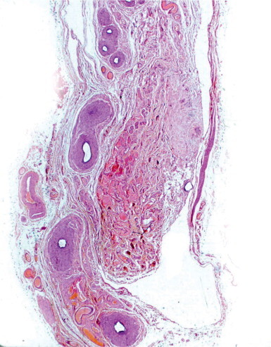
Spermatic cord in anorchidism. Fibrous connective tissue with dystrophic calcification surrounds the distal end of the vas deferens and replaces the testis.
The minimal requirement to diagnose vanishing testis is to find either a vascularized fibrous nodule with calcification or hemosiderin, or a fibrous nodule with cord elements.275 It has been proposed that removal of the testicular nubbin in this syndrome may not be required because the percentage of seminiferous tubules is very low and the presence of germ cells low, and thus the probability of a tumor is minimal.276, 277 The general recommendation is scrotal exploration as a first step, reserving laparoscopy for cases in which either the atrophic remnant cannot be identified during scrotal exploration or has a patent vaginal process.266
Etiology
The histologic findings suggest that most cases of unilateral and bilateral anorchidism are produced during the fetal period after the testis has inhibited the müllerian ducts and induced differentiation of wolffian duct derivatives. Two hypotheses account for the disappearance of the testes: primary anomaly of the gonad; and atrophy secondary to a vascular lesion such as thrombosis or intrauterine torsion. The presence of macrophages with hemosiderin and dystrophic calcification supports the latter. Absence of one testis may be associated with malformations of the urogenital system, such as absence of the kidney, cystic seminal vesicles, and ipsilateral renal dysgenesis.278, 279
Micro-orchidism
This clinical term refers to diverse conditions (Klinefelter's syndrome, hypogonadotropic hypogonadism, rudimentary testes syndrome, bilateral cryptorchidism, etc.) that share small testicular size.280, 281
A peculiar case is presented by some patients with Kenny–Caffey syndrome: short stature, cortical thickening and medullary stenosis of long bones, delayed closure of anterior fontanelles, hypoparathyroidism, and several ocular alterations. FSH serum levels are elevated, but only in some cases, whereas LH and testosterone are normal. Adult testes are small, with seminiferous tubules showing complete but diminished spermatogenesis. Leydig are hyperplastic. Unlike patients with the rudimentary testes syndrome, micro-orchidism patients have a normal-sized penis and no epididymal or prostatic atrophy.282
Polyorchidism
Polyorchidism is a rare condition, with approximately 100 reported cases.283, 284 It was first described in a postmortem study in 1880,285 and the first case treated surgically and confirmed histologically was reported in 1895.286 Although three testes are the most common,287 four testes have been reported in six patients,288, 289, 290, 291, 292 and five in one case but without histologic confirmation.293 Age of diagnosis varies from newborn to 74 years, with a mean of 17 years. Testicular duplication is usually an incidental finding during surgery for inguinal hernia, cryptorchidism, or testicular torsion, but has also been detected in patients with infertility or unexplained fertility after bilateral vasectomy.294 The extra testis is often intrascrotal (75%) and less frequently inguinal (20%), abdominal,295 or retroperitoneal (5%).296, 297 Duplication is three times more frequent on the left than on the right.298 High-resolution ultrasound is the appropriate diagnostic technique.284, 299 Testicular maldescent (40%), inguinal hernia (30%), hydrocele, varicocele, and contralateral cryptorchidism are the most frequently associated anomalies.300, 301, 302 Testicular torsion (13%)303 and testicular cancer (5.4%) are occasional complications. Although the extra testis may be histologically normal,304, 305, 306 usually it is not,300, 307 and displays lesions such as Sertoli cell-only tubules, hypospermatogenesis, or maturation arrest. The lack of spermatogenesis has been attributed to the anomalous location of the testis and the absence of communication between the testis and excretory ducts.308
The embryologic origin of polyorchidism remains uncertain, and the following have been proposed to account for the variety of findings in different cases (Fig. 12-28 ):
-
•
Longitudinal division of all the structures of the genital ridge and mesonephric ducts. Each of the two testes resulting from the duplication has an excretory duct and develops active spermatogenesis.286, 294, 309, 310, 311
-
•
Longitudinal division of the genital ridge. Of the two resulting testes, the medial loses its connection with the mesonephric ducts and undergoes atrophy.
-
•
High transverse division of the genital ridge. The two resulting portions are in continuity with the mesonephric ducts that give rise to the ductuli efferentes. Each testis has its own ductus epididymidis or shares a common one, but there is a single vas deferens for both.302, 312
-
•
Low transverse division of the genital ridge. The more caudal testis has no excretory ducts.302
Fig. 12-28.
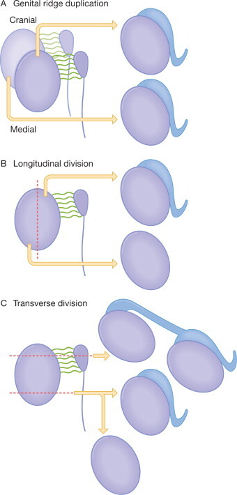
Possible mechanisms of polyorchidism. (A) Genital ridge duplication gives rise to two testes with their respective epididymides. (B) Longitudinal division of the genital ridge. The testis derived from the medial region has no epididymis. (C) Transverse division of the genital ridge. The resulting testes either share a single epididymis or one testis is devoid of epididymis.
The clinical differential diagnosis of polyorchidism includes most of pathologic conditions that enlarge the scrotum and spermatic cords: spermatocele, hydrocele, cysts and tumors of the spermatic cord, crossed testicular ectopia, adrenal cortical ectopia, and splenogonadal fusion. Orchidectomy used to be the treatment of choice for all atrophic and non-scrotal testes. Today, most surgeons undertake fixation of the testis to the scrotal pouch and the re-creation of a ‘simple testis’ if it is permitted by the anatomical condition and malignancy has been precluded. This treatment may allow spermatogenesis as well as additional psychologic and cosmetic benefits.313 Intrascrotal rhabdomyosarcoma, testicular teratoma, and seminoma have been reported in patients with polyorchidism.314, 315
Testicular hypertrophy (macroorchidism)
Macro-orchidism may be uni- or bilateral and be associated with chromosomal anomalies or endocrine alterations. An increase in the testicular parenchyma occurs in several conditions,316 including congenital Leydig cell hyperplasia, compensatory hypertrophy, benign idiopathic macroorchidism, bilateral megalotestes with low gonadotropins, fragile X chromosome, and the testicular hypertrophy observed in juvenile hypothyroidism.
Congenital Leydig cell hyperplasia
Congenital Leydig cell hyperplasia is uncommon and may be diffuse or nodular. The diagnosis of diffuse Leydig cell hyperplasia requires quantification of Leydig cells by morphometry, using normal newborn testes as controls (Fig. 12-29 ). Nodular Leydig cell hyperplasia is characterized by the presence of non-encapsulated Leydig cell nodules in the mediastinum testis, adjacent testicular parenchyma and connective tissue among the ductuli efferentes (Fig. 12-30 ).
Fig. 12-29.
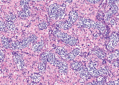
Congenital Leydig cell hyperplasia. Multiple nodules of Leydig cells are present in the mediastinum testis as well as deep in the parenchyma.
Fig. 12-30.
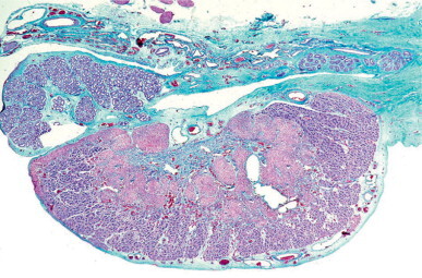
Congenital Leydig cell hyperplasia. Fetal Leydig cells form large clusters surrounding groups of seminiferous tubules.
The differential diagnosis of nodular Leydig cell hyperplasia includes intratesticular adrenal rests and bilateral Leydig cell tumor. Except for patients with adrenogenital syndrome, intratesticular adrenal rests are rare. These rests are encapsulated, with the exception of the adrenogenital tumors, and consist of radially arranged cells with vesicular nuclei and small nucleoli displacing the rete testis or seminiferous tubules. Leydig cell tumors may be bilateral, poorly circumscribed, and surrounded by testicular parenchyma, features making it difficult to distinguish from Leydig cell hyperplasia. However, Leydig cell tumors are rarely congenital, whereas those occurring at infancy often induce precocious maturation of the adjacent seminiferous tubules and early macrogenitosomia.
Leydig cell hyperplasia is caused by large quantities of hCG entering the fetal circulation. Diabetic mothers, particularly those with hypertension, may develop hyperplacentosis; the resulting edema in the placental villi alters the vascular permeability and allows the passage of hCG to the fetus. Congenital Leydig cell hyperplasia decreases rapidly during the first months of postnatal life, after maternal human chorionic gonadotropin is gone. Combined diffuse and nodular Leydig cell hyperplasia occurs in several malformative syndromes, such as Beckwith–Wiederman, leprechaunism, triploid fetuses, fetuses with Rh isoimmunization,317 and in several complications of pregnancy.
Compensatory hypertrophy of the testis
Compensatory hypertrophy has been observed in monor-chidism,318 cryptorchidism319 (Fig. 12-31 ), varicocele,320 and after testicular injury. Hypertrophy persists and may increase during childhood and puberty, but ceases thereafter; the hypertrophied testis then becomes normal or remains slightly enlarged.321, 322 The degree of hypertrophy is determined by three factors: the volume of the remaining testicular parenchyma, the age at which the injury occurred, and the functional ability of the descended testis.323 Compensatory hypertrophy results from an alteration in the hypophyseal hormonal feedback mechanism, followed by an increase in secretion of FSH, evidence that the contralateral testis is normal. In monorchidism, the testis is initially normal.237 When a 50% reduction of testicular mass occurs (probably before birth), the endocrine feedback changes and the resulting secretion of FSH (before or immediately after birth) causes accelerated growth of the contralateral testis. In cryptorchidism, the reduction in testicular mass is less severe than in monorchidism, and the scrotal testis may also be abnormal, inducing a lesser compensatory hypertrophy. Compensatory hypertrophy develops between birth and 3 years of age, and the testis may reach a volume twice normal when the other testis is absent.243
Fig. 12-31.
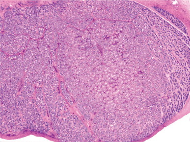
Contralateral scrotal testis from a cryptorchid patient showing a group of large seminiferous tubules that stands out from the surrounding small tubules.
Idiopathic benign macroorchidism
Some prepubertal and pubertal patients have pronounced unilateral324 or bilateral325, 326, 327 testicular hypertrophy in the absence of other pathologic findings. This probably results from hormonal receptivity in the testicular parenchyma. Morphometric studies have shown that the testicular enlargement is chiefly due to an increase in the length of the seminiferous tubules, although increases in tubular diameter and Sertoli cell numbers have also been observed. Elevated FSH serum levels, reported in some cases, or hyperactive FSH receptors might be the cause of the excessive Sertoli cell proliferation and the lengthening and thickening of seminiferous tubules.328, 329, 330 In addition, Leydig cell hyperplasia and deficient spermatogenesis are frequent findings in adult life. As the development of the two testes may be asynchronous during puberty, some unilateral macroorchidisms may represent cases in which these differences are unusually exaggerated.
Bilateral megalotestes with low gonadotropins
About 2% of adults with fertility problems have enlarged testes, with volumes over 25 mL, and low levels of FSH, LH, testosterone, prolactin, and estradiol.331 Despite the important hormonal changes, sperm concentrations and total numbers of spermatozoa are higher than normal. Low FSH levels may be attributable to increased inhibin secretion because the number of Sertoli cells is elevated in these testes, but no explanation for the reduction in the other hormone levels has been found.
Fragile X chromosome; Martin–Bell syndrome
Fragile X chromosome is the best-known form of inherited mental retardation, with an incidence of 1 in 1500 males and 1 in 2500 females.332 In addition to facial dysmorphia (large ears, prognathism, high forehead, and arched palate), macroorchidism (Martin–Bell syndrome) is often an associated finding.333, 334, 335, 336, 337 The impaired gene (FMR1 gene) is mapped to Xq27,3 which is genetically fragile. The gene alteration is due to a lengthening of a trinucleotide CGG repeat that results in FMR1 gene silencing. If the CGG sequence is repeated fewer than 200 times, the disorder is considered a premutation and males show no symptoms; if the number of repetitions exceeds 200, mutation is complete and all show the disorder.338, 339, 340 In men with this syndrome, the average testicular volume is more than 70 mL (four times greater than normal). The penis also is larger than normal, and both anomalies are apparent in infancy. The scrotum is also enlarged and prematurely pigmented. This precocious genital development is difficult to explain because the hypothalamopituitary axis is normal, but it may be caused by increased sensitivity to stimulation by FSH.341
Testicular biopsies from adults may be normal or show interstitial edema and hypospermatogenesis (Fig. 12-32 ). Usually, there is normal testicular parenchyma with focal reduced spermatogenesis and Sertoli cell hyperplasia (Fig. 12-33 ) or tubules containing only immature Sertoli cells. Morphometry indicates that testicular enlargement is chiefly the result of lengthening of seminiferous tubules.328 The low number of spermatids is attributed to atrophy caused by compression of the seminiferous epithelium by marked increase in intratubular fluid.342 Meiotic anomalies have been excluded.343 The fragile X syndrome is second in frequency only to Down's syndrome as a cause of mental retardation.344, 345, 346 However, this chromosomal anomaly is not always associated with mental retardation or macroorchidism, and there are men with fragile X syndrome who are otherwise normal.347
Fig. 12-32.
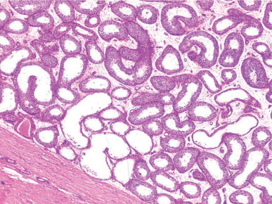
Martin–Bell syndrome (fragile X chromosome). The seminiferous tubules show variable degrees of dilatation and marked hypospermatogenesis.
Fig. 12-33.
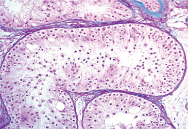
Martin–Bell syndrome (fragile X chromosome). The seminiferous tubules show marked hypospermatogenesis. Several groups of dysgenetic Sertoli cells are seen near the lumen.
The terms ‘fragile X-negative Martin–Bell syndrome’ or ‘mental retardation–macro-orchidism’ refer to X-linked (MRMO) or XLMR+MO patients who have the Martin–Bell syndrome phenotype but do not present the fragile X site. The gene responsible for this disorder is mapped to Xq12-q21.348
Other testicular hypertrophies
Testicular hypertrophy appears associated with FSH-secreting pituitary adenoma,349 hyperprolactinemia, hypoprolactinemia, and hypothyroidism.350, 351 The most frequent association of testicular hypertrophy is with hypothyroidism. Children with hypothyroidism often show testicular enlargement without virilization.350 About 80% have macroorchidism,352 most have elevated FSH levels, and half have increased LH levels.353, 354 Testosterone levels are normal during infancy. The response of FSH and LH to GnRH is altered and no pulsatile LH release occurs (Fig. 12-34 ).355
Fig. 12-34.
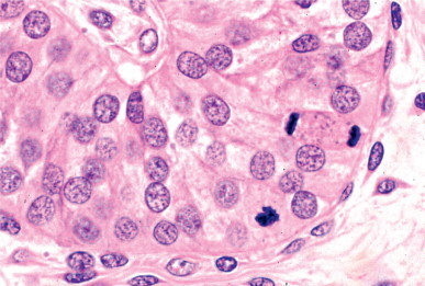
Macroorchidism in a 3-year-old infant with hypothyroidism. The Sertoli cells have spherical nuclei which contain small heterochromatin granules. Two mitotic figures are seen. The testicular interstitium has no Leydig cells.
Testicular biopsies before puberty show an accelerated development of the testis with pubertal maturation of seminiferous tubules but not Leydig cells. Testicular biopsies in untreated adults show tubular and interstitial hyalinization with few Leydig cells.356, 357 Testicular size in this type of macroorchidism diminishes as soon as the substitutive therapy starts.353, 358, 359 The etiopathogenesis has been explained by three hypotheses: an increase in gonadotropin secretion caused by TRH stimulation of gonadotropic cells;360, 361 a direct TSH effect on the testis due to the structural similarity between TSH receptors and FSH receptors present in the testis;362 and a lack of steroid hormones that are required for testicular maturation (in their absence, Sertoli cell proliferation is excessive, giving rise to testicular enlargement).363, 364, 365, 366
Precocious puberty
Precocious puberty is defined by onset of secondary sex characteristics at a chronologic age that is below the mean middle age for the population. For practical purposes, this is considered to be before 8 years of age in girls and 9 years in boys. The incidence is estimated at between 1 in 5000 and 1 in 10 000, with a female:male ratio higher than 20:1. In boys, the first symptom is rapid testicular enlargement followed by growth of pubic and axillary hair, enlargement of the penis, and acceleration of skeletal growth.367
According to hypothalamopituitary–gonadal axis function, precocious puberty can be classified into three groups: central or gonadotropin-dependent, which results from the activation of this axis; peripheral or gonadotropin-independent, mediated by sex steroid hormones secreted by the testis or adrenal glands; and a mixed group that first appears as peripheral precocious puberty and thereafter, because of the secondary response of the hypothalamus, becomes gonadotropin dependent.
Other possible causes of precocious puberty are hypoprolactinemia, pituitary tumor, and alteration of testicular steroid metabolism.
Central precocious puberty (CPP)
Central precocious puberty, also known as true precocious puberty, is isosexual. It is the most common form of precocious puberty in girls and accounts for more than 50% of cases in boys. The age of presentation is between 4 and 10 years.368 The cause is only known in 60% of cases; most are related to lesions in the central nervous system, whereas the others are usually idiopathic.
Lesions in the central nervous system that causes CPP share alterations of specific areas, including the posterior hypothalamus (eminencia media and tuber cinereum), mammillary bodies, the bottom of the third ventricle, or the pineal gland.369, 370 The most frequent causes are:
-
•
Tumor of the hypothalamus (astrocytoma, ganglio-neuroma, ganglioglioma, craniopharyngioma, cyst of the third ventricle, and suprasellar cyst of the arachnoid space),371, 372, 373 hamartoma (gangliocytoma) of the tuber cinereum and mammillary body, tumor of the pineal gland (teratoma and pinealoma), tumor of the optic nerve (glioma), and cerebral and cerebellar astrocytoma.
-
•
Cerebral trauma (including postpartum and accidental trauma) that stimulates the extrahypothalamic areas responsible for hypothalamic activation.374, 375, 376
-
•
Infections such as meningitis, encephalitis, toxoplasmosis, and syphilis.
-
•
Cerebral malformations, including hydrocephaly, microcephaly, and craniosynostosis.377
-
•
Hereditary diseases as neurofibromatosis and tuberous sclerosis. Children with type I neurofibromatosis often have also optic pathway tumors.
-
•
Cerebral irradiation, as occurs in hypothalamopituitary selective irradiation,378 prophylactic irradiation in children with acute lymphoblastic leukemia,379 and irradiation of cerebral tumor that is far from the hypothalamopituitary region.
The diagnosis of central precocious puberty is easy if the hormonal findings show elevated gonadotropin levels (both basal values and in response to GnRH), associated with high testosterone levels and an increase in either LH/FSH ratio or in LH and FSH values after stimulation with GnRH agonists. However, in some cases it is necessary to measure nocturnal LH secretion to find secretion pulses before a dynamic test can reveal the pubertal pattern.
Knowledge of the etiology in males has improved with the use of CT and MRI.380, 381 One of the most important contributions of these techniques is the finding of a high number of hamartomas in children with precocious puberty.382, 383, 384 These lesions, also known as gangliocytomas, consist of abnormally located neurons and glial cells. Lesions are usually multiple, small, and located on the hypothalamus between the anterior part of the mammillary body and the posterior part of the tuber cinereum. These neurons contain LHRH-positive neurosecretory granules, suggesting that this hormone can be released into the blood draining the hypophyseal portal system and reach the gonadotropic cells.385
Precocious puberty owing to cerebral tumors usually occurs with advanced stage of the tumor, preceded by cerebral symptoms such as hydrocephaly, papillary edema, or psychic alterations. The same occurs when precocious puberty results from cerebral inflammation or cerebral malformation.
Although pineal gland tumor is rare in children, 30% produce precocious puberty, principally in boys. This tumor is usually a teratoma or non-parenchymatous tumor that destroys the pineal gland, hindering its antigonadotropic action and initiating puberty.386 In contrast, pinealocyte-derived tumor secretes great amounts of melatonin that delay the onset of puberty.
Peripheral precocious puberty (PPP)
Peripheral precocious puberty is also known as precocious pseudopuberty. It may be caused by a primary testicular disorder, a lesion in other endocrine glands, or hormonal treatment. Primary testicular disorders causing precocious pseudopuberty include familial testotoxicosis, functioning testicular tumor, excessive aromatase activity, or Leydig cell hyperplasia with focal spermatogenesis. The principal secondary anomalies include adrenal cortical anomaly (congenital adrenal hyperplasia, virilizing tumor of the adrenal, and Nelson's syndrome), and lesion secondary to hCG-secreting tumor (hepatoblastoma accounts for half of precocious pseudopuberty cases, and testicular germ cell tumor and the tumors of the retroperitoneum, mediastinum, and pineal gland are responsible for the other half of cases).387
Familial testotoxicosis: gonadotropin-independent precocious puberty (GIPP) or familial male-limited precocious puberty (FMPP) Familial testotoxicosis is a form of male sexual precocity characterized by early differentiation of Leydig cells and the initiation of spermatogenesis in the absence of stimulation by pituitary gonadotropin. This is a primary testicular abnormality with autosomal dominant inheritance.388, 389 Ultrastructural studies confirm an adult Leydig cell pattern and complete spermatogenesis, although many spermatids are abnormal.390 The cause of familial testotoxicosis is a constitutive activating mutation of the LH receptor gene.391 This gene comprises 11 exons and has been mapped to 2p21. Hormonal measurements show elevated serum levels of testosterone, and low levels of dihydroepiandrosterone sulfate, androstenedione, 17-hydroxyprogesterone, gonadotropin-releasing hormone (GRH), and LH, as well as absence of a pulsatile pattern. In addition, serum levels of inhibin B appear elevated before the normal age of onset of puberty.392 In some patients, a mutation in LH receptor induces Leydig cell adenoma.393
Precocious puberty secondary to functioning testicular tumor A syndrome of precocious puberty can be the result of different tumors, including Leydig cell tumor, sex cord tumor, adrenal cortex virilizing carcinoma, and extratesticular hCG-secreting germ cell tumor.
Leydig cell tumor may cause precocious puberty. The testis is enlarged owing to tumor growth and maturation of the seminiferous tubules adjacent to the tumor; such maturation results from androgen secretion by tumor cells (Fig. 12-35 ). In most cases, the contralateral testis is not enlarged.394, 395
Fig. 12-35.
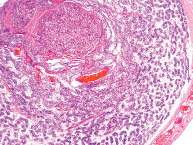
Precocious maturation of seminiferous tubules, which surround a virilizing Leydig cell tumor.
Sex cord tumor with annular tubules and large cell calcifying Sertoli cell tumor may give rise to precocious pseudopuberty that is isosexual (development of musculature and axillary and pubic hair) and heterosexual (gynecomastia). This precocious testicular maturation and the development of the tumor itself cause testicular enlargement. It has been suggested that tumor cells stimulate Leydig cells to produce androgens that are aromatized to estrogens by the tumor cells themselves, thus accounting for the clinical symptoms. These tumors are frequently observed in Peutz–Jeghers syndrome396, 397 and Carney's complex.398
Most infants with adrenal cortex virilizing tumors have small testes, but some cases of testicular hypertrophy have also been observed.399 Testicular development in these cases is attributed to adrenal androgenic action on seminiferous tubules.400 In untreated (or maltreated) congenital adrenal hyperplasia, both testes can be enlarged because they contain growing masses of adrenal cortex-like cells.401 A similar condition is observed in Nelson's syndrome.
Testicular enlargement is modest in paraneoplastic precocious pseudopuberty secondary to hepatoblastoma402 or extratesticular hCG-secreting germ cell tumor, although nodular or diffuse precocious maturation has been occasionally reported.403
Precocious pseudopuberty secondary to excessive aromatase activity Biosynthesis of C18 estrogens from C19 androgens occurs by three consecutive oxidative reactions that are catalyzed by an enzymatic complex known as estrogen synthetase or aromatase.404 This complex has two components: P450 arom (a product from the CYP19 gene located on 15p21.1),405 which joins C19 substrate and catalyzes the insertion of oxygen in C19 to form C18 estrogens; and NADPH-cytochrome P450 reductase, a ubiquitous flavoprotein that conveys reducing equivalents to any form of cytochrome P450 it meets.
Aromatase is in the endoplasmic reticulum of estrogen-synthesizing cells and expressed in placenta, ovarian granulosa, Sertoli cells, Leydig cells, adipose tissue, and several central nervous system regions, including the hypothalamus, amygdala, and hippocampus. Excessive aromatase causes excessive conversion of androgens to estrogen,406 and is a heterogeneous genetic disorder with an autosomal dominant inheritance. The disorder leads to heterosexual precocious pseudopuberty with gynecomastia in males, and to isosexual precocity and macromastia in females. Ultimately, patient stature is short because of the potent ability of androgens to accelerate epiphyseal closure. Most males are fertile and have normal libido.407 Generally, the inhibitory estrogenic effect on testicular function is less than that observed with estrogen-producing tumors or in patients treated with exogen estrogens.
Excessive aromatase caused by P450 mutation induces alterations in both males and females. In females lacking estrogens owing to desmolase deficiency, excessive aromatase leads to pseudohermaphroditism and progressive virilization at puberty; conversely, pubertal development is normal in males. In children, FSH and LH levels and gonadotropin response to GnRH are normal, suggesting that the role of estrogens in pituitary regulation is weak during infancy.408 In both genders, epiphyseal closure is delayed and a eunuchoid habitus results. Adult males have small testes, severe oligozoospermia, and complete asthenozoospermia; FSH and LH levels are high, testosterone levels are normal, and serum estrogen levels are very low.
All patients with excessive aromatase have short stature, with continuing linear growth into adulthood, unfused epiphyses, osteoporosis, bilateral genu valgum, and eunuchoid proportions. The testes show macroorchidism with normal testicular consistency in some cases,409 and are small with severe oligozoospermia and 100% immotile spermatozoa in other cases.410
A syndrome similar to that of excessive aromatase production is found in patients with estrogen resistance caused by disruptive mutations of the ER gene. These patients show macroorchidism, elevated testosterone levels, and increased levels of FSH, LH, estradiol, and estrona.411
Precocious pseudopuberty secondary to Leydig cell hyperplasia with focal spermatogenesis This entity can present with clinical symptoms similar to those of a functioning Leydig cell tumor; this is a precocious pseudopuberty with ipsilateral testicular enlargement.412 The testes contains hypertrophic Leydig cell nests in association with normal spermatogenesis. No tumoral mass is seen. Leydig cells do not contain Reinke's crystalloids and do not compress the seminiferous tubules. There is a clear delimitation between tubules with spermatogenesis and infantile immature tubules. The differential diagnosis between this entity and Leydig cell tumor with precocious pseudopuberty is based on the histological pattern. Open excisional testicular biopsy is recommended; if there is Leydig cell tumor, or the diagnosis by frozen section is not conclusive, removal is advisable.413 There are no data to suggest that this hyperplasia might develop into Leydig cell tumor.
Mixed precocious puberty
The best known form is the McCune–Albright syndrome (MAS), characterized by the association of ‘coffee and milk’ pigmentary lesions in the skin, bone lesions (polyostotic fibrous dysplasia), enlarged testes, prepubertal size of the penis, and absence of pubic and axillary hair. Although testicular enlargement is usually bilateral, unilateral macroorchidism may be the first symptom.414 An interesting finding is that the onset of testicular maturation is induced by the testis itself, which produces steroid secretion due to autonomous hyperfunction of Sertoli cells without evidence of Leydig cell involvement.415 This secretion causes early maturation of the hypothalamopituitary–testicular axis and, subsequently, true precocious puberty.416 Serum levels of testosterone are low, but those of inhibin B and AMH are abnormally increased. This syndrome is caused by mutations that activate the GNAS-1 gene, which encodes the α subunit of the trimeric G-protein. Because mutations are lethal in the uterus, those subjects producing AMH bear a mosaicism chromosomal constitution for this deficiency.
Testicular ectopia and testicular fusion
Testicular ectopia
A testis is ectopic when it is in a location outside the normal path of descent. Unlike cryptorchid testes, ectopic testes are nearly normal in size and are accompanied by a spermatic cord that is normal or even longer than normal, and by a normal scrotum.417
Testicular ectopia is classified according to location;418, 419, 420, 421, 422 in decreasing order of frequency, the major types are:
-
•
Interstitial or inguinal superficial ectopia. This is the most frequent form and may be confused with inguinal cryptorchidism. After passing through the outer genital opening, the testis ascends to the anterosuperior iliac spine and remains on the aponeurosis of the major oblique muscle. These testes often are more nearly normal histologically than are cryptorchid testes.
-
•
Femoral or crural ectopia. After passing through the inguinal canal, the testis lodges in the high crural cone in Scarpa's triangle.
-
•
Perineal. The testis is located between the raphe and the genitocrural fold.
-
•
Transverse or crossed ectopia. Both testes descend through the same inguinal canal and lodge in the same scrotal pouch. Each possesses its own vascular supply, epididymis, and vas deferens. In addition, there is ipsilateral hernia.423, 424, 425, 426, 427, 428, 429, 430 Between 20% and 40% of patients with this ectopia have persistent müllerian duct syndrome431, 432 and show a high incidence of testicular germ cell tumor.433
-
•
Pubopenile ectopia. The ectopic testis is on the back of the penis near the symphysis pubis.434
-
•
Pelvic ectopia. The testis is in the pelvis, usually in the depth of Douglas’ cul-de-sac.
-
•
Other unusual testicular ectopias include retroumbilical, craniolateral to the inner inguinal opening between the outer and inner oblique muscles, and subumbilical.435 Rarely, the testis and its spermatic cord may protrude through a defect in the scrotal skin, a condition called testicular exstrophy.436
The term testicular dislocation refers to testes that secondarily disappear from the scrotum and lodge around the superficial inguinal ring, within the inguinal ring, or inside the abdominal cavity as a result of testicular trauma. The formation of canalicular and intra-abdominal dislocation requires the presence of previous inguinal hernia.437
Testicular fusion
Testicular fusion is a rare anomaly characterized by fusion of the testes to form a single structure, usually in the midline. Each has its own epididymis and vas deferens. This anomaly is often associated with other malformations, such as fusion of the adrenal glands or horseshoe kidney.
Hamartomatous testicular lesions
Cystic dysplasia
Cystic dysplasia of the testis is a congenital lesion characterized by cystic transformation of an excessively developed rete testis that may extend to the tunica albuginea of the opposite pole.438 To date, fewer than 40 cases have been reported.439, 440 The seminiferous tubules may be dilated and atrophic; this is more evident after puberty. Ultrasound images are characteristic.441, 442 Cysts arise in the septal and mediastinal rete testis (Fig. 12-36 ); they are interconnected and contain acellular, eosinophilic, periodic acid–Schiff-positive material. They are lined by cuboidal cells that resemble those of the normal rete testis.443, 444, 445 The connective tissue between the cysts is scant and histologically similar to the interstitial connective tissue. There may be small groups of cysts limited to the region of the mediastinum testis, or cysts extending throughout the entire testis. In extensive cases, residual seminiferous tubules occupy only a small crescent beneath the tunica albuginea and the testis is grossly spongy. Cystic dysplasia occurs in normally descended and cryptorchid testes in children and adults, and may affect one or both testes.446 In adults, the residual parenchyma often shows complete tubular sclerosis or hypospermatogenesis with intratubular accumulation of spermatozoa and Leydig cell pseudohyperplasia.
Fig. 12-36.
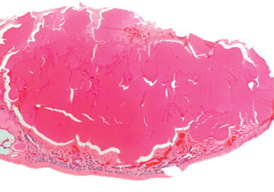
Cystic dysplasia of the testis. There is cystic transformation of the rete testis and adjacent seminiferous tubules.
In most cases the epididymis is altered.447 The head of the epididymis is small and contains few ductuli efferentes with irregular, usually dilated lumina. The ductus epididymidis is dilated, has an atrophic epithelium, and thick connective tissue replaces the muscular layer (Fig. 12-37 ).
Fig. 12-37.
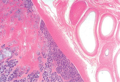
Marked luminal dilation of the ductus epididymidis in an infant with cystic dysplasia of the rete testis.
Testicular cystic dysplasia is frequently associated with severe anomalies of the urinary system. Renal agenesis,446, 447, 448, 449 renal dysplasia,446 hydroureter, and urethral stenosis450 have been reported ipsilateral to cystic dysplasia. The clinical differential diagnosis should consider all cystic testicular lesions impairing prepubertal testes, including epidermoid cyst, cystic teratoma, juvenile granulosa cell tumor, testicular lymphangiectasis, and simple cyst of the testis.451 The presence of ipsilateral renal anomalies during ultrasound exploration provides an important diagnostic clue.452 Previously, orchidectomy was the treatment of choice, but testis-sparing surgery453 is now recommended.454, 455
The etiology and pathogenesis of cystic dysplasia are uncertain. Given that the rete testis is a mesonephric derivative and most of the associated renal malformations are apparently caused by failure in the induction of renal blastema by the mesonephros, cystic dysplasia is considered to be the result of an abnormal mesonephros.
During childhood, the normal rete testis has no lumina, and these form during puberty. The adult rete testis is a conduit for the passage of tubular fluid and spermatozoa and also actively reabsorbs part of this fluid while adding ions, proteins and steroids to it. Malfunction of the rete testis cells may cause the formation of excessive fluid of abnormal composition, resulting in a condition morphologically similar to cystic dysplasia of the rete testis induced in fowl by sodium intoxication or the administration of the salt-retaining hormone deoxycorticosterone acetate.
Gonadoblastoid testicular dysplasia
Gonadoblastoid testicular dysplasia refers to an abnormally differentiated testicular parenchyma beneath the tunica albuginea.456 The anomaly consists of large tubular or nodular structures within a dense stroma, reminiscent of ovarian stroma (Fig. 12-38 ). Each structure is composed of three cell types: cells with vesicular nuclei and vacuolated cytoplasm; cells with hyperchromatic nuclei; and germ cell-like cells. The former two types are arranged at the periphery, forming a pseudostratified epithelium. The third type resembles fetal spermatogonia and are fewer in number. These structures contain eosinophilic, periodic acid–Schiff-positive material, similar to Call–Exner bodies (Fig. 12-39 ). There may be continuity between these structures and normal seminiferous tubules. The differential diagnosis includes conditions showing anomalous seminiferous tubules at the gonadal periphery, including testicular dysgenesis and gonadoblastoma. Testicular dysgenesis also presents tubular or cord-like structures, but these are differentiated (some form true seminiferous tubules) and may also be present within a poorly collagenized tunica albuginea; patients with testicular dysgenesis are male pseudohermaphrodites with müllerian remnants. Gonadoblastoma usually appears in a streak gonad or dysgenetic gonad and contains granulosa–Sertoli cells and germ cells that are similar to those of dysgerminoma or seminoma; these cells are absent in gonadoblastoid testicular dysplasia. Several cases with this disorder have been reported in patients with Walker–Warburg syndrome.457, 458
Fig. 12-38.
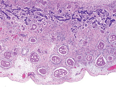
Gonadoblastoid testicular dysplasia. Several nodules are present at the periphery of the testicular parenchyma.
Fig. 12-39.
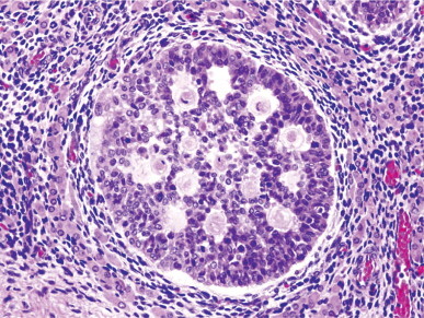
Gonadoblastoid testicular dysplasia. A nodule contains numerous Sertoli-like cells, Call–Exner bodies, and isolated germ cells. The nodule is surrounded by two cell layers: fusiform cells (inner layer) and Leydig cells (outer layer).
Sertoli cell nodule (hypoplastic zones or dysgenetic tubules)
This disorder refers to the presence, in an adult testis, of one or several foci of infantile (immature) seminiferous tubules. Each group of tubules appears well delimited but unencapsulated. Nodule size varies from microscopic to 5 mm. On section, each nodule is distinguished by its whitish color. Sertoli cell nodule is found in most adult cryptorchid testes, regardless of when the testes descended. It is also present in 22% of normal scrotal testes in some series,459 and is an occasional finding in males with idiopathic infertility.
The seminiferous tubules have a prepubertal diameter and may be anastomotic. The epithelium is columnar or pseudostratified, devoid of lumina, and usually consists only of Sertoli cells (Fig. 12-40 ). The cells have elongated hyperchromatic nuclei with one or several peripherally placed small nucleoli.459 The interstitium varies from scant to well collagenized. Leydig cells are usually absent in these areas and, if present, their numbers are low. Study of serial sections reveals continuity between some of these tubules and normal tubules. Sertoli cell nodule changes with advancing age. The Sertoli cells produce large amounts of basal lamina that protrudes inside the hypoplastic tubules. In transverse and oblique sections, these protrusions might be misinterpreted as intratubular accumulations of basal lamina material (Fig. 12-41 ). This material can undergo calcification to form microliths. Immunohistochemical study reveals two basic components of the basal lamina (collagen IV and laminin), confirming its extracellular origin; the protrusions consist mainly of laminin, whereas collagen IV delimits the outer profile of the seminiferous tubules. So, while the amount of collagen IV is uniform around the tubules, the depth of laminin varies within the same tubule.
Fig. 12-40.
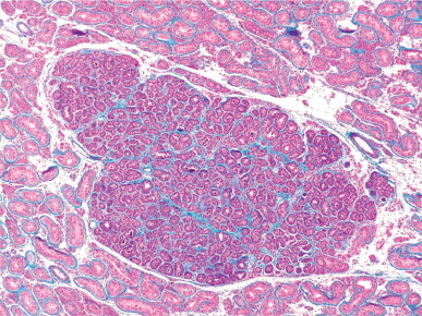
Sertoli cell nodule. This adult cryptorchid testis contains compact groups of small seminiferous tubules with pseudostratified cell layers without lumina.
Fig. 12-41.
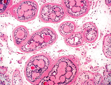
Sertoli cell nodule. Sertoli cell-produced material, similar to the basal lamina material, forms fingerlike protrusions inside the hypoplastic tubules. The Sertoli cells are arranged in a ring around this material.
Tubular hypoplasia is assumed to be a primary testicular lesion, and refers to the presence of seminiferous tubules that are unable to undergo pubertal development despite the same hormonal stimuli of adjacent normal tubules. This dysgenesis includes immature Sertoli cell pattern, low inhibin secretion, absence of androgen receptors,460 and lack of maturation of peritubular myoid cells that fail to synthesize elastic fibers. The presence of hypoplastic zones in a testicular biopsy is an adverse prognostic sign for fertility.
The differential diagnosis includes tubular hamartoma in androgen insensitivity syndrome, sex cord tumor with annular tubules, and mixed atrophy of the testis. Tubular hamartoma in androgen insensitivity syndrome is multiple, similar to the hypoplastic zones of tubular hypoplasia; however, the Sertoli-like cells of hamartoma have spherical nuclei (rather of elongated nuclei), form a cuboidal epithelium, and contain numerous Leydig cells among the tubules (see Androgen insensitivity syndrome, below). Sex cord tumor with annular tubules may present with multiple foci of intratubular neoplasia, similar in distribution to that of hypoplastic zones; however, sex cord tumor appears in undescended testes, and in patients with Peutz–Jeghers syndrome, and consists of cuboidal or spherical cells that express cytokeratins that are not expressed in hypoplastic tubules.
It is possible that hypoplastic tubules contain some germ cells that may be spermatogonia or gonocytes. There are scant spermatogonia that fail to display signs of maturation or proliferation. Also, some of the tubules contain intratubular undifferentiated germ cell neoplasia that usually also appears in the adjacent, non-hypoplastic seminiferous tubules. The histologic picture is similar to that of gonadoblastoma, but such a tumor can be easily excluded because it arises in malformed gonads (gonadal dysgenesis and testicular dysgenesis) characteristic of intersex stages, unlike patients with tubular hypoplasia.
Congenital testicular lymphangiectasis
Congenital testicular lymphangiectasis is characterized by abnormal and excessive development of lymphatic vessels in the tunica albuginea, mediastinum testis, interlobular septa, and testicular interstitium.461, 462, 463 Ultrastructurally these dilated vessels are similar to normal lymphatic capillaries, although some are markedly dilated and the testicular interstitium is slightly edematous (Fig. 12-42 ). Testicular lymphangiectasis occurs in both cryptorchid and scrotal testes; in one of the latter cases, the patient had Noonan's syndrome. The disease does not seem to affect the seminiferous tubules, and low numbers of spermatogonia and reduced tubular diameters are observed only in cryptorchid testes. The epididymis and spermatic cord are not affected, and congenital testicular lymphangiectasis is not associated with pulmonary, intestinal, or systemic lymphangiectasis. During fetal life, lymphatic vessels are visible only immediately beneath the tunica albuginea and in the interlobular septa.464 During childhood, the number and size of the septal lymphatic vessels decreases;465 by adulthood they are inconspicuous.466 In lymphangiectasis, the septal lymphatic vessels are large and often massively dilated. Testicular lymphangiectasis occurs only in the childhood testis, suggesting that these dilated vessels undergo involution at puberty or that pubertal development of the seminiferous tubules masks the lymphangiectasis. One exceptional case of epididymal lymphangiectasis, with dilated epididymal blood vessels, was reported in a 59-year-old man.467 The vessels distort the architecture of the ductuli efferentes, which in turn become irregularly dilated by mechanical compression.
Fig. 12-42.
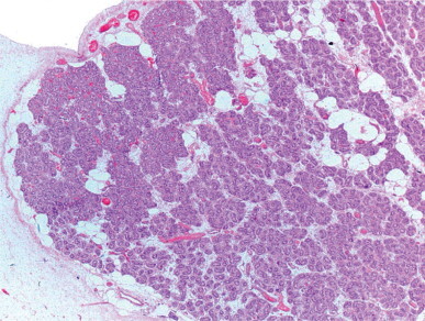
Congenital testicular lymphangiectasis. Ectatic lymphatic vessels are seen in the tunica vasculosa and interlobular septa, as well as among the seminiferous tubules, causing compression.
Other hamartomatous testicular lesions
Other hamartomas of the testis include hamartoma of the rete testis and smooth muscle hamartoma. Hamartoma of the rete testis is a disordered proliferation of tubular structures in a loose connective tissue.468 Cystic transformation of the rete testis associated with proliferation of smooth muscle cells and abundant myxoid stroma was reported in a 26-year-old man.469
Smooth muscle hamartoma is located in the inferior testicular pole, the cauda of the epididymis, and the proximal segment of the vas deferens (Fig. 12-43 ),470 and is similar to that reported in the digestive and respiratory tracts.471, 472 Smooth muscle hyperplasia also occurs in the androgen insensitivity syndrome, forming nodules up to 1 cm in diameter. The muscular proliferation is located in the lower testicular pole, and involves the tunica albuginea and adjacent soft tissues.
Fig. 12-43.
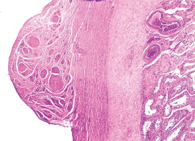
Smooth muscle hamartoma within enlarged tunica albuginea.
Testicular ectopia
Gonadal blastema ectopia
This infrequent finding has been observed in newborns and consists of gonadal blastema in otherwise normal testes. The blastema is located in the vicinity of the upper testicular pole, near the implantation of the caput of the epididymis, displays a crescent shape, and extends throughout the depth of the tunica albuginea and the adjacent testicular parenchyma.
The blastema consists of epithelial cords of cells or solid masses in continuity with the mesothelium (Fig. 12-44 ). These cells are intermingled with others that are larger, with pale cytoplasm, vesicular nuclei, and prominent nucleoli. The blastematous epithelial cells display immunoreactivity for vimentin, laminin, type IV collagen, and cytokeratin; the expression of the latter in the most superficial cells is similar to that of mesothelial cells and decreases in intensity in the deeper cells. This suggests that these may be pre-Sertoli cells. The cord-like structures are delimited by laminin and type IV collagen. The second larger cell type is immunoreactive for placenta-like alkaline phosphatase (PLAP) on the surface, suggesting that it is related to the gonocyte. Leydig cells have not been observed among the cords of gonadal blastema.
Fig. 12-44.
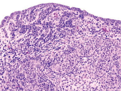
Persistent testicular blastema in a newborn. The blastema forms anastomosic cord-like formations which connect to the superficial cells. Several gonocytes are observed at the periphery of the blastema.
The differential diagnosis of gonadal blastema ectopia is with ovotestes. The small size of the gonocytes distinguishes them from ovocytes, which are several times larger. In addition, no intersex condition is observed.
Seminiferous tubule ectopia
The presence of seminiferous tubules within the tunica albuginea is rare and usually an incidental histologic finding.473 Ectopic tubules are present in approximately 0.8% of pediatric autopsies and 0.3% of adult autopsies. The lower incidence in adults may be explained by proportionally less sampling. The lesion ranges from microscopic size to a few millimeters in diameter, and may be visible as minute bulges in which multiple small vesicles protrude through a thin tunica albuginea.474 Histologically there are groups of seminiferous tubules in the tunica albuginea, sometimes accompanied by Leydig cells. In children, the ectopic tubules appear normal (Fig. 12-45 ), whereas in adults they are usually slightly dilated, although some may be hyalinized. Serial sections reveal continuity with the intraparenchymatous seminiferous tubules.
Fig. 12-45.
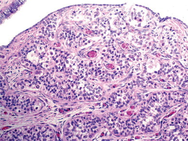
Testis from 2-month-old infant showing ectopic seminiferous tubules within the tunica albuginea in the upper testicular pole.
Ectopia of the seminiferous tubule is probably congenital, although it has been found in elderly men.475 It does not appear to be the result of trauma. The malformation probably arises in the sixth week of gestation, when the primordial sex cords have formed and are branching toward the gonadal surface, and the developing testes is covered by only one to three layers of celomic epithelium. Later, the tunica albuginea forms around the sex cords and under the celomic epithelium. Failure of insertion of the tunica albuginea between the sex cords and celomic epithelium may entrap seminiferous tubules.
Ectopia differs from testicular dysgenesis, a distinctive form of male pseudohermaphroditism with müllerian remnants. Numerous features, characteristic of ectopic seminiferous tubules, distinguish it from other conditions, including normal thickness and collagenization of the tunica albuginea, absence of interstitial tissue resembling ovarian stroma (characteristic of testicular dysgenesis), and clear delimitation of the tunica albuginea and testicular parenchyma (see discussion on male pseudohermaphroditism with müllerian remnants, below).
In a unique case, there were multiple clusters of seminiferous tubules in the wall of a hernia sac that accompanied an undescended testis removed from an adult man. The ectopic tubules were not surrounded by tunica albuginea and were similar to those in cryptorchid testicular parenchyma with only dysgenetic Sertoli cells.
Leydig cell ectopia
Leydig cells occur normally in the testicular interstitium (interstitial Leydig cells) and in the wall of the seminiferous tubules (peritubular Leydig cells). However, clusters of Leydig cells are often observed in other locations in the testis, or in the epididymis or spermatic cord.476
Ectopic Leydig cells may be found in the interlobular septa,477, 478, 479 rete testis, tunica albuginea,480, 481, 482 or within hyalinized seminiferous tubules.478, 483, 484, 485 Intratubular Leydig cells are found only in tubules with advanced atrophy and marked thickening of the tunica propria, including the tubules in adult cryptorchid testes, those of men with Klinefelter's syndrome, and in some other primary hypogonadisms (Fig. 12-46 ). Immunohistochemical studies suggest that the endocrine function of these Leydig cells is low.486 Several theories have been offered to account for these ectopic cells, including in situ differentiation, migration from the testicular interstitium, and trapping of peritubular Leydig cells in the tunica propria during its thickening.487 Leydig cells are commonly found in the epididymis487 and spermatic cord;488, 489 26 of 64 autopsies had such foci.490 Extratesticular Leydig cells usually form small groups within or adjacent to nerves (Fig. 12-47 ).477, 490
Fig. 12-46.
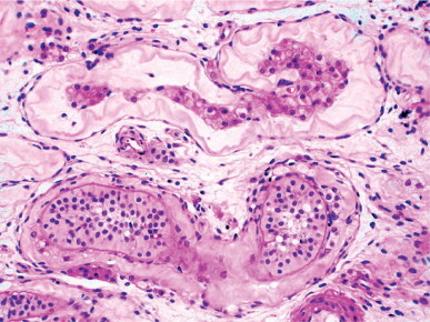
Ectopic Leydig cells inside a hyalinized seminiferous tubule. This picture contrasts with that of dysgenetic Sertoli-cell-only tubule, which shows a patent basal membrane located between the dysgenetic Sertoli cells and the tubular wall.
Fig. 12-47.
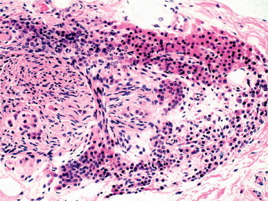
Ectopic Leydig cells around and inside a spermatic cord nerve.
The occurrence of ectopic Leydig cells in the albuginea, epididymis, or spermatic cord may account for the rare cases of Leydig cell tumor in these paratesticular structures. Ectopic Leydig cells should not be misinterpreted as tumor cells (infiltration or metastasis) when malignancy of a testicular Leydig cell tumor is suspected.
Other ectopias
Other rare forms of ectopia are found within and outside the testis. Intratesticular ectopia includes adrenal cortical ectopia, osseous and adipose tissue heterotopia, and ectopia of the ductus epididymidis. Extratesticular ectopia includes splenic ectopia (splenogonadal fusion), hepatic ectopia (hepatotesticular fusion), and renal blastema ectopia (see discussion in Chapter 12).
Adrenal cortical ectopia may be important in two conditions that develop tumoral masses: adrenogenital syndrome and Nelson's syndrome. Tumors in adrenogenital syndrome appear in 8.2% of patients with congenital adrenal hyperplasia, appearing as bilateral testicular masses of synchronous growth. These tumors consist of well delimited but non-encapsulated yellow nodules, several centimeters long, composed of large microvacuolated cells. The cause seems to be prolonged stimulation by elevated ACTH secretion. The differential diagnosis includes Leydig cell tumor. The diagnosis of tumors in adrenogenital syndrome is supported by a family or personal history of salt-lost syndrome or hypertension, demonstration of 11 β-hydroxysteroids (a specific marker for adrenal cortex) in spermatic vein blood, or a rapid positive response of tumor to corticoid treatment. Nelson's syndrome occurs in patients who, after adrenalectomy for treatment of Cushing's syndrome, develop an ACTH-secreting pituitary adenoma. These patients may develop testicular tumor growth similar to that in adrenogenital syndrome. Most Nelson's syndrome tumors do not respond to dexametasone treatment.
Cartilaginous heterotopia may be found in the caput of the epididymis and has been attributed to metaplasia of metanephric rests. Osseous heterotopia (testicular osteoma) is a metaplasia occurring in areas of the testicular parenchyma with fibrosis or ischemia.491 Adipose metaplasia is frequent in undescended testis, elderly men, and those with Cowden's syndrome (Fig. 12-48 ).492 Groups of tubular formations that resembles the epididymis have been reported inside the testicular parenchyma in testes with marked tubular atrophy, and probably represent a rare form of metaplasia.493
Fig. 12-48.
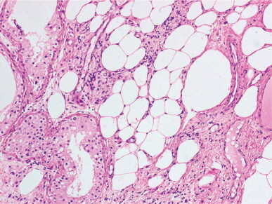
Adult cryptorchid testis showing metaplastic fat cells between the seminiferous tubules and the rete testis.
Undescended testes
Testicular descent is not always complete at birth, and about 3.2% of full-term newborns have incompletely descended testes. Most of these descend within 3 months, and only 0.8% of infants have incompletely descended testes 12 months after birth. Spontaneous testicular descent is exceptional after the first year. In recent decades, a significant increase in the incidence of cryptorchidism has been detected.494
Only 5% of patients with impalpable testes are actually devoid of testes. Other causes include true cryptorchidism, testicular ectopia, and retractile testes. True cryptorchidism includes abdominal, inguinal, and high scrotal testes that cannot be moved to the scrotum. Ectopic testes are those located out of the normal path of testicular descent; the most frequent site is the superficial inguinal pouch. Other rare locations of ectopia include the abdominal wall, the upper thigh, the perineum, and the base of the penis. Retractile testes may be moved to the scrotum at exploration and account for about one third of undescended testes.
True cryptorchidism
Patients with true cryptorchidism account for about 25% of cases of empty scrotum. These testes most frequently are found in the inguinal canal or upper scrotum; arrest within the abdomen is less frequent. Cryptorchidism is slightly more frequent on the right than the left, and in approximately 18% of cases is bilateral. There is a family history of cryptorchidism in 14% of cases.495 The cryptorchid testis is usually smaller than the contralateral one, and this difference is often discernible at 6 months of age.496 One-third of cryptorchid testes are soft.
Etiology
Several conditions are predictive of high risk of cryptorchidism, including increased maternal age, maternal obesity, pregnancy toxemia, bleeding during late pregnancy, and smoking, tallness, subfertility antecedents, cesarean birth, low birthweight, preterm newborn, twin birth, hypospadias497 and other congenital malformations, and children born from September to November, and in May and June.498, 499 Of these associations, low birth weight seems to be the most important.500
There are two types of cryptorchidism: congenital and acquired.
Congenital cryptorchidism
This cryptorchidism is caused by anomalies in anatomic development or hormonal mechanisms involved in testicular descent (described above). Impalpable undescended testes are infrequent because the transabdominal phase follows the simple mechanism of relative movement of the testis, whereas displacement of the ovary is more complex.501 Conversely, palpable undescended testes are more frequent because the second phase of testicular descent is more complex. Unilateral cryptorchidism may be caused by androgen failure, which leads to either an ipsilateral lesion in the development of genitofemoral nerve neurons or a defect in CGRP release that hinders normal migration of the gubernaculum.
Acquired cryptorchidism
A normally descended testis may become cryptorchid and locate even in the abdominal cavity. Two categories of acquired undescended testis have been described.
The postoperative trapped testis 502 is a normally descended testis that leaves the scrotal pouch after surgery owing to an inguinal hernia or hydrocele.503, 504, 505 This iatrogenic cryptorchidism occurs in 1.2% of children after herniotomy. Adherence of the testis or the cremasteric muscle to the surgical incision causes testicular ascent when the incision heals and undergoes retraction.
Spontaneous ascent from unknown causes. Various mechanisms have been proposed, including inability of the spermatic blood vessels to grow adequately,506 anomalous insertion of the gubernaculum,507 failure in reabsorption of the vaginal process508, 509 and failure in postnatal elongation of the spermatic cord.510, 511 The spermatic cord measures 4–5 cm at birth and reaches 8–10 cm at 10 years of age. This growth does not occur if the peritoneal–vaginal duct has become a fibrous remnant. The cause might be a defect in postnatal CGRP release by the genitofemoral nerve.501, 512, 513
Pathogenesis
The most frequent findings in congenital and acquired cryptorchidism at infancy are decreased germ cell numbers and diminished tubular diameter.514, 515 There are multiple causes of testicular maldescent, including anatomical anomalies of the gubernaculum testis, hormonal dysfunction (hypogonadotropic hypogonadism), mechanical impairment (insufficient intra-abdominal pressure, short spermatic cord, underdeveloped processus vaginalis), dysgenetic (primary anomaly of the testis), and heredity.
Most cryptorchidism appears to be caused by either a deficit of fetal androgens or an excess of maternal estrogens. Androgen insufficiency seems to be slight and transient because anomalies other than hypoplasia of the epididymis are not seen. Elevated maternal estrogens level could cause diminution of FSH secretion by the fetal pituitary, inducing low müllerian-inhibiting hormone production that would hinder testicular descent.516
Three mechanisms seem to be involved in the process:
-
•
Primary testicular anomaly. Cryptorchid testes may bear an anomalous germ cell population, as suggested many years ago.517 More than 40% of cryptorchid patients have a marked decrease in the tubular fertility index,518 even with nearly normal numbers of spermatogonia; these cells also have abnormal DNA content.519
-
•
Lesions secondary to transient perinatal hypogonadotropic hypogonadism. Cryptorchid patients do not have gonadotropin elevation, which normally occurs between 60 and 90 days after birth, and this deficiency of LH could cause Leydig cell involution. The subsequent androgen deficiency could account for failure of gonocytes to differentiate into spermatogonia.520, 521, 522
-
•
Injury caused by increased temperature. This was suggested in the past on the basis of experimental studies in laboratory animals. In follow-up biopsies from testes that were descended surgically or with hormonal treatment, the sole parameter that improved during childhood was tubular diameter. Because this depends on Sertoli cells, it may be that temperature is more important for Sertoli cells than for spermatogonia.518
In the normal testis there is transient formation of spermatocytes at 4–5 years of age. This meiotic attempt is probably an androgenic event that does not occur in cryptorchid testes and agrees with the characteristic low numbers of spermatogonia in the prepubertal age.523
Histology of cryptorchid testes
Prepubertal testes
Undescended testes are usually smaller than the contralateral ones. This difference is already significant at 6 months of age.524, 525 Although there have been a number of biopsy studies in the first years of life, there is no agreement about the severity of damage or the time of its onset.523, 526, 527 Based on the tubular fertility index (TFI) and mean tubular diameter (MTD), most testicular biopsies from cryptorchid testes of children can be classified into one of three groups:
-
•
Type I (testes with slight alterations). The tubular fertility index is higher than 50, and the mean tubular diameter is normal or slightly (<10%) decreased. Approximately 31% of cryptorchid testes are in this group (Fig. 12-49 ).
-
•
Type II (testes with marked germinal hypoplasia). Tubular fertility index is between 30 and 50, and mean tubular diameter is 10–30% lower than normal. The spermatogonia are distributed irregularly and most are in tubular sections that are grouped in the same testicular lobule. These testes comprise approximately 29% of cryptorchid testes (Fig. 12-50 ).528
-
•
Type III (testes with severe germinal hypoplasia). Tubular fertility index is less than 30, and mean tubular diameter less than 30% of normal. Many of the spermatogonia are giant with dark nuclei (Fig. 12-51 ). These testes often contain ring-shaped tubules, megatubules (with or without eosinophilic bodies or microliths) (Fig. 12-52 ), and focal granular changes in the Sertoli cells (Fig. 12-53 ). The testicular interstitium is wide and edematous. These comprise about 40% of cryptorchid testes.
Fig. 12-49.
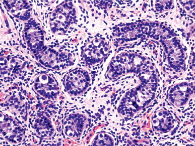
Cryptorchidism. Seminiferous tubules with type I lesions show slightly decreased diameters and a normal tubular fertility index.
Fig. 12-50.
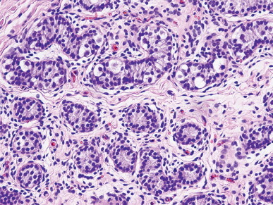
Cryptorchidism. Seminiferous tubules with type II lesions show markedly decreased diameters and an irregular distribution of germ cells.
Fig. 12-51.
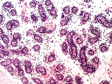
Cryptorchidism. Seminiferous tubules with type III lesions show severe reduction in both tubular diameter and tubular fertility index.
Fig. 12-52.
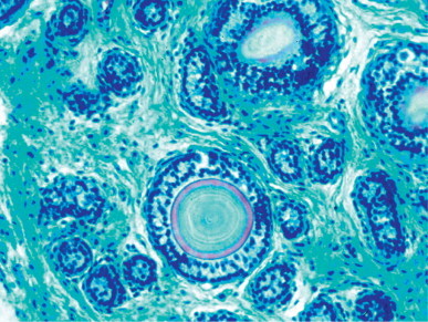
Microlithiasis in an infant cryptorchid testis. The seminiferous tubules show type III lesions and contain numerous microliths.
Fig. 12-53.
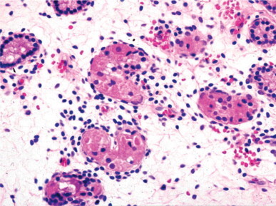
Cryptorchidism. Type III lesions, in which the interstitium is expanded by edema. The cytoplasm of the Sertoli cells contains numerous eosinophilic granules of variable size.
About 8% of tests with type I lesions show many multinucleated spermatogonia (with three or more nuclei) (Fig. 12-54 ).529 The seminiferous tubules of testes with types II or III lesions have a thickened lamina propria during childhood and, at puberty, Sertoli cell hyperplasia.526 Patients with bilateral cryptorchidism have a higher incidence of type II and III lesions than those with unilateral cryptorchidism.
Fig. 12-54.
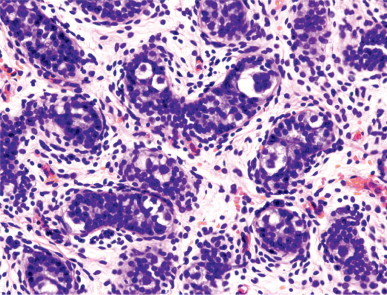
Prepubertal cryptorchid testis. The seminiferous tubules have Sertoli cells with elongated nuclei, pseudostratified growth pattern, and isolated spermatogonia, some of which are multinucleate or contain hypertrophic nuclei.
Type I lesions are comparable to those seen in experimental cryptorchidism; normal testes in which lesions were induced by increased temperature.527 Testes with type II or III lesions bear variable degrees of dysgenesis that, in addition to germ cells, involve Sertoli cells, peritubular myofibroblasts, and Leydig cells. The dysgenesis of these other cell types is evident only after puberty. In about 25% of cases the contralateral scrotal testis also has histologic lesions of variable severity. This finding supports the hypothesis of a bilateral defect in many cases of unilateral cryptorchidism. Microdeletions in the long arm of the Y chromosome are present in 27% of patients with corrected unilateral cryptorchidism who present with azoospermia or severe oligospermia.530 These findings are similar to those observed in patients with azoospermia or severe idiopathic oligospermia. Unilateral cryptorchidism with a normal contralateral testis could be due to an end-organ failure.531 In cryptorchidism secondary to spontaneous ascent, lesions are similar to those of congenital cryptorchidism, whereas in cryptorchidism secondary to herniotomy, germ cell depletion is slight532 and becomes important only after 5 years of age.533
Adult testes
Most pubertal and adult cryptorchid testes have anomalies in all testicular structures. The seminiferous tubules have decreased diameters and deficient spermatogenesis. In decreasing order of frequency, the most common germ cell lesions are tubules with Sertoli cell and spermatogonia-only pattern; tubules with Sertoli cells (dysgenetic) only; tubular hyalinization; and mixed atrophy. The lamina propria has scant elastic fibers and increased collagen fibers.534 Sertoli cells are present in increased numbers and do not mature normally except in tubules with germ cells (Fig. 12-55 ).528, 535 Often, groups of tubules containing only Sertoli cells with a prepubertal pattern (very small diameter and total absence of maturation) are present and are considered hypoplastic, dysgenetic, or hamartomatous. Areas of apparent Leydig cell hyperplasia are frequent, and many of these cells contain vacuolated lipid-laden cytoplasm (Fig. 12-56 ).
Fig. 12-55.
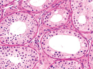
Adult ex-cryptorchid testis which was surgically descended at the age of 2 years. Tubular sections show a pattern varying from spermatogonial maturation arrest to complete, although decreased, spermatogenesis.
Fig. 12-56.
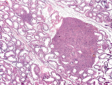
Nodular Leydig cell hyperplasia in an adult ex-cryptorchid testis.
The rete testis is hypoplastic in most cases and is lined by columnar epithelium with rare areas of flattened cells. Cystic dilation is common, and adenomatous hyperplasia has been found in some cases. Near the rete testis, the testicular parenchyma frequently contains metaplastic fat. In some cryptorchid testes, several tubular segments are destroyed by inflammation that probably has an autoimmune cause (focal orchitis).536 Epididymal tubules are poorly developed and peritubular tissue is immature.
Blood flow is associated with testicular histology. For example, testicular volume, histologic pattern, and testicular artery resistive index are lower in undescended testes than in controls, and testicular artery resistive index is inversely proportional to testicular histology score in undescended testes.537 There is also an apparent correlation between testicular size, spermiogram, and hormone levels. Assuming that a significant reduction in testicular size (>12 mL) is only observed in 9.3% of cases, and that serum levels of FSH, LH, and testosterone are normal, an inverse correlation is seen between FSH and testicular volume, sperm concentration, sperm motility, and normally shaped sperm. In addition, there is a direct relation between testicular volume and sperm concentration, sperm motility, and normally shaped sperm. These findings indicate the cause of tubular impairment in young men operated on in childhood for cryptorchidism.538
Obstructed testes
Obstructed testes are located in the superficial inguinal pouch (Denis–Browne pouch) and are considered ectopic by some authors and cryptorchid by others.539, 540 Histologic studies reveal that most obstructed testes bear the same lesions as true cryptorchid testes. Type I lesions are observed in half, type II in more than one-third, and the remainder show type III lesions. The higher proportion of type I lesions suggests a better prognosis than in true cryptorchid testes.
Retractile testes
Some authors assume that retractile testes are normal and exclude them from studies of cryptorchidism.541, 542 However, these testes may present important lesions and many consider them to be a form of cryptorchidism.543, 544, 545 Retractile testes may not always be movable to the lower scrotum (70–75 mm from the pubic tubercle) and in 50% of cases are smaller than scrotal testes. Approximately 50% of retractile testes remain high after age 6 years, when cremasteric activity declines.546 Retractile testes have a 32% risk of becoming ascending or acquired undescended testes. The risk is higher in boys younger than 7 years, or when the spermatic cord is tight or inelastic.547 During childhood, tubular diameter and tubular fertility index decrease.544 Adults with retractile testes that descended spontaneously but late may be fertile548 or infertile.549 Usually there is germ cell atrophy that varies in severity from lobule to lobule.544 Regular examination of retractile testes is advisable during childhood and, if complete testicular descent does not occur, orchidopexy is indicated.
Congenital anomalies associated with undescended testes
Most cryptorchid patients have a patent processus vaginalis, and 65–75% have a hernia sac, although most hernias are not clinically visible. Urologic anomalies are present in 10.5% of patients, the most frequent being hypospadias, complete duplication of the urinary tract, non-obstructive ureteral dilatation, kidney malrotation, and posterior urethral valves. Cryptorchidism is more frequent in patients with microcephaly, myelomeningocele, bifid spine, omphalocele, gastroschisis, micropenis, and imperforate anus.
Cryptorchidism may appear isolated or associated with congenital anomalies, endocrine dysfunction, chromosomal disorders, or intersex conditions. Thus, cryptorchidism is found in the Kallmann, Prader–Willi, Klinefelter, Noonan, Smith–Lemli–Opitz, Aarskog–Scott, Rubinstein–Taybi, prune belly, and caudal regression syndromes, anomalies of the androgen receptor, absence of anti-müllerian hormone, CHARGE association, and trisomies 13, 18, and 21.
Sperm excretory duct anomalies occur in 9–36% of cryptorchid patients,550, 551 and are classified into three types:552
-
•
Ductal fusion anomalies (25% of cases). These consist of anomalous fusion of the caput of the epididymis to the testis or segmental atresia of the epididymis and vas deferens. This is chiefly associated with intra-abdominal or high scrotal cryptorchid testes.
-
•
Ductal suspension anomalies (59% of cases). The caput of the epididymis is attached to the testis, whereas the corpus and the cauda of the epididymis are separated from the testis by a mesentery. A variant consists of an excessively long cauda of the epididymis that descends along the inguinal duct to the scrotum.
-
•
Anomalies associated with absent or vanishing testes (16% of cases).
Cryptorchidism is part of the testicular dysgenesis syndrome. This consists of abnormal testicular development that predisposes to cryptorchidism, hypospadias, spermatogenetic alterations, and testicular cancer. The association of these disorders with cryptorchidism has been corroborated by numerous clinical, epidemiological and genetic studies. The least severe form of this syndrome is a defect in spermatogenesis; the most severe is testicular cancer. A constellation of histologic lesions is common in the testes of men with testicular dysgenesis; these lesions include Sertoli cell-only pattern, mixed atrophy, hypoplastic tubules (Sertoli cell nodules), microlithiasis, malformed tubules, granular changes in Sertoli cells, nodular Leydig cell hyperplasia, and intratubular germ cell neoplasia. It is assumed that there is a prenatal development of the lesions as a result of several genetic, environmental, or endocrine disruptor factors that would interfere with the estrogen/androgen ratio.553, 554, 555, 556
Complications of cryptorchidism
The main complications of cryptorchidism are testicular cancer, infertility, testicular torsion, and psychological problems.
Testicular cancer
Approximately 0.8% of 1-year-old males have cryptorchidism, and about 10% of testicular cancer patients had cryptorchidism. The risk of testicular cancer in cryptorchid males is four to10 times higher than that of the general population. Testes with elevated number of multinucleated spermatogonia seem to have a higher risk of cancer and adulthood.557 About 5% of biopsies in children contain cells similar to those seen in undifferentiated intratubular germ cell neoplasia, and these cells may evolve toward germ cell tumor (Fig. 12-57 ).558 The most frequent tumor in undescended testes is seminoma.559, 560 Regardless of timing, orchidopexy does not reduce the risk of cancer, although it facilitates early detection as the intrascrotal testis is palpable. One in five testicular tumors arises in properly descended testes contralateral to cryptorchid testes, suggesting that there is a primary bilateral testicular anomaly in cryptorchidism. Intra-abdominal testes also have a higher incidence of tumors.560
Fig. 12-57.
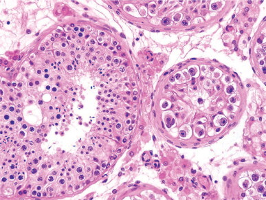
Adult ex-cryptorchid testis which was surgically descended at infancy. The patient was infertile. The smallest seminiferous tubule shows intratubular germ cell neoplasia, undifferentiated type. The relative tumor cell homogeneity contrasts with the variety in shape and size of the cells in the adjacent seminiferous tubule.
Infertility
Infertility is the most frequent problem caused by cryptorchidism. In a series of patients with infertility, nearly 9% had cryptorchidism.561 Infertility is influenced by several factors, including bilaterality, number of germ cells, location and size of the testis, and age at time of orchidopexy. The most important risk factors are bilaterality and germ cell number. Only 16%562 to 25%563 of men with bilateral cryptorchidism have normal sperm counts (20 million/mL or more). The highest sperm counts occur with testes in the superficial inguinal pouch. Patients with bilaterally impalpable testes are usually azoospermic.563 Fertility rates in unilateral cryptorchidism vary from 25% to 81%.564
The number of germ cells per cross-sectioned tubule is the most important prognostic factor. Patients with no increase in inhibin B during the postoperative period usually have a low number of spermatogonia per cross-sectioned tubule and a low tubular fertility index. In unilateral cryptorchidism, fertility depends on the number of spermatogonia in the contralateral testis. However, if the number of germ cells per cross-sectioned tubule in the cryptorchid testis is lower than 1% of normal, the risk of infertility is 33%. In bilateral cryptorchidism the risk of infertility rises from 75% to 100% when one or both testes have less than 1% of germ cells per cross-sectioned tubule. Neither the preoperative location of the testis in patients with unilateral cryptorchidism nor the small size of the testis at the time of orchidopexy is relevant for fertility.565, 566, 567 An important fertility factor is the permeability of sperm excretory ducts. The age at orchidopexy may also influence fertility, although this has not been proven. In patients over 4 years of age orchidopexy does not enhance fertility.568, 569
Benefit of testicular biopsy in patients with cryptorchidism
Testicular biopsies of infantile testes at orchidopexy are useful for determining baseline germ cell status and whether surgery should be completed with hormonal treatment.570 However, even if biopsy supplies important data, it is not considered a routine procedure.
Even in the best cases when the number of spermatogonia is nearly normal, spermatogenesis may never occur owing to deficient spermatogonium development during childhood, failure of spermatogenesis at puberty, and, if complete spermatogenesis occurs, this might be associated with obstruction of sperm excretory ducts.
In childhood, the chance of a biopsy finding an occult cancer or precancer is low because intratubular germ cell neoplasia is not diffusely distributed throughout the testis. Testicular biopsy is recommended in patients with intra-abdominal testes, abnormal external genitalia, or abnormal karyotype.571 The situation is different in adults because intratubular germ cell neoplasia is present in 2–3% of cases and is diffuse.572, 573 When intratubular germ cell neoplasia is detected in a child, further examination of the testis and rebiopsy after puberty are recommended.574 In adults, if intratubular germ cell neoplasia is unilateral orchidectomy should be performed, but if it is bilateral, radiation may be used to eradicate the neoplasia while maintaining Leydig cell function.575
Testicular microlithiasis
Testicular microlithiasis (TM) is characterized by the presence of numerous calcifications diffusely distributed throughout the testicular parenchyma. The number and size of the calcifications often is great enough to be detected radiographically or by ultrasound.576 Isolated microliths have been reported in undescended testes, prepubertal Klinefelter's syndrome, male pseudohermaphroditism, and otherwise normal children and patients studied for other diseases.577 In adults, microliths are frequently observed in cryptorchid and ex-cryptorchid testes,578 seminiferous tubules located at the periphery of germ cell tumor,579 infertile patients,580, 581, 582 and in some patients complaining of orchialgia583, 584 or testicular asymmetry.585
Testicular microlithiasis occurs in 0.3% of cryptorchid testes and is slightly more common in prepubertal than adult testes. In adults, it usually is diagnosed when men seek help for infertility, pain, or testicular asymmetry.581 Microlithiasis has been observed in 1.4–2% of testicular echographies of different disorders.586, 587 In infertile patients the incidence is slightly higher. Microlithiasis is present in 35% of testis having a malignant tumor.588
Ultrasound studies reveal two types of microlithiasis: classic TM, in which the number of microliths is five or more; and limited TM, when there are fewer than five microliths (Fig. 12-58 ). The incidences of TM in these studies are lower than 1% in infants, 5.6% in the general population aged between 18 and 35 years (bilateral in 66% of patients showing microliths,589 0.68–4.1% in patients with other disorders,586, 587, 590, 591, 592 from 4.6%593 to 20%594 in subfertile patients, 9.52% in ex-cryptorchid testes,595 and more than 30% in adult testes with germ cell tumors).588, 596, 597, 598, 599 Several cases of testicular microlithiasis have also been observed in infant testes with germ cell tumor or gonadal stroma tumor.600, 601 The incidence is higher in whites than in blacks.
Fig. 12-58.
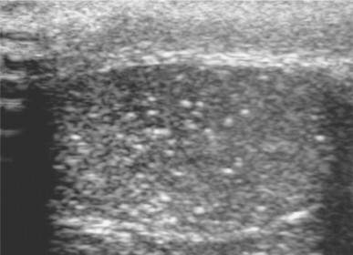
Testicular microlithiasis showing the characteristic ‘snowstorm’ pattern.
Pain is the most common clinical symptom in patients without a palpable testicular mass, and has been attributed to dilation of seminiferous tubules secondary to obstruction by microliths.
Microliths are made by hydroxyapatite, according to X-ray diffraction studies602 and Raman spectroscopy.603 In the prepubertal testis, microliths are surrounded by a double layer of Sertoli cells and measure up to 300 μm in diameter. When they are very large, the seminiferous epithelium may be destroyed and the microlith is surrounded by peritubular cells (Fig. 12-59 ). Testes with microliths have subnormal mean tubular diameters and tubular fertility index.604 In adult testes with microliths there is incomplete spermatogenesis. Some seminiferous tubules with microliths are cystically dilated (Fig. 12-60 ). Microliths arise as extratubular eosinophilic bodies that mineralize and pass into the tubular lumina.605 Microlithiasis may be a disorder of the tunica propria. Also, testicular microlithiasis is occasionally associated with pulmonary microlithiasis and with calcifications in the parasympathetic nervous system.606, 607
Fig. 12-59.
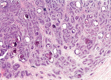
Testicular microlithiasis. Infantile cryptorchid testis with type III lesions and numerous microliths within the seminiferous tubules.
Fig. 12-60.
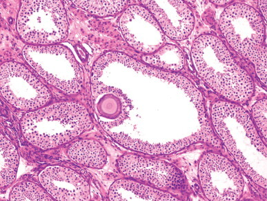
Testicular microlithiasis. Seminiferous tubules with dilated lumina in a patient biopsied for infertility. The central tubule contains a microlith which developed in the tubular wall and protrudes into the lumen.
The association of microlithiasis and testicular cancer is controversial.608, 609 Although the development of testicular cancer has been observed in several patients whose testicular microlithiasis had been previously diagnosed by ultrasound studies,610, 611, 612, 613, 614 it is also thought that patients with testicular microlithiasis not associated with other disorder do not require any follow-up.615 When microlithiasis is associated with infertility the incidence of cancer varies according to the unilaterality or bilaterality of microlithiasis:594 subfertile patients with unilateral microlithiasis show no intratubular germ cell neoplasia, whereas this is present in 20% of those with bilateral microlithiasis. The risk of malignancy is higher in classic than in limited TM.616 The nexus between microlithiasis and cancer does not seem to be the predisposition of one disorder towards the other but rather the predisposition of both to develop in abnormal testes. This may also explain the association between microlithiasis and infertility.
Yearly ultrasound examination, perhaps with testicular biopsy, is recommended in those with testicular microlithiasis associated with cryptorchidism, infertility, atrophic testes, or contralateral testis bearing germ cell tumor.617
Microlithiasis also occurs in the rete testis or sperm excretory ducts. Epididymal rupture and extravasation of microliths into the interductal tissue may cause a histiocytic reaction resembling malakoplakia (Fig. 12-61 ). The disorder is asymptomatic and not associated with testicular cancer.618
Fig. 12-61.
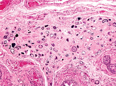
Epididymal microlithiasis. Numerous microliths displaying a psammoma body-like appearance are set in hyalinized stroma.
Gonadal dysgenesis
Gonadal dysgenesis refers to disorders characterized by amenorrhea and streak gonads in phenotypically female patients. In adults, streak gonads are elongated masses of fibrous tissue resembling ovarian stroma (Fig. 12-62 ). They may contain hilar cells and rete or epithelial cords with variable degrees of maturation, and may result from failure in gonad formation, failure of gonadal differentiation to ovary, or failure of gonadal differentiation to testis. Some streak gonads contain a few ovocytes or primordial follicles, but all germ cells disappear at puberty. Patients with streak gonads have a hypoplastic uterus and fallopian tubes. Four types of gonadal dysgenesis have been described: 46XY pure, 46XX pure, 45X0, and mixed.
Fig. 12-62.
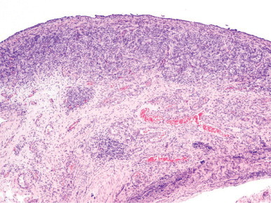
Gonadal dysgenesis. The elongate formation consists of an outer cellular part devoid of ovarian follicles and a central part with numerous blood vessels.
46XY Gonadal dysgenesis
46XY gonadal dysgenesis (Swyer's syndrome) is characterized by female phenotype, absence of Turnerian stigmata, and female external genitalia, sometimes with fused labia majora, a hypertrophic clitoris, and hypospadias. The breasts develop at puberty. Sexual infantilism persists in adulthood, and eunuchoidism and amenorrhea appear. These patients have elevated serum gonadotropin levels and low serum estradiol.
There are two types of gonadal dysgenesis: complete and incomplete. Patients with the complete type have female external genitalia and classic streak gonads, although cases with ovarian tissue have been reported. The cause is unknown in about 80% of cases,619 and is due to alterations in the SRY gene in the remainder (a mutation in 10–15% of cases, and a SRY deletion as a result of an aberrant X/Y interchange in 10–15%).620 The consequence of failure is very early gonadal alteration (sixth to eighth week of gestation). With the subsequent absence of müllerian inhibiting factor, testosterone, and dihydrotestosterone, a female phenotype develops.
Patients with incomplete 46XY gonadal dysgenesis have ambiguous external genitalia and variable degree of development of the müllerian and wolffian structures. Although they have streak gonads, testicular development is usually observed. This gonadal dysgenesis does not seem to be caused by SRY alterations.621 These findings suggest that in the first type ovarian differentiation was canceled, and that in the second type testicular differentiation failed. The first is similar to the gonad of 45X0 Turner's syndrome, whereas the second resembles the gonad of mixed gonadal dysgenesis.622 The clitoromegaly may be caused by androgens secreted by hyperplastic Leydig cells in the streak gonad.
Some patients with 46XY gonadal dysgenesis present with extragonadal anomalies and multiple syndromes, including camptomelic dysplasia and renal disorder,623 myotonic dystrophy and terminal renal disease,624 progressive renal insufficiency and gonadoblastoma (Frasier's syndrome) (Fig. 12-63, Fig. 12-64 ),625, 626, 627, 628, 629 mental retardation with630 or without631 facial anomalies or short stature,632 renal insufficiency and Wilms’ tumor (Denys–Drash syndrome), the combination of cleft palate, micrognathia, kyphosis, scoliosis, and clubfoot (Gardner–Silengo–Wachtel syndrome or genitopalatocardiac syndrome),633 pterygium multiple syndrome,634 Graves’ disease,635, 636 and congenital universalis alopecia, microcephaly, cutis marmorata, and short stature.637, 638
Fig. 12-63.
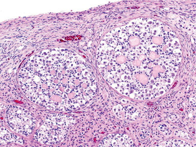
Frasier's syndrome in a 16-year-old patient. The two streak gonads contain gonadoblastoma.
Fig. 12-64.
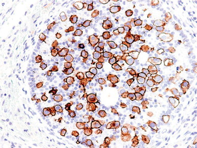
Frasier's syndrome. The cell surface and Golgi zone of the atypical gonadoblastoma germ cells are immunoreactive for c-kit.
Most cases are sporadic,639 although the syndrome has been reported in several members of the same family,640, 641, 642, 643 and several forms of inheritance (X-linked, autosomal recessive, and male-limited autosomal dominant) have been proposed.644 In addition to infertility, patients with 46XY gonadal dysgenesis have a high risk of germ cell tumor. This risk is about 5% in the first decade of life, and 25–30% overall,645, 646, 647, 648 and, thus, prophylactic gonadectomy is recommended.
46XX Gonadal dysgenesis
Patients with 46XX gonadal dysgenesis have normal stature, female phenotype, well-developed external genitalia, and hypoplastic ovaries rather than streak gonads (Fig. 12-65 ). The anomaly is usually detected when patients present with primary amenorrhea or infertility. This syndrome is sporadic and familial, and it may be linked to recessive autosomal inheritance.649, 650 Patients have no predisposition to gonadal neoplasia. Associated somatic anomalies such as neurosensory hearing loss (Perrault's syndrome) are rare.
Fig. 12-65.
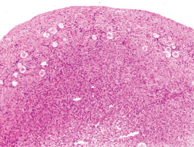
46XX gonadal dysgenesis. The streak gonads contain isolated ovocytes.
Some familial cases have shown a balanced translocation of the X chromosome (from the long arm to the short arm)651, 652 or between chromosomes 1 and 11.653 Because the development of ovarian follicles requires FSH, mutations have been sought in the FSHR gene. Mutations have been detected in familial cases and also in unrelated patients,654, 655 whereas other patients have shown no mutations in this gene.656 The incidence of tumors in these patients is very low, and the most common is dysgerminoma.657, 658, 659
45X0 Gonadal dysgenesis
This is one of the most common chromosomal anomalies (from 1/2500 to 1/5000 in female newborns),660 although 99% of zygotes with this karyotype are aborted in the first stages of embryonal development.661
Patients with 45XO gonadal dysgenesis have characteristic stigmata of Turner's syndrome, including short stature, pterygium coli, lymphedema, and cardiac malformations. The external genitalia are female and infantile; the gonads are typical streak gonads. Today, Turner's syndrome is defined by the combination of physical features and the complete or partial absence of one of both X chromosomes, frequently associated with mosaicism. Turnerian stigmata may be classified into four groups:662 skeletal anomalies such as cubitus valgus, shortening of the fourth metacarpal and Madelund's deformity characteristic of Leri–Weill dyschondrosteosis; soft tissue anomalies such as webbed neck, low posterior hair line, and puffy hands and feet; visceral anomalies such as aortic coarctation, horseshoe kidney, polycystic kidney, urethral stenosis and vesicourethral reflux; and miscellaneous anomalies such as nevus pigmentosus.663
During embryonic life, these gonads show normal germ cell numbers up to the third month, when germ cell proliferation ceases.665, 666 Ovogenesis stops in meiosis I, usually before the pachytene stage. The cause seems to be generalized meiotic pairing errors with the start of an apoptotic mechanism to avoid the formation of abnormal gametes.667 Massive apoptosis of ovocytes occurs between the 15th and the 20th weeks.668 Surviving germ cells disappear throughout fetal life, and their numbers at birth are usually low (Fig. 12-66 ).669
Fig. 12-66.
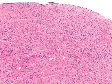
Gonadal dysgenesis. Streak fibroblastic stroma resembling ovarian cortex.
Patients with mosaicism have fewer anomalies than pure 45X0 individuals; 12% have menstruation (compared to 3% of pure 45X0 patients), and 18% have breast development (compared to 5% of pure 45X0 patients). In 10–20% of 45X0 patients the SRY gene is demonstrable by in-situ hybridization. It has been proposed that patients with SRY expression should undergo gonadectomy, because this gene is also a marker of gonadoblastoma.670 These patients may develop gonadoblastoma, dysgerminoma, and mixed germ cell tumor.670, 671
Mixed gonadal dysgenesis
Mixed gonadal dysgenesis is characterized by the presence of a streak gonad and a contralateral testis (often cryptorchid) or streak testis (see discussion on male pseudohermaphroditism with müllerian remnants, below).
True hermaphroditism
True hermaphroditism is a disorder of gonadal differentiation characterized by the presence in the same individual of both testicular and ovarian tissue. This condition is rare and usually difficult to diagnose, so only 25% of male hermaphrodites are diagnosed before age 20.672 Failure to recognize this disorder may lead to surgical intervention for hernia repair or orchidopexy. Most hermaphrodites raised as males display symptoms for the first time at puberty because of breast development673 (95% of hermaphrodites have some degree of gynecomastia), periodic hematuria674 (if they have a uterus ending in the urinary tract), or cryptorchidism.675 Hermaphrodites raised as females initially present with irregular menstruation or clitoromegaly. True hermaphroditism should be suspected in all children with ambiguous sex characteristics (Fig. 12-67 ). The gonads of these patients are ovotestes, ovaries, or testes, with all possible combinations.676 True hermaphroditism can be (1) unilateral, if there are both testicular and ovarian tissues (forming one ovotestes or two separated gonads) on one side, and a testis or an ovary in the other side; if there is no gonadal tissue in this latter side, unilateral hermaphroditism is incomplete; (2) bilateral, if testicular and ovarian tissues are present on both sides of the body; and (3) alternate, if there is a testis on one side, and an ovary on the other side.
Fig. 12-67.
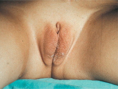
True hermaphrodite showing external genitalia which display transverse folds and a slightly hypertrophic clitoris.
Ovotestis is the most frequent gonadal type in true hermaphroditism. It is more frequent on the right side and is located in the abdomen (50% of cases), labioscrotal folds, inguinal canal, or the external inguinal ring. The ovotestis has a bilobated or ovoid shape (Fig. 12-68 ). In the bilobated ovotestis the testis and ovary are connected by a pedicle, whereas in the ovoid ovotestis the ovarian tissue forms a crescent capping the testicular parenchyma. The proportion of ovary to testis varies widely (Fig. 12-69 ). At adulthood, the ovarian follicles mature and corpora lutea or corpora albicantia may be seen. The seminiferous tubules rarely develop complete spermatogenesis. The interstitium usually contains Leydig cells. Ovotestis is associated with a fallopian tube in 65% of cases, and with a vas deferens in the remainder. If the patient has ovotestis/ovary, a completely developed uterus is present. If the patient has bilateral ovotestis (13%), uterine agenesis is frequent (Fig. 12-70 ).677
Fig. 12-68.
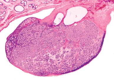
True hermaphroditism. The ovotestis contains ovarian follicles arranged in a crescent. There is cystic transformation of the rete testis. The epididymis is hypoplastic.
Fig. 12-69.
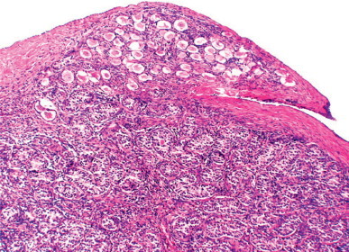
True hermaphroditism. Ovotestis from a 2-year-old. The ovarian and testicular tissues are sharply demarcated.
Fig. 12-70.
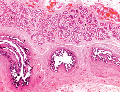
True hermaphroditism. Epididymis and fallopian tube in an adult hermaphrodite raised as a female.
The testis of hermaphrodites is most often on the right side (60%) and is located anywhere from the abdomen to the scrotum. These testes have low tubular fertility indices during childhood. After puberty, the seminiferous tubules remain small, often containing only dysgenetic Sertoli cells, similar to the tubules of cryptorchid testes. Incomplete spermatogenesis has been reported, but complete spermatogenesis is exceptional. The ovary of hermaphrodites is most frequently on the left side (63%) and usually is hypoplastic with few primordial follicles. However, in occasional patients the ovary is histologically and functionally normal.
The most frequent karyotype is 46XX (60%), followed by several mosaicisms (33%) which, in decreasing order of frequency, are 46XX/46XY, 46XY/47XXY, 45X0/46XY, 46XX/47XXY. The 46XY karyotype is the least common (7%). There is variation in the incidence of some karyotypes around the world. Mosaicism is found in 40.5% of European cases, but in only 21% of North America cases. Conversely, most African true hermaphrodites (97%) have 46XX karyotype. The karyotype 46XY is rare and its frequency is similar in Europe, Asia, and North America.678, 679 Most cases are sporadic, and families with several affected members also have 46XX males. This finding suggests that both genetic anomalies are alternative forms of a single genetic defect.680 The following mechanisms681, 682 have been proposed to explain the occurrence of testicular parenchyma: true hermaphroditism 46XX, a hidden mosaicism with a cell line having a Y chromosome; transfer from a Y chromosome fragment (including SRY gene) to the X chromosome; autosomal mutation of variable penetrance; and X-linked mutation coupled with rare X inactivation or X mutation that permits testicular differentiation in the absence of SRY. Some 46XX hermaphrodites with SRY-negative leukocytes are positive for this gene in DNA from the testicular parenchyma in the ovotestis.683 Over 22 pregnancies in true hermaphrodites have been reported,684 in contrast to the exceptional cases of paternity. Ovules may arise from the ovotestes or the ovary.
Management of true hermaphroditism depends on the patient's age at the time of diagnosis, the nature and location of the gonads, and the developmental stage of the external genitalia. Although bilateral castration may be justified in order to avoid the risk of neoplasia, gonadal preservation may be desirable until adulthood. In this case, if the patient is raised as a girl, puberty will occur spontaneously and there is a small chance of fertility.685 However, the high risk of malignancy (estimated at 4.6%) should be taken into account. The most frequent tumors are gonadoblastoma, dysgerminoma, and yolk sac tumor.676 The risk of cancer may be reduced if some precautions are taken, including removal of the testis if it has not descended and surveillance of the residual gonad with periodic ultrasound studies, especially in cases of chromosomal mosaicisms.
Male pseudohermaphroditism
Normal male development requires adequate differentiation of the testes in the fetal period, synthesis and secretion of testicular hormones, and proper response of target organs to these hormones. Anti-müllerian hormone produced by Sertoli cells inhibits the development of müllerian derivatives that would otherwise form the uterus and fallopian tubes. Testosterone produced by Leydig cells stimulates differentiation of the wolffian ducts into male genital ducts. The conversion of testosterone into dihydrotestosterone by the enzyme 5α-reductase ensures the development of male external genitalia. Alterations in these processes may cause male pseudohermaphroditism.
Impaired Leydig cell activity
Androgen synthesis deficiencies
These autosomal recessive syndromes are characterized by an error in testosterone synthesis that results in incomplete or absent virilization. Cholesterol is the source for the synthesis of androgens, estrogens, and other steroid hormones through multiple steps. First, the steroidogenic acute regulatory protein (StAR) generates cholesterol into mitochondria; StAR gene mutations cause congenital lipoid adrenal hyperplasia. Second, within mitochondria, the cholesterol side-chain cleavage enzyme P450scc transforms cholesterol into pregnenolone; a disorder in this enzyme is rare because it is highly lethal in embryonic life. Third, pregnenolone undergoes 17α-hydroxylation by microsomal P450c17; deficiency in 17α-hydroxylase causes female sexual infantilism and hypertension. Fourth, 17-OH-pregnenolone is converted into DHEA by 17,20-lyase activity of P450c17. The ratio of 17,20-lyase to 17α-hydroxylase activity of P450c17 determines the ratio of C21 to C19 steroids produced. The ratio is regulated by at least three factors, including the electron-donating protein P450 oxidoreductase (POR), cytochrome b5, and serine phosphorylation of P450c17. Mutations in POR are present in the Antley–Bixler skeletal dysplasia syndrome as well as a variant of polycystic ovarian syndrome. Figure 12-71 shows the enzymes involved in the abovementioned steps. The enzyme 3β-hydroxysteroid dehydrogenase transforms DHEA to androstenedione, and the enzymatic complex called aromatase transforms androstenedione into estrone and testosterone into estradiol.
Fig. 12-71.
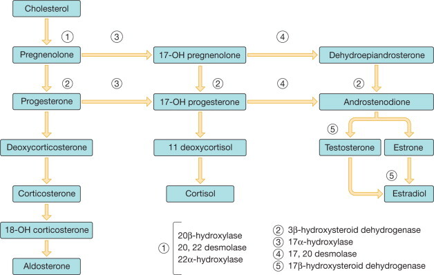
Enzymatic defects in impaired testosterone biosynthesis.
In some patients cholesterol synthesis is also impaired, and congenital adrenal hyperplasia is superimposed on androgen deficiency. Deficient testosterone synthesis may result from abnormalities in the enzymes involved in pregnenolone formation (congenital lipoid adrenal hyperplasia), including 3β hydroxysteroid dehydrogenase, 17α-hydroxylase, 17,20-desmolase, and 17β-hydroxysteroid dehydrogenase (Fig. 12-71).
Congenital lipoid adrenal hyperplasia
Congenital lipoid adrenal hyperplasia is the most severe form of congenital adrenal hyperplasia.686 The disorder is characterized by a deficit in steroid hormone synthesis in the adrenal cortex and gonads, producing a female phenotype with severe salt-loss syndrome. Conversion of cholesterol to pre-gnenolone requires the enzymes 20α-hydroxylase, 20,22-desmolase, and 22α-hydroxylase. Failure of any of these leads to deficits in cortisol, aldosterone, and testosterone.687
The enzymatic defect is usually is caused by a deficit in the steroidogenic acute regulatory (StAR) protein; in other cases, the deficit is in P450ssc. The mitochondrial protein StAR promotes cholesterol transfer from the outer to inner mitochondrial membrane, where cholesterol serves as a substrate for P450scc and initiates steroido-genesis. More than 35 different mutations in the StAR gene have been identified.690 As a result, cholesterol is not converted to pregnenolone, which is required for the synthesis of mineralocorticoids, glucocorticoids, and sex hormones.
The disorder is rare in most countries, but is common in Japan, Korea, and the Arabian countries.691, 692 Patients usually present with salt-losing crisis in the first 2 months of life.693, 694 In most cases, males have female or ambiguous external genitalia and a blind-sac vagina, hypoplastic wolffian derivatives, absence of müllerian structures, and cryptorchidism.695 The adrenals usually appear enlarged and contain lipid accumulations,696, 697 but these diminish with age and the adrenals shrink.
In the testes, lipid accumulations may be present or absent in Leydig cells686, 696, 698, 699, 700, 701, 702 or Sertoli cells.703 An 8-year-old child had partially hyalinized seminiferous tubules with Sertoli cell-only pattern.704 The testes of pubertal patients are usually normal for age.701, 702 Intratubular germ cell neoplasia has been reported in one case.705
Most patients die from adrenal insufficiency. Survivors have female phenotype704 and require the administration of glucocorticoids, mineralocorticoids, and gonadal steroids.703
3β-Hydroxysteroid dehydrogenase deficiency
Patients with this defect have two main problems: salt-loss syndrome produced by reduced aldosterone secretion, and incomplete virilization.706 At puberty, virilization increases and gynecomastia develops.707, 708
The enzyme 3BHSD catalyzes the conversion of 5-3β-hydroxysteroids such as pregnenolone, 17-hydroxypregnenolone, and dehydroepiandrosterone into respectively 4-3β-ketosteroid, progesterone, 17-hydroxyprogesterone, and androstenedione.709 There are two 3BHSD genes located on the p11-p13 region of chromosome 1. The type I gene is expressed in the placenta, kidney, and skin, whereas the type II gene 3BHSD is expressed only in the gonads and adrenal glands. Complete absence of the 3BHSD gene is lethal; therefore, most reported cases have only partial 3BHSD deficits.710, 711, 712 It is assumed that these deficits account for 10% of cases of congenital adrenal hyperplasia.
The classic form of salt-losing 3BHSD deficit is diagnosed in the first months of life because of insufficient aldosterone synthesis and subsequent loss of salt. The other 3BHSD deficit, without salt loss, is due to mutations in the type II 3BHSD gene708 and its diagnosis may be delayed until puberty.
Severe forms of 3BHSD deficiency are associated with deficits in aldosterone, cortisol, and estradiol. Symptoms may vary widely, as enzymatic activity in the adrenal gland is not the same as in the testis. Most patients show salt loss and adrenal crisis; they have incomplete masculinization and may develop spontaneous puberty and gynecomastia.706, 707, 713 Patients with mild forms have normal genitalia and normal mineralocorticoid levels. Some patients have only hypospadias714 or micropenis.715 The testes are smaller and softer than normal.
17α-Hydroxylase deficiency
The cause of deficits in the enzymes 17α-hydroxylase and 17,20-lyase are mutations of the CYP17 gene that encodes cytochrome P450c17.716 The CYP17 gene is located on chromosome 10q24-q25,717 and 50 different mutations have been described.718 P450c17 catalyzes the 17α-hydroxylation of pregnenolone to 17OH-pregnenolone and of progesterone to 17α-OH-progesterone. This enzyme also catalyzes 17,20-lyase activity, transforming 17OH-pregnenolone to DHEA. The classic form of 17α-hydroxylase deficit is caused by severe deficiencies in CYP17; less severe defects give rise to the isolated 17,20-lyase deficit. 17α-Hydroxylase deficit impairs the synthesis of both cortisol and testosterone.719 Low cortisol levels stimulate ACTH secretion, causing hypersecretion of aldosterone precursors and the development of hypokalemic hypertension and male pseudohermaphroditism in males.720 Patients usually have hypospadias and develop gynecomastia at puberty.721
17,20-Desmolase deficiency
The enzyme 17,20-desmolase cleaves the side chain of 17-hydroxypregnenolone and 17-hydroxyprogesterone to form dehydroepiandrosterone and androstenedione, respectively. Varying degrees of 17,20-desmolase deficiency are seen, resulting in varied development of external genitalia, ranging from female phenotype to virilization with microphallus, bifid scrotum, and perineal hypospadias. In childhood, the testes contain reduced numbers of spermatogonia (Fig. 12-72, Fig. 12-73 ).722, 723 The cause may be mutations in one of the genetic loci encoding P450c17, flavoprotein OR or b5.724
Fig. 12-72.
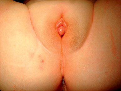
Male pseudohermaphrodite with androgen synthesis deficiency. The external genitalia are ambiguous.
Fig. 12-73.
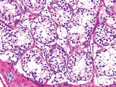
Intense Leydig cell vacuolation in an infant with androgen synthesis deficiency.
17β-Hydroxysteroid dehydrogenase deficiency
This enzyme transforms androstenedione into testosterone and also converts estrone into estradiol. The enzymatic defects are sex-linked. Most patients have female phenotype at birth and are raised as girls, but at puberty undergo virilization.725 One or both testes may be cryptorchid or are located in the labia majora. Normal spermatogenesis has never been observed. The most common testicular patterns are hypoplasia or absence of germ cells and Leydig cell hyperplasia.726 The germ cell injury was initially attributed to cryptorchidism, but it is now thought to be a primary testicular lesion because even very young patients lack germ cells.727 This deficit is due to mutations in the HSD17B3 gene located on 9q22.728, 729
Leydig cell hypoplasia
This variant of male pseudohermaphroditism is defined by insufficient testosterone secretion422 and the following characteristics: predominance of female external genitalia; absence of male secondary sex characteristics at puberty; absence of uterus and fallopian tubes and the presence of epididymis and vas deferens; 46XY karyotype; lack of response to human chorionic gonadotropin stimulation; absence of an enzymatic defect in testosterone synthesis; and small undescended testes that are gray and mucous on section.730, 731, 732, 733 Age at diagnosis varies from 4 months to 35 years. The syndrome is sporadic and familial.734, 735
The best-known cause of Leydig cell hypoplasia is inactivating mutation of the LH receptor in these cells.736, 737, 738 During fetal life, there is an inadequate response to placental hCG initially and to pituitary LH subsequently. Phenotypes vary widely according to the presence of complete or partial loss of receptor function. These changes range from male pseudohermaphroditism with female external genitalia (type I of Leydig cell hypoplasia) to male phenotype with micropenis, hypospadias, pubertal delay, and primary hypogonadism (type II of Leydig cell hypoplasia).
In type I hypoplasia, the testes contain small seminiferous tubules with Sertoli cells, spermatogonia, and thickened basement membranes. Leydig cells are rare or absent, in contrast to Leydig cell hyperplasia seen in other types of male pseudohermaphroditism, such as those arising from defects in androgen synthesis or androgen action on peripheral tissues.739, 740 Leydig cell hypoplasia accounts for low serum testosterone levels, lack of virilization, and lack of spermatogenesis. The absence of müllerian derivatives suggests a normal function of Sertoli cells, which synthesize müllerian inhibiting factor. In type II hypoplasia, adult testes show maturation arrest of spermatogonia and a few incompletely differentiated Leydig cells.741, 742
Impaired androgen metabolism in peripheral tissues
Androgen insensitivity syndromes
Resistance to androgen stimulation is the cause of several syndromes with phenotypes varying from complete testicular feminization743 to normal male.744, 745 These syndromes are caused by partial or complete lack of response of the target organs to androgens746 due to the absence, diminution, or impairment of androgen receptors or post-receptor anomaly.740 The gene for the androgen receptor is located on the X chromosome (Xq11-q12), and X-linked transmission occurs in two-thirds of cases. The karyotype is usually 46XY, but 47XXY and several mosaicisms have been observed.747
These syndromes affect 1:20 000–1:40 000 newborns. The diverse phenotypes associated with androgen insensitivity may be classified as: complete androgen insensitivity syndrome (CAIS) or testicular feminization syndrome; partial androgen insensitivity syndrome (PAIS) or partial testicular feminization syndrome, which includes the syndromes of Lubs, Gilbert–Dreyfus, Reifenstein, and Rossewater; and mild androgen insensitivity syndrome (MAIS), infertile men with light androgen insensitivity, and Kennedy's disease.
Complete androgen insensitivity syndrome (complete testicular feminization syndrome)
This form of male pseudohermaphroditism is characterized by female phenotype with testes. Complete testicular feminization syndrome is rarely diagnosed during childhood except in patients who present with hernia, inguinal tumor, or with a family history of pseudohermaphroditism. Primary amenorrhea is the principal presentation in adults.
The testes may be in the abdomen, inguinal canal, or labia majora, and during the first year of life may be normal histologically except for reduced tubular diameter and low tubular fertility index. After the first year, decreased germ cell numbers become evident and the few remaining spermatogonia are concentrated in clusters of seminiferous tubules. The testicular interstitium contains numerous spindle cells arranged in bundles, and during the first year of life has Leydig cells with abundant eosinophilic or vacuolated cytoplasm. At puberty, patients have female external genitalia, a short blind-ended vagina, feminine breast development; and scarce pubic and axillary hair. Serum testosterone is at the normal male level and LH is markedly increased.
In adults, the testes vary in size from small to large, are tan-brown, and contain small seminiferous tubules without lumina which usually contain only Sertoli cells.748, 749 In one-third of patients both Sertoli cells and spermatogonia are present.750 Ultrastructurally, Sertoli cells lack Charcot–Böttcher crystals and annulated lamellae; inter-Sertoli cell specialized junctions are not well developed, and in cryofracture studies the arrangement of membrane particles has an immature pattern.751 Leydig cells are abundant, but few contain Reinke's crystalloids. Often, there are areas resembling ovarian stroma in the testicular interstitium.
In about two-thirds of cases the testes contain grossly visible white nodules that stand out from the surrounding testicular parenchyma (Fig. 12-74, Fig. 12-75 ). Histologically, the nodules consist of clusters of small seminiferous tubules with immature Sertoli cells, hyalinized lamina propria, numerous Leydig cells, and an absence of elastic fibers (Fig. 12-76 ). These have been referred to as Sertoli–Leydig cell hamartoma. About 25% of testes have Sertoli cell adenoma, sometimes very large, consisting of tubules resembling infantile testis but lacking in germ cells and peritubular myofibroblasts. No Leydig cells are present between the tubules (Fig. 12-77, Fig. 12-78 ).752 Other benign tumors include Sertoli cell tumor (large cell calcifying Sertoli cell tumor and sex cord tumor with annular tubules), Leydig cell tumor, leiomyoma, and fibroma.746
Fig. 12-74.
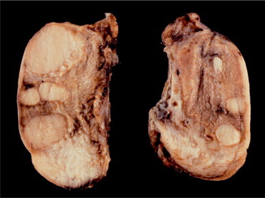
Testicular feminization syndrome. Both testes are enlarged and contain several gray-white nodules.
Fig. 12-75.
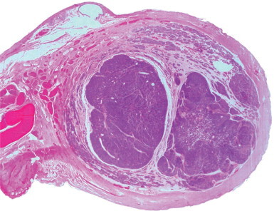
Testicular feminization syndrome. Cross-sectioned testis with multiple well-demarcated nodules.
Fig. 12-76.
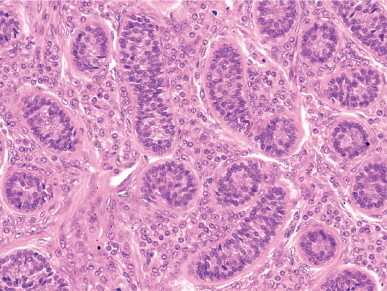
Testicular feminization syndrome. Small seminiferous tubules with immature Sertoli cells surrounded by thick basement membranes and numerous Leydig cells.
Fig. 12-77.
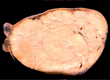
Large Sertoli cell adenoma in an abdominal testis from a 65-year-old patient with testicular feminization syndrome.
Fig. 12-78.
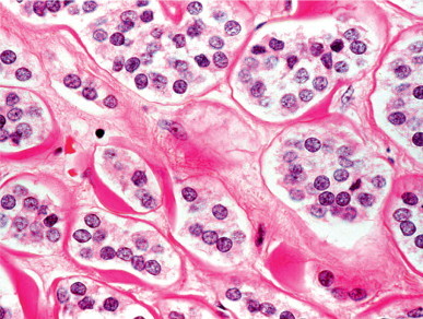
Sertoli cell adenoma showing tubular clusters with a hyalinized wall in a stroma devoid of Leydig cells.
Approximately 60% of cases have small cystic structures closely apposed to the testes, and about 80% of patients have thick bundles of smooth muscle fibers resembling myometrium near the testis. True myometrium has been demonstrated in only one case. Hypoplastic fallopian tubes are present in about one-third of cases. In about 70% of patients the epididymis and vas deferens are rudimentary; the only explanation for this is residual activity of the mutated androgen receptor.753 Approximately 10% of testes from patients with testicular feminization syndrome develop cancer. The frequency increases with age, but tumors rarely appear before puberty. These tumors include intratubular germ cell neoplasia (Fig. 12-79 ),749 several types of germ cell tumor,750, 754 and sex cord tumor.441 Thus, the gonads should be removed immediately after puberty.755
Fig. 12-79.
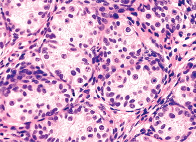
Intratubular germ cell neoplasia, undifferentiated type, in a phenotypically female patient with inguinal testes. The tumor cells stand out by virtue of their large size, pale cytoplasm, and prominent nucleoli.
Partial androgen insensitivity syndrome (partial testicular feminization syndrome)
The phenotype of patients with partial testicular feminization varies from normal female to normal male. The disorder includes four classic syndromes: Lubs' syndrome,756 characterized by partial fusion of labioscrotal folds, a definitive introitus, clitoromegaly, pubic and axillary hair, and poor breast development;757 Gilbert–Dreyfus syndrome, characterized by progressively greater male phenotypic features that include small phallus, hypospadias, incomplete development of wolffian derivatives, and gynecomastia;758 Reifenstein's syndrome, characterized by hypospadias, weak or absent virilization, testicular atrophy, gynecomastia, azoospermia, and infertility;759 and Rosewater–Gwinup–Hamwi syndrome, characterized by infertile men whose only abnormal feature is gynecomastia.760
Mild androgen insensitivity syndrome
Spermatogenesis requires high levels of intratesticular testosterone. A minor form of androgen insensitivity may be observed in some patients with male phenotype who present with infertility.761 The frequency of androgen resistance among azoospermic and oligozoospermic men is estimated at about 19%762 or lower.763, 764 Some patients have lost exon 4765 or mutated exons 6764 or 7.766
Kennedy's disease
Kennedy's disease (spinal and bulbar muscular atrophy, SBMA) is an X-linked recessive disorder of the adult male767, 768 characterized by loss of motor neurons in the spinal cord and brain stem and associated with less important loss of sensory neurons and atrophy caused by skeletal muscle denervation.767, 769 Disease onset around 20 years of age includes muscular weakness, cramps, and fasciculations.770 In most cases the male reproductive system is impaired.770, 771, 772 The testes may be normal in the initial stages of the disease, and many patients are fertile; however, with progression, there is onset of secondary testicular atrophy and gynecomastia. Testosterone levels are decreased in some cases.
The disease results from mutations in the first exon of the androgen receptor (AR) gene.773 The SMBA gene, located on Xq11-12, has expansion of a repetitive CAG sequence in exon A. The number of CAG repeats is 21 (range, 17–26) in control men and more than 40 in men with Kennedy's disease.768, 774, 775, 776, 777
5α-Reductase deficiency
This disorder is a variant of male pseudohermaphroditism caused by a lack of the enzyme 5α-reductase with failure of conversion of testosterone to dihydrotestosterone.778 In patients with the 46XY karyotype there are two isoenzymes: isoenzyme 1 is encoded by the gene SRD5A, located on 5p15, and isoenzyme 2 is encoded by the gene SRD5A2 on 2p23. Most reported cases result from defects in SRD5A2.779 Many mutations in different exons have been reported.780, 781, 782, 783, 784
During childhood, patients have a clitoriform penis, bifid scrotum, urogenital sinus, and testes in the inguinal canal or labioscrotal folds (Fig. 12-80 ). Müllerian derivatives are absent. At puberty they acquire the male phenotype, with development of the penis and scrotum. Adults have erections, ejaculations, and normal libido, scant body hair and a thin beard, a very small prostate, and lack of temporal hairline recession (male pattern baldness). Serum levels of FSH, LH, and testosterone are increased, but dihydrotesto-sterone is decreased.785, 786
Fig. 12-80.
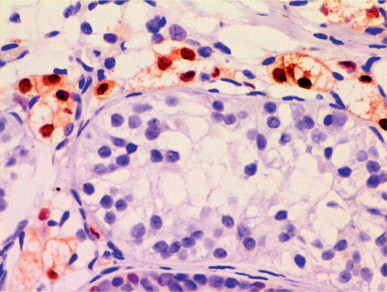
Testis from an infant with 5α-reductase deficiency showing hyperplastic Leydig cells that have marked cytoplasmic vacuolation and surround a seminiferous tubule lacking germ cells. (Immunostain for calretinin.)
The disorder is autosomal recessive and has been observed in many consanguineous families from the Dominican Republic.787
Defective regression of müllerian ducts
This group of male pseudohermaphrodites is characterized by the presence of müllerian derivatives and unilateral or bilateral testicular dysgenesis. These two features depend on anti-müllerian hormone gene mutations and end-organ insensitivity.788, 789, 790, 791
In normal development, anti-müllerian hormone is responsible for inhibition of the ipsilateral müllerian ducts and collagenization of the tunica albuginea. Patients with deficient secretion of this hormone may also have androgen deficiency. Three variants of defective müllerian duct regression have been reported: mixed gonadal dysgenesis, dysgenetic male pseudohermaphroditism, and persistent müllerian duct syndrome.
Mixed gonadal dysgenesis
Mixed gonadal dysgenesis (asymmetric gonadal differentiation) is characterized by the presence of a testis on one side of the body and a streak gonad on the other.792 If the gonads are intra-abdominal, the labioscrotal folds may appear as either normal labia or empty scrotal sacs (Fig. 12-81 ). In the former, the syndrome cannot be recognized in the newborn unless a peniform clitoris is present. If the gonad is descended, it is usually a testis. Müllerian derivatives such as fallopian tubes are usually associated with streak gonad (95% of cases), but may also be associated with testicular tissue (74%). Ipsilateral to the testis there is one epididymis and one vas deferens. On the contralateral side, no gonad or a streak gonad and a fallopian tube are present. A hypoplastic uterus and a poorly developed vagina are frequent findings.
Fig. 12-81.
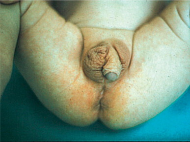
Mixed gonadal dysgenesis in a 3-year-old infant with ambiguous external genitalia, hypoplastic uterus, testicular dysgenesis on the right side, and streak gonad on the left side.
This syndrome accounts for about 15% of intersex conditions. Some patients are raised as males, although their external genitalia are usually ambiguous as a result of fetal virilization. The penis is clitoriform, and the urethra opens in the perineum. Most have cryptorchid testes and are raised as girls, becoming virilized at puberty. Infertility is a common symptom.793 The etiology is heterogeneous:794 one-third of patients have turnerian features, in accordance with the presence of the 45X0/46XY karyotype in more than 50% of patients. Other observed karyotypes are 46XY and 45X0/47XYY. Approximately 81% of patients have one Y chromosome. Mutation in the SRY gene has not been found.795
The testes can show two different patterns: testicular dysgenesis and streak testis. Testicular dysgenesis is characterized by a tunica albuginea that varies in width and is reminiscent of ovarian stroma by the storiform distribution of cells and fibers; there are also malformed seminiferous tubules (Fig. 12-82 ) that are small, usually lack lumina, and contain only immature Sertoli cells. In adults, spermatogenesis has been observed occasionally. The testicular interstitium contains increased numbers of Leydig cells.
Fig. 12-82.
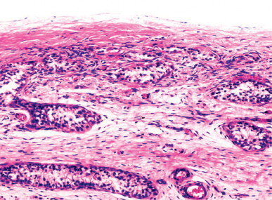
Testicular dysgenesis. Several irregularly shaped seminiferous tubules are observed within a thin, poorly collagenized tunica albuginea.
Streak testes are complex gonads in which testicular dysgenesis is associated with a fibrous streak. Most of the gonad consists of a testis showing the characteristic lesions of testicular dysgenesis. In a pole of the gonad, or in continuity with it, there is a fibrous streak whose structure may correspond to any of the varieties mentioned above (Fig. 12-83 ). This peculiar gonad can also be observed in some dysgenetic male pseudohermaphrodites as well as in the persistent müllerian duct syndrome. In these cases, the streak contains no ovocytes. Light microscopy indicates a wide spectrum of testicular lesions, ranging from those of patients with 46XY pure gonadal dysgenesis to true hermaphroditism. Differentiation of the ovocyte-containing streak testis and ovotestis remains controversial.796, 797
Fig. 12-83.
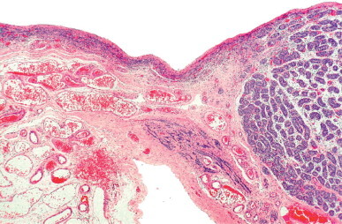
Streak testis consisting of a streak gonad connected to a testis which shows the characteristic lesions of testicular dysgenesis.
The testes in mixed gonadal dysgenesis are incapable of müllerian duct inhibition and allow complete differentiation of wolffian derivatives, virilization of external genitalia, and, in most cases, testicular descent. The risk of germ cell neoplasia reaches 50% in the third decade of life, usually beginning with gonadoblastoma. The testes should be removed after puberty.
Dysgenetic male pseudohermaphroditism
Dysgenetic male pseudohermaphroditism is a disorder of sexual differentiation characterized by bilateral dysgenetic testes or streak testis, persistent müllerian structures, and cryptorchidism. This syndrome is considered a variant of mixed gonadal dysgenesis (Fig. 12-84 ).791, 798 The karyotype may be 46XY or 45X0/46XY, and turnerian stigmata may be present. The uterus and fallopian tubes are present and both are usually hypoplastic (Fig. 12-85 ).799 The testes show lesions characteristic of testicular dysgenesis, with few germ cells during childhood (Fig. 12-85).799 In adults, spermatogenesis is poorly developed and the testicular interstitium shows Leydig cell hyperplasia. About 25% of patients develop gonadoblastoma.800
Fig. 12-84.
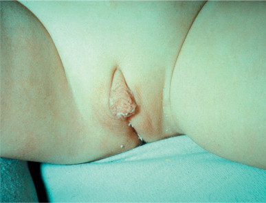
Male pseudohermaphrodite with bilateral testicular dysgenesis.
Fig. 12-85.
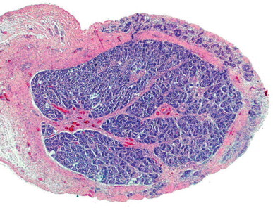
Testicular dysgenesis. The gonad has a central portion showing a testicular pattern and a peripheral band consisting of poorly collagenized connective tissue that contains seminiferous tubules that reach the gonadal surface.
Persistent müllerian duct syndrome
Persistent müllerian duct syndrome has many names, including male with uterus, tubular hermaphroditism, persistent oviduct syndrome, and hernia uteri inguinalis.801 It is a rare form of pseudohermaphroditism, with müllerian derivatives in an otherwise phenotypically normal male, and is the most characteristic form of isolated anti-müllerian hormone deficiency.
The molecular basis of this syndrome is heterogeneous. Three hypotheses have been proposed, including a defect in anti-müllerian hormone synthesis, caused by mutation in the anti-müllerian hormone gene (45% of cases); resistance of target organs to this hormone, caused by mutation in the receptor II for this hormone (39% of cases); and failure in the action of this hormone immediately before the eighth week of gestation (16% of cases).802
Although the external genitalia are male, one (35% of cases) or both testes (75% of cases) are cryptorchid. The syndrome usually also includes inguinal hernia contralateral to the undescended testis, with a uterus and fallopian tubes within the hernia sac (Fig. 12-86, Fig. 12-87 ).803 Several cases with transverse testicular ectopia and persistent müllerian duct structures have been reported.804, 805 Patients usually have inguinal hernia, but others have cryptorchidism, infertility,806 and testicular tumor.807
Fig. 12-86.
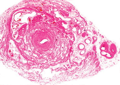
Persistent müllerian duct syndrome. Cross sectioned hypoplastic uterus. In its tunica adventitia and parallel to it, a folded ductus deferens is seen.
Fig. 12-87.
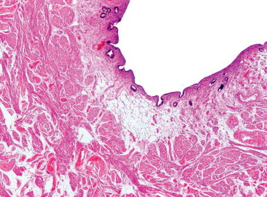
Persistent müllerian duct syndrome. Uterus with atrophic endometrium and hypoplastic myometrium within a hernia sac.
In childhood, the testes have a low tubular fertility index and decreased tubular diameter. In adults, the tunica albuginea is variably thickened, contains connective tissue resembling ovarian stroma, and may contain tubular structures – alterations typical of testicular dysgenesis. The seminiferous tubules are usually atrophic and hyalinized. Tubules with reduced spermatogenesis or patterns suggesting mixed atrophy (seminiferous tubules with spermatogenesis intermingled with Sertoli cell-only tubules) have also been reported. The Leydig cells appear hyperplastic. Azoospermia or oligozoospermia are common, and paternity is exceptional.808
The syndrome is sporadic or familial, with autosomal recessive or X-linked inheritance.809, 810 These patients have a higher risk of testicular tumor than that attributed to cryptorchidism,811 and all types of germ cell tumor have been observed.812, 813
Other forms of male pseudohermaphroditism
Of the dysmorphic syndromes associated with incomplete virilization of external genitalia, the best-known are those of RSH, Denys–Drash, WAGR, Opiz, camptomelic dysplasia, ATR-X, Gardner–Silengo–Wachtel, Meckel, branchioskeletal–genital, Down's, and other trisomies.
RSH (Smith–Lemli–Opitz) syndrome is a malformative recessive autosomal syndrome caused by mutations in the gene encoding for 7-dehydrocholesterol reductase (DHCR7), responsible for the synthesis of cholesterol from its immediate precursor 7-dehydrocholesterol.814, 815, 816 The disorder is common in Europe and rare in other countries.817
The most severe form is lethal before birth. Fetuses show postaxial oligodactyly (instead of polydactyly) and sometimes severe hydrops.818 Non-lethal forms are characterized after birth by severe growth failure; a semi-obtunded state; absence of psychomotor development; microcephaly; congenital cataracts; peculiar facies; broad anteriorly rugose alveolar ridges with cleft palate, edema of the nape of the neck, and unilobulate lungs; male pseudohermaphroditism or female external genitalia in 46,XY patients; postaxial polydactyly of the hands and feet; congenital heart defects; and renal anomalies.819 Hepatic and renal insufficiencies are frequent.820
The less severe forms in the male have genital anomalies (70%) varying from normal genitalia to severe hypospadias with or without cryptorchidism, and numerous small anomalies whose collection characterizes the syndrome. Most patients also show mental retardation and severe behavioral problems.821
The DHCR7 gene maps to chromosome 11q12-13. Its product is a microsomal, membrane-bound protein. Many different missense, nonsense, and splice-site mutations as well as duplications and deletions have been reported.822, 823, 824, 825, 826, 827 Prenatal diagnosis is possible by relating ultrasound and cytogenetic studies and carrying out a biochemical analysis in the second trimester in those pregnant women who have low levels or no conjugated estriol.828
In Denys–Drash syndrome, male pseudohermaphroditism is associated with nephroblastoma and renal insufficiency.829 The pseudohermaphroditism is usually either mixed gonadal dysgenesis, dysgenetic male pseudohermaphroditism, 46XY pure gonadal dysgenesis, or true hermaphroditism.830 The most common nephropathy is diffuse mesangial sclerosis.831 Most patients have mutations in the WT-1 gene,832 which is expressed in the genital ridge in the sixth week of gestation and gives rise to either streak gonads or testicular dysgenesis, but, if a delay in testicular determination occurs, normal testes are formed.833
The term WAGR syndrome refers to Wilms’ tumor, aniridia, genital anomalies, and mental retardation. Prevalence is estimated at between 0.75% and 2% of Wilms’ tumor patients. The syndrome is related to the syndrome of Denys–Drash and that of Frasier (a variety of 46,XY gonadal dysgenesis).834, 835 All have in common mutations in the WT-1 gene located on chromosome 11 (11p13).
WT-1 product is a transcription factor expressed in different tissues that participates in embryogenesis and cell differentiation. Mutations lead to the production of an anomalous protein that causes alterations in renal function, gonadal anomalies, and the loss of tumor suppressor function. Six variants of alleles have been described: isolated Wilms’ tumor, mesothelioma, isolated diffuse mesangial sclerosis, Denis–Drash syndrome, Frasier syndrome, and WAGR syndrome. Frasier syndrome is caused by mutations in the donor zone of the intron 9 link, with the subsequent loss of the +KTS isoform (the patient has an imbalance in KTS isoforms), whereas large deletions or loss of genetic material that comprises the WT-1 gene and other contiguous genes (PAX6 or AN) lead to the WAGR syndrome.836, 837
Patients with Opitz's syndrome are mainly boys with hypertelorism and, in the severe forms, unilateral or bilateral lip cleft, laryngeal cleft, severe dysphagia with more or less life-threatening aspiration, hypospadias and, occasionally, imperforate anus. The most important internal anomalies are those in the tracheobronchial tree, cardiovascular system (defects in cardiac septation), and gallbladder, with a subjacent defect of the developing embryonal ventral midline. The syndrome is genetically heterogeneous and consists of two entities that were described as different in the past: ADOS, or autosomal dominant Opitz syndrome or G syndrome838 with a mutated gene that maps to 22q11.2; and XLOS, or X-linked Opitz syndrome or BBB syndrome839 with a mutated gene that maps to Xp22.3.840, 841
Camptomelic dysplasia is an autosomal dominant syndrome with multiple osseous malformations. Patients have 46XY karyotype and external genitalia that are ambiguous or female. Gonadal histology varies from testes to dysgenetic ovaries with primary follicles. The cause is a haploinsufficiency of SOX9, located on 17q.842 The incidence of gonadoblastoma is low.
ATR-X syndrome is characterized by mild α-thalassemia, mental retardation, facial dysmorphism, and hypospadias.843, 844 The disorder is X-linked, and is caused by mutation in the ART-X gene (synonymous XNP, HX2).845
Infertility
Testicular biopsy
Testicular biopsy to diagnose infertility began in the 1940s,846, 847 and most of the diagnostic terms used today were created then.848 These terms are usually descriptive and, except for a few (normal testes, Sertoli cell-only tubules, tubular hyalinization, for example), do not specify the degree of tubular abnormality that is evaluated by each pathologist subjectively. The terms maturation arrest and hypospermatogenesis have been applied to biopsies in more than 50% of cases of infertility,849, 850, 851 but the criteria for these vary widely among pathologists.
Two forms of maturation arrest have been described: spermatogenic arrest, and spermatocytic arrest, or its equivalent, meiotic arrest. True spermatogenic arrest is rare because germ cell maturation usually does not arrest at the level of a defined germ cell type.852 To avoid confusion the term irregular hypospermatogenesis has been proposed853 for testicular biopsies with decreased numbers of germ cells, subclassified as slight, moderate, or severe. However, this diagnosis is of little help to clinicians. The reported frequency of spermatocytic (meiotic) arrest in infertile men varies from 12%854 to 32.1%855 and is present in one or both testes of about 18% of oligozoospermic or azoospermic patients.856 If observed in only one testis, the contralateral testis may show histologic changes ranging from normal spermatogenesis to hyalinized tubules.
Disorganization of the seminiferous tubular cell layers is another frequent diagnosis in testicular biopsies,848, 857, 858 but this term is rejected by many pathologists. Actual disorganization of the seminiferous tubular cells is unlikely and has not been demonstrated in ultrastructural studies. In most cases, the apparent disorganization is an artifact induced by handling or fixation.859, 860
The term tubular blockage was introduced by Meinhard and co-workers858 for testes with at least 50% of seminiferous tubules devoid of a central lumen and showing spatial disorganization of germ cells. This morphology was found in 28% of testicular biopsies from infertile men, mainly those with obstructive azoospermia.861 Although this appearance can result from improper fixation,862 the accumulation of Sertoli cells and immature germ cells in the centers of tubules suggests a specific lesion, a variant of germ cell sloughing.
Diagnostic confusion decreased the interest and trust of urologists and andrologists in the study of testicular biopsies. Subsequent studies attempted to correlate semen spermatozoa concentration with testicular size and biochemical findings such as serum levels of FSH, and testicular biopsies were undertaken in only a limited number of oligozoospermic and azoospermic patients.859, 862, 863 However, these studies were also discouraging because FSH was found to correlate poorly with numbers of spermatozoa in the semen but better with numbers of spermatogonia in the seminiferous tubules,864 and normal numbers of spermatozoa can be produced by relatively small testes whereas some large testes have no spermatogenesis. In recent years, serum levels of inhibin B have been shown to have a positive correlation with spermatozoon numbers and serum FSH level.865, 866
The development of morphometry caused a resurgence of interest in biopsies. Many semiquantitative853, 867, 868, 869 and quantitative870, 871, 872, 873, 874, 875 studies were carried out. The greatest achievements of these studies were enhancement of the reproducibility of results and better evaluation of the reversibility of lesions. Morphometry emerged as the best method to objectively evaluate the seminiferous tubular cells.876 The scoring method of Johnsen,868 estimation of the germ cell/Sertoli cell ratio for each germ cell type,871 and calculation of germ cell number per unit length of seminiferous tubules870 are reliable and useful.
Several methods are available to evaluate the Leydig cell population, including the mean number of cells per seminiferous tubule and per cell cluster; the mean number of Leydig cell clusters per seminiferous tubule; the ratio of Leydig cell area to seminiferous tubule area;877 and the ratio of Leydig cells to Sertoli cells.878 These methods have shown that the appearance of Leydig cell hyperplasia described in many conditions is false, and that true Leydig cell hyperplasia is extremely rare.
Optimal interpretation of testicular biopsies depends on the surgical technique by which the tissue sample is taken, the care and delicacy with which the specimen is manipulated, and proper fixation and processing of the tissue. The size of the biopsy should not be greater than a grain of rice: that is, no diameter should be greater than 3 mm. This amounts to about 0.12% of testicular volume (normal volume is approximately 20 mL). The biopsy should be bilateral because in more than 28% of patients the findings differ between the testes. At the time of biopsy, the testicular axes should be measured as the basis of quantitative studies. The tissue should be taken opposite to the rete testis through a 4–5 mm incision in the tunica albuginea. This parenchyma herniates through the incision and can be carefully snipped off. If only light microscopy is to be performed, the specimen should be fixed in Bouin's fluid for 24 hours. If electron microscopy is indicated, a small biopsy fragment should be fixed in glutaraldehyde–osmium tetroxide or a similar fixative. To perform meiotic studies, testicular biopsy should be processed by air-drying or surface-spreading methods. The examination of testicular biopsies includes qualitative and quantitative evaluation of the testis and correlation between the biopsy and spermiogram.
Qualitative and quantitative evaluation of testicular biopsy
Light microscopy immediately reveals whether the lesion is focal or diffuse. If focal, the percentage of tubules showing each lesion (Sertoli cell-only, hyalinization, tubular hypoplasia, etc.) should be calculated. It is useful to evaluate elastic fibers with a special stain because this highlights groups of small tubules that may be missed with hematoxylin and eosin. A minimum of 30 cross-sectioned tubules should be studied (this is usually possible when five or six histological sections are available). The diameter of each tubule should be measured, and the number of spermatogonia, primary spermatocytes, young spermatids (also called round spermatids or Sa + Sb spermatids), mature spermatids (also called elongated or Sc + Sd spermatids), Sertoli cells, and, in some cases, peritubular cells counted. The presence of tubular diverticula,879, 880 the maturation of Sertoli cells, and morphologic anomalies in germ cells should also be noted. Evaluation of the testicular interstitium should include the number of Leydig cells per tubule (or number of Leydig cell clusters per tubule), the presence of angiectasis (phlebectasis), and the occurrence of peritubular or perivascular inflammation. Normal values are tabulated in Table 12-6 . For a clear and rapid understanding of the results, data can be presented using cartesian axes (see Fig. 12-96, Fig. 12-103 ).
Table 12-6.
Testicular parameters in normal adult testes (means ± SD)
| Values per cross-sectioned tubule | Means ± SD |
|---|---|
| Seminiferous Tubules | |
| Mean tubular diameter (μm) | 193 ± 8 |
| Number of spermatogonia | 21 ± 4 |
| Number of primary spermatocytes | 31 ± 6 |
| Number of young (Sa + Sb) spermatids | 37 ± 7 |
| Number of mature (Sc + Sd) spermatids | 25 ± 4 |
| Number of Sertoli cells | 10.4 ± 2 |
| Number of Sertoli cell vacuoles | 0.8 ± 0.3 |
| Lamina propria thickness (μm) | 5.3 ± 1 |
| Number of peritubular cells | 21 ± 4 |
| Testicular Interstitium | |
| Number of Leydig cell clusters per tubule | 1.2 ± 0.3 |
| Number of Leydig cells per tubule | 5 ± 0.2 |
Fig. 12-96.
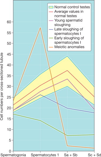
Germ cell number per cross-sectioned tubule in patients with lesions in the adluminal compartment of the seminiferous tubules.
Fig. 12-103.
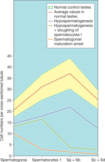
Germ cell number per cross-sectioned tubule in patients with lesions in the basal and adluminal compartments of the seminiferous tubules.
Common lesions
The most frequently observed lesions are Sertoli cell-only tubules, tubular hyalinization, alterations in spermatogenesis in either the adluminal or the basal compartments of seminiferous tubules, and mixed tubular atrophy.
Sertoli cell-only syndrome
Sertoli cell-only syndrome includes all azoospermias in which the seminiferous epithelium consists only of Sertoli cells. To better understand this syndrome, it is necessary to consider the morphological and functional changes induced in the Sertoli cell by hypophyseal gonadotropin secretion during puberty. During childhood, Sertoli cells are pseudostratified and their nuclei are dark, small, and round or elongated, with regular outlines and one or two small peripherally placed nucleoli. The cytoplasm lacks specialized organelles.881 Adult Sertoli cells have characteristically pale, triangular nuclei with irregular, indented outlines. The nucleoli are large and have tripartite structures. The cytoplasm contains abundant smooth endoplasmic reticulum and specialized structures, including annulate lamellae, Charcot–Böttcher crystals, and specialized junctional complexes with other Sertoli cells. The pubertal increase in length and width of the seminiferous tubules replaces the infantile pseudostratified pattern with a simple columnar distribution.
Five variants of the Sertoli cell-only syndrome are identified by Sertoli cell morphology, the degree of development of the seminiferous tubules, and the presence or absence of interstitial lesions.882 These variants are designated by the appearance of the predominant Sertoli cell population: immature Sertoli cells, dysgenetic Sertoli cells, adult Sertoli cells, involuting Sertoli cells, and dedifferentiated Sertoli cells (Fig. 12-88 ). Each type is associated with other tubular and interstitial alterations (Table 12-7 ).
Fig. 12-88.
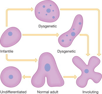
Sertoli cell types.
Table 12-7.
Variants of Sertoli cell-only syndrome
| Testis pattern | Variants of the Sertoli cell-only syndrome |
||||
|---|---|---|---|---|---|
| Immature Sertoli cells | Dysgenetic Sertoli cells | Adult Sertoli cells | Involuting Sertoli cells | Dedifferentiated Sertoli cells | |
| Tubular diameter | Very decreased | Decreased | Decreased | Decreased | Decreased |
| Tubular lumen | Small or absent | Small or absent | Normal | Normal | Normal |
| Lamina propria thickness | Thin | Enlarged | Normal or enlarged | Normal or enlarged | Enlarged |
| Elastic fibers in lamina propria | Absent | Decreased | Normal | Normal | Normal |
| Sertoli cells | |||||
| Number | Very increased | Increased | Normal or increase | Normal or increased | Increased |
| Distribution | Pseudostratified | Pseudostratified | Columnar | Columnar | Columnar or pseudostratified |
| Nuclear shape | Ovoid | Round or ovoid | Triangular | Lobated | Round |
| Nuclear outline | Regular | Regular | Few indented | Very indented | Regular |
| Chromatin | Dark | Pale with granules | Pale | Pale | Pale |
| Nucleolus | Small, peripheral | Developed, central | Developed, central | Developed, central | Small, central or peripheral |
| Vacuoles | Absent | Present | Present | Abundant | Abundant |
| Lipids | Absent | Absent | Decreased | Abundant | Abundant |
| Vimentin filaments | Basal | Basal | Basal and perinuclear | Basal and perinuclear | Basal |
| Antimüllerian hormone | Present | Present | Absent | Absent | Absent |
| Interstitium | Scanty | Increased | Normal | Normal/fibrosis | Fibrosis |
| Leydig cells | Absent | Pleomorphic, vacuolated, increased or decreased | Normal | Decreased, many lipofuscin granules | Decreased, many lipofuscin granules |
| Clinical symptoms | Hypogonadotropic hypogonadism | Infertility | Infertility, orchitis | Infertility, hypergonadotropic hypogonadism, chemo- or radiotherapy | Treatment with estrogens, antiandrogens or cisplatinum, chronic hepatopathy |
The most frequent types of Sertoli cell-only syndrome in infertility patients are dysgenetic Sertoli cells, adult Sertoli cells, and involuting Sertoli cells. The clinical manifestations are similar, including normal external genitalia, well-developed secondary male characteristics, azoospermia, elevated serum FSH level, normal or elevated serum LH level, and normal or slightly low testosterone. These clinical and histologic features were long thought to constitute a single syndrome, Del Castillo's syndrome, but recent ultrastructural, histochemical, immunohistochemical, and cytogenetic studies have shown that this results from a variety of syndromes that may have primary or secondary causes (Table 12-7).883, 884, 885, 886, 887
Some patients with the adult or dysgenetic Sertoli cell-only syndrome variants have a few spermatozoa in their spermiograms. This discrepancy between oligozoospermia and the biopsy histology is caused by the presence of some seminiferous tubules with complete spermatogenesis elsewhere in the testicular parenchyma.
Sertoli cell-only syndrome with immature Sertoli cells
Sertoli cells in adult testes with this variant of Sertoli cell-only syndrome have an immature prepubertal appearance with pseudostratification. The number of cells per cross-sectioned tubule is greater than normal. Other tubular and interstitial features suggest immaturity, including small tubular diameters (<80 μm), tubules lacking central lumina, thin lamina propria lacking elastic fibers, and interstitium lacking mature Leydig cells.888, 889, 890
This syndrome is caused by a deficiency of both FSH and LH which begins in childhood and is responsible for the lack of maturation of the Sertoli cells, tubular walls, and interstitium. Subsequently, there is no renewal or differentiation of germ cells, and these eventually disappear. When these patients are treated with hormones, the biopsy may show some degree of spermatogenesis or thickening and hyalinization of the tubular basement membrane.
Sertoli cell-only syndrome with dysgenetic Sertoli cells
Dysgenetic Sertoli cells begin pubertal differentiation but variably deviate from normal maturation, so that the morphology of dysgenetic Sertoli cells differs among tubules and even among Sertoli cells within the same tubule. Nuclei usually have both mature features (pale chromatin and a centrally located, tripartite nucleolus) and features of immaturity (ovoid or round shape; regular outline; and small, dense chromatin granules) (Fig. 12-89 ).891 In addition to vimentin, Sertoli cells immunoexpress anti-müllerian hormone (AMH)892 and cytokeratin 18.893 Immunoreaction to these two substances is assumed to be a sign of immaturity, as under normal conditions it is not detected after puberty. Other signs of immaturity are poor development of the hematotesticular barrier894 and the absence of tubular lumina.
Fig. 12-89.
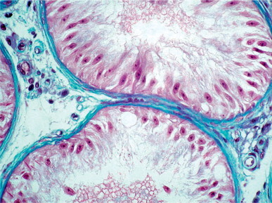
Sertoli cell-only syndrome with dysgenetic Sertoli cells. Seminiferous tubules show slightly thickened tunica propria. The Sertoli cells are increased in number and have elongated nuclei and abundant apical cytoplasm.
Tubular lumina are very small or absent in most dysgenetic Sertoli cell-containing tubules, because the ability to produce testicular fluid is greatly reduced. Sertoli cell numbers per cross-sectioned tubule are very high, and mean tubular diameter is lower than 120 μm. The tubular walls have few elastic fibers,534 and most tubules show a variable degree of tunica propria hyalinization.
Completely hyalinized tubules are frequent. The testicular interstitium contains a variable number of Leydig cells (normal, decreased, or apparently increased), many of which are pleomorphic with abundant paracrystalline inclusions.895, 896
Most patients have normal or slightly subnormal testosterone level and elevated levels of FSH and LH. This syndrome can be observed in men with cryptorchid testes, at the periphery of germ cell tumors, in men with idiopathic infertility,897 and in men with Y chromosome anomalies.898
Sertoli cell-only syndrome with mature Sertoli cells
In this variant, most Sertoli cells appear mature but are present in increased numbers (14 ± 0.8 per cross-sectioned tubule). The seminiferous tubules have small diameters, but are still larger than in the two variants described above, and central lumina are visible. The cytoplasm contains abundant vacuoles that communicate with the tubular lumina (Fig. 12-90 ). The lateral cell surfaces have many unfolding and extensive specialized junctions with other Sertoli cells (from the basement membrane to the apical cytoplasmic portion). Lipid droplets, usually derived from phagocytosis of spermatid tubulobulbar complexes and dead germ cells, are scant.884 Vimentin filaments are abundant in the basal and perinuclear cytoplasm.899 The lamina propria is normal or slightly thickened. Leydig cells are normal.
Fig. 12-90.
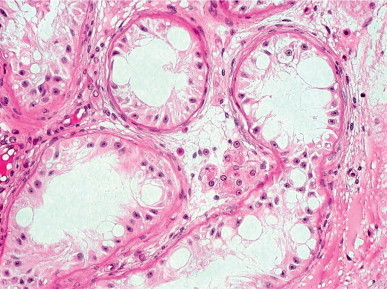
Sertoli cell-only syndrome with mature Sertoli cells. The seminiferous tubules are lined by normal adult Sertoli cells, many with cytoplasmic vacuoles.
Serum testosterone level is normal or nearly normal, and FSH and LH levels are elevated.900, 901, 902 This syndrome is probably caused by failure of migration of primordial germ cells from the primitive yolk sac to the gonadal ridge.903 This failure may be due to a deletion in the AZFa region in Yq11904 or a mutation in the genes that encodes c-KIT or its ligand (stem cell factor), responsible for migration, proliferation, and survival of germ cells.
Sertoli cell-only syndrome with involuting Sertoli cells
Testes with this variant of Sertoli cell-only syndrome have numerous changes. Sertoli cell nuclei may have lobulated shapes with irregular outlines, coarse chromatin granules, and inconspicuous nucleoli. Seminiferous tubules have central lumina, decreased diameters, and variable thickening of the basement membrane (Fig. 12-91 ). Elastic fibers are present in normal or diminished amounts. Leydig cells are variably involuted.
Fig. 12-91.
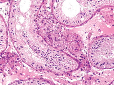
Sertoli cell-only syndrome with involuting Sertoli cells. The Sertoli cell nuclei are hyperchromatic and have irregular outlines.
This syndrome may be a primary disorder or secondary to irradiation or cytotoxic therapy, such as cancer chemotherapy or treatment for nephrotic syndrome.905 It is not usually possible to determine the etiology from the biopsy findings alone. Changes in the tubular walls are more pronounced in patients with a history of cyclophosphamide treatment, combination chemotherapy, or radiotherapy. The testicular interstitium may be fibrotic in patients treated with cis-platinum or cyclophosphamide.906 Some syndromes with involuting Sertoli cells, mainly those associated with decreased number of elastic fibers, probably express a primary testicular anomaly with involuting and dysgenetic Sertoli cells within the same tubule.
Sertoli cell-only syndrome with dedifferentiated Sertoli cells
The presence of immature-appearing Sertoli cells in otherwise mature tubules is the most striking feature of this variant of Sertoli cell-only syndrome. Sertoli cells appear abnormally numerous due to shortening of the tubule, and nuclei are either round or elongated. Round nuclei have single, small, central or peripheral nucleoli, whereas elongated nuclei have dense clumped chromatin and small peripheral nucleoli.
The tubular wall is thickened and contains elastic fibers, increased amounts of collagen fibers, and elevated numbers of peritubular cells as a result of tubular shortening. Mean tubular diameter is markedly decreased to less than 90 μm. The testicular interstitium contains few Leydig cells, and these appear dedifferentiated or contain an increased amount of lipofuscin.
This variant has been observed in surgical specimens from patients receiving androgen deprivation therapy for prostatic cancer, estrogen treatment for transsexuality, and cancer chemotherapy with cis-platinum. There is a correlation between the degree of Sertoli cell dedifferentiation and the dose and timing of treatment with estrogens or anti-androgens. Brief treatment induces germ cell loss and inconspicuous Sertoli cell changes; long-term treatment causes pronounced Sertoli cell changes, including initial nuclear rounding followed by nuclear elongation and the development of dark chromatin masses.907 Eventually, the nuclei come to resemble those of infantile Sertoli cells, including pseudostratification. At the same time, the tubules become hyalinized and peritubular cells increase whereas Leydig cells disappear.908, 909
Estrogens act on the pituitary by inhibiting LH secretion, and on Leydig cells.910 The action of gonadotropin-releasing hormone agonist analogs is only on the pituitary, whereas cis-platinum acts only on the testis.
Tubular hyalinization
A few azoospermic patients have diffuse hyalinization of seminiferous tubules. The incidence of this lesion is difficult to estimate, as these patients usually are not biopsied because their testes are small. Hyalinization of seminiferous tubules is the endpoint of tubular atrophy and includes the absence of both germ cells and Sertoli cells with alterations in the lamina propria and Leydig cells. Etiology can be determined from several histologic features and clinical data, including:
-
•
General histologic appearance: extent and topography of the hyalinized tubules and presence of isolated tubules containing germ cells or Sertoli cells only (dysgenetic, adult, involuting, or dedifferentiated).
-
•
Appearance of atrophic tubules, all showing the same pattern or variable degrees of atrophy: tubular diameter; trophism of peritubular cells; presence of elastic fibers; degree of collagenization of the lamina propria, and the presence of cell remnants or unusual cells in the tubules.
-
•
Appearance of the interstitium: number and morphology of Leydig cells; vascular lesions; and lymphoid infiltrate.
-
•
Chronology of testicular shrinkage.
The most common causes of tubular hyalinization include dysgenetic hyalinization, hormonal deficit, ischemia, ob-struction, inflammation, and physical or chemical agents. The differential diagnosis is given in Table 12-8 .
Table 12-8.
Differential diagnosis of tubular hyalinization
| Dysgenetic | Hormonal deficit | Ischemia | Excretory duct obstruction | Postinflammatory hyalinization | Physical or chemical agents | |
|---|---|---|---|---|---|---|
| Hyalinized tubule size | Minimum | Minimum | Minimum | Very decreased | Minimum | Very decreased |
| Tubular lumen | Absent | Absent | Absent | Present | Absent | Absent |
| Peritubular cells | Decreased | Decreased | Decreased | Increased | Decreased or increased | Decreased |
| Elastic fibers | Decreased | Normal | Normal | Normal | Normal | Normal |
| Leydig cells | Increased or decreased, pleomorphic | Absent | Absent | Normal | Pseudo-hyperplasia | Decreased |
| FSH | Increased | Decreased | Increased | Increased | Increased | Increased |
| LH | Increased | Decreased | Increased | Increased | Increased | Increased |
| Testosterone | Normal or decreased | Decreased | Normal or decreased | Normal | Normal | Normal or decreased |
Dysgenetic hyalinization
Dysgenetic hyalinization is a diffuse lesion in which most tubules are uniformly hyalinized (Fig. 12-92 ). Tubules lack seminiferous tubular cells and have a reduced number of peritubular cells. The few preserved tubules usually contain only Sertoli cells, although rarely a few with spermatogenesis are present. Dysgenetic hyalinization is seen in Klinefelter's syndrome, testes that remain cryptorchid through puberty, and some hypergonadotropic hypogonadisms associated with myopathy. Focal lesions are seen in mixed atrophy of the testis.
Fig. 12-92.
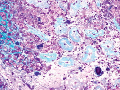
Dysgenetic hyalinization. Fully hyalinized seminiferous tubules and a few peritubular cells among Leydig cell clusters.
Tubular hyalinization is pronounced in Klinefelter's syndrome, and from infancy the seminiferous tubules are small, containing reduced numbers of Sertoli cells and few or no spermatogonia. At puberty, the dysgenetic Sertoli cells fail to mature and soon disappear. The tubules collapse, giving the appearance of phantom tubules.911 Peritubular cells fail to differentiate and their number is low.912 They form a discontinuous ring around the hyalinized tubules and are incapable of synthesizing elastic fibers and other components of the lamina propria. Dysgenesis also involves the interstitium: Leydig cells exhibit a characteristic adenomatous pattern, although their total number is decreased. The morphology of the Leydig cell is not uniform, and there are shrunken, normal, and large forms. Most contain reduced amounts of lipofuscin granules and lipid droplets. Reinke's crystalloids are uncommon, and paracrystalline inclusions are abundant.896 In spite of the hyperplastic adenomatous appearance of the Leydig cells, testosterone secretion is markedly decreased, and the resulting hypogonadism is the most important clinical feature of Klinefelter's syndrome.
Tubular hyalinization in the cryptorchid testis is also dysgenetic. However, in contrast to the atrophic collapse seen in Klinefelter's syndrome, cross-sections of the hyalinized tubules in cryptorchidism are targetoid. This results from the arrangement of the peritubular cells into two layers, suggesting an atrophic process that has evolved over a longer period than in Klinefelter's syndrome, or a lower degree of dysgenesis.913 Elastic fibers are diminished.534 In the interstitium Leydig cells appear hyperplastic, forming large aggregates, although their absolute numbers are decreased. Leydig cell pleomorphism is less intense than in Klinefelter's syndrome. Many Leydig cells have abundant vacuolated cytoplasm. Whereas tubular hyalinization in Klinefelter's syndrome is secondary to the effect of pubertal gonadotropin secretion on dysgenetic tubules, tubular hyalinization in cryptorchidism probably results from the effect of increased temperature on the dysgenetic tubules. However, other mechanisms are also involved in cryptorchid tubular hyalinization, including obstruction of sperm excretory ducts (anomalies in these ducts are frequent in cryptorchidism) and ischemia (principally in testes that could only be incompletely descended by surgery).
Hyalinization caused by hormonal deficit
Hormonal deficit causes diffuse tubular hyalinization, although the tubules may be recognized for a time as cellular cords surrounded by hyaline material. Sertoli cell, a few spermatogonia, and rare primary spermatocytes may be identified in these cords. When hyalinization is complete, only the elastic fibers in the lamina propria indicate the structure of the previously normal adult testis. Peritubular myofibroblasts decrease in number and form a ring at the periphery of the lamina propria. Leydig cells disappear as hyalinization progresses, and the few that remain have pyknotic nuclei and shrunken cytoplasm with abundant lipofuscin granules.
This process manifests clinically as postpubertal hypogonadotropic hypogonadism and is usually caused by a lesion in or near the pituitary, such as pituitary adenoma, craniopharyngioma, and trauma to the cranial base or sella turcica (see discussion on hypogonadotropic hypogonadism in this chapter).
Ischemic hyalinization
Ischemic atrophy is usually caused by torsion of the spermatic cord, vascular injury during inguinal surgery,914 polyarteritis nodosa, and severe arteriosclerosis.915 Except for cases caused by torsion of the cord, these patients usually are not referred to infertility clinics.
Torsion of the spermatic cord often is not listed as a cause in large series of infertile patients. However, follow-up of men with torsion reveals marked alteration in their spermiograms. Several hypotheses have been offered to explain the low number of sperm produced by the contralateral normal testis; the most promising include response to the release of antigens by the ischemic testis, and primary lesions of the contralateral testis916 (see discussion on testicular torsion in this chapter).
Testicular anoxia caused by torsion rapidly produces severe lesions that are irreversible without adequate treatment. Eight hours after torsion, there is intense hemorrhagic infarction of the seminiferous tubular cells. Chronic anoxia leads to tubular hyalinization and loss of Leydig cells (Fig. 12-93 ).
Fig. 12-93.
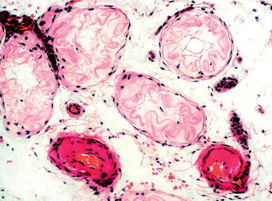
Ischemic tubular hyalinization. Fully hyalinized seminiferous tubules are surrounded by peritubular cells. The testicular interstitium lacks Leydig cells and shows arteriolar hyalinization.
Testicular atrophy secondary to inguinal hernia surgery occurs in 0.03–0.5% of patents in the first repair, and in 0.8–5% in surgery for recurrent hernia. Atrophy is most frequent in cases that require extensive dissection of the spermatic cord.
Postobstructive hyalinization
Obstruction of sperm excretory ducts may cause atrophy of seminiferous tubules. In order to produce tubular hyalinization, the obstruction must be close to the testis because the ductuli efferentes in the caput epididymis absorb about 90% of tubular fluid and protect the testis from excessive intratubular pressure. Obstructive tubular hyalinization is usually focal and secondary to varicocele and other disorders involving dilation of the channels of the rete testis. These may be congenital, as in epididymis–testis dissociation, or acquired, as in rete testis dilation secondary to epididymal atrophy caused by arteritis, arteriosclerosis, or androgen insufficiency. Obstructive tubular hyalinization also occurs in the seminiferous tubules at the periphery of the testis in patients who have had orchitis.917
Obstructive hyalinization has a mosaic distribution: lobules of completely hyalinized tubules are intermingled with lobules of normal tubules (Fig. 12-94 ). The diameter of the hyalinized tubules is not as small as in other causes of hyalinization, and the tubules occasionally contain Sertoli cells. In the centers of many of the tubules there is a small lumen or vacuole, the latter in the cytoplasm of a residual Sertoli cell.918 The lamina propria is thick and contains hypertrophic peritubular cells and abundant extracellular material. Finally, the peritubular cells dedifferentiate and only fibroblasts remain.919 The interstitium contains a normal number of Leydig cells forming small clusters, some of which are among hyalinized tubules. This is not seen in other patterns such as ischemic hyalinization. In addition, dilated veins with eccentrically hyalinized walls can be seen in testes associated with varicocele. This lobular pattern of tubular atrophy causes a peculiar ultrasound image which has been described as a striated pattern.920, 921
Fig. 12-94.
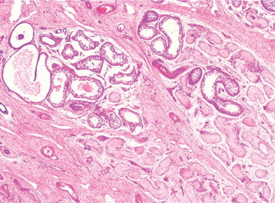
Post-obstructive hyalinization. Seminiferous tubules with marked ectasis with hyalinized tubules. Leydig cell clusters are seen among the hyalinized tubules.
Postinflammatory hyalinization
Many infections of the testis cause irreversible lesions in the seminiferous tubules. In bacterial infection the epididymis is usually involved, resulting in obstructive azoospermia. In viral infection the testis is often affected, even without symptoms. Two types of viral orchitis often cause infertility, including mumps orchitis and Coxsackie B orchitis.
Tubular atrophy caused by viral infection has a mosaic topography in which hyalinized and normal tubules are intermingled. In fully hyalinized tubules, the only recognizable cells are peritubular cells that form an incomplete, peripheral ring around the hyalinized material. The presence of elastic fibers in these tubules distinguishes this from dysgenetic hyalinization. Leydig cells form clusters of variable size, but their total number is normal. In bacterial infection the pattern of tubular hyalinization is variable.
Tubular atrophy of unknown etiology may be caused by an autoimmune response. This appears to occur in hypogonadism associated with disorders in other endocrine glands, such as Addison's disease associated with gonadal insufficiency; adrenal–thyroid–gonadal insufficiency; and the association of diabetes, hypogonadism, adrenal insufficiency, and hypothyroidism. The testicular lesions are morphologically similar to those seen in the seminiferous tubules at the periphery of germ cell tumor and in testes with burn-out germinal cancer. In the initial stages of hyalinization associated with germ cell neoplasm, the tubules are small, contain intratubular germ cell neoplasia and dedifferentiating Sertoli cells, and the lamina propria is infiltrated by macrophages, lymphocytes, and plasma cells. In the final stages, the intratubular cells have degenerated, the inflammation has disappeared, and the seminiferous tubules are replaced by areas of hypocellular or acellular fibrosis (Fig. 12-95 ). It should be noted that autoimmune hyalinization is not the most common type of hyalinization associated with testicular tumors: the obstructive, ischemic, and dysgenetic variants are more common.
Fig. 12-95.
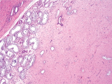
Post-inflammatory hyalinization. Most of the testis consists of cicatricial tissue with no recognizable seminiferous tubules.
Hyalinization caused by physical or chemical agents
Radiation and a wide variety of chemicals cause tubular hyalinization. Lengthy cancer chemotherapy combined with radiotherapy invariably causes hyalinization. Children's testes are more sensitive to radiation than those of adults. Radiation for testicular leukemia frequently causes tubular hyalinization. In addition, radiation induces dense interstitial fibrosis and loss of peritubular cells, obscuring the borders between the interstitium and the tubules. This makes the tubules hard to see in hematoxylin–eosin-stained sections. Leydig cells are atrophic and decreased in number. Ischemia secondary to radiation-induced vascular injury also contributes to hyalinization.
In tubular hyalinization associated with cancer chemotherapy, in addition to the direct toxicity of drugs on seminiferous tubular cells (see discussion on Sertoli cell-only syndrome with involuting Sertoli cells in this chapter), nutritional deficiencies cause hypogonadotropic hypogonadism.922, 923
Diffuse lesions in spermatogenesis
Histophysiological studies have distinguished two compartments in the seminiferous tubules: basal and adluminal. The blood–testis barrier separates these, and each contains different cell types with diverse hormonal and nutritional requirements. On this basis, lesions may be classified as involving only the adluminal compartment or both the basal and the adluminal compartments. The following discussion of spermatogenic lesions uses this new concept of tubular pathophysiology, conserving as much as possible of the classic terminology.
Lesions in the adluminal compartment of seminiferous tubules
This category includes all infertile testes with normal numbers of spermatogonia per cross-sectioned tubule, normal or decreased numbers of spermatocytes and young spermatids, and variable numbers of adult spermatids. A descriptive term for this disorder is immature germ cell sloughing.
A few immature germ cells are normally found in the lumina of the seminiferous tubules,923 a finding that correlates with their presence in the ejaculates of fertile men.924 When these cells make up more than 4% of the cells in the ejaculate, it is abnormal and the result of premature sloughing of spermatids and, in some cases, of spermatocytes.925, 926 Some authors have attempted to establish a correlation between the number of sloughed immature germ cells and the severity of lesions of the seminiferous tubules using light927 and electron928 microscopy.
Lesions in the adluminal compartment are classified according to the most abundant type of germ cell whose maturation is arrested and which then sloughs: young spermatids, late primary spermatocytes, or early primary spermatocytes (Fig. 12-96).
Young spermatid sloughing Young spermatid sloughing is present when the ratio of elongated (Sc + Sd) spermatids to round (Sa + Sb) spermatids is lower than normal. The implication of this pattern is that many round spermatids are incapable of further differentiation and are sloughed (Fig. 12-97 ).
Fig. 12-97.
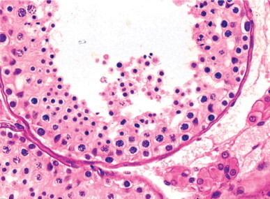
Seminiferous tubule with dilated lumen and moderate young spermatid sloughing.
Late primary spermatocyte sloughing In this condition, spermatogenesis develops normally up to the level of interphase primary spermatocytes, and these are present in normal numbers. Afterwards, these spermatocytes degenerate without achieving meiosis and slough into the tubular lumen. All types of spermatid are greatly reduced in number. When biopsies of these testes are not properly fixed, the seminiferous tubules may have a target-like appearance, with numerous cells in the lumen. This appearance sometimes has been referred to as tubular blockage. Another descriptive term, spermatogenic arrest, also has been applied to this morphology. The latter term is inadequate in most cases, because some spermatids are present, and the number of primary spermatocytes is usually not increased as would occur if the transformation of spermatocyte into spermatid were blocked (Fig. 12-98 ). Late spermatocyte sloughing is a more accurate term for this condition and is preferred. Primary spermatocyte sloughing occurs at the pachytene or diplotene stage of meiosis.
Fig. 12-98.
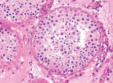
Seminiferous tubule with sloughing of both primary spermatocytes and young spermatids.
Early primary spermatocyte sloughing This lesion is characterized by the presence of a normal number of spermatogonia and decreased numbers of primary spermatocytes (Fig. 12-99 ). The seminiferous tubules may contain a few spermatids. The term early primary spermatocyte sloughing does not necessarily imply an early meiotic lesion, which is quite rare.856, 926 Rather, it refers to the sloughing of newly formed spermatocytes. The Sertoli cells may show vacuolation of the apical cytoplasm as an expression of germ cell loss. This lesion is more severe than that in testes with late primary spermatocyte sloughing, and is considered to result from failure of the Sertoli cells to maintain the adluminal compartment.
Fig. 12-99.
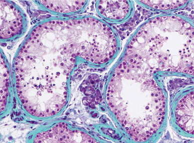
Seminiferous tubule with dilated lumen, apical vacuolation of Sertoli cells, normal number of spermatogonia, and decreased number of other germ cell types.
Etiology The mechanisms causing adluminal compartment lesions can be classified into obstructive and non-obstructive. Obstruction is present in more than 70% of cases, and is characterized by variability of involvement among lobules and the presence of at least two of the following abnormalities: enlargement of tubular diameter and a lumen with remarkable differences among lobules; Sertoli cells with adherens germ cells protruding into the lumen, giving an indented outline; intense apical vacuolation of Sertoli cell cytoplasm; accumulation of spermatozoa in the lumen of some tubules; or number of spermatids Sc + Sd is higher that that of Sa + Sb (see Testicular lesions resulting from obstruction of sperm excretory ducts).929
The three levels of severity of adluminal compartment lesions emphasized by the terms young spermatid sloughing, later primary spermatocytes sloughing, and early primary spermatocyte sloughing, depend on the degree (total or partial) of obstruction and the level of sperm excretory duct obstruction: as the obstruction gets nearer to the testis, the greater the severity. Obstruction may be extratesticular (epididymis, vas deferens, and ejaculatory ducts) or intratesticular (rete testis or any level of the seminiferous tubule length). The most frequent causes of extratesticular excretory duct obstruction are vasectomy, inflammation (epididymitis, prostatitis), mucoviscidosis (congenital bilateral absence of vas deferens), and testis–epididymis dissociation.
Rete testis obstruction. Varicocele is the most frequent cause of obstruction of the rete testis. More than 50% of testes with varicocele have a mosaic pattern of tubular lesions, together with marked dilation and eccentric mural fibrosis of intratesticular veins. In normal testes, the walls of veins are extremely thin and the lumina nearly collapsed. Varicocele patients also often have spermatozoa with characteristically elongated heads with thin bases.930 Initially, abnormalities are confined to the testis ipsilateral to the varicocele, but eventually both testes are affected, although abnormalities are more severe in the ipsilateral testis. Elevated pressure in the pampiniform plexus is transmitted to the veins within the testes, principally to the centripetal veins that cross the testicular mediastinum and drain most of the testicular parenchyma (Fig. 12-100 ).931 The dilated centripetal veins compress the intratesticular sperm excretory ducts, explaining the mosaic distribution of the tubular lesions.932
Fig. 12-100.
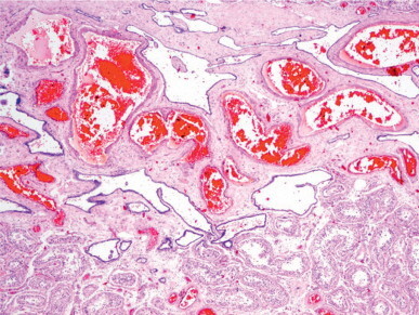
Mediastinum testis from a young man with varicocele. Marked venous dilation (intratesticular varicocele) disrupts and compresses the rete testis cavities, causing partial obstruction of the tubuli recti.
Seminiferous tubule obstruction. Obstruction at the level of the seminiferous tubules can be dysgenetic or post-orchitic. A dysgenetic cause may be suspected in specimens with a mosaic distribution of lesions and seminiferous tubules with small diameters, thickened lamina propria, and an unusual seminiferous tubular cell layer consisting of cuboidal Sertoli cells and spermatozoa that clog the lumina (Fig. 12-101 ). The diagnosis is confirmed if study of serial sections demonstrates continuity between these tubules and those with conserved spermatogenesis. The structure of seminiferous tubules has been observed with scanning microscopy at such points of continuity.858, 933 Tubular stenosis appears to be due to a primary anomaly of Sertoli cells and peritubular cells.
Fig. 12-101.
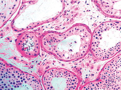
Segmentary dysgenesis of seminiferous tubules. The two central tubules, which only display dysgenetic Sertoli cells, contain numerous spermatozoa from adjacent seminiferous tubules with normal spermatogenesis.
Post-orchitic obstruction should be suspected in cases of tubular atrophy with a mosaic pattern without dysgenetic tubules or varicocele. Some patients have a history of orchitis associated with parotiditis;934 in others the only findings are oligozoospermia and small testes. Testicular biopsy, sampling only the testicular periphery, reveals only the consequences of obstruction, lesions similar to those observed with varicocele. However, some postinflammatory changes should also be present, including hyalinized tubules, dilated tubules lined by cuboidal Sertoli cells, or complete spermatogenesis. Occasionally, there is modest perivascular or peritubular inflammation and angiectasis.935, 936
About 30% of testes with lesions in the adluminal compartment have no obstruction, and most have primary anomalies of germ cells. This claim is supported by the following: pronounced decrease of germ cell type when the preceding type is greatly increased in number; normal correlation between the number of mature spermatids in biopsy and number of spermatozoa in the spermiogram; and the presence of numerous malformed germ cells in the adluminal compartment.
Decrease in the number of a germ cell type may be so important that spermatogenesis is arrested, with subsequent azoospermia. In some cases, maturation arrest is only partial and results in severe oligozoospermia. This maturation arrest is observed mainly in primary spermatocytes and young spermatids.
Primary spermatocyte sloughing may also be owing to meiotic anomalies (Fig. 12-102 ). The observation of increased numbers of spermatocytes arrested in preleptotene–leptotene926 or, more frequently, pachytene856 suggests the diagnosis. The lesion is always bilateral. Spermatocytes arrested in pachytene are usually increased in size and later degenerate. In addition, some spermatids have large, diploid, spherical, hyperchromatic nuclei. The anomaly does not always affect all spermatocytes, and then a higher number of spermatids are produced.856
Fig. 12-102.
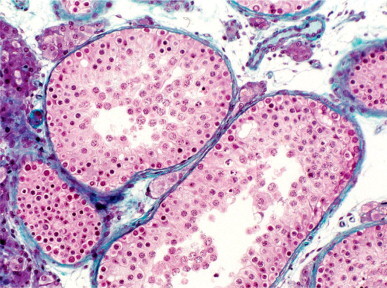
Meiotic abnormalities. The seminiferous tubules contain normal number of spermatogonia and disproportionately high number of primary spermatocytes which do not complete meiosis. No spermatids are seen.
Young spermatid sloughing not associated with obstruction may be due to either meiotic anomalies or defective spermiogenesis. The former gives rises to the appearance of many multinucleate, polyploid, hyperchromatic young spermatids. In the second cause, young spermatids are incapable of transforming into mature spermatids, and only round spermatids appear in the ejaculate.
Lesions in basal and adluminal compartments of seminiferous tubules
Lesions in the basal and adluminal compartments of seminiferous tubules are the most frequent histological findings in testicular biopsies from infertile men. These testes may be classified into two major subgroups: hypospermatogenesis and spermatogonial maturation arrest (Fig. 12-103).
Hypospermatogenesis: Types and etiology Hypospermatogenesis is defined as a reduced number of spermatogonia and primary spermatocytes, with primary spermatocytes outnumbering the spermatogonia. Most seminiferous tubules contain few spermatids. About 8% of patients with hypospermatogenesis have focal tubular hyalinization.937 Two variants of hypospermatogenesis have been quantitatively distinguished: pure hypospermatogenesis, and hypospermatogenesis associated with sloughing of primary spermatocytes.
Pure hypospermatogenesis is defined as a proportionate decrease in the number of all types of germ cell. The number of spermatogonia per cross-sectioned tubule is less than 17 and usually more than 10. The number of primary spermatocytes is equal to or higher than that of spermatogonia. The number of round spermatids is higher than that of primary spermatocytes, and the number of elongated spermatids is similar to that of spermatogonia (Fig. 12-104 ).
Fig. 12-104.
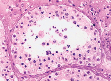
Pure hypospermatogenesis in a patient with severe oligozoospermia. The seminiferous tubule shows slight ectasis and a proportionate decrease of all germ cell types.
Hypospermatogenesis associated with primary spermatocyte sloughing is characterized by two features: low numbers of spermatogonia and primary spermatocytes (with spermatocytes more numerous than spermatogonia), and degeneration and sloughing of many primary spermatocytes. The remaining spermatocytes give rise to the few spermatids observed in the tubules (Fig. 12-105 ).
Fig. 12-105.
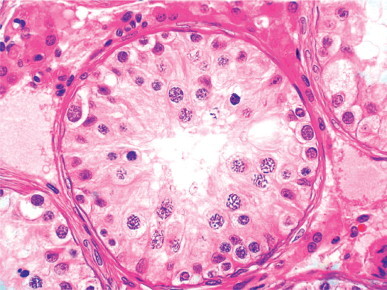
Hypospermatogenesis associated with primary spermatocyte sloughing in an azoospermic patient. Spermatogonia and primary spermatocytes are the sole germ cell types.
Etiology of hypospermatogenesis.
Hypospermatogenesis may result from hormonal dysfunction, congenital germ cell deficiency, Sertoli cell dysfunction, Leydig cell dysfunction, androgen insensitivity, exposure to chemical or physical agents, and vascular malfunction.
Hormonal dysregulation. Although complete spermatogenesis may be observed in men with low levels of FSH and LH, the production of a normal number of spermatozoa requires normal gonadotropin levels. Hypospermatogenesis has been reported in patients with abnormal pulsatile secretion of FSH and LH,938 low gonadotropin secretion,939 biologically inactive gonadotropins, mutation in the gonadotropin β subunit,940 inactivating mutation of FSH receptor gene,941 hyperprolactinemia, and adrenal and thyroid dysfunction (see discussion on hypogonadisms secondary to endocrine gland dysfunction in this chapter).
Congenital germ cell deficiency. Biopsy of cryptorchid patients after orchidopexy reveals that spermatogonia proliferation is decreased and germ cell development is insufficient in adulthood even if the number of spermatogonia was normal in infancy. Is it likely that this poorly understood primary anomaly of germ cells is present in some cases of hypospermatogenesis.
Sertoli cell dysfunction. For many years, primary germ cell deficiency was considered the most common cause of hypospermatogenesis; today, it is known that Sertoli cell failure is the cause of many cases of germ cell deficiency. This conclusion is based on several findings. Sertoli cells in many infertile patients are markedly abnormal, with an increase in the number of glycogen granules942 and acid phosphatase activity;884 a decrease in the number of lipid droplets; and alterations in the cytoskeleton,943 the nucleus,944 and cytoplasmic organelles.945 In some cases Sertoli cells have abnormal maturation, with elongated nuclei containing coarse clumped chromatin instead of triangular-shaped nuclei with pale chromatin. Anomalies in Sertoli cell FSH receptors may be present in idiopathic oligozoospermia associated with elevated levels of FSH.946 Serum inhibin B concentration may be used as a marker to estimate Sertoli cell function.947
Leydig cell dysfunction. Testosterone synthesis by Leydig cells is necessary for normal spermatogenesis,948 and abnormal Leydig cell function is a frequent finding in idiopathic oligozoospermia.949, 950, 951 Leydig cell dysfunction should be suspected when the cells appear diffusely hyperplastic. Patients have elevated serum LH level with depletion of rapid-release testosterone, revealing a lack of early response of Leydig cells to gonadotropin-releasing hormone stimulation. The ratio of testosterone to LH in the plasma indicates the degree of Leydig cell dysfunction. Decreased ratio with normal testosterone level suggests compensated dysfunction. Patients with a ratio of less than 1:5 and normal other parameters may have complete spermatogenesis.951
Androgen insensitivity. Some patients with severe oligozoospermia or azoospermia have a defect in androgen receptor responsiveness, similar to that in Reifenstein's syndrome.952, 953, 954 The abnormality may arise from a genetic defect in the eight exons that code for this receptor, mapped to Xq11-12,955 or from post-translational errors.956, 957 This defect is also referred to as infertile male syndrome and mild androgen insensitivity, and the patients have male phenotype with somatic features of slight androgen deficit.958 Histologically, the testis is similar to that observed with Leydig cell dysfunction or mixed atrophy, although the mechanism causing the Leydig cell hyperplasia is quite different (Fig. 12-106 ). Peripheral resistance to testosterone action alters regulation of the hypothalamohypophyseal–testicular axis, and LH and testosterone levels are elevated. Androgen insensitivity causes between 10%959 and 40%960 of all cases of severe oligozoospermia or azoospermia. In such cases spermatogenesis improves with the administration of tamoxifen citrate,960 clomiphene citrate, or androgen therapy.961, 962 Calculation of the index of androgen insensitivity can be helpful: plasma LH (mIU/mL) × plasma testosterone levels (ng/mL). In patients with androgen insensitivity, the index is higher than 200 (normal is about 102).
Fig. 12-106.
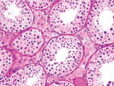
Hypospermatogenesis due to androgen receptor defect. The seminiferous tubules show hypospermatogenesis associated with diffuse Leydig cell hyperplasia.
Physical and chemical agents. The number of chemicals implicated in infertility increases daily. A detailed history is invaluable in evaluating these patients. The same is true of physical agents such as prolonged exposure to heat, ionizing radiation, or microwave radiation.963
Etiology of hypospermatogenesis associated with primary spermatocyte sloughing. Most testes with primary spermatocyte sloughing have varicocele, and this is commonly associated with infertility.964, 965, 966, 967 Varicocele is found in 15% of the general population, and is present in 30–40% of infertile men. The mechanism by which varicocele affects fertility is unknown. Clinical varicocele may occur without a testicular lesion (or only phlebectasis), and subclinical varicocele may be associated with severe spermatogenic lesions. Increased testicular temperature968, 969 and compression of intratesticular sperm excretory ducts by dilated veins932 are the most plausible mechanisms. In other cases, primary spermatocyte sloughing results from anomalies of primary spermatocytes and spermatids, suggesting a meiotic anomaly. Finally, in some patients the cause may be the presence of involuting Sertoli cells.
Spermatogonial maturation arrest Spermatogonial maturation arrest is a disorder defined by the presence of fewer than 17 spermatogonia per cross-sectioned tubule and even fewer primary spermatocytes. Spermatids are usually absent. There have been attempts to correlate the etiology of spermatogonial maturation arrest with the Sertoli cell type present.970 Immature Sertoli cells are characteristic of hypogonadotropic hypogonadism and some syndromes with androgen insensitivity (Fig. 12-107 ). Mature Sertoli cells, if their presence is unilateral, are observed in varicocele, epididymitis, and ipsilateral testicular traumatism, but if they appear in both testes the etiology is unknown. Involuting Sertoli cells are usually present bilaterally; some cases are idiopathic, whereas others are associated with a history of alcoholism or chemotherapy. Dedifferentiated Sertoli cells are found in spermatogonial maturation arrest caused by gonadotropin inhibition in treatment with estrogen, -releasing hormone agonist, or anti-androgen.971
Fig. 12-107.
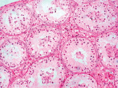
Spermatogonial maturation arrest. The seminiferous tubules have increased numbers of Sertoli cells and nearly normal number of spermatogonia, while the remaining germ cell types are scant. The testicular interstitium shows diffuse Leydig cell hyperplasia.
Focal lesions in spermatogenesis (mixed atrophy)
Mixed atrophy is a descriptive term for the coexistence, in the same testis, of tubules containing only Sertoli cells and tubules with complete or incomplete spermatogenesis.972 This disorder includes patchy failure of spermatogenesis and partial del Castillo's syndrome.
The extent of Sertoli cell-only tubules varies widely. Tubules with spermatogenesis may be normal or partially atrophic. Tubular hyalinization is occasionally seen (Fig. 12-108 ). Mixed atrophy is more common than suggested by the literature, and many cases are included under other diagnoses, such as ‘hypospermatogenesis with a severe germ cell depletion in such a way that some Sertoli cell-only tubules are seen,’859 and ‘Sertoli cell-only syndromes with focal spermatogenesis.’973
Fig. 12-108.
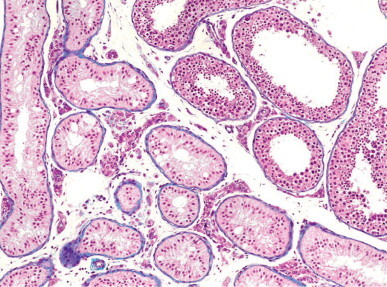
Mixed atrophy. Seminiferous tubules with slight ectasis and complete spermatogenesis adjacent to Sertoli-cell-only pattern. The tubular lesions probably belong to different lobules.
Serial sections from testes with mixed atrophy reveal that the two different types of tubule are grouped according to their histologic pattern, suggesting that the distribution is by testicular lobules. In cases of mixed atrophy, the percentage of tubules with spermatogenesis, the degree of spermatogenic development in the tubules, and the type of Sertoli cell present should be reported. Correlation of the first two with the spermiogram gives an indication of prognosis, and the Sertoli cell types identifies the nature (primary or secondary) of the lesion.974
Mixed atrophy (probably primary) is observed in idiopathic infertility, cryptorchidism (even if orchidopexy was done at infancy, in both the cryptorchid and the contralateral descended testis), retractile testes, macroorchidism, intravaginal torsion of the spermatic cord (in both twisted and contralateral testis), and chromosomal anomalies such as Down's syndrome, 47/XYY karyotype, 46/XX karyotype, giant Y chromosome, Klinefelter's syndrome with chromosomal mosaicism, partial androgen insensitivity, and some male pseudohermaphrodites. Secondary mixed atrophy may be seen in patients undergoing chemotherapy, corticoid therapy,975 or in those with a history of viral orchitis.
Germ cell anomalies in infertile patients
In addition to anomalies in the seminiferous tubules, examination of the biopsy should include a description of the morphology of the germ cells.
Giant spermatogonia.
Giant spermatogonia are a normal component of the seminiferous epithelium. These cells may be altered spermatogonia in the S or G2 phases of the cell cycle. They rest on the basal lamina and have pale cytoplasm and an ovoid nucleus measuring at least 13 μm in diameter. The frequency of these cells in normal and infertile men is about 0.65 cells per 50 cross-sectioned tubules, although their number is usually higher in mixed atrophy. These cells should not be mistaken for intratubular germ cell neoplasia; they are also present in normal numbers in tubules at the periphery of germ cell tumor (Fig. 12-109 ).976
Fig. 12-109.
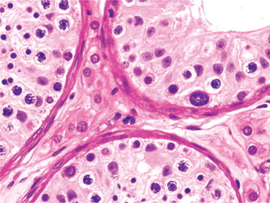
Hypertrophic spermatogonia in a seminiferous tubule showing marked decrease in the number of spermatogenetic cells.
Multinucleate spermatogonia
Multinucleate spermatogonia are a common finding in cryptorchid testes that were surgically corrected, infertile patients, and old men. Nuclei of both Ad and Ap spermatogonial types may be seen within the same cell.
Dislocated spermatogonia
Normally, spermatogonia are present only in the transition zone between the seminiferous tubule basal layer and the tubuli recti. Dislocated spermatogonia have been found throughout the testis in old age,977 in infertile patients with a variety of lesions, after long-term estrogen therapy,978 and in seminiferous tubules with intratubular germ cell neoplasia.979
Megalospermatocytes
Megalospermatocytes are large primary spermatocytes arrested in the leptotene stage (Fig. 12-110 )980 that exhibit asynapsis of chromosomes.981 Joined by cytoplasmic bridges, they form small groups. These cells may be clones of synchronously degenerating spermatocytes.982 They are frequently found in elderly men and are a non-specific finding in infertile patients.
Fig. 12-110.
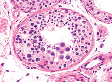
Megalospermatocytes. The seminiferous tubule contains a group of very large primary spermatocytes displaying fine chromatin and eosinophilic cytoplasm.
Multinucleated spermatids
The presence of spermatids with multiple nuclei (from 2 to 86) is frequent is old age.983 Similar cells with fewer nuclei have also been reported in infertility due to cryptorchidism,984 hyperprolactinemia, and idiopathic infertility (Fig. 12-111 ).
Fig. 12-111.
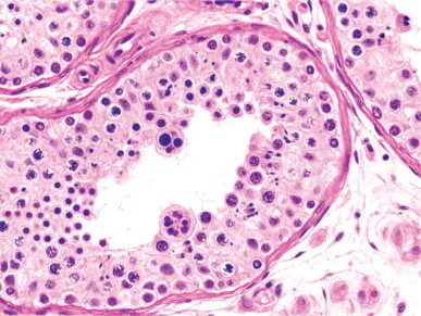
Multinucleation of both spermatids and spermatocytes.
Malformed spermatids
There are at least four teratozoospermic syndromes that may be easily identified by testicular biopsy, although in most the diagnosis previously relied on morphologic study of the spermiogram: (1) round-headed spermatids (characteristic of spermatozoa lacking acrosomes) (Fig. 12-112 ), (2) Sc + Sd spermatids with a very elongated head (characteristic of varicocele) (Fig. 12-113 ), (3) macrocephalic Sc + Sd spermatids whose DNA content suggests an anomaly in the first meiotic division, and (4) Sc + Sd spermatids with voluminous eosinophilic cytoplasmic droplets (syndrome of spermatozoa with short thick flagella985 or fibrous sheath dysplasia).
Fig. 12-112.
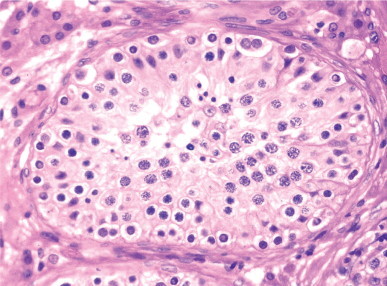
Testicular biopsy showing spermatids with small spherical nuclei, a finding characteristic of round spermatozoa lacking acrosomes. The remaining germ cells are morphologically normal.
Fig. 12-113.
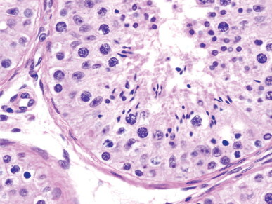
Elongated spermatids showing bell-clapper nuclei in a patient with varicocele.
In some patients, Sa + Sb spermatids rest in these initial phases of spermiogenesis and eventually become sloughed in the tubular lumina.986 In other testes there are macrocephalic Sc and Sd spermatids with anomalous DNA content, suggesting an anomaly in the first meiotic division.
Morphologically abnormal spermatozoa
Ultrastructural study of spermatozoa is sometimes necessary to determine the cause of male infertility. A number of morphologically abnormal spermatozoa are present in all semen samples, including those from fertile men, but abnormal spermatozoa are very numerous in infertile patients. Ultrastructural study is advised in all cases of asthenozoospermia, in teratozoospermia when the number of spermatozoa showing the same morphological anomaly is high, and in cases with apparently normal spermatozoa that fail to fertilize in vitro.987 The classification of ultrastructural anomalies in spermatozoa is based on light microscopy findings988 of lesions in the head and tail.
Anomalies of the spermatozoal head
These are defined by changes in the shape of the head, and usually involve both the nucleus and the acrosome. Some anomalies, such as pear-shaped, candle-shaped, or egg-shaped heads,989, 990 are regarded as minor variants of normal. More significant abnormalities are the elongated, microcephalic, macrocephalic, and crater-defect forms.
The most frequent abnormal head shape is elongated with a narrow base (tapered head spermatozoa). This anomaly is frequently associated with varicocele.991
Microcephalic spermatozoa have spherical (globozoospermia) or irregularly shaped heads. The former have spherical nuclei with poorly condensed chromatin and lack acrosomes, postacrosomal sheaths, and a nuclear ring (Fig. 12-114 ). Most cases are sporadic, but this lesion was also reported in two pairs of infertile brothers.992, 993 Microcephalic spermatozoa with irregularly shaped heads have small and irregularly shaped acrosomes that usually are not in contact with the nucleus. This anomaly may be congenital, as in Aarskog–Scott syndrome,994 or secondary to heat exposure or hashish smoking. In both types of microcephaly loss of connection between the acrosomal vesicle and the spermatozoal head is attributed to a deficiency in basic proteins of the sperm perinuclear theca that promotes nuclear envelope organization and adhesion of the acrosomal vesicle.995 Acrosin is reduced or absent in spermatozoa lacking acrosomes and those with small acrosomes.996 Motility may be normal. The occurrence of aneuploidy997 and disomy of sex chromosomes998, 999 in some cases should be evaluated before performing intracytoplasmic sperm injection (ICSI).
Fig. 12-114.
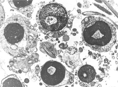
Microcephalic spermatozoa with a spherical nucleus lacking an acrosome and poorly condensed chromatin. Ultrastructural anomalies are observed.
The cause of round-headed spermatozoa might be the lack of Golgi-associated protein known in male mice as Golgi-associated PDZ- and coiled-coil motif-containing protein (GOPC). This protein is principally localized in the trans-Golgi region in round spermatids, and its loss produces globozoospermia. The primary defect consists of an inability of acrosomal vesicles to fuse to each other to create the acrosome.1000
Macrocephalic spermatozoa (macronuclear spermatozoa) have enlarged, irregularly shaped heads and deficient chromatin condensation. There are two types (multiple tails1001, 1002 and aflagellate), both of which have abnormal DNA content (many are tetraploid), suggesting a meiotic anomaly.1003, 1004
Irregularly shaped spermatozoa are characterized by irregularity in the shape of the nucleus or acrosome.1005 In the crater defect syndrome, there is invagination of the nuclear envelope in which the acrosome penetrates. The tail is morphologically normal, and motility is only slightly reduced. In spermatozoa with spoon-shaped nuclei, the defect is probably genetic. Other anomalies include double-headed spermatozoa with two nuclei sharing a single acrosome.1006
Anomalies in the spermatozoal tail
Spermatozoal tail anomalies are classified as generalized anomalies of the tail or anomalies in defined tail components, such as the connecting piece, the axoneme, or the periaxonemal structure.1007
Generalized anomalies in the tail Cytoplasmic remnants. The presence of cytoplasmic droplets is normal during spermiogenesis. An elevated number of spermatozoa with cytoplasmic droplets in semen is associated with premature sloughing of spermatozoa, as occurs in varicocele, and should not be misinterpreted as spermatozoa with excess residual cytoplasm.1008 These spermatozoa are very often abnormal and the residual cytoplasm may be located around the intermediate piece or surrounding the head. These spermatozoa also have other flagellar anomalies.
Bent tail. A bend in the tail may occur at the level of the connecting piece or the intermediate piece. In bends of the connecting piece, the tail is laterally implanted and forms an angle with a nucleus that displays a thin base. Bends of the intermediate piece are associated with cytoplasmic droplets, malposition of mitochondria, and loss of the parallel arrangement of the dense outer fibers.
Coiled tail. Spermatozoa with a coiled tail are a frequent finding in centrifuged semen, but they may also be a true abnormality. These spermatozoa have a perinuclear cytoplasmic remnant containing a flagellum that is coiled around the nucleus and along the middle or principal pieces (Fig. 12-115 ). This is frequently associated with abnormalities of the periaxonemal structures.
Fig. 12-115.
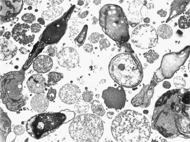
Spermatozoa with coiled tails. The anomaly occurs in the principal pieces. The intermediate pieces show variable lengths, absence of parallelism in the outer dense fibers, and large cytoplasmic droplets. This teratozoospermia was found in two infertile brothers.
Tail stump (short-tail spermatozoa). The presence of many spermatozoa with short, thick tails in semen represents a well-defined teratozoospermic syndrome.1009 Ultrastructural examination reveals hypertrophy and hyperplasia of the fibrous sheath,1010 hence this syndrome has also been termed ‘fibrous sheath dysplasia.’1011 Additional axonemal malformations, including absence of the central pair of microtubules (Fig. 12-116 )1012 and, less frequently, lack of dynein arms, are observed in 50% of cases. About 24% of patients have respiratory disease, such as rhinosinusitis, bronchitis, and bronchiectasis from an early age. Similar findings have been reported in the cilia of the upper respiratory tract, and thus a relationship between fibrous sheath dysplasia and immotile cilia syndrome has been assumed. Clinical presentation may be sporadic or familial. The cause of fibrous sheath dysplasia and the subsequent lack of motility in these spermatozoa is probably related to the occurrence of deletions in Akap3 and Akap4 genes, as well as the absence of Akp4 protein in the fibrous sheath.1013
Fig. 12-116.
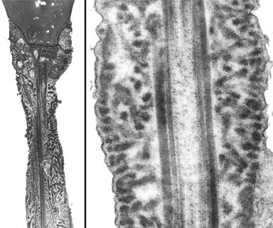
Tail-stump spermatozoal malformation. Longitudinal section of two spermatozoa showing a marked thickening of the principal piece with both hypertrophy and hyperplasia of the fibrous sheath. One of them also shows a very short intermediate piece.
Multiple tails. The presence of more than two tails is associated with macrocephalic spermatozoa.1014
Sperm tail agenesis. Teratozoospermia with 100% sperm tail agenesis has been reported in patients with a high degree of consanguinity. These spermatozoa also have defects in chromatin condensation and residual cytoplasmic droplets.1015
Anomalies of the connecting piece Anomalies of the connecting piece are classified as acephalic spermatozoa, deficient organization of the connecting piece, and separation between the head and the tail.
Acephalic spermatozoa are known as ‘pin-headed,’ although they lack a true head; the small cephalic knob-like thickening is actually a cytoplasmic droplet with a variable degree of mitochondrial organization giving rise to a variable degree of motility.1016 This anomaly is due to an early failure in spermiogenesis. It may be familial in some cases.1017, 1018 Spermatozoa with deficient organization of the connecting piece have narrowing at this level, with loss of alignment of the head and flagellum axes. Spermatozoa with a separated head and flagellum, known as decapitated and decaudated spermatozoa, are also the result of an anomaly in spermiogenesis, but the separation between heads and tails can occur during spermiation or at any level of the sperm excretory ducts.1019, 1020
Anomalies in axoneme Abnormalities of the axoneme are classified as numerical anomalies, microtubular ectopia, and immotile cilia syndrome.
The most common numerical anomalies are the absence of one or both microtubules of the central pair and complete lack of the axoneme. Spermatozoa lacking the central microtubule pair also lack the central sheath and are immotile, although they are normal by light microscopy. Familial cases have been reported.1021 This anomaly may be associated with ciliary dyskinesia.1022
Immotile cilia syndrome (primary ciliary dyskinesia)1023 refers to patients having low mucociliary clearance associated with otitis, sinusitis, bronchitis, bronchiectasis, and immotile spermatozoa. Most patients have the same defect in the axoneme and cilia of the respiratory mucosa. The frequency of this syndrome is estimated at between 1 in 20 000 and 1 in 60 000 men. Clinical symptoms consist of reduced clearance of ciliary mucus in the airway, with onset at infancy. In order to prevent the later development of bronchiectasis, ultrastructural study of the respiratory mucosa is advisable if other disorders have been excluded, including cystic fibrosis, allergy and other immune disorders, α1-antitrypsin deficiency, and cardiovascular and metabolic diseases.1024 The most frequent anomalies of this syndrome are the absence of microtubule doublets and peripheral junctions, the central microtubule pair, the outer dynein arms, the central junctions, the two dynein arms, and the inner dynein arm plus the peripheral junctions (Fig. 12-117 ). Spermatozoa lacking the two dynein arms or the peripheral junctions are immotile. Reduced motility is seen in spermatozoa with only one dynein arm. Kartagener's syndrome is a variant of the immotile cilia syndrome characterized by the classic triad of situs inversus, bronchiectasis, and chronic sinusitis. The syndrome has autosomal recessive inheritance1025 and is found in 20–25% of patients with situs inversus.1026
Fig. 12-117.
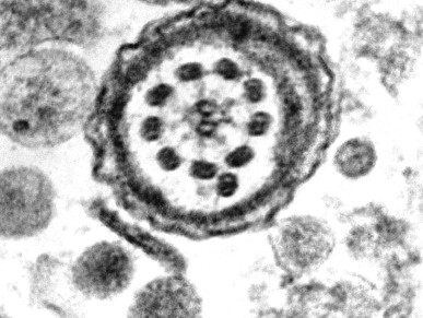
Cross-section of the intermediate piece from a spermatozoon lacking dynein arms and showing a supernumerary microtubule doublet.
Anomalies in periaxonemal structures Periaxonemal abnormalities include mitochondrial sheath defects,1027 malposition of the annulus, alteration in number, shape, or length of the outer dense fibers, and absence, thickening, or disruption of the fibrous sheath.1011, 1028
Many cases of asthenozoospermia, present in 30% of infertile men, may be attributable to deficient mitochondrial function, possibly caused by mutations in their DNA.1029 Abnormalities of the dense fibers are associated with deficient motility. Abnormalities of the fibrous sheath include, in addition to the abovementioned dysplasia of the fibrous sheath, absence of the fibrous sheath, and redundant fibrous sheath material associated with a deficit or lack of mitochondria.1030 The three defects are probably inherited.
Presence of intratubular germ cell neoplasia
The incidence of intratubular germ cell neoplasia (IGCN) in infertile patient is 0.4% in England,1031 0.7% in Spain,1932 0.73% in Germany,1033 and 1.1% in Denmark.1034 A higher risk occurs in patients with severe oligozoospermia (fewer than 10 million spermatozoa per milliliter), azoospermia associated with unilaterally or bilaterally diminished testicular volume,1035 a history of testicular maldescent,1036, 1037 or unilateral testicular cancer.1038
The cells of IGCN are located in seminiferous tubules with decreased tubular diameter and lacking spermatogenesis. These cells are large and have pale cytoplasm and large and irregularly outlined nuclei, with one or several prominent nucleoli. They stain intensely with periodic acid–Schiff and express placenta-like alkaline phosphatase, c-kit, and the cell adhesion molecule CD44.1039
Anomalies in Leydig cells
A reduction in the number or absence of Leydig cells is infrequent in infertility, and only occurs in hypogonadotropic hypogonadism secondary to LH deficit and in patients with biologically inactive LH. Leydig cell hyperplasia is very common,1040 and has been observed in Klinefelter's syndrome, cryptorchidism, male pseudohermaphroditism, minor androgen insensitivity, infertility secondary to Leydig cell dysfunction, varicocele, after treatment with 5α-reductase inhibitors or non-steroidal anti-androgens, and in some elderly men. Such hyperplasia may give rise to hypoechoic or hyperechoic images that may be misdiagnosed as tumor.1041
Mast cells
There is a close relationship between testicular dysfunction and elevated mast cell numbers in the testis. An increase in interstitial and peritubular mast cells occasionally occurs in infertile patients.1042, 1043 This increase is higher than that observed in inflammatory or neoplastic process.1044 Daily administration of ketotifen, an antihistamine-like drug with a mast cell-stabilizing effect, significantly improves the spermiogram parameters in some patients.1045
Correlation between testicular biopsy and spermiogram
For effective therapy, it is important to know whether or not the azoospermia or oligozoospermia is the result of obstruction.863, 1046
Obstructive azoospermia and oligozoospermia
Azoospermia caused by obstruction is usually easily diagnosed, but this determination is more difficult with oligozoospermia. Obstruction of the ductal system should be suspected when there are more than 20 mature spermatids (Sc + Sd) per cross-sectioned tubule and fewer than 10 million spermatozoa in the spermiogram (Fig. 12-118 ).1047, 1048 Ob-structive azoospermia is implicated in 7.4–14.3% of cases of male infertility.
Fig. 12-118.
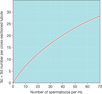
Power curve showing the correlation between the number of spermatozoa in the spermiogram and the number of mature spermatids (Sc + Sd) per cross-sectioned tubule. If the number of mature spermatids is correlated to that of spermatozoa in spermiogram, the oligozoospermia is of the pure secretory type. If the number of mature spermatids is higher than that of spermatozoa in spermiogram, the disorder is either an obstructive azoospermia with ‘normal’ testicular biopsy or a mixed obstructive secretory oligozoospermia.
Classification of obstructive azoospermia by location
Obstruction is classified as proximal, distal, and mixed, according to the distance from the testis to the point of obstruction in the ductal system.
Proximal obstruction Obstruction is considered proximal when the lesion lies between the seminiferous tubules and the distal end of the ampulla of the vas deferens. Epididymal obstruction, principally of the caput-corpus transition zone, accounts for 66% of cases. Rarely, there is a defective connection between the rete testis and epididymal ductuli efferentes. Because the seminal vesicles are normal, men with proximal obstruction have a normal volume of semen (the testicular contribution to semen is about 5% of the total volume). When obstruction is in the cauda of the epididymis, epididymal markers, including carnitine, glycerophosphorylcholine and α-glycosidase are low.1049 The nearer the obstruction is to the caput of the epididymis, the higher the level of these markers.
Distal obstruction Distal obstruction is located between the ampulla of the vas deferens and the junction of the ejaculatory ducts and urethra. These patients present with sacral, perineal, or scrotal pain on ejaculation. Rectal examination often reveals enlarged seminal vesicles. The volume of semen is low and consists of watery fluid that fails to coagulate. Seminal vesicle secretions are lacking. The concentration of prostatic secretions, such as acid phosphatase and citric acid, is increased owing to the lack of semen dilution. Vasography may help in diagnosis, as higher segments fail to fill.1050 Transrectal ultrasonography is the most accurate imaging modality for the diagnosis of ejaculatory duct obstruction. Needle aspiration of seminal vesicle fluid may show spermatozoa that have entered the seminal vesicles by reflux.
Mixed obstruction Mixed obstruction refers to lack of patency of the vas deferens or the epididymis and alterations in the ejaculatory ducts or seminal vesicles (low ejaculate volume, and absence of fructose). The most frequent cause is mucoviscidosis. One-third of patients with congenital bilateral absence of the vas deferens have agenesis or hypoplasia of the seminal vesicles. The cause of epididymal obstruction in patients with anomalies of the prostate–vesiculo–deferential junctions is difficult to determine.
Etiology of obstructive azoospermia
Obstructive azoospermia may be caused by congenital or acquired lesions.
Congenital azoospermia The most frequent anomalies associated with congenital azoospermia are testis–epididymis dissociation, epididymal malformation in cryptorchidism, bilateral absence of the vas deferens, congenital unilateral absence of the vas deferens associated with pathology of the contralateral testis or its sperm excretory ducts, seminal vesicle agenesis, and ejaculatory duct obstruction (Table 12-9 ).
Table 12-9.
Congenital anomalies of the male mesonephric ducts
|
|
|
|
|
Agenesis of all mesonephric duct derivatives. Agenesis of all mesonephric duct derivatives is a rare disorder that gives rise to varied anatomical anomalies, depending on the stage of embryonic development at which the mesonephric duct derivatives disappear. If failure occurs before the fourth week the ipsilateral kidney and ureter are absent, although the testis may be present, or there may be other renal anomalies. If failure occurs in the fourth week, and the ureteral bud is already formed, the ureter and kidney may develop normally. If failure occurs between the fourth and the 13th weeks, there is a variable constellation of anomalies that most frequently include normal development of the testis and globus major and hypoplasia of the other excretory duct segments, or agenesis of an excretory duct segment (epididymis, vas deferens, or seminal vesicle).
Epididymal anomalies. The most frequent epididymal anomalies are absence of the epididymis, testis–epididymis dissociation, defective connection of the vas deferens and epididymis, epididymal cyst, and anatomical abnormalities of the epididymis.
Complete absence of the epididymis is frequent in monorchidism and anorchidism. The epididymis is replaced by a small mass of cellular connective tissue with abundant blood vessels at the blind end of the vas deferens.
Partial absence of the epididymis is more frequent than complete absence. Absence of the corpus of the epididymis gives rise to a characteristic malformation called bilobated epididymis. This varies from simple strangulation to complete separation of the caput and cauda. These anomalies are often associated with absence of the vas deferens.
Testis–epididymis dissociation is found in 1% of cases of obstructive azoospermia and is usually associated with cryptorchidism.
Defects in connection between the ductuli efferentes and the ductus epididymidis are rarely complete. In the incomplete form, some of the five to 30 ductuli efferentes in the epididymis are short and end blindly.
Epididymal cysts usually arise from blind-ended ductuli efferentes and contain spermatozoa. These spermatoceles retain their epithelial lining, although it becomes atrophic (Fig. 12-119 ). Spermatozoa may be obtained from these cysts. Some epididymal cysts arise from embryonic remnants, do not contain spermatozoa, and are lined by columnar or pseudostratified epithelium. Wolffian cyst, unlike müllerian cyst, is immunoreactive in the apical border of epithelial cells with CD10.1051 Cyst lined by clear cells with or without papillae raises concern for von Hippel–Lindau disease.1052 Large epididymal cyst requires removal and must be excised with great care to avoid damaging the ductuli efferentes and resulting in obstruction. Epididymal cyst is present in about 5% of males, and the incidence is high (21%) in those exposed to diethylstilbestrol during gestation. The incidence of epididymal cyst in those with hepatorenal polycystosis is similar to that in the general population.1053
Fig. 12-119.
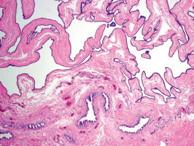
Infertile patient with bilateral epididymal cysts. The cystic wall appears collapsed and folded on the epididymis.
Anomalies in epididymal configuration, altering its shape and location, are frequent in men with cryptorchidism and uncommon with descended testes. The most common malformations are elongated epididymis, angulated epididymis, and free epididymis. Elongated epididymis is found in approximately 68% of undescended testes. The length of the epididymis may be several times that of the testis, and, in abdominal or inguinal cryptorchidism, the epididymis extends several centimeters below the testis. Angulated epididymis is characterized by a long epididymis that has a sharp bend in the corpus with or without stenosis. With free epididymis, all or part of the epididymis is unattached to the testis. The most common variant is epididymis with free cauda.
Vas deferens anomalies. The most frequent anomalies are of the vas deferens are congenital absence, segmental aplasia, ectopia, duplication, diverticula, and crossed dystopia.1054
Congenital absence is defined as unilateral or bilateral absence of either the whole vas deferens or only a segment. Obviously, azoospermia occurs with bilateral absence. The frequency of this malformation varies among populations. At autopsy, the prevalence is 0.5%, but the clinical incidence is 1–1.3% in infertile men1055 and 10–25% in patients with obstructive azoospermia. Unilateral complete absence is three times more frequent than bilateral absence, and absence of only a segment is even more frequent. The affected segment may be absent or reduced to a fibrous cord. Absence of the vas deferens may be associated with other malformations of the sperm excretory ducts or urinary system. The most frequent malformations of the excretory ducts are absence of the ejaculatory ducts (33% of cases) and, less frequently, absence of the seminal vesicles. About 71% of patients with bilateral absence of the vas deferens have partial aplasia of the epididymis. The most frequent malformations of the urinary system are absence of the ipsilateral kidney and other renal anomalies. Complete or partial absence of the vas deferens occurs frequently in patients with cystic fibrosis.
Persistent mesonephric duct consists of the ureter joined to the vas deferens, forming a single duct that opens in an ectopic orifice between the trigone and the verumontanum. This malformation may be associated with cystic transformation or absence of the seminal vesicle. The kidney may be normal or dysplastic.
Anomalies of seminal vesicle and ejaculatory duct. The most frequent anomalies are agenesis of the seminal vesicles or ejaculatory ducts, cyst of the seminal vesicle, and ectopic opening of the ureter into the seminal vesicle. The last is the most common and often is associated with ipsilateral renal dysplasia.
Acquired azoospermia Inflammation and trauma are the main causes of acquired azoospermia. Epididymitis is a frequent cause; Chlamydia trachomatis1056, 1057 and Escherichia coli are the most common infectious causes in developed countries.1958 Infections with Neisseria gonorrheae and mycobacteria are also implicated, and non-specific epididymitis is important.1059 Apart from elective vasectomy, the most frequent traumatic causes of azoospermia are surgical accidents during herniorrhaphy in chidren,1060 orchidopexy, varicocelectomy, hydrocelectomy, deferentography,1061 and removal of epididymal cyst. Obstructive azoospermia may also result from blockage of the ejaculatory ducts following transurethral resection, or as a result of chronic urethral catheterization.
Testicular and epididymal lesions resulting from obstruction of sperm excretory ducts
Lesions of the testis and epididymis may result from obstructed sperm excretory ducts, depending on the location, origin (congenital or acquired), and duration of the obstruction.
Location of obstruction Obstruction at the level of the ampulla of the vas deferens, seminal vesicles, or ejaculatory ducts does not usually cause significant lesions in the testis or epididymis. More proximal obstruction at the level of the vas deferens, epididymis, or testis–epididymis junction usually causes severe lesions in both the sperm excretory ducts and the testicular parenchyma. Obstruction of the vas deferens causes increased pressure within the ductus epididymis. As a result, epididymal lumina dilate, the epithelium atrophies, and fluid containing few spermatozoa and some spermiophages accumulates in the lumen (Fig. 12-120 ). The most dilated epididymal segment is the caput. The ductuli efferentes often become cystically dilated and filled with spermatozoa and macrophages. From reabsorption and lysosomal degradation of this protein-rich fluid, the epithelium accumulates lipofuscin granules or aquires apical eosinophilic granules (Paneth cell-like change).1062 Rupture of the vas deferens gives rise to microgranulomas and ceroid granuloma (Fig. 12-121 ). Macrophages and lymphocytes are often present in the intertubular connective tissue.1063
Fig. 12-120.
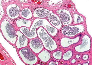
Obstructive azoospermia in a patient with history of epididymitis. The caput epididymidis shows marked dilation of the ductuli efferentes with numerous spermatozoa.
Fig. 12-121.
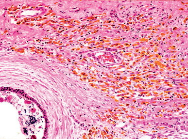
Ceroid granuloma in a patient with history of sperm excretory duct obstruction.
The most frequent testicular lesions in proximal obstruction involve the adluminal compartment, and are the result of the negative effect of hydrostatic pressure on the seminiferous tubular cell layers and, in particular, on the Sertoli cell (Fig. 12-122, Fig. 12-123, Fig. 12-124 ).
Fig. 12-122.
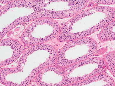
Seminiferous tubules with marked luminal dilation, moderate decrease in cellularity, and occasional vacuolation of Sertoli cell cytoplasm.
Fig. 12-123.
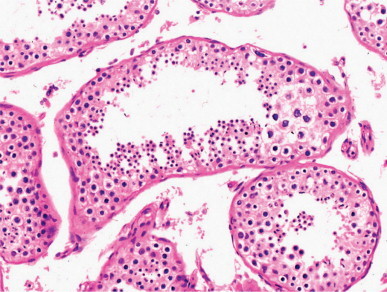
Seminiferous tubules with slight luminal dilation. The seminiferous tubular cell layers have a ‘toothed’ pattern. Degenerating megalospermatocytes can be seen in the seminiferous epithelium.
Fig. 12-124.
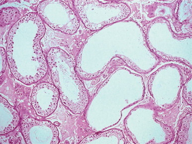
Seminiferous tubules with marked ectasis and atrophy of the seminiferous epithelium in a patient with epididymal obstruction.
Etiology of obstruction Obstruction secondary to congenital absence of the vas deferens usually causes little testicular injury, mainly dilation of the seminiferous tubules and an increase in the number of mature (Sc + Sd) spermatids.1064 Lesions resulting from vasectomy are more important. Increased intraluminal pressure in the epididymis1065 may give rise to pain (late post-vasectomy syndrome).1066 Testicular lesions depend on the surgical technique used: they are slight if the proximal end of the vas deferens is not ligated or sperm granuloma forms at the site of vasectomy. The spermatogenic rhythm in the testis is slower than before vasectomy, and lesions characteristic of testicular obstruction develop, including thickening of the lamina propria and fibrosis of the interstitium.1067, 1068 In testicular obstruction secondary to herniorrhaphy in infancy, testicular lesions are mild. Testicular lesions may be important if the epididymis is damaged by hydrocelectomy, and consist mainly of primary spermatocyte sloughing. In addition to these lesions, hyalinized tubules may be observed when obstruction is caused by inflammation.
Duration of obstruction In acquired obstruction the testicular lesions worsen with time. Obstruction in the caput of the epididymis leads to disappearance of all germ cells in the adluminal compartment of seminiferous tubules. The tubules become dilated and Sertoli cells appear vacuolated. Testicular alterations after vasectomy may not be related to the duration of the obstruction but rather to the initial injury, and may disappear with time as the intraluminal pressure decreases.1069 However, if a significant amount of time has elapsed after vasectomy, the possibility of attaining a normal spermiogram with vasovasostomy is very low. Vasal patency is restored in most cases of reanastomosis, but paternity rates are markedly lower (25–51%)1069 than normal (85%).1070
Functional azoospermia and oligozoospermia
Some azoospermic patients have testicular biopsy with minimal histologic abnormality or minor tubular dilation without detectable excretory duct obstruction. These findings are characteristic of two main conditions: Young's syndrome, and alterations in spermatozoal transport.
Young's syndrome
Young's syndrome is defined by the following constellation of findings: azoospermia, sinusitis, bronchitis or bronchiectasis, and normal spermatozoal flagella.1071 The incidence is probably higher than that recorded in the literature, and Young's syndrome should be suspected in all patients with obstructive azoospermia without a history of epididymitis or scrotal trauma. These patients have a lesion at the junction of the caput and corpus of the epididymis that gives the epididymis a characteristic gross appearance. The caput of the epididymis is distended, the ductuli efferentes contain yellowish fluid and numerous spermatozoa, and the remaining epididymal segments are normal. The ductus epididymidis is blocked by thick fluid.1072 Young's syndrome should be distinguished from other causes of infertility also associated with chronic sinusitis and pulmonary infections, including ciliary dyskinesia and cystic fibrosis. Ciliary dyskinesia consists of morphological, biochemical, and functional alterations in cilia and flagella, and includes several diseases such as the immotile cilia syndrome, Kartagener's syndrome, and miscellaneous syndromes characterized by imperfectly defined abnormalities of cilia and flagella.1073 In Young's syndrome, sinusitis and pulmonary infections develop in childhood and stabilize or improve in adolescence; in other conditions, the pulmonary damage increases with age and the cilia and flagella are ultrastructurally abnormal.1074
Alterations in spermatozoon transport
Normally, spermatozoa detach from the Sertoli cells and are transported through the intratesticular and extratesticular excretory ducts, where they are stored, mainly in the cauda of the epididymis, and finally released from the corpus by ejaculation or eliminated by phagocytosis. Only about 50% of spermatozoa are ejaculated. Whereas the release of spermatozoa from the corpus is intermittent, their transport through the sperm excretory ducts is continuous. Transport is accomplished by the myofibroblasts in the wall of the seminiferous tubules and ductuli efferentes and the smooth muscle cells in the wall of the ductus epididymidis and vas deferens. These cells cause peristaltic contraction, propelling spermatozoa along the length of the epididymis in a mean of 12 days (range, 1–21 days). The walls of the seminiferous tubules and extratesticular excretory ducts are under hormonal and neural control. The myofibroblasts in the seminiferous tubules have oxytocinic, α1-β-adrenergic, and muscarinic receptors. Unmyelinated nerve fibers penetrate the tubular lamina propria, pass among the myofibroblasts, and end near the Sertoli cells.1075 Along their length these nerve fibers have varicosities containing sympathetic vesicles.
The ductus epididymis is innervated by sympathetic adrenergic nerve fibers that end among the smooth muscle cells. Several hormones, including oxytocin, endothelin-1, vasopressin, and prostaglandins, act on the musculature of the ductus epididymis. The peristaltic contractions begin in the caput and propagate toward the cauda. The frequency and amplitude of contractions vary from region to region, being higher in frequency near the caput and of maximal amplitude in the initial portion of the cauda. The progressive increase in amplitude parallels the progressive increase in thickness of the muscular wall and the requirement for greater force to propel the fluid as it becomes progressively more viscous with a higher concentration of spermatozoa. The distal portion of the cauda is unusually at rest because it is the main reservoir of spermatozoa between ejaculations. Several times daily, vigorous contractions of the distal cauda impel the spermatozoa from the cauda toward the vas deferens.1076
Several drugs that favor contraction of the muscular wall (α1 blocking and F2α prostaglandins) have been successfully used in the treatment of alterations in the spermatozoon transport.1077
Infertility and chromosomal anomalies
Knowledge of the incidence of chromosomal abnormalities in male infertility has progressed in parallel with advances in technology: karyotypic studies in peripheral blood, meiotic and chromosomal studies of testicular biopsies, analysis of chromosomes in spermatozoa, and analysis of DNA in blood and spermatozoa for the detection of chromosome Y deletions.1078 The incidence of chromosomal anomalies in infertile men is 2.2–6.6%, whereas in the general population it is lower than 0.5%. The frequency of chromosomal abnormalities increases with the decrease in number of spermatozoa in the ejaculate.1079
Abnormalities of sex chromosomes
Klinefelter's syndrome
Genetic and clinical aspects
Klinefelter's syndrome is characterized by an abnormal number of X chromosomes and primary gonadal insufficiency. The original description was of a man with eunuchoidism, gynecomastia, small testes, mental retardation, and elevated level of serum gonadotropins.1080 The frequency of this syndrome varies according to the population studied: 1 in 1000 to 1 in 1400 surviving newborns; 1 in 100 patients in mental institution; 3.4 in 100 infertile men; and 11% of patients who are azoospermic.1081
In 80% of cases, the karyotype is 47XXY. The remaining 20% have chromosomal mosaicism with at least two X chromosomes. The most common are XY/XXY, XY/XXXY, XX/XXY, XXY/XX/XY, XY/XO/XXY, XX/XXY/XXXY, and XXXY/XXXXY. The 47XXY lesion is due to non-disjunction in sex chromosome migration during the first or second meiotic division of the spermatocyte or ovule, or during the first meiotic division of the zygote.1082 Study of the Xg antigen in blood revealed that the extra X chromosome is from the mother in 73% of cases. Advanced maternal age increases the incidence of children with the 47XXY karyotype.
In 47XXY patients, the most common clinical findings are:1083
-
•
Eunuchoid phenotype with increased stature. The increased height is due to a disproportionate lengthening of the lower extremities. The ratio of span to height is less than 1.
-
•
Incomplete virilization. This is variable and ranges from normal development to absence of secondary sex characteristics.
-
•
Gynecomastia, usually bilateral, present in 50% of patients.
-
•
Mental retardation.
Other commonly associated conditions include chronic bronchitis; varicose veins; cervical rib; kyphosis; scoliosis or pectus excavatum; and a high incidence of hypothalamic, hypophyseal, thyroid, and pancreatic dysfunction.1084
The external genitalia usually are normally developed. The testes are usually less than 2.5 cm long, although in some cases of chromosomal mosaicism they are of normal size.1085 The incidence of cryptorchidism is low in 47XXY patients but increased in mosaicism.1086
Supernumerary X-chromosome material is associated with a reduction of gray matter in the left temporal lobule, a finding correlated with verbal and language deficits.1087
Histologically, the testes show the classic picture of tubular dysgenesis with small hyalinized seminiferous tubules lacking elastic fibers and pseudoadenomatous clustering of Leydig cells (Fig. 12-125, Fig. 12-126 ).1080 Most biopsies show some tubules with a few Sertoli cells.1088 These cells may be dysgenetic (pseudostratified distribution of nuclei that are dark and elongate and contain small peripherally placed nucleoli in tubules without apparent lumina). Sex chromatin may only be observed in dysgenetic Sertoli cells.1089 This suggests that either there is testicular mosaicism of the X chromosome, or that both X chromosomes are heterochromatinized. In mosaicism, Sertoli cell-only tubules may be more numerous than hyalinized ones.
Fig. 12-125.
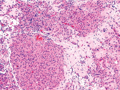
Klinefelter's syndrome. Leydig cell nodules mingle with hyalinized tubules.
Fig. 12-126.
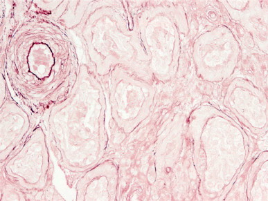
Klinefelter's syndrome. Most seminiferous tubules, even those with Sertoli cell only, have scant elastic fibers that can be demonstrated with orcein stain. The intense staining observed in the inner elastic lamina of arterioles provides a positive control.
The reduced testicular volume gives an appearance of Leydig cell hyperplasia,1090 although quantitative studies have shown that the total number of Leydig cells is lower than normal.1091 Many of the Leydig cells are pleomorphic and some are multivacuolated. Immature fibroblast-like Leydig cells may be present. The abnormally differentiated Leydig cells have nuclei with coarse masses of dense chromatin, deep unfolding of the nuclear envelope, multiple paracrystalline inclusions instead of Reinke's crystalloids, multilayered concentric cisternae of smooth endoplasmic reticulum, large masses of microfilaments, and scant lipid droplets.1092 Sex chromatin is apparent in 40–70% of Leydig cells. Leydig cell function is insufficient and androgen levels are less than 50% of normal. Basal FSH and LH are markedly increased.1084, 1093, 1094 In a few patients the testicular damage is less severe, with some tubules showing spermatogenesis and less prominence of Leydig cells.1095 Exceptionally, complete spermatogenesis and even paternity have been reported.1096
The XY/XXY karyotype is the most frequent variant of Klinefelter's syndrome with chromosomal mosaicism. In this condition, the clinical abnormalities may be attenuated. Gynecomastia is present in 33% of cases, compared to a frequency of 55% in men with the 47XXY karyotype. Azoospermia is found in 50% of cases (93% in XXY men). The testes are larger and spermatogenesis is more developed in men with XXY (Fig. 12-127 ). Patients with the 47XXY karyotype who have spermatozoa in seminiferous tubules are bearers of 46XY spermatogonia and also of 47XXY spermatogonia, whereas those who have no spermatozoa have 47XXY spermatogonia only; these 47XXY spermatogonia may include some spermatozoa with 23X or 23Y chromosomal complement, elevated numbers of both 24XY and 24XX spermatozoa, and also a high frequency of spermatozoa with 21 disomy; this could be an important risk for gonosomy1097 and also for trisomy 21.1098 Genetic counseling is advisable in patients seeking intracytoplasmic sperm injection therapy. Genetic diagnosis before implantation of the zygote or prenatal diagnosis have been recommended, except for parents who assume the risk of gonosomy.
Fig. 12-127.
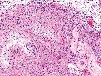
Klinefelter's syndrome mosaicism showing focal spermatogenesis in two seminiferous tubules located within a Leydig cell nodule.
The incidence of the 48XXYY karyotype is estimated to be 0.04 per 1000 live births.1099, 1100, 1101, 1102 This karyotype may be associated with aggressive character, antisocial behavior, more severe mental retardation, and a higher frequency of congenital malformations than the 47XXY karyotype. Men with the 48XXYY karyotype also have characteristic dermatoglyphics with an increase in arches, a decrease in total finger ridge count, and ulnar triradiuses associated with changes in the hypothenar region.1103 Concentric lamellae of smooth endoplasmic reticulum in Leydig cells are a characteristic finding (Fig. 12-128 ).1104 Men with the 48XXXY or 49XXXYY karyotype often have skeletal malformations, principally radioulnar synostosis, and cryptorchidism.1105 In addition to the characteristic symptoms of 47XXY Klinefelter's syndrome,1106 men with the 49XXXXY karyotype have other abnormalities, including severe mental retardation, hypoplasia of external genitalia, cardiac malformations, radioulnar synostosis, microcephaly, and a high arched palate.1107
Fig. 12-128.
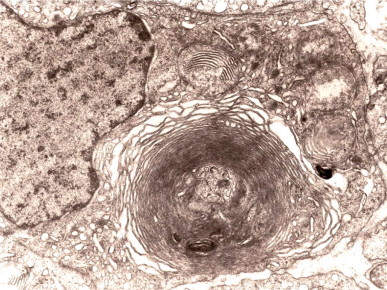
48XXYY Klinefelter's syndrome showing a Leydig cell that contains giant mitochondria and a wheel of smooth endoplasmic reticulum.
Association of Klinefelter's syndrome with malignancy
Patients with Klinefelter's syndrome have a higher incidence of malignancy than the general population. The association was first discovered with breast carcinoma,1108 which had an incidence 20 times greater than in the general male population,1109 and is related to hormonal stimulation.1110 Although testicular germ cell tumor is rare in these patients,1111 extragonadal germ cell tumor is 30–40 times more frequent than in the general population. Most occur in the mediastinum (about 71%) and are less frequent in the pineal gland, central nervous system, and retroperitoneum. The most frequent types are teratoma and choriocarcinoma; embryonal carcinoma and seminoma are rare.1112, 1113, 1114 The extragonadal origin of germ cell tumors has been attributed to abnormal germ cell migration from the yolk sac. The high incidence has been attributed to elevated hormone levels and chromosomal anomaly.1115 In a patient with the XY/XXY chromosomal mosaic and bronchogenic carcinoma, cultured XXY fibroblasts transformed three times more frequently when exposed to SV40 virus than did fibroblasts from normal men.1116
Other tumors reported in patients with Klinefelter's syndrome (lymphoma, leukemia, bronchogenic carcinoma, urothelial carcinoma of the bladder, adrenal carcinoma, prostatic adenocarcinoma, testicular Leydig cell tumor, and epidermoid cyst) do not appear to have a higher incidence than in the general population.1117, 1118, 1119, 1120
Occurrence of Klinefelter's syndrome in childhood
Early identification of this syndrome is possible with systematic cytogenetic study of newborns with positive sex chromatin or mental retardation.1121 Several clinical symptoms suggest Klinefelter's syndrome. Initial symptoms include decreased muscle tone, delayed speech, and poor language skills with an increased incidence of reading difficulties and dyslexia.1122 Later, there may be recognition of mental retardation,1123 psychiatric problems, excessive stature for age, disproportionately long legs, micropenis, and small testes.1124, 1125, 1126, 1127 Androgen deficiency is an early finding.1128 Testicular biopsy reveals scant or absent germ cells. Quantitative studies indicate that the number of germ cells in 47XXY fetuses is significantly lower than in normal 46XY fetuses. The seminiferous tubules have reduced diameter, particularly those devoid of germ cells. The number of Sertoli cells per cross-sectioned tubule is reduced. Megatubules, ring-shaped tubules, and intratubular eosinophilic bodies are common (Fig. 12-129 ). In some cases of Klinefelter's syndrome associated with Down's syndrome, tubular hyalinization is observed in childhood.1129 The interstitium is wide and contains few Leydig cell precursors. If one testis is undescended, its histology does not differ from that of the contralateral testis. The testicular pattern remains constant through childhood.1130 At puberty, before maturation of the tunica propria occurs, the seminiferous tubules rapidly hyalinize and Leydig cell precursors differentiate into Leydig cells.1131
Fig. 12-129.
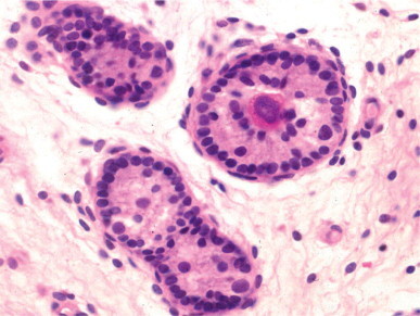
Klinefelter's syndrome at infancy. Seminiferous tubules showing decreased diameters, isolated germ cells, and a ring-shaped tubule that contains a microlith.
Association of Klinefelter's syndrome with precocious puberty
Although precocious puberty is not a characteristic finding in Klinefelter's syndrome, karyotyping in older boys with mental retardation, gynecomastia, small testes, and precocious puberty is advisable. In most cases, the cause of precocious puberty is a hCG-secreting germ cell tumor in the mediastinum.1132 Infrequently, precocious puberty is idiopathic, and only in isolated cases is there a hamartoma in the third ventricle.1133
Association of Klinefelter's syndrome with hypogonadotropic hypogonadism
Klinefelter's syndrome is often associated with pituitary disorders such as panhypopituitarism1134 or incomplete hypopituitarism.1135 Deficits in FSH,1136 LH,1137 or both1138, 1139 have been reported. The cause of this association is unknown, and diverse etiologies such as trauma, immunologic disorders, and genetic deficiencies have been postulated. Alternatively, it may be due to exhaustion of pituitary gonadotropin-secreting cells after years of gonadotropin-releasing hormone stimulation.1135
In patients deficient in both gonadotropins, testicular biopsy shows diffuse tubular hyalinization and a marked reduction in or absence of Leydig cells. The histological picture is similar to that of hypogonadotropic hypogonadism occurring after puberty, except for the presence of isolated tubules containing only dysgenetic Sertoli cells and absence of elastic fibers in the hyalinized tubular wall (Fig. 12-130 ).1139 Biopsy of patients with a deficit only in FSH is similar to that of the dysgenetic Sertoli cell variant of the Sertoli cell-only syndrome, although some hyalinized tubules are present. The testicular biopsy of patients deficient only in LH resembles that of men with classic 47XXY Klinefelter's syndrome.
Fig. 12-130.
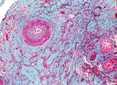
Klinefelter's syndrome with hypogonadotropic hypogonadism showing diffuse tubular hyalinization associated with absence of Leydig cells. Only tubules with dysgenetic Sertoli cells are present.
46XX males
The 46XX karyotype may be present in three phenotypes: male phenotype, including normal external genitalia; male pseudohermaphrodites, with a variable degree of ambiguity in external genitalia, ranging from hypospadias to micropenis; and true male hermaphrodites.
46XX males with male phenotype and normal external genitalia
Men with the 46XX karyotype having male phenotype and normal external genitalia have clinical features similar to those of Klinefelter's syndrome, including small testes, small or normal penis, azoospermia, gynecomastia, and minimal development of secondary sex characteristics. However, these men have harmonious body proportions, normal or slightly low stature, and normal intelligence.1140 The incidence of 46XX males varies from 1:10 000 to 1:25 000 live births, accounting for about 0.2% of infertile men.1141, 1142 Males with 46XX karyotype have hypergonadotropic hypogonadism with elevated serum levels of FSH and, to a lesser degree, elevated LH, with normal or slightly decreased testosterone. Familial cases have been reported.1143
During childhood, biopsy of 46XX males reveals decreased numbers of germ cells.1144, 1145 Biopsies from adults show one of three patterns: histology similar to that of 47XXY men, including diffuse tubular hyalinization with prominent Leydig cells;1146 Sertoli cell-only tubules;1147, 1148 and both patterns intermingled with less prominent Leydig cells. The last is the most frequent (Fig. 12-131 ). Ultrastructural studies reveal an increase in intermediate filaments, absence of annulate lamellae in Sertoli cells,1149 absence of Reinke's crystalloids, and abundance of intracytoplasmic and intranuclear paracrystalline inclusions in Leydig cells.1147
Fig. 12-131.
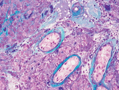
Testis from a 46XX male showing Sertoli-cell-only tubules together with hyalinized tubules, and nodular and diffuse Leydig cell hyperplasia.
46XX males with ambiguous external genitalia
Some patients with the 46XX karyotype have ambiguous external genitalia or hypospadias and are assumed to have a variation of male pseudohermaphroditism.1150 These males, together with true hermaphrodites, may be found in the same family, suggesting that both disorders are different manifestations of the same genetic defect.
Etiology of 46XX males
The origin of 46XX males may be difficult to determine. However, as testicular differentiation requires genes located on the Y chromosome, 46XX males have been classified by cytogenetics as those having the SRY gene, those lacking the SRY gene, and XX/XY mosaicism. Males with the SRY gene comprise 80% of 46XX males.1151 It is likely that this occurs when the genetic material from the short arm of the Y chromosome is translocated to the X chromosome.1152 During paternal meiosis, the homolog pseudoautosomal regions of chromosomes X and Y interchange the terminal portions of their short arms, giving rise to an X chromosome with the SRY gene but lacking the azoospermia factor.1153, 1154, 1155, 1156, 1157, 1158 Alternatively, the SRY region may be inserted in an autosome.1159 Most 46XX patients who are SRY positive have a normal male phenotype. About 10% of 46XX males are SRY negative and most have ambiguous genitalia. Some patients have a normal male phenotype1161 and only infertility.1162 Although SRY is assumed to be the most important regulator factor of testicular determination, these patients may have mutation of one of the downstream non-Y testis-determining genes.1163, 1164, 1165, 1166 About 10% of 46XX males have XX/XY mosaicism or other karyotype with the chromosomal complement Y. In these cases, detection of the specific DNA sequences of Y chromosome may be difficult because this chromosome may be only in some tissues and in a small number of cells.1160
47XYY syndrome
The 47XYY syndrome was first described in 1961 in the father of a girl with Down's syndrome.1167 The only clinical findings were excessive height and pustular acne. Study of other cases suggests that these men are predisposed to a psychopathic personality and antisocial behavior, although most have a normal personality and are socially adapted. The incidence of 47XYY patients is estimated to be 0.01% of the general population, 0.7–0.9% of men in prison, and 1.8% of sexual homicide criminals.1168 The extra Y chromosome originates from non-disjunction during the paternal second meiotic division.
In the past decade, many cases have been diagnosed prenatally. From birth, the patients have weight, stature and cephalic circumference above mean values and a higher risk for delayed language and/or motor development. About 50% of children have psychological and psychiatric problems such as autism; although their intelligence is normal, many patients are referred to special education programs.1169 As adults, they have normal external genitalia and secondary sex characteristics. Fertility is reduced,1170 although many have been fathers. Usually, testicular biopsy reveals mixed atrophy characterized by tubules with spermatogenesis associated with Sertoli cell-only tubules (Fig. 12-132 ).1171, 1172 Those tubules with spermatogenesis may show normal spermatogenesis or have lesions in the adluminal or basal compartments. In these tubules, many XXY spermatocytes degenerate during meiosis. About 64 % of pachytene cells have three sex chromosomes.1173 The number of normal spermatozoa in the ejaculate is low. There is a high incidence of both YY and XY spermatozoa and disomy 18.
Fig. 12-132.
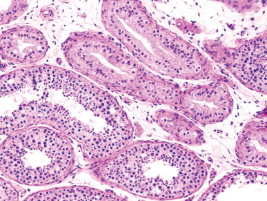
47/XYY syndrome. The testis show tubules with complete spermatogenesis, Sertoli-cell-only tubules, and tubules with spermatogonial maturation arrest.
The variability in germ cell development is apparently due to elimination of germ cells that could not pair their sex chromosomes during the first or second meiotic divisions1174 or, later, during the round spermatid stage.1175 Spermatocytes that succeed in forming trivalent chromosomes are initially viable.1176 The ultimate trivalent chromosome segregation yields aneuploid and euploid cells in equal numbers. Sertoli cell-only tubules are attributed to either spermatogonial damage by substances released from degenerated spermatocytes1177 or absence of testicular colonization by primordial germ cells. These men have normal serum levels of testosterone and LH. The latter may be slightly increased in 47XYY men with severe spermatogenic alterations.1178
47XYY men with mosaicism (47XYY/46XY) have a higher risk of fathering children with hyperdiploid chromosomal constitution, and spermatozoa should be studied genetically to evaluate the risk of intracytoplasmic sperm injection.1179
Men with three and four Y chromosomes have been reported. Men with the 48XYYY karyotype are tall and have normal male phenotype, slight mental retardation, azoospermia and, during childhood, frequent infections of the upper respiratory tract.1180 Testicular biopsy shows Sertoli cell-only tubules, severe hyalinization of tubular basement membrane, and diffuse Leydig cell hyperplasia. The chromosomal complement of parents can be normal.1181 Men with 49XYYYY also have no significant phenotypic abnormalities (except for cases of chromosomal mosaicism). Slight mental retardation, infertility, and antisocial behavior are the most significant clinical findings.1182 Rarely patients have facial dysmorphism and various skeletal abnormalities.1183
Structural anomalies of the Y chromosome
The Y chromosome is essential for gender determination and spermatogenesis, and abnormalities often lead to infertility. The relationship between Y chromosome abnormalities and infertility is best understood in azoospermic men with alterations in Yq11, the distal region of the euchromatic part of the long Y arm, the location of a male fertility gene complex called azoospermia factor. Infertility may result from deletion of any of four subregions in which the azoospermia factor has been divided (AZFa, AZFb, AZFc and AZFd).1184, 1185 The best-known Y chromosome genes involved in spermatogenesis are RBM. DAZ, DFFRY, CDY, SMCY, and ZFY. Six different partial deletions of this region have been found in azoospermic patients (Table 12-10 ). Other genes related to spermatogenesis are BPY2, PRY, TTY1, TTY2, and VCY.1186
Table 12-10.
Pathologic findings in infertile men with Y chromosome anomalies in the YQ11 region
| Karyotype | External genitalia | Testicular lesions | Associated anomalies |
|---|---|---|---|
| 46XYq | Small testes | Tubular hyalinization, Sertoli cell-only, spermatogenetic maturation arrest | Low stature, mental retardation, gynecomastia |
| 46XYnf | Small and soft testes, small penis, ambiguous genitalia, cryptorchidism, hypospadias | Tubular hyalinization, Sertoli cell-only, Leydig cell hyperplasia, decreased spermatid number | Low stature, gynecomastia |
| 45X0 | |||
| 46Xr(Y) | Small testes, cryptorchidism, hypospadias | Sertoli cell-only, spermatogenetic arrest in premeiotic spermatocytes | Low stature |
| 45X0 | |||
| 46Xt(Yp11,Yq11) 45X0 | Small and soft testes, hypospadias | Spermatogenetic arrest in premeiotic spermatocytes, decreased spermatid number | Low stature |
| 46Yt(Xp22,Yq11) | Small testes, small penis | Spermatogenetic arrest in premeiotic spermatocytes | Mental retardation, digital anomalies, facial dysmorphism |
| 46XYqt(Yq11-qter,A) | Normal or small testes | Spermatogenetic arrest in premeiotic spermatocytes | |
| 46Xt(Yq11-pter,A) | Normal or hypoplastic testes, cryptorchidism, hypospadias | Sertoli cell-only, immature seminiferous tubules | |
Monocentric deleted Yq chromosome
Partial deletion of the distal portion of the Yq11 euchromatic region is associated with azoospermia owing to loss of the azoospermia factor. These men have normal external genitalia except for small testes,1187 normal testosterone and LH serum levels, and increased FSH serum level. The most frequent histological finding is Sertoli cell-only pattern, although many other patterns have been reported.1188 The number of Leydig cells is normal or increased. These findings suggest that the azoospermia factor is required for early spermatogenesis.1189 If the breakpoint of Yq11 is proximal to the centromere, patients are short because the gene that controls stature is close to that for the azoospermia factor.1190
Dicentric Yq isochromosomes
Sterility is frequent in men with dicentric Yq isochromosomes.1191 This anomaly is usually associated with a 45X cell line. The proportion of this line varies between patients and between cell types (fibroblasts or lymphocytes). When the point of breakage and fusion of the two Y chromosomes is in the distal region Yq11, and the second centromere is inactivated, the Y isochromosome is normal in size but does not stain with quinacrine, and thus is called non-fluorescent Y chromosome (Ynf). As the breakpoint is in the Yq11 region, the azoospermia factor function is altered. Development of external genitalia varies from ambiguous to normal, and is probably related to the extent of XO present.1192 Testicular biopsies are similar to those of men with monocentric deleted Yq chromosomes (Fig. 12-133 ).1193, 1194
Fig. 12-133.
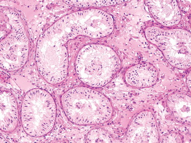
Testis from a male with dicentric Yq isochromosome showing seminiferous tubules with Sertoli cell-only pattern and slight Leydig cell hyperplasia.
Ring Y chromosome
Men with ring Y chromosomes have normal male phenotype, azoospermia, and, in some cases, short stature. Most have mosaic karyotype with a 45X line. In some cases, testicular biopsy resembles that of men with monocentric deleted Yq chromosome, but in others there is premeiotic arrest of spermatocyte maturation.1195 This is attributed to difficulty in pairing the X and Y chromosomes during meiosis. Many patients have deletion of some AZF regions.1196, 1197
Y/Y translocation chromosome
Patients with this anomaly have small soft testes and primary spermatocyte maturation arrest owing to defective pairing of the X and Y chromosomes. The karyotype may be mosaic with a 45X line.1198
Translocation of Y chromosome to X chromosome
Most frequently this translocation is cytogenetically undetectable, and patients present with infertility and are found to have 46XX karyotype.1199 The phenotype is similar to that of men with Klinefelter's syndrome except for shorter stature, absence of mental retardation, and smaller teeth. Testicular biopsy shows Sertoli cell-only pattern. Men with cytogenetically detectable translocations have short stature, small testes, tubular hyalinization, and prominent clustered Leydig cells similar to Klinefelter's syndrome.
Autosomal translocation of Y chromosome
Translocation of the distal heterochromatic portion of the Y chromosome to the short arm of an acrocentric chromosome occurs occasionally. The most frequent are translocations to chromosomes 5, 18, 13, 15, and 22. The fertility of these men depends on the point of breakage.1200, 1201 If this occurs in the Yq12 heterochromatic region, the patient has a male phenotype and is fertile. If the point of breakage is in the Yq11 region, the patient is infertile and has small testes. Seminiferous tubules may show only Sertoli cells, spermatogenetic arrest in early stages of meiosis, or an infantile pattern.1202, 1203
Interstitial microdeletion in Yq11
Yq11 microdeletion is the most frequent congenital cause of infertility. The frequency of Y chromosome microdeletion in infertile patients varies widely (1–35%).1204 In azoospermic men, the frequency is between 18%1205 and 37%.1206 In oligozoospermic males the incidence drops. Most microdeletions are in the AZFc subregion.1207, 1208 Testicular biopsy shows only Sertoli cells, maturation arrest, or mixed atrophy. There is no correlation between site of AZF subregion alteration and histological pattern.1209 There is no exact correlation between genotype and phenotype,1209 but most microdeletions in AZFa are associated with azoospermia, most microdeletions in AZFb are associated with maturation arrest, and most microdeletions of AZFc are associated with spermatid maturation arrest or mixed testicular atrophy. Partial deletion of AZFc has a mild effect on fertility.1210
Structural anomalies of the X chromosome
External genitalia in 46XY patients with duplication of distal Xp vary from male, ambiguous, to female, and gonadal dysgenesis is frequent. If the patient has male genitalia, these are usually hypoplastic with hypogonadotropic hypogonadism and, frequently, multiple congenital anomalies and mental retardation.1211
Males with translocation of the X chromosome to an autosome may have disturbed spermatogenesis with subfertility or infertility.1212, 1213 47XXX males show mental retardation, gynecomastia, normal stature, hypoplastic scrotum, a well-configured but small penis, small testes, and poorly developed pubic hair. Serum testosterone levels are very low. Seminiferous tubules appear severely hyalinized. 47XXX males result from an abnormal X–Y interchange during paternal meiosis and X–X non-disjunction during maternal meiosis.1214
Anomalies in autosomes
There have been many reports on the relationship between autosomal anomalies and infertility, although the causes are not fully understood because the same anomaly is associated with infertility in some patients but not in others.
Chromosomal translocations and inversions
Robertsonian translocations are found in 0.7% of infertile men (8.5% higher than in the normal population) and are more frequent in oligozoospermic than in azoospermic men. The most frequent translocations are 13;14 and 14;21.
The incidence of reciprocal translocations in infertile patients is 0.5% (0.1% in the general population) and increases to 0.8% in patients with azoospermia or severe oligozoospermia.1215 The most frequent in infertile men are 11;22 and 17;21.
Paracentric and pericentric inversions (except for the pericentric inversion of the heterochromatic region in chromosome 9) are eight times greater in infertile patients (0.16%) than in the general population. The highest risk for infertility occurs in the pericentric inversion of chromosome 1.1216, 1217
The most common testicular lesions in men with auto-somal anomalies are spermatogonial maturation arrest, primary spermatocyte sloughing sometimes associated with hypospermatogenesis, and Sertoli cell-only pattern.1218
Down's syndrome
The only autosomal anomaly with prolonged survival is Down's syndrome. In addition to trisomy of chromosome 21 and the characteristic appearance, patients with Down's syndrome usually have cryptorchidism, small testes, hypoplasia of the penis and scrotum, and hypospadias.1219 Adults have oligozoospermia or azoospermia secondary to primary testicular deficiency. Levels of FSH and LH are elevated, but testosterone is normal or slightly diminished.1220 Isolated cases of paternity have been reported.1221, 1222
In utero, there is marked delay in germ cell development.1223 Histologic studies of prepubertal testes at autopsy reveal decreased tubular diameter and tubular fertility index. Eosinophilic bodies or microliths may be present in some tubules (Fig. 12-134 ). Adult testes have deficient spermatogenesis and mixed atrophy, with some tubules showing complete spermatogenesis and others containing Sertoli cell-only pattern.1224
Fig. 12-134.
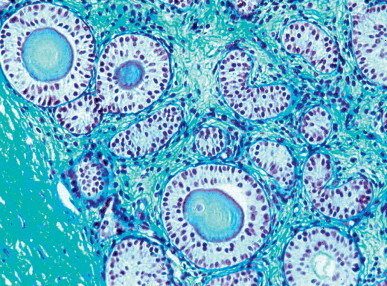
Prepubertal testis in Down's syndrome. There are megatubules, ring-shaped tubules, and small tubules. Germ cell number is very low in all these tubules. Eosinophilic bodies or microliths are present in some tubules.
Other syndromes associated with hypergonadotropic hypogonadism
Hypergonadotropic hypogonadism is found in several myopathies (myotonic dystrophy and progressive muscular dystrophy) and dermopathies (Bloom's, Rothmund–Thomson, Werner's, Cockayne's, and Tay's syndromes), with testicular histology that resembles that of Klinefelter's syndrome. Hypogonadism is also observed in Noonan's syndrome, cerebellar ataxia (with milder testicular lesions), and a miscellaneous group of syndromes with variable histological findings.1225
Myotonic dystrophy accounts for approximately 30% of men with muscular disorders, and about 80% have testicular atrophy. The estimated incidence is 1 in 8000 live births. The abnormality involves the distal muscles of the extremities. In addition, patients may have premature baldness, posterior subcapsular cataracts, cardiac conduction defects, impotence, gynecomastia (rarely), and dementia (at later stages). Myotonic dystrophy is an autosomal dominant inherited disease with variable penetrance. Two loci are associated with the disease phenotype: DM1 in 19q13.3, and DM2 in chromosome 3. Mutation in DM1 results in a serine/threonine protein kinase deficiency that causes expansion of a CTG repeat (from 50 to several hundred repeats) located on the 3′-untranslated region of the dystrophy myotonic-protein kinase gene. The number of repeats is positively correlated to severity of the disease and negatively correlated to age of clinical onset.1226, 1227, 1228 DM2 is caused by a mutation in 3q21.3 of the ZNF9 gene and accounts for CCTG-repeat expansion (from 75 to 11 000 repeats) in intron 1 of this gene. The common clinical symptoms are due to gain of function of RNA mechanism in CUG and CCUG repeats altering cellular function, including alternative splicing of various genes.1229 The severity of the disease increases in the successive generations.1230 The number of CTG repeats is not associated with male subfertility.1231
Hypogonadism is hypergonadotropic in most cases and is not related to the number of CTG repeats.1232 Testicular lesions probably begin late because 65% of patients are fathers. Testicular biopsy shows different degrees of severity, ranging from nearly normal to fully hyalinized seminiferous tubules, with the number of Leydig cells varying from increased to decreased. In some patients the hypogonadism is hypogonadotropic, and the testes show an infantile pattern. Infertility may be the first symptom of myotonic dystrophy.1233
Progressive muscular dystrophy is a multisystemic X-linked disease. It is usually associated with gonadal atrophy caused by a defective locus in chromosome 19. Patients rarely live more than 20 years. The incidence is approximately 1 in 4000 live births. In both Duchenne and Becker forms the cause is a defect in the dystrophin gene.1234, 1235
Bloom's, Rothmund–Thomson, and Werner's syndromes are caused by a homozygous defect in human RECEQ helicases in chromosome 15. Of the five members of this gene family (RECQ1, BLM, WRN, RECQ4, and RECQ5), three produce autosomal recessive inherited diseases. Mutations of BLM have been identified in patients with Bloom's syndrome, WRN has been shown to be mutated in Werner's syndrome, and mutations of RecQ4 have been associated with Rothmund–Thomson syndrome.1236, 1237 Despite the close genetic origin of the three syndromes, symptoms are very different. Bloom's syndrome is characterized by short stature, narrow face with prominent nose, facial ‘patchy’ skin color changes that become more marked with sun light exposure, and increased susceptibility to respiratory diseases, cancer and leukemia. Severe oligozoospermia and azoospermia are common. Leydig cell function is conserved.1238 Rothmund–Thomson syndrome presents with poikiloderma, juvenile cataracts, sparse hair, short stature, skeletal defects, dystrophic teeth and nails, and hypogonadism. These patients are predisposed to cancer and osteogenic sarcoma.1239 Werner's syndrome (progeria) is characterized by short stature, prematurely graying hair, baldness, cataracts, atrophy and calcification of muscle and fat, wrinkling of the skin, keratosis, osteoporosis, telangiectasis, atheroma, diabetes mellitus, gynecomastia, and hypergonadotropic hypogonadism. The lifespan of fibroblasts and other cells is shortened in this syndrome. The mutation is in the RECQ3 helicase gene.
Cockayne's syndrome is a rare autosomal recessive neurodegenerative disorder. Signs and symptoms include infantile failure to thrive, short stature, poorly developed trunk, premature aging, neurological alterations, retinitis pigmentosa, optic atrophy, cataract, deafness, microcephaly, micrognathia, photosensitivity, delayed eruption of primary teeth, congenital absence of some permanent teeth, partial macrodontia, atrophy of the alveolar process and caries, limited articular movements in elbows, knees, and fingers,1240 abnormally small eccrine glands,1241 and hypergonadotropic hypogonadism. It may be caused by two gene mutations: CNK1 (ERCC8) and ERCC6, located respectively on chromosomes 5 and 10, and causing two variations of Cockayne's syndrome, including CS-A, secondary to a ERCC8 mutation, and CS-B with ERCC6 mutation. CS-B patients have hypersensitivity to ultraviolet light secondary to a DNA repair defect.1242
Tay's syndrome (trichothiodystrophy) has two presentations: IBSD (ichthyosis, brittle hair, impaired intelligence, short stature) and IBISD (photosensitivity, ichthyosis, brittle hair, impaired intelligence, short stature). In both forms, patients have decreased fertility. One case of hypergonadotropic hypogonadism has been reported.1243
Noonan's syndrome is characterized by multiple malformations reminiscent of Turner's syndrome, including short statute, pterygium coli, and cubitus valgus, although there is normal male karyotype. The disease has an incidence of 1 in 1000 to 1 in 2500 live births and autosomal dominant inheritance, with sporadic occurrence in about 50% of cases. A locus for dominant forms has been mapped to 12q24.1.1244 Mutation in PTPN11 (protein–tyrosine phosphatase, non-receptor-type 11) accounts for half of cases, although similar germline mutations also cause Leopard's syndrome and certain pediatric hematopoietic malignancies.1245 Cryptorchidism is present in about 70% of cases and is usually bilateral. During childhood, testicular biopsy shows a low tubular fertility index. Puberty is often delayed, and, at adulthood, hypogonadotropic or hypergonadotropic hypogonadism occurs. Ultrastructural studies reveal morphologic anomalies in germ cells.1246 Although spermatogenesis is generally impaired, some patients have been fertile (Fig. 12-135 ).
Fig. 12-135.
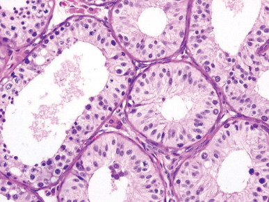
Testis from a 15-year-old boy with Noonan's syndrome. Most seminiferous tubules are small and contain Sertoli cells and isolated spermatogonia. The most dilated tubules have complete although quantitatively decreased spermatogenesis.
Cerebellar atrophy may be associated with hypogonadism. Patients are infertile and have moderate ataxia without endocrine disorder. Infertility is due to morphological abnormalities of spermatozoa caused by decreased expression of MAP2 (the most important microtubule-associated protein), and a defect in erythroid ankyrin.1247, 1248
Many other syndromes also present with primary hypogonadism. The best known are Alström's, Weinstein's, Borjenson–Forssman–Lehmann, Marinesco–Sjögren, Richards–Rundle, Robinow's, and Silver–Russell syndromes.
Secondary idiopathic hypogonadism
Hypogonadotropic hypogonadism or hypogonadism of hypothalamo–hypophyseal origin is classified according to whether the hypothalamo–hypophyseal failure occurs before or after puberty. Eunuchoidism, present only in the former group, is the basis of the distinction. The most frequent types of hypogonadism caused by hypothalamo–hypophyseal failure are those caused by a deficit of gonadotropin-releasing hormone, bioinactive FSH and LH, deficit in growth hormone, those associated with Prader–Willi syndrome, and Laurence–Moon–Rozabal–Bardet–Biedl syndrome.
GnRH deficit
The onset and maintenance of the hypothalamo–hypophyseal–gonadal axis is due to pulsatile gonadotropin-releasing hormone (GnRH) secretion by neurons of the nucleus arcuatus hypothalamus, with release into the pituitary portal system and subsequent stimulation of gonadotropin-releasing hormone receptors on the surface of gonadotropin-secreting cells. The GnRH gene is located on 4q13.1249
Patients with GnRH deficit have partial or complete absence of GnRH-induced pulsatile LH secretion, and normalization of pituitary and gonadal secretions after exogenous GnRH administration. Imaging studies of the hypothalamo–hypophyseal region are normal. Clinical symptoms vary with age at presentation (congenital or acquired) and severity (complete or partial deficit). Clinical presentations include delayed puberty, idiopathic hypogonadotropic hypogonadism (isolated gonadotropin deficit), Kallmann's syndrome, isolated FSH deficit, and isolated LH deficit (fertile eunuch syndrome).
Constitutional delayed puberty
Constitutional delayed puberty is assumed to be a minor form of GnRH deficit,1250 and is characterized by delayed sexual maturation in otherwise healthy males. Patients are short and usually have a family history of delayed puberty. Puberty usually begins at 13–14 years of age and progresses over 2 years. If a 14-year-old boy has not begun pubertal changes (testicular enlargement, growth in height, and development of secondary sex characteristics), delayed puberty should be suspected.1251 Simple pubertal delay that is overcome naturally in a short time without treatment must be distinguished from hypogonadotropic hypogonadism. The latter should be suspected when any of the following symptoms are present in the patient or his family: a midline defect, anosmia, or pubic hair without testicular development. Hormone assays may also assist in diagnosis. If a patient between 16 and 18 years old has prepubertal gon-adotropin levels, he probably has hypogonadotropic hypogonadism.
Isolated gonadotropin deficit
A variant of hypogonadotropic hypogonadism, isolated gonadotropin deficit is characterized by defects in the synthesis or release of FSH and LH; other hypophyseal functions are normal. Patients have eunuchoid phenotype, with small testes and penis, scanty body hair and beard, a high-pitched voice, and poorly developed muscles. Presentation may be sporadic, autosomal dominant, autosomal recessive, or X-linked. The cause might be a mutation in the GnRH receptor gene.1252 Patients have very low levels of FSH, LH, testosterone, and estrogen. Clomiphene citrate treatment fails to stimulate hormonal secretion.1253 Pulsatile administration of GnRH is useful to promote both androgen production and spermatogenesis. The LH–Leydig cell–testosterone axis is normal in most cases, but normalization of the FSH–Sertoli cell–inhibin axis is not achieved in all cases. Basal inhibin levels higher than 60 pg/mL and absence of cryptorchidism are favorable predictor factors for the acquisition of normal testicular size and acceptable spermatogenesis.1254
Testicular biopsy reveals an immature pattern. The seminiferous tubules have neither lumina nor elastic fibers (Fig. 12-136 ). Sertoli cells are immature, and no differentiated Leydig cells are seen. Spermatogonia are rare. In some patients the pattern is similar to that of Sertoli cell-only testes with immature Sertoli cells.1255
Fig. 12-136.
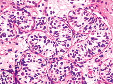
Isolated gonadotropin deficit. The seminiferous tubules have prepubertal diameter, pseudostratified distribution of the Sertoli cells, and several spermatogonia per tubular section.
Hypogonadism associated with anosmia
Hypogonadism associated with anosmia is also known as Maestre de San Juan,1256 Kallman,1257 or De Morsier1258 syndromes. The two most important features are hypogonadotropic hypogonadism and anosmia. Members of affected families may have both features or only one. Associated abnormalities include olfactory bulb agenesis, cryptorchidism, mental retardation, color blindness, facial asymmetry, nerve deafness, epilepsy, shortening of the fourth metacarpal, tarsal navicular fibrous dysplasia, familial cerebellar ataxia, diabetes mellitus, hyperlipidemia, gynecomastia, cleft lip, maxillary or palate, unilateral renal aplasia, and cardiovascular abnormalities. The syndrome may be X-linked or autosomal. The gene for the X-linked form is mapped to Xp22.3 and may have different mutations (termed Kal-X, KALIG-1, and ADMLX), complete deletion, and point mutations. This gene encodes the protein anosmina-1, which is similar to other nerve cell adhesion molecules and is involved in axonal growth and development. KAL protein, secreted by mitral cells, permits the passage of olfactory neurons into the olfactory bulbs and is lacking in Kallmann's syndrome. This failure also inhibits migration of neuroblasts from the olfactory epithelium to the hypothalamus to form GnRH-secreting neurons.1259 The autosomal dominant presentation (occurring in 10% of cases) is due to loss of function of fibroblastic growth factor receptor 1 (FGFR1).1260 Interaction between KAL1 and FGFR1 is required for neuronal migration.1261
Patients are classified into two groups according to the partial or complete absence of GnRH. Partial absence of GnRH is diagnosed by the presence of spontaneous pulses of LH, FSH, and testosterone during a 24-hour period. Complete absence is diagnosed by the absence of spontaneous pulses of LH, FSH, and testosterone during a 24-hour period. These patients show an increase in FSH only after GnRH administration.1262 Testes are histologically infantile; the tubules have a small diameter, lack lumina, and contain immature Sertoli cells and isolated spermatogonia.1263 The interstitium is wide and consists of acellular connective tissue with no recognizable Leydig cell precursors (Fig. 12-137 ).1264
Fig. 12-137.
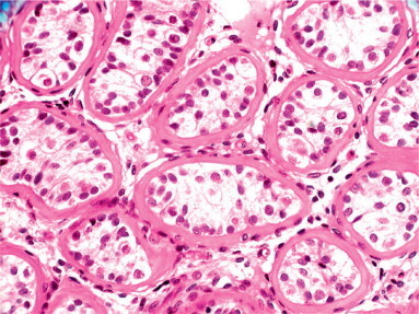
Hypogonadism associated with anosmia in a previously treated patient. The testis shows marked hyalinization of the tubular wall. Some spermatogonia can be observed among the Sertoli cells. The testicular interstitium lacks Leydig cells.
Autopsy studies in patients with anosmia and hypogonadism reveal agenesis of the olfactory bulbs that may be partial or complete and unilateral or bilateral, together with an apparently normal hypophysis and normal or hypoplastic hypothalamus. This syndrome is the least severe form of holoprosencephaly–hypopituitarism complex, a spectrum of developmental anomalies associated with impaired midline cleavage of the embryonic forebrain, aplasia of the olfactory bulbs and tracts, and midline dysplasia of the face. Testicular seminoma has been reported in a patient with anosmia with hypogonadotropic hypogonadism.1265
Isolated FSH deficiency
This rare syndrome is characterized by azoospermia or oligozoospermia in normally virilized patients with normal sexual potency. Serum levels of LH and testosterone are normal, but FSH levels are very low or undetectable. The clomiphene stimulation test gives variable results. The GnRH test induces a normal response only of LH. Mutations in the FSH-β gene are exceptional.1266, 1267
Testicular biopsy shows maturation arrest at the spermatocyte level, hypospermatogenesis, or partial Sertoli cell-only pattern.1268 Gonadotropin treatment increases spermatozoal numbers in most cases, and fertility may be induced.
Isolated LH deficiency
Isolated LH deficiency, also known as Pasqualini's or fertile eunuch syndrome,1269, 1270 is characterized by hypogonadism secondary to LH deficit with preservation of spermatogenesis. Patients have eunuchoid habitus, small testes, decreased libido, female distribution of pubic hair, and a high-pitched voice. Other frequent findings include gynecomastia, anosmia, ocular lesions, and pituitary tumor.1271 FSH level is normal, but LH and testosterone levels are very low. Mutations in the LH-β subunit gene1272 and the GnRH receptor have been reported.1273
The clomiphene test is usually negative, and GnRH stimulation increases LH and, to a lesser degree, FSH. Testicular biopsy shows seminiferous tubules with normal or slightly decreased diameters and complete spermatogenesis; however, the number of all germ cell types is below normal. Leydig cells are rare or absent (Fig. 12-138 ). Maintenance of spermatogenesis in the absence of Leydig cells and serum testosterone can only be explained by assuming the occurrence of testosterone secretion sufficient for spermatogenesis but not to be detectable in the blood.
Fig. 12-138.
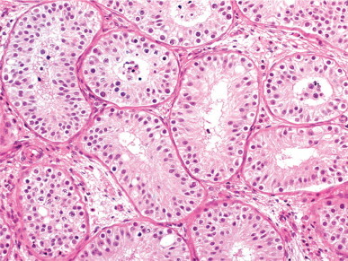
Isolated deficiency of luteinizing hormone. Most seminiferous tubules have a central lumen, numerous spermatogonia, and increased number of Sertoli cells. Spermatocytes and spermatids are observed only in isolated tubules. The testicular interstitium lacks Leydig cells.
Bioinactive FSH and LH
In addition to adequate hypothalamic function, spermatogenesis requires that FSH and LH are biologically active. LH is a heterodimer, composed of two subunits: α (common to FSH and LH) and β (specific for LH). The genes for the β subunit are on 19q13.32. If both alleles are mutated for this subunit, the LH produced in biologically inactive although it may be detectable in standard hormone assay. Homozygous patients have elevated serum level of LH and low testosterone levels, lack of puberty, and infantile testes. Heterozygous patients are only infertile.1274 Patients with mutation in the β subunit of the FSH gene are oligozoospermic or azoospermic.1275
Mutations in gonadotropin receptor genes
Activating and inactivating mutations of gonadotropin receptor genes have been reported. Activating mutation of the LH/human chorionic gonadotropin receptor gene causes familial precocious puberty (see discussion on familial testotoxicosis, below). Inactivating mutation of this gene causes male pseudohermaphroditism (see discussion on Leydig cell hypoplasia in this chapter).
Inactivating mutation of the FSH receptor gene produces only mild spermatogenetic lesions, emphasizing the relative value assumed for FSH in spermatogenesis. Activating mutation of this gene gives rise to spermatogenesis even in the absence of pituitary function.
Growth hormone deficit
Patients with isolated growth hormone deficit and those with resistance to growth hormone action may have delayed puberty and hypogonadotropic hypogonadism.1276 Some patients with spermatogenetic maturation arrest or idiopathic oligozoospermia have a relative deficit of growth hormone. This hormone probably acts on the testis by stimulating local secretion of insulin-like growth factor-1, which cooperates with testosterone.
Prader–Willi syndrome
Prader–Willi syndrome is characterized by hypogonadism, obesity, muscular hypotonia, mental and physical retardation, and acromicria.1277 Other frequent findings include strabismus and non-insulin-dependent diabetes mellitus. The incidence is estimated at between 1 in 12 000 and 1 in 15 000 newborns in 25 000 live births, and is higher in males. Patients have low serum levels of LH, testosterone, estradiol, and inhibin B, and high levels of FSH. These hormonal findings suggest the occurrence of a mixed form of central (low LH) and peripheral (low inhibin B and high FSH) hypogonadism.1278
The penis and testes are hypoplastic, and cryptorchidism is present in about 70% of cases (bilateral in 45% of cases) (Fig. 12-139 ).1279 During infancy and childhood, the testes have reduced tubular diameters; adults have an infantile pattern.1280 This syndrome is caused by an anomaly of chromosome 15, usually in the 15p11-12 band. Other chromosomal anomalies include Robertsonian translocations, reciprocal translocations, small supernumerary metacentric chromosomes, and partial deletion of the long arm of chromosome 15.
Fig. 12-139.
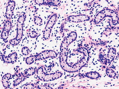
Testis from a 7-year-old child with Prader–Willi syndrome. The seminiferous tubules have a reduced diameter and lack germ cells.
Bardet–Biedl syndrome
This syndrome is a pleiotropic disorder characterized by obesity, infantilism, short stature, diabetes insipidus, mental retardation, retinitis pigmentosa, polydactyly, and syndactyly. It is more frequent in males than in females. Men with this syndrome are infertile, and about 74% show hypogonadism. The testes are prepubertal, the scrotum is hypoplastic or bifid, and the penis is small. Cryptorchidism is found in 42% of males, and is bilateral in 28%. At least 11 genes responsible for this syndrome have been cloned, and it is probable that additional genes are involved. The function of the products of these gene is to mediate and regulate microtubule-based transport processes.1281, 1282
Hypogonadotropic hypogonadism associated with dermatologic diseases
Several dermatopathies are associated with hypogonadotropic hypogonadism, including ichthyosis and Johnson's neuroectodermic syndrome. Most cases of ichthyosis associated with hypogonadism are X-linked. About 15% of these patients have cryptorchidism, small testes, micropenis, and high risk of testicular cancer. The cause is a defective microsomal enzyme, steroid sulfatase, causing the accumulation of cholesterol sulfate that hinders sloughing of the cornified layer of the epidermis. The gene responsible for this enzyme is mapped to Xp22,3. Some of these patients also have anosmia or hyposmia owing to involvement of the neighboring genes, causing a contiguous gene defect.1283
Johnson–McMillin neuroectodermic syndrome is a rare autosomal dominant disorder characterized by alopecia, hypogonadotropic hypogonadism, anosmia or hyposmia, deafness, prominent ears, microtia and/or atresia of the external auditory meatus, and a pronounced tendency to dental caries.1284
Hypogonadotropic hypogonadism associated with ataxia
Hypogonadism associated with ataxia is rare. Most patients are the offspring of a consanguineous marriage. Inheritance is autosomal recessive. The most frequent syndromes are Louis–Bar's syndrome (ataxia–telangiectasia) and Friedreich's ataxia.
Ataxia–telangiectasia is the most common inherited ataxia and is characterized by cerebellar ataxia that starts in infancy and develops progressively; mucocutaneous telangiectasis; anomalies of the immune system that cause pulmonary infection; hypersensitivity to ionizing radiation owing to impairment of DNA repair; and a high risk of lymphoid neoplasia. The gene responsible is on 11q22-q23.1.1285 This ataxia results from inactivation of the A-T mutated (ATM) kinase, a critical protein kinase that regulates the response to DNA double-strand breaks by selective phosphorylation of a variety of substrates.1286
Friedreich's ataxia is a neurodegenerative disorder characterized by degeneration of dorsal root ganglia and spinocerebellar tracts. Hypertrophic myocardiopathy is also observed in many of these patients. The incidence is estimated at 1 in 40 000 children. It is caused by defects in the gene encoding frataxin, a protein required for vesicular traffic in cell and synaptic transmission.1287 About 95% of patients are homozygous for an unstable trinucleoid (GAA) expansion in intron 18 of STM7 on 9q13. The normal gene has up to 35 or 40 triplet repeats, whereas patients with this ataxia carry 70 to more than 1000 GAA triplets.1288 The normal gene has seven to 22 GAA repeats, whereas the mutated gene has over 120 repeats. The extent of the expanded allele is directly proportional to the severity of disease, early onset of disease, and development of cardiac abnormalities.
Other ataxias associated with hypogonadism are Kearns–Sayre, Boucher–Neuhauser, and Gordon–Holmes syndromes.
Other forms of hypogonadotropic hypogonadism
Hypogonadotropic hypogonadism may also be present in Carpenter's, Biemond's, Fraser's, and Moebius’ syndromes, and in patients with mental retardation.
Hypogonadism secondary to endocrine gland dysfunction and other disorders
Maintenance of spermatogenesis requires the harmonious cooperation of several endocrine glands and proper functioning of other tissues. Symptomatic endocrinopathy is present in only 1.7% of infertile men, but over 9% of infertile patients have abnormalities in their endocrine studies.1289 Hypogonadism may be present in disorders involving the hypothalamus–hypophysis, thyroid, adrenals, pancreas, liver, kidney, and gastrointestinal tract, and may be associated with AIDS, chronic anemia, obesity, lysosomal and peroxisomal diseases, and neoplasia. Hypogonadotropic hypogonadism can also be found in some (usually women) who perform rigorous sports (long-distance runners, swimmers, dancers, and rhythmic gymnasts).1290
Hypothalamus–hypophysis
Hypopituitarism
Hypogonadism may result from destruction of the hypothalamus or hypophysis by primary or secondary hypothalamic tumor; granulomatous disease (Fig. 12-140 ); fracture of the cranial base; radiotherapy for malignancy of the nasopharynx, central nervous system, or the eye orbit; pituitary adenoma and cyst; aneurysm of the inner carotid artery; and chronic and nutritional disease. Many of these processes cause panhypopituitarism with varied symptoms.1291
Fig. 12-140.
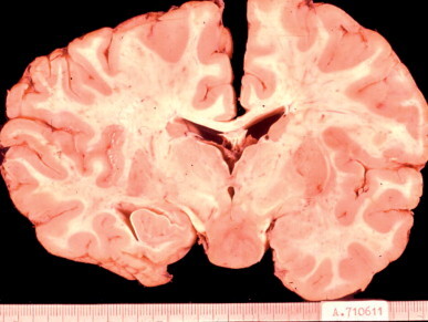
Frontal section from an 18-year-old patient showing destruction of the hypothalamus caused by Langerhans’ cell granulomatosis.
Clinical manifestations of hypogonadism in patients with pituitary lesions vary according to time of onset (childhood, or after puberty). In prepubertal hypopituitarism the testes retain an infantile appearance into adulthood, and there is rarely proliferation of spermatogonia and the development of primary spermatocytes. Biopsy shows variable hyalinization of tubules. In postpubertal hypopituitarism the appearance ranges from complete spermatogenesis to tubular hyalinization (Fig. 12-141 ). The presence of elastic fibers in tubular walls indicates that pubertal maturation occurred before the development of hypopituitarism. Leydig cells have pyknotic nuclei and retracted cytoplasm with abundant lipofuscin. In some patients, recovery of spermato-genesis occurs after administration of human chorionic gonadotropin.1292
Fig. 12-141.
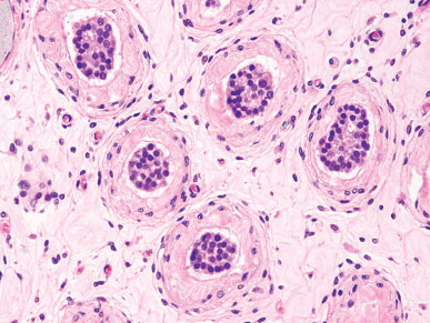
Tubular hyalinization caused by hormonal deprivation and decreased Leydig cell number in a 28-year-old patient who underwent surgery owing to pituitary adenoma. The seminiferous tubules contain dedifferentiated Sertoli cells and isolated spermatogonia.
There are cases in which pituitary adenoma secretes both FSH and LH, inducing testosterone hypersecretion and an elevated sperm count.1293 FSH-secreting pituitary adenoma associated with large testes and increased serum inhibin concentration has been reported.1294
Hyperprolactinemia
Prolactin inhibits GnRH secretion and hence FSH and LH secretion. In addition, prolactin has a direct inhibitory effect on androgens in target tissues. In men, hyperprolactinemia causes impairment of spermatogenesis, impotence, loss of libido, and depressed serum testosterone.1295 Some patients seek treatment because of oligozoospermia and infertility. Hyperprolactinemia is also associated with dysfunction of prolactin receptors.1296 Spermiograms usually show oligozoospermia and an elevated level of fructose,1287 although not all males with hyperprolactinemia have subnormal testicular function.1298
Testicular biopsy reveals variable testicular atrophy. The most frequent lesion is in the tubular adluminal compartment, with degenerative changes in the apical cytoplasm of Sertoli cells, sloughing of young spermatids,1297 and increased lipid droplets in Leydig cells.1299 In boys, two different conditions associated with abnormal prolactin secretion have been reported: hyperprolactinemia, testicular enlargement, and primary hypothyroidism; and prolactin deficiency, obesity, and enlarged testes.
Thyroid gland
Infertility caused by thyroid gland malfunction is rare but reversible. It accounts for about 0.5% of male infertility Testicular function is impaired more by hypo- than by hyperthyroidism. Patients with hyperthyroidism may have gynecomastia, impotence, and infertility. Levels of FSH and LH serum are normal or increased, with elevated sex hormone-binding globulin, increased testosterone concentration, reduced non-sex hormone-binding globulin-bound testosterone, and little or no change in free testosterone.1300, 1301 In Graves’ disease there is a pronounced inhibition of gonadal steroidogenesis.1302 In patients with hyperthyroidism, spermatozoa may be normal or reduced in number, and in both cases progressive motility is low.
Prepubertal hypothyroidism may impair testicular function by causing precocious or delayed puberty. In delayed puberty, hypothyroidism leads to hypogonadotropic hypogonadism, with testes showing incomplete maturation arrest and, in severe myxedematous hypothyroidism, hydrocele.1303 In experimental hypothyroidism, testicular enlargement is frequently associated with increased spermatid production.1304 Primary hypothyroidism in adults causes hypergonadotropic, hypogonadotropic, or normogonadotropic hypogonadism,1305 but testicular function is rarely impaired and patients are usually infertile.1306 The cause of testicular damage is decreased gonadotropins or hyperprolactinemia.1307 Children with hypothyroidism usually have precocious pseudopuberty.1308
Adrenals
About 11% of infertile patients reportedly have subclinical adrenal dysfunction, but the true incidence is probably lower. Adrenal disorders most frequently associated with infertility are adrenal hypoplasia, adrenal hyperplasia, and adrenal carcinoma.
Congenital adrenal hypoplasia
Congenital adrenal hypoplasia with hypogonadotropic hypogonadism is an X-linked recessive disorder that gives rise to adrenal insufficiency in the first months of life. In later presentations, patients have cryptorchidism and delayed puberty.1309 The responsible gene, DAX1 on Xp21, is expressed in the adrenals, testes, pituitary, and hypothalamus. The resulting hypogonadism may be either pure or mixed (hypophyseal and testicular). In the last case, hypogonadism is partial.1310 Testicular biopsy from one adult with adrenal hypoplasia showed an apparent primary lesion, including tubules with dysgenetic Sertoli cells and others with spermatogonial maturation arrest in associated with hypertrophy and hyperplasia of Leydig cells.1311
Congenital adrenal hyperplasia
Infertility is frequent in patients with minor forms of congenital adrenal hyperplasia. Those with deficiency of 21-hydroxylase1312 or 11β-hydroxylase usually have complete spermatogenesis but with reduced numbers of all germ cells. The characteristic histologic finding is decreased numbers of Leydig cells.1313, 1314, 1315, 1316 In untreated patients, the testes become enlarged by ‘tumors’ of the adrenogenital syndrome that consist of cells similar to adrenal cortical cells (Fig. 12-142 ).1316, 1317, 1318, 1319
Fig. 12-142.
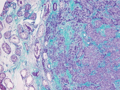
Intratesticular adrenal choristoma near the rete testis from a newborn. The seminiferous tubules are rejected by the mass. The adrenal cortex cells show bizarre nuclei and eosinophilic cytoplasm.
Adrenal cortical carcinoma
Adrenal carcinoma is often associated with excessive secretion of several hormones, causing hyperaldosteronism, Cushing's syndrome, virilization, or feminization. Virilizing tumors in infancy have their own characteristics, which differ from those of the same adult tumors as the infantile form may be associated with other disorders, such as hemihypertrophy and Beckwith–Wiederman syndrome, may be included in the spectrum of ‘families with cancer predisposition’ (mutations in p53 gene), and produce precocious pseudopuberty syndrome. In adults, adrenal carcinoma may cause marked spermatogenic depletion owing to the conversion of large amounts of dehydroepiandrosterone produced by the tumor into estrogen. Feminizing tumor in infancy causes gynecomastia and pubic hair development.1320 Feminizing tumor presents more striking clinical characteristics, including progressive loss of secondary sex characteristics and feminization due to elevated estrogen. Testicular atrophy results from the inhibitory effect of estrogen on pituitary gonadotropins. Similar symptoms may be observed in patients with prostatic carcinoma treated with estrogens (Fig. 12-143 ) and in other conditions with excessive estrogen production, such as Sertoli cell or Leydig cell tumor.
Fig. 12-143.
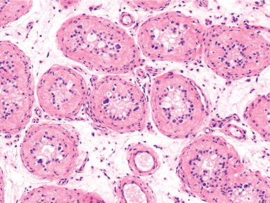
Hypogonadism caused by estrogen therapy for prostate cancer. The seminiferous tubules contain isolated spermatogonia and dedifferentiated Sertoli cells with spherical nuclei, small nucleoli, and pseudostratified infantile distribution. The interstitium contains scattered Leydig cells.
Cushing's syndrome
Patients with Cushing's syndrome or diseases that require long-term corticoid therapy, such as ulcerous colitis, rheumatoid arthritis, or asthma, have reversible reduction of fertility. The explanation for this is that most testicular receptors for corticoids are in Leydig cells, and thus glucocorticoids are powerful inhibitors of testosterone synthesis.
Pancreas
Diabetes mellitus
Alterations in the carbohydrate, lipid and protein metabolism characteristic of diabetes mellitus involve the genital system, although most diabetic patients are fertile. Gonadal impairment depends on the type of diabetes and the time of disease onset (infancy and childhood, puberty, or adulthood).1321, 1322 Testicular lesions in newborns with diabetic mothers are discussed in the section on congenital anomalies of the testis.317
Puberty may be delayed in diabetic patients, although the cause is unknown. Other gonadal alterations appear at puberty, and diabetic men who have not been adequately treated may be infertile and have sexual dysfunction. Serum levels of FSH, LH, and testosterone are decreased.1323 Spermiograms reveal low numbers and poor motility of spermatozoa.1324 Prolactin levels are increased and testosterone levels low or near normal.
The seminiferous tubules have reduced diameters, thickening of the lamina propria, and alterations in the adluminal compartment. These consist of degenerative changes in the Sertoli cell apical cytoplasm and sloughing of immature germ cells. The major lesion is in the interstitial connective tissue and Leydig cells. Small interstitial blood vessels show diabetic microangiopathy characterized by enlargement and duplication of the basal lamina, pericyte degeneration, and endothelial cell alterations. The number of fibroblasts and the amount of collagen and ground substance in the interstitial connective tissue are increased.1325 Leydig cells are decreased in number and show increased amounts of lipid droplets and lysosomes, accounting for the reduced function of these cells.
The tubular lesions are attributed to low serum testosterone, probably owing to deficient Leydig cell stimulation by insulin (or a decrease in insulin-dependent FSH) and abnormal carbohydrate metabolism of Sertoli cells. Sexual dysfunction is present in more than half of patients and consists of impotence, decreased libido, disorders of intercourse, and retrograde ejaculation. The causes of impotence are multiple, including microangiopathy and macroangiopathy, hormonal deficiencies, psychological factors, and autonomic neuropathy affecting the parasympathetic system. Neuropathy is probably chiefly responsible for erectile failure in diabetic men.1326 Alterations in sperm excretory ducts may be associated with diabetes. The most frequent are enlarged seminal vesicles and calcification of both seminal vesicles and vasa deferentia. Calcifications are found in the muscular layers and display a concentric arrangement (Fig. 12-144 ).1327
Fig. 12-144.
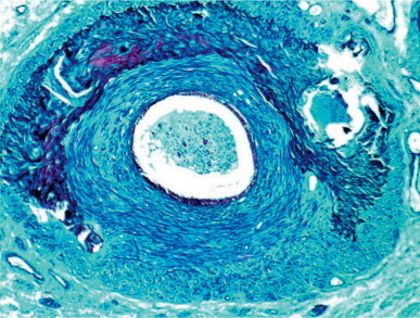
Diabetic patient with dystrophic calcification in the ductus deferens muscular wall.
Mucoviscidosis
Although cystic fibrosis (mucoviscidosis) was recognized as a disease prior to 1940, its effects on the male genital system were not recognized until the 1970s. This may be explained by improvements in medical care during childhood, allowing the survival of many patients to adulthood, and the recognition of cystic fibrosis in patients who had been diagnosed with chronic bronchitis and hepatic or digestive dysfunction. In the US, cystic fibrosis is the most lethal congenital disease, with a prevalence of 1 in 2500 children, and a carrier status of 1 in 25 white men.1328 Lesions in sperm excretory ducts involve (in decreasing order of frequency) the vas deferens (congenital bilateral absence, unilateral absence), ejaculatory ducts (bilateral obstruction), epididymis (diffuse or segmental hypoplasia), and seminal vesicles (incomplete development). Thus, it appears that most patients with cystic fibrosis have infertility due to obstruction.1329, 1330
Histologic studies in children, even at an early age, reveal that the vas deferens and ductus epididymis are absent or reduced to small ductuli with reduced or absent lumina and thin, poorly muscular walls (Fig. 12-145 ). The testes are normal during childhood, but show hypospermatogenesis and spermatid malformations by adulthood. The spermiogram is characteristic of obstructive azoospermia, with acid pH, decreased semen volume and fructose concentration, and increased citric acid and acid phosphatase.1331
Fig. 12-145.
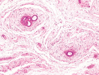
Epididymis in cystic fibrosis. Sections of the ductus epididymidis show decreased lumen diameter with surrounding concentric rings of loose connective tissue.
The disease is a genetic disorder with autosomal recessive inheritance. The impaired gene (cystic fibrosis gene) is on chromosome 7 (7q31),1332 and encodes a protein termed cystic fibrosis transmembrane regulator (CFTR). Alterations in this protein cause cystic fibrosis. Although more than 800 mutations of this gene have been identified,1333 the most frequent mutation in Caucasians is D-F508, responsible for 70% of cases. Congenital bilateral obstructive azoospermia secondary to bilateral absence of the vas deferens, even in the absence of other symptoms, is often a forme fruste of cystic fibrosis.1334 Before initiating treatment for infertility, the possibility that the patient is a carrier of the cystic fibrosis gene should be evaluated.1335
Malformation of the genital system plays the most important role in infertility in cystic fibrosis.1336 The lesions begin in the 10th week of gestation, when the wolffian duct forms the sperm excretory ducts.1337 Variable penetrance of the cystic fibrosis gene accounts for the diversity of malformations affecting different regions of the male genital system.
Liver
The liver has a primary role in metabolism, detoxification, and excretion of sex steroid hormones. Chronic hepatic failure damages the hypothalamo–hypophyseal–testicular axis, and subsequently all related endocrine glands. Hypogonadism is frequent in the final stages of severe chronic liver diseases, including alcoholism, non-alcoholic liver disease, and hemochromatosis.
Hypogonadism, liver disease, and excessive alcohol consumption
The association of testicular atrophy with gynecomastia and hepatic cirrhosis is well known and is referred to as Silvestrini–Corda syndrome.1338, 1339
Alcohol has a direct toxic effect on Leydig cells. Acute alcoholic intoxication suppresses serum testosterone in voluntary non-alcoholic men and laboratory animals. Chronic alcohol ingestion, even in the absence of cirrhosis, causes hypogonadism, with symptoms of Leydig cell failure, including testicular atrophy, infertility, decreased libido, impotence, and reduced size of the prostate and seminal vesicles.1340 Chronic alcoholic patients with cirrhosis also have symptoms of hyperestrogenism, including gynecomastia, female escutcheon, and female fat distribution pattern.
Most chronic alcoholic men, with or without cirrhosis, have significant testicular lesions. The seminiferous tubules have reduced diameters, thickened lamina propria, and decreased or absent germ cells. Leydig cells are reduced in number and contain abundant lipofuscin granules (Fig. 12-146 ). The epididymis becomes atrophic, mainly in the ductuli efferentes, owing to androgen deprivation. The epithelium of the rete testis becomes cuboidal or columnar due to estrogens. The spermiogram correlates with the variability of histologic findings, usually showing a marked reduction in the number and motility of spermatozoa and an increase in the percentage of morphologically abnormal spermatozoa.1341, 1342 About 20% of patients initially have an increase in serum testosterone; with advanced disease, testosterone level decreases. The initial increase is due to an elevation in sex hormone-binding globulin concentration and reduced testosterone metabolism by the liver.1343 Serum estrogen level also increases owing to increased conversion of testosterone into estrogen in peripheral adipose and muscular tissue.1344
Fig. 12-146.
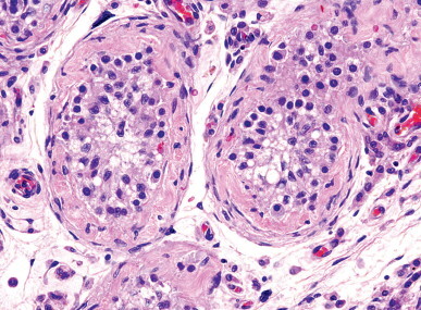
Testis from a patient with alcoholic cirrhosis. The seminiferous tubules show decreased diameter, thickening of the tubular wall, and spermatogonia, isolated spermatocytes, and Sertoli cells exhibiting intense vacuolation of the adluminal compartment. The testicular interstitium shows marked Leydig cell atrophy and numerous macrophages.
Non-alcoholic hepatic disease and infertility
Non-alcoholic liver disease impairs gonadal function according to the severity of the disease.1345 Patients have decreased levels of total and biologically active free testosterone. Hormonal alterations are not as severe as in alcoholic patients, emphasizing the direct action of alcohol on Leydig cells. In α1-antitrypsin deficiency testicular function and fertility are conserved; only in advanced stages of the disease do minor biochemical alterations occur.1346 In Alagille's syndrome (intrahepatic biliary duct hypoplasia), hypogonadism is associated with cholestasis, frequent vertebral, cardiac, and facial malformations, and mental retardation. Hypogonadism is manifest by small testes, delayed puberty, and, in adults, lack of germ cell development.
Hemochromatosis and infertility
Hereditary hemochromatosis is the most frequent genetic disease in the northern hemisphere and results from excessive iron absorption and accumulation in multiple tissue and organs, leading to cirrhosis, diabetes, hypogonadism, and arthralgia. Four types of hereditary hemochromatosis have been reported.1347 Type 1, the most frequent, is caused by mutation in the HFE gene (C282Y), leading to increased intestinal absorption of iron, supersaturation of iron deposits, and damage in multiples organs. The type I hereditary hemochromatosis gene (HFE) is located on the short arm of chromosome 6,1348, 1349 is present in 85–100% of hemochromatosis patients with northern European ancestry, and its protein product is mainly expressed in the epithelium of Lieberkühn crypts. This protein interacts with the transferrin receptor, reducing its affinity for iron-bound transferrin; therefore, HFE becomes a negative regulator of transferrin-bound iron uptake. Type 2 gene is a juvenile form that expresses before the age of 30 years in both sexes, and is associated with severe cardiomyopathy and hypogonadism.1350 The type 2 hemochromatosis locus is on chromosome 1q21, but this gene has not yet been isolated.1351, 1352 Type 3 is on chromosome 7q22, impairs the transferrin 2 receptor, and its consequences are similar to those of type 1 receptor defect. Type 4 is autosomal dominant, on 2q32, and affects the basolateral iron carrier ferroportin 1, resulting in iron deposition in macrophages. Types 1, 2, and 3 have recessive autosomal inheritance and show a similar distribution pattern of iron deposits. In these three types, alteration of gonadal function has also been reported.
Iron homeostasis depends on many genes that act in a coordinated manner, and their exact function is not well known. It is assumed that normal individuals absorb 1–2 mg/day of iron, whereas homozygous patients with hereditary hemochromatosis absorb up to 3–4 mg/day. Once iron deposits become saturated (cells of liver, pancreas, hypophysis, heart, adrenals, and gastric mucosa), the toxic effects of iron cause dysfunction of the liver (cirrhosis and cancer in 5–10% of patients), the pancreas (diabetes in 80% of patients), the heart (myocardiopathy), musculoskeletal system (arthritis), and hypophysis (hypogonadism) (Fig. 12-147 ).
Fig. 12-147.
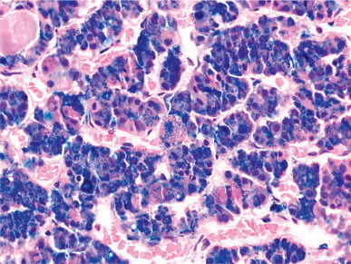
Perl stains decorates the voluminous iron deposits in cells of the anterior pituitary in a patient with hemochromatosis.
Hypogonadism may be the first sign of disease when it starts in adult life.1353 With age, hypogonadism becomes hypogonadotropic, with low serum levels of testosterone, LH, and FSH in more than 40% of patients,1354 except if early treatment is initiated.1355 The most frequent findings are testicular atrophy with diminished tubular diameter, tubular wall thickening, a progressive decrease in spermatogenesis, and increased lipofuscin granules in Leydig cells. The cause of these testicular disorders might be preferential deposition of iron in gonadotropic cells.1356 Iron deposits are not observed in the testis. Hypogonadism decreases after aggressive therapy.1357
Kidney
Polycystic renal disease
Polycystic renal disease in adults is a dominant autosomal disorder that appears with 1 in 1000 frequency in the general population. Patients with this disease comprise 10% of end-stage renal failure cases.1358 Infertility is common, even before the beginning of renal insufficiency. Oligoteratozoospermia and necrospermia are frequent findings.1359, 1360 Serum levels of FSH, LH, prolactin, testosterone, and estradiol remain normal for a long time before the onset of renal insufficiency. The causes of spermiogram alterations have been related to partial obstruction of ejaculatory ducts (based on finding cystic dilations in seminal vesicles in 60% of patients) or seminal vesicle cyst.1361 The incidence of these two disorders in patients with polycystic renal disease is very high compared to andrological patients without this disease (5.2%).1362
Chronic renal insufficiency
Chronic renal insufficiency is associated with disturbed endocrine function in the pituitary, thyroid, parathyroids, and testes. The associated sexual dysfunction consists of erectile impotence, diminution of libido and semen volume, oligozoospermia or azoospermia, and infertility. In children, skeletal development and puberty are delayed.1363
Hormonal studies reveal elevated levels of FSH, LH, and prolactin, but testosterone levels are low.1364 Testicular biopsy shows seminiferous tubules with reduced diameters and reduced or absent germ cells (Fig. 12-148 ).1365, 1366 The interstitium contains a normal number of Leydig cells and increased numbers of macrophages. Additionally, patients with chronic renal insufficiency due to glomerulonephritis have thickening of the tubular lamina propria and decreased number of Leydig cells. Patients with end-stage renal disease who undergo dialysis show calcifications in several organs and tissues, including the male genital system (epididymidis, tunica albuginea, and cavernous tissue) in 87% of cases, and, in isolated cases, calcification of the testicular parenchyma and microlithiasis.1367 Elevated serum levels of phosphorus, increased calcium–phosphorus product, severe hyperparathyroidism secondary to other disorders, older age, and prolonged time on dialysis contribute to this disorder. Uremic calcification is a cell-mediated process in which elevated levels of TGF, vitamin K-dependent proteins such as osteocalcin and atherocalcin, and defects in calcium-regulatory proteins such as fetuin are implicated.1368 When these patients are dialyzed, accumulations of urate and oxalate crystals are deposited in the rete testes and ductuli efferentes. These crystals are deposited beneath the epithelium and often sloughed into the lumen. Reactive changes in the rete testis, including cystic transformation, are frequent (see Disorders of the rete testis).1369
Fig. 12-148.
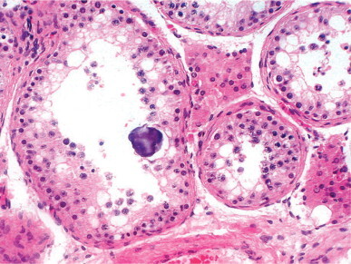
Testis from a patient with chronic renal insufficiency. The seminiferous tubules show premature sloughing of primary spermatocytes. An intraepithelial microlith is present.
The cause of gonadal dysfunction is unclear and probably involves several factors, including impaired testicular steroidogenesis,1370 reduced clearance of pituitary hormones,1371 and secretory defects of the pituitary and hypothalamus.1372 Dialysis does not improve testicular function. The response to renal transplantation is not immediate and is related to the glomerular filtration rate. Patients with rates lower than 50 mL/min develop atrophy of the seminiferous tubular cells.1370
Chronic inflammatory bowel disease
Hypogonadism is a frequent finding in men with celiac disease, and results in clinical symptoms in 5–10% of untreated patients. Celiac disease causes infertility in some cases. Spermiograms show reduced motility and numerous morphologic anomalies in spermatozoa. Hormonal studies show elevated serum FSH levels in more than 25% of men with celiac disease. LH also is increased in more than 50% of these men. The response of FSH and LH to GnRH stimulation is excessive. The cause of this pituitary derangement is unknown. Sperm anomalies are not always corrected by a gluten-free diet. Studies in patients with ulcerative colitis and regional enteritis reveal a low sperm count, impaired motility, and ultrastructural alterations, including nuclear pleomorphism and chromatin malcondensation and decondensation. Zinc deficit may be responsible for these alterations in Crohn's disease.1373 The alterations apparently are related to the extent of the intestinal lesions and the severity of symptoms.1374 Patients with ulcerative colitis treated with salazopyrine,1375 mesalazine1376 or fasalazine1377 present with significant impairment of spermatogenesis and subfertility. Spermiogram parameters improve when treatment ceases.
Acquired immunodeficiency syndrome (AIDS)
More than 17% of HIV-infected men have hypogonadism,1378 which can be observed even in those whose viral replication is under control and show normal numbers of CD4 lymphocytes. Patients frequently develop ‘early andropause,’ marked by dysregulation of the hypothalamopituitary–testicular axis.1379
Hypogonadism is more frequent in HIV-infected men with wasting syndrome, and therefore these patients should undergo screening for hypogonadism and, if necessary, physiologic androgen replacement therapy.1380, 1381, 1382, 1383
The incidence of hypogonadism in males with AIDS is estimated to be 50%.1384, 1385 According to autopsy studies this increases to 100% in the 3–24 months prior to death.1386 Histological studies reveal that 28% have complete but quantitatively abnormal spermatogenesis, and the remainder have spermatocytic arrest or Sertoli cell-only pattern.
Chronic anemia
Patients with chronic anemia requiring multiple transfusions develop iron deposits in the pituitary and polyglandular insufficiency, with atrophy of the thyroid, adrenals, and testes (Fig. 12-149 ). The most frequent conditions are β-thalassemia and sickle cell anemia (see Fig. 12-119).
Fig. 12-149.
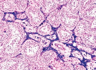
Major thalassemia in a patient who underwent multiple blood transfusions. The testicular interstitium and atrophic tubules show Perl's stain-positive iron deposits.
β-Thalassemia is an autosomal dominant disease with three types: thalassemia trait (heterozygous β-thalassemia), intermediate thalassemia, and major β-thalassemia. The cause is mutation in the β-globin gene resulting in ineffective erythropoiesis, hemolysis, and anemia. Nearly 20% of patients with major thalassemia have delayed puberty,1387, 1388, 1389 and 69% have hypogonadotropic hypogonadism.1390 Gonadal dysfunction persists in most patients after healing of the thalassemia.1391
Sickle cell anemia is an autosomal recessive disorder with a constellation of findings resulting from abnormal synthesis of hemoglobin, with over 90% of hemoglobin being type A. Most patients have hypogonadotropic hypogonadism.1392
Obesity
The majority of people in developed countries are currently overweight, and the incidence of obesity seems to be increasing. Infertility is frequently associated with obesity. Very obese males have increased levels of serum estradiol and decreased levels of free testosterone and inhibin B.1393 Testosterone reduction is not followed by a compensatory increase in gonadotropins, resulting in hypogonadotropic hypogonadism.1394, 1395 Testicular abnormalities begin with the adluminal compartment and later involve the basal compartment; also, there are Leydig cell atrophy, cuboidal metaplasia of the rete testis, and epididymal atrophy.
Autoimmune polyglandular syndrome
There are three types of autoimmune polyglandular insufficiency syndrome. Type I is defined by the presence of at least two of three characteristic features: Addison's disease, hypoparathyroidism, and chronic mucocutaneous candidiasis. The AIRE gene (autoimmune regulator), responsible for type l disease, is on 21q22.3,1396, 1397 and the disorder is recessive autosomal. Hypergonadotropic hypogonadism is frequent.1398 Patients with type I syndrome have antibodies against many autoantigens, intracellular enzymes including the P450 side-chain cleavage enzyme, 17α-hydroxylase1399, 1400 and 21-hydroxylase, glutamic acid decarboxylase 65, aromatic l-amino acid decarboxylase, tyrosine phosphatase-like protein IA-2, tryptophan hydroxylase (TPH), tyrosine hydroxylase, and cytochrome P450 1A2.1401
Type II autoimmune polyglandular syndrome is characterized by the presence of diabetes mellitus, hyperthyroidism, Hashimoto's thyroiditis, Addison's disease, vitiligo, alopecia, pernicious anemia, and hypogonadism (listed in decreasing order of frequency). Type III syndrome includes thyroiditis, diabetes mellitus, pernicious anemia, and vitiligo or alopecia. About 14% of patients have hypogonadism owing to autoimmune destruction of the testis or pituitary gonadotropin-secreting cells (Fig. 12-150 ).1402, 1403
Fig. 12-150.
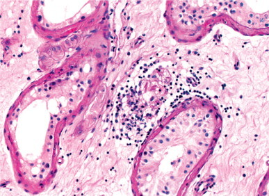
Testis from a man with autoimmune polyglandular syndrome showing selective lymphoid infiltrates in a Leydig cell cluster. Reinke's crystalloid can be recognized. The seminiferous tubules contain Sertoli cells and isolated spermatogonia.
Lysosomal and peroxisomal diseases
There are at least four diseases caused by metabolic deposits in lysosomes or peroxisomes associated with testicular alterations, including Fabry's disease, adrenal leukodystrophy, Wolman's disease, and cystinosis.
Fabry's disease
Fabry's disease is an X-linked metabolic disorder characterized by intralysosomal deposits of globotriaosylceramide (Gb3) owing to α-galactosidase deficiency. Clinical symptoms begin with painful neuropathy and progressive renal, cardiovascular, and cerebrovascular dysfunction. All endocrine glands may accumulate Gb3 as a result of well-developed vasculature and low rate of cell proliferation.1404 Testes and sperm excretory ducts are always damaged. Some alterations, including those of endothelial cells, smooth muscle cells, and fibroblasts, are non-specific; others, such as those of myofibroblasts, Leydig cells, and epididymal epithelium, are specific (Fig. 12-151, Fig. 12-152 ). Spermatogenesis is deficient.1405 Enzyme replacement therapy with recombinant human α-galactosidase eliminates existing glycosphingolipid deposits and blocks new ones, and is thus recommended for implementation as soon as possible after diagnosis.1406, 1407, 1408
Fig. 12-151.
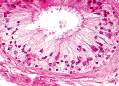
Fabry's disease. Both basal and principal cells of the epididymis show pale and vacuolated cytoplasms, due to lipid deposits.
Fig. 12-152.
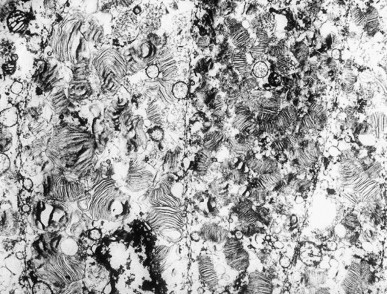
Fabry's disease. The deposits observed in the ductus epididymidis epithelium consist of multiple, parallelly arranged laminae.
Adrenoleukodystrophy (adrenal testicular myeloneuropathy)
This disorder is caused by mutation in the adrenoleukodystrophy gene on Xq28.1409 Mutation at this site produces three peroxisomal diseases: adrenoleukodystrophy, adrenomyeloneuropathy, and Addison's disease.
Adrenoleukodystrophy is characterized by progressive demyelinization of the central nervous system, usually in children and young adults, often with adrenal insufficiency and testicular failure. Peroxisomal β-oxidation is deficient and, as a result, very long-chain fatty acids accumulate inside peroxisomes in many tissues, causing the signs and symptoms of the disease.1410, 1411
Adrenomyeloneuropathy begins at a later age (about 30 years) with progressive paraparesis, peripheral neuropathy, and adrenal cortical failure. Males usually have gonadal dysfunction with oligozoospermia or azoospermia and hypergonadotropic hypogonadism.1412 Testicular atrophy develops slowly, the seminiferous epithelium disappear, and Leydig cells contain characteristic cytoplasmic lamellar inclusions, with similar inclusions in adrenal cortical cells and cerebral cells.1413
Wolman's disease
Wolman's disease is a rare inherited lysosomal disease characterized by a deficit in acid lipase/cholesteryl ester hydrolase. The genetic mutation has been mapped to 10q23.2-q23.3.1414 Complete enzymatic deficiency (Wolman's disease) causes death in infancy as a result of the accumulation of cholesterol esters and triglycerides in numerous organs such as the liver, adrenal cortex, and intestines.1415 Partial deficiency is known as cholesteryl ester storage disease, and the testis accumulates triglycerides and cholesterol in Leydig cells and, to a lesser degree, in interstitial macrophages. Delayed disruption of spermatogenesis by this storage disease probably accounts for the frequent lack of fertility problems in men with this disease.1416 Early treatment of children with Wolman's disease by transplantation of umbilical cord blood-derived stem cells may successfully restore acid lipase level in some.1417
Cystinosis
Cystinosis is an autosomal recessive metabolopathy characterized by alterations in cystine transport from the lysosomes to the cytosol that results in intralysosomal accumulation of cystine. There are several genes responsible, all on chromosome 17p13. Cystine storage occurs in all body tissues. Deposits in the renal parenchyma cause the main complication of cystinosis, namely renal insufficiency (nephropathic cystinosis). Patients also develop hypergonadotropic hypogonadism. Testicular involvement may be massive, with interstitial macrophages filled with cystine crystals that are visible by polarized light.1418
Niemann–Pick disease
Niemann-Pick disease consists of a heterogeneous group of inherited recessive autosomal diseases characterized by deposition of lipids in macrophages and other tissues. There are four reported types (A, B, C, D). The most common, type A, results from excessive storage of sphingomyelin owing to a mutation in the acid sphingomyelin gene that encodes a lysosomal hydrolase, located on 11p15.1-4 region.1419
Interstitial macrophages in the testes have wide eosinophilic, granular cytoplasm. Ultrastructural studies reveal a large number of lysosomes filled with laminate bodies.
Infertility secondary to physical and chemical agents
Physical and chemical agents may impair testicular function by direct action on the pituitary, the testis, or the sperm excretory ducts. In the pituitary, damage to gonadotropic cells may be caused by estrogen. In the testes, gonadotoxic agents may selectively impair a select cell type, but later, global dysfunction occurs. For example, there is direct toxicity to Sertoli cells by phthalates used as plasticizers, nitroaromatic compounds intermediate in the production of dyes and explosives, and γ-diketones used as solvents. Direct toxicity on spermatogenesis is seen wtih ionizing radiation. Many drugs that impair epididymal fluid or spermatozoon transport damage sperm excretory ducts, with subsequent loss of fertility.1420
Occupational exposure
The relationship between infertility or subfertility and certain professions or exposures to environmental agents is well known.1421 Adverse effects of the following agents on spermatogenesis has been demonstrated: organic solvents such as chlorinated solvents, aromatic solvents and varnishes, degreasers, thinners, and adhesives; this is also the case with carbon disulfide exposure; pesticides such as DDT, linuron, and polychlorinated biphenyls;1422 heavy metals such as lead, cadmium, mercury, and copper; industrial wastes such as dioxins and ethylene dibromide; phthalates and polyvinyl chloride; oral contraceptives; exposure to radiation or high temperature; and recreational drugs and doping. There is also a long list of potentially harmful agents that disrupt testicular function.1423
Carbon disulfide
Carbon disulfide is used as a solvent in the production of rayon. Continuous exposure is toxic to the nervous system, and causes a decrease in spermatogenesis and libido and an increase in FSH and LH serum levels.1424, 1425
Dibromochloropropane
Dibromochloropropane is used as a soil fumigant to control nematodes. Lengthy exposure causes oligozoospermia, azoospermia, increased FSH and LH levels, and Y-chromosome non-dysjunction.1426
Lead
Of the two natural forms of lead, organic and inorganic, the inorganic form is more dangerous. Exposure to inorganic lead by workers in smelting, battery, and stained-glass plants causes direct spermatogenic damage.1427 Patients have asthenospermia, teratozoospermia, and oligozoospermia.1428, 1429
Oral contraceptive manufacture
Workers in pharmaceutical plants using synthetic estrogens and progestins develop hyperestrogenism with gynecomastia, decreased libido, and impotence.1430
Neonatal exposure of males to diethylstilbestrol may induce cryptorchidism, testicular hypoplasia, epididymal cyst, and severe anomalies in semen production.1431
Endocrine-disrupting compounds
There is increasing evidence to suggest that estrogen-like effects are produced by a variety of naturally occurring estrogens (so-called phytoestrogens) and numerous synthetic compounds such as phthalates,1432 pesticides,1433 and polychlorinated biphenyls.1434 The principal methods of contact with potential endocrine-disrupting compounds is dietary ingestion of milk, fish, meat, fruits and vegetables, or environmental exposure.1435 The increasing incidence of cryptorchidism, hypospadias, testicular cancer, and poor semen quality may be related to the negative influence of environmental factors on the testis during fetal life. The term ‘testicular dysgenesis syndrome’ has been proposed to designate this constellation of putative syndromes.1436
Estrogen exposure in utero may disrupt development of the testes and the entire male reproductive tract. Estrogen may hinder FSH secretion by the fetal pituitary, and also interfere with subsequent Sertoli cell proliferation, and hence the secretion of AMH required for the regression of müllerian ducts. Persistence of müllerian derivatives is associated with lack of testicular descent. Changes in AMH secretion may also account for altered germ cell proliferation during fetal life. Exposure to high concentrations of estrogen might compromise testosterone production as well as masculinization of external genitalia (hypospadias) and inguinal descent of the testis (cryptorchidism). Abnormal development of Sertoli cells and low germ cell numbers could cause diminished spermatozoon production and infertility.1437
Recreational drugs and doping
Marijuana decreases sperm density and motility and increases the number of morphologically abnormal spermatozoa.1438 Cocaine induces apoptosis in the rat testis (Fig. 12-153 ).1439 About 20% of injection drug users have low serum testosterone levels. Consumption of more than 80 g alcohol per day adversely affects spermatogenesis in two-thirds of patients.1440 Women smoking more than 20 cigarettes per day have fertility problems, neonatal and perinatal mortality, miscarriage, and congenital malformations.1441 Abuse of anabolic steroids by athletes causes hypogonadotropic hypogonadism and transient azoospermia.1442
Fig. 12-153.
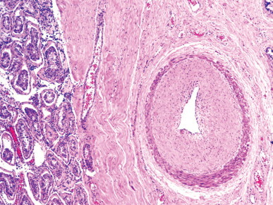
Testis from a 40-year-old patient who consumed cocaine from the age of 16 years. In the tunica albuginea, a branch of the testicular artery shows intense fibrosis in the tunica intima. The seminiferous tubules have marked germ cell atrophy.
Radiation
Ionizing radiation causes alterations in spermatogenesis and hormonal regulation of the testes. Some patients recover fertility a few years after exposure.1443 The effects of non-ionizing radiation are less severe; however, reduced libido and reduced numbers of spermatozoa have been reported in men exposed to microwaves.1444
Heat
Normal intratesticular temperature is 31–33°C, about 4–6°C lower than core body temperature. Conditions causing higher testicular temperature, such as varicocele and cryptorchidism, also cause testicular damage, with decreased numbers of spermatozoa and an elevated percentage of spermatozoa with abnormal forms and low motility.1445, 1446 Primary spermatocytes at the end of the pachytene stage are most sensitive to heat. The mechanism by which heat produces testicular lesions is unknown; hyperthermia affects the activity of enzymes such as ornithine decarboxylase1447 and carnitine acetyl transferase,1448 both necessary for metabolism and proliferation of the seminiferous tubular cells.1449 The synthesis of DNA and RNA by germ cells also depends on temperature. DNA synthesis by spermatogonia and preleptotene primary spermatocytes is higher at 31°C than at 37°C. RNA and protein synthesis are normal at temperatures between 28°C and 37°C, but decrease markedly at 40°C.1450
Testicular trauma
Testicular trauma is especially frequent among athletes. Trauma results in a wide variety of lesions, including contusion with or without hematocele, rupture, dislocation, and eventually spermatogenetic alteration that may lead to infertility. Dislocation involves the displacement of one or both testes to a non-scrotal location1451, 1452 such as the inguinal canal, abdominal cavity, acetabular area, or distant locations such as the perineum, subcutaneous tissues, or superficial to the outer oblique fascia.1453, 1454 Spermatogenetic recovery by orchidopexy has been successfully performed up to 13 years after bilateral traumatic dislocation.1455
Cancer therapy
Sexual dysfunction is found in 25–50% of patients who are treated for cancer.1456 Testicular cancer, Hodgkin's disease, and leukemia are the most frequent malignancies during the reproductive years. Therefore, preservation of fertility requires careful selection of less gonadotoxic therapeutic regimens; if paternity is planned, cryopreservation of semen before treatment may be considered. The most destructive treatments for gonadal function are radiation therapy and alkylating agents.1457
Radiation therapy
The testicular parenchyma is one of the most radiosensitive tissues of the body, and the germ cells are the most radiosensitive cells of the testis. Experimental irradiation of volunteers with a single dose revealed that late spermatogonia (Ap and B) are more radiosensitive than early (Ad) spermatogonia. Ap and B spermatogonia may be destroyed with doses as low as 0.3 Gy (1 Gy = 100 rad), whereas Ad spermatogonia tolerate doses higher than 4 Gy. Type A spermatogonia, spermatids, and spermatozoa are respectively 100, 200, and 10 000 times less radiosensitive than B spermatogonia. Doses higher than 6 Gy produce a Sertoli cell-only pattern. Leydig cells tolerate up to 8 Gy and Sertoli cells up to 60 Gy, although Sertoli cells show ultrastructural alterations and increased phagocytosis of germ cell remnants after low doses of radiation.
Even with optimal protection, the contralateral testis absorbs from 0.2 to 1.4 Gy in adjuvant therapy for rectal cancer1458 or when the opposite testis is irradiated,1459 a dose sufficient to cause temporary azoospermia. Likewise, irradiation of iliac or inguinal lymph nodes for Hodgkin's disease or other forms of lymphoma exposes the testes to about 5 Gy.1460 Restoration of testicular function is time-dependent,1461 requiring at least 2 years.1462 Fertility in thyroid cancer patients who received radioiodine-131 (131I) therapy decreases briefly, but infertility is not permanent.1463 Electromagnetic radiation from cell phones impairs spermatozoon motility according to one study.1464
Prepubertal testes also are sensitive to radiation therapy. Patients treated for Wilms’ tumor may have delayed puberty and, at adulthood, oligoospermia or azoospermia with elevated levels of FSH; this finding suggests that Leydig cells are also damaged. A special case is that of children with acute lymphoblastic leukemia involving the testis. Radiotherapy with doses of 20–25 Gy, either alone or with chemotherapy, causes irreversible damage to the seminiferous tubules and Leydig cells. These patients develop azoospermia and hypogonadotropic hypogonadism with low serum testosterone (Fig. 12-154 ).
Fig. 12-154.
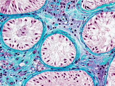
Testis from a 26-year-old patient who, at the age of 9 years, underwent surgery followed by radiotherapy for paratesticular rhabdomyosarcoma. The testicular biopsy shows post-irradiation lesions, including germ cell aplasia and peritubular and interstitial fibrosis.
Chemotherapy
Widespread use of cytotoxic chemotherapy has created a number of adverse side effects, including gonadotoxicity. Combination chemotherapy makes it difficult to ascertain which specific agent is responsible for azoospermia and Leydig cell dysfunction. Comparative studies of chemotherapy for acute lymphoblastic leukemia,1465 extragonadal solid tumors,1466 Hodgkin's disease,1467 Ewing's sarcoma, and other soft tissue sarcomas1468 in children and pubertal boys have shown that alkylating agents cause the most severe testicular damage. Alkylating agents destroy the seminiferous tubular cells and induce tubular atrophy, shrinking the testis and increasing FSH serum concentration.1469 These agents also impair Leydig cell function, causing low testosterone, normal or elevated serum levels of LH, and an exaggerated response of LH to GnRH administration.1470 Testicular damage may be increased by combination with other agents (Fig. 12-155 ).
Fig. 12-155.
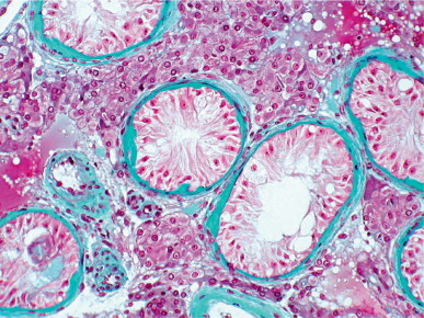
Testis from a patient with Hodgkin's disease after chemotherapy. The seminiferous tubules are small and contain only vacuolated Sertoli cells. The testicular interstitium has pseudohyperplasia of Leydig cells.
Cyclophosphamide appears to be responsible for the greatest number of permanent or temporary cases of azoospermia after chemotherapy. This agent acts directly on the spermatogenic stem cells,1468 and recovery depends on the number of surviving cells. In children, cyclophosphamide reduces seminiferous tubule diameter and germ cell numbers; in the residual spermatogonia nuclei are enlarged. Puberty may progress, even during treatment, and the adult testis may show a Sertoli cell-only pattern.1465 In adults, cyclophosphamide treatment may cause irreversible testicular damage. Administered alone, a dose of 20 000 mg/m2 produces permanent azoospermia in 50% of men. If cyclophosphamide is administered with doxorubicin, vincristine, dacarbazine, or dactinomycin (drugs that alone do not cause azoospermia), doses of 7500 mg/m2 cause azoospermia in 50% of patients. Fludarabine, used for the treatment of chronic lymphocytic leukemia, produces testicular damage with diminution of ejaculate volume, oligozoospermia, increase in serum levels of FSH and LH, and decreased testosterone level. DNA in spermatozoa is markedly abnormal, an effect that persists for several months.1471
Procarbazine, used to treat Hodgkin's disease, causes permanent azoospermia in 30% of patients, even when not combined with alkylating agents.1472 Patients treated with a combination of cyclophosphamide and procarbazine in the COPP protocol (cyclophosphamide, vincristine, procarbazine, and prednisone) do not recover spermatogenesis even if the cyclophosphamide dose does not exceed 4800 mg/m2.
Chemotherapy without both alkylating agents and procarbazine, such as the ABVD (dexorubicin, bleomycin, vinblastine and dacarbazine) or VBM (vinblastine, bleomycin and methotrexate) regimens, produces reversible azoospermia in 36% of patients. The alternating use of MOPP (mechlorethamine, vincristine, procarbazine and prednisone) and ABVD treatments causes testicular dysfunction in 87% of patients, but spermatogenesis recovers in 40%.1473
Patients with germ cell cancer who received chemo-therapy with BEP regimens (cisplatinum, etoposide, and bleomycin) become azoospermic 7–8 weeks after starting treatment. When the total doses reaches 600 mg/m2, infertility is irreversible; at lower dosages, fertility might be recovered over a period of about 2 (50% of patients) to 5 (80%) years,1474 although a high percentage of spermatozoa with DNA abnormalities persists.1475
An important consideration in patients with testicular cancer or Hodgkin's disease is the existence of testicular dysfunction before treatment. In some series1476 dysfunction is present at diagnosis in more than 50% of patients; its cause is unknown. Proposed mechanisms include primary germ cell deficiency, release of toxic substances by tumor cells, and alteration in the hypothalamo–hypophyseal–testicular axis.
Surgery
Sexual function is often lost in patients who undergo bilateral retroperitoneal lymph node dissection for non-seminomatous testicular cancer. Up to 90% lose antegrade ejaculation, although libido, erection, and orgasm are normal. Loss of antegrade ejaculation results from the removal of or injury to sympathetic ganglia and the hypogastric nervous plexus during surgery. Unilateral surgery, especially if the left side is not operated on, reduces this complication.1477, 1478 Hypospermatogenesis sometimes occurs after surgery for rectal cancer, perhaps due to vascular compromise.
Infertility in patients with spinal cord injury
Spinal cord injury is a frequent finding, with more than 10 000 cases annually in the US, mostly in young adults.1479 Fertility is impaired in 90% of males with spinal cord injury. The major sexual dysfunctions in these patients are the lack of erection and ejaculation and poor semen quality.1480, 1481, 1482, 1483, 1484, 1485 Failure of ejaculation occurs in 95% of patients. Semen may be obtained by means of vibratory stimulation of the penis or electroejaculation in more than 90%, but its quality is low, with increased numbers of dead spermatozoa, markedly low motility, and reduced fertilization rate.1486, 1487, 1488 Possible explanations include genitourinary tract infection, endocrine anomaly, and impaired spermatogenesis. Recurrent infection occurs in 60–70% of patients. Compared to controls, a significant increase in the numbers of neutrophils and macrophages occurs, with a marked increase in the production of reactive oxygen species.1489, 1490 This finding and the presence of elevated cytokine levels1491 are assumed to be involved in pathogenesis. Endocrine anomalies are transient, and hormonal levels return to normal after a few months. More than 50% of patients have abnormalities of the adluminal compartment of the seminiferous tubules, with variable degrees of immature germ cell sloughing;1482 in 50% of patients the number of mature spermatids per cross-sectioned tubule is less than 10 (normal >21).
Possible etiologies include an increase in testicular temperature due to vascular dilation, or an alteration in scrotal thermoregulation secondary to impaired sympathetic innervation from prolonged wheelchair restraint; alteration in sperm transport secondary to nerve injury, resulting in sperm stagnation in seminal vesicles, a hostile environment that normally is devoid of spermatozoa;1493 and abnormal composition of seminal fluid, causing deterioration of spermatozoa that in the epididymis and ductus deferens had good motility.1494
More than 25% of patients with spinal cord injury have brown-tinged semen in some ejaculations.1495 Although the cause is unknown, it might be related to seminal vesicle dysfunction.
When spermatozoa cannot be obtained by electroejaculation or vibratory stimulation, vasal aspiration or testicular biopsy are recommended. Most patients have at least a few mature spermatids in some seminiferous tubules; therefore, testicular sperm extraction followed by intracytoplasmic sperm injection is a reasonable consideration in azoospermic patients.1492
Inflammation and infection
Infectious agents may reach the testis and epididymis through blood vessels, lymphatics, sperm excretory ducts, or directly from a superficial wound. Infection transmitted through the blood mainly affects the testis and causes orchitis, whereas infection ascending through the sperm excretory ducts usually causes epididymitis. Acute inflammation is accompanied by enlargement of the testis or epididymis. The tunica albuginea is covered by a fibrinous exudate, and the testicular parenchyma is yellow or brown. Bacterial infection may cause abscess. In some cases the infection begins to heal, with the deposition of granulation tissue and fibrosis; in others, the infection may persist as an active process for a long time, resulting in chronic orchidoepididymitis.
Orchitis
Viral orchitis
The most frequent causes of viral orchidoepididymitis are mumps virus and Coxsackie B virus. Other viral infections that occasionally cause acute orchitis include influenza, infectious mononucleosis, echovirus, lymphocytic choriomeningitis, adenovirus, coronavirus, bat salivary gland virus, smallpox, varicella, vaccinia, rubella, dengue, and phlebotomous fever. Subclinical orchitis probably occurs during other viral infections (Fig. 12-156 ).
Fig. 12-156.
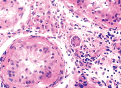
Orchitis caused by cytomegalovirus in a patient with HIV. The inflammatory infiltrate of the testicular interstitium has two characteristic intranuclear inclusions.
Before vaccination was commonly used, mumps orchidoepididymitis complicated 14–35% of adult mumps cases and was bilateral in 20–25% of cases. Nevertheless, mini-epidemics still occasionally occur.1496, 1497 As expected, the incidence remains high in countries where vaccination is not obligatory.1498 In about 85% of cases of mumps orchitis the epididymis is also involved, but epididymal involvement alone is rare.1499 Clinical symptoms of orchitis usually appear 4–6 days after symptoms of parotiditis, but orchitis may also appear without parotid involvement.1500 Testicular involvement is multifocal, and consists of acute inflammation of the interstitium and seminiferous tubules. The tubular lining is destroyed, and eventually only hyalinized tubules and clusters of Leydig cells remain.1501 With time, the testes shrink and become soft. If the infection is bilateral the patient is usually infertile, with severe oligozoospermia or azoospermia, although biopsy may reveal the presence of mature spermatids in some tubules, allowing sperm extraction for paternity.1502 If only one testis was affected, the sperm concentration may be normal or slightly decreased and fertility is maintained. Occasionally the testicular damage is so severe that testicular endocrine function is impaired, causing hypergonadotropic hypogonadism, with low testosterone levels and regression of secondary sex characteristics. Mumps orchidoepididymitis is infrequent in childhood.
Bacterial orchitis
Most bacterial orchitis is associated with bacterial epididymitis. Orchitis secondary to suppurative epididymitis caused by Escherichia coli is most common.1503 On light microscopy, the tubules are effaced by intense acute inflammation. Chronic orchitis with microabscesses is caused by E. coli, streptococci, staphylococci, pneumococci, Salmonella enteritidis,1504 and Actinomyces israeli.1505, 1506 In some cases of chronic bacterial orchitis, the testis contains an inflammatory infiltrate consisting of numerous histiocytes with foamy cytoplasm (xanthogranulomatous orchitis) (Fig. 12-157 ),1507 similar to that of idiopathic granulomatous orchitis but lacking intratubular giant cells. Rarely, as in Whipple's disease, large numbers of bacilli are present in histiocytes in the interstitium, vascular walls, and seminiferous tubules.
Fig. 12-157.
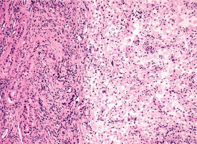
Xanthogranulomatous orchitis showing a dense infiltrate of macrophages with vacuolated cytoplasm surrounded by atrophic seminiferous tubules.
The most frequent complications of pyogenic bacterial orchidoepididymitis are scrotal pyocele and chronic draining scrotal sinus. Small fragments of testicular parenchyma may be eliminated through the scrotal skin, known clinically as fungus testis. Another complication is testicular infarct, resulting from compression or thrombosis of the veins of the spermatic cord, in the scrotal neck, or the superficial inguinal ring.
Granulomatous orchidoepididymitis
Most cases of chronic orchidoepididymitis are associated with granulomas in the testis. Specific causes may require special stains, cultures, or serologic tests, and include tuberculosis, syphilis, leprosy, brucellosis, mycoses, and parasitic diseases. In sarcoidosis and idiopathic granulomatous orchitis, the agent is unknown.
Tuberculosis
The incidence of tuberculous orchidoepididymitis declined after the development of effective antibiotics, but it has recently undergone a resurgence among people who have emigrated from countries with a high incidence of the disease and the increasing population of immunologically compromised patients.
Most cases of tuberculous orchidoepididymitis are associated with involvement elsewhere in the genitourinary system.1508 Tuberculous epididymitis is usually the result of ascent from tuberculous prostatitis, which in turn is often secondary to renal or pulmonary tuberculosis. The pattern of spread is different in children: more than half have advanced pulmonary tuberculosis, and the testis is infected through the blood.1509 More than 50% of patients with renal tuberculosis develop tuberculous epididymitis, and orchitis occurs in approximately 3% of patients with genital tuberculosis, usually secondary to epididymal tuberculosis. It has been suggested that some cases of tuberculous orchidoepididymitis are sexually transmitted.1510 Tuberculous orchidoepididymitis occurs mainly in adults: 72% of patients are older than 35 years, and 18% are over 65 years. The signs and symptoms may be mild, consisting only of testicular enlargement and scrotal pain. In such cases, fever is infrequent and constitutional symptoms may be absent.1511
Histologically, there are typical caseating and non-caseating granulomas that destroy the seminiferous tubules and interstitium (Fig. 12-158, Fig. 12-159 ). In immunosuppressed patients, the granulomas consist of epithelioid histiocytes and a few lymphocytes with rare giant cells. Acid-fast bacilli tend to be more numerous in immunosuppressed patients. Similar lesions may be observed in orchidoepididymitis caused by bacillus Calmette–Guérin, which is usually used for intravesical instillation in patients with vesicular urothelial carcinoma.1512
Fig. 12-158.
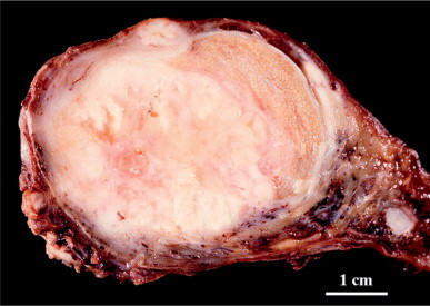
Tuberculous orchitis in a 38-year-old patient with a white-gray nodule which has a pseudotumoral pattern and caused testicular enlargement.
Fig. 12-159.
Tuberculous orchitis showing central necrosis surrounded by numerous granulomas, some of which contain giant cells in their centers.
Syphilis
Syphilitic orchitis may be congenital or acquired. In congenital orchitis, both testes are enlarged at birth. The histological findings are similar to those of the interstitial orchitis of acquired syphilis. If diagnosis is delayed until puberty, the testis often shows retraction and fibrosis. In adults, acquired orchitis is a complication of the tertiary stage of syphilis and has two characteristic histologic patterns: interstitial inflammation and gumma.
Early in the disease, patients with interstitial orchitis have painless enlargement. Grossly, the parenchyma is gray with translucent areas. Histologically, plasma cells are abundant. The inflammation begins in the mediastinum testis and testicular septa, later extending through the parenchyma as the seminiferous tubules lose their cellular lining and undergo sclerosis. Initially, the arteries show an obliterans type of endarteritis. Small gummas may be observed. Eventually, the inflammation subsides and is replaced by fibrosis. The epididymis is usually not affected.
Gummatous orchitis is characterized by the presence of one or several well-delineated grossly gray-yellow zones of necrosis.1513 Histologically, ghostly silhouettes of seminiferous tubules are visible within the gumma, surrounded by inflammation consisting of lymphocytes, plasma cells, and scattered giant cells. In most cases spirochetes may be demonstrated histochemically with Warthin–Starry silver stain, but the most specific diagnostic technique is genetic testing.
Leprosy
The testis may be infected in patients with lepromatous or borderline leprosy. Frequent involvement of the testis in lepromatous leprosy results from the low intrascrotal temperature that promotes growth of the bacilli. Orchitis is usually bilateral, although the degree of involvement may differ between the testes. Occasionally, testicular involvement may be the sole indication of the infection, and the diagnosis may be made by testicular biopsy.1514
The histologic findings in the testis vary with the duration of the infection. Initially, there is perivascular lymphocytic inflammation and interstitial macrophages that contain numerous acid-fast bacilli. Later, the seminiferous tubules undergo atrophy, the Leydig cells cluster, and blood vessels show endarteritis obliterans. Finally, the testis is replaced by fibrous tissue with a few lymphocytes and macrophages containing acid-fast bacilli. Most patients with lepromatous leprosy are infertile, even if the orchitis was clinically mild.1515, 1516
Brucellosis
Brucellosis is common in some parts of the world, including the Middle East.1517, 1518 Orchitis occurs in some patients and may be the first sign of disease. Brucellosis should be suspected when testicular enlargement occurs in patients with undulating fever, malaise, sweats, weight loss, and headache.1519 Occasionally this may mimic testicular tumor. Histologically, there is a dense lymphohistiocytic inflammation with occasional non-caseating granulomas in the interstitium. The seminiferous tubules are infiltrated by inflammatory cells and undergo atrophy. Diagnosis is made by clinical and laboratory findings, including blood culture, the Bengal rose test, and high brucella agglutination titers,1520, 1521 or by real-time polymerase chain reaction assay of urine.1522
Sarcoidosis
Sarcoidosis is a systemic granulomatous disease of unknown etiology that preferentially affects young black adults. The genitourinary tract is involved in only 0.5% of clinical cases and 5% of autopsy cases. Fewer than 30 cases of primary epididymal involvement have been reported, and about 12 of these also involved the testis.1523, 1524 Isolated testicular involvement is exceptional.1523, 1525, 1526 Testicular sarcoidosis is usually unilateral and nodular.947 It is often asymptomatic and found at autopsy.1527 The testis contains non-caseating granulomas similar to sarcoid granulomas at other locations. Before diagnosing testicular sarcoidosis, other granulomatous lesions should be excluded, including tuberculosis, sperm granuloma, granulomatous orchitis, and seminoma. Seminoma often has an intense sarcoid-like reaction, and examination of multiple histologic sections may be necessary to find diagnostic foci of seminoma. An association of mediastinal sarcoidosis and testicular cancer has been reported.1528 Genital involvement of sarcoidosis may be the cause of intermittent azoospermia that benefits from corticoid therapy.1529
Malakoplakia
Malakoplakia is a chronic inflammatory disease that was initially described in the bladder1530 and subsequently in many other organs. The testes (alone or together with the epididymis) are involved in 12% of cases involving the urogenital system.1531, 1532 Grossly, the testes are enlarged and have a brown-yellow parenchymous discoloration,1533 often with abscesses. Malakoplakia causes tubular destruction that is associated with a dense infiltrate of macrophages with granular eosinophilic cytoplasm that often contains Michaelis–Gutmann bodies (Fig. 12-160 ).1534, 1535
Fig. 12-160.
Malakoplakia of the testis showing macrophages with granular and eosinophilic cytoplasm that contains several Michaelis–Gutmann bodies.
The differential diagnosis includes idiopathic granulo-matous orchitis and Leydig cell tumor. Inflammation in idiopathic granulomatous orchitis includes intratubular multinucleate giant cells; in malakoplakia it is difficult to identify the tubular outlines, and giant cells are usually absent. Leydig cell tumor is not usually associated with inflammation, but may contain mononucleated or binucleated cells with abundant eosinophilic cytoplasm. Reinke's crystalloids are identified in up to 40% of cases of Leydig cell tumor but absent in malakoplakia, and Michaelis–Gutmann bodies are absent.
Orchidoepididymitis caused by fungi and parasites
Fungal orchitis is rare; most cases are associated with blastomycosis, coccidiomycosis, histoplasmosis, and cryptococcocis.1536 The genital tract may be involved in widespread blastomycosis. In decreasing order, the organs most frequently affected are the prostate, epididymis, testis, and seminal vesicles. Grossly, there often are small abscesses that may have caseous centers. Fungi measuring 8–15 μm in diameter with double refringent contours are present in the giant cells in granulomas and stain positively with periodic acid–Schiff and methenamine silver stains.
Coccidioidomycosis is endemic in California, the southwestern United States, and Mexico, and may present as epididymal disease after remission of systemic symptoms.1537 The granulomas are similar to those of tuberculosis and contain 30–60 μm sporangia with endospores that stain with periodic acid–Schiff. Dissemination of histoplasmosis and cryptococcosis frequently occurs after steroid therapy and may give rise to granulomatous orchitis with extensive necrosis.1538 Histoplasma capsulatum measures 1–5 μm in diameter and may be demonstrated with silver stain. Cryptococcus is identified by its thick wall that stains with mucicarmine.
Most parasites that reach the genital tract, such as Phyllaria and Schistosoma, are in the spermatic cord, and testicular lesions are secondary to vascular injury.1539 Testicular infection has also been reported in patients with visceral leishmaniasis, congenital and acquired toxoplasmosis (Fig. 12-161 ),1540 Echinococcus infection,1541 and orchitis due to Trichomonas vaginalis.
Fig. 12-161.
Orchitis caused by toxoplasmosis. The giant cells in the testicular interstitium and those in the seminiferous tubules or walls contain numerous organisms.
Idiopathic granulomatous orchitis
Idiopathic granulomatous orchitis is a chronic inflammatory condition of older adults (mean, 59.2 years). The most prominent clinical symptom is testicular enlargement, suggesting malignancy.1542 Most patients have a history of scrotal trauma, 66% have symptoms of urinary tract infection with negative cultures, and 40% have sperm granuloma in the epididymis. An autoimmune etiology has been suggested.
The testis is enlarged, with a nodular cut surface and areas of necrosis or infarction. There are two histologic forms, according to whether the lesion is predominantly in the tubules (tubular orchitis) or the interstitium (interstitial orchitis). In tubular orchitis, germ cells degenerate and the Sertoli cells have vacuolated cytoplasm and vesicular nuclei. Plasma cells and lymphocytes infiltrate the walls of the seminiferous tubules, forming concentric rings. Multinucleated giant cells are present in the tubular lumina and sometimes in the interstitium (Fig. 12-162 ). Vascular thrombosis and arteritis are common. In interstitial orchitis, the inflammation is predominantly interstitial. Ultimately, tubular atrophy and interstitial fibrosis prevail in both forms, which may arise from different immune mechanisms.1543 Tubular orchitis histologically resembles experimental orchitis caused by injection of serum from animals with orchitis, whereas interstitial orchitis resembles orchitis produced by the transfer of cells from immunized animals.
Fig. 12-162.
Idiopathic granulomatous orchitis showing seminiferous tubules with peritubular fibrosis. Numerous lymphocytes and macrophages are present in the interstitium and within seminiferous tubules. Multinucleated giant cells are present in some tubules.
The differential diagnosis of idiopathic granulomatous orchitis is infectious orchitis caused by bacteria, spirochetes, fungi, or parasites. A useful clue in the tubular form is the presence of giant cells within seminiferous tubules.
Focal orchitis
The occurrence of focal lymphoid cell infiltrates in the testicular interstitium is common in infertile patients,1544, 1545 patients who have undergone surgery for bilateral inguinal hernia,1546 vasectomized patients who developed post-infection obstruction,1547 after testicular piercing,1548 and cryptorchidism.1549 Inflammatory infiltrates usually involve the seminiferous tubules, and this suggests the disorder is due to an immunologic response (Fig. 12-163 ).
Fig. 12-163.
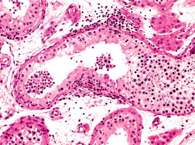
Focal orchitis showing infiltrates of lymphoid cells and macrophages within a seminiferous tubule. There is persistence of Sertoli cells and isolated spermatogonia.
Testicular pseudolymphoma
Pseudolymphoma is a benign reactive process with a lymphoid cell proliferation so intense that it may be mistaken for lymphoma. Testicular pseudolymphoma consists of inflammatory infiltrates with numerous lymphocytes and plasma cells that partially or totally destroy testicular parenchyma.1550, 1551
The differential diagnosis includes lymphoma, various forms of orchitis, and seminoma. The diagnosis of lymphoma may be excluded by the lack of atypia and polyclonal nature of the inflammation. Syphilitic orchitis also contains a plasma cell-rich inflammatory infiltrate, but pseudolymphoma does not have other characteristic features of syphilitic orchitis, such as endarteritis obliterans; spirochetes cannot be demonstrated by special stains. The lack of granulomas or significant numbers of macrophages, together with the negative results of specific histochemical stains, also helps to exclude idiopathic granulomatous orchitis, tuberculosis, leprosy, sarcoidosis, and fungal infection. Finally, although the presence of a prominent inflammatory infiltrate and, in many cases, numerous lymphoid follicles, may suggest the diagnosis of seminoma, the presence of seminoma cells should be easily demonstrated with Best's carmine stain, periodic acid–Schiff, or placenta-like alkaline phosphatase. The term plasma cell granuloma1152 refers to a reactive process characterized by the presence of polyclonal adult plasma cells that are absent in testicular plasmacytoma.1553
Histiocytosis with testicular involvement
Sinus histiocytosis with massive lymphadenopathy (Rosai–Dorfman disease) is a benign proliferation of macrophages that uniquely contain numerous lymphocytes in their cytoplasm. The disease was reported in a kidney and testis of a patient in remission from malignant lymphoma in association with monoclonal IgA gammopathy,1554 and in a second patient with diabetes mellitus who had been previously treated for pulmonary tuberculosis.1555
Increased numbers of interstitial macrophages may also be observed in more than two-thirds of autopsies from adult patients, but the cause is unknown. One condition associated with this disorder is treatment with hydroxyethylstarch plasma expander. In this lesion, the interstitial macrophages stand out by virtue of their large size and multivacuolated cytoplasm, suggesting thesaurosis. There is no evidence of mucin glycoproteins, proteoglycans, starch, lipids, glycogen, or foreign body material. Most patients have no clinical symptoms other than pruritus and persistent erythrema.1556
Other testicular and epididymal lesions
Epididymitis nodosa
Epididymitis nodosa is a proliferation of small irregular ducts whose epithelium lacks the characteristic features of the epididymal epithelium. The disorder is associated with inflammation and fibrosis, similar to vasitis nodosa.1557
Epididymitis induced by amiodarone
In several tissues, including the testis, amiodarone is concentrated up to 300 times its plasma level,1558 causing testicular atrophy and increased serum levels of FSH and LH in some patients.1559 The incidence of epididymitis during amiodarone therapy varies from 3% to 11%,1560, 1561 and more than 35 cases (in several cases involvement was bilateral) have been reported, although there are probably many others.1562, 1563 The disorder may occur at any age.1564 When amiodarone dosage is reduced to 300 mg/day the epididymitis heals within a few weeks.1565 Autopsy studies show focal areas of fibrosis and lymphoid cell infiltrates not related to infection. Recognition is important to avoid unnecessary antibiotics or aggressive surgery.
Ischemic granulomatous epididymitis
This term describes a lesion located in the epididymal head characterized by non-infectious necrosis with polypoid masses of inflamed granulation tissue in peripheral ductal structures. Granulomas containing multinucleated giant cells present within efferent ductuli or form sperm microgranulomas with ductal neoformation similar to that of epididymitis nodosa. The cause is unknown, but may result from ischemia.1566
Calculus (stone in the testis)
The terms ‘testicular calculus’ and ‘stone in the testis’ have been used to describe a lesion characterized by the presence of nodular testicular calcification that is not related to ischemia, orchitis, vasculitis, hematoma, or tumor.1567, 1568
Polyarteritis nodosa
The testicular arteries may be affected by systemic disorders such as Schönlein–Henoch purpura,1569 Wegener's disease,1570, 1571 Kogan's disease,1572 Behçet's disease,1573 relapsing polychondritis, rheumatoid arthritis, and dermatomyositis, but the most frequent involvement is with polyarteritis nodosa.1574 Approximately 80% of patients with polyarteritis nodosa have testicular or epididymal involvement,1575 but only 2–18% are diagnosed during life. Rarely, testicular or epididymal polyarteritis nodosa is the first manifestation of the disease. In these cases the symptoms may suggest orchitis, epididymitis, testicular torsion, or tumor.1576, 1577, 1578
The testis usually shows arterial lesions in different stages of evolution, including fibrinoid necrosis, inflammatory reaction, thrombosis, or aneurysm. The parenchyma initially has zones of infarction (Fig. 12-164 ). Histologic and immunohistochemical findings similar to those of polyarteritis nodosa may occasionally be observed in the testis or the epididymis without lesions elsewhere; this condition is referred to as isolated arteritis of the testis and epididymis,1579 and differs from classic polyarteritis by a lack of vascular thrombosis, aneurysm, or infarct. The etiology of isolated arteritis is unknown, but the prognosis is excellent.1580 The histologic findings of necrotizing arteritis in the testis or epididymis should be followed by clinical, hematologic, and biochemical studies to exclude systemic arteritis.1581, 1582
Fig. 12-164.
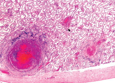
Polyarteritis nodosa involving several intraparenchymal arteries.
Testicular infarct
Torsion of the spermatic cord is the most frequent cause of testicular infarct, followed by trauma, incarcerated inguinal hernia, epididymitis, and vasculitis.
Spermatic cord torsion
Spermatic cord torsion is a surgical emergency. If repair is delayed more than 8 hours, testicular viability is usually compromised. This disorder may appear at any age, but the peaks of maximal incidence are the perinatal period and puberty.1583
Factors that predispose to testicular torsion are anatomical anomalies in testicular suspension and abnormal position of the testis. Many men with testicular torsion have an abnormally high reflection of the tunica vaginalis, giving rise to the deformity known as ‘bell-clapper.’ Other anomalies include elongated mesorchium, separation between the epididymis and testis, and absent or very elongated gubernaculum. The frequency of testicular torsion is higher in cryptorchid and retractile testes than in normal testes.
There are two classic anatomic forms of testicular torsion: high (supravaginal or extravaginal) and low (intravaginal). Each appears at a different age. Extravaginal torsion typically occurs in infancy and childhood, whereas intravaginal torsion is more frequent at puberty and adulthood.
Neonatal torsion is bilateral in 12–21% of cases.1584 Most torsion observed on the first of life is intrauterine.1585 Pubertal and adult torsion causes testicular pain that may radiate to the abdomen or other sites. About 36% of patients have a previous history of pain or swelling in one or both testes. The differential diagnosis includes all causes of acute scrotum.1586, 1587
Torsion causes hemorrhagic infarction of the testis (Fig. 12-165 ). In old neonatal torsion, the histological findings are so advanced that only collagenized tissue containing calcium and hemosiderin deposits is seen. In adults, three degrees of histological lesion may be distinguished.1588 Degree I (26.5% of adult twisted testes) is characterized by edema, vascular congestion, and focal hemorrhage. Seminiferous tubules are dilated, with sloughed immature germ cells, apical vacuolation of Sertoli cells, and dilated lymphatic vessels.1589 Degree II (26.5% of testes) has pronounced interstitial hemorrhage and sloughing of all germ cell types in the seminiferous tubules. The lesion is more severe in the center of the testis, and thus biopsy might provide erroneous information (Fig. 12-166 ). Degree III lesions (45% of testes) are characterized by necrosis of the seminiferous tubular cell layers. There is often a correlation between the time interval of torsion and the degree of the histologic lesion.1590 Degree I appears in torsion of less than 4 hours’ duration, degree II in torsion of between 4 and 8 hours, and degree III in torsion of more than 12 hours. Nevertheless, there are some exceptions that could probably be related, among other factors, to the number of twists in the torsed spermatic cord (degrees of testicular rotation). The testicular salvage rate, defined as testicular growth and development that reflects the age of the patient and the contralateral testis, is around 50% in all cases of testicular torsion.1591 Testes that do not bleed into the albugineal incision within 10 minutes are assumed to be non-viable and should be removed.1592
Fig. 12-165.
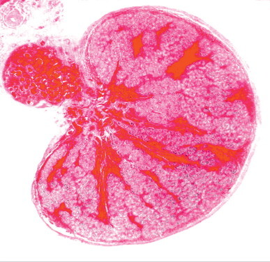
Hemorrhagic infarct in a newborn testis. The hemorrhagic areas are near the rete testis and follow the course of the centripetal veins.
Fig. 12-166.
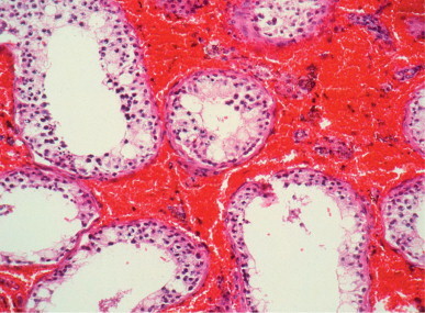
Hemorrhagic infarct grade II in a 13-year-old boy. There is interstitial hemorrhage, focal sloughing of the seminiferous tubular cells, and intense Sertoli cell vacuolation.
Little attention has been paid to intermittent testicular torsion. Early orchiopexy may save these testes, but after surgery, the testis becomes small and excessively mobile, and most have the bell-clapper deformity.1593 Seminiferous tubules are devoid of germ cells and have hyalinized walls.
Some adults with untreated testicular torsion develop lipomembranous fat necrosis of the spermatic cord.1594 Patients seek help for pain in the high scrotum. At this level, there is a small nodule that corresponds to remnants of the twisted testis. The epididymis and proximal spermatic cord characteristically contain fat necrosis (Fig. 12-167 ).
Fig. 12-167.
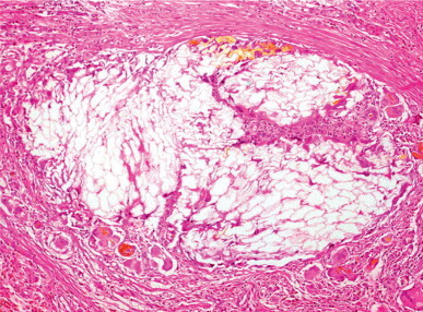
Lipomembranous fat necrosis in a 14-year-old boy who presented with a history of several weeks of intense scrotal pain. Giant cell granulomatous reaction around the membranes had developed.
Adults with prior spermatic cord torsion often consult for infertility. The mechanism causing spermiogram alteration is controversial, and three hypotheses have been proposed:
-
•
Autoimmune process. It has been suggested that the ischemic injury breaks the blood–testis barrier, and antigens released from the necrotic germ cells activate macrophages and lymphocytes in the interstitium, stimulating the formation of antibodies against these antigens. These antibodies that enter in the blood circulation may presumably damage the contralateral testis.1595
-
•
Alterations in microcirculation. After testicular torsion, blood flow decreases in the contralateral testis, causing an increase in the characteristic products of hypoxia, such as lactic acid and hypoxanthine.1596 Intense apoptosis involving mainly spermatocytes I and II has been observed.1597 Long-term effects are yet unknown.
-
•
Primary testicular lesions. Many twisted testes have lesions that cannot be formed in a few hours, such as hypoplastic tubules, microlithiasis, and focal spermatogenesis. In addition, more than half of biopsies of the contralateral testis show marked spermatogenetic lesions.1598 These findings suggest that torsion occurs in testes with congenital lesions.
Other causes of testicular infarct
Trauma1599 and lesions of the vessels of the spermatic cord may also cause testicular infarct. Ischemic atrophy is a risk of inguinal surgery, including herniorrhaphy, varicocelectomy, hydrocelectomy, and descent of cryptorchid testis (Fig. 12-168 ). The incidence of atrophy after inguinal herniorrhaphy varies from 0.06% in primary herniorrhaphy1600 to 7.9% after surgery for recurrent herna,1601 depending on the difficulty and extent of the hernia. Atrophy occurs in some cases of thrombosis of the vena cava or spermatic artery.1602 Focal infarction of the testis is associated with polycythemia, sickle cell disease, trauma,1603, 1604 and laparoscopic inguinal hernia repair.
Fig. 12-168.
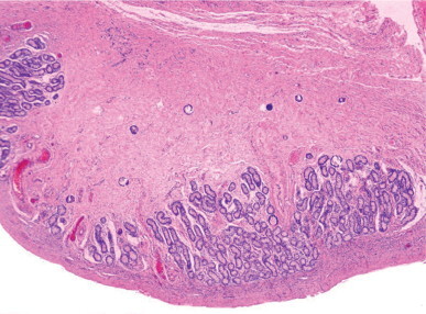
Longitudinally sectioned testis from a 4-year-old infant who had previously undergone orchidopexy. The testis shows marked fibrosis and numerous calcifications except for the periphery of the testicular parenchyma.
Focal infarction may also be spontaneous. Clinical symptoms of testicular infarct mimic testicular tumor. Color Doppler ultrasound reveals the diagnosis in most cases.1605
Other testicular diseases
Cystic malformation
Cystic malformation of the tunica albuginea and testicular parenchyma was first described in the 19th century,1606 and was long considered rare and mainly present in the tunica albuginea.1607, 1608 With the systematic use of ultrasonography, the incidence of cysts has been found to be much higher:1609 non-neoplastic cysts are found in 2.1%1610 to 9.8%1611 of testes.1612, 1613
Cyst of the tunica albuginea is usually an incidental finding in patients in the fifth or sixth decade of life. It is located in the anterolateral aspect of the testis and may be unilocular or multilocular,1614 ranging from 2 to 4 mm and containing clear fluid without spermatozoa. The cyst may be embedded within the connective tissue of the tunica albuginea, protrude from the inner surface of the tunica albuginea into the testicular parenchyma, or protrude from the outer surface forming a blue lump in the tunica albuginea. The epithelium lining the cyst may be simple columnar or stratified cuboidal, and is supported by a thin layer of collagenized connective tissue. The columnar epithelium usually includes some ciliated cells,1615 and the cuboidal epithelium is composed of two layers of non-ciliated cells (Fig. 12-169 ).
Fig. 12-169.
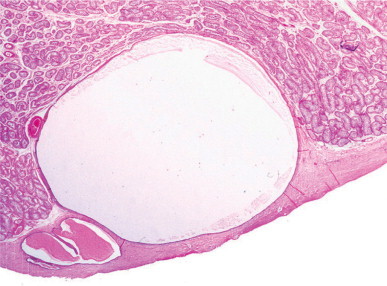
Multilocular cyst in the tunica albuginea. The largest cavity protrudes into the testicular parenchyma.
Cyst of the rete testis is identified by a distinctive epithelial lining of areas of flattened cells intermingled with areas of tall columnar cells. Spermatozoa are frequently found within the cyst,1616 and hence the cyst is also called intratesticular spermatocele.1617 It may be associated with cystic transformation of the rete testis and multiple epididymal cysts. Rete testis cyst is not always attached to the rete and may be found at a distance.
Simple cyst of the testis constitutes the remaining intraparenchymal cyst. It is usually lined by cuboidal epithelium and contains no spermatozoa.1618, 1619 Simple cyst ranges from 2 nm to 18 mm in diameter.1620, 1621 The disorders occurs at any age, from 5 months to 80 years, with a bimodal distribution with peaks at 8 month and 60 years.1622 It may occur bilaterally,1623 and may present as two cysts in the same testis.1624
Origin of the three types of testicular cyst is uncertain. Previously, traumatic1625 and inflammatory1626 origins were attributed to tunica albuginea cyst, but most now believe that they are derived from embryonal remnants of the mesonephric ducts1615, 1627 or mesothelial cells embedded in the tunica albuginea during embryogenesis.1614, 1628, 1629 Simple cyst of the testis may also have a mesothelial origin, but it is possible that some arise from ectopic rete testis epithelium. These cysts are unrelated to epidermoid cyst, differing in the ultrasonographic1630, 1631 and histologic features (see discussion on cystic dysplasia and testicular tumors in the section on hamartomatous testicular lesions). Ultrasound studies indicate that testicular cyst has little potential for growth.1630, 1632 Currently, excision is recommended only in children when the cyst may impair testicular development.1633
Disorders of the rete testis
Dysgenesis
Dysgenesis of the rete testis is characterized by inadequate maturation and persistence of infantile or pubertal characteristics in adults.1634 This disorder is frequent in undescended adult testes. The lesion involves the rete testis segments referred to as septal, mediastinal, and extratesticular. There is poor development of the cavities and their epithelial lining, which becomes cuboidal or columnar instead of flattened with areas of columnar cells. The lumina of the rete testis cavities may be completely absent (simple hypoplasia) or, conversely, undergo microcystic dilation (cystic hypoplasia). In a few cases, the rete testis develops papillary, cribriform, or tubular formations (adenomatous hyperplasia).
Metaplasia
The epithelium of the rete testis is usually flattened, with scattered areas of columnar cells. In estrogen-treated patients, those with chronic hepatic insufficiency, functioning tumor that secretes estrogens or human chorionic gonadotropin, and other disorders that are described as hyperplasia of the rete testis it may undergo diffuse transformation into tall columnar epithelium. Except for the latter group, metaplasia of the rete testis seems to be an estrogen-dependent process, and estrogen receptors are present in the rete testis epithelium.1635
Cystic ectasia of the rete testis (acquired cystic transformation)
Acquired cystic transformation of the rete testis is common, and its incidence increases with age and associated disorders.1636 Ultrasound1637, 1638 and magnetic resonance1639 studies reveal characteristic images that may suggest malignancy. The lesion has three forms: simple, associated with epithelial metaplasia, and with crystalline deposits.
Simple cystic transformation consists of dilated cavities with normal epithelium. It results from obstruction of the epididymis or the initial portion of the vas deferens due to ischemia (aging men); compression by epididymal and spermatic cord tumor, or by congestive veins in varicocele; inflammation in patients with previous epididymitis; malformation (testis–epididymis dissociation, malformed epididymis and absence of the vas deferens);1640 or iatrogenic causes (surgery for epididymoectomy or removal of epididymal cyst) (Fig. 12-170 ).1641
Fig. 12-170.
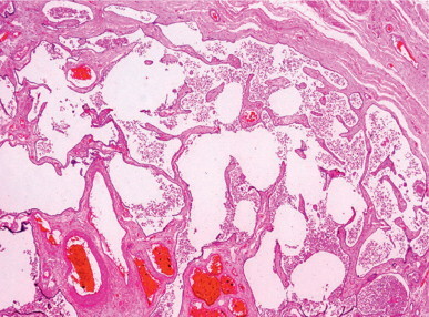
Cystic transformation of the rete testis secondary to a lesion in the caput epididymidis in a patient with chronic epididymitis.
Cystic transformation with epithelial metaplasia is a frequent finding at autopsy.1369 Its development is probably due to the concurrence of sperm excretory duct obstruction and conditions involved in increased serum estrogen levels, such as chronic liver insufficiency. Another possible cause is inflammation involving the rete testis.
Cystic transformation with crystalline deposits has also been called cystic transformation of the rete testis secondary to renal insufficiency.1642 It is a bilateral lesion of adult testes characterized by the concurrence of three findings: cystic transformation of the rete testis, cuboidal or columnar metaplasia of its epithelium, and the presence of urate and oxalate crystalline deposits that may be recognized by polarized light. The lesion is pathognomonic of dialyzed patients with chronic renal insufficiency. Crystalline deposits are initially formed beneath the epithelia of the rete testis and ductuli efferentes; later they protrude into the lumina, where they are finally released. Inflammation is absent or slight, although a few giant cells and small fibrotic areas are often seen (Fig. 12-171, Fig. 12-172 ).
Fig. 12-171.
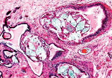
Changes in the rete testis associated with dialysis. Dilation of the rete testis and initial portion of the ductuli efferentes can be observed. Crystalline structures, mainly rhomboidal in shape, accumulate inside and outside the tubules.
Fig. 12-172.
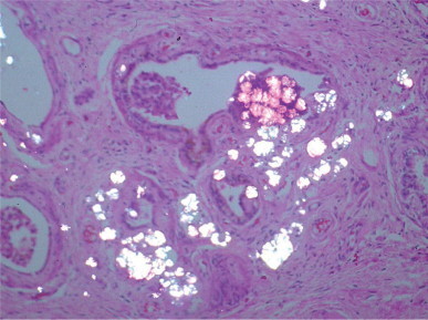
Renal dialysis-associated cystic transformation of the rete testis with oxalate crystals demonstrated by polarized light.
Adenomatous hyperplasia
This lesion is characterized by diffuse or nodular proliferation of tubular or papillary structures that are derived from the rete testis1643 and are observed in cryptorchid or normally descended testes. Cases have been reported in newborns, children, and adults.1644
Adenomatous hyperplasia in newborn and infantile testes consists of enlargement of the mediastinum testis by cord-like or tubular structures derived from the rete testis. The lesion may extend up to one-third of testicular volume. Despite excessive development of the rete testis, the normal connections with seminiferous tubules and efferent ductuli remain. Presentation may be unilateral or bilateral. Unilateral presentation is associated with cryptorchidism or vanishing testis. Bilateral cases may also present with bilateral renal dysplasia. Efferent ductuli may show luminal dilation and irregular outlines. The etiopathogenesis might be similar to that of cystic dysplasia of the testis.1644
Adenomatous hyperplasia in adults is usually an incidental finding at autopsy,1645 in cryptorchid testes,1646 or in testes with germ cell tumor. The rete testis epithelium forms non-encapsulated nodular outgrowths or a diffuse pattern. Nodule size may be large enough to suggest tumor. The epithelium consists of cuboidal cells with ovoid nuclei, deep nuclear folds, and peripheral nucleoli. Atypias and mitotic figures are lacking (Fig. 12-173 ). The ultrastructure and immunophenotype of the epithelium are similar to those of the normal rete testis. Spermatozoa may be seen inside the cavities in some cases, suggesting that such a proliferation is connected with the seminiferous tubules. Most of the testes show a certain degree of seminiferous tubular atrophy.
Fig. 12-173.
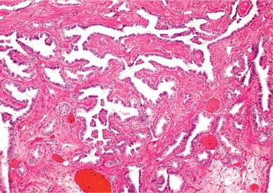
Adenomatous hyperplasia of the rete testis. The epithelium is columnar and supported by a well-collagenized stroma.
In incidental autopsy cases the etiology is unknown, although it may be related to hormonal or chemical agent effects.1647, 1648, 1649 In cryptorchid testes and with many testicular tumors, the most probable cause is a primary anomaly that is part of the testicular dysgenesis syndrome.1650
Adenomatous hyperplasia should be distinguished from three entities: rete testis pseudohyperplasia, which appears in atrophic testes; primary rete testis tumor; and metastasis of adenocarcinoma. In pseudohyperplasia, lesions are focal, microscopic, and usually located in the septal rete, although the mediastinal rete shows few or no alterations. Benign rete testis tumor such as adenoma (solid and papillary variants) and cystoadenoma are isolated and focal,1651 whereas rete testis hyperplasia is diffuse. Adenocarcinoma of the rete testis is a tumor that displays numerous mitotic figures and infiltrates adjacent structures.1652 Metastasis of prostatic adenocarcinoma may be excluded because these metastases alter the rete testis architecture and are immunoreactive for prostatic acid phosphatase and PSA.
Hyperplasia with hyaline globule formation
This reactive lesion is characterized by the presence of intracytoplasmic accumulation of hyaline eosinophilic globules in the epithelial cells of the rete testis. The epithelium may be hyperplastic, but does not contain mitotic figures or nuclear atypia. The globules are up to 15 μm in diameter (Fig. 12-174 ). This lesion is associated with tumor and inflammatory processes occurring near the mediastinum testis, and can be observed in association with 75% of mixed testicular germ cell tumors, 47% of seminomas, and 20% of non-germ cell testicular tumors, such as epididymal tumor that infiltrates the testis (adenomatoid tumor).1653 Yolk sac tumor infiltrating the rete testis may closely resemble this type of rete testis hyperplasia. Positive immunoreactions for α-fetoprotein and placenta-like alkaline phosphatase, as well as nuclear atypia, are helpful to distinguish germ cell neoplasia from this rete testis hyperplasia.1654
Fig. 12-174.
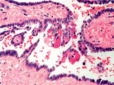
Rete testis hyperplasia with hyaline globules. The globule-containing cells protrude into the lumina of the rete testis channels.
Intracavitary polypoid nodular proliferation
This lesion, described as nodular proliferation of calcifying connective tissue in the rete testis, is characterized by the presence of multiple nodules that originate from the rete testis lining and subjacent connective tissue, protruding into the channels of the rete testis. These consist of cellular connective tissue covered by several layers of a fibrin-like material, which in turn is covered by rete testis epithelium. The nodules may be totally or partially calcified (Fig. 12-175 ).1655 The lesion is an incidental finding at autopsy in patients with impaired peripheral perfusion.
Fig. 12-175.
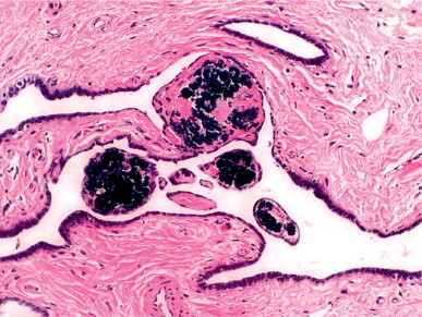
Nodular proliferation of calcifying connective tissue in the rete testis with large calcium deposits.
Selective location of the lesion in the walls of the cavities and chordae rete testis is probably related to poor vascularization of these structures. The etiopathogenetic mechanism may be anoxia, necrosis, fibrin deposition, proliferation of connective tissue, or dystrophic calcification. The intracavitary growth of the lesion might be due to the lower intracavitary pressure and also to the stiff structure of the mediastinum testis.
REFERENCES
- 1.Hiort O, Holterhus PM. The molecular basis of male sexual differentiation. Eur J Endocrinol. 2000;142:101–110. doi: 10.1530/eje.0.1420101. [DOI] [PubMed] [Google Scholar]
- 2.Gessler M, Poustka A, Cavenee W. Homozygous deletions in Wilms'tumours of a zinc-finger gene identified by chromosome jumping. Nature. 1990;343:774–778. doi: 10.1038/343774a0. [DOI] [PubMed] [Google Scholar]
- 3.Call KM, Glaser T, Ito CY. Isolation and characterization of a zinc finger polypeptide gene at the human chromosome 11 Wilms’ tumor locus. Cell. 1990;60:509–520. doi: 10.1016/0092-8674(90)90601-a. [DOI] [PubMed] [Google Scholar]
- 4.Kreidberg JA, Sariola H, Loring JM. WT-1 is required for early kidney development. Cell. 1993;74:679–691. doi: 10.1016/0092-8674(93)90515-r. [DOI] [PubMed] [Google Scholar]
- 5.Pelletier J, Bruening W, Kashtan CE. Germline mutations in the Wilms’ tumor suppressor gene are associated with abnormal urogenital development in Denys–Drash syndrome. Cell. 1991;67:437–447. doi: 10.1016/0092-8674(91)90194-4. [DOI] [PubMed] [Google Scholar]
- 6.Barbaux S, Niaudet P, Gubler MC. Donor splice-site mutations in WT1 are responsible for Frasier syndrome. Nature Genet. 1997;17:467–470. doi: 10.1038/ng1297-467. [DOI] [PubMed] [Google Scholar]
- 7.Hanley NA, Ball SG, Clement-Jones M. Expression of steroidogenic factor 1 and Wilms’ tumour 1 during early human gonadal development and sex determination. Mech Dev. 1999;87:175–180. doi: 10.1016/s0925-4773(99)00123-9. [DOI] [PubMed] [Google Scholar]
- 8.Achermann JC, Ito M, Hindmarsh PC. A mutation in the gene encoding steroidogenic factor-1 causes XY sex reversal and adrenal failure in humans. Nature Genet. 1999;22:125–126. doi: 10.1038/9629. [DOI] [PubMed] [Google Scholar]
- 9.Biason-Lauber A, Schoenle EJ. Apparently normal ovarian differentiation in a prepubertal girl with transcriptionally inactive steroidogenic factor 1 (NR5A1/SF-1) and adrenal cortical insufficiency. Am J Hum Genet. 2000;67:1563–1568. doi: 10.1086/316893. [DOI] [PMC free article] [PubMed] [Google Scholar]
- 10.Lim HN, Hawkins JR. Genetic control of gonadal differentiation, Baillière's. Clin Endocrinol Metab. 1998;12:1–16. doi: 10.1016/s0950-351x(98)80410-2. [DOI] [PubMed] [Google Scholar]
- 11.Bishop CE, Guellaen G, Gelowerth D. Single-copy DNA sequences specific for the human Y chromosome. Nature. 1984;309:253–255. doi: 10.1038/303831a0. [DOI] [PubMed] [Google Scholar]
- 12.McElreavey K, Fellous M. Sex determination and the Y chromosome. Am J Med Genet. 1999;89:176–185. doi: 10.1002/(sici)1096-8628(19991229)89:4<176::aid-ajmg2>3.0.co;2-b. [DOI] [PubMed] [Google Scholar]
- 13.Blyth B, Duckett JW. Gonadal differentiation: a review of the physiological process and influencing factors based on recent experimental evidence. J Urol. 1991;145:689–694. doi: 10.1016/s0022-5347(17)38426-4. [DOI] [PubMed] [Google Scholar]
- 14.Salas-Cortes L, Jaubert F, Barbaux S. The human SRY protein is present in fetal and adult Sertoli cells and germ cells. Int J Dev Biol. 1999;43:135–140. [PubMed] [Google Scholar]
- 15.Haqq CM, King CY, Ukiyama E. Molecular basis of mammalian sexual determination: activation of Müllerian inhibiting substance gene expression by SRY. Science. 1999;266:1494–1500. doi: 10.1126/science.7985018. [DOI] [PubMed] [Google Scholar]
- 16.Huang B, Wang S, Ning Y. Autosomal XX sex reversal caused by duplication of SOX-9. Am J Med Genet. 1999;87:349–353. doi: 10.1002/(sici)1096-8628(19991203)87:4<349::aid-ajmg13>3.0.co;2-n. [DOI] [PubMed] [Google Scholar]
- 17.Foster JW, Dominguez-Steglich MA, Guioli S. Campomelic dysplasia and autosomal sex reversal caused by mutations in an SRY-related gene. Nature. 1994;372:525–530. doi: 10.1038/372525a0. [DOI] [PubMed] [Google Scholar]
- 18.Chaboissier MC, Kobayashi A, Vidal VI. Functional analysis of Sox8 and Sox9 during sex determination in the mouse. Development. 2004;131:1891–1901. doi: 10.1242/dev.01087. [DOI] [PubMed] [Google Scholar]
- 19.Wagner T, Wirth J, Meyer J. Autosomal sex reversal and campomelic dysplasia are caused by mutations in and around the SRY-related gene SOX9. Cell. 1994;79:1111–1120. doi: 10.1016/0092-8674(94)90041-8. [DOI] [PubMed] [Google Scholar]
- 20.Zhou R, Liu L, Guo Y. Similar gene structure of two Sox9a genes and their expression patterns during gonadal differentiation in a teleost fish, rice field eel (Monopterus albus) Mol Reprod Dev. 2003;66:211–217. doi: 10.1002/mrd.10271. [DOI] [PubMed] [Google Scholar]
- 21.Koopman P. Sex determination: a tale of two Sox genes. Trends Genet. 2005;21:367–370. doi: 10.1016/j.tig.2005.05.006. [DOI] [PubMed] [Google Scholar]
- 22.Wolf U. The molecular genetics of human sex determination. J Mol Med. 1995;73:325–331. doi: 10.1007/BF00192884. [DOI] [PubMed] [Google Scholar]
- 23.Baumstark A, Barbi G, Djalali M. X-p duplications with and without sex reversal. Hum Genet. 1996;97:79–86. doi: 10.1007/BF00218838. [DOI] [PubMed] [Google Scholar]
- 24.Bennett CP, Docherty Z, Robb SA. Deletion 9p and sex reversal. J Med Genet. 1993;30:518–520. doi: 10.1136/jmg.30.6.518. [DOI] [PMC free article] [PubMed] [Google Scholar]
- 25.Wilkie AOM, Campbell FM, Daubeney P. Complete and partial XY sex reversal associated with terminal deletion of 10q: report of 2 cases and literature review. Am J Med Genet. 1993;46:597–600. doi: 10.1002/ajmg.1320460527. [DOI] [PubMed] [Google Scholar]
- 26.Merchant-Larios H, Moreno-Mendoza N, Buehr M. The role of the mesonephros in cell differentiation and morphogenesis of the mouse fetal testis. Int J Dev Biol. 1993;37:407–415. [PubMed] [Google Scholar]
- 27.Moreno-Mendoza N, Herrera-Muñoz J, Merchant-Larios H. Limb bud messenchyme permits seminiferous cord formation in the mouse fetal testis but subsequent testosterone output is markedly affected by the sex of the donor stromal tissue. Dev Biol. 1995;169:51–56. doi: 10.1006/dbio.1995.1125. [DOI] [PubMed] [Google Scholar]
- 28.Tilmann C, Capel B. Mesonephric cell migration induces testis cord formation and Sertoli cell differentiation in the mammalian gonad. Development. 1999;126:2883–2890. doi: 10.1242/dev.126.13.2883. [DOI] [PubMed] [Google Scholar]
- 29.Capel B, Albrecht K, Washburn LL. Migration of mesonephric cells into the mammalian gonad depends on Sry. Mech Dev. 1999;84:127–131. doi: 10.1016/s0925-4773(99)00047-7. [DOI] [PubMed] [Google Scholar]
- 30.Karl J, Capel B. Sertoli cells of the mouse testis originate from the coelomic epithelium. Dev Biol. 1998;203:323–333. doi: 10.1006/dbio.1998.9068. [DOI] [PubMed] [Google Scholar]
- 31.Magre S, Jost A. Sertoli cells and testicular differentiation in the rat fetus. J Electron Microsc Tech. 1991;19:172–188. doi: 10.1002/jemt.1060190205. [DOI] [PubMed] [Google Scholar]
- 32.Wartenberg H, Kinsky I, Viebahn C, Schmolke C. Fine structural characteristics of testicular cord formation in the developing rabbit gonad. J Electron Microsc Tech. 1991;19:133–157. doi: 10.1002/jemt.1060190203. [DOI] [PubMed] [Google Scholar]
- 33.Satoh M. Histogenesis and organogenesis of the gonad in human embryos. J Anat. 1991;177:85–107. [PMC free article] [PubMed] [Google Scholar]
- 34.Merchant-Larios H, Moreno-Mendoza N. Mesonephric stromal cells differentiate into Leydig cells in the mouse fetal testis. Exp Cell Res. 1998;244:230–238. doi: 10.1006/excr.1998.4215. [DOI] [PubMed] [Google Scholar]
- 35.Merchant-Larios H, Taketo T. Testicular differentiation in mammals under normal and experimental conditions. J Electron Microsc Tech. 1991;19:158–171. doi: 10.1002/jemt.1060190204. [DOI] [PubMed] [Google Scholar]
- 36.Takedo T. Production of müllerian-inhibiting substance (MIS) and sulfated glycoprtein-2 (SGP-2) associated with testicular differentiation in the XX mouse gonadal graft. Ann NY Acad Sci. 1991;637:74–89. doi: 10.1111/j.1749-6632.1991.tb27302.x. In: Robaire B, ed. The male germ cell. [DOI] [PubMed] [Google Scholar]
- 37.Byskov AG. Differentiation of the mammalian embryonic gonad. Physiol Rev. 1986;66:71–117. doi: 10.1152/physrev.1986.66.1.71. [DOI] [PubMed] [Google Scholar]
- 38.Haider SG, Laue D, Schwochau G, Hilscher B. Morphological studies on the origin of adult-type Leydig cells in rat testis. Ital J Anat Embryol. 1995;100:535–541. [PubMed] [Google Scholar]
- 39.Lee MM, Donahoe PK. Müllerian inhibiting substance: a gonadal hormone with multiple functions. Endocrinol Rev. 1993;14:152–164. doi: 10.1210/edrv-14-2-152. [DOI] [PubMed] [Google Scholar]
- 40.Picard JY, Josso N. Purification of testicular anti-müllerian hormone allowing direct visualization of the pure glycoprotein and determination of yield and purification factor. Mol Cell Endocrinol. 1984;34:23–29. doi: 10.1016/0303-7207(84)90155-2. [DOI] [PubMed] [Google Scholar]
- 41.Josso N, Picard JY. Anti-müllerian hormone. Physiol Rev. 1986;66:1038–1090. doi: 10.1152/physrev.1986.66.4.1038. [DOI] [PubMed] [Google Scholar]
- 42.Behringer RR, Finegold MJ, Cate RL. Müllerian-inhibiting substance function during mammalian sexual development. Cell. 1994;79:415–425. doi: 10.1016/0092-8674(94)90251-8. [DOI] [PubMed] [Google Scholar]
- 43.Tran D, Muesy-Dessole N, Josso N. Anti-müllerian hormone is a functional marker of foetal Sertoli cells. Nature. 1977;269:411–412. doi: 10.1038/269411a0. [DOI] [PubMed] [Google Scholar]
- 44.Josso N, Cate RL, Picard JY. Anti-Müllerian hormone, the Jost factor. Recent Prog Hormone Res. 1993;48:1–59. doi: 10.1016/b978-0-12-571148-7.50005-1. [DOI] [PubMed] [Google Scholar]
- 45.Schwindt B, Doyle LW, Hutson JM. Serum levels of müllerian inhibiting substance in preterm and term male neonates. J Urol. 1997;158:610–612. [PubMed] [Google Scholar]
- 46.Shen WH, Moore CC, Ikeda Y. Nuclear receptor steroidogenic factor 1 regulates the müllerian inhibiting substance gene: a link to the sex determination cascade. Cell. 1994;77:651–661. doi: 10.1016/0092-8674(94)90050-7. [DOI] [PubMed] [Google Scholar]
- 47.Barbara PS, Moniot B, Poulat F. Steroidogenic factor-1 regulates transcription of the human anti-müllerian hormone receptor. J Biol Chem. 1998;273:29654–29660. doi: 10.1074/jbc.273.45.29654. [DOI] [PubMed] [Google Scholar]
- 48.Rey R, Josso N. Regulation of testicular anti-Müllerian hormone secretion. Eur J Endocrinol. 1996;135:144–152. doi: 10.1530/eje.0.1350144. [DOI] [PubMed] [Google Scholar]
- 49.Jirasek JE. Development of the genital system and male pseudohermaphroditism. Johns Hopkins; Baltimore: 1971. [Google Scholar]
- 50.Musnsterberg A, Lovell-Badge R. Expression of the mouse anti-müllerian hormone gene suggests a role in both male and female sexual differentiation. Development. 1991;113:613–624. doi: 10.1242/dev.113.2.613. [DOI] [PubMed] [Google Scholar]
- 51.Catlin EA, Powell SM, Manganaro TF. Sex-specific fetal lung development and Müllerian inhibiting substance. Am Rev Respir Dis. 1990;141:466–470. doi: 10.1164/ajrccm/141.2.466. [DOI] [PubMed] [Google Scholar]
- 52.Cunha GR, Alarid ET, Turner T. Normal and abnormal development of the male urogenital tract. Role of androgens, mesenchymal–epithelial interactions, and growth factors. J Androl. 1992;13:465–475. [PubMed] [Google Scholar]
- 53.Larsen WJ. Human embryology. Churchill Livingstone; London: 1993. p. 247. [Google Scholar]
- 54.Orth JM. The role of follicle-stimulating hormone in controlling Sertoli cell proliferation in testes of fetal rats. Endocrinology. 1984;115:1248–1255. doi: 10.1210/endo-115-4-1248. [DOI] [PubMed] [Google Scholar]
- 55.Eskola V, Nikula H, Huhtaniemi I. Age-retated variation of follicle-stimulating hormone stimulated cAMP production, protein kinase C activity and their interactions on the rat testis. Mol Cell Endocrinol. 1993;93:143–148. doi: 10.1016/0303-7207(93)90117-3. [DOI] [PubMed] [Google Scholar]
- 56.Wensing CJ. The embryology of testicular descent. Hormone Res. 1988;30:144–152. doi: 10.1159/000181051. [DOI] [PubMed] [Google Scholar]
- 57.Heyns CF, De Klerk DP. The gubernaculum during testicular descent in the pig fetus. J Urol. 1985;133:694–699. doi: 10.1016/s0022-5347(17)49163-4. [DOI] [PubMed] [Google Scholar]
- 58.Heyns CF. The gubernaculum during testicular descent in the human fetus. J Anat. 1987;153:93–112. [PMC free article] [PubMed] [Google Scholar]
- 59.Heyns CF, Human HJ, De Klerk DP. Hyperplasia and hypertrophy of the gubernaculum during testicular descent in the fetus. J Urol. 1986;135:1043–1047. doi: 10.1016/s0022-5347(17)45972-6. [DOI] [PubMed] [Google Scholar]
- 60.Frey HL, Rajfer J. Incidence of cryptorchidism. Urol Clin North Am. 1982;9:327–329. [PubMed] [Google Scholar]
- 61.Baumans V, Dijkstra G, Wensing CJ. The role of a non-androgenic testicular factor in the process of testicular descent in the dog. Int J Androl. 1983;6:541–552. doi: 10.1111/j.1365-2605.1983.tb00345.x. [DOI] [PubMed] [Google Scholar]
- 62.Backhouse KM. Mechanism of testicular descent. Karger Prog Reprod Biol Med. 1984;10:16–23. [Google Scholar]
- 63.Backhouse KM. The gubernaculum testis Hunteri, testicular descent and maldescent. Ann Roy Coll Surg Engl. 1964;35:227–233. [PMC free article] [PubMed] [Google Scholar]
- 64.Frey HL, Rajfer J. Role of the gubernaculum and intraabdominal pressure in the process of testicular descent. J Urol. 1984;131:574–579. doi: 10.1016/s0022-5347(17)50507-8. [DOI] [PubMed] [Google Scholar]
- 65.Hutson JM, Hasthorpe S. Testicular descent and cryptorchidism: the state of the art in 2004. J Pediatr Surg. 2005;40:297–302. doi: 10.1016/j.jpedsurg.2004.10.033. [DOI] [PubMed] [Google Scholar]
- 66.Baker LA, Nef S, Nguyen MT. The insulin-3 Gene: Lack of a genetic basis for human cryptorchidism. J Urol. 2002;167:2534–2537. [PubMed] [Google Scholar]
- 67.Tomiyama H, Hutson JM, Truong A, Agoulnik AI. Transabdominal testicular descent is disrupted in mice with deletion of insulinlike factor 3 receptor. J Pediatr Surg. 2003;38:1793–1798. doi: 10.1016/j.jpedsurg.2003.08.047. [DOI] [PubMed] [Google Scholar]
- 68.Foresta C, Bettella A, Vinanzi C. A novel circulating hormone of testis origin in humans. J Clin Endocrinol Metab. 2004;89:5952–5958. doi: 10.1210/jc.2004-0575. [DOI] [PubMed] [Google Scholar]
- 69.Yamanaka J, Metcalf SA, Hutson JM, Mendelsohn FA. Testicular descent II. Ontogeny and response to denervation of calcitonin gene-related peptide receptors in neonatal rat gubernaculum. Endocrinology. 1993;132:280–284. doi: 10.1210/endo.132.1.8380378. [DOI] [PubMed] [Google Scholar]
- 70.Hutson JM, Beasley SW. The mechanism of testicular descent. Aust Paediatr J. 1987;23:215–216. doi: 10.1111/j.1440-1754.1987.tb00252.x. [DOI] [PubMed] [Google Scholar]
- 71.Goh DW, Middlesworth W, Farmer PJ, Hutson JM. Prenatal androgen blockade with flutamide inhibits masculinization of the genitofemoral nerve and testicular descent. J Pediatr Surg. 1994;29:836–838. doi: 10.1016/0022-3468(94)90383-2. [DOI] [PubMed] [Google Scholar]
- 72.Tanyel FC, Erdem S, Büyükpamukcu N, Tan E. Cremaster muscles obtained from boys with an undescended testis show significant neurological changes. BJU Int. 2000;85:116–119. doi: 10.1046/j.1464-410x.2000.00362.x. [DOI] [PubMed] [Google Scholar]
- 73.Bernstein L, Pike MC, Depue RH. Maternal hormone levels in early gestation of cryptorchid males: a case-control study. Br J Cancer. 1988;58:379–381. doi: 10.1038/bjc.1988.223. [DOI] [PMC free article] [PubMed] [Google Scholar]
- 74.Spencer JR. The endocrinology of testicular descent. AUA Update Series Lesson. 1994;12:94–99. [Google Scholar]
- 75.Staub C, Rauch M, Ferriere F. Expression of estrogen receptor ESR1 and its 46-kDa variant in the gubernaculum testis. Biol Reprod. 2005;73:703–707. doi: 10.1095/biolreprod.105.042796. [DOI] [PubMed] [Google Scholar]
- 76.Cain MP, Kramer SA, Tindall DJ, Husmann DA. Expression of androgen receptor protein within the lumbar spinal cord during ontologic development and following antiandrogen induced cryptorchidism. J Urol. 1994;152:766–769. doi: 10.1016/s0022-5347(17)32703-9. [DOI] [PubMed] [Google Scholar]
- 77.Belman AB. Acquired undescended (ascended) testis: Effects of human chorionic gonadotropin. J Urol. 1988;140:1189–1190. doi: 10.1016/s0022-5347(17)41997-5. [DOI] [PubMed] [Google Scholar]
- 78.Vilar O. Histology of the human testis from neonatal period to adolescence. Adv Exp Med Biol. 1970;10:95–111. [Google Scholar]
- 79.Müller J, Skakkebaek NE. Fluctuations in the number of germ cells during late foetal and early postnatal periods in boys. Acta Endocrinol. 1984;105:271–274. doi: 10.1530/acta.0.1050271. [DOI] [PubMed] [Google Scholar]
- 80.Müller J, Skakkebaek NE. Quantification of germ cells and seminiferous tubules by stereological examination of testicles of 50 boys who suffered from sudden death. Int J Androl. 1983;6:143–156. doi: 10.1111/j.1365-2605.1983.tb00333.x. [DOI] [PubMed] [Google Scholar]
- 81.Cortes D, Müller J, Skakkebaek NE. Proliferation of Sertoli cells during development of the human testis assessed by stereological methods. Int J Androl. 1987;10:589–596. doi: 10.1111/j.1365-2605.1987.tb00358.x. [DOI] [PubMed] [Google Scholar]
- 82.Nistal M, Abaurrea MA, Paniagua R. Morphological and histometric study of human Sertoli cells from birth to the onset of puberty. J Anat. 1982;134:351–363. [PMC free article] [PubMed] [Google Scholar]
- 83.Lin LH, DePhilip RM. Dfferential expression of placental (P)-cadherin in Sertoli cells and peritubular myoid cells during postnatal development of the mouse testis. Anat Rec. 1996;244:155–164. doi: 10.1002/(SICI)1097-0185(199602)244:2<155::AID-AR3>3.0.CO;2-0. [DOI] [PubMed] [Google Scholar]
- 84.Prince FP. Ultrastructural evidence of mature Leydig cells and Leydig cell regression in the neonatal human testis. Anat Rec. 1990;228:405–417. doi: 10.1002/ar.1092280406. [DOI] [PubMed] [Google Scholar]
- 85.Hadziselimovic F. Ultrastructure of normal and cryptorchid testis development. Adv Anat Embryol Cell Biol. 1977;53:47–50. [PubMed] [Google Scholar]
- 86.Nistal M, Santamaría L, Paniagua R. Mast cells in the human testis and epididymis from birth to adulthood. Acta Anat. 1984;139:535–552. doi: 10.1159/000145878. [DOI] [PubMed] [Google Scholar]
- 87.Faiman C, Reyes FI, Winter JSD. Serum gonadotropin patterns during the perinatal period in man and in the chimpanzee. INSERM. 1974;32:281–298. [Google Scholar]
- 88.Forest MG, Sizonenko PC, Cathiard AM. Hypophyso-gonadal function in humans during the first year of life. 1. Evidence for testicular activity in early infancy. J Clin Invest. 1974;53:819–824. doi: 10.1172/JCI107621. [DOI] [PMC free article] [PubMed] [Google Scholar]
- 89.Bidlingmaier F, Dörr HG, Eisenmenger W. Testosterone and androstenedione concentrations in human testis and epididymis during the first two years of live. J Clin Endocrinol Metabol. 1983;57:311–315. doi: 10.1210/jcem-57-2-311. [DOI] [PubMed] [Google Scholar]
- 90.Pelliniemi LK, Kuopio T, Fröjdman K. The cell biology and function of the fetal Leydig cell. In: Payne A, Hardy M, Russel L, editors. The Leydig cell. Cache River Press; Vienna, FL: 1996. pp. 141–174. [Google Scholar]
- 91.Codesal J, Regadera J, Nistal M. Involution of human fetal Leydig cells. An immunohistochemical, ultrastructural and quantitative study. J Anat. 1990;172:103–114. [PMC free article] [PubMed] [Google Scholar]
- 92.Nistal M. Paraganglios del epidídimo humano. An Anat. 1977;1:307–316. [Google Scholar]
- 93.Müller J, Skakkebaek NE. Quantification of germ cells and seminiferous tubules by stereological examination of testicles from 50 boys who suffered from sudden death. Int J Androl. 1983;6:143–156. doi: 10.1111/j.1365-2605.1983.tb00333.x. [DOI] [PubMed] [Google Scholar]
- 94.Paniagua R, Nistal M. Morphological and histometric study of human spermatogonia from birth to the onset of puberty. J Anat. 1984;139:535–552. [PMC free article] [PubMed] [Google Scholar]
- 95.Codesal J, Santamaría L, Paniagua R. Proliferative activity of human spermatogonia from fetal period to senility measured by cytophotometric DNA quantification. Arch Androl. 1989;22:209–215. doi: 10.3109/01485018908986774. [DOI] [PubMed] [Google Scholar]
- 96.Rey R. Assessment of seminiferous tubule function (anti-müllerian hormone) Baillière's Best Pract Res Clin Endocrinol Metab. 2000;14:399–408. doi: 10.1053/beem.2000.0087. [DOI] [PubMed] [Google Scholar]
- 97.Andersson AM, Toppari J, Haavisto AM. Longitudinal reproductive hormone profiles in infants: Peak of inhibin B levels in infant boys exceeds levels in adult men. J Clin Endocrinol Metab. 1998;83:675–681. doi: 10.1210/jcem.83.2.4603. [DOI] [PubMed] [Google Scholar]
- 98.Racine C, Rey R, Forest MG. Receptors for anti-Müllerian hormone on Leydig cell are responsible for its effects on steroidogenesis and cell differentiation. Proc Natl Acad Sci USA. 1998;95:594–599. doi: 10.1073/pnas.95.2.594. [DOI] [PMC free article] [PubMed] [Google Scholar]
- 99.Frasier SD, Horton R. Androgens in the peripheral plasma of prepubertal children and adults. Steroids. 1966;8:777–784. doi: 10.1016/0039-128x(66)90017-1. [DOI] [PubMed] [Google Scholar]
- 100.Forti G, Santoro S, Andrea-Grisolia G. Spermatic and peripheral plasma concentration of testosterone and androstenedione in prepubertal boys. J Clin Endocrinol Metab. 1981;53:883–886. doi: 10.1210/jcem-53-4-883. [DOI] [PubMed] [Google Scholar]
- 101.Lee VMK, Burger HG. Pituitary testicular axis during puberal development. In: De Kretser DM, Burger HG, Hudson B, editors. The pituitary and testis: Clinical and experimental studies. Springer Verlag; Heidelberg: 1983. pp. 44–70. [Google Scholar]
- 102.Mancini RE, Vilar O, Lavieri JC. Development of Leydig cells in the normal human testis. A cytological, cytochemical and quantitative study. Am J Anat. 1963;112:203–214. [Google Scholar]
- 103.Nistal M, Paniagua R, Regadera J. A quantitative morphology study of human Leydig cells from birth to adulthood. Cell Tissue Res. 1986;246:229–236. doi: 10.1007/BF00215884. [DOI] [PubMed] [Google Scholar]
- 104.Zachmann M, Kind HP, Häfliger H. Testicular volume during adolescence. Cross-sectional and longitudinal studies. Helv Paediatr Acta. 1974;29:61–72. [PubMed] [Google Scholar]
- 105.Daniel WA, Feinstein RA, Howard-Peebles P. Testicular volumes of adolescent. J Pediatr. 1982;101:1010–1012. doi: 10.1016/s0022-3476(82)80034-6. [DOI] [PubMed] [Google Scholar]
- 106.Hansen P, With TH. Clinical measurement of the testes in boys and men. Acta Med Scand. 1952;226:457–465. doi: 10.1111/j.0954-6820.1952.tb13395.x. [DOI] [PubMed] [Google Scholar]
- 107.Orvis BR, Bottle SK, Kogan BA. Testicular histology in fetuses with the prune belly syndrome and posterior urethral valves. J Urol. 1988;139:335–337. doi: 10.1016/s0022-5347(17)42403-7. [DOI] [PubMed] [Google Scholar]
- 108.Cortes D, Müller J, Skakkebaek NE. Proliferation of Sertoli cells during development of the human testis assessed by stereological methods. Int J Androl. 1987;10:589–596. doi: 10.1111/j.1365-2605.1987.tb00358.x. [DOI] [PubMed] [Google Scholar]
- 109.Nistal M, Paniagua R, Abaurrea MA. Hyperplasia and the immature appearance of Sertoli cells in primary testicular disorders. Hum Pathol. 1982;13:3–12. doi: 10.1016/s0046-8177(82)80132-9. [DOI] [PubMed] [Google Scholar]
- 110.Nistal M, Garcia-Rodeja E, Paniagua R. Granular transformation of Sertoli cells in testicular disorders. Hum Pathol. 1991;22:131–137. doi: 10.1016/0046-8177(91)90034-m. [DOI] [PubMed] [Google Scholar]
- 111.Nistal M, Paniagua R, Regadera J. A quantitative morphological study of human Leydig cells from birth to adulthood. Cell Tissue Res. 1986;246:229–236. doi: 10.1007/BF00215884. [DOI] [PubMed] [Google Scholar]
- 112.Nistal M, González-Peramato P, Paniagua R. Congenital Leydig cell hyperplasia. Histopathology. 1988;12:307–317. doi: 10.1111/j.1365-2559.1988.tb01945.x. [DOI] [PubMed] [Google Scholar]
- 113.Doshi N, Surti UI, Szulman AE. Morphologic anomalies in triploid liveborn fetuses. Hum Pathol. 1983;14:716–723. doi: 10.1016/s0046-8177(83)80145-2. [DOI] [PubMed] [Google Scholar]
- 114.Behre HM, Nashan D, Nieschlag E. Objective measurement of testicular volume by ultrasonography: evaluation of the technique and comparison with orchidometer estimates. J Androl. 1989;12:395–403. doi: 10.1111/j.1365-2605.1989.tb01328.x. [DOI] [PubMed] [Google Scholar]
- 115.Diamond JM. Ethnic differences. Variation in human testis size. Nature. 1986;320:488–489. doi: 10.1038/320488a0. [DOI] [PubMed] [Google Scholar]
- 116.Handelsman DJ, Stara S. Testicular size: The effects of ageing, malnutrition, and illness. J Androl. 1985;6:144–151. doi: 10.1002/j.1939-4640.1985.tb00830.x. [DOI] [PubMed] [Google Scholar]
- 117.Prader A. Testicular size: assesment and clinical importance. Triangle. 1966;7:240–243. [PubMed] [Google Scholar]
- 118.Sosnik H. Studies of the participation of the tunica albuginea and rete testis (TA and RT) in the quantitative structure of human testis. Gegenbaurs Morphol Jahrb. 1985;131:347–356. [PubMed] [Google Scholar]
- 119.Leeson CR, Forman DE. Postnatal development and differentiation of contractile cells within the rabbit testis. J Anat. 1981;132:491–511. [PMC free article] [PubMed] [Google Scholar]
- 120.Middendorff R, Muller D, Mewe M. The tunica albuginea of the human testis is characterized by complex contraction and relaxation activities regulated by cyclic GMP. J Clin Endocrinol Metab. 2002;87:3486–3499. doi: 10.1210/jcem.87.7.8696. [DOI] [PubMed] [Google Scholar]
- 121.Trainer TD. Histology of the normal testis. Am J Surg Pathol. 1987;11:107–171. doi: 10.1097/00000478-198710000-00007. [DOI] [PubMed] [Google Scholar]
- 122.Lennox B, Ahmad RN, Mack WS. A method for determining the relative total lenght of the tubules in the testis. J Pathol. 1970;102:229–238. doi: 10.1002/path.1711020407. [DOI] [PubMed] [Google Scholar]
- 123.Fawcett DW, Burgos MH. The fine structure of Sertoli cells in the human testis. Anat Res. 1956;124:401–402. [Google Scholar]
- 124.Nagano T. Some observations on the fine structure of the Sertoli cell in the human testis. Zeitschr Zellforsch. 1966;73:89–106. doi: 10.1007/BF00348468. [DOI] [PubMed] [Google Scholar]
- 125.Schulze C. On the morphology of the human Sertoli cell. Cell Tissue Res. 1974;153:339–355. doi: 10.1007/BF00229163. [DOI] [PubMed] [Google Scholar]
- 126.Schulze W, Rehder V. Organization and morphogenesis of the human seminiferous epithelium. Cell Tissue Res. 1984;237:395–407. doi: 10.1007/BF00228424. [DOI] [PubMed] [Google Scholar]
- 127.Fawcett DW. The mammalian spermatozoon. Dev Biol. 1975;44:394–436. doi: 10.1016/0012-1606(75)90411-x. [DOI] [PubMed] [Google Scholar]
- 128.Paniagua R, Nistal M, Amat P. Ultrastructural observations on nucleoli and related structures during human spermatogenesis. Anat Embryol. 1986;174:301–306. doi: 10.1007/BF00698780. [DOI] [PubMed] [Google Scholar]
- 129.Bustos-Obregón E, Esponda P. Ultrastructure of the nucleus of human Sertoli cells in normal and pathological testes. Cell Tissue Res. 1974;152:467–475. doi: 10.1007/BF00218932. [DOI] [PubMed] [Google Scholar]
- 130.Sáez JM, Avallet O, Lejeune H. Cell–cell communication in the testis. Hormone Res. 1991;36:104–115. doi: 10.1159/000182142. [DOI] [PubMed] [Google Scholar]
- 131.Dym M. The fine structure of the monkey (Macaca) Sertoli cell and its role in maintaining the blood–testis barrier. Anat Rec. 1973;175:639–656. doi: 10.1002/ar.1091750402. [DOI] [PubMed] [Google Scholar]
- 132.Fawcett DW, Leak LV, Heidger PM. Electron microscopic observations on the structural components of the blood testis barrier. J Reprod Fertil. 1979;22:105–122. [PubMed] [Google Scholar]
- 133.Russell LD, Peterson RN. Sertoli cell junctions: morphological and functional correlates. Int Rev Cytol. 1985;94:177–211. doi: 10.1016/s0074-7696(08)60397-6. [DOI] [PubMed] [Google Scholar]
- 134.Russell LD. Morphological and functional evidence for Sertoli-germ cell relationship. In: Russell LD, Griswold MD, editors. The Sertoli cell. Cache River Press; Clearwater, FL: 1993. pp. 365–390. [Google Scholar]
- 135.Ritzen EM, Hansson V, French FS. The Sertoli cell. In: Burger H, De Kretser DM, editors. The testis. Raven Press; New York: 1981. pp. 171–194. [Google Scholar]
- 136.Pfeiffer DC, Vogl AW. Evidence that vinculin is codistributed with actin bundles in ectoplasmic (‘junctional’) specializations of mammalian Sertoli cells. Anat Rec. 1991;231:89–100. doi: 10.1002/ar.1092310110. [DOI] [PubMed] [Google Scholar]
- 137.Paniagua R, Rodríguez MC, Nistal M. Changes in the lipid inclusion/Sertoli cell cytoplasm area ratio during the cycle of the human seminiferous epithelium. J Reprod Fertil. 1987;80:335–341. doi: 10.1530/jrf.0.0800335. [DOI] [PubMed] [Google Scholar]
- 138.Bawa SR. Fine structure of the Sertoli cell of the human testis. J Ultrastruct Res. 1963;9:459–474. doi: 10.1016/s0022-5320(63)80078-7. [DOI] [PubMed] [Google Scholar]
- 139.Paniagua R, Martín A, Nistal A. Testicular involution in elderly men: comparison of histologic quantitative studies with hormone patterns. Fertil Steril. 1987;47:671–679. doi: 10.1016/s0015-0282(16)59120-1. [DOI] [PubMed] [Google Scholar]
- 140.Schulze W, Schulze C. Multinucleate Sertoli cells in aged human testes. Cell Tissue Res. 1981;217:259–266. doi: 10.1007/BF00233579. [DOI] [PubMed] [Google Scholar]
- 141.Johnson L, Petty CS, Neaves WB. Influence of age on sperm production and testicular weights in men. J Reprod Fertil. 1984;70:211–218. doi: 10.1530/jrf.0.0700211. [DOI] [PubMed] [Google Scholar]
- 142.Bockers TM, Nieschlag E, Kreutz MR. Localization of follicle-stimulating hormone (FSH) immunoreactivity and hormone receptor mRNA in testicular tissue of infertile men. Cell Tissue Res. 1994;278:595–600. doi: 10.1007/BF00331379. [DOI] [PubMed] [Google Scholar]
- 143.Simoni M, Weinbauer GF, Gromoll J. Role of FSH in male gonadal function. Ann Endocrinol. 1999;60:102–106. [PubMed] [Google Scholar]
- 144.Suarez-Quian CA, Martinez-García F, Nistal M. Androgen receptor distribution in adult human testis. J Clin Endocrinol Metab. 1999;84:350–358. doi: 10.1210/jcem.84.1.5410. [DOI] [PubMed] [Google Scholar]
- 145.Lejeune H, Skalli M, Chatelain PG. The paracrine role of Sertoli cells on Leydig cell function. Cell Biol Toxicol. 1992;8:73–83. doi: 10.1007/BF00130513. [DOI] [PubMed] [Google Scholar]
- 146.Petrie RG, Morales CR. Receptor-mediated endocytosis of testicular transferrin by germinal cells of the rat testis. Cell Tissue Res. 1992;267:45–55. doi: 10.1007/BF00318690. [DOI] [PubMed] [Google Scholar]
- 147.Anderson RA, Sharpe RM. Regulation of inhibin production in the human male and its clinical applications. Int J Androl. 2000;23:136–144. doi: 10.1046/j.1365-2605.2000.00229.x. [DOI] [PubMed] [Google Scholar]
- 148.Khan SA, Schmidt K, Hallin P. Human testis cytosol an ovarian follicular fluid contain high amounts of interleukin-1-like factor(s) Mol Cell Endocrinol. 1988;58:221–230. doi: 10.1016/0303-7207(88)90158-x. [DOI] [PubMed] [Google Scholar]
- 149.Parvinen M, Vihko KK, Toppari J. Cell interactions during the seminiferous epithelial cycle. Int Rev Cytol. 1986;104:115–151. doi: 10.1016/s0074-7696(08)61925-7. [DOI] [PubMed] [Google Scholar]
- 150.Paniagua R, Nistal M, Amat P. Quantitative differences between variants of A spermatogonia in man. J Reprod Fertil. 1986;77:669–673. doi: 10.1530/jrf.0.0770669. [DOI] [PubMed] [Google Scholar]
- 151.Rowley MJ, Berlin JD, Heller CG. The ultrastructure of the four types of human spermatogonia. Zeitschr Zellforsch. 1971;112:139–157. doi: 10.1007/BF00331837. [DOI] [PubMed] [Google Scholar]
- 152.Nistal M, Codesal J, Paniagua R. Decrease in the number of Ap and Ad spermatogonia and the Ap/Ad ratio with advancing age. J Androl. 1987;8:64–68. doi: 10.1002/j.1939-4640.1987.tb00950.x. [DOI] [PubMed] [Google Scholar]
- 153.Paniagua R, Codesal J, Nistal M. Quantification of cell types throughout the cycle of the human seminiferous epithelium and their DNA content. Anat Embryol. 1987;176:225–230. doi: 10.1007/BF00310055. [DOI] [PubMed] [Google Scholar]
- 154.Schulze W. Normal and abnormal spermatogonia in the human testis. Fortschr Androl. 1981;7:33–45. [Google Scholar]
- 155.Nistal M, Paniagua R, Esponda E. Development of the endoplasmic reticulum during human spermatogenesis. Acta Anat. 1980;108:238–249. doi: 10.1159/000145305. [DOI] [PubMed] [Google Scholar]
- 156.Heller CG, Clermont Y. Kinetics of the germinal epithelium in man. Recent Prog Hormone Res. 1964;20:545–571. [PubMed] [Google Scholar]
- 157.Holstein AF, Roosen-Runge EC. Atlas of human spermatogenesis. Grosse-Verlag; Berlin: 1981. [Google Scholar]
- 158.De Kretser DM. Ultrastructural features of human spermiogenesis. Zeitschr Zellforsch. 1969;98:477–505. doi: 10.1007/BF00347027. [DOI] [PubMed] [Google Scholar]
- 159.Fawcett DW, Anderson WA, Phillips DM. Morphogenetic factors influencing the shape of the sperm head. Dev Biol. 1971;26:220–251. doi: 10.1016/0012-1606(71)90124-2. [DOI] [PubMed] [Google Scholar]
- 160.Fawcett DW, Phillips DM. The fine structure and development of the neck region of the mammalian spermatozoon. Anat Rec. 1969;165:153–184. doi: 10.1002/ar.1091650204. [DOI] [PubMed] [Google Scholar]
- 161.Bruecker H, Shafe E, Holstein AF. Morphogenesis and fate of the residual body in human spermiogenesis. Cell Tissue Res. 1985;240:303–309. doi: 10.1007/BF00222339. [DOI] [PubMed] [Google Scholar]
- 162.Chemes M. The phagocytic function of Sertoli cells. A morphological, biochemical, and endocrinological study of lysosomes and acid phosphatase localization in the rat testis. Endocrinology. 1986;119:1673–1686. doi: 10.1210/endo-119-4-1673. [DOI] [PubMed] [Google Scholar]
- 163.Clermont Y. Renewal of spermatogonia in man. Am J Anat. 1966;118:509–524. doi: 10.1002/aja.1001180211. [DOI] [PubMed] [Google Scholar]
- 164.Holstein AF. Ultrastructural observations on the differentiation of spermatids in man. Andrologia. 1976;8:157–165. doi: 10.1111/j.1439-0272.1976.tb02126.x. [DOI] [PubMed] [Google Scholar]
- 165.Dym M, Fawcet DW. Further observations on the numbers of spermatogonia, spermatocytes and spermatids connected by intercellular bridges in the mammalian testis. Biol Reprod. 1971;4:195–215. doi: 10.1093/biolreprod/4.2.195. [DOI] [PubMed] [Google Scholar]
- 166.Schulze W. Evidence of a wave of spermatogenesis in the human testis. Andrologia. 1982;14:200–207. doi: 10.1111/j.1439-0272.1982.tb03124.x. [DOI] [PubMed] [Google Scholar]
- 167.Schulze W, Reimer M, Rehder V. Computeraided three-dimensional reconstructions of the arrangement of primary spermatocytes in human seminiferous tubules. Cell Tissue Res. 1986;244:1–8. doi: 10.1007/BF00218375. [DOI] [PubMed] [Google Scholar]
- 168.Clermont Y. The cycle of the seminiferous epithelium in man. Am J Anat. 1963;2:35–51. doi: 10.1002/aja.1001120103. [DOI] [PubMed] [Google Scholar]
- 169.Ueno H, Mori H. Morphometrical analysis of Sertoli cell ultrastructure during the seminiferous epithelial cycle in rats. Biol Reprod. 1990;43:769–776. doi: 10.1095/biolreprod43.5.769. [DOI] [PubMed] [Google Scholar]
- 170.Grandjean V, Sage J, Ranc F. Stage-specific signals in germ line differentiation control of Sertoli cell phagocytic activity by spermatogenic cells. Dev Biol. 1997;184:165–174. doi: 10.1006/dbio.1997.8518. [DOI] [PubMed] [Google Scholar]
- 171.Paniagua R, Rodriguez MC, Nistal M. Changes in the lipid inclusions/Sertoli cell cytoplasm area ratio during the cycle of the Sertoli cell of the human seminiferous epithelium. J Reprod Fertil. 1987;80:335–341. doi: 10.1530/jrf.0.0800335. [DOI] [PubMed] [Google Scholar]
- 172.Griswold MD. Interaction between germ cells and Sertoli cells in the testis. Biol Reprod. 1995;52:211–216. doi: 10.1095/biolreprod52.2.211. [DOI] [PubMed] [Google Scholar]
- 173.Bartke A. Apoptosis of male germ cells, a generalized or a cell type-specific phenomenon? Endocrinology. 1995;136:3–4. doi: 10.1210/endo.136.1.7828545. [DOI] [PubMed] [Google Scholar]
- 174.Bustos-Obregón E. Ultrastructure and function of the lamina propia of mammalian seminiferous tubules. Andrologia. 1976;8:179–185. doi: 10.1111/j.1439-0272.1976.tb02132.x. [DOI] [PubMed] [Google Scholar]
- 175.De Kretser DM, Kerr JB, Paulsen CA. The peritubular tissue in the normal and pathological human testis: an ultrastructural study. Biol Reprod. 1975;12:317–324. doi: 10.1095/biolreprod12.3.317. [DOI] [PubMed] [Google Scholar]
- 176.Dym M. Basement membrane regulation of Sertoli cells. Endocrinol Rev. 1994;15:102–115. doi: 10.1210/edrv-15-1-102. [DOI] [PubMed] [Google Scholar]
- 177.Christl HW. The lamina propia of vertebrate seminiferous tubules: A comparative light and electron microscopic investigation. Andrologia. 1990;22:85–94. doi: 10.1111/j.1439-0272.1990.tb01946.x. [DOI] [PubMed] [Google Scholar]
- 178.Ross MH, Long IR. Contractile cells in human seminiferous tubules. Science. 1986;153:1271–1273. doi: 10.1126/science.153.3741.1271. [DOI] [PubMed] [Google Scholar]
- 179.Virtanen I, Kallojoki M, Narvanen O. Peritubular myoid cells of human and rat testis are smooth muscle cells that contain desmin-type intermediate filaments. Anat Rec. 1986;215:10–20. doi: 10.1002/ar.1092150103. [DOI] [PubMed] [Google Scholar]
- 180.De Menczes AP. Elastic tissue in the limiting membrane of the human seminiferous tubules. Am J Anat. 1977;150:349–374. doi: 10.1002/aja.1001500208. [DOI] [PubMed] [Google Scholar]
- 181.Gabbiani G. The biology of myofibroblasts. Kidney Int. 1992;41:530–532. doi: 10.1038/ki.1992.75. [DOI] [PubMed] [Google Scholar]
- 182.Miyake K, Yamamoto M, Narita H. Evidence for contractility of the human seminiferous tubule confirmed by its response to noradrenaline and acetylcholine. Fertil Steril. 1986;46:734–737. doi: 10.1016/s0015-0282(16)49663-9. [DOI] [PubMed] [Google Scholar]
- 183.Johnson L, Petty CS, Neaves WB. Age-related variations in seminiferous tubules in men. A stereologic evaluation. J Androl. 1986;7:316–322. doi: 10.1002/j.1939-4640.1986.tb00939.x. [DOI] [PubMed] [Google Scholar]
- 184.Kuroda N, Nakayama H, Miyazaki E. Distribution and role of CD34-positive stromal cells and myofibroblasts in human normal testicular stroma. Histol Histopathol. 2004;19:743–751. doi: 10.14670/HH-19.743. [DOI] [PubMed] [Google Scholar]
- 185.Nelson AA. Giant interstitial cells and extraparenchimal interstitial cells of the human testis. Am J Pathol. 1938;14:831–841. [PMC free article] [PubMed] [Google Scholar]
- 186.Okkels M, Sand K. Morphological relationship between testicular nerves and Leydig cells in man. J Endocrinol. 1940;2:38–49. [Google Scholar]
- 187.Schulze C. Sertoli cells and Leydig cells in man. Adv Anat Embryol Cell Biol. 1984;88:1–104. doi: 10.1007/978-3-642-69869-9. [DOI] [PubMed] [Google Scholar]
- 188.Paniagua R, Amat P, Nistal M. Ultrastructure of Leydig cells in human ageins testes. J Anat. 1986;146:173–183. [PMC free article] [PubMed] [Google Scholar]
- 189.Schulze W, Davidoff MS, Ivell R. Neuron-specific enolase-like inmunoreactivity in human Leydig cells. Andrologia. 1991;23:279–283. doi: 10.1111/j.1439-0272.1991.tb02560.x. [DOI] [PubMed] [Google Scholar]
- 190.Ivell R, Balvers M, Domagalski R. Relaxin-like factor: a highly specific and constitutive new marker for Leydig cells in the human testis. Mol Hum Reprod. 1997;3:459–466. doi: 10.1093/molehr/3.6.459. [DOI] [PubMed] [Google Scholar]
- 191.Barreiro ML, Tena-Sempere M. Ghrelin and reproduction: a novel signal linking energy status and fertility. Mol Cell Endocrinol. 2004;226:1–9. doi: 10.1016/j.mce.2004.07.015. [DOI] [PubMed] [Google Scholar]
- 192.Foresta C, Bettella A, Vinanzi C. A novel circulating hormone of testis origin in humans. J Clin Endocrinol Metab. 2004;89:5952–5958. doi: 10.1210/jc.2004-0575. [DOI] [PubMed] [Google Scholar]
- 193.Strauss KI, Isaacs KR, Ha QN, Jacobowitz DM. Calretinin is expressed in the Leydig cells of rat testis. Biochim Biophys Acta. 1994;1219:435–440. doi: 10.1016/0167-4781(94)90069-8. [DOI] [PubMed] [Google Scholar]
- 194.Movahedi-Lankarani S, Kurman RJ. Calretinin, a more sensitive but less specific marker than α-inhibin for ovarian sex cord-stromal neoplasms: an immunohistochemical study of 215 cases. Am J Surg Pathol. 2002;26:1477–1483. doi: 10.1097/00000478-200211000-00010. [DOI] [PubMed] [Google Scholar]
- 195.Holash JA, Harik SI, Perry G, Stewart PA. Barrier properties of testis microvessels. Proc Natl Acad Sci USA. 1993;90:11069–11073. doi: 10.1073/pnas.90.23.11069. [DOI] [PMC free article] [PubMed] [Google Scholar]
- 196.Davidoff MS, Schulze W, Middendorff R. The Leydig cell of the human testis – a new member of the diffuse neuroendocrine system. Cell Tissue Res. 1993;271:429–439. doi: 10.1007/BF02913725. [DOI] [PubMed] [Google Scholar]
- 197.Davidoff MS, Middendorff R, Pusch W. Sertoli and Leydig cells of the human testis express neurofilament triplet proteins. Histochem Cell Biol. 1999;111:173–187. doi: 10.1007/s004180050347. [DOI] [PubMed] [Google Scholar]
- 198.Chemes H, Cigorraga S, Bergada C. Isolation of human Leydig cell mesenchymal precursors from patients with the androgen insensitivity syndrome: testosterone production and response to human chorionic gonadotropin stimulation in culture. Biol Reprod. 1992;46:793–801. doi: 10.1095/biolreprod46.5.793. [DOI] [PubMed] [Google Scholar]
- 199.Davidoff MS, Middendorff R, Enikolopov G. Progenitor cells of the testosterone-producing Leydig cells revealed. Cell Biol. 2004;167:935–944. doi: 10.1083/jcb.200409107. [DOI] [PMC free article] [PubMed] [Google Scholar]
- 200.Kaler LW, Neaves WB. Attrition of the human Leydig cell population with advancing age. Anat Rec. 1978;192:513–518. doi: 10.1002/ar.1091920405. [DOI] [PubMed] [Google Scholar]
- 201.Kothari LK, Gupta AS. Effect of ageing on the volume, structure and total Leydig cell content of human testis. Int J Fertil. 1974;19:140–146. [PubMed] [Google Scholar]
- 202.Mori H, Hiromoto N, Nakahara M. Stereological analysis of Leydig cell ultrastructure in aged humans. J Clin Endocrinol Metab. 1982;55:634–641. doi: 10.1210/jcem-55-4-634. [DOI] [PubMed] [Google Scholar]
- 203.Nistal M, Santamaría L, Paniagua R. Multinucleate Leydig cells in normal human testes. Andrologia. 1986;18:268–272. doi: 10.1111/j.1439-0272.1986.tb01774.x. [DOI] [PubMed] [Google Scholar]
- 204.Amat P, Paniagua R, Nistal M. Mitosis in adult human Leydig cells. Cell Tissue Res. 1986;243:219–221. doi: 10.1007/BF00221871. [DOI] [PubMed] [Google Scholar]
- 205.Neaves WB, Johnson L, Porter JC. Leydig cell numbers, daily sperm production and serum gonadotropin levels in ageing men. J Clin Endocrinol Metab. 1984;55:756–763. doi: 10.1210/jcem-59-4-756. [DOI] [PubMed] [Google Scholar]
- 206.Neaves WB, Johnson L, Petty CS. Age-related change in numbers of other interstitial cells in testes of adult men: evidence bearing on the fate of Leydig cells lost with increasing age. Biol Reprod. 1985;33:259–269. doi: 10.1095/biolreprod33.1.259. [DOI] [PubMed] [Google Scholar]
- 207.Sharpe RM, Maddocks S, Millar M. Testosterone and spermatogenesis. Identification of stage-specific, androgen-regulated proteins secreted by adult rat seminiferous tubules. J Androl. 1992;13:172–184. [PubMed] [Google Scholar]
- 208.Parvinen M. Regulation of the semininiferous epithelium. Endocrinol Rev. 1982;3:404–417. doi: 10.1210/edrv-3-4-404. [DOI] [PubMed] [Google Scholar]
- 209.Paniagua R, Rodríguez MC, Nistal M. Changes in surface area and number of Leydig cells in relation to the 6 stages of the cycle of the human seminiferous epithelium. Anat Embryol. 1988;178:423–427. doi: 10.1007/BF00306048. [DOI] [PubMed] [Google Scholar]
- 210.Anthony CT, Rosselli M, Skinner MK. Actions of the testicular paracrine factor (P-Mod-S) on Sertoli cell transferrin secretion througout pubertal development. Endocrinology. 1991;129:353–360. doi: 10.1210/endo-129-1-353. [DOI] [PubMed] [Google Scholar]
- 211.Norton JN, Skinner MK. Regulation of Sertoli cell function and differentiation through the actions of a testicular paracrine factor P-Mod-S. Endocrinology. 1989;124:2711–2719. doi: 10.1210/endo-124-6-2711. [DOI] [PubMed] [Google Scholar]
- 212.Skinner MK. Cell–cell interactions in the testis. Endocrinol Rev. 1991;12:45–77. doi: 10.1210/edrv-12-1-45. [DOI] [PubMed] [Google Scholar]
- 213.Middendorff R, Müller D, Wichers S. Evidence for production and functional activity of nitric oxide in seminiferous tubules and blood vessels of the human testis. J Clin Endocrinol Metab. 1997;82:4154–4161. doi: 10.1210/jcem.82.12.4432. [DOI] [PubMed] [Google Scholar]
- 214.Prince FP. Ultrastructural evidence of indirect and direct autonomic innervation of human Leydig cells. Comparison of neonatal, childhood and pubertal ages. Cell Tissue Res. 1992;269:383–390. doi: 10.1007/BF00353893. [DOI] [PubMed] [Google Scholar]
- 215.Nistal M, Paniagua R. Leydig cell differentiation induced by stimulation with HCG and HMG in two patients affected with hypogonadotropic hypogonadism. Andrologia. 1979;11:211–222. doi: 10.1111/j.1439-0272.1979.tb02187.x. [DOI] [PubMed] [Google Scholar]
- 216.Miller SC, Bowman BM, Rowland HG. Structure, cytochemistry, endocytic activity, and immunoglobulin (Fc) receptors of rat testicular interstitial-tissue macrophages. Am J Anat. 1983;168:1–13. doi: 10.1002/aja.1001680102. [DOI] [PubMed] [Google Scholar]
- 217.Hutson JC. Changes in the concentration and size of testicular macrophages during development. Biol Reprod. 1990;43:885–890. doi: 10.1095/biolreprod43.5.885. [DOI] [PubMed] [Google Scholar]
- 218.Hutson JC. Testicular macrophages. Int Rev Cytol. 1994;149:99–143. doi: 10.1016/s0074-7696(08)62087-2. [DOI] [PubMed] [Google Scholar]
- 219.Jonsson CK, Setchell BP, Martinelle N. Endotoxin-induced interleukin 1 expression in testicular macrophages is accompanied by downregulation of the constitutive expression in Sertoli cells. Cytokine. 2001;14:283–288. doi: 10.1006/cyto.2001.0878. [DOI] [PubMed] [Google Scholar]
- 220.Gaytan F, Romero JL, Morales C. Response of testicular macrophages to EDS-induced Leydig cell death. Andrologia. 1995;27:259–265. doi: 10.1111/j.1439-0272.1995.tb01103.x. [DOI] [PubMed] [Google Scholar]
- 221.Afane M, Dubost J-J, Sauvezie B. Modulation of Leydig cell testosterone production by secretory products of macrophages. Andrologia. 1998;30:71–78. doi: 10.1111/j.1439-0272.1998.tb01149.x. [DOI] [PubMed] [Google Scholar]
- 222.Lukyanenko Y, Chen JJ, Hutson JC. Testosterone regulates 25-hydroxycholesterol production in testicular macrophages. Biol Reprod. 2002;67:1435–1438. doi: 10.1095/biolreprod.102.007575. [DOI] [PubMed] [Google Scholar]
- 223.Mayerhofer A, Frungieri MB, Fritz S. Evidence for catecholaminergic, neuron-like cells in the adult human testis: changes associated with testicular pathologies. J Androl. 1999;20:341–347. [PubMed] [Google Scholar]
- 224.Maseki Y, Mikaye K, Mitsuya H. Mastocytosis occurring in testes from patients with idiopathic male infertility. Fertil Steril. 1981;36:814–817. doi: 10.1016/s0015-0282(16)45931-5. [DOI] [PubMed] [Google Scholar]
- 225.Jarow JP, Ogle A, Kaspar J, Hopkins M. Testicular artery ramification within the inguinal canal. J Urol. 1992;147:1290–1292. doi: 10.1016/s0022-5347(17)37545-6. [DOI] [PubMed] [Google Scholar]
- 226.Kormano M, Suoranta H. Microvascular organization of the adult human testes. Anat Rec. 1971;170:31–40. doi: 10.1002/ar.1091700103. [DOI] [PubMed] [Google Scholar]
- 227.Suoranta M. Changes in the small blood vessels of the adult human testis in relation to age and to some pathological conditions. Virchows Arch A [Pathol Anat] 1971;352:165–181. doi: 10.1007/BF00548374. [DOI] [PubMed] [Google Scholar]
- 228.Takayama H, Tomoyoshi T. Microvascular architecture of rat and human testes. Invest Urol. 1981;18:341–344. [PubMed] [Google Scholar]
- 229.Suzuki F, Nagano T. Microvasculature of the human testis and excurrent duct system. Resin-casting and scanning electron-microscopic studies. Cell Tissue Res. 1986;243:79–89. doi: 10.1007/BF00221855. [DOI] [PubMed] [Google Scholar]
- 230.Ergun S, Davidoff M, Holstein AF. Capillaries in the lamina propria of human seminiferous tubules are partly fenestrated. Cell Tissue Res. 1996;286:93–102. doi: 10.1007/s004410050678. [DOI] [PubMed] [Google Scholar]
- 231.Holstein AF, Orlandini GE, Moller R. Distribution and fine structure of the lymphatic system in the human testis. Cell Tissue Res. 1979;200:15–27. doi: 10.1007/BF00236883. [DOI] [PubMed] [Google Scholar]
- 232.Nistal M, Paniagua R, Abaurrea MA. Varicose axons bearing ‘synaptic’ vesicles on the basal lamina of the human seminiferous tubules. Cell Tissue Res. 1982;226:75–82. doi: 10.1007/BF00217083. [DOI] [PubMed] [Google Scholar]
- 233.Santamaría L, Reoyo A, Regadera J. Histochemistry and ultrastructure of nerve fibres and contractile cells in the tunica albuginea of the rat testis. Acta Anat. 1990;139:126–133. doi: 10.1159/000146988. [DOI] [PubMed] [Google Scholar]
- 234.Jonte G, Holstein AF. On the morphology of the transitional zones from the rete testis into the ductuli efferentes and from the ductuli efferentes into the ductus epididymidis investigations on the human testis and epididymis. Andrologia. 1978;19:398–412. doi: 10.1111/j.1439-0272.1987.tb02321.x. [DOI] [PubMed] [Google Scholar]
- 235.Dinges HP, Zatloukal K, Schmid C. Co-expression of cytokeratin and vimentin filaments in rete testis and epididymis. An inmunohistochemical study. Virchows Archiv A [Pathol Anat] 1991;418:119–127. doi: 10.1007/BF01600287. [DOI] [PubMed] [Google Scholar]
- 236.Kogan SJ. Cryptorchidism. In: Kelalis PP, King LR, Belman AB, editors. 2nd edn. Vol 2. WB Saunders; Philadelphia: 1985. p. 864. (Clinical pediatric urology). [Google Scholar]
- 237.Oesch I, Ransley PG. Unilaterally impalpable testis. Eur Urol. 1987;13:324–325. doi: 10.1159/000472811. [DOI] [PubMed] [Google Scholar]
- 238.Levitt SB, Kogan SJ, Schnider KM. Endocrine tests in phenotypic children with bilateral impalpable testes can reliably predict ‘congenital’ anorchism. Urology. 1978;11:11–17. doi: 10.1016/0090-4295(78)90192-9. [DOI] [PubMed] [Google Scholar]
- 239.Aynsley-Green A, Zachmann M, Illig R, Prader A. Congenital bilateral anorchia in childhood: a clinical endocrine and therapeutic evaluation of twenty-one cases. Clin Endocrinol. 1976;5:381–391. doi: 10.1111/j.1365-2265.1976.tb01966.x. [DOI] [PubMed] [Google Scholar]
- 240.Rivarola MA, Bergada C, Cullen M. HCG estimulation test in prepubertal boys with cryptorchidism, in bilateral anorchia and in male pseudohermaphroditism. J Clin Endocrinol Metab. 1970;31:526–530. doi: 10.1210/jcem-31-5-526. [DOI] [PubMed] [Google Scholar]
- 241.Jarow JP, Berkovitz GD, Migeon CJ. Elevation of serum gonadotropins establishes the diagnosis of anorchism in prepubertal boys with bilateral cryptorchidism. J Urol. 1986;136:277–279. doi: 10.1016/s0022-5347(17)44840-3. [DOI] [PubMed] [Google Scholar]
- 242.Koff SA. Does compensatory testicular enlargement predict monorchism? J Urol. 1991;146:632–633. doi: 10.1016/s0022-5347(17)37877-1. [DOI] [PubMed] [Google Scholar]
- 243.Huff DS, Snyder HM, Hadziselimovic F. An absent testis is associated with contralateral testicular hypertrophy. J Urol. 1992;148:627–628. doi: 10.1016/s0022-5347(17)36673-9. [DOI] [PubMed] [Google Scholar]
- 244.Brothers LR, Weber CH, Ball TP. Anorchismus versus cryptorchidism; the importance of a diligent search for intra-abdominal testes. J Urol. 1971;119:207–209. doi: 10.1016/s0022-5347(17)57598-9. [DOI] [PubMed] [Google Scholar]
- 245.Plotzker ED, Rushton HG, Belman AB. Laparoscopy for nonpalpable testes in childhood: Is inguinal exploration also necessary when vas and vessels exit the inguinal ring? J Urol. 1992;148:635–638. doi: 10.1016/s0022-5347(17)36676-4. [DOI] [PubMed] [Google Scholar]
- 246.Edman CD, Winter AJ, Porter JC. Embryonic testicular regression: a clinical spectrum of XY agonadal individuals. Obstet Gynecol. 1977;2:208–217. [PubMed] [Google Scholar]
- 247.Coulam CB. Testicular regression syndrome. Obstet Gynecol. 1979;53:44–49. [PubMed] [Google Scholar]
- 248.Maciel-Guerra AT, Farah SB, Garmes HM. True agonadism: report of a case analyzed with Y-specific DNA probes. Am J Med Genet. 1991;41:444–445. doi: 10.1002/ajmg.1320410412. [DOI] [PubMed] [Google Scholar]
- 249.Devriendt K, Deloof E, Moerman P. Diaphragmatic hernia in Denys–Drash syndrome. Am J Med Genet. 1995;57:97–101. doi: 10.1002/ajmg.1320570120. [DOI] [PubMed] [Google Scholar]
- 250.Manouvrier-Hanu S, Besson R, Cousin L. Sex reversal and diaphragmatic hernia in phenotypically female sibs with normal XY chromosomes. J Med Genet. 2000;37:315–318. doi: 10.1136/jmg.37.4.315. [DOI] [PMC free article] [PubMed] [Google Scholar]
- 251.Zenteno JC, Jimenez AL, Canto P. Clinical expression and SRY gene analysis in XY subjets lacking gonadal tissue. Am J Med Genet. 2001;99:244–247. doi: 10.1002/1096-8628(2001)9999:9999<::aid-ajmg1170>3.0.co;2-a. [DOI] [PubMed] [Google Scholar]
- 252.Macayran JF, Doroshow RW, Phillips J. PAGOD syndrome: eighth case and comparison to animal models of congenital vitamin A deficiency. Am J Med Genet. 2002;108:229–234. doi: 10.1002/ajmg.10262. [DOI] [PubMed] [Google Scholar]
- 253.Kennerknecht L, Sorgo W, Oberhoffer R. Familial occurrence of agonadism and multiple internal malformations in phenotypically normal girls with 46,XY and 46,XX karyotypes, respectively: a new autosomal recessive syndrome. Am J Med Genet. 1993;47:1166–1170. doi: 10.1002/ajmg.1320470807. [DOI] [PubMed] [Google Scholar]
- 254.Silengo M, Del Monaco A, Linari A, Lala R. Low birth-weight, microcephalic malformation syndrome in a 46,XX girl and her 46,XY sister with agonadism: third report of the Kennerknecht syndrome or autosomal recessive Seckel-like syndrome with previously undescribed genital anomalies. Am J Med Genet. 2001;101:275–278. doi: 10.1002/ajmg.1384. [DOI] [PubMed] [Google Scholar]
- 255.Kushnick T, Wiley JE, Palmer SM. Agonadism in a 46,XY patient with CHARGE association. Am J Med Genet. 1992;42:96–99. doi: 10.1002/ajmg.1320420119. [DOI] [PubMed] [Google Scholar]
- 256.Najjar SS, Takla RJ, Nassar VH. The syndrome of rudimentary testes: occurrence in live siblings. J Pediatr. 1974;84:119–122. doi: 10.1016/s0022-3476(74)80571-8. [DOI] [PubMed] [Google Scholar]
- 257.Glass AR. Identical twins discordant for the ‘rudimentary testes’ syndrome. J Urol. 1982;127:140–141. doi: 10.1016/s0022-5347(17)53646-0. [DOI] [PubMed] [Google Scholar]
- 258.Acquafredda A, Vassal J, Job JC. Rudimentary testes syndrome revisited. Paediatrics. 1987;2:209–214. [PubMed] [Google Scholar]
- 259.Tzvetkov D, Tzvetkova P, Kanchev L. Congenital anorchism: diagnostic and therapeutic aspects. Arch Androl. 1994;32:243–249. doi: 10.3109/01485019408987792. [DOI] [PubMed] [Google Scholar]
- 260.De Rosa M, Lupoli G, Mennitti M. Congenital bilateral anorchia: clinical, hormonal and imaging study in 12 cases. Andrologia. 1996;28:281–285. doi: 10.1111/j.1439-0272.1996.tb02797.x. [DOI] [PubMed] [Google Scholar]
- 261.Lobaccaro JM, Medlej R, Berta P. PCR analysis and sequencing of the SRY sex determining gene in four patients with bilateral congenital anorchia. Clin Endocrinol. 1993;38:197–201. doi: 10.1111/j.1365-2265.1993.tb00993.x. [DOI] [PubMed] [Google Scholar]
- 262.Parigi GB, Bardoni B, Avoltini V. Is bilateral congenital anorchia genetically determined? Eur J Pediatr Surg. 1999;9:312–315. doi: 10.1055/s-2008-1072271. [DOI] [PubMed] [Google Scholar]
- 263.Tosi SE, Morin LS. The vanishing testis syndrome. Indications for conservative therapy. J Urol. 1976;115:758. doi: 10.1016/s0022-5347(17)59367-2. [DOI] [PubMed] [Google Scholar]
- 264.Sparnon A, Guiney EJ, Puri P. The vanishing testis. Pediatr Surg Int. 1986;1:227–228. [Google Scholar]
- 265.Abeyaratne MR, Aherne WA, Scott JES. The vanishing testis. Lancet. 1969;2:822–824. doi: 10.1016/s0140-6736(69)92275-2. [DOI] [PubMed] [Google Scholar]
- 266.Belman AB, Rushton HG. Is the vanished testis always a scrotal event? BJU Int. 2001;87:480–483. doi: 10.1046/j.1464-410x.2001.00101.x. [DOI] [PubMed] [Google Scholar]
- 267.Simpson JL, Horwith M, Morrillo-Cucci G. Bilateral anorchia: discordance in monozygotic twins. Birth Defects. 1971;7:196–200. [PubMed] [Google Scholar]
- 268.Vinci G, Anjot MN, Trivin C. An analysis of the genetic factors involved in testicular descent in a cohort of 14 male patients with anorchia. J Clin Endocrinol Metab. 2004;89:6282–6285. doi: 10.1210/jc.2004-0891. [DOI] [PubMed] [Google Scholar]
- 269.Amelar RD. Anorchism without eunuchoidismn. J Urol. 1956;76:174. doi: 10.1016/S0022-5347(17)66678-3. [DOI] [PubMed] [Google Scholar]
- 270.Honoré M. Unilateral anorchism. Report of 11 cases with discussion of etiology and pathogenesis. Urology. 1978;11:251–254. doi: 10.1016/0090-4295(78)90126-7. [DOI] [PubMed] [Google Scholar]
- 271.Nistal M, Paniagua R, Regadera J. Hyperplasia of spermatic cord nerves: a sign of testicular absence. Urology. 1987;29:411–415. doi: 10.1016/0090-4295(87)90511-5. [DOI] [PubMed] [Google Scholar]
- 272.Salle B, Hedinger C, Nicole R. Significance of testicular biopsies in cryptorchidism in children. Acta Endocrinol. 1968;58:67–76. doi: 10.1530/acta.0.0580067. [DOI] [PubMed] [Google Scholar]
- 273.Kogan SJ, Gill B, Bennett B. Human monorchism: A clinicopathological study of unilateral absent testes in 65 boys. J Urol. 1986;135:758–761. doi: 10.1016/s0022-5347(17)45842-3. [DOI] [PubMed] [Google Scholar]
- 274.Smith NM, Byard RW, Bourne AJ. Testicular regression syndrome a pathological study of 77 cases. Histopathology. 1991;19:269–272. doi: 10.1111/j.1365-2559.1991.tb00033.x. [DOI] [PubMed] [Google Scholar]
- 275.Spires SE, Woolums CS, Pulito AR, Spires SM. Testicular regression syndrome: a clinical and pathologic study of 11 cases. Arch Pathol Lab Med. 2000;124:694–698. doi: 10.5858/2000-124-0694-TRS. [DOI] [PubMed] [Google Scholar]
- 276.Grady RW, Mitchell ME, Carr MC. Laparoscopic and histologic evaluation of the inguinal vanishing testis. Urology. 1998;52:866–869. doi: 10.1016/s0090-4295(98)00326-4. [DOI] [PubMed] [Google Scholar]
- 277.Cendron M, Schned AR, Ellsworth PL. Histological evaluation of the testicular nubbin in the vanishing testis syndrome. J Urol. 1998;160:1161–1163. doi: 10.1097/00005392-199809020-00054. [DOI] [PubMed] [Google Scholar]
- 278.Das S, Amar AD. Ureteral ectopia into cystic seminal vesicle with ipsilateral renal dysgenesis and monorchia. J Urol. 1980;124:574–575. doi: 10.1016/s0022-5347(17)55551-2. [DOI] [PubMed] [Google Scholar]
- 279.Matsuoka LY, Wortsman J, McConnachie P. Renal and testicular agenesis in a patient with Darier's disease. Am J Med. 1985;78:873–877. doi: 10.1016/0002-9343(85)90298-0. [DOI] [PubMed] [Google Scholar]
- 280.Konig MP. Findings: small testicles. Schweiz Med Wschr. 1987;117:731–735. [PubMed] [Google Scholar]
- 281.Margery J, Le Berre JP, Bredin C. Klinefelter's syndrome diagnosed three years after surgery for mediastinal teratoma. Presse Med. 2005;34:1078–1079. doi: 10.1016/s0755-4982(05)84120-x. [DOI] [PubMed] [Google Scholar]
- 282.Hoffman WH, Kovacs K, Li S. Kenny–Caffey syndrome and microorchidism. Am J Med Genet. 1998;80:107–111. doi: 10.1002/(sici)1096-8628(19981102)80:2<107::aid-ajmg3>3.0.co;2-v. [DOI] [PubMed] [Google Scholar]
- 283.Thum G. Polyorchidism: case report and review of literature. J Urol. 1991;145:370–372. doi: 10.1016/s0022-5347(17)38344-1. [DOI] [PubMed] [Google Scholar]
- 284.Oner AY, Sahin C, Pocan S, Kizilkaya E. Polyorchidism: sonographic and magnetic resonance image findings. Acta Radiol. 2005;46:769–771. doi: 10.1080/02841850500216293. [DOI] [PubMed] [Google Scholar]
- 285.Ahlfeld F. Die Missbildungen des Menschen. Grunow; Leipzig: 1880. pp. 126–127. [Google Scholar]
- 286.Lane WA. A case of supernumerary testis. Trans Clin Soc Lond. 1895;28:59–60. [Google Scholar]
- 287.Khetan N, Torkington J, Jamison MH. Polyorchidism presenting as retractile testes. BJU Int. 1999;83:524. doi: 10.1046/j.1464-410x.1999.00035.x. [DOI] [PubMed] [Google Scholar]
- 288.Baker LL, Hajek PC, Burkhard TK. Polyorchidism: evaluation by MR. AJR Am J Roentgenol. 1987;148:305–307. doi: 10.2214/ajr.148.2.305. [DOI] [PubMed] [Google Scholar]
- 289.Singh A, Sobti MK. Polyorchidism. Br J Urol. 1988;61:458–459. doi: 10.1111/j.1464-410x.1988.tb06600.x. [DOI] [PubMed] [Google Scholar]
- 290.Snow BW, Tarry WF, Duckett JW. Polyorchidism: An unusual case. J Urol. 1985;133:483–484. doi: 10.1016/s0022-5347(17)49033-1. [DOI] [PubMed] [Google Scholar]
- 291.Spranger R, Gunst M, Kühn M. Polyorchidism: a strange anomaly with unsuspected properties. J Urol. 2002;168:198. [PubMed] [Google Scholar]
- 292.Deveci S, Aygun C, Agildere AM, Ozkardes H. Bilateral double by testis: evaluation magnetic resonance imaging. Int J Urol. 2004;11:813–815. doi: 10.1111/j.1442-2042.2004.00878.x. [DOI] [PubMed] [Google Scholar]
- 293.Day GH. One man with five testes: report of case. JAMA. 1918;71:2055–2057. [Google Scholar]
- 294.Hakami M, Mosavy SH. Triorchidism with normal spermatogenesis: an unusual cause for failure of vasectomy. Br J Surg. 1975;62:633. doi: 10.1002/bjs.1800620812. [DOI] [PubMed] [Google Scholar]
- 295.Yeniyol CO, Nergiz N, Tuna A. Abdominal polyorchidism: a case report and review of the literature. Int Urol Nephrol. 2004;36:407–408. doi: 10.1007/s11255-004-8870-3. [DOI] [PubMed] [Google Scholar]
- 296.Giyanani VL, McCarthy J, Venable DD. Ultrasound of polyorchidism: Case report and literature review. J Urol. 1987;138:863–864. doi: 10.1016/s0022-5347(17)43403-3. [DOI] [PubMed] [Google Scholar]
- 297.Hancock RA, Hodgins TE. Polyorchidism. Urology. 1984;24:303–307. doi: 10.1016/0090-4295(84)90196-1. [DOI] [PubMed] [Google Scholar]
- 298.Abbasoglu L, Salman FT, Gun F, Asicioglu C. Polyorchidism presenting with undescended testis. Eur J Pediatr Surg. 2004;14:355–357. doi: 10.1055/s-2004-821107. [DOI] [PubMed] [Google Scholar]
- 299.Chung T-J, Yao W-J. Sonographic features of polyorchidism. J Clin Ultrasound. 2002;30:106–108. doi: 10.1002/jcu.10031. [DOI] [PubMed] [Google Scholar]
- 300.Pelander WM, Luna G, Lilly JR. Polyorchidism: Case report and literature review. J Urol. 1978;119:705–706. doi: 10.1016/s0022-5347(17)57597-7. [DOI] [PubMed] [Google Scholar]
- 301.Gandia VM, Arrizabalaga M, Leiva O. Polyorchidism discovered as testicular torsion associated with undescended atrophic contralateral testis. A surgical solution. J Urol. 1987;137:743–744. doi: 10.1016/s0022-5347(17)44199-1. [DOI] [PubMed] [Google Scholar]
- 302.Verdú TF, Pérez-Bustamante I, Jiménez CM. Poliorquia. Revisión y aportación de un nuevo caso. Actas Urol Esp. 1986;10:277–278. [PubMed] [Google Scholar]
- 303.Feldman S, Drach GW. Polyorchidism discovered as testicular torsion. J Urol. 1983;130:976–977. doi: 10.1016/s0022-5347(17)51605-5. [DOI] [PubMed] [Google Scholar]
- 304.Khan C. Polyorchidism with normal spermatogenesis. Br J Urol. 1988;61:100–103. doi: 10.1111/j.1464-410x.1988.tb09175.x. [DOI] [PubMed] [Google Scholar]
- 305.Smart RH. Polyorchidism with normal spermatogenesis. J Urol. 1972;107:278. doi: 10.1016/s0022-5347(17)61001-2. [DOI] [PubMed] [Google Scholar]
- 306.Nocks BN. Polyorchidism with normal spermatogenesis and equal sized testes. A theory of embryonal development. J Urol. 1978;120:638–640. doi: 10.1016/s0022-5347(17)57308-5. [DOI] [PubMed] [Google Scholar]
- 307.Garat JM, Marina S, Sole-Balcells F. Polyorchidie. J d'Urol. 1981;87:175–176. [PubMed] [Google Scholar]
- 308.Mallafre JM, Janeiro MR, Corominas S. Testiculo supernumerario. Comunicación de un caso y revisión de la literatura. Arch Esp Urol. 1989;42:166–168. [PubMed] [Google Scholar]
- 309.Al-Hibbal Z, Izzidien AY. Polyorchidism: case report and review of the literature. J Pediatr Surg. 1984;19:212–214. doi: 10.1016/s0022-3468(84)80457-1. [DOI] [PubMed] [Google Scholar]
- 310.Darrow RP, Humes JJ. Polyorchidism: a case report. J Urol. 1954;72:53–54. doi: 10.1016/S0022-5347(17)67541-4. [DOI] [PubMed] [Google Scholar]
- 311.Thiessen NW. Polyorchidism: report of a case. J Urol. 1943;49:710–711. [Google Scholar]
- 312.Nistal M, Paniagua R, Martín-Lopez R. Polyorchidism in a newborn: case report and review of the literature. Pediatr Pathol. 1990;10:601–607. doi: 10.3109/15513819009067148. [DOI] [PubMed] [Google Scholar]
- 313.Holland AJ. Polyorchidism: A case report and review of the literature. J Pediatr Surg. 2005;40:1219. [Google Scholar]
- 314.Grechi G, Zampi GC, Selli C. Polyorchidism and seminoma in a child. J Urol. 1980;123:291–292. doi: 10.1016/s0022-5347(17)55905-4. [DOI] [PubMed] [Google Scholar]
- 315.Scott KW. A case of polyorchidism with testicular teratoma. J Urol. 1980;124:930–932. doi: 10.1016/s0022-5347(17)55739-0. [DOI] [PubMed] [Google Scholar]
- 316.Takihara H, Consentino MJ, Sakatoku J. Significance of testicular size measurement in andrology. II Correlation of testicular size with testicular function. J Urol. 1987;137:416–418. doi: 10.1016/s0022-5347(17)44053-5. [DOI] [PubMed] [Google Scholar]
- 317.Nistal M, González-Peramato P, Paniagua R. Congenital Leydig cell hyperplasia. Histopathology. 1988;12:307–317. doi: 10.1111/j.1365-2559.1988.tb01945.x. [DOI] [PubMed] [Google Scholar]
- 318.Huff DS, Wu HY, Snyder H. Evidence in favor of the mechanical (intrauterine torsion) theory over the endocrinopathy (cryptorchidism) theory in the pathogenesis of testicular agenesis. J Urol. 1991;146:630–631. doi: 10.1016/s0022-5347(17)37876-x. [DOI] [PubMed] [Google Scholar]
- 319.Laron Z, Zilka E. Compensatory hypertrophy of testicle in unilateral cnyptorchidism. J Clin Endocrinol Metab. 1969;29:1409–1413. doi: 10.1210/jcem-29-11-1409. [DOI] [PubMed] [Google Scholar]
- 320.Ku JH, Son H, Kwak C. Impact of varicocele on testicular volume in young men: significance of compensatory hypertrophy of contralateral testis. J Urol. 2002;168:1541–1544. doi: 10.1016/S0022-5347(05)64516-8. [DOI] [PubMed] [Google Scholar]
- 321.Laron Z, Dickerman Z, Prager-Lewin R. Plasma LH and FSH response to LRH in boys with compensatory testicular hypertrophy. J Clin Endocrinol Metab. 1975;40:977–981. doi: 10.1210/jcem-40-6-977. [DOI] [PubMed] [Google Scholar]
- 322.Laron Z, Dickerman Z, Ritterman I. Followup of boys with unilateral compensatory testicular hypertrophy. Fertil Steril. 1980;33:297–300. doi: 10.1016/s0015-0282(16)44598-x. [DOI] [PubMed] [Google Scholar]
- 323.Zachman M, Prader A, Kind HP. Testicular volume during adolencence. Helv Paediatr Acta. 1974;29:61–72. [PubMed] [Google Scholar]
- 324.Lee PA, Marshall FF, Greco JM. Unilateral testicular hypertrophy: An apparently benign occurrence without cryptorchidism. J Urol. 1982;127:329–331. doi: 10.1016/s0022-5347(17)53766-0. [DOI] [PubMed] [Google Scholar]
- 325.Nisula BC, Loriaux DL, Sherins RJ. Benign bilateral testicular enlargement. J Clin Endocrinol Metab. 1974;38:440–445. doi: 10.1210/jcem-38-3-440. [DOI] [PubMed] [Google Scholar]
- 326.Breen DH, Braunstein GD, Neufeld N. Benign macroorchidism in a pubescent boy. J Urol. 1981;125:589–591. doi: 10.1016/s0022-5347(17)55115-0. [DOI] [PubMed] [Google Scholar]
- 327.Truwit CHL, Jackson M, Thompson IM. Idiopathic macroorchidism. J Clin Ultrasound. 1989;17:200–205. doi: 10.1002/jcu.1870170308. [DOI] [PubMed] [Google Scholar]
- 328.Nistal M, Martínez-García F, Regadera J. Macro-orchidism: light and electron microscopic study of four cases. Hum Pathol. 1992;23:1011–1018. doi: 10.1016/0046-8177(92)90262-2. [DOI] [PubMed] [Google Scholar]
- 329.Somuncu S, Cakmak M, Caglayan F. Idiopathic benign bilateral testicular enlargement in a pubertal boy: a case report and review of literature. J Pediatr Surg. 2005;40:23–25. doi: 10.1016/j.jpedsurg.2005.07.037. [DOI] [PubMed] [Google Scholar]
- 330.Alvarez-Acevedo Garcia M, Molina Rodriguez MA, Gonzalez Casado I. Macroorchidism: a case report. An Pediatr. 2006;64:89–92. doi: 10.1016/s1695-4033(06)70015-7. [DOI] [PubMed] [Google Scholar]
- 331.Meschede D, Behre HM, Nieschlag E. Endocrine and spermatological characteristics of 135 patients with bilateral megalotestis. Andrologia. 1995;27:207–212. doi: 10.1111/j.1439-0272.1995.tb01094.x. [DOI] [PubMed] [Google Scholar]
- 332.Van Esch H. The Fragile X premutation: new insights and clinical consequences. Eur J Med Genet. 2006;49:1–8. doi: 10.1016/j.ejmg.2005.11.001. [DOI] [PubMed] [Google Scholar]
- 333.Lubs HA. A marker X chromosome. A J Hum Genet. 1969;21:231–234. [PMC free article] [PubMed] [Google Scholar]
- 334.Martin JP, Bell J. A pedigree of mental defect showing sex-linkage. J Neurol Psychiatr. 1943;6:154–157. doi: 10.1136/jnnp.6.3-4.154. [DOI] [PMC free article] [PubMed] [Google Scholar]
- 335.Turner G, Eastman C, Casey J. X-linked mental retardation associated with macroorchidism. J Med Genet. 1975;12:367–371. doi: 10.1136/jmg.12.4.367. [DOI] [PMC free article] [PubMed] [Google Scholar]
- 336.Cantú JM, Scaglia HE, González-Didi M. Inherited congenital normofunctional testicular hyperplasia and mental deficiency. A corroborative study. Hum Genet. 1975;12:367–371. doi: 10.1007/BF00284767. [DOI] [PubMed] [Google Scholar]
- 337.Ruvalcaba RHA, Myhre SA, Roosen-Runge EC. X-linked mental deficiency megalotestes syndrome. JAMA. 1977;238:1646–1650. [PubMed] [Google Scholar]
- 338.Sutherland GR, Ashforth PLC. X-linked mental retardation with macroorchidism and the fragile site at Xq 27 or 28. Hum Genet. 1979;48:117–120. doi: 10.1007/BF00273283. [DOI] [PubMed] [Google Scholar]
- 339.Howard-Peebles PN, Stoddard GR. Familial-X-linked mental retardation with a marker X chromosome and its relationship to macroorchidism. Clin Genet. 1980;17:125–128. doi: 10.1111/j.1399-0004.1980.tb00120.x. [DOI] [PubMed] [Google Scholar]
- 340.Hecht JT, Moore CM, Scott CI. A recognizable syndrome of sex-linked mental retardation, large testes and marker X chromosome. South Med J. 1981;74:1493–1495. doi: 10.1097/00007611-198112000-00020. [DOI] [PubMed] [Google Scholar]
- 341.Berkowitz GD, Wilson DP, Carpenter NJ. Gonadal function in men with the Martin–Bell (fragile X) syndrome. Am J Med Genet. 1986;23:227–239. doi: 10.1002/ajmg.1320230118. [DOI] [PubMed] [Google Scholar]
- 342.Johannisson R, Rehder H, Wendt V. Spermatogenesis in two patients with the fragile X syndrome. I. Histology: light and electron microscopy. Hum Genet. 1987;76:141–147. doi: 10.1007/BF00284911. [DOI] [PubMed] [Google Scholar]
- 343.Johannisson R, Froster-Iskenius U, Saadallah N. Spermatogenesis in two patients with the fragile X syndrome. Hum Genet. 1988;79:231–234. doi: 10.1007/BF00366242. [DOI] [PubMed] [Google Scholar]
- 344.Gerald PS. X-linked mental retardation and an X-chromosome marker. N Engl J Med. 1980;303:696–697. doi: 10.1056/NEJM198009183031209. [DOI] [PubMed] [Google Scholar]
- 345.Webb TP, Bundey SE, Thake AI. Population incidence and segregation ratios in Martin–Bell syndrome. Am J Hum Genet. 1986;23:573–580. doi: 10.1002/ajmg.1320230151. [DOI] [PubMed] [Google Scholar]
- 346.Vuelckel MA, Philip N, Piquet C. Study of a family with a fragile site of the X chromosome at Xq 27–28 without mental retardation. Hum Genet. 1989;81:353–357. doi: 10.1007/BF00283690. [DOI] [PubMed] [Google Scholar]
- 347.Rudelli RD, Jenkins EC, Wisniewscki K. Testicular size in fetal fragile X syndrome. Lancet. 1983;1:1221–1222. doi: 10.1016/s0140-6736(83)92498-4. [DOI] [PubMed] [Google Scholar]
- 348.Johnson JP, Nelson R, Schwartz CE. A family with mental retardation, variable macrocephaly and macro-orchidism, and linkage to Xq12-q21. J Med Genet. 1998;35:1026–1030. doi: 10.1136/jmg.35.12.1026. [DOI] [PMC free article] [PubMed] [Google Scholar]
- 349.Heseltine D, White MC, Kendall-Taylor P. Testicular enlargement and elevated serum inhibin concentrations occur in patients with pituitary macroadenomas secreting follicle stimulating hormone. Clin Endocrinol. 1989;31:411–423. doi: 10.1111/j.1365-2265.1989.tb01265.x. [DOI] [PubMed] [Google Scholar]
- 350.Laron Z, Karp M, Dolberg L. Juvenile hypothyroidism with testicular enlargement. Acta Paediatr Scand. 1970;59:317–322. doi: 10.1111/j.1651-2227.1970.tb09010.x. [DOI] [PubMed] [Google Scholar]
- 351.Cordero GL, Gracia R, Nistal M. Hipotiroidismo y maduración testicular precoz. Rev Clin Esp. 1973;128:83–88. [PubMed] [Google Scholar]
- 352.Hannini EA, Ulisse S, D'Armiento M. Thyroid hormone and male gonadal function. Endocrinol Rev. 1995;16:443–459. doi: 10.1210/edrv-16-4-443. [DOI] [PubMed] [Google Scholar]
- 353.Barnes ND, Hayles AB, Ryan RJ. Sexual maturation in juvenile hypothyroidism. Mayo Clin Proc. 1973;48:849–856. [PubMed] [Google Scholar]
- 354.Castro-Magana M, Angulo M, Canas A. Hypothalamic–pituitary gonadal axis in boys with primary hypothyroidism and macroorchidism. J Pediatr. 1988;112:397–402. doi: 10.1016/s0022-3476(88)80319-6. [DOI] [PubMed] [Google Scholar]
- 355.Maran RR. Thyroid hormones: their role in testicular steroidogenesis. Arch Androl. 2003;49:375–388. doi: 10.1080/01485010390204968. [DOI] [PubMed] [Google Scholar]
- 356.De La Balze F, Arrillaga F, Mancini RE. Male hypogonadism in hypothyroidism: A study of six cases. J Clin Endocrinol Metab. 1962;22:212–222. doi: 10.1210/jcem-22-2-212. [DOI] [PubMed] [Google Scholar]
- 357.Hoffman WH, Kovacs KT, Gala RR. Macroorchidism and testicular fibrosis associated with autoimmune thyroiditis. J Endocrinol Invest. 1991;14:609–616. doi: 10.1007/BF03346881. [DOI] [PubMed] [Google Scholar]
- 358.Franks RC, Stempfel RS. Juvenile hypothyroidism and precocious testicular maturation. J Clin Endocrinol Metab. 1963;23:805–810. doi: 10.1210/jcem-23-8-805. [DOI] [PubMed] [Google Scholar]
- 359.Hopwood NJ, Lockhart LH, Bryan GT. Acquired hypothyroidism with muscular hypertrophy and precocious testicular enlargement. J Pediatr. 1974;85:233–236. doi: 10.1016/s0022-3476(74)80402-6. [DOI] [PubMed] [Google Scholar]
- 360.Wierman ME, Bruder JM, Kepa JK. Regulation of gonadotropin-releasing hormone (GnRH) gene expression in hypothalamic neuronal cells. Cell Mol Neurobiol. 1995;15:79–88. doi: 10.1007/BF02069559. [DOI] [PMC free article] [PubMed] [Google Scholar]
- 361.Bruder JM, Samuels MH, Bremner WJ. Hypothyroidism-induced macroorchidism: Use of a gonadotropin-hormone agonist to understand its mechanism and augment adult stature. J Clin Endocrinol Metab. 1995;80:11–16. doi: 10.1210/jcem.80.1.7829598. [DOI] [PubMed] [Google Scholar]
- 362.Anasti JN, Flack MR, Froehlich J. A potential novel mechanism for precocious puberty in juvenile hypothyroidism. J Clin Endocrinol Metab. 1995;80:276–279. doi: 10.1210/jcem.80.1.7829625. [DOI] [PubMed] [Google Scholar]
- 363.Van Haaster LH, De Jong FH, Docter R, De Rooij DG. High neonatal triiodothironine levels reduce the period of Sertoli cell proliferation and accelerate tubular lumen formation in the rat testis, and increase serum inhibin levels. Endocrinology. 1993;133:755–760. doi: 10.1210/endo.133.2.8344214. [DOI] [PubMed] [Google Scholar]
- 364.Simorangkir DR, De Kretser DM, Wreford NG. Increased numbers of Sertoli and germ cells in adult rat testes induced by synergistic action of transient neonatal hypothyroidism and neonatal hemicastration. J Reprod Fertil. 1995;104:207–213. doi: 10.1530/jrf.0.1040207. [DOI] [PubMed] [Google Scholar]
- 365.Palmero S, Prati M, Bolla F, Fugassa E. Tri-iodothyronine directly affects rat Sertoli cell proliferation and differentiation. J Endocrinol. 1995;145:355–362. doi: 10.1677/joe.0.1450355. [DOI] [PubMed] [Google Scholar]
- 366.De Franca LR, Hess RA, Cooke PS, Russell LD. Neonatal hypothyroidism causes delayed Sertoli cell maturation in rats treated with propylthiouracil: evidence that the Sertoli cell controls testis growth. Anat Rec. 1995;242:57–69. doi: 10.1002/ar.1092420108. [DOI] [PubMed] [Google Scholar]
- 367.Lee PA. Central precocious puberty. An overview of diagnosis, treatment, and outcome. Endocrinol Metab Clin North Am. 1999;28:901–918. doi: 10.1016/s0889-8529(05)70108-0. [DOI] [PubMed] [Google Scholar]
- 368.Colaco P. Precocious puberty. Indian J Pediatr. 1997;64:165–175. doi: 10.1007/BF02752439. [DOI] [PubMed] [Google Scholar]
- 369.Cloutier MD, Hayles AB. Precocious puberty. Adv Pediatr. 1970;17:125–138. [PubMed] [Google Scholar]
- 370.Bierich JR. Sexual precocity. Clin Endocrinol Metab. 1975;4:107–142. doi: 10.1016/s0300-595x(75)80036-3. [DOI] [PubMed] [Google Scholar]
- 371.Brauner R, Pierre-Kahn A, Nemedy-Sandor E. Precocious puberty caused by a suprasellar arachnoid cyst. Analysis of 6 cases. Arch Fr Pediatr. 1987;44:489–493. [PubMed] [Google Scholar]
- 372.Mohn A, Schoof E, Fahlbusch R. The endocrine spectrum of arachnoid cysts in childhood. Pediatr Neurosurg. 1999;31:316–321. doi: 10.1159/000028882. [DOI] [PubMed] [Google Scholar]
- 373.Adan L, Bussieres L, Dinand V. Growth, puberty and hypothalamic–pituitary function in children with suprasellar arachnoid cyst. Eur J Pediatr. 2000;159:348–355. doi: 10.1007/s004310051285. [DOI] [PubMed] [Google Scholar]
- 374.Bovier-Lapierre M, Sempe M, David M. Aspects étiologiques cliniques et biologiques des pubertés précoces d'origine centrale. Pediatrie. 1972;27:587–609. [PubMed] [Google Scholar]
- 375.Shaul PW, Towbin RB, Chernausek SD. Precocious puberty following severe head trauma. Am J Dis Child. 1985;139:467–469. doi: 10.1001/archpedi.1985.02140070041029. [DOI] [PubMed] [Google Scholar]
- 376.Sockalosky JJ, Kriel RL, Krach LE, Sheehan M. Precocious puberty after traumatic brain injury. J Pediatr. 1987;110:373–377. doi: 10.1016/s0022-3476(87)80497-3. [DOI] [PubMed] [Google Scholar]
- 377.Morello A, Porcaro S, Lima J, Impallaria P. Endocrine disorder as the only sign of chronic ‘non-hypertensive’ hydrocephalus. J Neurosurg Sci. 2002;46:81–84. [PubMed] [Google Scholar]
- 378.Brauner R, Czernichow P, Rappaport R. Precocious puberty after hypothalamic and pituitary irradiation in young children. N Engl J Med. 1984;311:920. doi: 10.1056/NEJM198410043111414. [DOI] [PubMed] [Google Scholar]
- 379.Leiper AD, Stanhope R, Kitching P, Chessells JM. Precocious and premature puberty associated with treatment of acute lymphoblastic leukaemia. Arch Dis Child. 1987;62:1107–1112. doi: 10.1136/adc.62.11.1107. [DOI] [PMC free article] [PubMed] [Google Scholar]
- 380.Cacciari E, Zucchini S, Carla G. Endocrine function and morphological findings in patients with disorders of the hypothalamo–pituitary area: a study with magnetic resonance. Arch Dis Child. 1990;65:1191–1202. doi: 10.1136/adc.65.11.1199. [DOI] [PMC free article] [PubMed] [Google Scholar]
- 381.Fahmy JL, Kaminsky CK, Kaufman F. The radiological approach to precocious puberty. Br J Radiol. 2000;73:560–567. doi: 10.1259/bjr.73.869.10884758. [DOI] [PubMed] [Google Scholar]
- 382.Shah P, Patkar D, Patankar T. MR imaging features in hypothalamic hamartoma: a report of three cases and review of literature. J Postgrad Med. 1999;45:84–86. [PubMed] [Google Scholar]
- 383.Feuillan PP, Jones JV, Barnes KM. Boys with precocious puberty due to hypothalamic hamartoma: reproductive axis after discontinuation of gonadotropin-releasing hormone analog therapy. J Clin Endocrinol Metab. 2000;85:4036–4038. doi: 10.1210/jcem.85.11.6951. [DOI] [PubMed] [Google Scholar]
- 384.De Sanctis V, Corrias A, Rizzo V. Etiology of central precocious puberty in males: the results of the Italian Study Group for Physiopathology of Puberty. J Pediatr Endocrinol Metab. 2000;13:687–693. doi: 10.1515/jpem.2000.13.s1.687. [DOI] [PubMed] [Google Scholar]
- 385.Judge DM, Kulin HE, Page R. Hypothalamic hamartoma: a source of luteinizing-hormone-releasing factor in precocious puberty. N Engl J Med. 1977;296:7–10. doi: 10.1056/NEJM197701062960102. [DOI] [PubMed] [Google Scholar]
- 386.Tandon N, Chopra R, Ghoshal S. Mixed germ cell tumour of the pineal region: a case report. Neurol India. 1999;47:321–323. [PubMed] [Google Scholar]
- 387.Nogueira K, Liberman B, Pimentel-Filho FR. hCG-secreting pineal teratoma causing precocious puberty: report of two patients and review of the literature. J Pediatr Endocrinol Metab. 2002;15:1195–1201. doi: 10.1515/jpem.2002.15.8.1195. [DOI] [PubMed] [Google Scholar]
- 388.Wierman ME, Beardsworth DE, Mansfield MJ. Puberty without gonadotropins: A unique mechanism of sexual development. N Engl J Med. 1985;312:65–72. doi: 10.1056/NEJM198501103120201. [DOI] [PubMed] [Google Scholar]
- 389.Rosenthal SM, Grumbach MM, Kaplan SL. Gonadotropin-independent familial sexual precocity with premature Leydig and germinal cell maturation (familial testotoxicosis): Effects of a patent luteinizing hormone-releasing factor agonist and metroxyprogesterone acetate therapy in four cases. J Clin Endocrinol Metab. 1983;57:571–579. doi: 10.1210/jcem-57-3-571. [DOI] [PubMed] [Google Scholar]
- 390.Gondos B, Egli CA, Rosenthal SM, Grumbach MM. Testicular changes in gonadotropin-independent familial male sexual precocity. Familial testotoxicosis. Arch Pathol Lab Med. 1985;109:990–995. [PubMed] [Google Scholar]
- 391.Latronico AC, Shinozaki H, Guerra G., Jr Gonadotropin-independent precocious puberty due to luteinizing hormone receptor mutations in Brazillian boys: A novel constitutively activating mutation in the first transmembrane helix. J Clin Endocrinol Metab. 2000;85:4799–4805. doi: 10.1210/jcem.85.12.7071. [DOI] [PubMed] [Google Scholar]
- 392.Soriano-Guillen L, Mitchell V, Carel JC. Activating mutations in the LH receptor gene: a human model of non FSH-dependent inhibin production and germ cell maturation. J Clin Endocrinol Metab. 2006;91:3041–3047. doi: 10.1210/jc.2005-2564. [DOI] [PubMed] [Google Scholar]
- 393.Canto P, Soderlund D, Ramon G. Mutational analysis of the luteinizing hormone receptor gene in two individuals with Leydig cell tumors. Am J Med Genet. 2002;108:148–152. doi: 10.1002/ajmg.10218. [DOI] [PubMed] [Google Scholar]
- 394.Gracia R, Nistal M, Gallego ME. Tumor de células de Leydig con pseudopubertad precoz. An Esp Pediatr. 1980;13:593–598. [PubMed] [Google Scholar]
- 395.Polepalle SK, Shabaik A, Alagiri M. Leydig cell tumor in a child with spermatocyte maturation and no pseudoprecocious puberty. Urology. 2003;62:551–555. doi: 10.1016/s0090-4295(03)00469-2. vii. [DOI] [PubMed] [Google Scholar]
- 396.Coen P, Kulin H, Ballantine T. An aromatase-producing sex-cord tumor resulting in prepubertal gynecomastia. N Engl J Med. 1991;324:317–322. doi: 10.1056/NEJM199101313240507. [DOI] [PubMed] [Google Scholar]
- 397.Ros P, Nistal M, Alonso M. Sertoli cell tumour in a boy with Peutz–Jeghers syndrome. Histopathology. 1999;34:84–86. doi: 10.1046/j.1365-2559.1999.00552.x. [DOI] [PubMed] [Google Scholar]
- 398.Carney JA. Carney complex: the complex of mixomas, spotty pigmentation, endocrine overactivity, and schwannomas. Semin Dermatol. 1995;14:90–98. doi: 10.1016/s1085-5629(05)80003-3. [DOI] [PubMed] [Google Scholar]
- 399.Bonfig W, Bittmann I, Bechtold S. Virilising adrenal cortical tumours in children. Eur J Pediatr. 2003;162:623–628. doi: 10.1007/s00431-003-1230-y. [DOI] [PubMed] [Google Scholar]
- 400.Drago JR, Olstein JS, Tesluk H. Virilizing adrenal cortical carcinoma with hypertrophy of spermatic tubules in childhood. Urology. 1979;14:70–75. doi: 10.1016/0090-4295(79)90219-x. [DOI] [PubMed] [Google Scholar]
- 401.Cunnanh D, Perry L, Dacie JA. Bilateral testicular tumours in congenital adrenal hyperplasia: A continuing diagnostic and therapeutic dilemma. Clin Endocrinol. 1989;30:141–147. doi: 10.1111/j.1365-2265.1989.tb03735.x. [DOI] [PubMed] [Google Scholar]
- 402.Heimann A, White PF, Riely CA. Hepatoblastoma presenting as isosexual precocity. The clinical importance of histologic and serologic parameters. J Clin Gastroenterol. 1987;9:105–110. [PubMed] [Google Scholar]
- 403.Cohgen AR, Wilson JA, Sadeghi-Nejad A. Gonadotropin-secreting pineal teratoma causing precocious puberty. Neurosurgery. 1991;28:597–602. doi: 10.1097/00006123-199104000-00021. [DOI] [PubMed] [Google Scholar]
- 404.Nebert DW, Nelson DR, Adesnik M. The P450 gene superfamily: updated listing of all genes and recommended nomenclature for the chromosomal loci. DNA. 1989;8:1–13. doi: 10.1089/dna.1.1989.8.1. [DOI] [PubMed] [Google Scholar]
- 405.Chen SA, Besman MJ, Sparkes RS. Human aromatase: cDNA cloning, Southern blot analysis, and assignment of the gene to chromosome 15. DNA. 1988;7:27–38. doi: 10.1089/dna.1988.7.27. [DOI] [PubMed] [Google Scholar]
- 406.Martin RM, Lin CJ, Nishi MY. Familial hyperestrogenism in both sexes: clinical, hormonal, and molecular studies of two siblings. J Clin Endocrinol Metab. 2003;88:3027–3034. doi: 10.1210/jc.2002-021780. [DOI] [PubMed] [Google Scholar]
- 407.Stratakis CA, Vottero A, Brodie A. The aromatase excess syndrome is associated with feminization of both sexes and autosomal dominant transmission of aberrant P450 aromatase gene transcription. J Clin Endocrinol Metab. 1998;83:1348–1357. doi: 10.1210/jcem.83.4.4697. [DOI] [PubMed] [Google Scholar]
- 408.Deladoëy J, Flück C, Bex M. Aromatase deficiency caused by a novel P450arom gene mutation: impact of absent estrogen production on serum gonadotropin concentration in a boy. J Clin Endocrinol Metab. 1999;84:4050–4054. doi: 10.1210/jcem.84.11.6135. [DOI] [PubMed] [Google Scholar]
- 409.Morishima A, Grumbach MM, Simpson ER. Aromatase deficiency in male and female siblings caused by a novel mutation and the physiological role of estrogens. J Clin Endocrinol Metab. 1995;80:3689–3698. doi: 10.1210/jcem.80.12.8530621. [DOI] [PubMed] [Google Scholar]
- 410.Carani C, Qin K, Simoni M. Effect of testosterone and estradiol in a man with aromatase deficiency. N Engl J Med. 1997;337:91–95. doi: 10.1056/NEJM199707103370204. [DOI] [PubMed] [Google Scholar]
- 411.Grumbach MM, Auchus RJ. Estrogen: consequences and implications of human mutations in synthesis and action. J Clin Endocrinol Metab. 1999;84:4677–4694. doi: 10.1210/jcem.84.12.6290. [DOI] [PubMed] [Google Scholar]
- 412.Wilson BE, Netzloff ML. Primary testicular abnormalities causing precocious puberty Leydig cell tumor, Leydig cell hyperplasia, and adrenal rest tumor. Ann Clin Lab Sci. 1983;13:315–320. [PubMed] [Google Scholar]
- 413.Leung AC, Kogan SJ. Focal lobular spermatogenesis and pubertal acceleration associated with ipsilateral Leydig cell hyperplasia. Urology. 2000;56:508–509. doi: 10.1016/s0090-4295(00)00636-1. [DOI] [PubMed] [Google Scholar]
- 414.Arrigo T, Pirazzoli P, De Sanctis L. McCune–Albright syndrome in a boy may present with a monolateral macroorchidism as an early and isolated clinical manifestation. Hormone Res. 2006;65:114–119. doi: 10.1159/000091279. [DOI] [PubMed] [Google Scholar]
- 415.Coutant R, Lumbroso S, Rey R. Macroorchidism due to autonomous hyperfunction of Sertoli cells and G(s)alpha gene mutation: an unusual expression of McCune–Albright syndrome in a prepubertal boy. J Clin Endocrinol Metab. 2001;86:1778–1781. doi: 10.1210/jcem.86.4.7391. [DOI] [PubMed] [Google Scholar]
- 416.Giovannelli G, Bernasconi S, Banchini G. McCune–Albright syndrome in a male child: A clinical and endocrinologic enigma. J Pediatr. 1978;92:220–226. doi: 10.1016/s0022-3476(78)80008-0. [DOI] [PubMed] [Google Scholar]
- 417.Campbel MF. Anomalies of the testicle. In: Campbell MF, Harrisson JH, editors. Urology. 3rd edn. WB Saunders; Philadelphia: 1970. p. 1632. [Google Scholar]
- 418.Dieckmann KP, Düe W, Fiedler U. Perineale Hodenektopie. Urologe (A) 1988;27:358–362. [PubMed] [Google Scholar]
- 419.Murphy DM, Butler MR. Preperitoneal ectopic testis: a case report. J Pediatr Surg. 1985;20:93–94. doi: 10.1016/s0022-3468(85)80404-8. [DOI] [PubMed] [Google Scholar]
- 420.Paramo PG, Nacarino L, Polo G. Ectopia epidídimo-perineal. Arch Esp Urol. 1971;24:61–63. [Google Scholar]
- 421.Tramoyeres A, Esteve J, Fernández A. Testículo ectópico perineal. Arch Esp Urol. 1980;33:357. [PubMed] [Google Scholar]
- 422.Wattenberg CA, Rape MG, Beare JB. Perineal testicle. J Urol. 1949;62:858–861. doi: 10.1016/S0022-5347(17)69013-X. [DOI] [PubMed] [Google Scholar]
- 423.Oludiran OO, Sakpa CL. Crossed ectopic testis: a case report and review of the literature. Pediatr Surg Int. 2005;21:672–673. doi: 10.1007/s00383-005-1436-3. [DOI] [PubMed] [Google Scholar]
- 424.Doraiswamy NV. Crossed ectopic testis. Case report and review. Zeitschr Kinder Chir. 1983;38:264–268. doi: 10.1055/s-2008-1059985. [DOI] [PubMed] [Google Scholar]
- 425.Fujita J. Transverse testicular ectopia. Urology. 1980;16:400–402. doi: 10.1016/0090-4295(80)90150-8. [DOI] [PubMed] [Google Scholar]
- 426.Miura T, Takahashi G. Crossed ectopic testis with common vas deferens. J Urol. 1985;134:1206–1208. doi: 10.1016/s0022-5347(17)47688-9. [DOI] [PubMed] [Google Scholar]
- 427.Gornall PG, Pender DJ. Crossed testicular ectopia detected by laparoscopy. Br J Urol. 1987;59:283. doi: 10.1111/j.1464-410x.1987.tb04627.x. [DOI] [PubMed] [Google Scholar]
- 428.Peters JH, Sing S. Transverse testicular ectopia: a case report. Del Med J. 1987;59:333–335. [PubMed] [Google Scholar]
- 429.Beasley SW, Auldist AW. Crossed testicular ectopia in association with double incomplete testicular descent. Aust NZ J Surg. 1985;55:301–303. doi: 10.1111/j.1445-2197.1985.tb00090.x. [DOI] [PubMed] [Google Scholar]
- 430.Dogruyol H, Özcan M, Balkan E. Two rare genital abnormalities: Crossed testicular and scroto-testicular ectopia. Br J Urol. 1992;70:201–203. doi: 10.1111/j.1464-410x.1992.tb15705.x. [DOI] [PubMed] [Google Scholar]
- 431.Tiryaki T, Hucumenoglu S, Atayurt H. Transverse testicular ectopia associated with persistent Müllerian duct syndrome. A case report. Urol Int. 2005;74:190–192. doi: 10.1159/000083295. [DOI] [PubMed] [Google Scholar]
- 432.Josso N, Picard JY, Imbeaud S. The persistent müllerian duct syndrome: a rare cause of cryptorchidism. Eur J Pediatr. 1993;152:S76–S78. doi: 10.1007/BF02125444. [DOI] [PubMed] [Google Scholar]
- 433.Manassero F, Cuttano MG, Morelli G. Mixed germ cell tumor after bilateral orchidopexy in persistent Müllerian duct syndrome with transverse testicular ectopia. Urol Int. 2004;73:81–83. doi: 10.1159/000078809. [DOI] [PubMed] [Google Scholar]
- 434.Middleton GW, Beamon CR, Guillenwater JY. Two rare cases of ectopic testes. J Urol. 1976;115:445–446. doi: 10.1016/s0022-5347(17)59241-1. [DOI] [PubMed] [Google Scholar]
- 435.Gaur DD, Purohit KC, Joshi AS. Subumbilical ectopic testis. BJU Int. 1999;84:887. doi: 10.1046/j.1464-410x.1999.00339.x. [DOI] [PubMed] [Google Scholar]
- 436.Heyns CF. Exstrophy of the testis. J Urol. 1990;144:724–725. doi: 10.1016/s0022-5347(17)39566-6. [DOI] [PubMed] [Google Scholar]
- 437.O'Donnell C, Kumar U, Kiely EA. Testicular dislocation after scrotal trauma. Br J Urol. 1998;82:768. doi: 10.1046/j.1464-410x.1998.00792.x. [DOI] [PubMed] [Google Scholar]
- 438.Leissring JC, Oppenheimer ROF. Cystic dysplasia of the testis: a unique anomaly studied by microdissection. J Urol. 1973;110:362–363. doi: 10.1016/s0022-5347(17)60218-0. [DOI] [PubMed] [Google Scholar]
- 439.Kajo K, Matoska J, Javorka K. Cystic dysplasia of the rete testis. Case report. APMIS. 2005;113:720–723. doi: 10.1111/j.1600-0463.2005.apm_342.x. [DOI] [PubMed] [Google Scholar]
- 440.Nanni L, Buonuomo V, Gessi M. Cystic dysplasia of the rete testis associated to cryptorchidism: a case report. Arch Ital Urol Androl. 2005;77:199–201. [PubMed] [Google Scholar]
- 441.Cho CS, Kosek J. Cystic dysplasia of the testis: sonographic and pathologic findings. Radiology. 1985;156:777–778. doi: 10.1148/radiology.156.3.3895294. [DOI] [PubMed] [Google Scholar]
- 442.Garrett JE, Cartwright PC, Snow BW. Cystic testicular lesions in the pediatric population. J Urol. 2000;163:928–936. [PubMed] [Google Scholar]
- 443.Roosen-Runge EC, Holstein AF. The human rete testis. Cell Tissue Res. 1978;189:409–433. doi: 10.1007/BF00209130. [DOI] [PubMed] [Google Scholar]
- 444.Bustos-Obregón E, Holstein AF. The rete testis in man: Ultrastructural aspects. Cell Tissue Res. 1976;175:1–15. doi: 10.1007/BF00220819. [DOI] [PubMed] [Google Scholar]
- 445.Dym M. The mammalian rete testis. A morphological examination. Anat Rec. 1976;186:493–524. doi: 10.1002/ar.1091860404. [DOI] [PubMed] [Google Scholar]
- 446.Nistal M, Regadera J, Paniagua R. Cystic displasia of the testis: light and electron microscopic study of three cases. Arch Pathol Lab Med. 1984;108:579–583. [PubMed] [Google Scholar]
- 447.Robson WL, Thomason MA, Minette LJ. Cystic dysplasia of the testis associated with multicystic dysplasia of the kidney. Urology. 1998;51:477–479. doi: 10.1016/s0090-4295(97)00688-2. [DOI] [PubMed] [Google Scholar]
- 448.Fischer JE, Jewett TC, Nelson ST. Ectasia of the rete testis with ipsilateral renal agenesis. J Urol. 1982;128:1040–1043. doi: 10.1016/s0022-5347(17)53335-2. [DOI] [PubMed] [Google Scholar]
- 449.Glantz I, Hansen K, Caldamone A. Cystic dysplasia of the testis. Hum Pathol. 1993;24:1141–1145. doi: 10.1016/0046-8177(93)90197-o. [DOI] [PubMed] [Google Scholar]
- 450.Tesluk H, Blankenberg TA. Cystic dysplasia of testis. Urology. 1987;29:47–49. doi: 10.1016/0090-4295(87)90597-8. [DOI] [PubMed] [Google Scholar]
- 451.Garrett JE, Cartwright PC, Snow BW, Coffin CM. Cystic testicular lesions in the pediatric population. J Urol. 2000;163:928–936. [PubMed] [Google Scholar]
- 452.Bonnet JP, Aigrain Y, Ferkadji L. Cystic dysplasia of the testis with ipsilateral renal agenesis. A case report and review of the literature. Eur J Pediatr Surg. 1997;7:57–59. doi: 10.1055/s-2008-1071054. [DOI] [PubMed] [Google Scholar]
- 453.Noh PH, Cooper CS, Snyder HM., III Conservative managemet of cystic dysplasia of the testis. J Urol. 1999;162:2145. doi: 10.1016/S0022-5347(05)68145-1. [DOI] [PubMed] [Google Scholar]
- 454.Toffolutti T, Gamba PG, Cecchetto G. Testicular cystic dysplasia: evaluation of 3 new cases treated without surgery. J Urol. 1999;162:2146–2148. doi: 10.1016/S0022-5347(05)68146-3. [DOI] [PubMed] [Google Scholar]
- 455.Cimador M, Rosone G, Castagnetti M. Cystic dysplasia of rete testis associated with ipsilateral renal agenesis. Case report. Minerva Pediatr. 2003;55:175–179. [PubMed] [Google Scholar]
- 456.Spear GS, Martin CG. Fetal gonadoblastoid testicular dysplasia. Hum Pathol. 1986;17:531–533. doi: 10.1016/s0046-8177(86)80045-4. [DOI] [PubMed] [Google Scholar]
- 457.Hung NA, Silver MM, Chitayat D. Gonadoblastoid testicular dysplasia in Walker–Warburg syndrome. Pediatr Dev Pathol. 1998;1:393–404. doi: 10.1007/s100249900054. [DOI] [PubMed] [Google Scholar]
- 458.Nistal M, Rodriguez JI, García-Fernandez E, et al. Fetal gonadoblastoid testicular dysplasia: a focal failure of testicular development. Pediatr Dev Pathol 2007 (in press). [DOI] [PubMed]
- 459.Hedinger CE, Huber R, Weber E. Frequency of so-called hypoplastic or dysgenetic zones in scrotal and otherwise normal testes. Virchows Arch [Pathol Anat Physiol Klin Med] 1967;342:165–168. doi: 10.1007/BF00960585. [DOI] [PubMed] [Google Scholar]
- 460.Regadera J, Martínez-García F, González-Peramato P. Androgen receptor expression in Sertoli cells as a function of seminiferous tubule maturation in the human cryptorchid testis. J Clin Endocrinol Metab. 2001;86:413–421. doi: 10.1210/jcem.86.1.7109. [DOI] [PubMed] [Google Scholar]
- 461.Nistal M, Paniagua R. Congenital testicular lymphangiectasis. Virchows Arch A [Pathol Anat Histol] 1977;377:79–84. doi: 10.1007/BF00432700. [DOI] [PubMed] [Google Scholar]
- 462.Nistal M, Paniagua R, Bravo MP. Congenital testicular lymphangiectasis in Noonan's syndrome. J Urol. 1984;131:759–761. doi: 10.1016/s0022-5347(17)50612-6. [DOI] [PubMed] [Google Scholar]
- 463.Nistal M, Garcia-Rojo M, Paniagua R. Congenital testicular lymphangiectasis in children with otherwise normal testes. Histopathology. 1990;17:335–338. doi: 10.1111/j.1365-2559.1990.tb00737.x. [DOI] [PubMed] [Google Scholar]
- 464.Ostroverkhova VG. Macro-microscopischeskoe is sledavanie vnutriorgannoi limfaticheskoi sistemy muzhskoi polovoi zhelezy [Macro/microscopic study of intraorgan lymphatic system of male gonad in man] Arch Anat Gist Embriol. 1960;39:59–65. [Google Scholar]
- 465.Holstein AF, Orlandini GE, Moller R. Distribution and fine structure of the lymphatic system in the human testis. Cell Tissue Res. 1979;200:15–27. doi: 10.1007/BF00236883. [DOI] [PubMed] [Google Scholar]
- 466.Fawcett DW, Heidger PM, Leak LV. Lymph vascular system of the interstitial tissue of the testis as revealed by electron microscopy. J Reprod Fertil. 1969;19:109–119. doi: 10.1530/jrf.0.0190109. [DOI] [PubMed] [Google Scholar]
- 467.Kaido M, Iwai S, Ide Y. Epididymal lymphangiectasis. J Urol. 1993;150:1251–1252. doi: 10.1016/s0022-5347(17)35746-4. [DOI] [PubMed] [Google Scholar]
- 468.Srigley JR, Hartwick RWJ. Tumors and cysts of the paratesticular region. Pathol Annu. 1990;25:51–108. [PubMed] [Google Scholar]
- 469.Fridman E, Skarda J, Ofek-Moravsky E, Cordoba M. Complex multilocular cystic lesion of rete testis, accompanied by smooth muscle hyperplasia, mimicking intratesticular Leydig cell neoplasm. Virchows Arch. 2005;447:768–771. doi: 10.1007/s00428-005-0021-4. [DOI] [PubMed] [Google Scholar]
- 470.Barton JH, Davis CJ, Jr, Sesterhenn IA. Smooth muscle hyperplasia of the testicular adnexa clinically mimicking neoplasia. Clinicopathologic study of sixteen cases. Am J Surg Pathol. 1999;23:903–909. doi: 10.1097/00000478-199908000-00007. [DOI] [PubMed] [Google Scholar]
- 471.Tanaka N, Seya T, Onda M. Myoepithelial hamartoma of the small bowel: report of a case. Surg Today. 1996;26:1010–1013. doi: 10.1007/BF00309963. [DOI] [PubMed] [Google Scholar]
- 472.Benisch BM, Wood WG, Kroeger GB. Focal muscular hyperplasia of the trachea. Arch Otolaryngol. 1974;99:226–227. doi: 10.1001/archotol.1974.00780030234015. [DOI] [PubMed] [Google Scholar]
- 473.Nistal M, Paniagua R, León L. Ectopic seminiferous tubules in the tunica albuginea of normal and dysgenetic testes. Appl Pathol. 1985;3:123–128. [PubMed] [Google Scholar]
- 474.Schmidt SS, Minckler TM. Pseudocysts of the tunica albuginea: Benign invasion by testicular tubules. J Urol. 1987;138:151. doi: 10.1016/s0022-5347(17)43029-1. [DOI] [PubMed] [Google Scholar]
- 475.Nistal M, Paniagua R. Development of the testis from birth to puberty. In: Nistal M, Paniagua R, editors. Testicular and epididymal pathology. Thieme-Stratton; New York: 1984. pp. 14–25. [Google Scholar]
- 476.Regadera J, Cobo O, Martínez-García C. Testosterone immunoexpression in human Leydig cells of the tunica albugiena testis and spermatic cord. A quantitative study in normal fetuses, young adults, elderly men and patients with cryptorchidism. Andrologia. 1993;25:115–122. doi: 10.1111/j.1439-0272.1993.tb02693.x. [DOI] [PubMed] [Google Scholar]
- 477.Nelson AA. Giant interstitial cells and extraparenchymal interstitial cells of the human testis. Am J Pathol. 1938;14:831–841. [PMC free article] [PubMed] [Google Scholar]
- 478.Halley JBW. The infiltrative activity of Leydig cells. J Pathol Bacteriol. 1961;81:347–353. doi: 10.1002/path.1700810206. [DOI] [PubMed] [Google Scholar]
- 479.Halley JBW. Relation of Leydig cells in the human testicle to the tubules and testicular function. Nature. 1960;185:865–866. doi: 10.1038/185865a0. [DOI] [PubMed] [Google Scholar]
- 480.McDonald JH, Calams JA. A histological study of extraparenchymal Leydig-like cells. J Urol. 1958;79:850–858. doi: 10.1016/S0022-5347(17)66355-9. [DOI] [PubMed] [Google Scholar]
- 481.Berbingler H. Über die Zwischenzellen des Hodens. Verh D Path Ges. 1921;18:186–197. [Google Scholar]
- 482.Brack E. Zur pathologischen Anatomie der Leydig Zelle. Virchows Arch [Pathol Anat Physiol] 1923;240:127–143. [Google Scholar]
- 483.Schulze C, Holstein AF. Leydig cells within the lamina propria of seminiferous tubules in four patients with azoospermia. Andrologia. 1978;10:444–452. doi: 10.1111/j.1439-0272.1978.tb03068.x. [DOI] [PubMed] [Google Scholar]
- 484.Mori H, Shiraishi T, Matsumoto K. Ectopic Leydig cells in seminiferous tubules of an infertile human male with a chromosomal aberration. Andrologia. 1978;10:434–443. doi: 10.1111/j.1439-0272.1978.tb03066.x. [DOI] [PubMed] [Google Scholar]
- 485.Mori H, Tamai M, Fushimi H. Leydig cells within the spermatogenic seminiferous tubules. Hum Pathol. 1987;18:1227–1231. doi: 10.1016/s0046-8177(87)80405-7. [DOI] [PubMed] [Google Scholar]
- 486.Regadera J, Codesal J, Paniagua R. Immunohistochemical and quantitative study of interstitial and intratubular Leydig cells in normal men, cryptorchidism, and Klinefelter's syndrome. J Pathol. 1991;164:299–306. doi: 10.1002/path.1711640405. [DOI] [PubMed] [Google Scholar]
- 487.Priesel A. Über das Verhalten von Hoden und Nebenhoden bei angeborenem Fehlen des Ductus deferens, zugleich zur Frage des Vorkommens von Zwischenzellen in menschliche Nebenhoden. Virchows Arch [Pathol Anat Physiol] 1924;249:246–304. [Google Scholar]
- 488.Berger L. Sur l'existence de glands sympathicotropes dans l'ovaire et le testicule humains; leur rapport avec la glande interstitielle du testicule. Compt Rend Acad Sci Paris. 1922;175:907–909. [Google Scholar]
- 489.Peters KH. Zur Ultrastrusktur der Leydigzellen im Funiculus spermaticus des Menschen. Verh Anat Ges. 1977;71:555–559. [PubMed] [Google Scholar]
- 490.Nistal M, Paniagua R. Histogenesis of human extraparenchymal Leydig cells. Acta Anat. 1979;105:188–197. doi: 10.1159/000145122. [DOI] [PubMed] [Google Scholar]
- 491.Lespi PJ, Gregorini SD, De Lasa AT, D'Orazio O. Osteoma testicular. Presentación de un caso y revisión de la literatura. Patologia. 1999;37:289–290. [Google Scholar]
- 492.Woodhouse JB, Delahunt B, English SF. Testicular lipomatosis in Cowden's syndrome. Mod Pathol. 2005;18:1151–1156. doi: 10.1038/modpathol.3800448. [DOI] [PubMed] [Google Scholar]
- 493.Nistal M, Garcia-Cabezas MA, Castello MC. Age-related epididymis-like intratesticular structures: benign lesions of Wolffian origin that can be misdiagnosed as testicular tumors. J Androl. 2006;27:79–85. doi: 10.2164/jandrol.04190. [DOI] [PubMed] [Google Scholar]
- 494.Storgaard L, Bonde JP, Olsen J. Male reproductive disorders in humans and prenatal indicators of estrogen exposure. A review of published epidemiological studies. Reprod Toxicol. 2006;21:4–15. doi: 10.1016/j.reprotox.2005.05.006. [DOI] [PubMed] [Google Scholar]
- 495.Suetomi T, Kawai K, Sekido N. Testicular cancers occurring in brothers with cryptorchism. Int J Urol. 2002;9:67–70. doi: 10.1046/j.1442-2042.2002.00419.x. [DOI] [PubMed] [Google Scholar]
- 496.Cendron M, Huff DS, Keating MA. Anatomical, morphological and volumetric analysis: a review of 759 cases of testicular maldescent. J Urol. 1993;149:570–573. doi: 10.1016/s0022-5347(17)36151-7. [DOI] [PubMed] [Google Scholar]
- 497.Akre O, Lipworth L, Cnattingius S. Risk factor patterns for cryptorchidism and hypospadias. Epidemiology. 1999;10:364–369. [PubMed] [Google Scholar]
- 498.Hjertkvist M, Dauber JE, Berhg A. Cryptorchidism: a registry based study in Sweden on some factors of some possible etiological importance. J Epidemiol Commun Health. 1989;43:324–329. doi: 10.1136/jech.43.4.324. [DOI] [PMC free article] [PubMed] [Google Scholar]
- 499.Berkowitz GS, Lapinski RH, Godbold JH. Maternal and neonatal risk factors for cryptorchidism. Epidemiology. 1995;6:127–131. doi: 10.1097/00001648-199503000-00007. [DOI] [PubMed] [Google Scholar]
- 500.Weidner IS, Moller H, Jensen TK, Skakkebaek NE. Risk factors for cryptorchidism and hypospadias. J Urol. 1999;161:1606–1609. [PubMed] [Google Scholar]
- 501.Hutson JM, Hasthorpe S. Abnormalities of testicular descent. Cell Tissue Res. 2005;322:155–158. doi: 10.1007/s00441-005-1126-4. [DOI] [PubMed] [Google Scholar]
- 502.Eardley KC, Saw KC, Whitaker RH. Surgical outcome of orchidopexy II. Trapped and ascending testes. Br J Urol. 1994;73:204–206. doi: 10.1111/j.1464-410x.1994.tb07494.x. [DOI] [PubMed] [Google Scholar]
- 503.Kaplan GW. Iatrogenic cryptorchidism resulting from hernia repair. Surg Gynecol Obstet. 1976;142:671–672. [PubMed] [Google Scholar]
- 504.Surana R, Puri P. Iatrogenic ascent of the testis: an under-recognized complication of inguinal hernia operation in children. Br J Urol. 1994;73:580–581. doi: 10.1111/j.1464-410x.1994.tb07648.x. [DOI] [PubMed] [Google Scholar]
- 505.Colodny AH. Iatrogenic ascent of the testis: an underrecognized complication of inguinal hernia operation in children. Br J Urol. 1994;74:531–532. [PubMed] [Google Scholar]
- 506.Docimo SG. Testicular descent and ascent in the first year of life. Urology. 1996;48:458–460. doi: 10.1016/S0090-4295(96)00211-7. [DOI] [PubMed] [Google Scholar]
- 507.Rabinowitz R, Hulbert WC., Jr Late presentation of cryptorchidism: The etiology of testicular re-ascent. J Urol. 1997;157:1892–1894. doi: 10.1016/s0022-5347(01)64895-x. [DOI] [PubMed] [Google Scholar]
- 508.Atwell JD. Ascent of the testis: fact or fiction. Br J Urol. 1985;57:474–477. doi: 10.1111/j.1464-410x.1985.tb06315.x. [DOI] [PubMed] [Google Scholar]
- 509.Clarnette TD, Rowe D, Hasthorpe S, Hutson JM. Incomplete disappearance of the processus vaginalis as a cause of ascending testis. J Urol. 1997;157:1889–1891. [PubMed] [Google Scholar]
- 510.Myers NA, Officer CB. Undescended testis: congenital or acquired? Aust Paediatr J. 1975;11:76–80. doi: 10.1111/j.1440-1754.1975.tb02284.x. [DOI] [PubMed] [Google Scholar]
- 511.Gracia J, Navarro E, Guirado F. Spontaneous ascent of the testis. Br J Urol. 1997;79:113–115. doi: 10.1046/j.1464-410x.1997.26223.x. [DOI] [PubMed] [Google Scholar]
- 512.Schiffer KA, Kogan SJ, Reda EF. Acquired undescended testis. Am J Dis Child. 1987;141:106–107. doi: 10.1001/archpedi.1987.04460010106038. [DOI] [PubMed] [Google Scholar]
- 513.Shono T, Zakaria O, Imajima T. Does proximal genitofemoral nerve division induce testicular maldescent or ascent in the rat? BJU Int. 1999;83:323–326. doi: 10.1046/j.1464-410x.1999.00903.x. [DOI] [PubMed] [Google Scholar]
- 514.Konstantinos S, Alevizos A, Anargiros M. Association between testicular microlithiasis, testicular cancer, cryptorchidism and history of ascending testis. Int Braz J Urol. 2006;32:434–438. doi: 10.1590/s1677-55382006000400008. [DOI] [PubMed] [Google Scholar]
- 515.Rusnack SL, Wu H-Y, Huff DS. The ascending testis and the testis undescended since birth share the same histopathology. J Urol. 2002;168:2590–2591. doi: 10.1016/S0022-5347(05)64223-1. [DOI] [PubMed] [Google Scholar]
- 516.Hadziselimovic F, Geneto R, Emmons LR. Elevated placental estradiol: a possible etiological factor of human cryptorchidism. J Urol. 2000;164:1694–1695. [PubMed] [Google Scholar]
- 517.Farrington GH. Histologic observations in cryptorchidism: the congenital germinal-cell deficiency of the undescended testis. J Pediatr Surg. 1969;4:606–613. doi: 10.1016/0022-3468(69)90487-4. [DOI] [PubMed] [Google Scholar]
- 518.Nistal M, Paniagua R, Díez-Pardo JA. Histologic classification of undescended testes. Hum Pathol. 1980;11:666–673. doi: 10.1016/s0046-8177(80)80078-5. [DOI] [PubMed] [Google Scholar]
- 519.Codesal J, Paniagua R, Queizán A. Cytophotometric DNA quantification in human spermatogonia of cryptorchid testes. J Urol. 1993;149:382–385. doi: 10.1016/s0022-5347(17)36099-8. [DOI] [PubMed] [Google Scholar]
- 520.Huff DS, Hadziselimovic F, Snyder HMC. Early postnatal testicular maldevelopment in cryptorchidism. J Urol. 1991;146:624–626. doi: 10.1016/s0022-5347(17)37874-6. [DOI] [PubMed] [Google Scholar]
- 521.Christiansen P, Müller J, Buhl S. Hormonal treatment of cryptorchidism – hCG or GnRH – a multicentre study. Acta Paediatr. 1992;81:605–608. doi: 10.1111/j.1651-2227.1992.tb12310.x. [DOI] [PubMed] [Google Scholar]
- 522.Bica DTG, Hadsizelimovic F. Busereline treatment of cryptorchidism: a randomized, double-blind, placebo-controlled study. J Urol. 1992;148:617–621. doi: 10.1016/s0022-5347(17)36670-3. [DOI] [PubMed] [Google Scholar]
- 523.Huff DS, Fenig DM, Canning DA. Abnormal germ cell development in cryptorchidism. Hormone Res. 2001;55:11–17. doi: 10.1159/000049957. [DOI] [PubMed] [Google Scholar]
- 524.Cendron M, Huff DS, Keating MA. Anatomical, morphological and volumetric analysis: A review of 759 cases of testicular maldescent. J Urol. 1993;149:570–573. doi: 10.1016/s0022-5347(17)36151-7. [DOI] [PubMed] [Google Scholar]
- 525.Nagar H, Haddad R. Impact of early orchidopexy on testicular growth. Br J Urol. 1997;80:334–335. doi: 10.1046/j.1464-410x.1997.t01-1-00301.x. [DOI] [PubMed] [Google Scholar]
- 526.Kogan S, Tennenbaum S, Gill B. Efficacy of orchiopexy by patient age 1 year for cryptorchidism. J Urol. 1990;144:508–509. doi: 10.1016/s0022-5347(17)39505-8. [DOI] [PubMed] [Google Scholar]
- 527.Thorup J, Cortes D, Nielsen H. Clinical and histopathologic evaluation of operated maldescended testes after luteinizing hormone-releasing hormone treatment. Pediatr Surg Int. 1993;8:419–422. [Google Scholar]
- 528.Nistal M, Paniagua R, Riestra ML, et al. Bilateral prepubertad testicular biopsias predict significance of cryptorchisism-associated mixed testicular atrophy, and allow assessment of fertility. Am J Surg Pathol 2007 (in press). [DOI] [PubMed]
- 529.Cortes D, Thorup J, Visfeldt J. Multinucleated spermatogonia in cryptorchid boys: a possible association with an increased risk of testicular malignancy. APMIS. 2003;111:25–30. doi: 10.1034/j.1600-0463.2003.11101051.x. [DOI] [PubMed] [Google Scholar]
- 530.Foresta C, Moro E, Garolla A. Y chromosome microdeletions in cryptorchidism and idiopathic infertility. J Clin Endocrinol Metab. 1999;84:3660–3665. doi: 10.1210/jcem.84.10.6077. [DOI] [PubMed] [Google Scholar]
- 531.Hadziselimovic F, Snyder HM, Huff DS. An unusual subset of cryptorchidism: possible end organ failure. J Urol. 1999;162:983–985. doi: 10.1016/S0022-5347(01)68039-X. [DOI] [PubMed] [Google Scholar]
- 532.Mayr J, Rune GM, Holas A. Ascent of the testis in children. Eur J Pediatr. 1995;154:893–895. doi: 10.1007/BF01957500. [DOI] [PubMed] [Google Scholar]
- 533.Imthurn T, Hadziselimovic F, Herzog B. Impaired germ cells in secondary cryptorchid testis after herniotomy. J Urol. 1995;153:780–781. [PubMed] [Google Scholar]
- 534.Gotoh M, Miyake K, Mitsuya H. Elastic fibers in tunica propria of undescended and contralateral scrotal testes from cryptorchid patients. Urology. 1987;30:359–363. doi: 10.1016/0090-4295(87)90301-3. [DOI] [PubMed] [Google Scholar]
- 535.Nistal M, Paniagua R, Abaurrea MA, Santamaria L. Hyperplasia and the immature appearance of Sertoli cells in primary testicular disorders. Hum Pathol. 1982;13:3–12. doi: 10.1016/s0046-8177(82)80132-9. [DOI] [PubMed] [Google Scholar]
- 536.Nistal M, Riestra ML, Paniagua R. Focal orchitis in undescended testes: discussion of pathogenetic mechanisms of tubular atrophy. Arch Pathol Lab Med. 2002;126:64–69. doi: 10.5858/2002-126-0064-FOIUT. [DOI] [PubMed] [Google Scholar]
- 537.Atilla MK, Sargin H, Yilmaz Y. Undescended testes in adults: clinical significance of resistive index values of the testicular artery measured by Doppler ultrasound as a predictor of testicular histology. J Urol. 1997;158:841–843. [PubMed] [Google Scholar]
- 538.Vinardi S, Magro P, Manenti M. Testicular function in men treated in childhood for undescended testes. J Pediatr Surg. 2001;36:385–388. doi: 10.1053/jpsu.2001.20723. [DOI] [PubMed] [Google Scholar]
- 539.Nistal M, Paniagua R, Queizán A. Histologic lesions in undescended ectopic obstructed testes. Fertil Steril. 1985;43:455–462. doi: 10.1016/s0015-0282(16)48449-9. [DOI] [PubMed] [Google Scholar]
- 540.Herzog B, Steigert M, Hadziselimovic F. Is a testis located at the superficial inguinal pouch (Denis Browne pouch) comparable to a true cryptorchid testis? J Urol. 1992;148:622–623. doi: 10.1016/s0022-5347(17)36671-5. [DOI] [PubMed] [Google Scholar]
- 541.Huff DS, Hadziselimovic F, Snyder HM., III Histologic maldevelopment of unilaterally cryptorchid testes and their descended partners. Eur J Pediatr. 1993;152:S10–S14. [PubMed] [Google Scholar]
- 542.Cendron M, Huff DS, Keating MA. Anatomical, morphological and volumetric analysis: a review of 759 cases of testicular maldescent. J Urol. 1993;149:570–573. doi: 10.1016/s0022-5347(17)36151-7. [DOI] [PubMed] [Google Scholar]
- 543.Alexandre C. Les testicules oscillants. forme degradée de cryptorchidie? JGynecol Obstet Biol Reprod (Paris) 1977;6:71–74. [PubMed] [Google Scholar]
- 544.Nistal M, Paniagua R. Infertility in adult males with retractile testes. Fertil Steril. 1984;41:395–403. [PubMed] [Google Scholar]
- 545.Ito H, Katauni Z, Kuwamura K. Changes in the volume and histology of retractile testes in prepubertal boys. Int J Androl. 1986;9:161–169. doi: 10.1111/j.1365-2605.1986.tb00879.x. [DOI] [PubMed] [Google Scholar]
- 546.Wyllie GW. The retractile testis. Med J Aust. 1984;140:403–405. doi: 10.5694/j.1326-5377.1984.tb108099.x. [DOI] [PubMed] [Google Scholar]
- 547.Agarwal PK, Diaz M, Elder JS. Retractile testis – is it really a normal variant? J Urol. 2006;175:1496–1499. doi: 10.1016/S0022-5347(05)00674-9. [DOI] [PubMed] [Google Scholar]
- 548.Puri P, Nixon HH. Bilateral retractile testes – subsequent effects on fertility. J Pediatr Surg. 1967;12:563–566. doi: 10.1016/0022-3468(77)90197-x. [DOI] [PubMed] [Google Scholar]
- 549.Caroppo E, Niederberger C, Elhanbly S. Effect of cryptorchidism and retractile testes on male factor infertility: a multicenter, retrospective, chart review. Fertil Steril. 2005;83:1581–1584. doi: 10.1016/j.fertnstert.2005.01.088. [DOI] [PubMed] [Google Scholar]
- 550.Elder JS. Epididymal anomalies associated with hydrocele/hernia and cryptorchidism: implications regarding testicular descent. J Urol. 1992;148:624–626. doi: 10.1016/s0022-5347(17)36672-7. [DOI] [PubMed] [Google Scholar]
- 551.Scorer CG, Farrington GH. Congenital deformities of the testis and epididymis. Appleton-Century-Crofts; New York: 1971. p. 136. [Google Scholar]
- 552.Mollaeian M, Nehrabi V, Elahi V. Significance of epididymal and ductal anomalies associated with undescended testis. Urology. 1994;43:857–860. doi: 10.1016/0090-4295(94)90152-x. [DOI] [PubMed] [Google Scholar]
- 553.Ferlin A, Bogatcheva NV, Gianesello L. Insulin-like factor 3 gene mutations in testicular dysgenesis syndrome: clinical and functional characterization. Mol Hum Reprod. 2006;12:401–406. doi: 10.1093/molehr/gal043. [DOI] [PubMed] [Google Scholar]
- 554.Bay K, Asklund C, Skakkebaek NE, Andersson AM. Testicular dysgenesis syndrome: possible role of endocrine disrupters. Best Pract Res Clin Endocrinol Metab. 2006;20:77–90. doi: 10.1016/j.beem.2005.09.004. [DOI] [PubMed] [Google Scholar]
- 555.Giwercman A, Rylander L, Hagmar L, Giwercman YL. Ethnic differences in occurrence of TDS – genetics and/or environment? Int J Androl. 2006;29:291–297. doi: 10.1111/j.1365-2605.2005.00621.x. [DOI] [PubMed] [Google Scholar]
- 556.Nistal M, Regadera J, Winitzky P. Granular changes in Sertoli cells in children and pubertal patients. Fertil Steril. 2005;83:1489–1499. doi: 10.1016/j.fertnstert.2004.12.032. [DOI] [PubMed] [Google Scholar]
- 557.Cortes D, Visfeldt J, Thorup JM. Erythropoietin may reduce the risk of germ cell loss in boys with cryptorchidism. Hormone Res. 2001;55:41–45. doi: 10.1159/000049963. [DOI] [PubMed] [Google Scholar]
- 558.Engeler DS, Hösli PO, John H. Early orchiopexy: prepubertal intratubular germ cell neoplasia and fertility outcome. Urology. 2000;56:144–148. doi: 10.1016/s0090-4295(00)00560-4. [DOI] [PubMed] [Google Scholar]
- 559.Abrat RP, Reddi VB, Sarembock LA. Testicular cancer and cryptorchidism. Br J Urol. 1992;70:656–659. doi: 10.1111/j.1464-410x.1992.tb15838.x. [DOI] [PubMed] [Google Scholar]
- 560.Giwercman A, Müller J, Skakkebaek NE. Cryptorchidism and testicular neoplasia. Hormone Res. 1988;30:157–163. doi: 10.1159/000181053. [DOI] [PubMed] [Google Scholar]
- 561.Larizza C, Antiba A, Palazzi J. Testicular maldescent and infertility. Andrologia. 1990;22:285–288. doi: 10.1111/j.1439-0272.1990.tb01982.x. [DOI] [PubMed] [Google Scholar]
- 562.Cortes D, Thorup J. Histology of testicular biopsies taken at operation for bilateral maldescended testes in relation to fertility in adulthood. Brit J Urol. 1991;68:285–291. doi: 10.1111/j.1464-410x.1991.tb15325.x. [DOI] [PubMed] [Google Scholar]
- 563.Puri P, O'Donnell B. Semen analysis in patients operated on for impalpable testes. Br J Urol. 1990;66:646–647. doi: 10.1111/j.1464-410x.1990.tb07201.x. [DOI] [PubMed] [Google Scholar]
- 564.Kogan SJ. Fertility in cryptorchidism: an overview in 1987. Eur J Pediatr. 1987;146:S21. doi: 10.1007/BF00452863. [DOI] [PubMed] [Google Scholar]
- 565.Lee PA, Coughlin MT, Bellinger MF. Paternity and hormone levels after unilateral cryptorchidism: association with pretreatment testicular location. J Urol. 2000;164:1697–1701. [PubMed] [Google Scholar]
- 566.Lee PA, Coughlin MT, Bellinger MF. No relationship of testicular size at orchiopexy with fertility in men who previously had unilateral cryptorchidism. J Urol. 2001;166:236–239. [PubMed] [Google Scholar]
- 567.Wilkerson ML, Bartone FF, Fox L, Hadziselimovic F. Fertility potential: a comparison of intra-abdominal and intracanalicular testes by age groups in children. Hormone Res. 2001;55:18–20. doi: 10.1159/000049958. [DOI] [PubMed] [Google Scholar]
- 568.Okuyama A, Nonomure N, Nakamura M. Surgical mangeament of undescended testis: retrospective study of potential fertility in 274 cases. J Urol. 1989;142:749–751. doi: 10.1016/s0022-5347(17)38876-6. [DOI] [PubMed] [Google Scholar]
- 569.Cendron M, Keating MA, Huff DS. Cryptorchidism, orchiopexy and infertility: a critical long-term retrospective analysis. J Urol. 1989;142:559–562. doi: 10.1016/s0022-5347(17)38815-8. [DOI] [PubMed] [Google Scholar]
- 570.Hadziselimovic F, Zivkovic D, Bica DT, Emmons LR. The importance of mini-puberty for fertility in cryptorchidism. J Urol. 2005;174:1536–1539. doi: 10.1097/01.ju.0000181506.97839.b0. [DOI] [PubMed] [Google Scholar]
- 571.Cortes D, Thorup JM, Visfeldt J. Cryptorchidism: aspects of fertility and neoplasms. A study including data of 1,335 consecutive boys who underwent testicular biopsy simultaneously with surgery for cryptorchidism. Hormone Res. 2001;55:21–27. doi: 10.1159/000049959. [DOI] [PubMed] [Google Scholar]
- 572.Pedersen KV, Boisen P, Zetterlund CG. Experience of screening for carcinoma-in-situ of the testis among young men with surgically corrected maldescended testes. Int J Androl. 1987;10:181–185. doi: 10.1111/j.1365-2605.1987.tb00181.x. [DOI] [PubMed] [Google Scholar]
- 573.Berthelsen JG, Sakakkebaek NE. Distribution of carcinoma-in-situ in testes from infertile men. Int J Androl. 1981;4:172–184. doi: 10.1111/j.1365-2605.1981.tb00672.x. [DOI] [PubMed] [Google Scholar]
- 574.Giwercman A, Clausen OPF, Skakkebaek NE. Carcinoma-in-situ of the testis: aneuploid cells in semen. Br Med J. 1988;296:1762–1764. doi: 10.1136/bmj.296.6639.1762. [DOI] [PMC free article] [PubMed] [Google Scholar]
- 575.Giwercman A, Müller J, Skakkebaek NE. Cryptorchidism and testicular neoplasia. Hormone Res. 1988;30:157–163. doi: 10.1159/000181053. [DOI] [PubMed] [Google Scholar]
- 576.Smith SW, Brammer HM, Henry M. Testicular microlithiasis: sonographic features with pathologic correlation. AJR Am J Roentgenol. 1991;157:1003–1004. doi: 10.2214/ajr.157.5.1927785. [DOI] [PubMed] [Google Scholar]
- 577.Kwan DJ, Kirsch AJ, Chang DT. Testicular microlithiasis in a child with torsion of the appendix testis. J Urol. 1995;153:183–184. doi: 10.1097/00005392-199501000-00072. [DOI] [PubMed] [Google Scholar]
- 578.Mullins TL, Sant GR, Ucci AA, Jr, Doherthy JF. Testicular microlithiasis occurring in a postorchidopexy testis. Urology. 1986;27:144–146. doi: 10.1016/0090-4295(86)90371-7. [DOI] [PubMed] [Google Scholar]
- 579.Ikinger U, Wuster K, Terwey B, Mohring K. Microcalcifications in testicular malignancy: diagnostic tool in occult tumor? Urology. 1982;19:525–528. doi: 10.1016/0090-4295(82)90611-2. [DOI] [PubMed] [Google Scholar]
- 580.Schantz A, Milsten R. Testicular microlithiasis with sterility. Fertil Steril. 1976;27:801–805. doi: 10.1016/s0015-0282(16)41956-4. [DOI] [PubMed] [Google Scholar]
- 581.Sasagawa I, Nakada T, Kazama T. Testicular microlithiasis in male infertility. Urol Int. 1988;43:368–369. doi: 10.1159/000281397. [DOI] [PubMed] [Google Scholar]
- 582.Gonzalez Sanchez FJ, Encinas Gaspar MB, Napal Lecumberri S. Microlitiasis testicular asociada a infertilidad. Arch Esp Urol. 1997;50:71–74. [PubMed] [Google Scholar]
- 583.Duchek M, Bergh A, Oberg L. Painful testicular lithiasis. Scand J Urol Nephrol. 1991;138:231–233. [PubMed] [Google Scholar]
- 584.Jara Rascon J, Escribano Patino G, Herranz Amo F. Testicular microlithiasis: diagnosis associated with orchialgia. Arch Esp Urol. 1998;51:82–85. [PubMed] [Google Scholar]
- 585.Moran JM, Moreno F, Climent V, Nistal M. Idiopathic testicular microlithiasis. Ultrastructural study. Br J Urol. 1993;72:252–253. doi: 10.1111/j.1464-410x.1993.tb00702.x. [DOI] [PubMed] [Google Scholar]
- 586.Skyrme RJ, Fenn NJ, Jones AR. Testicular microlithiasis in a UK population: its incidence, associations and follow-up. BJU Int. 2000;86:482–485. doi: 10.1046/j.1464-410x.2000.00786.x. [DOI] [PubMed] [Google Scholar]
- 587.Ganem JP, Workman KR, Shaban SF. Testicular microlithiasis is associated with testicular pathology. Urology. 1999;53:209–213. doi: 10.1016/s0090-4295(98)00438-5. [DOI] [PubMed] [Google Scholar]
- 588.Berger A, Brabrand K. Testicular microlithiasis – a possibly premalignant condition. Report of five cases and a review of the literature. Acta Radiol. 1998;39:583–586. doi: 10.1080/02841859809172231. [DOI] [PubMed] [Google Scholar]
- 589.Peterson AC, Bauman JM, Light DE. The prevalence of testicular microlithiasis in an asymptomatic population of men 18 to 35 years old. J Urol. 2001;166:2061–2064. [PubMed] [Google Scholar]
- 590.Cast JE, Nelson WM, Early AS. Testicular microlithiasis: prevalence and tumor risk in a population referred for scrotal sonography. Am J Roentgenol. 2000;175:1703–1706. doi: 10.2214/ajr.175.6.1751703. [DOI] [PubMed] [Google Scholar]
- 591.Miller FN, Rosairo S, Clarke JL. Testicular calcification and microlithiasis: association with primary intra-testicular malignancy in 3,477 patients. Eur Radiol. 2007;17:363–369. doi: 10.1007/s00330-006-0296-0. [DOI] [PubMed] [Google Scholar]
- 592.Derogee M, Bevers RF, Prins HJ. Testicular microlithiasis, a premalignant condition: prevalence, histopathologic findings, and relation to testicular tumor. Urology. 2001;57:1133–1137. doi: 10.1016/s0090-4295(01)00957-8. [DOI] [PubMed] [Google Scholar]
- 593.Mazzilli F, Delfino M, Imbrogno N. Seminal profile of subjects with testicular microlithiasis and testicular calcifications. Fertil Steril. 2005;84:243–245. doi: 10.1016/j.fertnstert.2005.01.107. [DOI] [PubMed] [Google Scholar]
- 594.De Gouveia Brazao CA, Pierik FH, Oosterhuis JW. Bilateral testicular microlithiasis predicts the presence of the precursor of testicular germ cell tumors in subfertile men. J Urol. 2004;171:158–160. doi: 10.1097/01.ju.0000093440.47816.88. [DOI] [PubMed] [Google Scholar]
- 595.Nicolas F, Dubois R, Laboure S. Testicular nicrolithiasis and cryptorchidism: ultrasound analysis after orchidopexy. Prog Urol. 2001;11:357–361. [PubMed] [Google Scholar]
- 596.Miller RL, Wissman R, White S, Ragosin R. Testicular microlithiasis. a benign condition with a malignant association. J Clin Ultrasound. 1996;24:197–202. doi: 10.1002/(SICI)1097-0096(199605)24:4<197::AID-JCU6>3.0.CO;2-A. [DOI] [PubMed] [Google Scholar]
- 597.Rashid HH, Cos LR, Weinberg E. Testicular microlithiasis: a review and its association with testicular cancer. Urol Oncol. 2004;22:285–289. doi: 10.1016/S1078-1439(03)00177-7. [DOI] [PubMed] [Google Scholar]
- 598.Arrigo T, Messina MF, Valenzise M. Testicular microlithiasis heralding mixed germ cell tumor of the testis in a boy. J Endocrinol Invest. 2006;29:82–85. doi: 10.1007/BF03349182. [DOI] [PubMed] [Google Scholar]
- 599.Zastrow S, Hakenberg OW, Wirth MP. Significance of testicular microlithiasis. Urol Int. 2005;75:3–7. doi: 10.1159/000085918. [DOI] [PubMed] [Google Scholar]
- 600.Drut R. Yolk sac tumor and testicular microlithiasis. Pediatr Pathol Mol Med. 2003;22:343–347. doi: 10.1080/pdp.22.4.343.347. [DOI] [PubMed] [Google Scholar]
- 601.Leenen AS, Riebel TW. Testicular microlithiasis in children: sonographic features and clinical implications. Pediatr Radiol. 2002;32:575–579. doi: 10.1007/s00247-002-0724-5. [DOI] [PubMed] [Google Scholar]
- 602.Smith GD, Steele L, Barnes RB, Levine LA. Identification of seminiferous tubule aberrations and a low incidence of testicular microliths associated with the development of azoospermia. Fertil Steril. 1999;72:467–471. doi: 10.1016/s0015-0282(99)00271-x. [DOI] [PubMed] [Google Scholar]
- 603.De Jong BW, De Gouveia Brazao CA, Stoop H. Raman spectroscopic analysis identifies testicular microlithiasis as intratubular hydroxyapatite. J Urol. 2004;171:92–96. doi: 10.1097/01.ju.0000101948.98175.94. [DOI] [PubMed] [Google Scholar]
- 604.Priebe CJ, Garret R. Testicular calcification in a 4-year-old boy. Pediatrics. 1970;46:785–789. [PubMed] [Google Scholar]
- 605.Nistal M, Martinez-Garcia C, Paniagua R. The origin of testicular microliths. Int J Androl. 1995;18:221–229. doi: 10.1111/j.1365-2605.1995.tb00414.x. [DOI] [PubMed] [Google Scholar]
- 606.Nistal M, Paniagua R, Díez-Pardo JA. Testicular microlithiasis in 2 children with bilateral cryptorchidism. J Urol. 1979;121:535–537. doi: 10.1016/s0022-5347(17)56857-3. [DOI] [PubMed] [Google Scholar]
- 607.Coetzee T. Pulmonary alveolar microlithiasis with involvement of the sympathetic nervous system and gonads. Thorax. 1970;25:637–642. doi: 10.1136/thx.25.5.637. [DOI] [PMC free article] [PubMed] [Google Scholar]
- 608.Wegner HE, Hubotter A, Andresen R, Miller K. Testicular microlithiasis and concomitant testicular intraepithelial neoplasia. Int Urol Nephrol. 1998;30:313–315. doi: 10.1007/BF02550316. [DOI] [PubMed] [Google Scholar]
- 609.Holm M, Hoei-Hansen CE, Rajpert-De Meyts E, Skakkebaek NE. Increased risk of carcinoma in situ in patients with testicular germ cell cancer with ultrasonic microlithiasis in the contralateral testicle. J Urol. 2003;170:1163–1167. doi: 10.1097/01.ju.0000087820.94991.21. [DOI] [PubMed] [Google Scholar]
- 610.Winter TC, III, Zunkel DE, Mack LA. Testicular carcinoma in a patient with previously demostrated testicular microlithiasis. J Urol. 1996;155:648. [PubMed] [Google Scholar]
- 611.Frush DP, Kliewer MA, Madden JF. Testicular microlithiasis and subsequent development of metastatic germ cell tumor. Am J Roentgenol. 1996;167:889–890. doi: 10.2214/ajr.167.4.8819375. [DOI] [PubMed] [Google Scholar]
- 612.Gooding GAW. Detection of testicular microlithiasis by sonography. Am J Roentgenol. 1997;168:281–282. doi: 10.2214/ajr.168.1.8976966. [DOI] [PubMed] [Google Scholar]
- 613.Golash A, Parker J, Ennis O. The interval of development of testicular carcinoma in a patient with previously demonstrated testicular microlithiasis. J Urol. 2000;163:239. [PubMed] [Google Scholar]
- 614.Hoei-Hansen CE, Sommer P, Meyts ER. A rare diagnosis: testicular dysgenesis with carcinoma in situ detected in a patient with ultrasonic microlithiasis. Asian J Androl. 2005;7:445–447. doi: 10.1111/j.1745-7262.2005.00020.x. [DOI] [PubMed] [Google Scholar]
- 615.Ravichandran S, Smith R, Cornford PA, Fordham MV. Surveillance of testicular microlithiasis? Results of an UK based national questionnaire survey. BMC Urol. 2006;6:8. doi: 10.1186/1471-2490-6-8. [DOI] [PMC free article] [PubMed] [Google Scholar]
- 616.Bennett HF, Middleton WD, Bullock AD, Teefey SA. Testicular microlithiasis: US follow-up. Radiology. 2001;218:359–363. doi: 10.1148/radiology.218.2.r01fe25359. [DOI] [PubMed] [Google Scholar]
- 617.Parra BL, Venable DD, Gonzalez E. Testicular microlithiasis as a predictor of intratubular germ cell neoplasia. Urology. 1996;48:797–799. doi: 10.1016/S0090-4295(96)00304-4. [DOI] [PubMed] [Google Scholar]
- 618.Nistal M, García-Cabezas MA, Regadera J, Castillo MC. Microlithiasis of the epididymis and the rete testis. Am J Surg Pathol. 2004;28:514–522. doi: 10.1097/00000478-200404000-00011. [DOI] [PubMed] [Google Scholar]
- 619.Scherer G, Held M, ErdeL M. Three novel SRY mutations in XY gonadal dysgenesis and the enigma of XY dysgenesis cases without SRY mutations. Cytogenetic Cell Genet. 1998;80:188–192. doi: 10.1159/000014978. [DOI] [PubMed] [Google Scholar]
- 620.Bilbao JR, Loridan L, Castaño J. A novel postzygotic nonsense mutation in SRY in familial XY gonadal dysgenesis. Hum Genet. 1996;97:537–539. doi: 10.1007/BF02267082. [DOI] [PubMed] [Google Scholar]
- 621.Tagliarini EB, Assumpcao JG, Scolfaro MR. Mutations in SRY and WT1 genes required for gonadal development are not responsible for XY partial gonadal dysgenesis. Braz J Med Res. 2005;38:17–25. doi: 10.1590/s0100-879x2005000100004. [DOI] [PubMed] [Google Scholar]
- 622.Vilain E, Jaubert F, Fellous M. Pathology of 46,XY pure gonadal dysgenesis: absence of testis differentiation associated with mutations in the testis-determining factor. Differentiation. 1993;52:151–159. doi: 10.1111/j.1432-0436.1993.tb00625.x. [DOI] [PubMed] [Google Scholar]
- 623.Simpson JL, Blagowidow N, Martin AO. XY gonadal dysgenesis: Genetic heterogeneity based upon clinical observations, H-Y antigen status and segregations analysis. Hum Genet. 1981;58:91–97. doi: 10.1007/BF00284155. [DOI] [PubMed] [Google Scholar]
- 624.Simpson JL, Chaganti RSK, Mouradian J, German J. Chronic renal disease, myotonic dystrophy, and gonadoblastoma in XY gonadal dysgenesis. J Med Genet. 1982;19:73–76. doi: 10.1136/jmg.19.1.73. [DOI] [PMC free article] [PubMed] [Google Scholar]
- 625.Harkins PG, Haning RV, Jr, Shapiro SS. Renal failure with XY gonadal dysgenesis: Report of the second case. Obstet Gynecol. 1980;56:751–752. [PubMed] [Google Scholar]
- 626.Haning RV, Chesney RW, Moorthy AV, Gilbert EF. A syndrome of chronic renal failure and XY gonadal dysgenesis in young phenotypic females without genital ambiguity. Am J Kidney Dis. 1985;6:40–48. doi: 10.1016/s0272-6386(85)80036-6. [DOI] [PubMed] [Google Scholar]
- 627.Machin GA. Atypical presentation of Denys–Drash syndrome in a female with a novel WT1 gene mutation. Birth Defects. 1996;30:269–286. [PubMed] [Google Scholar]
- 628.Perez de Nanclares G, Castaño L, Bilbao JR. Molecular analysis of Frasier syndrome: mutation in the WT1 gene in a girl with gonadal dysgenesis and nephronophthisis. J Pediatr Endocrinol Metab. 2002;15:1047–1050. doi: 10.1515/jpem.2002.15.7.1047. [DOI] [PubMed] [Google Scholar]
- 629.Zugor V, Zenker M, Schrott KM, Schott GE. Frasier syndrome: a rare syndrome with WT1 gene mutation in pediatric urology. Aktuelle Urol. 2006;37:64–66. doi: 10.1055/s-2005-870912. [DOI] [PubMed] [Google Scholar]
- 630.Hoffman RP, Steele MW, Lee PA. 46,XY siblings with inadequate virilization and CNS deficiency. Hormone Res. 1988;29:207–210. doi: 10.1159/000181004. [DOI] [PubMed] [Google Scholar]
- 631.Schipper JA, Delemarre-vd Waal HA, Hansen M, Sprangers MAJ. Testicular dysgenesis and mental retardation in two incompletely masculinized XY-siblings. Acta Paediatr Scand. 1991;80:125–128. doi: 10.1111/j.1651-2227.1991.tb11745.x. [DOI] [PubMed] [Google Scholar]
- 632.Tsutsumi O, Iida T, Nakahori Y, Taketani Y. Analysis of the testis-determining gene SRY in patients with XY gonadal dysgenesis. Hormone Res. 1996;46(Suppl 1):6–10. doi: 10.1159/000185168. [DOI] [PubMed] [Google Scholar]
- 633.Greenberg F, Gresik MV, Carpenter RJ. The Gardner–Silengo–Wachtel or genitor–palato–cardiac syndrome: Male pseudohermaphroditism with micrognathia, cleft palate, and conotruncal cardiac defects. Am J Med Genet. 1987;26:59–64. doi: 10.1002/ajmg.1320260111. [DOI] [PubMed] [Google Scholar]
- 634.Angle B, Hersh JH, Yen F, Verdi GD. XY gonadal dysgenesis associated with a multiple pterygium syndrome phenotype. Am J Med Genet. 1997;68:7–11. [PubMed] [Google Scholar]
- 635.Kawamura M, Owada M, Kimura Y. 46,XY pure gonadal dysgenesis: a case with Graves’ disease. Intern Med. 2001;40:740–743. doi: 10.2169/internalmedicine.40.740. [DOI] [PubMed] [Google Scholar]
- 636.Tanwani LK, Chudgar D, Murphree SS. A case of gonadal dysgenesis, breast development, Graves’ disease, and low bone mass. Endocrinol Pract. 2003;9:220–224. doi: 10.4158/EP.9.3.220. [DOI] [PubMed] [Google Scholar]
- 637.El-Shanti H, Ahmad M, Ajlouni K. Alopecia universalis congenita, XY gonadal dysgenesis and laryngomalacia: a novel malformation syndrome. Eur J Pediatr. 2003;162:36–40. doi: 10.1007/s00431-002-1108-4. [DOI] [PubMed] [Google Scholar]
- 638.Teebi AS, Dupuis L, Wherrett D. Alopecia congenita universalis, microcephaly, cutis marmorata, short stature and XY gonadal dysgenesis: variable expression of El-Shanti syndrome. Eur J Pediatr. 2004;163:170–172. doi: 10.1007/s00431-003-1380-y. [DOI] [PubMed] [Google Scholar]
- 639.Neri G, Opitz J. Syndromal (and nonsyndromal) forms of male pseudohermaphroditism. Am J Med Genet. 1999;89:201–209. [PubMed] [Google Scholar]
- 640.Kempe A, Engels H, Schubert R. Familial ovarian dysgerminomas (Swyer syndrome) in females associated with 46 XY-karyotype. Gynecol Endocrinol. 2002;6:107–111. [PubMed] [Google Scholar]
- 641.Brosnan PG, Lewandowski RC, Toguri AG. A new familial syndrome of 46 XY gonadal dysgenesis with anomalies of ectodermal and mesodermal structural. J Pediatr. 1980;97:586–590. doi: 10.1016/s0022-3476(80)80013-8. [DOI] [PubMed] [Google Scholar]
- 642.Sternberg WH, Barclay DL, Klopfer HW. Familial XY gonadal dysgenesis. N Engl J Med. 1968;278:695–700. doi: 10.1056/NEJM196803282781302. [DOI] [PubMed] [Google Scholar]
- 643.Chemke J, Carmichael R, Stewart JM. Familial XY gonadal dysgenesis. J Med Genet. 1970;7:105–111. doi: 10.1136/jmg.7.2.105. [DOI] [PMC free article] [PubMed] [Google Scholar]
- 644.Simpson JL, Blagowidow N, Martin AO. XY gonadal dysgenesis: Genetic heterogeneity based upon clinical observations, H-Y antigen status and segregations analysis. Hum Genet. 1981;58:91–97. doi: 10.1007/BF00284155. [DOI] [PubMed] [Google Scholar]
- 645.Mann JR, Corkery JJ, Fisher HJW. The X linked recessive form of XY gonadal dysgenesis with high incidende of gonadal cell tumors: Clinical and genetic studies. J Med Genet. 1983;20:264–270. doi: 10.1136/jmg.20.4.264. [DOI] [PMC free article] [PubMed] [Google Scholar]
- 646.Simpson JL, Photopulos G. The relationship of neoplasia to disorders of abnormal sexual diferentiation. Birth Defects. 1976;12:15–50. [PubMed] [Google Scholar]
- 647.Le Caignec C, Baron S, McElreavey K. 46,XY gonadal dysgenesis: evidence for autosomal dominant transmission in a large kindred. Am J Med Genet. 2003;116A:37–43. doi: 10.1002/ajmg.a.10820. [DOI] [PubMed] [Google Scholar]
- 648.Hoepffner W, Horn LC, Simon E. Gonadoblastomas in 5 patients with 46XY gonadal dysgenesis. Exp Clin Endocrinol Diabetes. 2005;113:231–235. doi: 10.1055/s-2005-837556. [DOI] [PubMed] [Google Scholar]
- 649.Portuondo JA, Neyro JL, Benito JA. Familial 46,XX gonadal dysgenesis. Int J Fertil. 1987;32:56–58. [PubMed] [Google Scholar]
- 650.Marrakchi A, Belhaj L, Boussouf H. Pure gonadal dysgenesis XX and XY: observations in fifteen patients. Ann Endocrinol (Paris) 2005;66:553–556. doi: 10.1016/s0003-4266(05)82117-1. [DOI] [PubMed] [Google Scholar]
- 651.Carpenter NJ, Say B, Browning D. Gonadal dysgenesis in a patient with an X; 3 translocation: case report and review. J Med Genet. 1980;17:216–221. doi: 10.1136/jmg.17.3.216. [DOI] [PMC free article] [PubMed] [Google Scholar]
- 652.Grass FS, Schwartz RP, Deal JO, Parke JC., Jr Gonadal dysgenesis, intra-X chromosomal insertion, and possible position effect in an otherwise normal female. Clin Genet. 1981;20:28–35. doi: 10.1111/j.1399-0004.1981.tb01802.x. [DOI] [PubMed] [Google Scholar]
- 653.Tullu MS, Arora P, Parmar RC. Ovarian dysgenesis with balanced autosomal translocation. J Postgrad Med. 2001;47:113–115. [PubMed] [Google Scholar]
- 654.Doherty E, Pakarinen P, Tiitinen A. A novel mutation in the FSH receptor inhibiting signal transduction and causing primary ovarian failure. J Clin Endocrinol Metab. 2002;87:1151–1155. doi: 10.1210/jcem.87.3.8319. [DOI] [PubMed] [Google Scholar]
- 655.Allen LA, Achermann JC, Pakarinen P. A novel loss of function mutation in exon 10 of the FSH receptor gene causing hypergonadotropic hypogonadism: clinical and molecular characteristics. Hum Reprod. 2003;18:251–256. doi: 10.1093/humrep/deg046. [DOI] [PubMed] [Google Scholar]
- 656.De la Chesnaye E, Canto P, Ulloa-Aguirre A, Mendez JP. No evidence of mutations in the follicle-stimulating hormone receptor gene in Mexican women with 46,XX pure gonadal dysgenesis. Am J Med Genet. 2001;98:125–128. doi: 10.1002/1096-8628(20010115)98:2<125::aid-ajmg1020>3.0.co;2-i. [DOI] [PubMed] [Google Scholar]
- 657.Letterie GS, Page DC. Dysgerminoma and gonadal dysgenesis in a 46,XX female with no evidence of Y chromosomal DNA. Gynecol Oncol. 1995;57:423–425. doi: 10.1006/gyno.1995.1166. [DOI] [PubMed] [Google Scholar]
- 658.Norimura Y, Nishiyama H, Yanagida K, Sato A. Dysgerminoma with syncytiotrophoblastic giant cells arising from 46,XX pure gonadal dysgenesis. Obstet Gynecol. 1998;92:654–656. doi: 10.1016/s0029-7844(98)00117-3. [DOI] [PubMed] [Google Scholar]
- 659.Namavar-Jahromi B, Mohit M, Kumar PV. Familial dysgerminoma associated with 46, XX pure gonadal dysgenesis. Saudi Med J. 2005;26:872–874. [PubMed] [Google Scholar]
- 660.Gravholt CH, Juul S, Naeraa R, Hansen J. Prenatal and postnatal prevalence of Turner's syndrome: a registry study. Br Med J. 1996;312:16–21. doi: 10.1136/bmj.312.7022.16. [DOI] [PMC free article] [PubMed] [Google Scholar]
- 661.Hook EB, Warburton D. The distribution of chromosomal genotypes associated with Turner's syndrome: livebirth prevalence and evidence for dismissed fetal mortality and severity in genotypes associated with structural X abnomalities or mosaicism. Hum Genet. 1983;64:24–27. doi: 10.1007/BF00289473. [DOI] [PubMed] [Google Scholar]
- 662.Lippe BM. Primary ovarian failure. In: Kaplan SA, editor. Clinical pediatric endocrinology. WB Saunders; Philadelphia: 1990. pp. 325–366. [Google Scholar]
- 663.Ogata T, Matsuo N. Turner syndrome and female sex chromosome aberrations: deduction of the principal factors involved in the development of clinical features. Hum Genet. 1995;95:607–629. doi: 10.1007/BF00209476. [DOI] [PubMed] [Google Scholar]
- 664.Ranke MB, Saenger P. Turner's syndrome. Lancet. 2001;358:309–314. doi: 10.1016/S0140-6736(01)05487-3. [DOI] [PubMed] [Google Scholar]
- 665.Singh RP, Carr DH. The anatomy and histology of XO human embryos and fetuses. Anat Rec. 1966;155:369–383. doi: 10.1002/ar.1091550309. [DOI] [PubMed] [Google Scholar]
- 666.Jirasek J. Principles of reproductive embryology. In: Simpson JL, editor. Disorders of sexual differentiation. Academic Press; New York: 1976. pp. 51–111. [Google Scholar]
- 667.Edelmann W, Cohen PE, Kneitz B. Mammalian MutS homologue 5 is required for chromosome pairing in meiosis. Nature Genet. 1999;21:123–127. doi: 10.1038/5075. [DOI] [PubMed] [Google Scholar]
- 668.Modi DN, Sane S, Bhartiya D. Accelerated germ cell apoptosis in sex chromosome aneuploid fetal human gonads. Mol Hum Reprod. 2003;9:219–225. doi: 10.1093/molehr/gag031. [DOI] [PubMed] [Google Scholar]
- 669.Ohno S. Sex chromosomes and sex-linked genes. Springer-Verlag; Berlin: 1967. [Google Scholar]
- 670.Tanaka Y, Sasaki Y, Tachibana K. Gonadal mixed germ cell tumor combined with a large hemangiomatous lesion in a patient with Turner's syndrome and 45,X/46,X, + mar karyotype. Arch Pathol Lab Med. 1994;118:1135–1138. [PubMed] [Google Scholar]
- 671.Brant WO, Rajimwale A, Lovell MA. Gonadoblastoma and Turner syndrome. J Urol. 2006;175:1858–1860. doi: 10.1016/S0022-5347(05)00932-8. [DOI] [PubMed] [Google Scholar]
- 672.Aaronson IA. True hermaphoditism. A review of 41 cases with observations on testicular histology and function. Br J Urol. 1985;57:775–779. doi: 10.1111/j.1464-410x.1985.tb07052.x. [DOI] [PubMed] [Google Scholar]
- 673.Ouhilal S, Turco J, Nangia A. True hermaphroditism presenting as bilateral gynecomastia in an adolescent phenotypic male. Fertil Steril. 2005;83:1041. doi: 10.1016/j.fertnstert.2004.09.036. [DOI] [PubMed] [Google Scholar]
- 674.Osorio Acosta VA, Alonso Dominguez FJ. True hermaphroditism. Arch Esp Urol. 2004;57:856–860. [PubMed] [Google Scholar]
- 675.Morel Y, Rey R, Teinturier C. Aetiological diagnosis of male sex ambiguity: a collaborative study. Eur J Pediatr. 2002;161:49–59. doi: 10.1007/s00431-001-0854-z. [DOI] [PubMed] [Google Scholar]
- 676.Nichter LS. Seminoma in a 46 XX true hermaphrodite with positive H-Y antigen. A case report. Cancer. 1984;53:1181–1184. doi: 10.1002/1097-0142(19840301)53:5<1181::aid-cncr2820530525>3.0.co;2-5. [DOI] [PubMed] [Google Scholar]
- 677.Van Niekert WA, Retief AE. The gonads of human true hermaphrodites. Hum Genet. 1981;58:117–122. doi: 10.1007/BF00284158. [DOI] [PubMed] [Google Scholar]
- 678.Van Niekert WA. Clinical morphological and cytogenetic aspects. Harper & Row; New York: 1974. True hermaphroditism. [Google Scholar]
- 679.Krob G, Braun A, Kuhnle U. True hermaphroditism: geographical distribution, clinical findings, chromosomes and gonadal histology. Eur J Pediatr. 1994;153:2–10. doi: 10.1007/BF02000779. [DOI] [PubMed] [Google Scholar]
- 680.Yordam N, Alikasifoglu N, Caglar M. True hermaprhoditism: clinical features, genetic variants and gonadal histology. J Pediatr Endocrinol Metab. 2001;14:421–427. doi: 10.1515/jpem.2001.14.4.421. [DOI] [PubMed] [Google Scholar]
- 681.Toublanc JE, Boucekkine C, Abbas N. Hormonal and molecular genetic fundings in 46, XX subjects with sexual ambiguity and testicular differentiation. Eur J Pediatr. 1993;152:S70–S75. doi: 10.1007/BF02125443. [DOI] [PubMed] [Google Scholar]
- 682.Greenfield SP. Familial 46XX males coexisting with familial 46,XX true hermaphrodites in same pedigree. J Pediatr. 1987;110:244–248. doi: 10.1016/s0022-3476(87)80162-2. [DOI] [PubMed] [Google Scholar]
- 683.Jimenez AL, Kofman-Alfaro S, Berumen J. Partially delected SRY gene confined to testicular tissue in a 46,XX true hermaphrodite without SRY in leukocytic DNA. Am J Med Genet. 2000;93:417–420. [PubMed] [Google Scholar]
- 684.Tanaka Y, Fujiwara K, Yamauchi H. Pregnancy in a woman with a Y chromosome after removal of an ovarian dysgerminoma. Gynecol Oncol. 2000;79:519–521. doi: 10.1006/gyno.2000.6004. [DOI] [PubMed] [Google Scholar]
- 685.Nihoul-Fekete C, Lortat-Jacob S, Cachin O. Preservation of gonadal function in true hermaphroditism. J Pediatr Surg. 1984;19:50–55. doi: 10.1016/s0022-3468(84)80015-9. [DOI] [PubMed] [Google Scholar]
- 686.Prader A, Gurtner HP. Das syndrom des Pseudohermaphroditismus masculinus bei kongenitaler Nebennierensiden-Hyperplasie ohne Androgenüberproduktion. Helvet Pediatr Acta. 1955;10:397–412. [PubMed] [Google Scholar]
- 687.Kirkland RT, Kirkland JL, Johnson CM. Congenital lipoid adrenal hyperplasia in an eight-year-old phenotype female. J Clin Endocrinol. 1973;36:488–496. doi: 10.1210/jcem-36-3-488. [DOI] [PubMed] [Google Scholar]
- 688.Katsumata N, Kawada Y, Yamamoto Y. A novel compound heterozygous mutation in the steroidogenic acute regulatory protein gene in a patient with congenital lipoid adrenal hyperplasia. J Clin Endocrinol Metab. 1999;84:3983–3987. doi: 10.1210/jcem.84.11.6118. [DOI] [PubMed] [Google Scholar]
- 689.Achermann JC, Meeks JJ, Heffs B. Molecular and structural analysis of two novel StAR mutation in patients with lipoid congenital adrenal hyperplasia. Mol Genet Metab. 2001;73:354–357. doi: 10.1006/mgme.2001.3202. [DOI] [PubMed] [Google Scholar]
- 690.Fluck CE, Maret A, Mallet D. A novel mutation L260P of the steroidogenic acute regulatory protein gene in three unrelated patients of Swiss ancestry with congenital lipoid adrenal hyperplasia. J Clin Endocrinol Metab. 2005;90:5304–5308. doi: 10.1210/jc.2005-0874. [DOI] [PubMed] [Google Scholar]
- 691.Bose HS, Sato S, Aisenberg J. Mutations in the steroidogenic acute regulatory protein (StAR) in six patients with congenital lipoid adrenal hyperplasia. J Clin Endocrinol Metab. 2000;85:3636–3639. doi: 10.1210/jcem.85.10.6896. [DOI] [PubMed] [Google Scholar]
- 692.Miller WL. Disorders of androgen synthesis – from cholesterol to dehydroepiandrosterone. Med Princ Pract. 2005;14:58–68. doi: 10.1159/000086185. [DOI] [PubMed] [Google Scholar]
- 693.Chen X, Baker BY, Abduljabbar MA, Miller WL. A genetic isolate of congenital lipoid adrenal hyperplasia with atypical clinical findings. J Clin Endocrinol Metab. 2005;90:835–840. doi: 10.1210/jc.2004-1323. [DOI] [PubMed] [Google Scholar]
- 694.Bhangoo A, Anhalt H, Ten S, King SR. Phenotypic variations in lipoid congenital adrenal hyperplasia. Pediatr Endocrinol Rev. 2006;3:258–271. [PubMed] [Google Scholar]
- 695.Saenger P. Abnormal sex differentiation. J Pediatr. 1984;104:1–17. doi: 10.1016/s0022-3476(84)80581-8. [DOI] [PubMed] [Google Scholar]
- 696.Saenger P, Klonari Z, Black SM. Prenatal diagnosis of congenital lipoid adrenal hyperplasia. J Clin Endocrinol Metab. 1995;80:200–205. doi: 10.1210/jcem.80.1.7829612. [DOI] [PubMed] [Google Scholar]
- 697.Caron KM, Soo SC, Wetsel WC. Targeted disruption of the mouse gene encoding steroidogenic acute regulatory protein provides insights into congenital lipoid adrenal hyperplasia. Proc Natl Acad Sci USA. 1997;94:11540–11545. doi: 10.1073/pnas.94.21.11540. [DOI] [PMC free article] [PubMed] [Google Scholar]
- 698.Tsutsui Y, Hirabayashi N, Ito G. An autopsy case of congenital lipoid hyperplasia of the adrenal cortex. Acta Pathol Jpn. 1970;20:227–237. doi: 10.1111/j.1440-1827.1970.tb02748.x. [DOI] [PubMed] [Google Scholar]
- 699.Müller J, Torsson A, Damkjaer Nielsen M. Gonadal development and grown in 46,XX and 46,XY individuals with P450scc deficiency (congenital lipoid adrenal hyperplasia) Hormone Res. 1991;36:203–208. doi: 10.1159/000182162. [DOI] [PubMed] [Google Scholar]
- 700.Saenger P. New developments in congenital lipoid adrenal hyperplasia and steroidogenic acute regulatory protein. Pediatr Clin North Am. 1997;44:397–421. doi: 10.1016/s0031-3955(05)70483-1. [DOI] [PubMed] [Google Scholar]
- 701.Dhom G. Zur Morphologie und Genese der kongenitalen Nebennierenrindenhyperplasie beim männlichen scheinzwitter. Zeitschr Allg Pathol Anat. 1958;97:346–357. [PubMed] [Google Scholar]
- 702.Ogata T, Matsuo N, Saito M, Prader A. The testicular lesion and sexual differentiation in congenital lipoid adrenal hyperplasia. Helv Paediatr Acta. 1989;43:531–538. [PubMed] [Google Scholar]
- 703.Hauffa BP, Miller WL, Grumbach MM. Congenital adrenal lipoid hyperplasia due to deficient cholesterol side-chain cleavage activity (20,22-desmolase) in a patient treated for 18 years. Clin Endocrinol. 1985;23:481–493. doi: 10.1111/j.1365-2265.1985.tb01107.x. [DOI] [PubMed] [Google Scholar]
- 704.Kirkland RT, Kirkland JL, Johnson CM. Congenital lipoid adrenal hyperplasia in a eight-year-old phenotype female. J Clin Endocrinol. 1973;36:488–496. doi: 10.1210/jcem-36-3-488. [DOI] [PubMed] [Google Scholar]
- 705.Korsch E, Peter M, Hiort O. Gonadal histology with testicular carcinoma in situ in a 15-year old 46,XY female patient with a premature termination in the steroidogenic acute regulatory protein causing congenital lipoid adrenal hyperplasia. J Clin Endocrinol Metab. 1999;84:1628–1632. doi: 10.1210/jcem.84.5.5694. [DOI] [PubMed] [Google Scholar]
- 706.Bongiovanni AM. Unusual steroid pattern in congenital adrenal hyperplasia: deficiency of 3β-hydroxydehydrogenase. J Clin Endocrinol. 1961;21:860–862. [Google Scholar]
- 707.Parks GA, Bermudez JA, Anast CS. Pubertal boy with the 3β-hydroxysteroid dehydrogenase defect. J Clin Endocrinol. 1971;33:269–278. [Google Scholar]
- 708.Zhang L, Mason JI, Naiki Y. Characterization of two novel homozygous missense mutations involving codon 6 and 259 of type II 3β-hydroxysteroid dehydrogenase (3βHSD) gene causing, respectively, nonsalt-wasting and salt-wasting 3βHSD deficiency disorder. J Clin Endocrinol Metab. 2000;85:1678–1685. doi: 10.1210/jcem.85.4.6539. [DOI] [PubMed] [Google Scholar]
- 709.Griffin JE, Wilson JD. Disorders of sexual differentiation. In: Walsh PC, Retik AB, Stamey TA, editors. Vol. 2. WB Saunders; Philadelphia: 1992. pp. 1496–1542. (Campbell's urology). [Google Scholar]
- 710.Rheaume E, Simard J, Morel Y. Congenital adrenal hyperplasia due to point mutations in the type II 3β-hydroxysteroid dehydrogenase gene. Nature Genet. 1992;1:239–245. doi: 10.1038/ng0792-239. [DOI] [PubMed] [Google Scholar]
- 711.Simard J, Rheaume E, Sanchez R. Molecular basis of congenital adrenal hyperplasia due to 3β-hydroxysteroid dehydrogenase deficiency. Mol Endocrinol. 1993;7:716–728. doi: 10.1210/mend.7.5.8316254. [DOI] [PubMed] [Google Scholar]
- 712.Sanchez R, Rheaume E, Laflamme N. Detection and functional characterization of the novel missense mutation Y254D in type II 3β-hydroxysteroid dehydrogenase (3βHSD) gene of a female patient with nonsalt-losing 3βHSD deficiency. J Clin Endocrinol Metab. 1994;78:561–567. doi: 10.1210/jcem.78.3.8126127. [DOI] [PubMed] [Google Scholar]
- 713.Pang S, Levine LS, Stoner E. Nonsalt-losing congenital adrenal hyperplasia due to 3β-hydroxysteroid dehydrogenase deficiency with normal glomerulosa function. J Clin Endocrinol Metab. 1983;56:808–818. doi: 10.1210/jcem-56-4-808. [DOI] [PubMed] [Google Scholar]
- 714.Aaronson IA, Carkmak MA, Key LL. Defects of the testosterone biosynthetic pathway in boys with hypospadias. J Urol. 1997;157:1884–1888. [PubMed] [Google Scholar]
- 715.Sapunar J, Vidal T, Bauer K. Abnormalities of adrenal steroidogenesis in Chilean boys with micropenis. Rev Med Chile. 2003;131:46–54. [PubMed] [Google Scholar]
- 716.Lam CW, Arlt W, Chan CK. Mutation of proline 409 to arginine in the meander region of cytochrome p450c17 causes severe 17 alpha-hydroxylase deficiency. Mol Genet Metab. 2001;72:254–259. doi: 10.1006/mgme.2000.3134. [DOI] [PubMed] [Google Scholar]
- 717.Fan YS, Sasi R, Lee C. Localization of the human CYP17 gene (cytochrome P450 (17 alpha)) to 10q24.3 by fluorescence ‘in situ’ hybridization and simultaneous chromosome banding. Genomics. 1992;14:1110–1111. doi: 10.1016/s0888-7543(05)80140-5. [DOI] [PubMed] [Google Scholar]
- 718.Brooke AM, Taylor NF, Shepherd JH. A novel point mutation in P450c17 (CYP17) causing combined 17alpha-hydroxylase/17,20-lyase deficiency. J Clin Endocrinol Metab. 2006;91:2428–2431. doi: 10.1210/jc.2005-2653. [DOI] [PubMed] [Google Scholar]
- 719.Auchus RJ, Gupta MK. Towards a unifying mechanism for CYP17 mutations that cause isolated 17,20-lyase deficiency. Endocrinol Res. 2002;28:443–447. doi: 10.1081/erc-120016821. [DOI] [PubMed] [Google Scholar]
- 720.Biglieri EG, Herron MA, Brust N. 17 alpha-hydroxylation deficiency in man. J Clin Invest. 1966;45:1946–1954. doi: 10.1172/JCI105499. [DOI] [PMC free article] [PubMed] [Google Scholar]
- 721.Sabage MO, Chausain JL, Evain D. Endocrine studies in male pseudohermaphroditism in childhood and adolescence. Clin Endocrinol. 1978;8:219–231. doi: 10.1111/j.1365-2265.1978.tb01498.x. [DOI] [PubMed] [Google Scholar]
- 722.Zachmann M, Vollman JA, Hamilton W. Steroid 17,20 desmolase deficiency. Clin Endocrinol. 1972;1:369–385. doi: 10.1111/j.1365-2265.1972.tb00407.x. [DOI] [PubMed] [Google Scholar]
- 723.Goebelsmann U, Davajan V, Isreal R. Male pseudohermaphroditism consistent with 17–20 desmolase deficiency. Gynecol Invest. 1974;5:60–64. doi: 10.1159/000301330. [DOI] [PubMed] [Google Scholar]
- 724.Gupta MK, Geller DH, Auchus RJ. Pitfalls in characterizing P450c17 mutations associated with isolated 17,20-lyase deficiency. J Clin Endocrinol Metab. 2001;86:4416–4423. doi: 10.1210/jcem.86.9.7812. [DOI] [PubMed] [Google Scholar]
- 725.Twesten W, Johannisson R, Holterhus PM, Hiort O. Severe 46,XY virilization deficit due to 17β-hydroxysteroid dehydrogenase deficiency. Klin Paediatr. 2002;214:314–315. doi: 10.1055/s-2002-34015. [DOI] [PubMed] [Google Scholar]
- 726.Millan M, Audi L, Martinez-Mora J. 17 ketosteroid reductase deficiency in an adult patient without gynecomastia but with female psychosexual orientation. Acta Endocrinol. 1983;102:633–640. doi: 10.1530/acta.0.1020633. [DOI] [PubMed] [Google Scholar]
- 727.Dumic M, Plavsic V, Fattorini I. Absent spermatogenesis despite early bilateral orchidopexy in 17-ketoreductase deficiency. Hormone Res. 1985;22:100–106. doi: 10.1159/000180080. [DOI] [PubMed] [Google Scholar]
- 728.Lindqvist A, Hughes IA, Andersson S. Substitution mutation C268Y causes 17β-hydroxysteroid dehydrogenase 3 deficiency. J Clin Endocrinol Metab. 2001;86:921–923. doi: 10.1210/jcem.86.2.7172. [DOI] [PubMed] [Google Scholar]
- 729.Richter-Unruh A, Korsch E, Hiort O. Novel insertion frameshift mutation of the LH receptor gene: problematic clinical distinction of Leydig cell hypoplasia from enzyme defects primarily affecting testosterone biosynthesis. Eur J Endocrinol. 2005;152:255–259. doi: 10.1530/eje.1.01852. [DOI] [PubMed] [Google Scholar]
- 730.Brown DM, Markland C, Dehner LP. Leydig cell hypoplasia: A cause of male pseudohermaphroditism. J Clin Endocrinol Metab. 1978;46:1–7. doi: 10.1210/jcem-46-1-1. [DOI] [PubMed] [Google Scholar]
- 731.Eil C, Austin RM, Sesterhenn I, Dunn JF. Leydig cell hypoplasia causing male pseudohermaphroditism: diagnosis 13 year after prepubertal castration. J Clin Endocrinol Metab. 1984;58:441–448. doi: 10.1210/jcem-58-3-441. [DOI] [PubMed] [Google Scholar]
- 732.Lee PA, Rock JA, Brown TR. Leydig cell hypofunction resulting in male pseudohermaphroditism. Fertil Steril. 1982;37:675–679. [PubMed] [Google Scholar]
- 733.Park IJ, Burnett LS, Jones HW. A case of male pseudohemaphroditism associated with elevated LH, normal FSH and low testosterone possibly due to the secretion of an abnormal LH molecula. Acta Endocrinol. 1976;83:173–181. doi: 10.1530/acta.0.0830173. [DOI] [PubMed] [Google Scholar]
- 734.Pérez-Palacios G, Scaglia HE, Kofman-Alpharo S. Inherited male pseudohermaphroditism due to gonadotropin irresponsiveness. Acta Endocrinol. 1981;98:148–155. doi: 10.1530/acta.0.0980148. [DOI] [PubMed] [Google Scholar]
- 735.Saldanha PH, Arnhold IJP, Mendonça BB. A clinico-genetic investigation of Leydig cell hypoplasia. Am J Med Genet. 1987;26:337–344. doi: 10.1002/ajmg.1320260212. [DOI] [PubMed] [Google Scholar]
- 736.Latronico AC. Naturally occurring mutations of the luteinizing hormone receptor gene affecting reproduction. Semin Reprod Med. 2000;18:17–20. doi: 10.1055/s-2000-13472. [DOI] [PubMed] [Google Scholar]
- 737.Salameh W, Choucair M, Guo TB. Leydig cell hypoplasia due to inactivation of luteinizing hormone receptor by a novel homozygous nonsense truncation mutation in the seventh transmembrane domain. Mol Cell Endocrinol. 2005;229:57–64. doi: 10.1016/j.mce.2004.09.005. [DOI] [PubMed] [Google Scholar]
- 738.Leung MY, Steinbach PJ, Bear D. Biological effect of a novel mutation in the third leucine-rich repeat of human luteinizing hormone receptor. Mol Endocrinol. 2006;20:2493–2503. doi: 10.1210/me.2005-0510. [DOI] [PubMed] [Google Scholar]
- 739.Wilson JD, Harrod MJ, Goldstein JL. Familial incomplete male pseudohermaphroditism, type I. Evidence for androgen resistance and variable clinical manifestations in a family with the Reifenstein syndrome. N Engl J Med. 1974;290:1097–1103. doi: 10.1056/NEJM197405162902001. [DOI] [PubMed] [Google Scholar]
- 740.Griffin JE, Durrant JL. Quantitative receptor defects in families with androgen resistance; failure of stabilization of the fibroblast cytosol androgen receptor. J Clin Endocrinol Metab. 1982;55:465–474. doi: 10.1210/jcem-55-3-465. [DOI] [PubMed] [Google Scholar]
- 741.Toledo SPA, Arnhold IJP, Luthold W. Leydig cell hypoplasia determining familial hypergonadotropic hypogonadism. Prog Clin Biol Res. 1985;200:311–314. [PubMed] [Google Scholar]
- 742.Arnhold IJP, Mendonça BB, Bloise W. Male pseudohermaphroditism resulting from Leydig cell hypoplasia. J Pediatr. 1985;106:1057–1060. doi: 10.1016/s0022-3476(85)80268-7. [DOI] [PubMed] [Google Scholar]
- 743.Morris JM. The syndrome of testicular feminization in male pseudohermaphrodites. Am J Obstet Gynecol. 1953;65:1192–1211. doi: 10.1016/0002-9378(53)90359-7. [DOI] [PubMed] [Google Scholar]
- 744.Nitsche EM, Hiort O. The molecular bases of androgen insensitivity. Hormone Res. 2000;54:327–333. doi: 10.1159/000053282. [DOI] [PubMed] [Google Scholar]
- 745.Brinkmann AO. Molecular basis of androgen insensitivity. Mol Cell Endocrinol. 2001;179:105–109. doi: 10.1016/s0303-7207(01)00466-x. [DOI] [PubMed] [Google Scholar]
- 746.Rutgers JL, Scully RE. The androgen insensitivity syndrome (testicular feminization). A clnicopathologic study of 43 cases. Int J Gynecol Pathol. 1991;10:126–145. doi: 10.1097/00004347-199104000-00002. [DOI] [PubMed] [Google Scholar]
- 747.Gerli M, Migliorini G, Bocchini V. A case of complete testicular feminization and 47 XXY karyotype. J Med Genet. 1979;16:480–483. doi: 10.1136/jmg.16.6.480. [DOI] [PMC free article] [PubMed] [Google Scholar]
- 748.Müller J. Morphometry and histology of gonads from twelve children and adolescents with the androgen insensitivity (testicular feminization) syndrome. J Clin Endocrinol Metab. 1984;59:485–789. doi: 10.1210/jcem-59-4-785. [DOI] [PubMed] [Google Scholar]
- 749.Müller J, Skakkebaek NE. Testicular carcinoma in situ in children with the androgen insensitivity (testicular feminization) syndrome. Br Med J. 1984;288:1419–1420. doi: 10.1136/bmj.288.6428.1419-a. [DOI] [PMC free article] [PubMed] [Google Scholar]
- 750.Nistal M, De la Roza C, Cano J. Síndrome de feminización testicular completa. Patología. 1979;12:119–125. [Google Scholar]
- 751.Aumuller G, Peter ST. Inmunohistochemical and ultrastructural study of Sertoli cells in androgen insensitivity. Int J Androl. 1986;9:99–108. doi: 10.1111/j.1365-2605.1986.tb00872.x. [DOI] [PubMed] [Google Scholar]
- 752.Ko HM, Chung JH, Jung IS. Androgen receptor gene mutation associated with complete androgen insensitivity syndrome and Sertoli cell adenoma. Int J Gynecol Pathol. 2001;20:196–199. doi: 10.1097/00004347-200104000-00014. [DOI] [PubMed] [Google Scholar]
- 753.Hannema SE, Scott IS, Hodapp J. Residual activity of mutant androgen receptors explains wolffian duct development in the complete androgen insensitivity syndrome. J Clin Endocrinol Metab. 2004;89:5815–5822. doi: 10.1210/jc.2004-0709. [DOI] [PubMed] [Google Scholar]
- 754.Sakai N, Yamada T, Asao T. Bilateral testicular tumors in androgen insensitivity syndrome. Int J Urol. 2000;7:390–392. doi: 10.1046/j.1442-2042.2000.00215.x. [DOI] [PubMed] [Google Scholar]
- 755.Papadimitriou DT, Linglart A, Morel Y, Chaussain JL. Puberty in subjects with complete androgen insensitivity syndrome. Hormone Res. 2006;65:126–131. doi: 10.1159/000091592. [DOI] [PubMed] [Google Scholar]
- 756.Lubs HA, Jr, Vilar O, Bergenstal DM. Familial male pseudohermaphroditism with labial testes and partial feminization: endocrine studies and genetic aspects. J Clin Endocrinol Metab. 1959;19:1110–1120. doi: 10.1210/jcem-19-9-1110. [DOI] [PubMed] [Google Scholar]
- 757.Gunasegaram R, Loganath A, Peh KL. Altered hypothalamic–pituitary–testicular function in incomplete testicular feminization syndrome. Aust NZ J Obstet Gynecol. 1984;24:288–292. doi: 10.1111/j.1479-828x.1984.tb01514.x. [DOI] [PubMed] [Google Scholar]
- 758.Gilbert-Dreyfus S, Sebaoum CIA, Belaisch J. Etude d'un cas familial d'androgynoïdisme avec hypospadias grave, gynécomastie et hyperoestrogénie. Ann Endocrinol. 1957;18:93–101. [PubMed] [Google Scholar]
- 759.Reifenstein EC., Jr Hereditary familial hypogonadism. Clin Res. 1947;3:86–89. [PubMed] [Google Scholar]
- 760.Rosewater S, Gwinup G, Hamwi GJ. Familial gynecomastia. Ann Intern Med. 1965;63:377–385. doi: 10.7326/0003-4819-63-3-377. [DOI] [PubMed] [Google Scholar]
- 761.Lombardo F, Sgro P, Salacone P. Androgens and fertility. J Endocrinol Invest. 2005;28:51–55. [PubMed] [Google Scholar]
- 762.Morrow AG, Gyorki S, Warne GL. Variable androgen receptor levels in infertile men. J Clin Endocrinol Metab. 1987;64:1115–1121. doi: 10.1210/jcem-64-6-1115. [DOI] [PubMed] [Google Scholar]
- 763.Schulster A, Ross L, Scommegna A. Frecuency of androgen insensitivity in infertile phenotypically normal men. J Urol. 1983;130:699–701. doi: 10.1016/s0022-5347(17)51412-3. [DOI] [PubMed] [Google Scholar]
- 764.Wang Q, Ghadessy FJ, Yong EL. Analysis of the transactivation domain of the androgen receptor in patients with male infertility. Clin Genet. 1998;54:185–192. doi: 10.1111/j.1399-0004.1998.tb04282.x. [DOI] [PubMed] [Google Scholar]
- 765.Akin JW, Behzadian A, Tho SPT, McDonough PG. Evidence for partial deletion in the androgen receptor gene in a phenotypic male with azoospermia. Am J Obstet Gynecol. 1991;165:1891–1894. doi: 10.1016/0002-9378(91)90052-s. [DOI] [PubMed] [Google Scholar]
- 766.Giwercman A, Kledal T, Schwartz M. Preserved male fertility despite decreased androgen sensitivity caused by a mutation in the ligand-binding domain of the androgen receptor gene. J Clin Endocrinol Metab. 2000;85:2253–2259. doi: 10.1210/jcem.85.6.6626. [DOI] [PubMed] [Google Scholar]
- 767.Kennedy WR, Alter M, Sung JH. Progressive proximal spinal and bulbar muscular atrophy of late onset. A sex-linked recessive trait. Neurology. 1968;18:671–680. doi: 10.1212/wnl.18.7.671. [DOI] [PubMed] [Google Scholar]
- 768.Tanaka F, Doyu M, Ito Y. Founder effect in spinal and bulbar muscular atrophy (SBMA) Hum Mol Genet. 1996;5:1253–1257. doi: 10.1093/hmg/5.9.1253. [DOI] [PubMed] [Google Scholar]
- 769.Sobue G, Hashizume Y, Mukai E. X-linked recessive bulbospinal neuronopathy: a clinicopathological study. Brain. 1989;112:209–232. doi: 10.1093/brain/112.1.209. [DOI] [PubMed] [Google Scholar]
- 770.Harding AE, Thomas PK, Baraitser M. X-linked recessive bulbospinal neuronopathy: a report of ten cases. J Neurol Neurosurg Psychiatry. 1982;45:1012–1019. doi: 10.1136/jnnp.45.11.1012. [DOI] [PMC free article] [PubMed] [Google Scholar]
- 771.Stefanis C, Papapetropoulos T, Scarpalezos S. X-Linked spinal and bulbar muscular atrophy of late onset. A separate type of motor neuron disease? J Neurol Sci. 1975;24:493–503. doi: 10.1016/0022-510x(75)90173-2. [DOI] [PubMed] [Google Scholar]
- 772.Arbizu T, Santamaria J, Gomez JM. A family with adult spinal and bulbar muscular atrophy, X-linked inheritance and associated testicular failure. J Neurol Sci. 1983;59:371–382. doi: 10.1016/0022-510x(83)90022-9. [DOI] [PubMed] [Google Scholar]
- 773.La Spada AR, Wilson EM, Lubahn DB. Androgen receptor gene mutations in X-linked spinal and bulbar muscular atrophy. Nature. 1991;352:77–79. doi: 10.1038/352077a0. [DOI] [PubMed] [Google Scholar]
- 774.La Spada AR, Roling DB, Harding AE. Meiotic stability and genotype-phenotype correlation of the tricucleotide repeat in X-linked spinal and bulbar muscular atrophy. Nature Genet. 1992;2:301–304. doi: 10.1038/ng1292-301. [DOI] [PubMed] [Google Scholar]
- 775.Chamberlain NL, Driver ED, Miesfeld RL. The length and location of CAG trinucleotide repeats in the androgen receptor N-terminal domain affect transactivation function. Nucleic Acids Res. 1994;22:3181–3186. doi: 10.1093/nar/22.15.3181. [DOI] [PMC free article] [PubMed] [Google Scholar]
- 776.MacLean HE, Choi WT, Rekaris G. Abnormal androgen receptor binding affinity in subjects with Kennedy's disease (spinal and bulbar muscular atrophy) J Clin Endocrinol Metab. 1995;80:508. doi: 10.1210/jcem.80.2.7852512. 516. [DOI] [PubMed] [Google Scholar]
- 777.Gottlieb B, Lombroso R, Beitel LK, Trifiro MA. Molecular pathology of the androgen receptor in male (in)fertility. Reprod Biomed Online. 2005;10:42–48. doi: 10.1016/s1472-6483(10)60802-4. [DOI] [PubMed] [Google Scholar]
- 778.Imperato-McGinley J, Guerrero J, Gautier T, Peterson RE. Steroid 5 alpha-reductase deficiency in man. An inherited form of male pseudohermaphroditism. Science. 1974;186:1213–1215. doi: 10.1126/science.186.4170.1213. [DOI] [PubMed] [Google Scholar]
- 779.Vilchis F, Mendez JP, Canto P. Identification of missense mutations in the SRD5A2 gene from patients with steroid 5alpha-reductase 2 deficiency. Clin Endocrinol. 2000;52:383–387. doi: 10.1046/j.1365-2265.2000.00941.x. [DOI] [PubMed] [Google Scholar]
- 780.Andersson S, Berman DM, Jenkins EP, Russell DW. Deletion of steroid 5 alpha reductase 2 gene in male pseudohermaphroditism. Nature. 1991;354:159–161. doi: 10.1038/354159a0. [DOI] [PMC free article] [PubMed] [Google Scholar]
- 781.Wilson JD, Griffin JE, Russell DW. Steroid 5 alpha-reductase 2 deficiency. Endocrinol Rev. 1993;14:577–593. doi: 10.1210/edrv-14-5-577. [DOI] [PubMed] [Google Scholar]
- 782.Boudon C, Lumbroso S, Lobaccaro JM. Molecular study of the 5 alpha-reductase type 2 gene in three european families with 5 alpha-reductase deficiency. J Clin Endocrinol Metab. 1995;80:2149–2153. doi: 10.1210/jcem.80.7.7608269. [DOI] [PubMed] [Google Scholar]
- 783.Skordis N, Patsalis PC, Bacopoulou I. 5alpha-reductase 2 gene mutations in three unrelated patients of Greek Cypriot origin: identification of an ancestral founder effect. J Pediatr Endocrinol Metab. 2005;18:241–246. doi: 10.1515/jpem.2005.18.3.241. [DOI] [PubMed] [Google Scholar]
- 784.Kim SH, Kim KS, Kim GH. A novel frameshift mutation in the 5alpha-reductase type 2 gene in Korean sisters with male pseudohermaphroditism. Fertil Steril. 2006;85:750. doi: 10.1016/j.fertnstert.2005.08.052. e9–750.e12. [DOI] [PubMed] [Google Scholar]
- 785.Schmidt JA, Schweikert HU. Testosterone and epitestosterone metabolism of single hairs in 5 patients with 5 alpha-reductase-deficiency. Acta Endocrinol. 1986;113:588–592. doi: 10.1530/acta.0.1130588. [DOI] [PubMed] [Google Scholar]
- 786.Okon E, Livni N, Rösler A. Male pseudohermaphroditism due to 5 alpha-reductase deficiency. Arch Pathol Lab Med. 1980;104:363–367. [PubMed] [Google Scholar]
- 787.Peterson RE, Imperato-Mcginley J, Gautier T. Male pseudohermaphroditism due to steroid 5 alpha-reductase deficiency. Am J Med. 1977;62:170–191. doi: 10.1016/0002-9343(77)90313-8. [DOI] [PubMed] [Google Scholar]
- 788.Josso N, Fekete C, Cachin O. Persistence of müllerian ducts in male pseudohermaphroditism, and its relationship to cryptorchidism. Clin Endocrinol. 1983;19:247–258. doi: 10.1111/j.1365-2265.1983.tb02987.x. [DOI] [PubMed] [Google Scholar]
- 789.Josso N, Boussin L, Knebelmann B. Anti-Müllerian hormone and intersex states. Trends Endocrinol Metab. 1991;2:227–233. doi: 10.1016/1043-2760(91)90029-m. [DOI] [PubMed] [Google Scholar]
- 790.Josso N, Cate RL, Picard JY. Anti-Müllerian hormone, the Jost factor. Rec Progr Hormone Res. 1993;48:1–59. doi: 10.1016/b978-0-12-571148-7.50005-1. [DOI] [PubMed] [Google Scholar]
- 791.Josso N, Picard JY, Imbeaud S. The persistent müllerian duct syndrome: a rare cause of cryptorchidism. Eur J Pediatr. 1993;152:S76–S78. doi: 10.1007/BF02125444. [DOI] [PubMed] [Google Scholar]
- 792.Sohval AR. Hermaphroditism with atypical or ‘mixed’ gonadal dysgenesis. Relationship to gonadal neoplasm. Am J Med. 1964;36:281–292. doi: 10.1016/0002-9343(64)90091-9. [DOI] [PubMed] [Google Scholar]
- 793.Zäh W, Kalderon AE, Tucci JR. Mixed gonadal dysgenesis. Acta Endocrinol. 1975;197:3–39. [PubMed] [Google Scholar]
- 794.Konrad D, Sossay R, Winklehner HL. Penoscrotal hypospadias and coarctation of the aorta with mixed gonadal dysgenesis. Pediatr Surg Int. 2000;16:226–228. doi: 10.1007/s003830050731. [DOI] [PubMed] [Google Scholar]
- 795.Alvarez-Nava F, Soto M, Borjas L. Molecular analysis of SRY gene in patients with mixed gonadal dysgenesis. Ann Genet. 2001;44:155–159. doi: 10.1016/s0003-3995(01)01081-4. [DOI] [PubMed] [Google Scholar]
- 796.Robboy SJ, Miller T, Donahoe PK. Dysgenesis of testicular and streak gonads in the syndrome of mixed gonadal dysgenesis. Perspective derived from a clinicopathologic analysis of twenty-one cases. Hum Pathol. 1982;13:700–716. doi: 10.1016/s0046-8177(82)80292-x. [DOI] [PubMed] [Google Scholar]
- 797.Berkovitz GD, Fechner PY, Zacur HW. Clinical and pathologic spectrum of 46,XY gonadal dysgenesis: its relevance to the understanding of sex differentiation. Medicine (Baltimore) 1991;70:375–383. doi: 10.1097/00005792-199111000-00003. [DOI] [PubMed] [Google Scholar]
- 798.Rajfer J, Mendelsohn G, Arnheim J. Dysgenetic male pseudohermaphroditism. J Urol. 1978;119:525–527. doi: 10.1016/s0022-5347(17)57536-9. [DOI] [PubMed] [Google Scholar]
- 799.Ribeiro-Scolfaro M, Aparecida-Cardinalli LY, Gabas-Stuchi-Perez E. Morphometry and histology of gonads from 13 children with dysgenetic male pseudohermaphroditism. Arch Pathol Lab Med. 2001;125:652–656. doi: 10.5858/2001-125-0652-MAHOGF. [DOI] [PubMed] [Google Scholar]
- 800.Slowikoska-Hilczer J, Szarras-Czapnik M. Testicular pathology in 46,XY dysgenetic male pseudohermaphroditism: an approach to pathogenesis of testis cancer. J Androl. 2001;22:781–792. [PubMed] [Google Scholar]
- 801.Nilson O. Hernia uteri inguinalis beim Manne. Acta Chirurg Scand. 1939;83:231–240. [Google Scholar]
- 802.Belville C, Josso N, Picard JY. Persistence of müllerian derivatives in males. Am J Med Genet. 1999;89:218–223. doi: 10.1002/(sici)1096-8628(19991229)89:4<218::aid-ajmg6>3.0.co;2-e. [DOI] [PubMed] [Google Scholar]
- 803.Sheehan SJ, Tobbia IN, Ismail MA. Persistent müllerian duct syndrome. Review and report of 3 cases. Br J Urol. 1985;57:548–551. doi: 10.1111/j.1464-410x.1985.tb05864.x. [DOI] [PubMed] [Google Scholar]
- 804.Beheshti M, Churchill BM, Hardy BE. Familial persistent müllerian duct syndrome. J Urol. 1984;131:968–969. doi: 10.1016/s0022-5347(17)50733-8. [DOI] [PubMed] [Google Scholar]
- 805.Mouli K, McCarthy P, Ray P. Persistent müllerian duct syndrome in a man with transverse testicular ectopia. J Urol. 1988;139:373–375. doi: 10.1016/s0022-5347(17)42421-9. [DOI] [PubMed] [Google Scholar]
- 806.Hershlag A, Spitz IM, Hochner-Celnikier D. Persistent müllerian structures in infertile male. Urology. 1986;28:138–141. doi: 10.1016/0090-4295(86)90106-8. [DOI] [PubMed] [Google Scholar]
- 807.Malayaman D, Armiger G, D'Arcangues C. Male pseudohermaphroditism with persistent müllerian and wolffian structures complicated by intra-abdominal seminoma. Urology. 1984;24:67–69. doi: 10.1016/0090-4295(84)90391-1. [DOI] [PubMed] [Google Scholar]
- 808.Belville C, Josso N, Picard J-Y. Persistence of müllerian derivatives in males. Am J Med Genet. 1999;89:218–223. doi: 10.1002/(sici)1096-8628(19991229)89:4<218::aid-ajmg6>3.0.co;2-e. [DOI] [PubMed] [Google Scholar]
- 809.Sloan WR, Walsh PC. Familial persistent müllerian duct syndrome. J Urol. 1976;115:459–461. doi: 10.1016/s0022-5347(17)59242-3. [DOI] [PubMed] [Google Scholar]
- 810.Carré-Eusèbe D, Imbeaud S, Harbison M. Variants of the anti-müllerian hormone gene in a compound heterozygote with the persistent müllerian duct syndrome and his family. Hum Genet. 1992;90:389–394. doi: 10.1007/BF00220465. [DOI] [PubMed] [Google Scholar]
- 811.Nistal M, Paniagua R, Isorna S. Diffuse intratubular undifferentiated germ cell tumor in both testes of a male subject with a uterus and ipsilateral testicular dysgenesis. J Urol. 1980;124:286–289. doi: 10.1016/s0022-5347(17)55411-7. [DOI] [PubMed] [Google Scholar]
- 812.Snow BW, Rowland RG, Seal GM. Testicular tumor in patient with persistent müllerian duct syndrome. Urology. 1985;26:495–497. doi: 10.1016/0090-4295(85)90164-5. [DOI] [PubMed] [Google Scholar]
- 813.Dueñas A, Saldivar C, Castillero C. A case of bilateral seminoma in the setting of persistent müllerian duct syndrome. Rev Invest Clin. 2001;53:193–196. [PubMed] [Google Scholar]
- 814.Kelley RI. RSH/Smith–Lemli–Opitz syndrome: mutations and metabolic morphogenesis. Am J Med Genet. 1998;63:322–326. doi: 10.1086/301987. [DOI] [PMC free article] [PubMed] [Google Scholar]
- 815.Angle B, Tint GS, Yacoub OA, Clark AL. Atypical case of Smith–Lemli–Opitz syndrome: implications for diagnosis. Am J Med Genet. 1998;80:322–326. [PubMed] [Google Scholar]
- 816.Nwokoro NA, Wassif CA, Porter FD. Genetic disorders of cholesterol biosynthesis in mice and humans. Mol Genet Metab. 2001;74:105–119. doi: 10.1006/mgme.2001.3226. [DOI] [PubMed] [Google Scholar]
- 817.Witsch-Baumgartner M, Ciara E, Loffer J. Frequency gradients of DHCR7 mutations in patients with Smith–Lemli–Opitz syndrome in Europe: evidence for different origins of common mutations. Eur J Hum Genet. 2001;9:45–50. doi: 10.1038/sj.ejhg.5200579. [DOI] [PubMed] [Google Scholar]
- 818.Putnam AR, Szakacs JG, Opitz JM, Byrne JL. Prenatal death in Smith–Lemli–Opitz/RSH syndrome. Am J Med Genet A. 2005;138:61–65. doi: 10.1002/ajmg.a.30246. [DOI] [PubMed] [Google Scholar]
- 819.Goldenberg A, Wolf C, Chevy F. Antenatal manifestations of Smith–Lemli–Opitz (RSH) syndrome: a retrospective survey of 30 cases. Am J Med Genet A. 2004;124:423–426. doi: 10.1002/ajmg.a.20448. [DOI] [PubMed] [Google Scholar]
- 820.Bradley LA, Palomaki GE Knight GJ. Levels of unconjugated estriol and other maternal serum markers in pregnancies with Smith–Lemli–Opitz (RSH) syndrome fetuses. Am J Med Genet. 1999;82:355–358. doi: 10.1002/(sici)1096-8628(19990212)82:4<355::aid-ajmg16>3.0.co;2-4. [DOI] [PubMed] [Google Scholar]
- 821.Neri G, Opitz J. Syndromal (and nonsyndromal) forms of male pseudohermaphroditism. Am J Med Genet. 1999;89:201–209. [PubMed] [Google Scholar]
- 822.Fitzky BU, Witsch-Baumgartner M, Erdel M. Mutations in the delta7-steroid reductase gene in patients with the Smith–Lemli–Opitz syndrome. Proc Natl Acad Sci USA. 1998;95:8181–8186. doi: 10.1073/pnas.95.14.8181. [DOI] [PMC free article] [PubMed] [Google Scholar]
- 823.Moebius FF, Fitzky BU, Lee JN. Molecular cloning and expression of the human delta7-sterol reductase. Proc Natl Acad Sci USA. 1998;95:1899–1902. doi: 10.1073/pnas.95.4.1899. [DOI] [PMC free article] [PubMed] [Google Scholar]
- 824.Wassif CA, Maslen C, Kachilele-Linjewile S. Mutations in the human sterol delta7 reductase gene at 11q12-13 cause Smith–Lemli–Opitz syndrome. Am J Hum Genet. 1998;63:55–62. doi: 10.1086/301936. [DOI] [PMC free article] [PubMed] [Google Scholar]
- 825.Waterham HR, Wijburg FA, Hennekam RC. Smith–Lemli–Opitz syndrome is caused by mutations in the 7-dehydrocholesterol reductase gene. Am J Hum Genet. 1998;63:329–338. doi: 10.1086/301982. [DOI] [PMC free article] [PubMed] [Google Scholar]
- 826.Porter FD. RSH/Smith–Lemli–Opitz syndrome: a multiple congenital anomaly/mental retardation syndrome due to an inborn error of cholesterol biosynthesis. Mol Genet Metab. 2000;71:163–174. doi: 10.1006/mgme.2000.3069. [DOI] [PubMed] [Google Scholar]
- 827.Krakowiak PA, Nwokoro NA, Wassif CA. Mutation analysis and description of sixteen RSH/Smith–Lemli–Opitz syndrome patients: polymerase chain reaction-based assays to simplify genotyping. Am J Med Genet. 2000;94:214–227. [PubMed] [Google Scholar]
- 828.Shinawi M, Szabo S, Popek E. Recognition of Smith–Lemli–Opitz syndrome (RSH) in the fetus: utility of ultrasonography and biochemical analysis in pregnancies with low maternal serum estriol. Am J Med Genet A. 2005;138:56–60. doi: 10.1002/ajmg.a.30898. [DOI] [PubMed] [Google Scholar]
- 829.Drash A, Sherman F, Hartmann WH. A syndrome of pseudohermaphroditism, Wilms’ tumor, hypertension and degenerative renal disease. J Pediatr. 1970;76:585–593. doi: 10.1016/s0022-3476(70)80409-7. [DOI] [PubMed] [Google Scholar]
- 830.Rajfer J. Association between Wilms’ tumor and gonadal dysgenesis. J Urol. 1981;125:388–390. doi: 10.1016/s0022-5347(17)55046-6. [DOI] [PubMed] [Google Scholar]
- 831.McCoy FE, Franklin WA, Aronson AJ. Glomerulonephritis associated with male pseudohermaphroditism and nephroblastoma. Am J Surg Pathol. 1983;7:387–395. doi: 10.1097/00000478-198306000-00011. [DOI] [PubMed] [Google Scholar]
- 832.Royer-Pokora B, Beier M, Henzler M. Twenty-four new cases of WT1 germline mutations and review of the literature: genotype/phenotype correlations for Wilms’ tumor development. Am J Med Genet. 2004;A 27:249–257. doi: 10.1002/ajmg.a.30015. [DOI] [PubMed] [Google Scholar]
- 833.Heppe RK, Koyle MA, Beckwith JB. Nephrogenic rests in Wilms’ tumor patients with the Drash syndrome. J Urol. 1991;145:1225–1228. doi: 10.1016/s0022-5347(17)38582-8. [DOI] [PubMed] [Google Scholar]
- 834.Koziell A, Charmandari E, Hindmarsh PC. Frasier syndrome, part of the Denys Drash continuum or simply a WT1 gene associated disorder of intersex and nephropathy? Clin Endocrinol. 2000;52:519–524. doi: 10.1046/j.1365-2265.2000.00980.x. [DOI] [PubMed] [Google Scholar]
- 835.Zugor V, Zenker M, Schrott KM, Schott GE. Frasier syndrome: a rare syndrome with WT1 gene mutation in pediatric urology. Aktuelle Urol. 2006;37:64–66. doi: 10.1055/s-2005-870912. [DOI] [PubMed] [Google Scholar]
- 836.Chao LY, Huff V, Strong LC, Saunders GF. Mutation in the PAX6 gene in twenty patients with aniridia. Hum Mutat. 2000;15:332–339. doi: 10.1002/(SICI)1098-1004(200004)15:4<332::AID-HUMU5>3.0.CO;2-1. [DOI] [PubMed] [Google Scholar]
- 837.Fischbach BV, Trout KL, Lewis J. WAGR syndrome: a clinical review of 54 cases. Pediatrics. 2005;116:984–988. doi: 10.1542/peds.2004-0467. [DOI] [PubMed] [Google Scholar]
- 838.Opitz JM, Frías JL, Gutenberger JE, Pellett JR. The G syndrome of multiple congenital anomalies. Birth Defects. 1969;5:95–101. [Google Scholar]
- 839.Opitz JM, Summitt RL, Smith DW. The BBB syndrome: familial telecanthus with associated congenital anomalies. Birth Defects. 1969;5:95–101. [Google Scholar]
- 840.Berti C, Fontanella B, Ferrentino R, Meroni G. Mig12, a novel Opitz syndrome gene product partner, is expressed in the embryonic ventral midline and co-operates with Mid1 to bundle and stabilize microtubules. BMC Cell Biol. 2004;5:9. doi: 10.1186/1471-2121-5-9. [DOI] [PMC free article] [PubMed] [Google Scholar]
- 841.Cho HJ, Shin MY, Ahn KM. X-linked Opitz G/BBB syndrome: identification of a novel mutation and prenatal diagnosis in a Korean family. J Korean Med Sci. 2006;21:790–793. doi: 10.3346/jkms.2006.21.5.790. [DOI] [PMC free article] [PubMed] [Google Scholar]
- 842.Giordano J, Prior HM, Bamforth JS. Genetic study of SOX9 in a case of campomelic dysplasia. Am J Med Genet. 2001;98:176–181. doi: 10.1002/1096-8628(20010115)98:2<176::aid-ajmg1027>3.0.co;2-q. [DOI] [PubMed] [Google Scholar]
- 843.Gibbons RJ, Higgs DR. Molecular-clinical spectrum of the ATR-X syndrome. Am J Med Genet. 2000;97:204–212. doi: 10.1002/1096-8628(200023)97:3<204::AID-AJMG1038>3.0.CO;2-X. [DOI] [PubMed] [Google Scholar]
- 844.Wada T, Fukushima Y, Saitoh S. A new detection method for ATRX gene mutations using a mismatch-specific endonuclease. Am J Med Genet A. 2006;140:1519–1523. doi: 10.1002/ajmg.a.31310. [DOI] [PubMed] [Google Scholar]
- 845.Fichera M, Silengo M, Spalletta A. Prenatal diagnosis of ATR-X syndrome in a fetus with a new G>T splicing mutation in the XNP/ATR-X gene. Prenat Diagn. 2001;21:747–751. doi: 10.1002/pd.142. [DOI] [PubMed] [Google Scholar]
- 846.Charny CW. Testicular biopsy, its value in male sterility. JAMA. 1940;115:1429–1432. [Google Scholar]
- 847.Charny CW, Meranze DR. Testicular biopsy. Further studies in male infertility. Surg Gynecol Obstet. 1942;74:836–842. [Google Scholar]
- 848.Nelson WO. Interpretation of testicular biopsy. JAMA. 1953;151:449–452. [PubMed] [Google Scholar]
- 849.Pesce C. Testicular biopsy in the evolution of male infertility. Semin Diagn Pathol. 1987;4:264–274. [PubMed] [Google Scholar]
- 850.Wong TW, Straus FH, II, Warner NE. Testicular causes of infertility. Arch Pathol. 1973;95:151–159. [PubMed] [Google Scholar]
- 851.Wong TW, Straus FH, II, Warner NE. Pretesticular causes of infertility. Arch Pathol. 1974;98:1–8. [PubMed] [Google Scholar]
- 852.Guarch R, Pesce C, Puras A. A quantitative approach to the classification of hypospermatogenesis in testicular biopsies for infertility. Hum Pathol. 1992;23:1032–1037. doi: 10.1016/0046-8177(92)90265-5. [DOI] [PubMed] [Google Scholar]
- 853.Honoré LJ. Testicular biopsy for infertility: a review of sixty-eight cases with a simplified histologic classification of lesions. Int J Fertil. 1979;24:49–52. [PubMed] [Google Scholar]
- 854.Girgis SM, Etriby A, Ibrahim AA. Testicular biopsy in azoospermia: a review of the last ten years. Experience of over 800 cases. Fertil Steril. 1969;20:467–477. [PubMed] [Google Scholar]
- 855.Wong TW, Straus FH, II, Warner NE. Posttesticular causes of infertility. Arch Pathol. 1973;95:160–164. [PubMed] [Google Scholar]
- 856.Söderstrom KO, Suominen J. Histopathology and ultrastructure of meiotic arrest in human spermatogenesis. Arch Pathol Lab Med. 1980;198:476–482. [PubMed] [Google Scholar]
- 857.Charny CW. Reflections on testicular biopsy. Fertil Steril. 1963;14:610–616. doi: 10.1016/s0015-0282(16)35044-0. [DOI] [PubMed] [Google Scholar]
- 858.Meinhard E, McRae CU, Chisholm GD. Testicular biopsy in evaluation of male infertility. Br Med J. 1973;3:577–581. doi: 10.1136/bmj.3.5880.577. [DOI] [PMC free article] [PubMed] [Google Scholar]
- 859.Levin HS. Testicular biopsy in the study of male infertility. Its current usefulness, histologic techniques, and prospects for the future. Hum Pathol. 1979;10:569–584. doi: 10.1016/s0046-8177(79)80100-8. [DOI] [PubMed] [Google Scholar]
- 860.Bairati A, Della Morte E, Giarola A. Testicular biopsy of azoospermic men with vas deferens malformation using two different techniques. Arch Androl. 1986;17:67–78. doi: 10.3109/01485018608986958. [DOI] [PubMed] [Google Scholar]
- 861.Narbaitz R, Tolnai G, Jolly E. Ultrastructural studies on testicular biopsies from eighteen cases of hypospermatogenesis. Fertil Steril. 1978;30:679–686. doi: 10.1016/s0015-0282(16)43696-4. [DOI] [PubMed] [Google Scholar]
- 862.Fossati P, Asfour M, Blacker C. Serum and seminal gonadotropins in normal and infertile men: correlations with sperm count, prolactinemia, and seminal prolactin. Arch Androl. 1979;2:247–252. doi: 10.3109/01485017908987320. [DOI] [PubMed] [Google Scholar]
- 863.Johnson L, Petty CS, Neaves WB. The relationship of biopsy evaluations and testicular measurements to overall daily sperm production in human testes. Fertil Steril. 1980;34:36–40. doi: 10.1016/s0015-0282(16)44836-3. [DOI] [PubMed] [Google Scholar]
- 864.De Kretser DM, Burger HG, Hudson B. The relationship between germinal cells and serum FSH levels in males with infertility. J Clin Endocrinol Metab. 1974;38:787–793. doi: 10.1210/jcem-38-5-787. [DOI] [PubMed] [Google Scholar]
- 865.Jensen TK, Andersson AM, Hjollund NH. Inhibin B as a serum marker of spermatogenesis: correlation to differences in sperm concentration and follicle-stimulating hormone levels. A study of 349 Danish men. J Clin Endocrinol Metab. 1997;82:4059–4063. doi: 10.1210/jcem.82.12.4456. [DOI] [PubMed] [Google Scholar]
- 866.Kumanov P, Nandipati K, Tomova A, Agarwal A. Inhibin B is a better marker of spermatogenesis than other hormones in the evaluation of male factor infertility. Fertil Steril. 2006;86:332–338. doi: 10.1016/j.fertnstert.2006.01.022. [DOI] [PubMed] [Google Scholar]
- 867.Makler A, Abramovici H. The correlation between sperm count and testicular biopsy using a new scoring system. Int J Fertil. 1978;23:300–304. [PubMed] [Google Scholar]
- 868.Johnsen SG. Testicular biopsy score count – a method for registration of spermatogenesis in human testes: Normal values and results in 335 hypogonadal males. Hormones. 1970;1:2–25. doi: 10.1159/000178170. [DOI] [PubMed] [Google Scholar]
- 869.Meyer JM, Roos M, Rumpler Y. Statistical study of a semiquantitative evaluation of testicular biopsies. Arch Androl. 1988;20:71. doi: 10.3109/01485018808987055. 71. [DOI] [PubMed] [Google Scholar]
- 870.Steinberger E, Tjioe DY. A method for quantitative analysis of human seminiferous epithelium. Fertil Steril. 1968;19:960–970. [PubMed] [Google Scholar]
- 871.Rowley MJ, Heller CG. Quantitation of the cells of the seminiferous epithelium of human testis employing Sertoli cells as a constant. Zeitschr Zellforsch. 1971;115:461–472. doi: 10.1007/BF00335713. [DOI] [PubMed] [Google Scholar]
- 872.Skakkebaek NE, Hammen R, Philip J. Quantification of human seminiferous epithelium. III Histological studies in 44 infertile men with normal chromosome complements. Acta Pathol Microbiol Scand (A) 1973;81:97–111. [PubMed] [Google Scholar]
- 873.Skakkebaek NE, Hulten M, Philip J. Quantification of human seminiferous epithelium. 4. Histological studies in 17 men with numerical and structural autosomal aberrations. Acta Pathol Microbiol Scand (A) 1973;81:112–124. [PubMed] [Google Scholar]
- 874.Skakkebaek NE, Heller CG. Quantification of human seminiferous epithelium. I Histological studies in twenty-one fertile men with normal chromosome complements. J Reprod Fertil. 1983;32:179–189. doi: 10.1530/jrf.0.0320379. [DOI] [PubMed] [Google Scholar]
- 875.Zuckerman Z, Rodriguez-Rigau LJ, Weiss DB. Quantitative analysis of the seminiferous epithelium in human testicular biopsies, and the relation of spermatogenesis to sperm density. Fertil Steril. 1978;30:448–455. doi: 10.1016/s0015-0282(16)43581-8. [DOI] [PubMed] [Google Scholar]
- 876.Johnson L, Zane RS, Petty CS. Quantification of the human Sertoli cell population: its distribution, relation to germ cell numbers, and age related decline. Biol Reprod. 1984;31:785–795. doi: 10.1095/biolreprod31.4.785. [DOI] [PubMed] [Google Scholar]
- 877.Weiss DB, Rodriguez-Rigau L, Smith KD. Quantitation of Leydig cells in testicular biopsies of oligospermic men with varicocele. Fertil Steril. 1978;30:305–312. doi: 10.1016/s0015-0282(16)43517-x. [DOI] [PubMed] [Google Scholar]
- 878.Heller CG, Lalli MF, Pearson JE. A method for the quantification of Leydig cells in man. J Reprod Fertil. 1971;25:177–184. doi: 10.1530/jrf.0.0250177. [DOI] [PubMed] [Google Scholar]
- 879.Averback P, Wight DGD. Seminiferous tubule hypercurvature: a newly recognized common syndrome of human male infertility. Lancet. 1979;1:181–183. doi: 10.1016/s0140-6736(79)90579-8. [DOI] [PubMed] [Google Scholar]
- 880.Averback P. Branching of seminiferous tubules associated with hypofertility and chronic respiratory infection. Arch Pathol Lab Med. 1980;104:361–362. [PubMed] [Google Scholar]
- 881.Nistal M, Abaurrea MA, Paniagua R. Morphological and histometric study on the human Sertoli cells from birth to the onset of puberty. J Anat. 1982;134:351–363. [PMC free article] [PubMed] [Google Scholar]
- 882.Nistal M, Jiménez F, Paniagua R. Sertoli cell types in Sertoli-cell-only syndrome: relationships between Sertoli cell morphology and aetiology. Histopathology. 1990;16:173–180. doi: 10.1111/j.1365-2559.1990.tb01086.x. [DOI] [PubMed] [Google Scholar]
- 883.Schulze C, Holstein AF, Schirden C. On the morphology of the human Sertoli cells under normal conditions and in patients with impaired fertility. Andrologia. 1976;8:167–178. doi: 10.1111/j.1439-0272.1976.tb02128.x. [DOI] [PubMed] [Google Scholar]
- 884.Chemes HE, Dym M, Fawcet DW. Pathophysiological observations of Sertoli cells in patients with germinal aplasia or severe germ cell depletion. Ultrastructural findings and hormone levels. Biol Reprod. 1977;17:108–128. doi: 10.1095/biolreprod17.1.108. [DOI] [PubMed] [Google Scholar]
- 885.Terada T, Hatakeyama S. Morphological evidence for two types of idiopathic ‘Sertoli-cell-only’ syndrome. Int J Androl. 1991;14:117–126. doi: 10.1111/j.1365-2605.1991.tb01073.x. [DOI] [PubMed] [Google Scholar]
- 886.Goslar HG, Hilscher B, Haider SG. Enzyme histochemical studies on the pathological changes in human Sertoli cells. J Histochem Cytochem. 1982;30:1268–1274. doi: 10.1177/30.12.6130114. [DOI] [PubMed] [Google Scholar]
- 887.Fabbrini A, Re M, Spera G. Behaviour of glycogen and related enzymes in the Sertoli cell syndrome. Experientia. 1969;25:647–651. doi: 10.1007/BF01896570. [DOI] [PubMed] [Google Scholar]
- 888.Nistal M. Testículo humano. Hypoplasia túbulo intersticial difusa (hipogonadismo hipogonadotrópico) Arch Esp Urol. 1973;3:252–280. [Google Scholar]
- 889.Nistal M, Paniagua R. Leydig cell differentiation induced by stimulation with HCG and HMG in two patients affected with hypogonadotropic hypogonadism. Andrologia. 1979;11:211–222. doi: 10.1111/j.1439-0272.1979.tb02187.x. [DOI] [PubMed] [Google Scholar]
- 890.De Kretser DM. The fine structure of the immature human testis in hypogonadotrophic hypogonadism. Virchows Arch [Cell Pathol] 1968;1:283–296. doi: 10.1007/BF02893724. [DOI] [PubMed] [Google Scholar]
- 891.Nistal M, Paniagua R, Abaurrea MA. Hyperplasia and the immature appearance of Sertoli cells in primary testicular disorders. Hum Pathol. 1982;13:3–12. doi: 10.1016/s0046-8177(82)80132-9. [DOI] [PubMed] [Google Scholar]
- 892.Steger K, Rey R, Kliesch S. Immunohistochemical detection of immature Sertoli cell markers in testicular tissue of infertile adult men: a preliminary study. Int J Androl. 1996;19:122–128. doi: 10.1111/j.1365-2605.1996.tb00448.x. [DOI] [PubMed] [Google Scholar]
- 893.Bar-Shira Maymon B, Paz G, Elliott DJ. Maturation phenotype of Sertoli cells in testicular biopsies of azoospermic men. Hum Reprod. 2000;15:1537–1542. doi: 10.1093/humrep/15.7.1537. [DOI] [PubMed] [Google Scholar]
- 894.Cavicchia JC, Sacerdote FL, Ortiz L. The human blood–testis barrier in impaired spermatogenesis. Ultrastruct Pathol. 1996;20:211–218. doi: 10.3109/01913129609016317. [DOI] [PubMed] [Google Scholar]
- 895.Steger K, Rey R, Louis F. Reversion of the differentiated phenotype and maturation block in Sertoli cells in pathological human testis. Hum Reprod. 1999;14:136–143. doi: 10.1093/humrep/14.1.136. [DOI] [PubMed] [Google Scholar]
- 896.Paniagua R, Nistal M, Bravo MP. Leydig cell types in primary testicular disorders. Hum Pathol. 1984;15:181–190. doi: 10.1016/s0046-8177(84)80059-3. [DOI] [PubMed] [Google Scholar]
- 897.Mack WS, Scott LS, Ferguson-Smith MA. Ectopic testis and the undescended testis: a histological comparison. J Pathol Bacteriol. 1961;82:439–443. doi: 10.1002/path.1700820221. [DOI] [PubMed] [Google Scholar]
- 898.Taniuchi I, Mizutani S, Namiki M. Short arm dicentric Y chromosome in a sterile man: a case report. J Urol. 1991;146:415–416. doi: 10.1016/s0022-5347(17)37810-2. [DOI] [PubMed] [Google Scholar]
- 899.Aumuller G, Schulze C, Viebahn C. Intermediate filaments in Sertoli cells. Microsc Res Tech. 1992;20:50–72. doi: 10.1002/jemt.1070200107. [DOI] [PubMed] [Google Scholar]
- 900.Christiansen P. Urinary gonadotropins in the Sertoli-cell-only syndrome. Acta Endocrinol. 1975;78:180–191. doi: 10.1530/acta.0.0780180. [DOI] [PubMed] [Google Scholar]
- 901.Hammar M, Berg AA. Impaired Leydig cell function in vitro in testicular tissue from human males with ‘Sertoli cell only’ syndrome. Andrologia. 1985;17:37–41. doi: 10.1111/j.1439-0272.1985.tb00957.x. [DOI] [PubMed] [Google Scholar]
- 902.Okuyama A, Nonomura N, Koh E. Testicular FSH and HCG receptors in Sertoli-cell-only syndrome. Arch Androl. 1989;23:119–124. doi: 10.3109/01485018908986833. [DOI] [PubMed] [Google Scholar]
- 903.Del Castillo EB, Trabucco A, De la Balze FA. Syndrome produced by absence of the germinal epithelium without impairment of the Sertoli or Leydig cells. J Clin Endocrinol Metab. 1947;7:493–497. doi: 10.1210/jcem-7-7-493. [DOI] [PubMed] [Google Scholar]
- 904.Blagosklonova O, Fellmann F, Clavequin MC. AZFa deletions in Sertoli cell-only syndrome: a retrospective study. Mol Hum Reprod. 2000;6:795–799. doi: 10.1093/molehr/6.9.795. [DOI] [PubMed] [Google Scholar]
- 905.Rothman MC, Sims SA, Stotts CI. Sertoli cell only syndrome in 1982. Fertil Steril. 1982;38:388–390. doi: 10.1016/s0015-0282(16)46526-x. [DOI] [PubMed] [Google Scholar]
- 906.Buchanan JD, Fairley KF, Barrie JU. Return of spermatogenesis after stopping cyclophosphamide therapy. Lancet. 1975;2:156–157. doi: 10.1016/s0140-6736(75)90059-8. [DOI] [PubMed] [Google Scholar]
- 907.Decensi AU, Guarneri D, Marroni P. Evidence for testicular impairment after long-term treatment with a luteinizing hormone-releasing hormone agonist in elderly men. J Urol. 1989;142:1235–1238. doi: 10.1016/s0022-5347(17)39042-0. [DOI] [PubMed] [Google Scholar]
- 908.Sapino A, Pagani A, Godano A. Effects of estrogens on the testis of transsexuals: a pathological and immunocytochemical study. Virchows Arch A. 1987;411:409–414. doi: 10.1007/BF00735221. [DOI] [PubMed] [Google Scholar]
- 909.Schulze C. Response of the human testis to long-term estrogen treatment: Morphology of Sertoli cells, Leydig cells and spermatogonial stem cells. Cell Tissue Res. 1988;251:31–43. doi: 10.1007/BF00215444. [DOI] [PubMed] [Google Scholar]
- 910.Daehlin L, Tomic R, Damber JE. Depressed testosterone release from testicular tissue in vitro after withdrawal of oestrogen treatment in patients with prostatic carcinoma. Scand J Urol Nephrol. 1988;22:11–13. doi: 10.1080/00365599.1988.11690376. [DOI] [PubMed] [Google Scholar]
- 911.Söderstrom KD. Tubular hyalinization in human testes. Andrologia. 1986;18:97–103. doi: 10.1111/j.1439-0272.1986.tb01746.x. [DOI] [PubMed] [Google Scholar]
- 912.Martín R, Santamaría L, Nistal M. The peritubular myofibroblasts in the testes from normal men and men with Klinefelter's syndrome. A quantitative, ultrastructural, and immunohistochemical study. J Pathol. 1992;168:59–66. doi: 10.1002/path.1711680111. [DOI] [PubMed] [Google Scholar]
- 913.Santamaría L, Martínez-Onsurbe P, Paniagua R. Laminin, type IV collagen, and fibronectin in normal and cryptorchid human testes. An immunohistochemical study. Int J Androl. 1990;13:470–487. doi: 10.1111/j.1365-2605.1990.tb00970.x. [DOI] [PubMed] [Google Scholar]
- 914.Wantz GI. Testicular atrophy as a sequela of inguinal herniorrhaphy. Int Surg. 1986;71:159–163. [PubMed] [Google Scholar]
- 915.Regadera J, Nistal M, Paniagua R. Testis epididymis, and spermatic cord in elderly men. Correlation of angiographic and histologic studies with systemic arteriosclerosis. Arch Pathol Lab Med. 1985;109:663–667. [PubMed] [Google Scholar]
- 916.Nistal M, Martínez C, Paniagua R. Primary testicular lesions in the twisted testis. Fertil Steril. 1992;57:381–386. [PubMed] [Google Scholar]
- 917.Morgan AD. Inflammation and infestation of the testis and paratesticular structures. In: Puch RCB, editor. Pathology of the testis. Blackwell; Oxford: 1976. pp. 79–138. [Google Scholar]
- 918.Mirsch IH, Choi H. Quantitative testicular biopsy in congenital and acquired genital obstruction. J Urol. 1990;143:311–312. doi: 10.1016/s0022-5347(17)39942-1. [DOI] [PubMed] [Google Scholar]
- 919.Santamaría L, Martín R, Nistal M. The peritubular myoid cells in the testes from men with varicocele. An ultrastructural, immunohistochemical and quantitative study. Histopathology. 1992;21:423–433. doi: 10.1111/j.1365-2559.1992.tb00426.x. [DOI] [PubMed] [Google Scholar]
- 920.Cohn EL, Watson L, Older R, Moran R. Striated pattern of the testicle on ultrasound: an appearance of testicular fibrosis. J Urol. 1996;156:180–181. [PubMed] [Google Scholar]
- 921.Casalino DD, Kim R. Clinical importance of a unilateral striated pattern seen on sonography of the testicle. AJR Am J Roentgenol. 2002;178:927–930. doi: 10.2214/ajr.178.4.1780927. [DOI] [PubMed] [Google Scholar]
- 922.Potashnic G, Ben-Aderet N, Israeli R. Suppressive effects of 1,2-dibromo-3–3-chloropropane on human spermatogenesis. Fertil Steril. 1978;30:444–447. doi: 10.1016/s0015-0282(16)43580-6. [DOI] [PubMed] [Google Scholar]
- 923.Barton M, Wiesner BP. Significance of testicular exfoliation in male infecundity. Br Med J. 1952;1:958–962. doi: 10.1136/bmj.2.4791.958. [DOI] [PMC free article] [PubMed] [Google Scholar]
- 924.Belsey MAR, Eliasson AJ, Gallegos KS. Laboratory manual for the examination of human semen and semen–cervical mucus interaction. In: Paulsen CA, Prasac MNR, editors. VHO special program in human reproduction. Press Concern; Singapore: 1980. pp. 74–94. [Google Scholar]
- 925.Sigg C, Hornstein OP. Zytologische Klassifikation in reifer Keimzellen im Luftgetrockneten Ejakulatanstrich beim spermatologische Syndrom der vermehrten Desquamation von Zellen der Spermatogenese (VDZS) Andrologia. 1987;19:378–391. [PubMed] [Google Scholar]
- 926.Breucker H, Hofmann N, Holstein AF. Transformed spermatocytes constituting the ejaculate of an infertile man. Andrologia. 1988;20:526–535. [PubMed] [Google Scholar]
- 927.Riedel HH, Schirren C. Studies on the differentiation of round cells in the human ejaculate. Zeitschr Hautkr. 1978;53:255–267. [PubMed] [Google Scholar]
- 928.Holstein C. Morphologie freier unreifer Keimzellen im menschlichen Hoden, Nebenhoden und Ejaculat. Andrologia. 1983;15:7–25. [PubMed] [Google Scholar]
- 929.Nistal M, Gonzalez-Peramato P, Paniagua R. Diagnostic value of differential quantification of spermatids in obstructive azoospermia. J Androl. 2003;24:721–726. doi: 10.1002/j.1939-4640.2003.tb02733.x. [DOI] [PubMed] [Google Scholar]
- 930.MacLeod J. Seminal cytology in the presence of varicocele. Fertil Steril. 1965;16:735–757. doi: 10.1016/s0015-0282(16)35765-x. [DOI] [PubMed] [Google Scholar]
- 931.Takihara M, Sakatoku J, Cockett ATK. The pathophysiology of varicocele in male infertility. Fertil Steril. 1991;55:861–868. doi: 10.1016/s0015-0282(16)54288-5. [DOI] [PubMed] [Google Scholar]
- 932.Nistal M, Paniagua R, Regadera J. Obstruction of the tubuli recti and ductuli efferentes by dilated veins in the testes of men with varicocele and its possible role in causing atrophy of the seminiferous tubules. Int J Androl. 1984;7:309–323. doi: 10.1111/j.1365-2605.1984.tb00788.x. [DOI] [PubMed] [Google Scholar]
- 933.Yamamoto M, Hashimoto J, Takaba H. Scanning electron microscopic study on the shape of infertile seminiferous tubules: A hypothesis of pathogenesis of idiopathic male infertility. Int J Fertil. 1988;33:265–272. [PubMed] [Google Scholar]
- 934.Gall EA. The histopathology of acute mumps orchitis. Am J Pathol. 1947;23:637–651. [PMC free article] [PubMed] [Google Scholar]
- 935.Sciurano RB, Rahn MI, Pigozzi MI. An azoospermic man with a double-strand DNA break-processing deficiency in the spermatocyte nuclei: case report. Hum Reprod. 2006;21:1194–1203. doi: 10.1093/humrep/dei479. [DOI] [PubMed] [Google Scholar]
- 936.Kostova E, Yeung CH, Luetjens CM. Association of three isoforms of the meiotic BOULE gene with spermatogenic failure in infertile men. Mol Hum Reprod. 2007;13:85–93. doi: 10.1093/molehr/gal101. [DOI] [PubMed] [Google Scholar]
- 937.Gulizia S, Vicari E, Aleffi A. Abnormal germ cell exfoliation in semen of hypogonadotrophic patients during a hCG treatment. Andrologia. 1981;13:74–77. doi: 10.1111/j.1439-0272.1981.tb00011.x. [DOI] [PubMed] [Google Scholar]
- 938.Scaglia HE, Timossi CM, Carrere CA. Altered luteinizing hormone pulsatility in infertile patients with idiopathic oligoasthenozoospermia. Hum Reprod. 1998;13:2782–2786. doi: 10.1093/humrep/13.10.2782. [DOI] [PubMed] [Google Scholar]
- 939.Dony JM, Smals AG, Rolland R. Differential effect of luteinizing hormone-releasing hormone infusion on testicular steroids in normal men and patients with idiopathic oligospermia. Fertil Steril. 1984;42:274–280. doi: 10.1016/s0015-0282(16)48026-x. [DOI] [PubMed] [Google Scholar]
- 940.Harsch IA, Simoni M, Nieschlag E. Molecular heterogeneity of serum follicle-stimulating hormone in hypogonadal patients before and during androgen replacement therapy and in normal men. Clin Endocrinol. 1993;39:173–180. doi: 10.1111/j.1365-2265.1993.tb01770.x. [DOI] [PubMed] [Google Scholar]
- 941.Tapanainen JS, Aittomaki K, Min J. Men homozygous for an inactivating mutation of the follicle-stimulating-hormone (FSH) receptor gene present variable suppression of spermatogenesis and fertility. Nature Genet. 1997;15:205–206. doi: 10.1038/ng0297-205. [DOI] [PubMed] [Google Scholar]
- 942.Sigg S. Klassifizierung tubulärer Hodenatrophien bei Sterilitätabklärungen. Schweiz Med Wschr. 1979;35:1284–1293. [PubMed] [Google Scholar]
- 943.Martinova Y, Kantcheva L, Tzvetkov D. Testicular ultrastructure in infertile men. Arch Androl. 1989;22:103–122. doi: 10.3109/01485018908986760. [DOI] [PubMed] [Google Scholar]
- 944.Bustos-Obregón E, Esponda P. Ultrastructure of the nucleus of human Sertoli cells in normal and pathological testes. Cell Tissue Res. 1974;152:467–475. doi: 10.1007/BF00218932. [DOI] [PubMed] [Google Scholar]
- 945.De Kretser DM, Kerr JB, Paulsen CA. Evaluation of the ultrastructural changes in the human Sertoli cell in testicular disorders and the relationship of the changes to the levels of serum FSH. Int J Androl. 1981;4:129–144. doi: 10.1111/j.1365-2605.1981.tb00698.x. [DOI] [PubMed] [Google Scholar]
- 946.Namiki M, Koide T, Okuyama A. Abnormality of testicular FSH receptors in infertile men. Acta Endocrinol. 1984;106:548–555. doi: 10.1530/acta.0.1060548. [DOI] [PubMed] [Google Scholar]
- 947.Kumanov P, Nandipati KC, Tomova A. Significance of inhibin in reproductive pathophysiology and current clinical applications. Reprod Biomed Online. 2005;10:786–812. doi: 10.1016/s1472-6483(10)61124-8. [DOI] [PubMed] [Google Scholar]
- 948.Gerris J, Comhaire F, Hellemans P. Placebo-controlled trial of high-dose mesterolone treatment of idiopathic male infertility. Fertil Steril. 1991;55:603–607. doi: 10.1016/s0015-0282(16)54193-4. [DOI] [PubMed] [Google Scholar]
- 949.Andersson AM, Jorgensen N, Frydelund-Larsen L. Impaired Leydig cell function in infertile men: a study of 357 idiopathic infertile men and 318 proven fertile controls. J Clin Endocrinol Metab. 2004;89:3161–3167. doi: 10.1210/jc.2003-031786. [DOI] [PubMed] [Google Scholar]
- 950.Stecker JF, Lloyd JW. Leydig and Sertoli cell function in normal and oligospermic males: a preliminary report. Fertil Steril. 1978;29:204–208. doi: 10.1016/s0015-0282(16)43100-6. [DOI] [PubMed] [Google Scholar]
- 951.Giagulli VA, Vermeulen A. Leydig cell function in infertile men with idiopathic oligospermic infertility. J Clin Endocrinol Metab. 1988;66:62–67. doi: 10.1210/jcem-66-1-62. [DOI] [PubMed] [Google Scholar]
- 952.Aiman J, Griffin JE, Gazak JM. Androgen insensitivity as a cause of infertility in otherwise normal men. N Engl J Med. 1979;300:223–227. doi: 10.1056/NEJM197902013000503. [DOI] [PubMed] [Google Scholar]
- 953.Aiman J, Griffin JE. The frequency of androgen receptor deficiency in infertile men. J Clin Endocrinol Metab. 1982;54:725–732. doi: 10.1210/jcem-54-4-725. [DOI] [PubMed] [Google Scholar]
- 954.O'Dowd J, Gaffney EF, Young RH. Malignant sex cord stromal tumor in a patient with the androgen insensitivity syndrome. Histopathology. 1990;16:279–282. doi: 10.1111/j.1365-2559.1990.tb01115.x. [DOI] [PubMed] [Google Scholar]
- 955.Knoke Y, Jakubiczka S, Lehnert H. A new point mutation of the androgen receptor gene in a patient with partial androgen resistance and severe oligozoospermia. Andrologia. 1999;31:199–201. doi: 10.1046/j.1439-0272.1999.00278.x. [DOI] [PubMed] [Google Scholar]
- 956.Akin JW. The use of clomiphene citrate in the treatment of azoospermia secondary to incomplete androgen resistance. Fertil Steril. 1993;59:223–224. doi: 10.1016/s0015-0282(16)55643-x. [DOI] [PubMed] [Google Scholar]
- 957.Akin JW, Behzadian A, Tho SPT. Evidence for a partial deletion in the androgen receptor gene in a phenotypic male with azoospermia. Am J Obstet Gynecol. 1991;165:1891–1894. doi: 10.1016/0002-9378(91)90052-s. [DOI] [PubMed] [Google Scholar]
- 958.Hiort O, Holterhus PM. Androgen insensitivity and male infertility. Int J Androl. 2003;26:16–20. doi: 10.1046/j.1365-2605.2003.00369.x. [DOI] [PubMed] [Google Scholar]
- 959.Schulster A, Ross L, Scommegna A. Frequency of androgen insensitivity infertile phenotypically normal men. J Urol. 1983;130:699–701. doi: 10.1016/s0022-5347(17)51412-3. [DOI] [PubMed] [Google Scholar]
- 960.Gooren L. Improvement of spermatogenesis after treatment with the antiestrogen tamoxifen in a man with the incomplete androgen insensitivity syndrome. J Clin Endocrinol Metab. 1989;68:1207–1210. doi: 10.1210/jcem-68-6-1207. [DOI] [PubMed] [Google Scholar]
- 961.Sokol RZ, Steiner BS, Bustillo M. A controlled comparison of the efficacy of clomiphene citrate in male infertility. Fertil Steril. 1988;49:865–870. doi: 10.1016/s0015-0282(16)59898-7. [DOI] [PubMed] [Google Scholar]
- 962.Yong EL, Ng SC, Roy AC. Pregnancy after hormonal correction of severe spermatogenic defect due to a mutation in androgen receptor gene. Lancet. 1994;344:826–827. [PubMed] [Google Scholar]
- 963.Schrag SD, Dixon RL. Occupational exposures associated with male reproductive dysfunction. Ann Rev Pharmacol Toxicol. 1985;25:567–592. doi: 10.1146/annurev.pa.25.040185.003031. [DOI] [PubMed] [Google Scholar]
- 964.Cameron DT, Snydle FE. Ultrastructural surface characteristics of seminiferous tubules from men with varicocele. Andrologia. 1982;14:425–433. doi: 10.1111/j.1439-0272.1982.tb02287.x. [DOI] [PubMed] [Google Scholar]
- 965.Takihara H, Cosentino MJ, Sakaratoku J. Significance of testicular size measurement in andrology. II. Correlation of testicular size with testicular function. J Urol. 1987;137:416–419. doi: 10.1016/s0022-5347(17)44053-5. [DOI] [PubMed] [Google Scholar]
- 966.Chehval MJ, Purcell MH. Deterioration of semen parameters over time in men with untreated varicocele: evidence of progressive testicular damage. Fertil Steril. 1992;57:174–177. doi: 10.1016/s0015-0282(16)54796-7. [DOI] [PubMed] [Google Scholar]
- 967.Agger P, Johnsen S. Quantitative evaluation of testicular biopsies in varicocele. Fertil Steril. 1978;29:52–57. doi: 10.1016/s0015-0282(16)43037-2. [DOI] [PubMed] [Google Scholar]
- 968.Dubin L, Hotchkiss RS. Testis biopsy in subfertile men with varicocele. Fertil Steril. 1969;20:50–57. [PubMed] [Google Scholar]
- 969.Etriby A, Girgis SM, Hefnawy H. Testicular changes in subfertile males with varicocele. Fertil Steril. 1967;18:666–671. doi: 10.1016/s0015-0282(16)36428-7. [DOI] [PubMed] [Google Scholar]
- 970.Nistal M, De Mora JC, Paniagua R. Classification of several types of maturational arrest of spermatogonia according to Sertoli cell morphology: an approach to aetiology. Int J Androl. 1998;21:317–326. doi: 10.1046/j.1365-2605.1998.00122.x. [DOI] [PubMed] [Google Scholar]
- 971.Denis L. Prostate cancer. Primary hormonal treatment. Cancer. 1993;71:1050–1058. doi: 10.1002/1097-0142(19930201)71:3+<1050::aid-cncr2820711425>3.0.co;2-#. [DOI] [PubMed] [Google Scholar]
- 972.Hatakeyama S, Takizawa T, Kawara Y. Focal atrophy of the seminiferous tubule in the human testis. Acta Pathol Jpn. 1979;29:901–905. [Google Scholar]
- 973.Sigg C, Hedinger C. Quantitative and ultrastructural study on germinal epithelium in testicular biopsies with ‘mixed atrophy.’. Andrologia. 1981;13:412–424. doi: 10.1111/j.1439-0272.1981.tb00074.x. [DOI] [PubMed] [Google Scholar]
- 974.Sharpe RM, McKinnell C, Kivlin C, Fisher JS. Proliferation and functional maturation of Sertoli cells, and their relevance to disorders of testis function in adulthood. Reproduction. 2003;125:769–784. doi: 10.1530/rep.0.1250769. [DOI] [PubMed] [Google Scholar]
- 975.Buchanan JD, Fairley KF, Barrie JU. Return of spermatogenesis after stopping cyclophosphamide therapy. Lancet. 1975;2:156–157. doi: 10.1016/s0140-6736(75)90059-8. [DOI] [PubMed] [Google Scholar]
- 976.Sigg C, Hedinger C. The frequency and morphology of ‘giant spermatogonia’ in the human testis. Virchows Arch B [Cell Pathol] 1983;44:115–134. doi: 10.1007/BF02890164. [DOI] [PubMed] [Google Scholar]
- 977.Paniagua R, Nistal M, Amat P. Seminiferous tubule involution in elderly men. Biol Reprod. 1987;36:939–947. doi: 10.1095/biolreprod36.4.939. [DOI] [PubMed] [Google Scholar]
- 978.Bergmann M, Nashan D, Nieschlag E. Pattern of compartmentation in human seminiferous tubules showing dislocation of spermatogonia. Cell Tissue Res. 1989;256:183–190. doi: 10.1007/BF00224733. [DOI] [PubMed] [Google Scholar]
- 979.Holstein AF, Bustos-Obregon E, Hartmann M. Dislocated type-A spermatogonia in human seminiferous tubules. Cell Tissue Res. 1984;236:35–40. doi: 10.1007/BF00216510. [DOI] [PubMed] [Google Scholar]
- 980.Miething A. Intercellular bridges between megalospermatocytes in the human testis. Andrologia. 1991;23:91–97. doi: 10.1111/j.1439-0272.1991.tb02509.x. [DOI] [PubMed] [Google Scholar]
- 981.Johannisson R, Schulze W, Holstein AF. Megalospermatocytes in the human testis exhibit asynapsis of chromosomes. Andrologia. 2003;35:146–151. doi: 10.1046/j.1439-0272.2003.00551.x. [DOI] [PubMed] [Google Scholar]
- 982.Holstein AF, Eckmann C. Megalospermatocytes: indicators of disturbed meiosis in man. Andrologia. 1986;18:601–609. doi: 10.1111/j.1439-0272.1986.tb01838.x. [DOI] [PubMed] [Google Scholar]
- 983.Nistal M. Codesal J, Paniagua R. Multinucleate spermatids in aging human testes. Arch Androl. 1986;16:125–129. doi: 10.3109/01485018608986931. [DOI] [PubMed] [Google Scholar]
- 984.Vegni-Talluri M, Bigliardi E, Soldani P. Unusual incidence of binucleate spermatids in human cryptorchidism. J Submicrosc Cytol. 1978;10:357–361. [Google Scholar]
- 985.Nistal M, Paniagua R. Testicular and epididymal pathology. Thieme-Stratton; New York: 1984. pp. 227–240. [Google Scholar]
- 986.Aumüller G, Fuhrmann W, Krause W. Spermatogenetic arrest with inhibition of acrosome and sperm tail development. Andrologia. 1987;19:9–17. doi: 10.1111/j.1439-0272.1987.tb01849.x. [DOI] [PubMed] [Google Scholar]
- 987.Baccetti B, Capitani S, Collodel G. Recent advances in human sperm pathology. Contraception. 2002;65:283–287. doi: 10.1016/s0010-7824(02)00290-1. [DOI] [PubMed] [Google Scholar]
- 988.David G, Bisson JP, Czyglik F. Anomalies morphologiques du spermatozoide humain. Propositions pour un système de classification. J Gynecol Obstetr Biol Reprod. 1975;4:17–36. [Google Scholar]
- 989.Holstein AF. Morphologische Studien an abnormen Spermatiden und Spermatozoen des Menschen. Virchows Arch [Pathol Anat Histol] 1975;367:92–112. doi: 10.1007/BF00430948. [DOI] [PubMed] [Google Scholar]
- 990.Holstein AF, Schirren C. Classification of abnormalities in human spermatids based on recent advances in ultrastructural research on spermatid differentiation. In: Fawcett DW, Bedford JM, editors. The spermatozoon. Maturation, mobility, surface properties and comparative aspects. Urban and Schwarzenberg; Baltimore: 1979. pp. 341–353. [Google Scholar]
- 991.Portuondo JA, Calabozo M, Echanojauregui AD. Morphology of spermatozoa in fertile man with and without varicocele. J Androl. 1983;4:312–315. doi: 10.1002/j.1939-4640.1983.tb02376.x. [DOI] [PubMed] [Google Scholar]
- 992.Kullander S, Rausing A. On round headed human spermatozoa. Int J Fertil. 1975;2:33–40. [PubMed] [Google Scholar]
- 993.Nistal M, Paniagua R. Morphogenesis of round headed human spermatozoa lacking acrosomes in a case of severe teratozoospermia. Andrologia. 1978;10:49–51. doi: 10.1111/j.1439-0272.1978.tb01313.x. [DOI] [PubMed] [Google Scholar]
- 994.Meschede D, Rolf C, Neugebauer D-C. Sperm acrosome defects in a patient with Aarskog–Scott syndrome. Am J Med Genet. 1996;66:340–342. doi: 10.1002/(SICI)1096-8628(19961218)66:3<340::AID-AJMG18>3.0.CO;2-M. [DOI] [PubMed] [Google Scholar]
- 995.Escalier D. Failure of differentiation of the nuclear-perinuclear skeletal complex in the round-headed human spermatozoa. Int J Dev Biol. 1990;34:287–297. [PubMed] [Google Scholar]
- 996.Reichart M, Lederman H, Har-Even D. Human sperm acrosin activity with relation to semen parameters and acrosomal ultrastructure. Andrologia. 1993;25:59–66. doi: 10.1111/j.1439-0272.1993.tb02683.x. [DOI] [PubMed] [Google Scholar]
- 997.Morel F, Douet-Guilbert N, Moerman A. Chromosome aneuploidy in the spermatozoa of two men with globozoospermia. Mol Hum Reprod. 2004;10:835–838. doi: 10.1093/molehr/gah111. [DOI] [PubMed] [Google Scholar]
- 998.Martin RH, Greene C, Rademaker AW. Sperm chromosome aneuploidy analysis in a man with globozoospermia. Fertil Steril. 2003;79:1662–1664. doi: 10.1016/s0015-0282(03)00401-1. [DOI] [PubMed] [Google Scholar]
- 999.Moretti E, Collodel G, Scapigliati G. ‘Round head’ sperm defect. Ultrastructural and meiotic segregation study. J Submicrosc Cytol Pathol. 2005;37:297–303. [PubMed] [Google Scholar]
- 1000.Yao R, Ito C, Natsume Y. Lack of acrosome formation in mice lacking a Golgi protein, GOPC. Proc Natl Acad Sci USA. 2002;99:11211–11216. doi: 10.1073/pnas.162027899. [DOI] [PMC free article] [PubMed] [Google Scholar]
- 1001.German J, Rasch EM, Huang CY. Human infertility due to production of multiple-tailed spermatozoa with excessive amounts of DNA. Am J Hum Genet. 1981;33:64–74. [Google Scholar]
- 1002.Nistal M, Paniagua R, Herruzo A. Multi-tailed spermatozoa in a case with asthenospermia and teratospermia. Virchows Arch B [Cell Pathol] 1977;26:111–118. doi: 10.1007/BF02889540. [DOI] [PubMed] [Google Scholar]
- 1003.Escalier D. Human spermatozoa with large heads and multiple flagella: a quantitative ultrastructural study of 6 cases. Biol Cell. 1983;48:65–74. doi: 10.1111/j.1768-322x.1984.tb00203.x. [DOI] [PubMed] [Google Scholar]
- 1004.Guthauser B, Vialard F, Dakouane M. Chromosomal analysis of spermatozoa with normal-sized heads in two infertile patients with macrocephalic sperm head syndrome. Fertil Steril. 2006;85:750. doi: 10.1016/j.fertnstert.2005.07.1334. e5. [DOI] [PubMed] [Google Scholar]
- 1005.Baccetti B, Burrini AG, Collodel G. Crater defect in human spermatozoa. Gamete Res. 1989;22:249–255. doi: 10.1002/mrd.1120220302. [DOI] [PubMed] [Google Scholar]
- 1006.Matano Y. Ultrastructural study of human binucleate spermatids. J Ultrastruct Res. 1971;34:123–134. doi: 10.1016/s0022-5320(71)90008-6. [DOI] [PubMed] [Google Scholar]
- 1007.Dadoune JP. Ultrastructural abnormalities of human spermatozoa. Hum Reprod. 1988;3:311–318. doi: 10.1093/oxfordjournals.humrep.a136701. [DOI] [PubMed] [Google Scholar]
- 1008.Cooper TG. Cytoplasmic droplets: the good, the bad or just confusing? Hum Reprod. 2005;20:9–11. doi: 10.1093/humrep/deh555. [DOI] [PubMed] [Google Scholar]
- 1009.Nistal M, Paniagua R, Herruzo A. Absence de la paire centrale du complexe axonemique dans une tératospermie avec flagelles courts et épais. J Gynecol Obstet Biol Reprod. 1979;8:47–50. [PubMed] [Google Scholar]
- 1010.Alexandre C, Bisson JP, David G. Asthenozoospermie totale avec anomalie ultrastructurale du flagelle dans deux frères stériles. J Gynecol Obstet Biol Reprod. 1978;7:31–38. [PubMed] [Google Scholar]
- 1011.Chemes HE, Brugo Olmedo S. Dysplasia of the fibrous sheath. An ultrastructural defect of human spermatozoa associated with sperm immotility and primary sterility. Fertil Steril. 1987;48:664–669. doi: 10.1016/s0015-0282(16)59482-5. [DOI] [PubMed] [Google Scholar]
- 1012.Barthelemy C, Tharanne MJ, Lebos C. Tail stump spermatozoa: morphogenesis of the defect. An ultrastructural study of sperm and testicular biopsy. Andrologia. 1990;22:417–425. doi: 10.1111/j.1439-0272.1990.tb02020.x. [DOI] [PubMed] [Google Scholar]
- 1013.Baccetti B, Collodel G, Estenoz M. Gene deletions in an infertile man with sperm fibrous sheath dysplasia. Hum Reprod. 2005;20:2790–2794. doi: 10.1093/humrep/dei126. [DOI] [PubMed] [Google Scholar]
- 1014.Nistal M, Paniagua R, Herruzo A. Multi-tailed spermatozoa in a case with asthenospermia and teratospermia. Virchows Arch B [Cell Pathol] 1977;26:111–118. doi: 10.1007/BF02889540. [DOI] [PubMed] [Google Scholar]
- 1015.Latini M, Gandini L, Lenzi A, Romanelli F. Sperm tail agenesis in a case of consanguinity. Fertil Steril. 2004;81:1688–1691. doi: 10.1016/j.fertnstert.2003.10.048. [DOI] [PubMed] [Google Scholar]
- 1016.Perotti ME, Giarola A, Gioria M. Ultrastructural study of the decapitates sperm defect in a infertile man. J Reprod Fertil. 1981;63:543–549. doi: 10.1530/jrf.0.0630543. [DOI] [PubMed] [Google Scholar]
- 1017.Baccetti B, Burrini AG, Collodel G. Morphogenesis of the decapitated and decaudated sperm defect in two brothers. Gamete Res. 1989;23:181–188. doi: 10.1002/mrd.1120230205. [DOI] [PubMed] [Google Scholar]
- 1018.Chemes HE, Puigdomenech ET, Carizza C. Acephalic spermatozoa and abnormal development of the head–neck attachment: a human syndrome of genetic origin. Hum Reprod. 1999;14:1811–1818. doi: 10.1093/humrep/14.7.1811. [DOI] [PubMed] [Google Scholar]
- 1019.Zamboni L. Sperm structure and its relevance to infertility. Arch Pathol Lab Med. 1992;116:325–344. [PubMed] [Google Scholar]
- 1020.Toyama Y, Iwamoto T, Yajima M. Decapitated and decaudated spermatozoa in man, and pathogenesis based on the ultrastructure. Int J Androl. 2000;23:109–115. doi: 10.1046/j.1365-2605.2000.t01-1-00217.x. [DOI] [PubMed] [Google Scholar]
- 1021.Okada H, Fujioka H, Tatsumi N. Assisted reproduction for infertile patients with 9 + 0 immotile spermatozoa associated with autosomal dominant polycystic kidney disease. Hum Reprod. 1999;14:110–113. doi: 10.1093/humrep/14.1.110. [DOI] [PubMed] [Google Scholar]
- 1022.Baccetti B, Burrini AG, Pallini V. Spermatozoa and cilia lacking axoneme in an infertile man. Andrologia. 1980;12:525–532. doi: 10.1111/j.1439-0272.1980.tb01344.x. [DOI] [PubMed] [Google Scholar]
- 1023.Afzelius BA, Eliasson R, Hoh O, Lindholmer C. Lack of dynein arms in immotile human spermatozoa. J Cell Biol. 1975;66:225–232. doi: 10.1083/jcb.66.2.225. [DOI] [PMC free article] [PubMed] [Google Scholar]
- 1024.Holzmann D, Ott PM, Felix H. Diagnostic approach to primary ciliary dyskinesia: a review. Eur J Pediatr. 2000;159:95–98. doi: 10.1007/pl00013813. [DOI] [PubMed] [Google Scholar]
- 1025.Carlen B, Stenram U. Primary ciliary dyskinesia: a review. Ultrastruct Pathol. 2005;29:217–220. doi: 10.1080/01913120590951220. [DOI] [PubMed] [Google Scholar]
- 1026.Douard R, Feldman A, Bargy F. Anomalies of lateralization in man: a case of total situs inversus. Surg Radiol Anat. 2000;22:293–297. doi: 10.1007/s00276-000-0293-y. [DOI] [PubMed] [Google Scholar]
- 1027.Schieferstein G, Wolburg H, Adam W. Stiff-tail-oder Mittelstück-Syndrom. Andrologia. 1987;1:5–8. [PubMed] [Google Scholar]
- 1028.Haidl G, Becker A, Henkel R. Pour development of outer dense fibres as a mayor cause of tail abnormalities in the spermatozoa of asthenoteratozoospermic men. Hum Reprod. 1991;6:1431–1438. doi: 10.1093/oxfordjournals.humrep.a137283. [DOI] [PubMed] [Google Scholar]
- 1029.Ruiz-Pesini E, Lapeña A-C, Díez-Sanchez C. Human mtDNA haplogroups associated with high or reduced spermatozoa motility. Am J Hum Genet. 2000;67:682–696. doi: 10.1086/303040. [DOI] [PMC free article] [PubMed] [Google Scholar]
- 1030.Carra E, Sangiorgi D, Gattuccio F, Rinaldi AM. Male infertility and mitochondrial DNA. Biochem Biophys Res Commun. 2004;322:333–339. doi: 10.1016/j.bbrc.2004.07.112. [DOI] [PubMed] [Google Scholar]
- 1031.Pryor JP, Cameron KM, Chilton CP. Carcinoma in situ in testicular biopsies from men presenting with infertility. Br J Urol. 1983;55:780–784. doi: 10.1111/j.1464-410x.1983.tb03425.x. [DOI] [PubMed] [Google Scholar]
- 1032.Nistal M, Codesal J, Paniagua R. Carcinoma in situ of the testis in infertile men. A histological, immunocytochemical and cytophotometric study of DNA content. J Pathol. 1989;159:205–210. doi: 10.1002/path.1711590306. [DOI] [PubMed] [Google Scholar]
- 1033.Schütte B. Early testicular cancer in severe oligozoospermia. In: Holstein AF, Leindenberger F, Hölzer KH, Bettendorf G, editors. Carl Schirren Symposium. Advances in andrology. Diesbach Verlag; Berlin: 1988. pp. 188–190. [Google Scholar]
- 1034.Skakkebaek NE, Berthelsen JG, Giwercman A. Carcinoma-in-situ of the testis: possible origin from gonocytes and precursor of all types of germ cell tumours except spermatocytoma. Int J Androl. 1987;10:19–28. doi: 10.1111/j.1365-2605.1987.tb00161.x. [DOI] [PubMed] [Google Scholar]
- 1035.Schutte B. Male subfertility. Continuing diagnostic and therapeutic measures. Ther Umsch. 1980;37:487–493. [PubMed] [Google Scholar]
- 1036.Giwercman A, Grindsted J, Hansen B. Testicular cancer risk in boys with maldescended testis: A cohort study. J Urol. 1987;138:1214–1216. doi: 10.1016/s0022-5347(17)43553-1. [DOI] [PubMed] [Google Scholar]
- 1037.Novero V, Jr, Goossens A, Tournaye H. Seminoma discovered in two males undergoing successful testicular sperm extraction for intracytoplasmic sperm injection. Fertil Steril. 1996;65:1051–1054. doi: 10.1016/s0015-0282(16)58286-7. [DOI] [PubMed] [Google Scholar]
- 1038.Berthelsen JC, Skakkebaek NE, Von Der Maase H. Screening for carcinoma in situ of the contralateral testis in patients with germinal testicular cancer. Br Med J. 1982;285:1683–1686. doi: 10.1136/bmj.285.6356.1683. [DOI] [PMC free article] [PubMed] [Google Scholar]
- 1039.Hadziselimovic F, Herzog B, Emmons LR. The incidence of seminoma and expression of cell adhesion CD44 in cryptorchid boys and infertile men. J Urol. 1997;157:1895–1897. [PubMed] [Google Scholar]
- 1040.Singh R, Shastry PK, Rasalkar AA. A novel androgen receptor mutation resulting in complete androgen insensitivity syndrome and bilateral Leydig cell hyperplasia. J Androl. 2006;27:510–516. doi: 10.2164/jandrol.05181. [DOI] [PubMed] [Google Scholar]
- 1041.Olumi AF, Garnick MB, Renshaw AA. Leydig cell hyperplasia mimicking testicular neoplasm. Urology. 1996;48:647–649. doi: 10.1016/S0090-4295(96)00222-1. [DOI] [PubMed] [Google Scholar]
- 1042.Hashimoto J, Nagay T, Takaba H. Increased mast cells in the limiting membrane of seminiferous tubules in the testes of patients with idiopathic infertility. Urol Int. 1988;43:129–132. doi: 10.1159/000281324. [DOI] [PubMed] [Google Scholar]
- 1043.Nagai T, Takaba H, Miyake K. Testicular mast cell heterogeneity in idiopathic male infertility. Fertil Steril. 1992;57:1331–1336. [PubMed] [Google Scholar]
- 1044.Kollur SM, Pattankar VL, El Hag IA. Mast cells in testicular lesions. Ups J Med Sci. 2004;109:239–245. doi: 10.3109/2000-1967-086. [DOI] [PubMed] [Google Scholar]
- 1045.Oliva A, Multigner L. Ketotifen improves sperm motility and sperm morphology in male patients with leukocytospermia and unexplained infertility. Fertil Steril. 2006;85:240–243. doi: 10.1016/j.fertnstert.2005.06.047. [DOI] [PubMed] [Google Scholar]
- 1046.Makler A, Geresh I. An attempt to explain occurrence of patent reproductive tract in azoospermic males with tubular spermatogenesis. Int J Fertil. 1979;24:246–250. [PubMed] [Google Scholar]
- 1047.Silber SJ, Rodriguez-Rigau LJ. Quantitative analysis of testicular biopsy: determination of partial obstruction and prediction of sperm count after surgery for obstruction. Fertil Steril. 1981;36:480–485. doi: 10.1016/s0015-0282(16)45798-5. [DOI] [PubMed] [Google Scholar]
- 1048.Nistal M, Codesal J, Santamaría L, Paniagua R. Correlation between spermatozoon numbers in spermiogram and seminiferous epithelium histology in testicular biopsies from subfertile men. Fertil Steril. 1987;48:507–509. doi: 10.1016/s0015-0282(16)59431-x. [DOI] [PubMed] [Google Scholar]
- 1049.Sandoval L, Diaz M, Rivas F. Alpha-1,4-glucosidase activity and the presence of germinal epithelium cells in the semen for differential diagnosis of obstructive and nonobstructive azoospermia. Arch Androl. 1995;35:155–158. doi: 10.3109/01485019508987867. [DOI] [PubMed] [Google Scholar]
- 1050.Pryor JP, Hendry WF. Ejaculatory duct obstruction in subfertile males: analysis of 87 patients. Fertil Steril. 1991;56:725–730. doi: 10.1016/s0015-0282(16)54606-8. [DOI] [PubMed] [Google Scholar]
- 1051.Nistal M, Gonzalez-Peramato P, Serrano A. Paratesticular cysts with benign epithelial proliferations of wolffian origin. Am J Clin Pathol. 2005;124:245–251. doi: 10.1309/0C8N-N2J3-CP6U-AMHB. [DOI] [PubMed] [Google Scholar]
- 1052.Sano T, Horiguchi H. Von Hippel–Lindau disease. Microsc Res Tech. 2003;60:159–164. doi: 10.1002/jemt.10253. [DOI] [PubMed] [Google Scholar]
- 1053.Belet U, Danaci M, Sarikaya S. Prevalence of epididymal, seminal vesicle, prostate, and testicular cysts in autosomal dominant polycystic kidney disease. Urology. 2002;60:138–141. doi: 10.1016/s0090-4295(02)01612-6. [DOI] [PubMed] [Google Scholar]
- 1054.Vohra S, Morgentaler A. Congenital anomalies of the vas deferens, epididymis, and seminal vesicles. Urology. 1997;49:313–321. doi: 10.1016/S0090-4295(96)00433-5. [DOI] [PubMed] [Google Scholar]
- 1055.Dubin L, Amelar RD. Etiologic factors in 1294 consecutive cases of male infertility. Fertil Steril. 1971;22:469–474. doi: 10.1016/s0015-0282(16)38400-x. [DOI] [PubMed] [Google Scholar]
- 1056.Auroux M, De Mouy DM, Acar JF. Male fertility and positive chlamydial serology. A study of 61 fertile and 82 subfertile men. J Androl. 1987;8:197–200. doi: 10.1002/j.1939-4640.1987.tb02431.x. [DOI] [PubMed] [Google Scholar]
- 1057.Hillier SL, Rabe LK, Muller CH. Relationship of bacteriologic characteristics to semen indices in men attending an infertility clinic. Obstet Gynecol. 1990;75:800–804. [PubMed] [Google Scholar]
- 1058.Wagenlehner FM, Naber KG. Treatment of bacterial urinary tract infections: presence and future. Eur Urol. 2006;49:235–244. doi: 10.1016/j.eururo.2005.12.017. [DOI] [PubMed] [Google Scholar]
- 1059.Delavierre D. Orchi-epididymitis. Ann Urol. 2003;37:322–338. [PubMed] [Google Scholar]
- 1060.Sandhu DPS, Osborn DE, Munson KW. Relationship of azoospermia to inguinal surgery. Int J Androl. 1992;15:504–506. doi: 10.1111/j.1365-2605.1992.tb01144.x. [DOI] [PubMed] [Google Scholar]
- 1061.Ross LS, Flom LS. Azoospermia. A complication of hydrocele repair in a fertile population. J Urol. 1991;146:852–853. doi: 10.1016/s0022-5347(17)37942-9. [DOI] [PubMed] [Google Scholar]
- 1062.Schned AR, Memoll VA. Coarse granular cytoplasmic change of the epididymis. An immunohistochemical and ultrastructural study. J Urol Pathol. 1994;2:213–222. [Google Scholar]
- 1063.Rajalakshmi M, Kumar BV, Ramakrishnan PR, Kapur MM. Histology of the epididymis in men with obstructive infertility. Andrologia. 1990;22:319–326. doi: 10.1111/j.1439-0272.1990.tb01995.x. [DOI] [PubMed] [Google Scholar]
- 1064.Hirsch IH, McCue P, Allen J. Quantitative testicular biopsy in spinal cord injured men: comparison to fertile controls. J Urol. 1991;146:337–341. doi: 10.1016/s0022-5347(17)37786-8. [DOI] [PubMed] [Google Scholar]
- 1065.Pardanini DS, Patil NG, Pawar HN. Some gross observations of the epididymides following vasectomy: a clinical study. Fertil Steril. 1976;27:267–270. doi: 10.1016/s0015-0282(16)41715-2. [DOI] [PubMed] [Google Scholar]
- 1066.McMahon AJ, Buckley J, Taylor A. Chronic testicular pain following vasectomy. Br J Urol. 1992;69:188–191. doi: 10.1111/j.1464-410x.1992.tb15494.x. [DOI] [PubMed] [Google Scholar]
- 1067.Jarow JP, Budin RE, Dym M. Quantitative pathologic changes in the human testis after vasectomy. A controlled study. N Engl J Med. 1985;313:1252–1256. doi: 10.1056/NEJM198511143132003. [DOI] [PubMed] [Google Scholar]
- 1068.Jenkins IL, Muir VY, Blacklock NJ. Consequences of vasectomy: an immunological and histological study related to subsequent fertility. Br J Urol. 1979;51:406–410. doi: 10.1111/j.1464-410x.1979.tb02898.x. [DOI] [PubMed] [Google Scholar]
- 1069.Marmar JL. The status of vasectomy reversals. Int J Fertil. 1991;36:352–357. [PubMed] [Google Scholar]
- 1070.Urry RL, Heaton JB, Moore M. A fifteen-year study of alterations in semen quality occurring after vasectomy reversal. Fertil Steril. 1990;53:341–345. doi: 10.1016/s0015-0282(16)53292-0. [DOI] [PubMed] [Google Scholar]
- 1071.Young D. Surgical treatment of male infertility. J Reprod Fertil. 1970;23:541–542. doi: 10.1530/jrf.0.0230541. [DOI] [PubMed] [Google Scholar]
- 1072.Handelsman DJ, Conway AJ, Boylan LM, Turtle JR. Young's syndrome: obstructive azoospermia and chronic sinopulmonary infections. N Engl J Med. 1984;310:3–4. doi: 10.1056/NEJM198401053100102. [DOI] [PubMed] [Google Scholar]
- 1073.Neville E, Brewis RAL, Yeates WK. Respiratory tract disease and obstructive azoospermia. Thorax. 1983;38:929–930. doi: 10.1136/thx.38.12.929. [DOI] [PMC free article] [PubMed] [Google Scholar]
- 1074.Hendry WF, Knight RK, Whitfield HN. Obstructive azoospermia: respiratory function tests, electron microscopy and the results of surgery. Br J Urol. 1978;50:598–604. doi: 10.1111/j.1464-410x.1978.tb06221.x. [DOI] [PubMed] [Google Scholar]
- 1075.Nistal M, Paniagua R, Abaurrea MA. Varicose axon bearing ‘synaptic’ vesicles on the basal lamina of the human seminiferous tubules. Cell Tissue Res. 1982;226:75–82. doi: 10.1007/BF00217083. [DOI] [PubMed] [Google Scholar]
- 1076.Filippi S, Morelli A, Vignozzi L. Oxytocin mediates the estrogen-dependent contractile activity of endothelin-1 in human and rabbit epididymis. Endocrinology. 2005;146:3506–3517. doi: 10.1210/en.2004-1628. [DOI] [PubMed] [Google Scholar]
- 1077.Yamamoto M, Hibi H, Miyake K. Comparison of the effectiveness of placebo and alpha-blocker therapy for the treatment of idiopathic oligozoospermia. Fertil Steril. 1995;63:396–400. doi: 10.1016/s0015-0282(16)57375-0. [DOI] [PubMed] [Google Scholar]
- 1078.Aran B, Blanco J, Vidal F. Screening for abnormalities of chromosomes X, Y, and 18 and for diploidy in spermatozoa from infertile men participating in an vitro fertilization-intracytoplasmic sperm injection program. Fertil Steril. 1999;72:696–701. doi: 10.1016/s0015-0282(99)00307-6. [DOI] [PubMed] [Google Scholar]
- 1079.Bertini V, Simi P, Valetto A. Cytogenetic study of 435 subfertile men: incidence and clinical features. J Reprod Med. 2006;51:15–20. [PubMed] [Google Scholar]
- 1080.Klinefelter HF, Reifenstein EC, Albright F. Syndrome characterized by gynecomastia, aspermatogenesis without aleydigism and increased excretion of follicle-stimulating hormone. J Clin Endocrinol. 1942;2:615–627. [Google Scholar]
- 1081.Glander HJ. Infertility in the Klinefelter syndrome. MMW Fortschr Med. 2005;147:39–41. [PubMed] [Google Scholar]
- 1082.Ohno S. Control of meiotic process. In: Troen P, Nankin HR, editors. The testis in normal and infertile men. Raven Press; New York: 1977. pp. 1–8. [Google Scholar]
- 1083.Smyth CM, Bremner WJ. Klinefelter syndrome. Arch Intern Med. 1998;158:1309–1314. doi: 10.1001/archinte.158.12.1309. [DOI] [PubMed] [Google Scholar]
- 1084.Hsueh WA, Hsu TH, Federman DD. Endocrine features of Klinefelter's syndrome. Medicine. 1978;57:447–461. doi: 10.1097/00005792-197809000-00004. [DOI] [PubMed] [Google Scholar]
- 1085.Kamischke A, Baumgardt A, Horst J, Nieschlag E. Clinical and diagnostic features of patients with suspected Klinefelter syndrome. J Androl. 2003;24:41–48. [PubMed] [Google Scholar]
- 1086.Becker KL. Clinical and therapeutic experiences with Klinefelter's syndrome. Fertil Steril. 1972;23:568–578. [PubMed] [Google Scholar]
- 1087.Patwardhan AJ, Eliez S, Bender B. Brain morphology in Klinefelter syndrome: extra X chromosome and testosterone supplementation. Neurology. 2000;54:2218–2223. doi: 10.1212/wnl.54.12.2218. [DOI] [PubMed] [Google Scholar]
- 1088.Mor C, Ben-Bassat M, Leiba S. Leydig and Sertoli cells. Their fine structure in three cases of Klinefelter's syndrome. Arch Pathol Lab Med. 1982;106:228–230. [PubMed] [Google Scholar]
- 1089.Frohland A, Skakkebaek NE. Dimorphism in sex chromatin pattern of Sertoli cells in adults with Klinefelter'ssyndrome: correlation with two types of ‘Sertoli-cell-only’ tubes. J Clin Endocrinol Metab. 1971;33:683–687. doi: 10.1210/jcem-33-4-683. [DOI] [PubMed] [Google Scholar]
- 1090.Ahmad KW, Dykes JRW, Ferguson-Smith MA. Leydig cell counts in chromatin-positive Klinefelter's syndrome. J Clin Endocrinol Metab. 1971;33:517–520. doi: 10.1210/jcem-33-3-517. [DOI] [PubMed] [Google Scholar]
- 1091.Nistal M, Santamaría L, Paniagua R. Quantitative and ultrastructural study of Leydig cells in Klinefelter'ssyndrome. J Pathol. 1985;146:323–331. doi: 10.1002/path.1711460405. [DOI] [PubMed] [Google Scholar]
- 1092.Rubin P, Mattei A, Cesarini JP. Etude en microscopie électronique de la cellule de Leydig dans la maladie de Klinefelter en périodes pre, per et postpubertaires. Ann Endocrinol. 1971;32:671–681. [PubMed] [Google Scholar]
- 1093.Gabrilove JC, Freiberg EK, Nicolis GC. Testicular function in Klinefelter's syndrome. J Urol. 1980;124:825–828. doi: 10.1016/s0022-5347(17)55685-2. [DOI] [PubMed] [Google Scholar]
- 1094.Wellen JJ, Smals AGH, Rijken JCW. Testosterone and δ4 androstenedione in the saliva of patients with Klinefelter's syndrome. Clin Endocrinol. 1983;18:51–59. doi: 10.1111/j.1365-2265.1983.tb03186.x. [DOI] [PubMed] [Google Scholar]
- 1095.Gómez-Acebo J, Parrilla R, Abrisqueta JA. Fine structure of spermatogenesis in Klinefelter's syndrome. J Clin Endocrinol Metab. 1968;28:1287–1292. doi: 10.1210/jcem-28-9-1287. [DOI] [PubMed] [Google Scholar]
- 1096.Steinberger E, Smith KD, Perloff WH. Spermatogenesis in Klinefelter's syndrome. J Clin Endocrinol Metab. 1965;25:1325–1330. doi: 10.1210/jcem-25-10-1325. [DOI] [PubMed] [Google Scholar]
- 1097.Staessen C, Tournaye H, Van Assche E. PDG in 47,XXY Klinefelter's syndrome patients. Hum Reprod Update. 2003;9:319–330. doi: 10.1093/humupd/dmg029. [DOI] [PubMed] [Google Scholar]
- 1098.Hennebicq S, Pelletier R, Bergues U, Rousseaux S. Risk of trisomy 21 in offspring of patients with Klinefelter's syndrome. Lancet. 2001;357:2104–2105. doi: 10.1016/S0140-6736(00)05201-6. [DOI] [PubMed] [Google Scholar]
- 1099.Mudal S, Ockey CH. The ‘double male.’ A new chromosome constitution in Klinefelter's syndrome. Lancet. 1960;2:492–493. doi: 10.1530/acta.0.0390183. [DOI] [PubMed] [Google Scholar]
- 1100.Ellis JR, Miller OJ, Penrose LS. A male with XXYY chromosomes. Ann Hum Genet. 1961;25:145–152. doi: 10.1111/j.1469-1809.1961.tb01512.x. [DOI] [PubMed] [Google Scholar]
- 1101.Borgaonkar DS, Muler E, Char F. Do the 48 XXYY males have a characteristic phenotype? Clin Genet. 1970;1:272–277. [Google Scholar]
- 1102.Bloomgarden ZT, Delozier CD, Cohen MP. Genetics and endocrine findings in a 48 XXYY male. J Clin Endocrinol Metab. 1980;50:740–743. doi: 10.1210/jcem-50-4-740. [DOI] [PubMed] [Google Scholar]
- 1103.Uchida IA, Miller JR, Soltan HC. Dermatoglyphics associated with the XXYY chromosome complement. Ann J Hum Genet. 1964;16:284–289. [PMC free article] [PubMed] [Google Scholar]
- 1104.Nistal M, Paniagua R, López-Pajares I. Ultrastructure of Leydig cells in Klinefelter's syndrome with 48 XXYY karyotype. Virchows Arch B [Cell Pathol] 1978;28:39–46. doi: 10.1007/BF02889055. [DOI] [PubMed] [Google Scholar]
- 1105.Ferguson-Smith MA, Johnston AW, Handmaker S. Primary amentia and microorchidism associated with an XXXY sex-chromosome constitution. Lancet. 1960;2:184–187. doi: 10.1016/s0140-6736(60)91329-5. [DOI] [PubMed] [Google Scholar]
- 1106.Kim HJ, Kim D, Shin JM. 49,XXXXY syndrome with diabetes mellitus. Hormone Res. 2006;65:14–17. doi: 10.1159/000090327. [DOI] [PubMed] [Google Scholar]
- 1107.Fraccaro M, Kaijser K, Lindsten J. A child with 49 chromosomes. Lancet. 1960;2:899–902. doi: 10.1016/s0140-6736(60)91963-2. [DOI] [PubMed] [Google Scholar]
- 1108.Jackson AW, Mudal S, Ockey CH. Carcinoma of the male breast in association with Klinefelter's syndrome. Br Med J. 1965;1:223–225. doi: 10.1136/bmj.1.5429.223. [DOI] [PMC free article] [PubMed] [Google Scholar]
- 1109.Scheike O. Male breast cancer. Acta Pathol Microbiol Scand. 1975;251:13–35. [PubMed] [Google Scholar]
- 1110.Mies R, Fischer H, Pfeiff B. Klinefelter's syndrome and breast cancer. Andrologia. 1982;14:317–321. doi: 10.1111/j.1439-0272.1982.tb02268.x. [DOI] [PubMed] [Google Scholar]
- 1111.Isurugi K, Imao S, Hirose K. Seminoma in Klinefelter's syndrome with 47 XXY, 15s+ karyotype. Cancer. 1977;39:2041–2047. doi: 10.1002/1097-0142(197705)39:5<2041::aid-cncr2820390521>3.0.co;2-x. [DOI] [PubMed] [Google Scholar]
- 1112.Vanfleteren E, Steeno O. Klinefelter's syndrome and mediastinal teratoma. Andrologia. 1981;13:573–577. [Google Scholar]
- 1113.Gohji K, Goto A, Takenaka A. Extragonadal germ cell tumor in the retrovesical region associated with Klinefelter's syndrome: A case report and review of literature. J Urol. 1989;141:133–136. doi: 10.1016/s0022-5347(17)40620-3. [DOI] [PubMed] [Google Scholar]
- 1114.McNeil MM, Leong A, Sage RE. Primary mediastinal embryonal carcinoma in association with Klinefelter's syndrome. Cancer. 1981;47:343–345. doi: 10.1002/1097-0142(19810115)47:2<343::aid-cncr2820470222>3.0.co;2-5. [DOI] [PubMed] [Google Scholar]
- 1115.Sogge MR, McDonald SDM, Cofold PB. The malignant potential of the dysgenetic germ cell in Klinefelter's syndrome. Am J Med. 1979;66:515–518. doi: 10.1016/0002-9343(79)91084-2. [DOI] [PubMed] [Google Scholar]
- 1116.Mukerjee D, Bowen J, Anderson DE. Simian papovavirus 40 transformation of cells from cancer patient with XY/XXY mosaic Klinefelter's syndrome. Cancer Res. 1970;30:1769–1772. [PubMed] [Google Scholar]
- 1117.Pascual J, Liaño F, García-Villanueva A. Isolated primary aldosteronism in a patient with adrenal carcinoma and XY/XXY Klinefelter's syndrome. J Urol. 1990;144:1454–1456. doi: 10.1016/s0022-5347(17)39765-3. [DOI] [PubMed] [Google Scholar]
- 1118.Penchansky L, Krause JR. Acute leukemia following a malignant teratoma in a child with Klinefelter's syndrome. Cancer. 1982;50:684–689. doi: 10.1002/1097-0142(19820815)50:4<684::aid-cncr2820500411>3.0.co;2-d. [DOI] [PubMed] [Google Scholar]
- 1119.Keung YK, Buss D, Chauvenet A, Pettenati M. Hematologic malignancies and Klinefelter syndrome. A chance association? Cancer Genet Cytogenet. 2002;139:9–13. doi: 10.1016/s0165-4608(02)00626-x. [DOI] [PubMed] [Google Scholar]
- 1120.Hwang JJ, Dharmawardana PG, Uchio EM. Prostate cancer in Klinefelter syndrome during hormonal replacement therapy. Urology. 2003;62:941. doi: 10.1016/s0090-4295(03)00693-9. [DOI] [PubMed] [Google Scholar]
- 1121.Ferguson-Smith MA. The prepubertal testicular lesion in chromatin-positive Klinefelter's syndrome (primary micro-orchidism). As seen in mentally handicapped children. Lancet. 1959;1:219–222. doi: 10.1016/s0140-6736(59)90049-2. [DOI] [PubMed] [Google Scholar]
- 1122.Samango-Sprouse C. Mental development in polysomy X Klinefelter syndrome (47,XXY; 48,XXXY): effects of incomplete X inactivation. Semin Reprod Med. 2001;19:193–202. doi: 10.1055/s-2001-15400. [DOI] [PubMed] [Google Scholar]
- 1123.Khalifa M, Struthers J. Klinefelter syndrome is a common cause for mental retardation of unknown etiology among prepubertal males. Clin Genet. 2002;61:49–53. doi: 10.1034/j.1399-0004.2001.610110.x. [DOI] [PubMed] [Google Scholar]
- 1124.Schibler D, Brook CG, Kind HP. Growth and body proportions in 54 boys and men with Klinefelter's syndrome. Helv Paediatr Acta. 1974;29:325–333. [PubMed] [Google Scholar]
- 1125.Grumbach MM, Conte FA. Disorders of sex differentiation. In: Wilson JD, Foster DW, editors. Williams’ textbook of endocrinology. 8th edn. WB Saunders; Philadelphia: 1992. pp. 853–951. [Google Scholar]
- 1126.Smyth CM, Bremner WJ. Klinefelter syndrome. Arch Intern Med. 1998;158:1309–1314. doi: 10.1001/archinte.158.12.1309. [DOI] [PubMed] [Google Scholar]
- 1127.Visootsak J, Aylstock M, Graham JM., Jr Klinefelter syndrome and its variants: an update and review for the primary pediatrician. Clin Pediatr. 2001;40:639–651. doi: 10.1177/000992280104001201. [DOI] [PubMed] [Google Scholar]
- 1128.Ross JL, Samango-Sprouse C, Lahlou N. Early androgen deficiency in infants and young boys with 47,XXY Klinefelter syndrome. Hormone Res. 2005;64:39–45. doi: 10.1159/000087313. [DOI] [PubMed] [Google Scholar]
- 1129.Lanman JT, Skalarin BS, Cooper HL. Klinefelter's syndrome in a ten month old mongolian idiot. N Engl J Med. 1960;263:887–888. doi: 10.1056/NEJM196011032631804. [DOI] [PubMed] [Google Scholar]
- 1130.Gracia R, Martín-Álvarez L, Figols J. Síndrome de Klinefelter's XXY en el periodo prepuberal. Estudio de ocho observaciones. An Esp Pediatr. 1974;7:510–523. [PubMed] [Google Scholar]
- 1131.Aksglaede L, Wikstrom AM, Rajpert-De Meyts E. Natural history of seminiferous tubule degeneration in Klinefelter syndrome. Hum Reprod Update. 2006;12:39–48. doi: 10.1093/humupd/dmi039. [DOI] [PubMed] [Google Scholar]
- 1132.Volkl TM, Langer T, Aigner T. Klinefelter syndrome and mediastinal germ cell tumors. Am J Med Genet A. 2006;140:471–481. doi: 10.1002/ajmg.a.31103. [DOI] [PubMed] [Google Scholar]
- 1133.Bertelloni S, Battini R, Baroncelli GL. Central precocious puberty in 48,XXYY Klinefelter syndrome variant. J Pediatr Endocrinol Metab. 1999;12:459–465. doi: 10.1515/jpem.1999.12.3.459. [DOI] [PubMed] [Google Scholar]
- 1134.Maisey DN, Mills IH, Middleton H. A case of Klinefelter's syndrome with acquired hypopituitarism. Acta Endocrinol. 1984;105:126–129. doi: 10.1530/acta.0.1050126. [DOI] [PubMed] [Google Scholar]
- 1135.Smals AGH, Kloppenborg PWC. Klinefelter's syndrome with hypogonadotropic hypogonadism. Br Med J. 1977;1:839. doi: 10.1136/bmj.1.6064.839-a. 839. [DOI] [PMC free article] [PubMed] [Google Scholar]
- 1136.Rabinowitz D, Cohen MM, Rosenmann E. Chromatin-positive Klinefelter's syndrome with undetectable peripheral FSH levels. Am J Med. 1975;59:584–590. doi: 10.1016/0002-9343(75)90266-1. [DOI] [PubMed] [Google Scholar]
- 1137.Shirai M, Matsuda S, Mitsukawa S. A case of hypogonadotrophic hypogonadism with an XY/XXY chromosome mosaicism. Tohoku J Exp Med. 1974;114:131–139. doi: 10.1620/tjem.114.131. [DOI] [PubMed] [Google Scholar]
- 1138.Carter JN, Wisseman DGH, Lee HB. Klinefelter's syndrome with hypogonadotrophic hypogonadism. Br Med J. 1977;1:212. doi: 10.1136/bmj.1.6055.212. 212. [DOI] [PMC free article] [PubMed] [Google Scholar]
- 1139.Nistal M, Paniagua R, Abaurrea MA. 47,XXY Klinefelter's syndrome with low FSH and LH levels and absence of Leydig cells. Andrologia. 1980;12:426–433. doi: 10.1111/j.1439-0272.1980.tb01693.x. [DOI] [PubMed] [Google Scholar]
- 1140.Fuse H, Ito H, Minagawa H. A case of XX-male syndrome. Jpn J Fertil Steril. 1982;27:77–82. [Google Scholar]
- 1141.Maeda O, Nakamura M, Namiki M. 45X/46XX boy with hypospadias: case report. J Urol. 1986;135:1249–1251. doi: 10.1016/s0022-5347(17)46059-9. [DOI] [PubMed] [Google Scholar]
- 1142.De la Chapelle A, Hortling H, Niemi M. XX sex chromosomes in a human male. First case. Acta Med Scand. 1964;175:25–38. doi: 10.1111/j.0954-6820.1964.tb04630.x. [DOI] [PubMed] [Google Scholar]
- 1143.Radi O, Parma P, Imbeaud S. XX sex reversal, palmoplantar keratoderma, and predisposition to squamous cell carcinoma: genetic analysis in one family. Am J Med Genet A. 2005;138:241–246. doi: 10.1002/ajmg.a.30935. [DOI] [PubMed] [Google Scholar]
- 1144.De la Chapelle A. Analytic review: Nature and origin of males with XX sex chromosomes. Am J Hum Genet. 1972;24:71–105. [PMC free article] [PubMed] [Google Scholar]
- 1145.Kovacs K, Singer W, Casal G. Leydig cell ultrastructure in an XX male. J Urol. 1974;112:651–654. doi: 10.1016/s0022-5347(17)59820-1. [DOI] [PubMed] [Google Scholar]
- 1146.Nistal M, Barreiro E, Herruzo A. Varón con cariotipo 46 XX. Arch Esp Urol. 1975;28:263–272. [Google Scholar]
- 1147.Nistal M, Paniagua R. Ultrastructure of testicular biopsy from an XX male. Virchows Arch B [Cell Pathol] 1979;31:45–55. doi: 10.1007/BF02889922. [DOI] [PubMed] [Google Scholar]
- 1148.Romani F, Terquem A, Dadoune JP. Le testicule chez l'homme 46, XX: A propos d'une observation ultrastructural. J Gynecol Obstet Biol Rep. 1977;6:1049–1059. [PubMed] [Google Scholar]
- 1149.Sasagawa I, Terada T, Katayama T. Ultrastructure of the testis in an XX male with normal plasma testosterone. Andrologia. 1986;18:361–367. doi: 10.1111/j.1439-0272.1986.tb01790.x. [DOI] [PubMed] [Google Scholar]
- 1150.Mittwoch U. Sex determination and sex reversal: genotype, phenotype, dogma and semantics. Hum Genet. 1992;89:467–479. doi: 10.1007/BF00219168. [DOI] [PubMed] [Google Scholar]
- 1151.Marquet F, Verloes A, Beckers A. Clinical case of the month. A male with 46,XX karyotype. Rev Méd Liège. 1998;53:515–517. [PubMed] [Google Scholar]
- 1152.Ferguson-Smith MA. X-Y chromosomal interchange in the etiology of true hermaphroditism and of XX Klinefelter's syndrome. Lancet. 1966;27:475–476. doi: 10.1016/s0140-6736(66)92778-4. [DOI] [PubMed] [Google Scholar]
- 1153.Butler MG, Walzak MP, Sanger WG. A possible etiology of the infertile 46 XX male subject. J Urol. 1983;130:154–156. doi: 10.1016/s0022-5347(17)51010-1. [DOI] [PMC free article] [PubMed] [Google Scholar]
- 1154.Muller U, Donlon T, Schmid M. Deletion mapping of the testis determining locus with DNA probes in 46 XX males and 46 XY and 46 X dic (Y) females. Nucleic Acids Res. 1986;14:6489–6505. doi: 10.1093/nar/14.16.6489. [DOI] [PMC free article] [PubMed] [Google Scholar]
- 1155.Fuse H, Satomi S, Kazama T. DNA hybridization study using Y-specific probes in an XX-male. Andrologia. 1991;23:237–239. doi: 10.1111/j.1439-0272.1991.tb02547.x. [DOI] [PubMed] [Google Scholar]
- 1156.Suzuki Y, Sasagawa l, Yazawa H. Localization of the sex-determining region-Y gene in XX males. Arch Androl. 2000;44:133–136. doi: 10.1080/014850100262308. [DOI] [PubMed] [Google Scholar]
- 1157.Castineyra G, Copelli S, Levalle O. 46,XX male: clinical, hormonal/genetic findings. Arch Androl. 2002;48:251–257. doi: 10.1080/01485010290031556. [DOI] [PubMed] [Google Scholar]
- 1158.Rigola MA, Carrera S, Ribas I. A comparative genomic hybridization study in a 46,XX male. Fertil Steril. 2002;78:186–188. doi: 10.1016/s0015-0282(02)03165-5. [DOI] [PubMed] [Google Scholar]
- 1159.Dauwerse JG, Hansson KB, Brouwers AA. An XX male with the sex-determining region Y gene inserted in the long arm of chromosome 16. Fertil Steril. 2006;86:463. doi: 10.1016/j.fertnstert.2005.12.062. e1–5. [DOI] [PubMed] [Google Scholar]
- 1160.Yoshida M, Kakizawa Y, Moriyama N. Deoxyribonucleic acid and cytological detection of Y-containing cells in a XX hypospadic boy with polyorchidism. J Urol. 1991;146:1356–1358. doi: 10.1016/s0022-5347(17)38093-x. [DOI] [PubMed] [Google Scholar]
- 1161.Abusheikha N, Lass A, Brinsden P. XX males without SRY gene and with infertility. Hum Reprod. 2001;16:717–718. doi: 10.1093/humrep/16.4.717. [DOI] [PubMed] [Google Scholar]
- 1162.Rajender S, Rajani V, Gupta NJ. SRY-negative 46,XX male with normal genitals, complete masculinization and infertility. Mol Hum Reprod. 2006;12:341–346. doi: 10.1093/molehr/gal030. [DOI] [PubMed] [Google Scholar]
- 1163.Ferguson-Smith MA, Cooke A, Affara NA. Genotype-phenotype correlations in XX males and their bearing on current theories of sex determination. Hum Genet. 1990;84:198–202. doi: 10.1007/BF00208942. [DOI] [PubMed] [Google Scholar]
- 1164.McElreavey K, Vilain E, Abbas N. A regulatory cascade hypothesis for mammalian sex determination: SRY represses a negative regulator of male development. Proc Natl Acad Sci USA. 1993;90:3368–3372. doi: 10.1073/pnas.90.8.3368. [DOI] [PMC free article] [PubMed] [Google Scholar]
- 1165.Seeherunvong T, Perera EM, Bao Y. 46,XX sex reversal with partial duplication of chromosome arm 22q. Am J Med Genet A. 2004;127:149–151. doi: 10.1002/ajmg.a.20630. [DOI] [PubMed] [Google Scholar]
- 1166.Ergun-Longmire B, Vinci G, Alonso L. Clinical, hormonal and cytogenetic evaluation of 46,XX males and review of the literature. J Pediatr Endocrinol Metab. 2005;18:739–748. doi: 10.1515/jpem.2005.18.8.739. [DOI] [PubMed] [Google Scholar]
- 1167.Sanberg AA, Koepf GF, Ishihara T. XYY human male. Lancet. 1961;2:888. doi: 10.1016/s0140-6736(61)92459-x. [DOI] [PubMed] [Google Scholar]
- 1168.Briken P, Habermann N, Berner W, Hill A. XYY chromosome abnormality in sexual homicide perpetrators. Am J Med Genet B Neuropsychiatr Genet. 2006;141:198–200. doi: 10.1002/ajmg.b.30279. [DOI] [PubMed] [Google Scholar]
- 1169.Geerts M, Steyaert J, Fryns JP. The XYY syndrome: a follow-up study on 38 boys. Genet Couns. 2003;14:267–279. [PubMed] [Google Scholar]
- 1170.Baghdassarian A, Bayard F, Borgaonkar DS. Testicular function in XYY men. Johns Hopkins Med J. 1975;136:15–24. [PubMed] [Google Scholar]
- 1171.Skakkebaek NE, Hulten M, Jacobsen P. Quantification of human seminiferous epithelium. II. Histological studies in eight 47, XYY men. J Reprod Fertil. 1973;32:391–401. doi: 10.1530/jrf.0.0320391. [DOI] [PubMed] [Google Scholar]
- 1172.Speed RM, Faed MJ, Batstone PJ. Persistence of two Y chromosomes through meiotic prophase and metaphase I in an XYY man. Hum Genet. 1991;87:416–420. doi: 10.1007/BF00197159. [DOI] [PubMed] [Google Scholar]
- 1173.Rives N, Milazzo JP, Miraux L. From spermatocytes to spermatozoa in an infertile XYY male. Int J Androl. 2005;28:304–310. doi: 10.1111/j.1365-2605.2005.00540.x. [DOI] [PubMed] [Google Scholar]
- 1174.Miklos GL. Sex chromosome pairing and male fertility. Cytogenet Cell Genet. 1974;13:558–577. doi: 10.1159/000130307. [DOI] [PubMed] [Google Scholar]
- 1175.Burgoyne PS, Sutcliffe MJ, Mahadevaiah SK. The role of unpaired sex chromosomes in spermatogenetic failure. Andrologia. 1992;24:17–20. doi: 10.1111/j.1439-0272.1992.tb02602.x. [DOI] [PubMed] [Google Scholar]
- 1176.Burgoyne PS. Evidence for an association between univalent Y chromosomes and spermatocyte loss in XYY mice and men. Cytogenet Cell Genet. 1979;23:84–89. doi: 10.1159/000131307. [DOI] [PubMed] [Google Scholar]
- 1177.Hulten M. Meiosis in XYY men. Lancet. 1970;1:717–718. doi: 10.1016/s0140-6736(70)90946-3. [DOI] [PubMed] [Google Scholar]
- 1178.Nielsen J, Johnsen SG. Pituitary gonadotrophins and 17-ketosteroids in patients with the XYY syndrome. Acta Endocrinol. 1973;72:191–196. doi: 10.1530/acta.0.0720191. [DOI] [PubMed] [Google Scholar]
- 1179.Lim AST, Fong Y, Yu SL. Analysis of the sex chromosome constitution of sperm in men with a 47,XYY mosaic karyotype by fluorescence in situ hybridization. Fertil Steril. 1999;72:121–123. doi: 10.1016/s0015-0282(99)00194-6. [DOI] [PubMed] [Google Scholar]
- 1180.Hori N, Kato T, Sugimura Y. A male subject with 3 Y chromosomes (48,XYYY): A case report. J Urol. 1988;139:1059–1061. doi: 10.1016/s0022-5347(17)42772-8. [DOI] [PubMed] [Google Scholar]
- 1181.Venkataraman G, Craft I. Triple-Y syndrome following ICSI treatment in a couple with normal chromosomes: case report. Hum Reprod. 2002;17:2560–2563. doi: 10.1093/humrep/17.10.2560. [DOI] [PubMed] [Google Scholar]
- 1182.Sirota L, Zlotogora Y, Shabtai F. 49 XYYYY. A case report. Clin Genet. 1981;19:87–93. doi: 10.1111/j.1399-0004.1981.tb00676.x. [DOI] [PubMed] [Google Scholar]
- 1183.DesGroseilliers M, Lemyre E, Dallaire L, Lemieux N. Tetrasomy Y by structural rearrangement: clinical report. Am Med Genet. 2002;111:401–404. doi: 10.1002/ajmg.10591. [DOI] [PubMed] [Google Scholar]
- 1184.Vogt PH. Human Y chromosome function in male germ cell development (review) Adv Dev Biol. 1996;4:191–257. [Google Scholar]
- 1185.Kent-First M, Muallem A, Shultz J. Defining regions of the Y-chromosome reponsible for male infertility and identification of a fourth AZF region (AZFd) by Y-chromosome microdeletion. Mol Reprod Dev. 1999;53:27–41. doi: 10.1002/(SICI)1098-2795(199905)53:1<27::AID-MRD4>3.0.CO;2-W. [DOI] [PubMed] [Google Scholar]
- 1186.Stuppia L, Gatta V, Fogh l. Characterization of novel genes in AZF regions. J Endocrinol Invest. 2000;23:659–663. doi: 10.1007/BF03343790. [DOI] [PubMed] [Google Scholar]
- 1187.Tiepolo L, Zuffardi O. Localization of factors controlling spermatogenesis in the nonfluorescent portion of the human Y chromosome long arm. Hum Genet. 1976;34:119–124. doi: 10.1007/BF00278879. [DOI] [PubMed] [Google Scholar]
- 1188.Reijo R, Alagappan RK, Patrizio P. Severe oligozoospermia resulting from deletions of azoospermia factor gene on Y chromosome. Lancet. 1996;347:1290–1293. doi: 10.1016/s0140-6736(96)90938-1. [DOI] [PubMed] [Google Scholar]
- 1189.Hartung M, Devictor M, Codaccioni JL. Yq deletion and failure of spermatogenesis. Ann Genet. 1988;31:21–26. [PubMed] [Google Scholar]
- 1190.Kosztolanyi G, Trixler M. Yq deletion with short stature, abnormal male development, and schizoid character disorder. J Med Genet. 1983;20:393–394. doi: 10.1136/jmg.20.5.393. [DOI] [PMC free article] [PubMed] [Google Scholar]
- 1191.Codina-Pascual M, Oliver-Bonet M, Navarro J. FISH characterization of a dicentric Yq (p11.32) isochromosome in an azoospermic male. Am J Med Genet A. 2004;127:302–306. doi: 10.1002/ajmg.a.30027. [DOI] [PubMed] [Google Scholar]
- 1192.Daniel A. Y isochromosomes and rings. In: Sandber AA, editor. The Y chromosome, Part B. Clinical aspects of Y chromosome abnormalities. Alan R Liss; New York: 1985. pp. 105–135. [Google Scholar]
- 1193.Chandley AC, Ambros P, McBeach S. Short arm dicentric Y chromosome with associated statural defect in a sterile man. Hum Genet. 1986;73:350–353. doi: 10.1007/BF00279099. [DOI] [PubMed] [Google Scholar]
- 1194.Giraud F, Mattei JF, Lucas C. Four new cases of dicentric Y chromosomes. Hum Genet. 1977;36:249–260. doi: 10.1007/BF00446273. [DOI] [PubMed] [Google Scholar]
- 1195.Chandley AC, Edmond P. Meiotic studies on a subfertile patient with a ring Y chromosome. Cytogenetics. 1971;10:295–304. doi: 10.1159/000130149. [DOI] [PubMed] [Google Scholar]
- 1196.Bertini V, Canale D, Bicocchi MP. Mosaic ring Y chromosome in two normal healthy men with azoospermia. Fertil Steril. 2005;84:1744. doi: 10.1016/j.fertnstert.2005.06.034. [DOI] [PubMed] [Google Scholar]
- 1197.Lin YH, Lin YM, Lin YH. Ring (Y) in two azoospermic men. Am J Med Genet A. 2004;128:209–213. doi: 10.1002/ajmg.a.30097. [DOI] [PubMed] [Google Scholar]
- 1198.Wahlström J. Y/Y translocations and their cytologic and clinical manifestation. In: Sandberg AA, editor. The Y chromosome Part B: Clinical aspects of Y chromosome abnormalities. Alan R Liss; New York: 1985. pp. 207–212. [Google Scholar]
- 1199.Petit C, De la Chapelle A, Levilliers J. An abnormal terminal X-Y interchange accounts for most but not all cases of human XX maleness. Cell. 1987;49:595–602. doi: 10.1016/0092-8674(87)90535-6. [DOI] [PubMed] [Google Scholar]
- 1200.Brisset S, Izard V, Misrahi M. Cytogenetic, molecular and testicular tissue studies in an infertile 45,X male carrying an unbalanced (Y; 22) translocation: case report. Hum Reprod. 2005;20:2168–2172. doi: 10.1093/humrep/dei034. [DOI] [PubMed] [Google Scholar]
- 1201.Pinho MJ, Neves R, Costa P. Unique t(Y; 1)(q12; q12) reciprocal translocation with loss of the heterochromatic region of chromosome 1 in a male with azoospermia due to meiotic arrest: a case report. Hum Reprod. 2005;20:689–696. doi: 10.1093/humrep/deh653. [DOI] [PubMed] [Google Scholar]
- 1202.Andersson M, Page DC, Pettay D. Y; autosome translocations and mosaicism in the aetiology of 45,X maleness: assignment of fertility factor to distal Yq ll. Hum Genet. 1988;79:2–7. doi: 10.1007/BF00291700. [DOI] [PubMed] [Google Scholar]
- 1203.Andersson M, Page DC, Brown LG. Characterization of a (Y; 4) translocation by DNA hybridization. Hum Genet. 1988;78:377–381. doi: 10.1007/BF00291741. [DOI] [PubMed] [Google Scholar]
- 1204.Vogt P, Keil R, Kohler M. Genome analysis: From sequence to function. Adv Mol Genet. 1991;4:277–280. [Google Scholar]
- 1205.Girardi SK, Mielnik A, Schlegel PN. Submicroscopic delections in the Y chromosome of infertile men. Hum Reprod. 1997;12:1635–1641. doi: 10.1093/humrep/12.8.1635. [DOI] [PubMed] [Google Scholar]
- 1206.Foresta C, Ferlin A, Garolla A. Y-chromosome deletions in idiopathic severe testiculopathies. J Clin Endocrinol Metab. 1997;82:1075–1080. doi: 10.1210/jcem.82.4.3798. [DOI] [PubMed] [Google Scholar]
- 1207.Oliva R, Margarit E, Ballesta JL. Prevalence of Y chromosome microdelections in oligospermic and azoospermic candidates for intracytoplasmic sperm injection. Fertil Steril. 1998;70:506–510. doi: 10.1016/s0015-0282(98)00195-2. [DOI] [PubMed] [Google Scholar]
- 1208.Dewan S, Puscheck EE, Coulam CB. Y-chromosome microdeletions and recurrent pregnancy loss. Fertil Steril. 2006;85:441–445. doi: 10.1016/j.fertnstert.2005.08.035. [DOI] [PubMed] [Google Scholar]
- 1209.Johnson MD, Tho SPT, Behzadian A. Molecular scanning of Yq 11 (interval 6) in men with Sertoli-cell-only syndrome. Am J Obstet Gynecol. 1989;161:1732–1737. doi: 10.1016/0002-9378(89)90959-9. [DOI] [PubMed] [Google Scholar]
- 1210.McElreavey K, Ravel C, Chantot-Bastaraud S, Siffroi JP. Y chromosome variants and male reproductive function. Int J Androl. 2006;29:298–303. doi: 10.1111/j.1365-2605.2005.00637.x. [DOI] [PubMed] [Google Scholar]
- 1211.Telvi LT, Ion A, Carel J-C. A duplication of distal Xp associated with hypogonadotrophic hypogonadism, hypoplastic external genitalia, mental retardation, and multiple congenital abnormalities. Med Genet. 1996;33:767–771. doi: 10.1136/jmg.33.9.767. [DOI] [PMC free article] [PubMed] [Google Scholar]
- 1212.Solari AJ, Rahn IM, Ferreyra ME, Carballo MA. The behavior of sex chromosomes in two human X-autosome translocations: failure of extensive-inactivation spreading. Biocell. 2001;25:155–166. [PubMed] [Google Scholar]
- 1213.Ma S, Yuen BH, Penaherrera M. ICSI and the transmission of X-autosomal translocation: a three-generation evaluation of X; 20 translocation: case report. Hum Reprod. 2003;18:1377–1382. doi: 10.1093/humrep/deg247. [DOI] [PubMed] [Google Scholar]
- 1214.Ogata T, Matsuo M, Muroya K. 47,XXX male: a clinical and molecular study. Am J Med Genet. 2001;98:353–356. doi: 10.1002/1096-8628(20010201)98:4<353::aid-ajmg1110>3.0.co;2-d. [DOI] [PubMed] [Google Scholar]
- 1215.Martin RH, Hulten M. Chromosome complements in 695 sperm from three men heterozygous for reciprocal translocations and a review of the literature. Hereditas. 1993;118:165–175. doi: 10.1111/j.1601-5223.1993.00165.x. [DOI] [PubMed] [Google Scholar]
- 1216.Martin RH, Chernos JE, Lowry RB. Analysis of sperm chromosome complements from a man heterozygous for a pericentric inversion of chromosome 1. Hum Genet. 1994;93:135–138. doi: 10.1007/BF00210597. [DOI] [PubMed] [Google Scholar]
- 1217.Bache I, Assche EV, Cingoz S. An excess of chromosome 1 breakpoints in male infertility. Eur J Hum Genet. 2004;12:993–1000. doi: 10.1038/sj.ejhg.5201263. [DOI] [PubMed] [Google Scholar]
- 1218.Shapiro CE. Unbalanced chromosomal translocation associated with Sertoli-cell-only histology. J Urol. 1991;145:563–564. doi: 10.1016/s0022-5347(17)38398-2. [DOI] [PubMed] [Google Scholar]
- 1219.Mercer ES, Broecker B, Smith EA. Urological manifestations of Down syndrome. J Urol. 2004;171:1250–1253. doi: 10.1097/01.ju.0000112915.69436.91. [DOI] [PubMed] [Google Scholar]
- 1220.Sasagawa I, Nakada T, Hasihimoto T. Hormone profiles and contralateral testicular histology in Down's syndrome with unilateral testicular tumor. Arch Androl. 1993;30:93–98. doi: 10.3109/01485019308987740. [DOI] [PubMed] [Google Scholar]
- 1221.Zuhlke C, Thies U, Braulke l. Down syndrome and male fertility: PCR-derived fingerprinting, serological and andrological investigations. Clin Genet. 1994;46:324–326. doi: 10.1111/j.1399-0004.1994.tb04171.x. [DOI] [PubMed] [Google Scholar]
- 1222.Kim ST, Cha YB, Park JM, Gye MC. Successful pregnancy and delivery from frozen-thawed embryos after intracytoplasmic sperm injection using round-headed spermatozoa and assisted oocyte activation in a globozoospermic patient with mosaic Down syndrome. Fertil Steril. 2001;75:445–447. doi: 10.1016/s0015-0282(00)01698-8. [DOI] [PubMed] [Google Scholar]
- 1223.Cools M, Honecker F, Stoop H. Maturation delay of germ cells in fetuses with trisomy 21 results in increased risk for the development of testicular germ cell tumors. Hum Pathol. 2006;37:101–111. doi: 10.1016/j.humpath.2005.09.021. [DOI] [PubMed] [Google Scholar]
- 1224.Johannisson R, Gropp A, Winking H. Down syndrome in the male. Reproductive pathology and meiotic studies. Hum Genet. 1983;63:132–138. doi: 10.1007/BF00291532. [DOI] [PubMed] [Google Scholar]
- 1225.Nistal M, Paniagua R. Hypogonadism due to primary testicular failure. In: Nistal M, Paniagua R, editors. Testicular and epididymal pathology. Thieme-Stratton; New York: 1984. pp. 145–167. [Google Scholar]
- 1226.Tsilfidis C, MacKenzie AE, Mettler G. Correlation between CTG trinucleotide repeat length and frequency of severe congenital myotonic dystrophy. Nature Genet. 1992;1:192–195. doi: 10.1038/ng0692-192. [DOI] [PubMed] [Google Scholar]
- 1227.Harley HG, Rundle SA, MacMillan JC. Size of the unstable CTG repeat sequence in relation to phenotype and prenatal transmission in myotonic dystrophy. Am J Hum Genet. 1993;52:1164–1174. [PMC free article] [PubMed] [Google Scholar]
- 1228.Redman JB, Fenwick RG, Jr, Fu YH. Relationship between parenteral trinucleotide CTG repeat length and severity of myotonic dystrophy in offspring. JAMA. 1993;269:1960–1965. [PubMed] [Google Scholar]
- 1229.Ranum LP, Day JW. Myotonic dystrophy: clinical and molecular parallels between myotonic dystrophy type 1 and type 2. Curr Neurol Neurosci Rep. 2002;2:465–470. doi: 10.1007/s11910-002-0074-6. [DOI] [PubMed] [Google Scholar]
- 1230.Brunner HG, Brüggenwirth HT, Nillesen W. Influence of sex of the transmitting parent as well as of parental allele size on the CTG expansion in myotonic dystrophy (DM) Am J Hum Genet. 1993;53:1016–1023. [PMC free article] [PubMed] [Google Scholar]
- 1231.Kunej T, Teran N, Zorn B, Peterlin B. CTG amplification in the DM1PK gene is not associated with idiopathic male subfertility. Hum Reprod. 2004;19:2084–2087. doi: 10.1093/humrep/deh382. [DOI] [PubMed] [Google Scholar]
- 1232.Marchini C, Lonigro R, Verriello L. Correlations between individual clinical manifestations and CTG repeat amplification in myotonic dystrophy. Clin Genet. 2000;57:74–82. doi: 10.1034/j.1399-0004.2000.570112.x. [DOI] [PubMed] [Google Scholar]
- 1233.Hauser W, Aulitzky W, Baltaci S, Frick J. Increasing infertility in myotonia dystrophica Curschmann–Steinert. A case report. Eur Urol. 1991;20:341–342. doi: 10.1159/000471729. [DOI] [PubMed] [Google Scholar]
- 1234.Hoffman EP, Fischbeck KH, Brown RH. Dystrophin characterization in muscle biopsies from Duchenne and Becker muscular dystrophy patients. N Engl J Med. 1988;318:1363–1368. doi: 10.1056/NEJM198805263182104. [DOI] [PubMed] [Google Scholar]
- 1235.Ervasti JM. Dystrophin, its interactions with other proteins, and implications for muscular dystrophy. Biochim Biophys Acta. 2007;1772:108–117. doi: 10.1016/j.bbadis.2006.05.010. [DOI] [PubMed] [Google Scholar]
- 1236.Kitao S, Ohsugi l, Ichikawa K. Cloning of two new human helicase genes of the RECQ family: biological significance of multiple species in higher eukaryotes. Genomics. 1998;54:443–452. doi: 10.1006/geno.1998.5595. [DOI] [PubMed] [Google Scholar]
- 1237.Yu CE, Oshima J, Fu YM. Positional cloning of the Werner's syndrome gene. Science. 1996;272:258–262. doi: 10.1126/science.272.5259.258. [DOI] [PubMed] [Google Scholar]
- 1238.Martin RH, Kademaker A, German J. Chromosomal breakage in human spermatozoa, a heterozygous effect of the Bloom syndrome mutation. Am J Hum Genet. 1994;55:1242–1246. [PMC free article] [PubMed] [Google Scholar]
- 1239.Werner SR, Prahalad AK, Yang J, Hock JM. RECQL4-deficient cells are hypersensitive to oxidative stress/damage: Insights for osteosarcoma prevalence and heterogeneity in Rothmund–Thomson syndrome. Biochem Biophys Res Commun. 2006;345:403–409. doi: 10.1016/j.bbrc.2006.04.093. [DOI] [PubMed] [Google Scholar]
- 1240.Hamdani M, El Kettani A, Rais L. Cockayne's syndrome with unusual retinal involvement (report of one family) J Fr Ophtalmol. 2000;23:52–56. [PubMed] [Google Scholar]
- 1241.Landing BH, Sugarman G, Dixon LG. Eccrine sweat gland anatomy in cockayne syndrome: a possible diagnostic aid. Pediatr Pathol. 1983;1:349–353. doi: 10.3109/15513818309040673. [DOI] [PubMed] [Google Scholar]
- 1242.Arenas-Sordo M de L, Hernandez-Zamora E, Montoya-Perez LA, Aldape-Barrios BC. Cockayne's syndrome: a case report. Literature review. Med Oral Patol Oral Cir Bucal. 2006;11:e236–e238. [PubMed] [Google Scholar]
- 1243.McCuaig C, Marcoux D, Rasmussen JE. Trichothiodystrophy associated with photosensitivity, gonadal failure, and striking osteosclerosis. J Am Acad Dermatol. 1993;28:820–826. doi: 10.1016/0190-9622(93)70109-7. [DOI] [PubMed] [Google Scholar]
- 1244.Jamieson CR, Van der Burgt I, Brady AF. Mapping a gene for Noonan syndrome to the long arm of chromosome 12. Nature Genet. 1994;8:357–360. doi: 10.1038/ng1294-357. [DOI] [PubMed] [Google Scholar]
- 1245.Tartaglia M, Gelb BD. Noonan syndrome and related disorders: genetics and pathogenesis. Annu Rev Genomics Hum Genet. 2005;6:45–68. doi: 10.1146/annurev.genom.6.080604.162305. [DOI] [PubMed] [Google Scholar]
- 1246.Nistal M, Paniagua R, Pallardo LF. Testicular biopsy and hormonal study in a male with Noonan's Syndrome. Andrologia. 1983;15:415–425. doi: 10.1111/j.1439-0272.1983.tb00162.x. [DOI] [PubMed] [Google Scholar]
- 1247.Peters LL, Birkenmeier CS, Bronson RT. Purkinje cell degeneration associated with erythroid ankyrin deficiency in nb/nb mice. J Cell Biol. 1991;114:1233–1241. doi: 10.1083/jcb.114.6.1233. [DOI] [PMC free article] [PubMed] [Google Scholar]
- 1248.Harada T, Pineda LL, Nakano A. Ataxia and male sterility (AMS) mouse. A new genetic variant exhibiting degeneration and loss of cerebellar Purkinje cells and spermatic cells. Pathol Int. 2003;53:382–389. doi: 10.1046/j.1440-1827.2003.01485.x. [DOI] [PubMed] [Google Scholar]
- 1249.Kakar SS. Molecular structure of the human gonadotropin-releasing hormone receptor gene. Eur J Endocrinol. 1997;137:183–192. doi: 10.1530/eje.0.1370183. [DOI] [PubMed] [Google Scholar]
- 1250.Seminara SB, Hayes FJ, Crowley WF., Jr Gonadotropin-Releasing hormone deficiency in the human (idiopathic hypogonadotropic hypogonadism and Kallmann's syndrome): pathophysiological and genetic considerations. Endocrinol Rev. 1998;19:521–539. doi: 10.1210/edrv.19.5.0344. [DOI] [PubMed] [Google Scholar]
- 1251.Pallardo LF, Santiago M, Cerdán A. Algunos aspectos del eunucoidismo hipogonadotrópico. Med Clin. 1972;59:390–396. [Google Scholar]
- 1252.Pralong FP, Gomez F, Castillo E. Complete hypogonadotropic hypogonadism associated with a novel inactivating mutation of the gonadotropin-releasing hormone receptor. J Clin Endocrinol Metab. 1999;84:3811–3816. doi: 10.1210/jcem.84.10.6042. [DOI] [PubMed] [Google Scholar]
- 1253.Boyar R, Finkelstein J, Roffwang H. Synchronization of augmented luteinizing hormone secretion with sleep during puberty. N Engl J Med. 1972;287:582–586. doi: 10.1056/NEJM197209212871203. [DOI] [PubMed] [Google Scholar]
- 1254.Pitteloud N, Hayes FD, Dwyer A. Predictors of outcome of long-term GnRH therapy in men with idiopathic hypogonadotropic hypogonadism. J Clin Endocrinol Metab. 2002;87:4128–4136. doi: 10.1210/jc.2002-020518. [DOI] [PubMed] [Google Scholar]
- 1255.Nistal M, Paniagua R. Hypogonadism due to secondary testicular failure. In: Nistal M, Paniagua R, editors. Testicular and epididymal pathology. Thieme-Stratton; New York: 1984. pp. 169–177. [Google Scholar]
- 1256.Maestre de San Juan A. Falta total de nervios olfatorios con anosmia en un individuo en quien existía una atrofia congénita de los testículos y miembro viril. Siglo Médico. 1856;131:211–214. [Google Scholar]
- 1257.Kallmann FJ, Shoenfeld WA, Barrera SE. The genetic aspects of primary eunuchoidism. Am J Ment Defic. 1944;48:203–236. [Google Scholar]
- 1258.De Morsier G, Gauthier G. La dysplasie olfacto-génitale. Pathol Biol. 1963;11:1267–1271. [PubMed] [Google Scholar]
- 1259.Yoshida K, Rutishauser U, Crandall JE. Polysialic acid facilitates migration of luteinizing hormone-releasing hormone neurons on vomeronasal axons. J Neurosci. 1999;19:794–801. doi: 10.1523/JNEUROSCI.19-02-00794.1999. [DOI] [PMC free article] [PubMed] [Google Scholar]
- 1260.Tsai PS, Gill JC. Mechanisms of disease: Insights into X-linked and autosomal-dominant Kallmann syndrome. Nature Clin Pract Endocrinol Metab. 2006;2:160–171. doi: 10.1038/ncpendmet0119. [DOI] [PubMed] [Google Scholar]
- 1261.Pitteloud N, Meysing A, Quinton R. Mutations in fibroblast growth factor receptor 1 cause Kallmann syndrome with a wide spectrum of reproductive phenotypes. Mol Cell Endocrinol. 2006;254–255:60–69. doi: 10.1016/j.mce.2006.04.021. [DOI] [PubMed] [Google Scholar]
- 1262.Happ J, Ditscheid W, Krause U. Pulsatile gonadotropin-releasing therapy in male patients with Kallmann's syndrome or constitutional delay of puberty. Fertil Steril. 1985;43:599–608. doi: 10.1016/s0015-0282(16)48504-3. [DOI] [PubMed] [Google Scholar]
- 1263.Enríquez L, Díaz-Rubio M, Zamarrón A. Hipogonadismo hipogonadotrópico con anosmia. Rev Clin Esp. 1973;131:383–392. [PubMed] [Google Scholar]
- 1264.Pervaiz N, Hagedoorn J, Mininberg DT. Electron microscopic studies of testes in Kallmann syndrome. Urology. 1979;14:267–269. doi: 10.1016/0090-4295(79)90498-9. [DOI] [PubMed] [Google Scholar]
- 1265.Albers DD, Males JL. Seminoma in hypogonadotropic hypogonadism associated with anosmia (Kallmann's syndrome) J Urol. 1981;126:57–58. doi: 10.1016/s0022-5347(17)54378-5. [DOI] [PubMed] [Google Scholar]
- 1266.Berger K, Souza H, Brito VN. Clinical and hormonal features of selective follicle-stimulating hormone (FSH) deficiency due to FSH β-subunit gene mutations in both sexes. Fertil Steril. 2005;83:466–470. doi: 10.1016/j.fertnstert.2004.06.069. [DOI] [PubMed] [Google Scholar]
- 1267.Lamminen T, Jokinen P, Jiang M. Human FSH β subunit gene is highly conserved. Mol Hum Reprod. 2005;11:601–605. doi: 10.1093/molehr/gah198. [DOI] [PubMed] [Google Scholar]
- 1268.Al-Ansari AAK, Khalil TH, Kelani Y, Mortimer CH. Isolated follicle-stimulating hormone deficiency in men: successful long-term gonadotropin therapy. Fertil Steril. 1984;42:618–626. doi: 10.1016/s0015-0282(16)48148-3. [DOI] [PubMed] [Google Scholar]
- 1269.Pasqualini RQ, Bur GE. Síndrome hipoandrogénico con gametogénesis conservada: clasificación de la insuficiencia testicular. Rev Asoc Med Argent. 1950;64:6–10. [Google Scholar]
- 1270.Pasqualini RQ, Bur GE. Hypoandrogenic syndrome with spermatogenesis. Fertil Steril. 1955;6:144–157. doi: 10.1016/s0015-0282(16)31924-0. [DOI] [PubMed] [Google Scholar]
- 1271.McCullagh EP, Beck JC, Schaffenburg CA. A syndrome of eunuchoidism with spermatogenesis, normal urinary FSH, and low-normal ICSH (‘fertile eunuchs’) J Clin Endocrinol Metab. 1953;13:489–509. doi: 10.1210/jcem-13-5-489. [DOI] [PubMed] [Google Scholar]
- 1272.Shiraishi K, Naito K. Fertile eunuch syndrome with the mutations (Trp8Arg and Ile15Thr) in the β subunit of luteinizing hormone. Endocrinol J. 2003;50:733–737. doi: 10.1507/endocrj.50.733. [DOI] [PubMed] [Google Scholar]
- 1273.Pitteloud N, Boepple PA, DeCruz S. The fertile eunuch variant of idiopathic hypogonadotropic hypogonadism: spontaneous reversal associated with a homozygous mutation in the gonadotropin-releasing hormone receptor. J Clin Endocrinol Metab. 2001;86:2470–2475. doi: 10.1210/jcem.86.6.7542. [DOI] [PubMed] [Google Scholar]
- 1274.Weiss J, Axelrod L, Whitcomb RW. Hypogonadism caused by a single amino acid substitution in the β subunit of luteinzing hormone. N Engl J Med. 1992;326:179–183. doi: 10.1056/NEJM199201163260306. [DOI] [PubMed] [Google Scholar]
- 1275.Layman LC, Porto AL, Xie J. FSH b gene mutations in a female with partial breast development and a male sibling with normal puberty and azoospermia. J Clin Endocrinol Metab. 2002;87:3702–3707. doi: 10.1210/jcem.87.8.8724. [DOI] [PubMed] [Google Scholar]
- 1276.Tato L, Zamboni G, Antoniazzi F. Gonadal function and response to growth hormone (GH) in boys with isolated GH deficiency and to GH and gonadotropins in boys with multiple pituitary hormone deficiencies. Fertil Steril. 1996;65:830–834. doi: 10.1016/s0015-0282(16)58222-3. [DOI] [PubMed] [Google Scholar]
- 1277.Prader A, Labhart A, Willi H. Ein syndrom von Adipositas, Kleinwuchs, Kryptorchismus, und Oligophrenie nach myotonieartigen Zustand in Neugeborenenalter. Schweiz Med Wschr. 1956;86:1260–1261. [Google Scholar]
- 1278.Eiholzer U, l'Allemand D, Rousson V. Hypothalamic and gonadal components of hypogonadism in boys with Prader–Labhart–Willi syndrome. J Clin Endocrinol Metab. 2006;91:892–898. doi: 10.1210/jc.2005-0902. [DOI] [PubMed] [Google Scholar]
- 1279.Uehling D. Cryptorchidism in the Prader–Willi syndrome. J Urol. 1980;124:103–104. doi: 10.1016/s0022-5347(17)55316-1. [DOI] [PubMed] [Google Scholar]
- 1280.Martín-Zurro A, Sánchez-Franco F, Cerdán-Vallejo A. Síndrome de Prader–Willi. Rev Clin Esp. 1972;125:5–15. [PubMed] [Google Scholar]
- 1281.Young TL, Penney L, Woods MO. A fifth locus for Bardet–Biedl syndrome maps to chromosome 2q31. Am J Hum Genet. 1999;64:900–904. doi: 10.1086/302301. [DOI] [PMC free article] [PubMed] [Google Scholar]
- 1282.Blacque OE, Leroux MR. Bardet–Biedl syndrome: an emerging pathomechanism of intracellular transport. Cell Mol Life Sci. 2006;63:2145–2161. doi: 10.1007/s00018-006-6180-x. [DOI] [PMC free article] [PubMed] [Google Scholar]
- 1283.Maya-Nuñez G, Torres L, Ulloa-Aguirre A. An atypical contiguous gene syndrome: molecular studies in a family with X-linked Kallmann's syndrome and X-linked ichthyosis. Clin Endocrinol. 1999;50:157–162. doi: 10.1046/j.1365-2265.1999.00588.x. [DOI] [PubMed] [Google Scholar]
- 1284.Cushman LJ, Torres-Martinez W, Weaver DD. Johnson–McMillin syndrome: report of a new case with novel features. Birth Defects Res A Clin Mol Teratol. 2005;73:638–641. doi: 10.1002/bdra.20178. [DOI] [PubMed] [Google Scholar]
- 1285.Savitsky K, Bar-Shira A, Gilad S. A single ataxia telangiectasia gene with a product similar to PI-3 kinase. Science. 1995;268:1749–1753. doi: 10.1126/science.7792600. [DOI] [PubMed] [Google Scholar]
- 1286.Frappart PO, McKinnon PJ. Ataxia-telangiectasia and related diseases. Neuromol Med. 2006;8:495–512. doi: 10.1385/NMM:8:4:495. [DOI] [PubMed] [Google Scholar]
- 1287.Carvajal JJ, Pook MA, Dos Santos M. The Friedreich's ataxia gene encodes a novel phosphatidylinositol-4-phosphatase 5-kinase. Nature Genet. 1996;14:157–162. doi: 10.1038/ng1096-157. [DOI] [PubMed] [Google Scholar]
- 1288.Pandolfo M. Friedreich ataxia: Detection of GAA repeat expansions and frataxin point mutations. Meth Mol Med. 2006;126:197–216. doi: 10.1385/1-59745-088-X:197. [DOI] [PubMed] [Google Scholar]
- 1289.Sigman M, Jarow JP. Endocrine evaluation of infertile men. Urology. 1997;50:659–664. doi: 10.1016/S0090-4295(97)00340-3. [DOI] [PubMed] [Google Scholar]
- 1290.Burge MR, Lanzi RA, Skarda ST. Idiopathic hypogonadotropic hypogonadism in a male runner is reversed by clomiphene citrate. Fertil Steril. 1997;67:783–785. doi: 10.1016/s0015-0282(97)81384-2. [DOI] [PubMed] [Google Scholar]
- 1291.Veldjuis JB, Hammond JM. Endocrine function after spontaneous infarction of the human pituitary. Report, review and reappraisal. Endocrinol Rev. 1980;1:100–107. doi: 10.1210/edrv-1-1-100. [DOI] [PubMed] [Google Scholar]
- 1292.Tash J, McGovern JH, Schlegel PN. Acquired hypogonadotropic hypogonadism presenting as decreased seminal volume. Urology. 2000;56:669. doi: 10.1016/s0090-4295(00)00716-0. [DOI] [PubMed] [Google Scholar]
- 1293.Zárate A, Fonseca ME, Mason M. Gonadotropin-secreting pituitary adenoma with concomitant hypersecretion of testosterone and elevated sperm count. Treatment with LRH agonists. Acta Endocrinol. 1986;113:29–34. doi: 10.1530/acta.0.1130029. [DOI] [PubMed] [Google Scholar]
- 1294.Heseltine D, White MC, Kendall-Taylor P. Testicular enlargement and elevated serum inhibin concentrations occur in patients with pituitary macroadenomas secreting follicle stimulating hormone. Clin Endocrinol. 1989;31:411–423. doi: 10.1111/j.1365-2265.1989.tb01265.x. [DOI] [PubMed] [Google Scholar]
- 1295.Corona G, Petrone L, Mannucci E. The impotent couple: low desire. Int J Androl. 2005;28:46–52. doi: 10.1111/j.1365-2605.2005.00594.x. [DOI] [PubMed] [Google Scholar]
- 1296.Bouhdiba M, Leroy-Martin B, Peyrat JP. Immunohistochemical detection of prolactin and its receptors in human testis. Andrologia. 1989;21:223–228. [PubMed] [Google Scholar]
- 1297.Cameron DF, Murray FT, Drylie D. Ultrastructural lesions in testes from hyperprolactinemic men. J Androl. 1984;5:285–293. doi: 10.1002/j.1939-4640.1984.tb00790.x. [DOI] [PubMed] [Google Scholar]
- 1298.Eggert-Kruse W, Schwalbach B, Gerhard I. Influence of serum prolactin on semen characteristics and sperm function. Int J Fertil. 1991;36:243–251. [PubMed] [Google Scholar]
- 1299.Murray FT, Cameron DF, Ketchum C. Return of gonadal function in men with prolactin-secreting tumors. J Clin Endocrinol Metab. 1984;59:79–85. doi: 10.1210/jcem-59-1-79. [DOI] [PubMed] [Google Scholar]
- 1300.Ford HC, Cooke RR, Keightley EA. Serum levels of free and bound testosterone in hyperthyroidism. Clin Endocrinol. 1992;36:187–192. doi: 10.1111/j.1365-2265.1992.tb00956.x. [DOI] [PubMed] [Google Scholar]
- 1301.Hudson RW, Edwards AL. Testicular function in hyperthyroidism. J Androl. 1992;13:117–124. [PubMed] [Google Scholar]
- 1302.Willey KP, Hunt N, Castel MA. Graves'autoimmune serum inhibits gonadal steroidogenesis. Development of a Leydig cell bioassay to identify broad spectrum anti-endocrine autoantibodies. J Reprod Immunol. 1993;24:45–63. doi: 10.1016/0165-0378(93)90035-g. [DOI] [PubMed] [Google Scholar]
- 1303.De La Balze F, Arillaga F, Mancini RE. Male hypogonodism in hypothyroidism: A study of six cases. J Clin Endocrinol Metab. 1962;22:212–222. doi: 10.1210/jcem-22-2-212. [DOI] [PubMed] [Google Scholar]
- 1304.Krassas GE, Pontikides N. Male reproductive function in relation with thyroid alterations. Best Pract Res Clin Endocrinol Metab. 2004;18:183–195. doi: 10.1016/j.beem.2004.03.003. [DOI] [PubMed] [Google Scholar]
- 1305.Donnelly P, White C. Testicular dysfunction in men with primary hypothyroidism; reversal of hypogonadotropic hypogonadism with replacement thyroxine. Clin Endocrinol. 2000;52:197–201. doi: 10.1046/j.1365-2265.2000.00918.x. [DOI] [PubMed] [Google Scholar]
- 1306.Corrales-Hernández JJ, Miralles García JM, García-Díez LC. Primary hypothyroidism and human spermatogenesis. Arch Androl. 1990;25:21–27. doi: 10.3109/01485019008987590. [DOI] [PubMed] [Google Scholar]
- 1307.Buitrago JM, Garía-Díez LC. Serum hormones and seminal parameters in males with thyroid disturbance. Andrologia. 1987;19:37–41. doi: 10.1111/j.1439-0272.1987.tb01854.x. [DOI] [PubMed] [Google Scholar]
- 1308.Weber G, Vigone MC, Stroppa L. Thyroid function and puberty. J Pediatr Endocrinol Metab. 2003;16:253–257. [PubMed] [Google Scholar]
- 1309.Loke KY, Larry KS, Lee YS. Prepubertal diagnosis of X-linked congenital adrenal hypoplasia presenting after infancy. Eur J Pediatr. 2000;159:671–675. doi: 10.1007/pl00008404. [DOI] [PubMed] [Google Scholar]
- 1310.Calvari V, Alpigiani mg, Poggi E. X-linked adrenal hypoplasia congenita and hypogonadotropic hypogonadism: report on new mutation of the DAX-1 gene in two siblings. J Endocrinol Invest. 2006;29:41–47. doi: 10.1007/BF03349175. [DOI] [PubMed] [Google Scholar]
- 1311.Seminara SB, Achermann JC, Gene LM. X-linked adrenal hypoplasia congenita: a mutation in DAX1 expands the phenotypic spectrum in males and females. J Clin Endocrinol Metab. 1999;84:4501–4509. doi: 10.1210/jcem.84.12.6172. [DOI] [PubMed] [Google Scholar]
- 1312.Sugino Y, Usui T, Okubo K. Genotyping of congenital adrenal hyperplasia due to 21-hydroxylase deficiency presenting as male infertility: Case report and literature review. J Assist Reprod Genet. 2006;23:377–380. doi: 10.1007/s10815-006-9062-0. [DOI] [PMC free article] [PubMed] [Google Scholar]
- 1313.Bonaccorsi AC, Adler I, Figueiredo JG. Male infertility due to congenital adrenal hyperplasia: testicular biopsy findings, hormonal evaluation, and therapeutic results in three patients. Fertil Steril. 1987;47:664–670. doi: 10.1016/s0015-0282(16)59119-5. [DOI] [PubMed] [Google Scholar]
- 1314.Burke EF, Gilbert E, Uehling DT. Adrenal rest tumors of the testes. J Urol. 1973;109:649–652. doi: 10.1016/s0022-5347(17)60505-6. [DOI] [PubMed] [Google Scholar]
- 1315.Oberman AS, Flatau E, Luboshitzki R. Bilateral testicular adrenal rests in a patient with 11-hydroxylase deficient congenital adrenal hyperplasia. J Urol. 1993;149:350–352. doi: 10.1016/s0022-5347(17)36079-2. [DOI] [PubMed] [Google Scholar]
- 1316.Cutfield RG, Bateman JM, Odell WD. Infertility caused by bilateral testicular masses secondary to congenital adrenal hyperplasia (21-hydroxylase deficiency) Fertil Steril. 1983;40:809–814. doi: 10.1016/s0015-0282(16)47485-6. [DOI] [PubMed] [Google Scholar]
- 1317.Cara JF, Moshang T, Jr, Bongiovanni AM. Elevated 17-hydroxyprogesterone and testosterone in a newborn with 3β-hydroxysteroid dehydrogenase deficiency. N Eng l J Med. 1985;313:618–621. doi: 10.1056/NEJM198509053131007. [DOI] [PubMed] [Google Scholar]
- 1318.Sasano H, Masuda T, Ojima M. Congenital 17 alfa-hydroxylase deficiency: A clinico-pathologic study. Hum Pathol. 1987;18:1002–1007. doi: 10.1016/s0046-8177(87)80216-2. [DOI] [PubMed] [Google Scholar]
- 1319.Battaglia M, Ditonno P, Palazzo S. Bilateral tumors of the testis in 21-alpha hydroxylase deficiency without adrenal hyperplasia. Urol Oncol. 2005;23:178–180. doi: 10.1016/j.urolonc.2004.12.011. [DOI] [PubMed] [Google Scholar]
- 1320.Watanabe T, Yasuda T, Noda H. Estrogen secreting adrenal adenocarcinoma in an 18-month-old boy: aromatase activity, protein expression, mRNA and utilization of gonadal type promoter. Endocrinol J. 2000;47:723–730. doi: 10.1507/endocrj.47.723. [DOI] [PubMed] [Google Scholar]
- 1321.Dinulovic D, Radonjic G. Diabetes mellitus/male infertility. Arch Androl. 1990;25:277–293. doi: 10.3109/01485019008987617. [DOI] [PubMed] [Google Scholar]
- 1322.Elamin A, Hussein O, Tuvemo T. Growth, puberty, and final height in children with type 1 diabetes. J Diabetes Complications. 2006;20:252–256. doi: 10.1016/j.jdiacomp.2005.07.001. [DOI] [PubMed] [Google Scholar]
- 1323.Ballester J, Munoz MC, Dominguez J. Insulin-dependent diabetes affects testicular function by FSH- and LH-linked mechanisms. J Androl. 2004;25:706–719. doi: 10.1002/j.1939-4640.2004.tb02845.x. [DOI] [PubMed] [Google Scholar]
- 1324.García-Díez LC, Corrales-Hernández J, Hernández-Díaz J. Semen characteristics and diabetes mellitus: significance of insulin in male infertility. Arch Androl. 1991;26:219–227. doi: 10.3109/01485019108987634. [DOI] [PubMed] [Google Scholar]
- 1325.Cameron DF, Murray FT, Drylie DD. Interstitial compartment pathology and spermatogenic disruption in testes from impotent diabetic men. Anat Rec. 1985;213:53–62. doi: 10.1002/ar.1092130108. [DOI] [PubMed] [Google Scholar]
- 1326.Quadri R, Veglio M, Flecchia D. Autonomic neuropathy and sexual impotence in diabetic patients: Analisis of cardiovascular reflexes. Andrologia. 1989;21:346–352. [PubMed] [Google Scholar]
- 1327.Regadera J, Nistal M, Paniagua R. Idiopathic vas deferens calcification. Morfol Normal Patol B. 1983;8:219–223. [Google Scholar]
- 1328.Lemna WK, Feldman GL, Kerem B. Mutation analysis for heterozygote detection and the prenatal diagnosis of cystic fibrosis. N Engl J Med. 1990;322:291–296. doi: 10.1056/NEJM199002013220503. [DOI] [PubMed] [Google Scholar]
- 1329.Stuhrmann M, Dork T. CFTR gene mutations and male infertility. Andrologia. 2000;32:71–83. doi: 10.1046/j.1439-0272.2000.00327.x. [DOI] [PubMed] [Google Scholar]
- 1330.Denning CR, Sommers SC, Herbert J. Infertility in male patients with cystic fibrosis. Pediatrics. 1968;41:7–17. [PubMed] [Google Scholar]
- 1331.Feigelson J, Pecau Y. Anomalies du sperme, des defferents et de l'epididyme dans la mucoviscidose. Presse Med. 1986;15:523–525. [PubMed] [Google Scholar]
- 1332.Osborne L, Knight RA, Santis G. A mutation in the second nucleotide binding fold of the cystic fibrosis gene. Am J Hum Genet. 1991;48:608–612. [PMC free article] [PubMed] [Google Scholar]
- 1333.Lewis-Jones Dl, Gazvani MR, Mountford R. Cystic fibrosis in infertility: screening before assisted reproduction: opinion. Hum Reprod. 2000;15:2415–2417. doi: 10.1093/humrep/15.11.2415. [DOI] [PubMed] [Google Scholar]
- 1334.Schellen TM, Van Straatten A. Autosomal recessive hereditary congenital aplasia of the vasa deferentia in four siblings. Fertil Steril. 1980;34:401–404. doi: 10.1016/s0015-0282(16)45030-2. [DOI] [PubMed] [Google Scholar]
- 1335.Anguiano A, Oates RD, Amos JA. Congenital bilateral absence of the vas deferens. A primary genital form of cystic fibrosis. JAMA. 1992;267:1794–1797. [PubMed] [Google Scholar]
- 1336.Olson JR, Weaver DK. Congenital mesonephric defects in male infants with mucoviscidosis. J Clin Pathol. 1969;22:725–730. doi: 10.1136/jcp.22.6.725. [DOI] [PMC free article] [PubMed] [Google Scholar]
- 1337.Holsclaw DS, Perlmutter AD, Jockin H. Genital abnormalities in male patients with cystic fibrosis. J Urol. 1971;106:568–574. doi: 10.1016/s0022-5347(17)61343-0. [DOI] [PubMed] [Google Scholar]
- 1338.Silvestrini R. La reviviscenza mammaria nell'uomo affecto da cirrosi del Laennec. Riforma Med. 1926;42:701–704. [Google Scholar]
- 1339.Corda L. Sulla c.d. reviviscenza della mammella maschile nella cirrosi epatica (nota preventiva) Minerva Medica. 1925;5:1064–1067. [Google Scholar]
- 1340.Van Thiel DH, Lester R, Sherins RJ. Hypogonadism in alcoholic liver direase: evidence for a double defect. Gastroenterology. 1974;67:1188–1199. [PubMed] [Google Scholar]
- 1341.Comathi C, Balasubramanian K, Vijayabhanu N. Effect of chronic alcoholism on semen – studies on lipid profiles. Int J Androl. 1993;16:175–181. doi: 10.1111/j.1365-2605.1993.tb01176.x. [DOI] [PubMed] [Google Scholar]
- 1342.Galvao-Teles A, Goncalves L, Carvalho H, Monteiro E. Alterations of testicular morphology in alcoholic disease. Alcohol Clin Exp Res. 1983;7:144–149. doi: 10.1111/j.1530-0277.1983.tb05428.x. [DOI] [PubMed] [Google Scholar]
- 1343.Terasaki T, Nowlin DM, Pardridge WM. Differential binding of testosterone and oestradiol to isoforms of sex hormone binding glolulim: selective alteration of estradiol binding in cirrhosis. J Clin Endocrinol Metab. 1988;67:639–643. doi: 10.1210/jcem-67-4-639. [DOI] [PubMed] [Google Scholar]
- 1344.Gluud C, Copenhagen Study Group for Liver Diseases Serum testosterone concentration in men with alcoholic cirrhosis: background for variation. Metabolism. 1987;36:373–378. doi: 10.1016/0026-0495(87)90210-1. [DOI] [PubMed] [Google Scholar]
- 1345.Zifroni A, Schiavi RC, Schaffner F. Sexual function and testosterone levels in men with nonalcoholic liver disease. Hepatology. 1991;14:479–482. [PubMed] [Google Scholar]
- 1346.Handelsman DJ, Conway AJ, Boylan LM. Testicular function and fertility in men with homozygous alpha-1 antitrypsin deficiency. Andrologia. 1986;18:406–412. doi: 10.1111/j.1439-0272.1986.tb01799.x. [DOI] [PubMed] [Google Scholar]
- 1347.Niederau C. Hereditary hemochromatosis. Internist. 2003;44:191–205. [PubMed] [Google Scholar]
- 1348.Jazwinska EC, Cullen LM, Busfield F. Haemochromatosis and HLA-H. Nature Genet. 1996;14:249–251. doi: 10.1038/ng1196-249. [DOI] [PubMed] [Google Scholar]
- 1349.Feder JN, Gnirke A, Thomas W. A novel MHC class 1-like gene is mutated in patients with hereditary haemochromatosis. Nature Genet. 1996;13:399–408. doi: 10.1038/ng0896-399. [DOI] [PubMed] [Google Scholar]
- 1350.Rivard SR, Mura C, Simard H. Clinical and molecular aspects of juvenile hemochromatosis in Saguenay-Lac-Saint-Jean (Quebec, Canada) Blood Cells Mol Dis. 2000;26:10–14. doi: 10.1006/bcmd.2000.0271. [DOI] [PubMed] [Google Scholar]
- 1351.Roetto A, Totaro A, Cazzola M. Juvenile hemochromatosis locus maps to chromosome 1q. Am J Hum Genet. 1999;64:1388–1393. doi: 10.1086/302379. [DOI] [PMC free article] [PubMed] [Google Scholar]
- 1352.Camaschella C, Roetto A, De Gobbi M. Juvenile hemochromatosis. Semin Hematol. 2002;39:242–248. doi: 10.1053/shem.2002.35635. [DOI] [PubMed] [Google Scholar]
- 1353.Berent R, Allinger S, Hobling W. A 24-year-old patient with decreased libido and erectile dysfunction as initial manifestations of hemochromatosis. Dtsch Med Wochenschr. 2000;125:1466–1468. doi: 10.1055/s-2000-8749. [DOI] [PubMed] [Google Scholar]
- 1354.Paris l, Hermans M, Buysschaert M. Endocrine complications of genetic hemochromatosis. Acta Clin Belg. 1999;54:334–345. doi: 10.1080/17843286.1999.11754257. [DOI] [PubMed] [Google Scholar]
- 1355.McDermott JH, Walsh CH. Hypogonadism in hereditary hemochromatosis. J Clin Endocrinol Metab. 2005;90:2451–2455. doi: 10.1210/jc.2004-0980. [DOI] [PubMed] [Google Scholar]
- 1356.Kontogeorgos G, Handy S, Kovacs K. The anterior pituitary in hemochromatosis. Endocrinol Pathol. 1996;7:159–164. doi: 10.1007/BF02739976. [DOI] [PubMed] [Google Scholar]
- 1357.Angelopoulos NG, Goula A, Dimitriou E. Reversibility of hypogonadotropic hypogonadism in a patient with the juvenile form of hemochromatosis. Fertil Steril. 2005;84:1744. doi: 10.1016/j.fertnstert.2005.05.070. [DOI] [PubMed] [Google Scholar]
- 1358.Manno M, Marchesan E, Tomei F. Polycystic kidney disease and infertility: case report and literature review. Arch Ital Urol Androl. 2005;77:25–28. [PubMed] [Google Scholar]
- 1359.Li Vecchi M, Cianfrone P, Damiano R. Infertility in adults with polycystic kidney disease. Nephrol Dial Transplant. 2003;18:190–191. doi: 10.1093/ndt/18.1.190. [DOI] [PubMed] [Google Scholar]
- 1360.Fang S, Baker HW. Male infertility and adult polycystic kidney disease are associated with necrospermia. Fertil Steril. 2003;79:643–644. doi: 10.1016/s0015-0282(02)04759-3. [DOI] [PubMed] [Google Scholar]
- 1361.Peces R, Venegas JL. Seminal vesicle cysts and infertility in autosomal dominant polycystic kidney disease. Nefrologia. 2005;25:78–80. [PubMed] [Google Scholar]
- 1362.Behre HM, Kliesch S, Schadel F. Clinical relevance of scrotal and transrectal ultrasonography in andrological patients. Int J Androl. 1995;2:27–31. [PubMed] [Google Scholar]
- 1363.Van Steenbergen MW, Wit JM, Donckerwolcke RA. Testosterone esters advance skeletal maturation more than growth in short boys with chronic renal failure and delayed puberty. Eur J Pediatr. 1991;150:676–680. doi: 10.1007/BF02072633. [DOI] [PubMed] [Google Scholar]
- 1364.Blackman MR, Weintraub BD, Kourides IA. Discordant elevation of the common alpha subunit of the glycoprotein hormones compared to β subunits in serum of uremic patients. J Clin Endocinol Metab. 1981;53:39–48. doi: 10.1210/jcem-53-1-39. [DOI] [PubMed] [Google Scholar]
- 1365.Holsdsworth S, Atkins RC, De Kretser DM. The pituitary testicular axis in men with chronic renal failure. N Engl J Med. 1977;296:1245–1249. doi: 10.1056/NEJM197706022962201. [DOI] [PubMed] [Google Scholar]
- 1366.Elias AN, Vaziri ND, Farooqui S. Pathology of endocrine organs in chronic renal failure. An autopsy analysis of 66 patients. Int J Artif Organs. 1984;7:251–256. [PubMed] [Google Scholar]
- 1367.Guvel S, Pourbagher MA, Torun D. Calcification of the epididymis and the tunica albuginea of the corpora cavernosa in patients on maintenance hemodialysis. J Androl. 2004;25:752–756. doi: 10.1002/j.1939-4640.2004.tb02851.x. [DOI] [PubMed] [Google Scholar]
- 1368.Ketteler M, Vermeer C, Wanner C. Novel insights into uremic vascular calcification: role of matrix Gla protein and alpha-2-heremans Schmid glycoprotein/fetuin. Blood Purif. 2002;20:473–476. doi: 10.1159/000063554. [DOI] [PubMed] [Google Scholar]
- 1369.Nistal M, Jimenez-Hefferman JA, Garcia-Viera M. Cystic transformation and calcium oxalate deposits in rete testis and efferent ducts in dialysis patients. Hum Pathol. 1996;27:336–341. doi: 10.1016/s0046-8177(96)90105-7. [DOI] [PubMed] [Google Scholar]
- 1370.Lim VS, Fang VS. Gonadal dysfunction in uremic men. A study of the hypothalamo-pituitary–testicular axis before and after renal transplantation. Am J Med. 1975;58:655–662. doi: 10.1016/0002-9343(75)90501-x. [DOI] [PubMed] [Google Scholar]
- 1371.Enmanouel DS, Lindheimer MD, Katz AI. Pathogenesis of endocrine abnormalities in uremia. Endocrinol Rev. 1980;1:28–44. doi: 10.1210/edrv-1-1-28. [DOI] [PubMed] [Google Scholar]
- 1372.Cowden EA, Ratcliffe WA, Ratcliffe JG. Hypothalamic–pituitary function in uremia. Acta Endocrinol. 1981;98:488–495. doi: 10.1530/acta.0.0980488. [DOI] [PubMed] [Google Scholar]
- 1373.El-Tawil AM. Zinc deficiency in men with Crohn's disease may contribute to poor sperm function and male infertility. Andrologia. 2003;35:337–341. doi: 10.1046/j.0303-4569.2003.00588.x. [DOI] [PubMed] [Google Scholar]
- 1374.Hrudka F, Singh A. Sperm nucleomalacia in men with inflamatory bowel disease. Arch Androl. 1984;13:37–57. doi: 10.3109/01485018408987499. [DOI] [PubMed] [Google Scholar]
- 1375.Cosentino MJ, Chey WY, Takihara H. The effects of sulphasalazine on human male fertility potencial and seminal prostaglandins. J Urol. 1984;132:682–686. doi: 10.1016/s0022-5347(17)49824-7. [DOI] [PubMed] [Google Scholar]
- 1376.Chermesh I, Eliakim R. Mesalazine-induced reversible infertility in a young male. Dig Liver Dis. 2004;36:551–552. doi: 10.1016/j.dld.2003.11.030. [DOI] [PubMed] [Google Scholar]
- 1377.Di Paolo MC, Paoluzi OA, Pica R. Sulphasalazine and 5-aminosalicylic acid in long-term treatment of ulcerative colitis: report on tolerance and side-effects. Dig Liver Dis. 2001;33:563–569. doi: 10.1016/s1590-8658(01)80108-0. [DOI] [PubMed] [Google Scholar]
- 1378.Hengge UR. Testosterone replacement for hypogonadism: clinical findings and best practices. AIDS Read. 2003;13:S15–S21. [PubMed] [Google Scholar]
- 1379.Cohan GR. HIV-associated hypogonadism. AIDS Read. 2006;16:341–345. [PubMed] [Google Scholar]
- 1380.Berger D, Muurahainen N, Wittert H, et al. Hypogonadism and wasting in the era of HAART in HIV-infected patients. Twelfth World AIDS Conference, Geneva, Switzerland, 1998 [Abstract 32174].
- 1381.Rietschel P, Corcoran C, Stanley T. Prevalence of hypogonadism among men with weight loss related to human immunodeficiency virus infection who were receiving highly active antiretroviral therapy. Clin Infect Dis. 2000;31:1240–1244. doi: 10.1086/317457. [DOI] [PubMed] [Google Scholar]
- 1382.Grinspoon S, Corcoran C, Lee K. Loss of lean body mass and muscle mass correlates with androgen levels in hypogonadal men with acquired immunodeficiency syndrome and wasting. J Clin Endocrinol Metab. 1996;81:4051–4058. doi: 10.1210/jcem.81.11.8923860. [DOI] [PubMed] [Google Scholar]
- 1383.Grinspoon S, Corcoran C, Stanley T. Effects of hypogonadism and testosterone administration on depression indices in HIV-infected men. J Clin Endocrinol Metab. 2000;85:60–65. doi: 10.1210/jcem.85.1.6224. [DOI] [PubMed] [Google Scholar]
- 1384.Wahlstrom JT, Tang A, Cofrancesco J. Gonadal hormone levels in injection drug users. Drug Alcohol Depend. 2000;60:311–313. doi: 10.1016/s0376-8716(99)00156-8. [DOI] [PubMed] [Google Scholar]
- 1385.Dobs S, Dempsey MA, Ladenson PW. Endocrine disorders in men infected with human immunodeficiency virus. Am J Med. 1988;84:611–616. doi: 10.1016/0002-9343(88)90144-1. [DOI] [PubMed] [Google Scholar]
- 1386.Salehian B, Jacobson D, Swerdloff RS. Testicular pathologic changes and the pituitary-testicular axis during human immunodeficiency virus infection. Endocrinol Pract. 1999;5:1–9. doi: 10.4158/EP.5.1.1. [DOI] [PubMed] [Google Scholar]
- 1387.Pintor C, Loche S, Puggioni R. Adrenal and testicular function in boys affected by thalassemia. J Endocrinol Invest. 1984;7:147–149. doi: 10.1007/BF03348406. [DOI] [PubMed] [Google Scholar]
- 1388.De Sanctis V, Roos M, Gasser T. Italian Working Group on Endocrine Complications in Non-Endocrine Diseases. Impact of long-term iron chelation therapy on growth and endocrine functions in thalassaemia. J Pediatr Endocrinol Metab. 2006;19:471–480. [PubMed] [Google Scholar]
- 1389.Al-Rimawi HS, Jallad MF, Amarin ZO. Pubertal evaluation of adolescent boys with β-thalassemia major and delayed puberty. Fertil Steril. 2006;86:886–890. doi: 10.1016/j.fertnstert.2006.02.118. [DOI] [PubMed] [Google Scholar]
- 1390.Moayeri H, Oloomi Z. Prevalence of growth and puberty failure with respect to growth hormone and gonadotropins secretion in β-thalassemia major. Arch Iran Med. 2006;9:329–334. [PubMed] [Google Scholar]
- 1391.De Sanctis V. Growth and puberty and its management in thalassaemia. Hormone Res. 2002;58:72–79. doi: 10.1159/000064766. [DOI] [PubMed] [Google Scholar]
- 1392.Soliman AT, El-Zalabany MM, Ragab M. Spontaneous and GnRH-provoked gonadotropin secretion and testosterone response to human chorionic gonadotropin in adolescent boys with thalassaemia major and delayed puberty. J Trop Pediatr. 2000;46:79–85. doi: 10.1093/tropej/46.2.79. [DOI] [PubMed] [Google Scholar]
- 1393.Globerman H, Shen-Orr Z, Karnieli E. Inhibin B in men with severe obesity and after weight reduction following gastroplasty. Endocrinol Res. 2005;31:17–26. doi: 10.1080/07435800500228971. [DOI] [PubMed] [Google Scholar]
- 1394.Norman RJ, Clark AM. Obesity and reproductive disorders: a review. Reprod Fertil Dev. 1998;10:55–63. doi: 10.1071/r98010. [DOI] [PubMed] [Google Scholar]
- 1395.Pasquali R. Obesity, fat distribution and infertility. Maturitas. 2006;20:363–371. doi: 10.1016/j.maturitas.2006.04.018. [DOI] [PubMed] [Google Scholar]
- 1396.Boe AS, Knappskog PM, Myhre AG. Mutational analysis of the autoimmune regulator (AIRE) gene in sporadic autoimmune Addison's disease can reveal patients with unidentified autoimmune polyendocrine syndrome type l. Eur J Endocrinol. 2002;146:519–522. doi: 10.1530/eje.0.1460519. [DOI] [PubMed] [Google Scholar]
- 1397.Meyer G, Badenhoop K. Autoimmune regulator (AIRE) gene on chromosome 21: implications for autoimmune polyendocrinopathy-candidiasis-ectodermal dystrophy (APECED) any more common manifestations of endocrine autoimmunity. J Endocrinol Invest. 2002;25:804–811. doi: 10.1007/BF03345516. [DOI] [PubMed] [Google Scholar]
- 1398.Smith BR, Furmaniak J. Adrenal and gonadal autoimmune diseases. J Clin Endocrinol Metab. 1995;80:1502–1505. doi: 10.1210/jcem.80.5.7744992. [DOI] [PubMed] [Google Scholar]
- 1399.Krohn K, Uibo R, Aavik E. Identification by molecular cloning of an autoantigen associated with Addison's disease as steroid 17 alpha-hydroxylase. Lancet. 1992;339:770–773. doi: 10.1016/0140-6736(92)91894-e. [DOI] [PubMed] [Google Scholar]
- 1400.Winqvist O, Gustafsson J, Rorsman F. Two different cytochrome p450 enzymes are the adrenal antigens in autoimmune polyendocrine syndrome type I and Addison's disease. J Clin Invest. 1993;92:2377–2385. doi: 10.1172/JCI116843. [DOI] [PMC free article] [PubMed] [Google Scholar]
- 1401.Soderbergh A, Myhre AG, Ekwall O. Prevalence and clinical associations of 10 defined autoantibodies in autoimmune polyendocrine syndrome type I. Clin Endocrinol Metab. 2004;89:557–562. doi: 10.1210/jc.2003-030279. [DOI] [PubMed] [Google Scholar]
- 1402.Trence DL, Morley JE, Handwerger BS. Polyglandular autoinmune syndromes. Am J Med. 1984;77:107–116. doi: 10.1016/0002-9343(84)90444-3. [DOI] [PubMed] [Google Scholar]
- 1403.Barkan AL, Kelch RP, Marshall JC. Isolated gonadotrope failure in the polyglandular autoimmune syndrome. N Engl J Med. 1985;312:1535–1540. doi: 10.1056/NEJM198506133122402. [DOI] [PubMed] [Google Scholar]
- 1404.Faggiano A, Pisani A, Milone F. Endocrine dysfunction in patients with Fabry disease. J Clin Endocrinol Metab. 2006;91:4319–4325. doi: 10.1210/jc.2006-0858. [DOI] [PubMed] [Google Scholar]
- 1405.Nistal M, Paniagua R, Picazo ML. Testicular and epididymal involvement in Fabry's disease. J Pathol. 1983;141:113–124. doi: 10.1002/path.1711410203. [DOI] [PubMed] [Google Scholar]
- 1406.Eng C, Guffon N, Wilcox WR. Safety and efficacy of recombinant human alpha-galactosidase A replacement therapy in Fabry's disease. N Engl J Med. 2001;345:9–16. doi: 10.1056/NEJM200107053450102. [DOI] [PubMed] [Google Scholar]
- 1407.Desnick RJ, Brady R, Barranger J. Fabry disease, an under-recognized multisystemic disorder: expert recommendations for diagnosis, management, and enzyme replacement therapy. Ann Intern Med. 2003;138:338–346. doi: 10.7326/0003-4819-138-4-200302180-00014. [DOI] [PubMed] [Google Scholar]
- 1408.Schwarting A, Dehout F, Feriozzi S. Enzyme replacement therapy and renal function in 201 patients with Fabry disease. Clin Nephrol. 2006;66:77–84. [PubMed] [Google Scholar]
- 1409.Mosser J, Douar AM, Sarde CO. Putative X-linked adrenoleukodystrophy gene shares unexpected homology with ABC transporters. Nature. 1993;361:726–730. doi: 10.1038/361726a0. [DOI] [PubMed] [Google Scholar]
- 1410.Graham GE, MacLeod PM, Lillicrap DP. Gonadal mosaicism in a family with adreno-leukodystrophy. Molecular diagnosis of carrier status among daughters of a gonadal mosaic when direct detection of the mutation is not possible. J Inherit Metab Dis. 1992;15:68–74. doi: 10.1007/BF01800346. [DOI] [PubMed] [Google Scholar]
- 1411.Jia Z, Pei Z, Li Y. X-linked adrenoleukodystrophy: role of very long-chain acyl-CoA synthetases. Mol Genet Metab. 2004;83:117–127. doi: 10.1016/j.ymgme.2004.06.015. [DOI] [PubMed] [Google Scholar]
- 1412.Libber SM, Migeon CJ, Brown FR. Adrenal and testicular function in 14 patients with adrenoleukodystrophy or adrenomyeloneuropathy. Hormone Res. 1986;24:1–8. doi: 10.1159/000180533. [DOI] [PubMed] [Google Scholar]
- 1413.Powers JM, Schaumburg HH. The testis in adreno-leukodystrophy. Am J Pathol. 1981;102:90–98. [PMC free article] [PubMed] [Google Scholar]
- 1414.Anderson RA, Rao N, Byrum RS. In situ localization of the genetic locus encoding the lysosomal acid lipase/cholesteryl esterase (LIPA) deficient in Wolman disease to chromosome 10q23.2-q23.3. Genomics. 1993;15:245–247. doi: 10.1006/geno.1993.1052. [DOI] [PubMed] [Google Scholar]
- 1415.Contreras F, Alvarez l, Nistal M. Enfermedad de Wolman. Estudio anatomopatológico de dos observaciones. Patología. 1974;7:189–200. [Google Scholar]
- 1416.Elleder M, Chlumská A, Ledvinová J. Testis – a novel storage site in human cholesteryl ester storage disease. Autopsy report of an adult case with a long-standing subclinical course complicated by accelerated atherosclerosis and liver carcinoma. Virchows Arch. 2000;436:82–87. doi: 10.1007/pl00008203. [DOI] [PubMed] [Google Scholar]
- 1417.Stein J, Garty BZ, Dror Y. Successful treatment of Wolman disease by unrelated umbilical cord blood transplantation. Eur J Pediatr. 2007;166:663. doi: 10.1007/s00431-006-0298-6. 555. [DOI] [PubMed] [Google Scholar]
- 1418.Chik CL, Friedman A, Merriam GR. Pituitary–testicular function in nephropathic cystinosis. Ann Intern Med. 1993;119:568–575. doi: 10.7326/0003-4819-119-7_part_1-199310010-00004. [DOI] [PubMed] [Google Scholar]
- 1419.Suzuki K, Suzuki K. Lysosomal diseases. In: Grahan DI, Lantos PL, editors. Greenfield's neuropathology. 7th edn. Arnold; Oxford: 2002. pp. 673–712. [Google Scholar]
- 1420.Brinkworth MH, Handelsman DJ. Environmental influences on male reproductive health. In: Nieschlag S, Behre HM, editors. Andrology. Male reproductive health and dysfunction. 2nd edn. Springer Verlag; Berlin: 2000. pp. 253–270. [Google Scholar]
- 1421.Schrag SD, Dixon RL. Occupational exposures associated with male reproductive dysfunction. Annu Rev Pharmacol Toxicol. 1985;25:567–592. doi: 10.1146/annurev.pa.25.040185.003031. [DOI] [PubMed] [Google Scholar]
- 1422.Swan SH. Semen quality in fertile US men in relation to geographical area and pesticide exposure. Int J Androl. 2006;29:62–68. doi: 10.1111/j.1365-2605.2005.00620.x. [DOI] [PubMed] [Google Scholar]
- 1423.De Celis R, Feria-Velasco A, González-Unzaga M. Semen quality of workers occupationally exposed to hydrocarbons. Fertil Steril. 2000;73:221–228. doi: 10.1016/s0015-0282(99)00515-4. [DOI] [PubMed] [Google Scholar]
- 1424.Meyer CR. Semen quality in workers exposed to carbon disulfide compared to a control group from the same plant. J Occup Med. 1981;23:435–439. doi: 10.1097/00043764-198106000-00018. [DOI] [PubMed] [Google Scholar]
- 1425.Wagar G, Tolonen M, Stenman UH, Helpio E. Endocrinologic studies in men exposed occupationally to carbon disulfide. J Toxicol Environ Health. 1981;7:363–371. doi: 10.1080/15287398109529987. [DOI] [PubMed] [Google Scholar]
- 1426.Kapp RW, Jr, Picciano DJ, Jacobson CB. Y-chromosomal nondysjunction in dibromochloropropane exposed workmen. Mutat Res. 1979;64:47–51. doi: 10.1016/0165-1161(79)90136-5. [DOI] [PubMed] [Google Scholar]
- 1427.Naha N, Chowdhury AR. Inorganic lead exposure in battery and paint factory: effect on human sperm structure and functional activity. J UOEH. 2006;28:157–171. doi: 10.7888/juoeh.28.157. [DOI] [PubMed] [Google Scholar]
- 1428.Lancranjan I, Popescu HI, Gavanescu O. Reproductive ability of workmen occupationally exposed to lead. Arch Environ Health. 1975;30:396–401. doi: 10.1080/00039896.1975.10666733. [DOI] [PubMed] [Google Scholar]
- 1429.Cullen MR, Kayne RD, Robins JM. Endocrine and reproductive dysfunction in men associated with occupational inorganic lead intoxication. Arch Environ Health. 1984;39:431–440. doi: 10.1080/00039896.1984.10545877. [DOI] [PubMed] [Google Scholar]
- 1430.Harrington JM. Occupational exposure to synthetic estrogens: some methodological problems. Scand J Work Environ Health. 1992;8:167–171. [PubMed] [Google Scholar]
- 1431.Gill WB, Schumacher GFB, Bibbo M. Association of diethylstilbestrol exposure in utero with cryptorchidism, testicular hypoplasia and semen abnormalities. J Urol. 1979;122:36–39. doi: 10.1016/s0022-5347(17)56240-0. [DOI] [PubMed] [Google Scholar]
- 1432.Hauser R, Meeker JD, Duty S. Altered semen quality in relation to urinary concentrations of phthalate monoester and oxidative metabolites. Epidemiology. 2006;17:682–691. doi: 10.1097/01.ede.0000235996.89953.d7. [DOI] [PubMed] [Google Scholar]
- 1433.Jurewicz J, Hanke W, Sobala W. Current use of pesticides in Poland and the risk of reproductive disorders. Med Pr. 2004;55:275–281. [PubMed] [Google Scholar]
- 1434.Hauser R. The environment and male fertility: recent research on emerging chemicals and semen quality. Semin Reprod Med. 2006;24:156–167. doi: 10.1055/s-2006-944422. [DOI] [PubMed] [Google Scholar]
- 1435.Andersson A-M, Skakkebaek NE. Exposure to exogenous estrogens in food: possible impact on human development and health. Eur J Endocrinol. 1999;140:477–485. doi: 10.1530/eje.0.1400477. [DOI] [PubMed] [Google Scholar]
- 1436.Skakkebaek NS, Rajpert-De Meyts E, Main KM. Testicular dysgenesis syndrome; an increasingly common developmental disorder with environmental aspects. Hum Reprod. 2001;16:972–978. doi: 10.1093/humrep/16.5.972. [DOI] [PubMed] [Google Scholar]
- 1437.Weber RF, Pierik FH, Dohle GR, Burdorf A. Environmental influences on male reproduction. BJU Int. 2002;89:143–148. doi: 10.1046/j.1464-4096.2001.00117.x. [DOI] [PubMed] [Google Scholar]
- 1438.Whan LB, West MC, McClure N, Lewis SE. Effects of delta-9-tetrahydrocannabinol, the primary psychoactive cannabinoid in marijuana, on human sperm function in vitro. Fertil Steril. 2006;85:653–660. doi: 10.1016/j.fertnstert.2005.08.027. [DOI] [PubMed] [Google Scholar]
- 1439.Yang GS, Wang W, Wang YM. Effect of cocaine on germ cell apoptosis in rats at different ages. Asian J Androl. 2006;8:569–575. doi: 10.1111/j.1745-7262.2006.00191.x. [DOI] [PubMed] [Google Scholar]
- 1440.Muthusami KR, Chinnaswamy P. Effect of chronic alcoholism on male fertility hormones and semen quality. Fertil Steril. 2005;84:919–924. doi: 10.1016/j.fertnstert.2005.04.025. [DOI] [PubMed] [Google Scholar]
- 1441.Hassa H, Yildirim A, Can C. Effect of smoking on semen parameters of men attending an infertility clinic. Clin Exp Obstet Gynecol. 2006;33:19–22. [PubMed] [Google Scholar]
- 1442.Karila T, Hovatta O, Seppala T. Concomitant abuse of anabolic androgenic steroids and human chorionic gonadotrophin impairs spermatogenesis in power athletes. Int J Sports Med. 2004;25:257–263. doi: 10.1055/s-2004-819936. [DOI] [PubMed] [Google Scholar]
- 1443.Ash P. The influence of radiation on fertility in man. Br J Radiol. 1980;53:271–278. doi: 10.1259/0007-1285-53-628-271. [DOI] [PubMed] [Google Scholar]
- 1444.Lancrajan I, Maicanescu M, Rafaila E. Gonadal function in workmen with long term exposure to microwaves. Health Phys. 1975;29:381–383. doi: 10.1097/00004032-197509000-00002. [DOI] [PubMed] [Google Scholar]
- 1445.Mieusset R, Bujan L, Mansat A. Effects of artificial cryptorchidism on sperm morphology. Fertil Steril. 1987;47:150–155. [PubMed] [Google Scholar]
- 1446.Dada R, Gupta NP, Kucheria K. Spermatogenic arrest in men with testicular hyperthermia. Teratog Carcinog Mutagen. 2003;(Suppl 1):235–243. doi: 10.1002/tcm.10050. [DOI] [PubMed] [Google Scholar]
- 1447.Peñafiel R, Solano F, Gramdes A. The effect of hyperthermia on ornithine decarboxylase activity in different rat tissue. Biochem Pharmacol. 1988;37:497–502. doi: 10.1016/0006-2952(88)90220-1. [DOI] [PubMed] [Google Scholar]
- 1448.Amendola R, Cordelli E, Mauro F. Effects of l-acetylcarnitine (LAC) on the post-injury recovery of mouse spermatogenesis monitored by flow cytometry. Recovery after hyperthermic treatment. Andrologia. 1991;23:135–140. doi: 10.1111/j.1439-0272.1991.tb02516.x. [DOI] [PubMed] [Google Scholar]
- 1449.Casillas ER, Erickson BJ. The role of carnitine in spermatozoa metabolism. Substrate induced elevations in the acetylation state of carnitine and coenzyme A in bovine and monkey spermatozoa. Biol Reprod. 1975;12:275–283. doi: 10.1095/biolreprod12.2.275. [DOI] [PubMed] [Google Scholar]
- 1450.Okuyama A, Koh E, Kondoh N. In vitro temperature sensitivity of DNA, RNA, and protein synthesis throughout puberty in human testis. Arch Androl. 1991;26:7–13. doi: 10.3109/01485019108987619. [DOI] [PubMed] [Google Scholar]
- 1451.Lee JY, Cass AS, Streitz JM. Traumatic dislocation of testes and bladder rupture. Urology. 1992;40:506–508. doi: 10.1016/0090-4295(92)90403-j. [DOI] [PubMed] [Google Scholar]
- 1452.Lopez Alcina E, Martin JC, Fuster A. Testicular dislocation. Report of 2 new cases and review of the literature. Actas Urol Esp. 2001;25:299–302. doi: 10.1016/s0210-4806(01)72619-8. [DOI] [PubMed] [Google Scholar]
- 1453.Nagarajan VP, Pranikoff K, Imahori SC. Traumatic dislocation of testis. Urology. 1983;22:521–524. doi: 10.1016/0090-4295(83)90233-9. [DOI] [PubMed] [Google Scholar]
- 1454.Kochakarn W, Choonhaklai V, Hotrapawanond P. Traumatic testicular dislocation a review of 36 cases. J Med Assoc Thai. 2000;83:208–212. [PubMed] [Google Scholar]
- 1455.Yoshimura K, Okubo K, Ichioka K. Restoration of spermatogenesis by orchiopexy 13 years after bilateral traumatic testicular dislocation. J Urol. 2002;167:649–650. doi: 10.1016/S0022-5347(01)69109-2. [DOI] [PubMed] [Google Scholar]
- 1456.Arai Y, Kawakita M, Okada Y. Sexuality and fertility in long-term survivors of testicular cancer. J Clin Oncol. 1997;15:1444–1448. doi: 10.1200/JCO.1997.15.4.1444. [DOI] [PubMed] [Google Scholar]
- 1457.Nicholson HS, Beyrne J. Fertility and pregnancy after treatment for cancer during childhood or adolescence. Cancer. 1993;71:3392–3399. doi: 10.1002/1097-0142(19930515)71:10+<3392::aid-cncr2820711743>3.0.co;2-f. [DOI] [PubMed] [Google Scholar]
- 1458.Piroth MD, Hensley F, Wannenmacher M. Male gonadal dose in adjuvant 3-d-pelvic irradiation after anterior resection of rectal cancer. Influence to fertility. Strahlenther Onkol. 2003;179:754–759. doi: 10.1007/s00066-003-1108-y. [DOI] [PubMed] [Google Scholar]
- 1459.Hahn EW, Feingold SM, Simpson L. Recovery from aspermia induced by low-dose radiation in seminoma patients. Cancer. 1982;50:337–340. doi: 10.1002/1097-0142(19820715)50:2<337::aid-cncr2820500229>3.0.co;2-6. [DOI] [PubMed] [Google Scholar]
- 1460.Speiser B, Rubin P, Casarett G. Aspermia following lower truncal irradiation in Hodgkin's disease. Cancer. 1973;32:692–698. doi: 10.1002/1097-0142(197309)32:3<692::aid-cncr2820320323>3.0.co;2-i. [DOI] [PubMed] [Google Scholar]
- 1461.Freund I, Zenzes MA, Muller RP. Testicular function in eight patients with seminoma after unilateral orchidectomy and radiotherapy. Int J Androl. 1987;10:447–455. doi: 10.1111/j.1365-2605.1987.tb00218.x. [DOI] [PubMed] [Google Scholar]
- 1462.Fossa SD, Almaas B, Jetne V. Paternity after irradiation for testicular cancer. Acta Radiol Oncol. 1986;25:33–36. doi: 10.3109/02841868609136374. [DOI] [PubMed] [Google Scholar]
- 1463.Esfahani AF, Eftekhari M, Zenooz N, Saghari M. Gonadal function in patients with differentiated thyroid cancer treated with (131)I. Hell J Nucl Med. 2004;7:52–55. [PubMed] [Google Scholar]
- 1464.Erogul O, Oztas E, Yildirim I. Effects of electromagnetic radiation from a cellular phone on human sperm motility: an in vitro study. Arch Med Res. 2006;37:840–843. doi: 10.1016/j.arcmed.2006.05.003. [DOI] [PubMed] [Google Scholar]
- 1465.Müller J, Hertz H, Skakkebaek NE. Development of the seminiferous epithelium during and after treatment for acute lymphoblastic leukemia in childhood. Hormone Res. 1988;30:115–120. doi: 10.1159/000181041. [DOI] [PubMed] [Google Scholar]
- 1466.Matus-Ridley M, Nicosia SV, Meadows AT. Gonadal effects of cancer therapy in boys. Cancer. 1985;55:2353–2363. doi: 10.1002/1097-0142(19850515)55:10<2353::aid-cncr2820551010>3.0.co;2-l. [DOI] [PubMed] [Google Scholar]
- 1467.Brämswig JH, Heimes U, Heiermann E. The effects of different cumulative doses of chemotherapy on testicular function. Cancer. 1990;65:1298–1302. doi: 10.1002/1097-0142(19900315)65:6<1298::aid-cncr2820650607>3.0.co;2-w. [DOI] [PubMed] [Google Scholar]
- 1468.Meistrich ML, Wilson G, Brown BW. Impact of cyclophosphamide on long-term reduction in sperm count in men treated with combination chemotherapy for Ewing and soft tissue sarcomas. Cancer. 1992;70:2703–2712. doi: 10.1002/1097-0142(19921201)70:11<2703::aid-cncr2820701123>3.0.co;2-x. [DOI] [PubMed] [Google Scholar]
- 1469.Bar-Shira Maymon B, Yogev L, Marks A. Sertoli cell inactivation by cytotoxic damage to the human testis after cancer chemotherapy. Fertil Steril. 2004;81:1391–1394. doi: 10.1016/j.fertnstert.2003.09.078. [DOI] [PubMed] [Google Scholar]
- 1470.Friedman NM, Plymate SR. Leydig cell dysfunction and gynaecomastia in adult males treated with alkylating agents. Clin Endocrinol. 1980;12:553–556. doi: 10.1111/j.1365-2265.1980.tb01375.x. [DOI] [PubMed] [Google Scholar]
- 1471.Chatterjee R, Haines GA, Perera DM. Testicular and sperm DNA damage after treatment with fludarabine for chronic lymphocytic leukaemia. Hum Reprod. 2000;15:762–766. doi: 10.1093/humrep/15.4.762. [DOI] [PubMed] [Google Scholar]
- 1472.Howell SJ, Shalet SM. Spermatogenesis after cancer treatment: damage and recovery. J Natl Cancer Inst Monogr. 2005;34:12–17. doi: 10.1093/jncimonographs/lgi003. [DOI] [PubMed] [Google Scholar]
- 1473.van den Berg H, Furstner F, van den Bos C, Behrendt H. Decreasing the number of MOPP courses reduces gonadal damage in survivors of childhood Hodgkin disease. Pediatr Blood Cancer. 2004;42:210–215. doi: 10.1002/pbc.10422. [DOI] [PubMed] [Google Scholar]
- 1474.Delbes G, Hales BF, Robaire B. Effects of the chemotherapy cocktail used to treat testicular cancer on sperm chromatin integrity. J Androl. 2007;28:241–249. doi: 10.2164/jandrol.106.001487. [DOI] [PubMed] [Google Scholar]
- 1475.Spermon JR, Ramos L, Wetzels AM. Sperm integrity pre- and post-chemotherapy in men with testicular germ cell cancer. Hum Reprod. 2006;21:1781–1786. doi: 10.1093/humrep/del084. [DOI] [PubMed] [Google Scholar]
- 1476.Viviani S, Ragni G, Santoro A. Testicular dysfunction in Hodgkin's disease before and after treatment. Eur J Cancer. 1991;27:1389–1392. doi: 10.1016/0277-5379(91)90017-8. [DOI] [PubMed] [Google Scholar]
- 1477.Fossa SD, Ous S, Byholm T. Posttreatment fertility in patients with testicular cancer. Part 1. Br J Urol. 1985;57:204–209. doi: 10.1111/j.1464-410x.1985.tb06425.x. [DOI] [PubMed] [Google Scholar]
- 1478.Nijman JM, Schraffordt Koops H. Sexual function after bilateral retroperitoneal lymph node dissection for nonseminomatous testicular cancer. Arch Androl. 1987;18:255–267. doi: 10.3109/01485018708988491. [DOI] [PubMed] [Google Scholar]
- 1479.Bennett CJ, Seager SW, Vasher EA. Sexual dysfunction and electroejaculation in men with spinal cord injury: review. J Urol. 1988;139:453–457. doi: 10.1016/s0022-5347(17)42491-8. [DOI] [PubMed] [Google Scholar]
- 1480.Murphy JB, Lipschultz LI, Vervoort SM. Infertilty in the spinal cord injuried male. World J Urol. 1986;4:83–87. [Google Scholar]
- 1481.Perkash I, Martin DE, Warner H. Reproductive biology of paraplegics: Results of semen collection, testicular biopsy and serum hormone evaluation. J Urol. 1985;134:284–288. doi: 10.1016/s0022-5347(17)47126-6. [DOI] [PubMed] [Google Scholar]
- 1482.Siösteen A, Steen Y, Forssman L. Auto-immunity to spermatozoa and quality of semen in men with spinal cord injury. Int J Fertil. 1993;38:117–122. [PubMed] [Google Scholar]
- 1483.Leriche A, Berard E, Vauzelle JL. Histological and hormonal testicular changes in spinal cord patients. Paraplegia. 1977–1978;15:274–279. doi: 10.1038/sc.1977.41. [DOI] [PubMed] [Google Scholar]
- 1484.Brown DJ, Hill ST, Baker HW. Male fertility and sexual function after spinal cord injury. Prog Brain Res. 2006;152:427–439. doi: 10.1016/S0079-6123(05)52029-6. [DOI] [PubMed] [Google Scholar]
- 1485.Kafetsoulis A, Brackett NL, Ibrahim E. Current trends in the treatment of infertility in men with spinal cord injury. Fertil Steril. 2006;86:781–789. doi: 10.1016/j.fertnstert.2006.01.060. [DOI] [PubMed] [Google Scholar]
- 1486.Holstein AF, Sauerwein D, Schirren U. Spermatogenese bei Patienten mit traumatischer Querschnittähmung. Urologe A. 1985;24:208–215. [PubMed] [Google Scholar]
- 1487.Brindley GS. Deep scrotal temperature and the effect on it of clothing, air, temperature, activity, posture and paraplegia. Br J Urol. 1982;54:49–55. doi: 10.1111/j.1464-410x.1982.tb13510.x. [DOI] [PubMed] [Google Scholar]
- 1488.Chapelle PA, Roby-Brami A, Yakovleff A. Neurological correlations of ejaculation and testicular size in men with a complete spinal cord section. J Neurol Neurosurg Psychiatry. 1988;51:197–202. doi: 10.1136/jnnp.51.2.197. [DOI] [PMC free article] [PubMed] [Google Scholar]
- 1489.Aird IA, Vince GS, Bates MD. Leukocytes in semen from men with spinal cord injuries. Fertil Steril. 1999;72:97–103. doi: 10.1016/s0015-0282(99)00154-5. [DOI] [PubMed] [Google Scholar]
- 1490.Trabulsi EJ, Shupp-Byrne D, Sedor J, Hirsch IH. Leukocyte subtypes in electroejaculates of spinal cord injured men. Arch Phys Med Rehab. 2002;83:31–34. doi: 10.1053/apmr.2002.26250. [DOI] [PubMed] [Google Scholar]
- 1491.Basu S, Aballa TC, Ferrel SM. Inflammatory cytokine concentrations are elevated in seminal plasma of men with spinal cord injuries. J Androl. 2004;25:250–254. doi: 10.1002/j.1939-4640.2004.tb02785.x. [DOI] [PubMed] [Google Scholar]
- 1492.Elliott SP, Orejuela F, Hirsch IH. Testis biopsy findings in the spinal cord injured patient. J Urol. 2000;163:792–795. [PubMed] [Google Scholar]
- 1493.Ohl DA, Menge AC, Jarow JP. Seminal vesicle aspiration in spinal cord injured men: insight into poor sperm quality. J Urol. 1999;162:2048–2051. doi: 10.1016/S0022-5347(05)68097-4. [DOI] [PubMed] [Google Scholar]
- 1494.Brackett NL, Lynne CM, Aballa TC. Sperm motility from the vas deferens of spinal cord injured men is higher than from the ejaculate. J Urol. 2000;164:712–715. doi: 10.1097/00005392-200009010-00022. [DOI] [PubMed] [Google Scholar]
- 1495.Wieder JA, Lynne CM, Ferrell SM. Brown-colored semen in men with spinal cord injury. J Androl. 1999;20:594–600. [PubMed] [Google Scholar]
- 1496.Casella R, Leibundgut B, Lehmann K. Mumps orchitis: report of a mini-epidemic. J Urol. 1997;158:2158–2161. doi: 10.1016/s0022-5347(01)68186-2. [DOI] [PubMed] [Google Scholar]
- 1497.Masarani M, Wazait H, Dinneen M. Mumps orchitis. J Roy Soc Med. 2006;99:573–575. doi: 10.1258/jrsm.99.11.573. [DOI] [PMC free article] [PubMed] [Google Scholar]
- 1498.Duszczyk E, Krynicka-Czech B, Talarek E, Popielska J. Mumps – an underestimated disease. Przegl Epidemiol. 2006;60:99–104. [PubMed] [Google Scholar]
- 1499.Shulman A, Shohat B, Gillis D. Mumps orchitis among soldiers: frequency, effect on sperm quality, and sperm antibodies. Fertil Steril. 1992;57:1344–1346. doi: 10.1016/s0015-0282(16)55099-7. [DOI] [PubMed] [Google Scholar]
- 1500.Diehl K, Hondl H. Mumps orchitis: symptoms and treatment possibilities. Zeitschr Urol Nephrol. 1990;83:243–247. [PubMed] [Google Scholar]
- 1501.Charny CW, Meranze DR. Pathology of mumps orchitis. J Urol. 1948;60:140–146. doi: 10.1016/S0022-5347(17)69213-9. [DOI] [PubMed] [Google Scholar]
- 1502.Lin YM, Hsu CC, Lin JS. Successful testicular sperm extraction and fertilization in an azoospermic man with postpubertal mumps orchitis. BJU Int. 1999;83:526–527. doi: 10.1046/j.1464-410x.1999.00034.x. [DOI] [PubMed] [Google Scholar]
- 1503.Mikuz G, Damjanov I. Inflammation of the testis, epididymis, peritesticular membranes and scrotum. Pathol Annu. 1982;1:101–128. [PubMed] [Google Scholar]
- 1504.Ejlertsen T, Jensen HK. Orchitis and testicular abscess formation caused by non-typhoid salmonellosis. APMIS. 1990;98:294–298. doi: 10.1111/j.1699-0463.1990.tb01035.x. [DOI] [PubMed] [Google Scholar]
- 1505.Jani AN, Casibang V, Mufarrij W. Disseminated actinomycosis presenting as a testicular mass. A case report. J Urol. 1990;143:1012–1014. doi: 10.1016/s0022-5347(17)40172-8. [DOI] [PubMed] [Google Scholar]
- 1506.Lin CY, Jwo SC, Lin CC. Primary testicular actinomycosis mimicking metastatic tumor. Int J Urol. 2005;12:519–521. doi: 10.1111/j.1442-2042.2005.01092.x. [DOI] [PubMed] [Google Scholar]
- 1507.Nistal M, Gonzalez-Peramato P, Serrano A, Regadera J. Xanthogranulomatous funiculitis and orchiepididymitis: report of 2 cases with immunohistochemical study and literature review. Arch Pathol Lab Med. 2004;128:911–914. doi: 10.5858/2004-128-911-XFAORO. [DOI] [PubMed] [Google Scholar]
- 1508.Almagro UA, Tresp M, Sheth NK. Tuberculous epididymitis occurring 35 years after renal tuberculosis. J Urol. 1989;141:1204–1205. doi: 10.1016/s0022-5347(17)41215-8. [DOI] [PubMed] [Google Scholar]
- 1509.Cabral DA, Johnson HW, Coleman GU. Tuberculous epididymitis as a cause of testicular pseudomalignancy in two young children. Pediatr Infect Dis. 1985;4:59–62. doi: 10.1097/00006454-198501000-00015. [DOI] [PubMed] [Google Scholar]
- 1510.Wolf JS, Jr, McAninch JW. Tuberculous epididymo-orchitis. Diagnosis by fine needle aspiration. J Urol. 1991;145:836–838. doi: 10.1016/s0022-5347(17)38469-0. [DOI] [PubMed] [Google Scholar]
- 1511.Stein AL, Miller DB. Tuberculous epididymo-orchitis. A case report. J Urol. 1983;129:613. doi: 10.1016/s0022-5347(17)52259-4. [DOI] [PubMed] [Google Scholar]
- 1512.Harada H, Seki M, Shinojima H. Epididymo-orchitis caused by intravesically instillated bacillus Calmette–Guérin: genetically proven using a multiplex polymerase chain reaction method. Int J Urol. 2006;13:183–185. doi: 10.1111/j.1442-2042.2006.01257.x. [DOI] [PubMed] [Google Scholar]
- 1513.Persaud V, Rao A. Gumma of testis. Br J Urol. 1977;49:142–143. doi: 10.1111/j.1464-410x.1977.tb04087.x. [DOI] [PubMed] [Google Scholar]
- 1514.Akhtar M, Alli MA, Mackey DM. Lepromatous leprosy presenting as orchitis. Am J Clin Pathol. 1980;73:712–715. doi: 10.1093/ajcp/73.5.712. [DOI] [PubMed] [Google Scholar]
- 1515.Kumar B, Raina A, Kraur S. Clinicopathological study of testicular involvement in leprosy. Indian J Lepr. 1982;54:48–55. [PubMed] [Google Scholar]
- 1516.Richens J. Genital manifestations of tropical diseases. Sex Transm Infect. 2004;80:12–17. doi: 10.1136/sti.2003.004093. [DOI] [PMC free article] [PubMed] [Google Scholar]
- 1517.Fallatah SM, Oduloju AJ, Al-Dusari SN, Fakunle YM. Human brucellosis in Northern Saudi Arabia. Saudi Med J. 2005;26:1562–1566. [PubMed] [Google Scholar]
- 1518.Yetkin MA, Erdinc FS, Bulut C, Tulek N. Epididymoorchitis due to brucellosis in central Anatolia, Turkey. Urol Int. 2005;75:235–238. doi: 10.1159/000087801. [DOI] [PubMed] [Google Scholar]
- 1519.Romero-Perez P, Navarro-Ibañez V, Amat-Cecilia M. Brucellar orchiepididymitis in acute brucellosis. Actas Urol Esp. 1995;19:330–332. [PubMed] [Google Scholar]
- 1520.Reisman EM, Colquitt LA, Childers J. Brucella orchitis: a rare cause of testicular enlargement. J Urol. 1990;143:821–822. doi: 10.1016/s0022-5347(17)40108-x. [DOI] [PubMed] [Google Scholar]
- 1521.Perez Fentes D, Blanco Parra M, Alende Sixto M. Orquioepididimitis brucelosa: A propósito de un caso. Arch Esp Urol. 2005;58:674–677. doi: 10.4321/s0004-06142005000700014. [DOI] [PubMed] [Google Scholar]
- 1522.Queipo-Ortuno MI, Colmenero JD, Munoz N. Rapid diagnosis of Brucella epididymo-orchitis by real-time polymerase chain reaction assay in urine samples. J Urol. 2006;176:2290–2293. doi: 10.1016/j.juro.2006.07.052. [DOI] [PubMed] [Google Scholar]
- 1523.Ryan DM, Lesser BA, Crumley LA. Epididymal sarcoidosis. J Urol. 1993;149:134–136. doi: 10.1016/s0022-5347(17)36023-8. [DOI] [PubMed] [Google Scholar]
- 1524.Handa T, Nagai S, Hamada K. Sarcoidosis with bilateral epididymal and testicular lesions. Intern Med. 2003;42:92–97. doi: 10.2169/internalmedicine.42.92. [DOI] [PubMed] [Google Scholar]
- 1525.Metcalfe MS, Rees Y, Morgan P. Sarcoidosis presenting as a testicular mass. Br J Urol. 1998;82:769–770. doi: 10.1046/j.1464-410x.1998.00776.x. [DOI] [PubMed] [Google Scholar]
- 1526.Wong JA, Grantmyre J. Sarcoid of the testis. Can J Urol. 2006;13:3201–3203. [PubMed] [Google Scholar]
- 1527.Singer AJ, Gavrell GJ, Leidich RB. Genitourinary involvement of systemic sarcoidosis confined to testicle. Urology. 1990;35:422–444. doi: 10.1016/0090-4295(90)80089-6. [DOI] [PubMed] [Google Scholar]
- 1528.Blacher EJ, Maynard JF. Seminoma and sarcoidosis: an unusual association. Urology. 1985;26:288–289. doi: 10.1016/0090-4295(85)90128-1. [DOI] [PubMed] [Google Scholar]
- 1529.Svetec DA, Waguespack RL, Sabanegh ES., Jr Intermittent azoospermia associated with epididymal sarcoidosis. Fertil Steril. 1998;70:777–779. doi: 10.1016/s0015-0282(98)00272-6. [DOI] [PubMed] [Google Scholar]
- 1530.Michaelis L, Gutmann C. Ueber Einschlüsse in Blastumoren. Ztschr Klin Med. 1902;47:208–215. [Google Scholar]
- 1531.McClure J. A case of malacoplakia of the epididymis associated with trauma. J Urol. 1980;124:934–935. doi: 10.1016/s0022-5347(17)55742-0. [DOI] [PubMed] [Google Scholar]
- 1532.Kusuma V, Niveditha SR, Krishnamurthy, Ramdev K. Epididymo-testicular malacoplakia – a case report. Indian J Pathol Microbiol. 2006;49:413–415. [PubMed] [Google Scholar]
- 1533.Saraf P, Disant'Agnese P, Valvo J. An unusual case of malakoplakia involving the testis and prostate. J Urol. 1983;129:149–151. doi: 10.1016/s0022-5347(17)51968-0. [DOI] [PubMed] [Google Scholar]
- 1534.Díaz-González R, Leiva D, Navas-Palacios JJ. Testicular malacoplakia. J Urol. 1982;127:325–328. doi: 10.1016/s0022-5347(17)53765-9. [DOI] [PubMed] [Google Scholar]
- 1535.Nistal M, Rodríguez Echandía EL, Paniagua R. Septate junctions between digestive vacuoles in human malakoplakia. Tissue Cell. 1978;10:137–142. doi: 10.1016/0040-8166(78)90012-5. [DOI] [PubMed] [Google Scholar]
- 1536.Orr WA, Mulholland SG, Walzak Jr MP. Genitourinary tract involvement with systemic mycosis. J Urol. 1972;107:1047–1150. doi: 10.1016/s0022-5347(17)61204-7. [DOI] [PubMed] [Google Scholar]
- 1537.Dykes TM, Stone AB, Canby-Hagino ED. Coccidioidomycosis of the epididymis and testis. AJR Am J Roentgenol. 2005;84:552–553. doi: 10.2214/ajr.184.2.01840552. [DOI] [PubMed] [Google Scholar]
- 1538.James CL, Lomax-Smith JD. Cryptococcal epididymoorchitis complicating steroid therapy for releasing polychondritis. Pathology. 1991;23:256–258. doi: 10.3109/00313029109063579. [DOI] [PubMed] [Google Scholar]
- 1539.Alves LS, Assis BP, Rezende MM. Schistosomal epididymitis. Int Braz J Urol. 2004;30:413–415. doi: 10.1590/s1677-55382004000500012. [DOI] [PubMed] [Google Scholar]
- 1540.Nistal M, Santana A, Paniagua R. Testicular toxoplasmosis in two men with the acquired immunodeficiency syndrome (AIDS) Arch Pathol Lab Med. 1986;110:744–746. [PubMed] [Google Scholar]
- 1541.Strohmaier WL, Bichler KH, Wilbert DM. Alveolar echinococcosis with involvement of the ureter and testes. J Urol. 1990;144:733–734. doi: 10.1016/s0022-5347(17)39569-1. [DOI] [PubMed] [Google Scholar]
- 1542.Martinez-Rodriguez M, Navarro Fos S, Soriano Sarrio P. Idiopathic granulomatous orchitis: pathologic study of one case. Arch Esp Urol. 2006;59:725–727. doi: 10.4321/s0004-06142006000700008. [DOI] [PubMed] [Google Scholar]
- 1543.Sato K, Hirokawa K, Hatakeyana S. Experimental allergic orchitis in mice. Histopathological and immunological studies. Virchows Arch [Pathol Anat] 1981;392:147–158. doi: 10.1007/BF00430817. [DOI] [PubMed] [Google Scholar]
- 1544.Lehmann D, Emmons LR. Immunological phenomena observed in the testis and their possible role in infertility. Am J Reprod Immunol. 1989;19:43–52. doi: 10.1111/j.1600-0897.1989.tb00547.x. [DOI] [PubMed] [Google Scholar]
- 1545.El-Demiry MI, Hargreave TB, Bussuttil A. Immunocompetent cells in human testis in health and disease. Fertil Steril. 1987;48:470–479. doi: 10.1016/s0015-0282(16)59421-7. [DOI] [PubMed] [Google Scholar]
- 1546.Suominen JJ. Sympathetic auto-immune orchitis. Andrologia. 1995;27:213–216. [PubMed] [Google Scholar]
- 1547.Hendry WF, Levison DA, Parkinson MC. Testicular obstruction: clinicopathological studies. Ann Roy Coll Surg Engl. 1990;72:396–407. [PMC free article] [PubMed] [Google Scholar]
- 1548.Lehmann J, Jancke C, Retz M. Hypoechoic Lesion found on testicular ultrasound after testicular piercing. J Urol. 2000;164:1651. [PubMed] [Google Scholar]
- 1549.Nistal M, Riestra ML, Paniagua R. Focal orchitis in undescended testes: discussion of pathogenetic mechanisms of tubular atrophy. Arch Pathol Lab Med. 2002;126:64–69. doi: 10.5858/2002-126-0064-FOIUT. [DOI] [PubMed] [Google Scholar]
- 1550.Jass JR, Farrell MA, Ellis H. Pseudolymphoma of the testis. Br J Urol. 1984;56:102–103. doi: 10.1111/j.1464-410x.1984.tb07178.x. [DOI] [PubMed] [Google Scholar]
- 1551.Algaba F, Santaularia JM, Garat JM, Cubells J. Testicular pseudolymphoma. Eur Urol. 1986;12:362–363. doi: 10.1159/000472658. [DOI] [PubMed] [Google Scholar]
- 1552.Aksoy PK, Ozdemir BH, Aygun C, Agildere M. Plasma cell granuloma of the testis: unusual localization. J Urol. 2001;166:1000. [PubMed] [Google Scholar]
- 1553.Anghel G, Petti N, Remotti D. Testicular plasmacytoma: report of a case and review of the literature. Am J Hematol. 2002;71:98–104. doi: 10.1002/ajh.10174. [DOI] [PubMed] [Google Scholar]
- 1554.Lossos IS, Okon E, Bogomolski-Yahalom V. Sinus histiocytosis with massive lymphadenopathy (Rosai–Dorfman disease): report of a patient with isolated renotesticular involvement after cure of non-Hodgkin's lymphoma. Ann Hematol. 1997;74:41–44. doi: 10.1007/s002770050254. [DOI] [PubMed] [Google Scholar]
- 1555.Fernandopulle SM, Hwang JS, Kuick CH. Rosai–Dorfman disease of the testis: an unusual entity that mimics testicular malignancy. J Clin Pathol. 2006;59:325–327. doi: 10.1136/jcp.2005.028423. [DOI] [PMC free article] [PubMed] [Google Scholar]
- 1556.Cox NH, Popple AW. Persistent erythema and pruritus, with a confluent histiocytic skin infiltrate, following the use of a hydroxyethylstarch plasma expander. Br J Dermatol. 1996;134:353–357. [PubMed] [Google Scholar]
- 1557.Schned AR, Selikowitz SM. Epididymitis nodosa. An epididymal lesion analogous to vasitis nodosa. Arch Pathol Lab Med. 1986;110:61–64. [PubMed] [Google Scholar]
- 1558.Adams PC, Holt P, Holt DW. Amiodarone in testis and semen. Lancet. 1985;1:341. doi: 10.1016/s0140-6736(85)91111-0. [DOI] [PubMed] [Google Scholar]
- 1559.Dobs AS, Sarma PS, Guarnieri T, Griffith L. Testicular dysfunction with amiodarone use. J Am Coll Cardiol. 1991;18:1328–1332. doi: 10.1016/0735-1097(91)90557-p. [DOI] [PubMed] [Google Scholar]
- 1560.Gasparich JP, Mason JT, Green HL. Non-infectious epididymitis associated with amiodarone therapy. Lancet. 1984;2:1211–1212. doi: 10.1016/s0140-6736(84)92765-x. [DOI] [PubMed] [Google Scholar]
- 1561.Sadek l, Biron P, Kus T. Amiodarone-induced epididymitis: report of a new case and literature review of 12 cases. Can J Cardiol. 1993;9:833–836. [PubMed] [Google Scholar]
- 1562.Kirkali Z. Amiodarone-induced sterile epididymitis. Urol Int. 1988;43:372–373. doi: 10.1159/000281399. [DOI] [PubMed] [Google Scholar]
- 1563.Gabal-Shehab LL, Monga M. Recurrent bilateral amiodarone induced epididymitis. J Urol. 1999;161:921. [PubMed] [Google Scholar]
- 1564.Hutcheson J, Peters CA, Diamond DA. Amiodarone induced epididymitis in children. J Urol. 1998;160:515–517. [PubMed] [Google Scholar]
- 1565.Hamoud Kaneti J, Smailowitz Z, Lissmer L. Amiodarone-induced epididymitis. Report of 2 cases. Eur Urol. 1996;29:497–498. [PubMed] [Google Scholar]
- 1566.Nistal M, Mate A, Paniagua R. Granulomatous epididymal lesion of possible ischemic origin. Am J Surg Pathol. 1997;21:951–956. doi: 10.1097/00000478-199708000-00010. [DOI] [PubMed] [Google Scholar]
- 1567.Ellis H, Hutton PA. A stone in the testicle. Br J Urol. 1983;55:449. [PubMed] [Google Scholar]
- 1568.Dayanc M, Kilciler M, Kibar Y. Testicular calculus. J Urol. 2000;163:1253–1254. doi: 10.1016/s0022-5347(05)67741-5. [DOI] [PubMed] [Google Scholar]
- 1569.O'Regan S, Robitaille P. Orchitis mimicking testicular torsion in Henoch–Schönlein's purpura. J Urol. 1981;126:834–835. doi: 10.1016/s0022-5347(17)54772-2. [DOI] [PubMed] [Google Scholar]
- 1570.Davenport A, Downey SE, Goel S. Wegener's granulomatosis involving the urogenital tract. Br J Urol. 1996;78:354–357. doi: 10.1046/j.1464-410x.1996.00166.x. [DOI] [PubMed] [Google Scholar]
- 1571.Barber TD, Al-Omar O, Poulik J, McLorie GA. Testicular infarction in a 12-year-old boy with Wegener's granulomatosis. Urology. 2006;67:846. doi: 10.1016/j.urology.2005.10.036. e9–10. [DOI] [PubMed] [Google Scholar]
- 1572.Vollerten RS, McDonald TJ, Younge BR. Cogan's syndrome: 18 cases and a review of the literature. Mayo Clin Proc. 1986;61:344–361. doi: 10.1016/s0025-6196(12)61951-x. [DOI] [PubMed] [Google Scholar]
- 1573.Karlamani VG, Vaiopoulos G, Markomichelakis N. Recurrent epididymo-orchitis in patients with Behçet's disease. J Urol. 2000;163:487–489. doi: 10.1016/s0022-5347(05)67908-6. [DOI] [PubMed] [Google Scholar]
- 1574.Nigro KG, Abdul-Karim FW, Maclennan GT. Testicular vasculitis. J Urol. 2006;176:2682. doi: 10.1016/j.juro.2006.09.005. [DOI] [PubMed] [Google Scholar]
- 1575.Persellin ST, Menke DM. Isolated polyarteritis nodosa of the male reproductive system. J Rheumatol. 1992;19:985–988. [PubMed] [Google Scholar]
- 1576.Mukamel E, Abarbane LJ, Savion M. Testicular mass as presenting symptom of isolated polyarteritis nodosa. Am J Clin Pathol. 1995;103:215–217. doi: 10.1093/ajcp/103.2.215. [DOI] [PubMed] [Google Scholar]
- 1577.Dahl EV, Baggenstoss AH, DeWeerd JH. Testicular lesions of periarteritis nodosa, with special reference to diagnosis. Am J Med. 1960;28:222–226. doi: 10.1016/0002-9343(60)90185-6. [DOI] [PubMed] [Google Scholar]
- 1578.Teichman JM, Mattrey RF, Demby AM, Schmidt JD. Polyarteritis nodosa presenting as acute orchitis: A case report and review of the literature. J Urol. 1993;149:1139–1140. doi: 10.1016/s0022-5347(17)36322-x. [DOI] [PubMed] [Google Scholar]
- 1579.Kessel A, Toubi E, Golan TD. Isolated epididymal vasculitis. Isr Med Assoc J. 2001;3:65–66. [PubMed] [Google Scholar]
- 1580.Fraenkel-Rubin M, Ergas D, Sthoeger ZM. Limited polyarteritis nodosa of the male and female reproductive systems: diagnostic and therapeutic approach. Ann Rheum Dis. 2002;61:362–364. doi: 10.1136/ard.61.4.362. [DOI] [PMC free article] [PubMed] [Google Scholar]
- 1581.Huisman TK, Collins WT, Voulgarakis GR. Polyarteritis nodosa masquerading as a primary testicular neoplasm. A case report and review of the literature. J Urol. 1990;114:1236–1238. doi: 10.1016/s0022-5347(17)39704-5. [DOI] [PubMed] [Google Scholar]
- 1582.Shurbaji MS, Epstein JI. Testicular vasculitis: Implication for systemic disease. Hum Pathol. 1988;19:186–189. doi: 10.1016/s0046-8177(88)80347-2. [DOI] [PubMed] [Google Scholar]
- 1583.Nistal M, Alcoba M, Contreras F. Torsión del cordón espermático. A propósito de 87 nuevos casos. Revisión de la literatura. Arch Esp Urol. 1971;24:385–398. [Google Scholar]
- 1584.Das S, Singer D. Controversies of perinatal torsion of the spermatic cord: a review, survey and recommendations. J Urol. 1990;143:231–233. doi: 10.1016/s0022-5347(17)39919-6. [DOI] [PubMed] [Google Scholar]
- 1585.Arena F, Nicotina PA, Romeo C. Prenatal testicular torsion: ultrasonographic features, management and histopathological findings. Int J Urol. 2006;13:135–141. doi: 10.1111/j.1442-2042.2006.01247.x. [DOI] [PubMed] [Google Scholar]
- 1586.Cuckow PM, Frank JD. Torsion of the testis. BJU Int. 2000;86:349–353. doi: 10.1046/j.1464-410x.2000.00106.x. [DOI] [PubMed] [Google Scholar]
- 1587.Davol P, Simmons J. Testicular torsion in a 68-year-old man. Urology. 2005;66:195. doi: 10.1016/j.urology.2005.02.001. [DOI] [PubMed] [Google Scholar]
- 1588.Mikuz G. Testicular torsion: simple grading for histological evaluation of tissues damage. Appl Pathol. 1985;3:134–139. [PubMed] [Google Scholar]
- 1589.Nistal M, Martinez C, Paniagua R. Primary testicular lesions in the twisted testis. Fertil Steril. 1992;57:381–386. [PubMed] [Google Scholar]
- 1590.Granados EA, Caicedo P, Garat JM. Torsión testicular antes de 6 horas. I Arch Esp Urol. 1998;51:971–974. [PubMed] [Google Scholar]
- 1591.Tryfonas G, Violaki A, Tsikopoulos G. Late postoperative results in males treated for testicular torsion during childhood. J Pediatr Surg. 1994;29:553–556. doi: 10.1016/0022-3468(94)90090-6. [DOI] [PubMed] [Google Scholar]
- 1592.Arda IS, Ozyaylali L. Testicular tissue bleeding as an indicator of gonadal salvageability in testicular torsion surgery. BJU Int. 2001;87:89–92. doi: 10.1046/j.1464-410x.2001.00021.x. [DOI] [PubMed] [Google Scholar]
- 1593.Johnston BI, Wiener JS. Intermittent testicular torsion. BJU Int. 2005;95:933–934. doi: 10.1111/j.1464-410X.2005.05482.x. [DOI] [PubMed] [Google Scholar]
- 1594.Nistal M, Gonzalez-Peramato P, Paniagua R. Lipomembranous fat necrosis in three cases of testicular torsion. Histopathology. 2001;38:443–447. doi: 10.1046/j.1365-2559.2001.01130.x. [DOI] [PubMed] [Google Scholar]
- 1595.Kosar A, Küpeli B, Alcigir G. Immunologic aspects of testicular torsion: detection of antisperm antibodies in contralateral testicle. Eur Urol. 1999;36:640–644. doi: 10.1159/000020060. [DOI] [PubMed] [Google Scholar]
- 1596.Akgür FM, Kilinç K, Tanyel FC. Ipsilateral and contralateral testicular biochemical acute changes after unilateral testicular torsion and detorsion. Urology. 1994;44:413–418. doi: 10.1016/s0090-4295(94)80105-3. [DOI] [PubMed] [Google Scholar]
- 1597.Hadziselimovic F, Geneto R, Emmons LR. Increased apoptosis in the contralateral testes of patients with testicular torsion as a factor for infertility. J Urol. 1998;160:1158–1160. doi: 10.1097/00005392-199809020-00053. [DOI] [PubMed] [Google Scholar]
- 1598.Laor E, Fisch S, Tennenbaum S. Unilateral testicular torsion: abnormal histological findings in the contralateral testis. Cause or effect? Br J Urol. 1990;65:520–523. doi: 10.1111/j.1464-410x.1990.tb14800.x. [DOI] [PubMed] [Google Scholar]
- 1599.Tomomasa H, Oshio S, Ameniya H. Testicular injury. Late results of semen analyses after orchiectomy. Arch Androl. 1992;29:59–63. doi: 10.3109/01485019208987709. [DOI] [PubMed] [Google Scholar]
- 1600.Wantz GE. Testicular atrophy as a sequela of inguinal hernioplasty. Int Surg. 1986;71:159–163. [PubMed] [Google Scholar]
- 1601.Rutledge RH. Cooper's ligament repair for adult growing hernias. Surgery. 1980;87:601–610. [PubMed] [Google Scholar]
- 1602.Roach R, Messing E, Starling J. Spontaneous thrombosis of left spermatic vein: Report of 2 cases. J Urol. 1985;134:369–373. doi: 10.1016/s0022-5347(17)47176-x. [DOI] [PubMed] [Google Scholar]
- 1603.Nistal M, Palacios J, Regadera J. Postsurgical focal testicular infarct. Urol Int. 1986;41:149–151. doi: 10.1159/000281185. [DOI] [PubMed] [Google Scholar]
- 1604.Nawrocki JD, Cook AJ. Localised infarction of the testis. Br J Urol. 1992;69:541. doi: 10.1111/j.1464-410x.1992.tb15606.x. [DOI] [PubMed] [Google Scholar]
- 1605.Sriprasad S, Kooiman GG, Muir GH, Sidhu PS. Acute segmental testicular infarction: differentiation from tumour using high frequency colour Doppler ultrasound. Br J Radiol. 2001;74:965–967. doi: 10.1259/bjr.74.886.740965. [DOI] [PubMed] [Google Scholar]
- 1606.Cooper AP. Observations on the structure and diseases of the testis. S McDowall; London: 1930. p. 79. [Google Scholar]
- 1607.Jenkins RH, Deming CL. Cysts of the testicle. N Engl J Med. 1935;213:57–59. [Google Scholar]
- 1608.Sethney HT, Albers DD. Tunica albuginea cyst: rare testicular mass. Urology. 1980;15:285–286. doi: 10.1016/0090-4295(80)90447-1. [DOI] [PubMed] [Google Scholar]
- 1609.Leung ML, Gooding GAW, Williams RD. High-resolution sonography of scrotal contents in asymptomatic subjects. AJR Am J Roentgenol. 1984;143:161–164. doi: 10.2214/ajr.143.1.161. [DOI] [PubMed] [Google Scholar]
- 1610.Haas GP, Shumaker BP, Cerny JC. The high incidence of benign testicular tumors. J Urol. 1987;138:1219–1220. doi: 10.1016/s0022-5347(17)45288-8. [DOI] [PubMed] [Google Scholar]
- 1611.Gooding GAW, Leonhardt W, Stein R. Testicular cysts. US findings. Radiology. 1987;163:537–538. doi: 10.1148/radiology.163.2.3550884. [DOI] [PubMed] [Google Scholar]
- 1612.Rubenstein RA, Dogra VS, Seftel AD, Resnick MI. Benign intrascrotal lesions. J Urol. 2004;71:1765–1772. doi: 10.1097/01.ju.0000123083.98845.88. [DOI] [PubMed] [Google Scholar]
- 1613.Bhatt S, Rubens DJ, Dogra VS. Sonography of benign intrascrotal lesions. Ultrasound Q. 2006;22:121–136. doi: 10.1097/00013644-200606000-00025. [DOI] [PubMed] [Google Scholar]
- 1614.Nistal M, Íñiguez L, Paniagua R. Cysts of the testicular parenchyma and tunica albuginea. Arch Pathol Lab Med. 1989;113:902–906. [PubMed] [Google Scholar]
- 1615.Bryant J. Efferent ductule cyst of tunica albuginea. Urology. 1986;27:172–173. doi: 10.1016/0090-4295(86)90380-8. [DOI] [PubMed] [Google Scholar]
- 1616.Tejada E, Eble JN. Simple cyst of the rete testis. J Urol. 1988;139:376–377. doi: 10.1016/s0022-5347(17)42422-0. [DOI] [PubMed] [Google Scholar]
- 1617.Davis RS. Intratesticular spermatocele. Urology. 1998;51:67–169. doi: 10.1016/s0090-4295(98)00087-9. [DOI] [PubMed] [Google Scholar]
- 1618.Tosi SE, Richardson J. Simple cyst of the testis: case report and review of literature. J Urol. 1975;114:473–475. doi: 10.1016/s0022-5347(17)67061-7. [DOI] [PubMed] [Google Scholar]
- 1619.Schmidt SS. Congenital simple cysts of the testis: a hitherto undescribed lesion. J Urol. 1966;96:236–238. [Google Scholar]
- 1620.Takihara H, Valvo JR, Tokuhara M. Intratesticular cysts. Urology. 1982;20:80–82. doi: 10.1016/0090-4295(82)90546-5. [DOI] [PubMed] [Google Scholar]
- 1621.Hamm B, Fobbe F, Loy V. Testicular cysts: Differentiation with US and clinical findings. Radiology. 1988;168:19–23. doi: 10.1148/radiology.168.1.3289090. [DOI] [PubMed] [Google Scholar]
- 1622.Ceylan H, Karaca I, Sari I. Simple testicular cyst: A rare cause of scrotal swelling in infancy. Int J Urol. 2004;11:352–354. doi: 10.1111/j.1442-2042.2004.00789.x. [DOI] [PubMed] [Google Scholar]
- 1623.Lam KY. Bilateral intratesticular cysts. A specific entity. Scand J Urol Nephrol. 1996;30:329–331. doi: 10.3109/00365599609182317. [DOI] [PubMed] [Google Scholar]
- 1624.Sahin A, Ozen H, Gedikoglu G. Two simple cysts of the same testis. Br J Urol. 1994;73:107–108. doi: 10.1111/j.1464-410x.1994.tb07472.x. [DOI] [PubMed] [Google Scholar]
- 1625.Frater K. Cysts of the tunica albuginea (cysts of the testis) J Urol. 1929;21:135–136. [Google Scholar]
- 1626.Arcadi JA. Cysts of the tunica albuginea testis. J Urol. 1952;68:631–632. doi: 10.1016/S0022-5347(17)68250-8. [DOI] [PubMed] [Google Scholar]
- 1627.Mennemeyer RP, Mason JT. Non-neoplastic cystic lesions of the tunica albuginea: an electron microscopic and clinical study of 2 cases. J Urol. 1979;121:373–375. doi: 10.1016/s0022-5347(17)56793-2. [DOI] [PubMed] [Google Scholar]
- 1628.Mancilla-Jiménez R, Matsuda GT. Cysts of the tunica albuginea: report of 4 cases and review of the literature. J Urol. 1975;114:730–733. doi: 10.1016/s0022-5347(17)67130-1. [DOI] [PubMed] [Google Scholar]
- 1629.Warner KE, Noyes DT, Ross JS. Cysts of the tunica albuginea: a report of 3 cases with a review of the literature. J Urol. 1984;132:131–132. doi: 10.1016/s0022-5347(17)49498-5. [DOI] [PubMed] [Google Scholar]
- 1630.Köbarth K, Kratzik CH. High resolution ultrasonography in the diagnosis of simple intratesticular cysts. J Urol. 1992;70:546–549. [PubMed] [Google Scholar]
- 1631.Rifkin MD, Jacobs JA. Simple testicular cyst diagnosed preoperatively by ultrasound. J Urol. 1983;129:982–983. doi: 10.1016/s0022-5347(17)52498-2. [DOI] [PubMed] [Google Scholar]
- 1632.Kratzik C, Hainz A, Kuber W. Surveillance strategy for intratesticular cysts: preliminary report. J Urol. 1990;143:313–315. doi: 10.1016/s0022-5347(17)39943-3. [DOI] [PubMed] [Google Scholar]
- 1633.Altadonna V, Snyder HM, Rosenberg HK. Simple cysts of the testis in children: Preoperative diagnosis by ultrasound and excision with testicular preservation. J Urol. 1988;140:1505–1507. doi: 10.1016/s0022-5347(17)42086-6. [DOI] [PubMed] [Google Scholar]
- 1634.Nistal M, Jimenez-Hefferman JA. Rete testis dysgenesis. A characteristic lesion of undescended testes. Arch Pathol Lab Med. 1997;121:1259–1264. [PubMed] [Google Scholar]
- 1635.Lee KH, Hess RA, Bahr JM. Estrogen receptor alpha has a functional role in the mouse rete testis and efferent ductules. Biol Reprod. 2000;63:1873–1880. doi: 10.1095/biolreprod63.6.1873. [DOI] [PubMed] [Google Scholar]
- 1636.Nistal M, Mate A, Paniagua R. Cystic transformation of the rete testis. Am J Surg Pathol. 1996;20:1231–1239. doi: 10.1097/00000478-199610000-00009. [DOI] [PubMed] [Google Scholar]
- 1637.Colangelo SM, Fried K, Hyacinthe LM. Tubular ectasia of the rete testis: an ultrasound diagnosis. Urology. 1995;45:532–534. doi: 10.1016/S0090-4295(99)80031-4. [DOI] [PubMed] [Google Scholar]
- 1638.Pascual Mateo C, Fernandez Gonzalez I, Lujan Galan M. Cystic ectasia of the rete testis. Arch Esp Urol. 2006;59:55–58. doi: 10.4321/s0004-06142006000100008. [DOI] [PubMed] [Google Scholar]
- 1639.Meyer DR, Huppe T, Lock U. Pronounced cystic transformation of the rete testis. MRI appearance. Invest Radiol. 1999;34:600–603. doi: 10.1097/00004424-199909000-00009. [DOI] [PubMed] [Google Scholar]
- 1640.Wilschanski M, Corey M, Durie P. The diversity of reproductive tract abnormalities in males with cystic fibrosis. JAMA. 1996;276:607–608. [PubMed] [Google Scholar]
- 1641.Tchetgen MB, Wolf JS., Jr Postsurgical changes in the testis: a diagnostic dilemma. Urology. 1998;51:333–334. doi: 10.1016/s0090-4295(97)00651-1. [DOI] [PubMed] [Google Scholar]
- 1642.Nistal M, Santamaría L, Paniagua R. Acquired cystic transformation of the rete testis secondary to renal failure. Hum Pathol. 1989;20:1065–1070. doi: 10.1016/0046-8177(89)90224-4. [DOI] [PubMed] [Google Scholar]
- 1643.Nistal M, Paniagua R. Adenomatous hyperplasia of the rete testis. J Pathol. 1988;154:343–346. doi: 10.1002/path.1711540410. [DOI] [PubMed] [Google Scholar]
- 1644.Nistal M, Castillo MC, Regadera J, Garcia-Cabezas MA. Adenomatous hyperplasia of the rete testis. A review and report of new cases. Histol Histopathol. 2003;18:741–752. doi: 10.14670/HH-18.741. [DOI] [PubMed] [Google Scholar]
- 1645.Nistal M, García-Villanueva M, Sánchez J. Displasia quística del testículo: anomalia en la diferenciación del parénquima testicular por probable fallo en la conexión entre los conductos de origen mesonéfrico y los cordones testiculares. Arch Esp Urol. 1976;29:431–444. [Google Scholar]
- 1646.Uguz A, Gonlusen G, Ergin M, Tunali N. Adenomatous hyperplasia of the rete testis: report of two cases. Int Urol Nephrol. 2002;34:87–89. doi: 10.1023/a:1021363013643. [DOI] [PubMed] [Google Scholar]
- 1647.Channer JL, MacIver AG. Glandular changes in the rete testis: metastatic tumour or adenomatous hyperplasia? [Letter] J Pathol. 1989;157:81–83. doi: 10.1002/path.1711570111. [DOI] [PubMed] [Google Scholar]
- 1648.Srigley JR, Hartwick RW. Tumors and cysts of the paratesticular region. Pathol Annu. 1990;25:51–108. [PubMed] [Google Scholar]
- 1649.Hartwick RW, Ro JY, Srigley JR. Adenomatous hyperplasia of the rete testis. A clinicopathologic study of nine cases. Am J Surg Pathol. 1991;15:350–357. doi: 10.1097/00000478-199104000-00003. [DOI] [PubMed] [Google Scholar]
- 1650.Nistal M, Gonzalez-Peramato P, Regadera J. Primary testicular lesions are associated with testicular germ cell tumors of adult men. Am J Surg Pathol. 2006;30:1260–1268. doi: 10.1097/01.pas.0000213361.10756.08. [DOI] [PubMed] [Google Scholar]
- 1651.Badoual C, Cohen C, Michenet P. Adénome du rete testis. Ann Pathol. 1999;19:80–81. [PubMed] [Google Scholar]
- 1652.Nochomovitz LE, Orenstein JM. Adenocarcinoma of the rete testis: case report, ultrastructural observations, and clinicopathologic correlates. Am J Surg Pathol. 1984;8:625–634. [PubMed] [Google Scholar]
- 1653.Skinnider BF, Young RH. Infarcted adenomatoid tumor: a report of five cases of a facet of a benign neoplasm that may cause diagnostic difficulty. Am J Surg Pathol. 2004;28:77–83. doi: 10.1097/00000478-200401000-00008. [DOI] [PubMed] [Google Scholar]
- 1654.Ulbright TM, Gersell DJ. Rete testis hyperplasia with hyaline globule formation. A lesion simulating yolk sac tumor. Am J Surg Pathol. 1991;15:66–74. doi: 10.1097/00000478-199101000-00008. [DOI] [PubMed] [Google Scholar]
- 1655.Nistal M, Paniagua R. Nodular proliferation of calcifying connective tissue in the rete testis: A study of three cases. Hum Pathol. 1989;20:58–61. doi: 10.1016/0046-8177(89)90203-7. [DOI] [PubMed] [Google Scholar]


