Abstract
The hybridization of Prosopis burkartii, a critically endangered endemic species, and the identification of its paternal species has not been genetically studied before. In this study we aimed to genetically confirm the origin of this species. To resolve the parental status of P. burkartii, inter-simple sequence repeat (ISSR), simple sequence repeats (SSR) and intron trnL molecular markers were used, and compared with Chilean species from the Algarobia and Strombocarpa sections. Out of seven ISSRs, a total of 70 polymorphic bands were produced in four species of the Strombocarpa section. An Multi-dimensional scaling (MDS) and Bayasian (STRUCTURE) analysis showed signs of introgression of genetic material in P. burkartii. Unweighted pair group method with arithmetic average (UPGMA) cluster analysis showed three clusters, and placed the P. burkartii cluster nested within the P. tamarugo group. Sequencing of the trnL intron showed a fragment of 535 bp and 529 bp in the species of the Algarobia and Strombocarpa sections, respectively. Using maximum parsimony (MP) and maximum likelihood (ML) trees with the trnL intron, revealed four clusters. A species-specific diagnostic method was performed, using the trnL intron Single Nucleotide Polymorphism (SNP). This method identified if individuals of P. burkartii inherited their maternal DNA from P. tamarugo or from P. strombulifera. We deduced that P. tamarugo and P. strombulifera are involved in the formation of P. burkartii.
Keywords: Prosopis burkartii, ISSR, SSR, DNA barcode, trnL intron, Strombocarpa section
1. Introduction
Species of the genus Prosopis L. inhabit the arid and semi-arid areas from the north to the center of Chile [1]. These regions harbor several native taxa: i.e., Prosopis chilensis (Molina) Stuntz emend. Burkart, P. flexuosa var. Flexuosa DC., P. flexuosa var. fruticosa (Meyen) F.A. Roig, P. alba Griseb, P. strombulifera (Lam.) Benth., P. tamarugo Phil. and P. burkartii Muñoz [1]. According to the systematic classification of the Prosopis genus, the 44 species of the genus Prosopis are grouped into five sections that are basically differentiated by the presence, type and distribution of the spines [1,2]. Prosopis chilensis, P. flexuosa and P. alba belong to the section Algarobia DC. Emend. Burk, to the Chilenses series [1,2]. P. strombulifera, P. tamarugo and P. burkartii belong to the section Strombocarpa Bentham. This last section is subdivided into two series, Strombocarpae Burkart to which P. strombulifera and P. burkartii belong, and Cavenicarpae Burkart, where P. tamarugo is found. Of the Strombocarpa taxa, the species P. tamarugo and P. burkartii and the variety P. flexuosa var. fruticosa are endemic to Chile [2,3], and in terms of conservation status, P. tamarugo is listed as endangered and P. burkartii as critically endangered [4]. Prosopis burkartii and P. tamarugo inhabit the Tarapacá Region, specifically the Pampa del Tamarugal [5]. Both species are morphologically similar in appearance, foliage and flower; however, P. burkartii differs from P. tamarugo in its shrub-like structure, intense basal branching, tangled habit and absence of rhizomes [5]. Fruits of P. burkartii are bi-spiral and have a septate endocarp, a diagnostic characteristic of Strombocarpa section; furthermore, they are compacted and grouped in glomerulus form. On the other hand, fruits of P. tamarugo are arranged in an isolated and individual manner, never forming compact masses [5]. In spite of its high risk of disappearing, with a population size of less than 50 mature individuals and located restrictively in the Pampa del Tamarugal (associated with the P. tamarugo forest) [6], P. burkartii has not been studied in depth for its relation towards other Prosopis species and its possible parent species. Burkart [2] noted that the species was a hybrid between P. tamarugo × P. strombulifera, which was supported by the polypeptide profile [7,8]. However, the hybrid status and parental assumptions of P. burkartii have, thus far, not been confirmed through genetic studies.
Natural hybridization is common in plants and plays a very important role in evolution. Understanding its origin requires genetic studies to compare hybrids with their (possible) parental species [9]. To identify hybrids, morphological characteristics were conventionally used. However, some of these characteristics are influenced by the environment and can be difficult to evaluate, resulting in subjective interpretations and eventually incorrect identification [10,11]. More recent studies have used genetic tools to identify natural hybrids and to study hybridization [12], and it has been shown that ISSR (inter-simple sequence repeat) and SSR (simple sequence repeats) markers could provide a high degree of resolution in the relationships and introgression patterns [10,13]. ISSRs are DNA markers that use a single primer composed of dinucleotide, trinucleotide, tetranucleotide and pentanucleotide repetitions; the amplified fragments come from regions of the genome located between two successive, inversely oriented microsatellites [14].
In most plant species, the chloroplast genome is inherited in part or completely from the mother [15,16]. To identify the maternal component of a species, DNA barcoding, which uses a standard chloroplast or nuclear gene or spacer to identify species, can be used [17]. This technique is useful for evolutionary studies and forensic analysis [18,19]. The chloroplast intron trnL (UAA) embodies a good region for identifying plant species [20,21]. Although it is not the most variable region of chloroplast DNA, it is the only intron that has a conserved secondary structure with alternating preserved and variable regions [22]. DNA barcoding has been used in several Prosopis studies, for example, internal transcribed spacers (ITS) of the nuclear ribosome were used to clarify phylogenetic relationships of populations of Prosopis species [23], matK-trnK, trnL-trnF, trnS-psbC, G3pdh and NIA markers were used to study diversification and evolution [24], and the rbcL gene was used to determinate and validate Prosopis species [25]. The objective of this work was to characterize the hybrid status of P. burkartii and its possible ancestry genetically, comparing it with native Prosopis species from Chile, using ISSR, SSR and DNA barcoding, to clarify the hybrid status of P. burkartii and resolve its parental relationship.
2. Results
2.1. ISSR Analysis
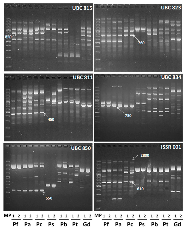
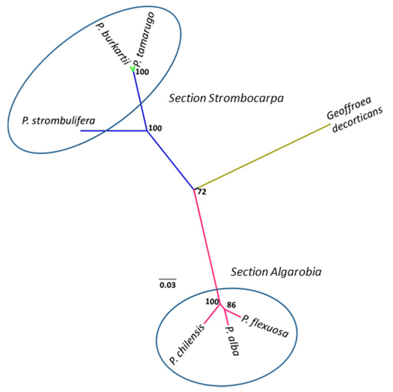
To identify the possible P. burkartii parental species, 18 ISSR markers were evaluated, of which 12 markers produced defined and robust polymorphic fragments, ranging from 220 to 2800 bp. A total of 235 fragments was produced, of which 232 were polymorphic. A comparative cluster analysis between species in the Algarobia section and species in the Strombocarpa section was carried out, using the 12 ISSR markers described before. In Figure 1, we show electrophoresis patterns of six ISSR markers that produced fragments exclusively for species of the Algarobia section (850 bp with the marker UBC815, 760 bp with UBC823, 450 bp with UBC811, 750 bp with UBC834, 550 bp with UBC850 and 610 bp and 2800 bp with ISSR001), which were absent for species of the Strombocarpa section. The cluster analysis (Figure 2) showed three main groups with good bootstrap support (>72), presenting high consistency and support of the clusters. One group was made up by the species P. strombulifera, P. burkartii and P. tamarugo from the Strombocarpa section, another group was made up of the species P. chilensis, P. alba and P. flexuosa from the Algarobia section and a third group was made up of the species Geoffroea decorticans Burkart (outgroup). High reproducibility was observed in most of the ISSR-PCR patterns (84 in total), shown by P. strombulifera and P. burkartii, except in two of them (UBC811 and UBC850).
Figure 1.
Electrophoresis patterns of six inter-simple sequence repeat (ISSR) markers obtained from DNA samples of P. flexuosa (Pf), P. alba (Pa), P. chilensis (Pc), P. strombulifera (Ps1), P. burkartii (Pb3), P. tamarugo (Pt6) and G. decorticans (Gd). Each species has two PCR profile repetitions to check its reproducibility. MP is a marker with a molecular weight between 100 and 3000 bp.
Figure 2.
Group analysis of the species in the Strombocarpa section (P. tamarugo, P. burkartii and P. strombulifera) and the Algarobia section (P. flexuosa, P. chilensis and P. alba), including one outgroup species, G. decorticans, based on 12 ISSR markers. The tree was constructed using the UPGMA (unweighted pair group method with arithmetic average) grouping method, and includes the bootstrap values.
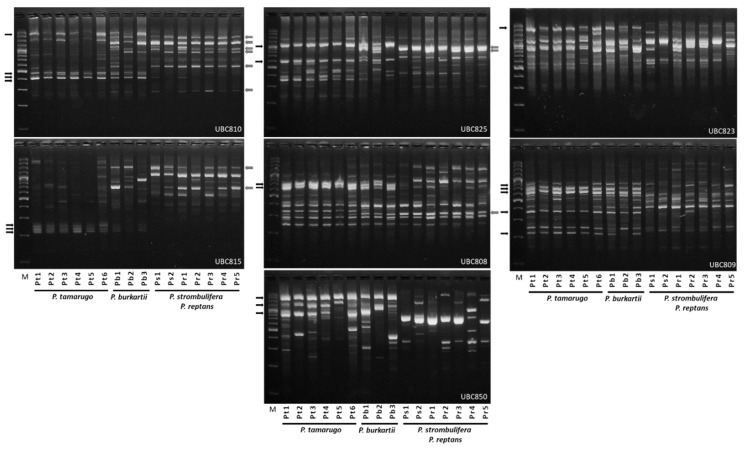
In Table 1, we present the information obtained from the seven ISSR primers belonging to four species in the Strombocarpa section, where we included Prosopis reptans Benth. var. chilensis Zoellner as a control species. ISSRs produced a total of 78 bands, of which 70 were polymorphic (89% polymorphism). Primers UBC810, UBC815 and UBC850 were 100% polymorphic, while primer UBC823 was 63% polymorphic. Primer UBC825 had the highest number of polymorphic bands (14 bands) and primer UBC823 the lowest number of polymorphic bands (five bands). The highest Rp value was observed with the UBC825 primer (9.33) and the lowest value with the UBC823 primer (3.33), while the highest PIC value was observed with the UBC815 primer (0.46) and the lowest value with the UBC823 primer (0.26) (Table 1). Figure 3 shows the electrophoresis patterns of four species in the Strombocarpa section, obtained with seven ISSR markers; next to the figure, indicated with arrows, the PCR bands which P. burkartii shares with P. tamarugo (20 bands) or P. strombulifera/P. reptans (11 bands) are shown.
Table 1.
ISSR primer information using P. tamarugo, P. burkartii, P. strombulifera and P. reptans species. Total number of bands (TNB), number of polymorphic bands (NPB) at 99%, percentage of polymorphic bands (P%) at 99%, number of different genotypes (NG), resolving power (Rp), number of exclusive bands (NEB) and polymorphic information content (PIC).
| Primer | TNB | NPB (99%) | P% (99%) | NG | Rp | NEB | PIC |
|---|---|---|---|---|---|---|---|
| UBC810 | 12 | 12 | 100 | 11 | 8.8571 | 0 | 0.4596 |
| UBC825 | 15 | 14 | 93 | 9 | 9.3333 | 1 | 0.3888 |
| UBC815 | 10 | 10 | 100 | 10 | 8.0000 | 0 | 0.4680 |
| UBC808 | 13 | 10 | 77 | 6 | 7.1429 | 0 | 0.3279 |
| UBC809 | 13 | 12 | 92 | 9 | 8.3810 | 0 | 0.3677 |
| UBC850 | 7 | 7 | 100 | 7 | 4.2500 | 0 | 0.4107 |
| UBC823 | 8 | 5 | 63 | 10 | 3.3333 | 0 | 0.2642 |
| TOTAL | 78 | 70 | 1 | ||||
| Average | 11 | 89 | 9 | 7.0425 | 0.1 | 0.3838 |
Figure 3.
Example of electrophoresis patterns obtained from seven ISSR markers and DNA of six individuals of P. tamarugo (Pt1, Pt2, Pt3, Pt4, Pt5 and Pt6), three of P. burkartii (Pb1, Pb2 and Pb3), two of P. strombulifera (Ps1 and Ps2) and five of P. reptans (Pr1, Pr2, Pr3, Pr4 and Pr5). The arrows (at the side of the image) indicate P. tamarugo (black) and P. strombulifera (grey) PCR fragments, which are shared with at least one individual of P. burkartii. M: is a marker with a molecular weight between 100 and 3000 bp.
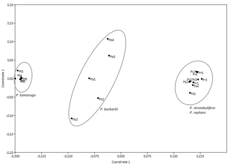
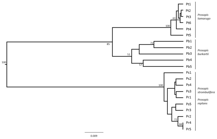
The multidimensional scaling (MDS) of the species from the Strombocarpa section clearly showed the separation between the P. tamarugo cluster and the P. strombulifera/P. reptans cluster, and P. burkartii individuals were found at an intermediate (hybrid) position (Figure 4). A UPGMA (unweighted pair group method with arithmetic average) clustering based on the ISSR band pattern showed three similar main clusters with high bootstrap support (>85%). The P. burkartii cluster was nested next to the P. tamarugo cluster, with good bootstrap support (85%) (Figure 5). A Bayesian analysis using STRUCTURE software, based on the highest probability K = 2 (Figure 6a), identified two main clusters, one with P. tamarugo individuals and another one with P. strombulifera/P. reptans. In addition, a cluster with hybrid genetics, containing P. burkartii individuals, was observed (Figure 6b). Furthermore, all the results (Figure 4, Figure 5 and Figure 6) clearly show that P. strombulifera and P. reptans are related and show a similar genetic structure.
Figure 4.
Multidimensional scaling (MDS) that clusters the 21 individuals, using seven ISSR markers.
Figure 5.
UPGMA dendrogram showing the relationships of individuals of P. tamarugo, P. burkartii, P. strombulifera and P. reptans. Bootstrap values greater than 50 percent are indicated.
Figure 6.
Relationship between number of clusters (K) and corresponding values of Delta K. (A). Genetic structure of P. tamarugo, P. burkartii, P. strombulifera and P. reptans populations, calculated with the program STRUCTURE. (B). The proportion of colors in each bar indicates the probability assigned to each individual.
2.2. Characterization of the trnL Intron
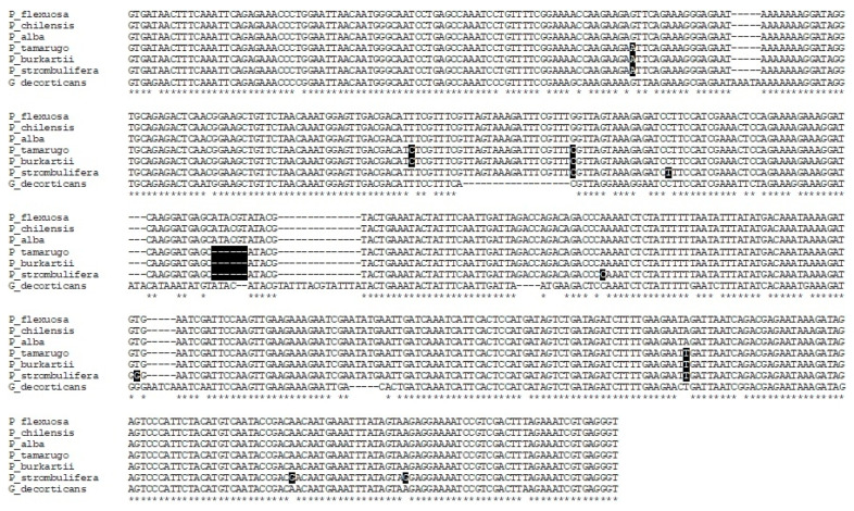
The sequencing of the trnL intron resulted in a fragment of 535 bp for the species of the Algarobia section, and a fragment of 529 bp for the species of the Strombocarpa section. In Figure 7, the alignment of the species sequences of the two sections is shown (it also includes the control species, G. decorticans as an outgroup), and three different haplotypes were found. One haplotype corresponded with the species of the Algarobia section (P. flexuosa, P. alba and P. chilensis), another with P. tamarugo and P. burkartii and a third with P. strombulifera. The species P. tamarugo and P. burkartii presented four SNPs of difference with the species of the Algarobia section, while P. strombulifera presented six SNPs of difference with the species P. tamarugo and P. burkartii and eight SNPs with the species of the Algarobia section (Figure 7). Six indels and three SNPs of difference were observed between the species of the Strombocarpa section and the species of the Algarobia section (Figure 7). Geoffroea decorticans had 44 SNPs of difference with the species of the genus Prosopis.
Figure 7.
Sequence alignment of the intron trnL (UAA) of the species P. flexuosa, P. chilensis, P. alba, P. tamarugo, P. burkartii, P. strombulifera and G. decorticans (outgroup). The polymorphisms (SNPs and Indels) observed among Prosopis species were shaded in black. Position 168 shows the SNP C/T.
2.3. Cluster Analysis Using trnL Intron
The cladograms constructed from the MP (Figure 8a) and ML (Figure 8b) methods, including the outgroup species G. decorticans, showed four main clusters. One cluster was formed by P. chilensis, P. alba and P. flexuosa, from the Algarobia section; another cluster was formed by P. burkartii and P. tamarugo from the Strombocarpa section (one species from the Strombocarpae series and the other from the Cavenicarpae series); another cluster was formed only by P. strombulifera from the Strombocarpa section; and the last cluster was formed by G. decorticans. All the clusters had high bootstrap values (>74), and showed high consistency of the clades.
Figure 8.
Cluster analysis of the trnL intron sequence data for the species P. flexuosa, P. chilensis, P. alba, P. tamarugo, P. burkartii, P. strombulifera and G. decorticans (outgroup). The trees were constructed with the maximum parsimony (a) and maximum likelihood (b) clustering methods; their corresponding bootstrap values are included in the nodes. The magnitude of the distance of the species G. decorticans was shortened to show the two clusters together.
2.4. Parent-Diagnostic cpDNA Markers
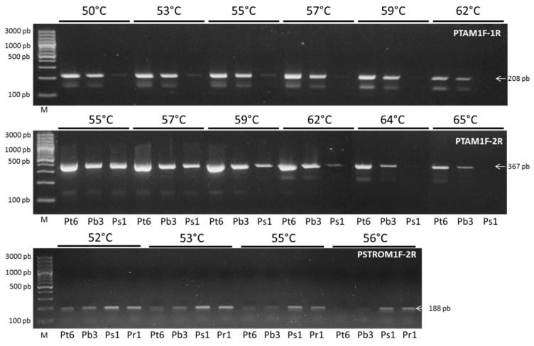
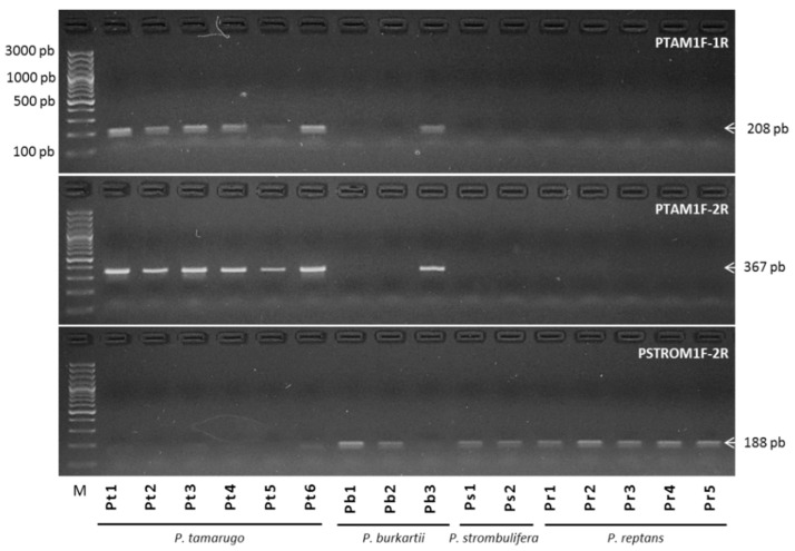
We used the trnL sequence information for a diagnostic, species-specific PCR method: we designed specific primers using SNPs of cpDNA haplotypes, considering specifically position 168 where the C/T SNP is located (see Figure 7), in order to evaluate the maternal load and optimize the Ta of the primers for the detection of the corresponding haplotype. Our results showed that the primer pairs PTAM1F-1R (at 62 °C) and PTAM1F-2R (at 65 °C), detected only cpDNA of P. tamarugo (Pt6) and P. burkartii, (Pb3) (Figure 9), but sequences of P. strombulifera (Ps1) were not amplified. Alternatively, a pair of PSTROM1F-2R primers was designed to amplify only P. strombulifera cpDNA. Our results clearly showed that at Ta of 56 °C, there was no amplification of P. tamarugo (Pt6) and P. burkartii (Pb3) DNA. However, P. strombulifera (Ps1) and P. reptans (Pr1) DNA was amplified (Figure 9). This procedure was then used on all the samples, with special focus on the amplification of the P. burkartii individuals. The results (Figure 10) showed amplification in all samples of P. tamarugo (Pt1 to Pt6) with the primer pairs PTAM1F-1R and PTAM1F-2R, while none of the samples of P. strombulifera (Ps1, Ps2) and P. reptans (Pr1–Pr5) showed amplification. It should be noted that DNA in the samples Ps3, Ps4 and Ps5 was not amplified either, but those samples were not included in the figure due to the reduced space on the gel. Surprisingly, DNA in some of the P. burkartii samples was amplified and produced the expected fragment (Pb3 and Pb5), while DNA in the others did not (Pb1, Pb2 and Pb4), as shown in Figure 10. We performed the same procedure with the PSTROM1F-2R primers, in order to confirm the results obtained by the PTAM1F-1R and PTAM1F-2R markers. Indeed, in all P. strombulifera and P. reptans samples, the expected fragment size was amplified, while in P. tamarugo samples, no fragments were amplified. Therefore, we confirmed the differences in the five P. burkartii samples with these primers. We concluded that P. burkartii individuals, Pb3 and Pb5, have P. tamarugo as their maternal parent, whereas P. burkartii individuals, Pb1, Pb2 and Pb4, have P. strombulifera as their maternal parent.
Figure 9.
Optimized PCR with specific designed primers for the trnL sequence of P. tamarugo, P. burkartii and P. strombulifera The annealing temperature (Ta) was optimized with DNA samples of Pt6, Pb3, Ps1 and Pr1, by means of a gradient between 50 °C to 65 °C, using the primer pairs PTAM1F-1R, PTAM1F-2R and PSTROM1F-2R. M: is a marker with a molecular weight between 100 to 3000 bp.
Figure 10.
PCR obtained from DNA of six individuals of P. tamarugo, three of P. burkartii, two of P. strombulifera and five of P. reptans, using specified PTAM1F-1R, PTAM1F-2R and PSTROM1F-2R primer pairs at an annealing temperature of 62 °C, 65 °C and 56.5 °C, respectively.
2.5. Species Diagnostic by SSR-PCR Markers
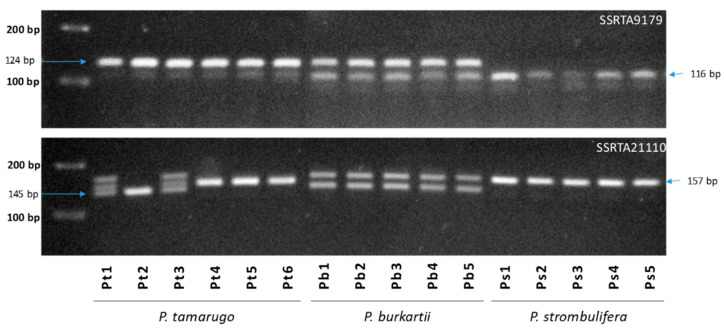
According to Figure 11, two nuclear SSR markers (SSRTA9179, SSRTA21110) showed different bands for the P. tamarugo, P. burkartii and P. strombulifera species. The SSRTA9179 marker showed a band, with a fragment of 124 bp in all P. tamarugo individuals, and a band with a fragment of 116 bp in all P. strombulifera individuals, while all P. burkartii individuals showed both fragments. Moreover, the SSRTA21110 marker showed a band with a fragment of 157 bp in all P. strombulifera individuals, and a band with a fragment of 145 bp in three P. tamarugo individuals (Pt1–Pt3), while all P. burkartii individuals could be seen in both fragments.
Figure 11.
Banding patterns of SSR-PCR of markers SSRTA9179, SSRTA21110 and SSRTA10814 on an agarose gel. The lanes containing six individuals of P. tamarugo (Pt1–Pt6), five individuals of P. burkartii (Pb1–Pb5) and five individuals of P. strombulifera (Ps1–Ps5).
3. Discussion
Although there was evidence that P. burkartii could have a hybrid origin involving P. tamarugo and P. strombulifera, based on morphological descriptions and electrophoresis studies of seed proteins [2,7,8], there is no genetic study to corroborate the hybridization and possible parent species thus far. Our results from the ISSR markers demonstrated a high support for the separation of two large clusters, the Algarobia section and the Strombocarpa section. Previous work has already shown that the species that make up each section are highly differentiated [26,27]. Similarly, the cluster analyses showed that the species of the Strombocarpa section are clearly different from the Algarobia section [28]. The electrophoresis patterns of the ISSR primers used in our study revealed the diagnostic markers of the species of the Strombocarpa and Algarobia section. A study carried out with RAPD primers found specific markers for species of the two sections as well [29]. Between these studies and our results, there is sufficient evidence that it would be unlikely to find ancestors of P. burkartii within the species from the Algarobia section. Therefore, for upcoming analyses, only species from the Strombocarpa section could be used to find potential parental candidates. It should also be noted that between species of the Algarobia section and species of the Strombocarpa section no hybrid formation has been reported [26].
The ISSR markers showed a high number of polymorphic bands when looking at the (21) individuals from the Strombocarpa section (P. tamarugo, P. burkartii, P. strombulifera and P. reptans), and specific primers (UBC810, UBC825, UBC815 and UBC850) showed a high content of polymorphic information. Of the various genetic tools used to study hybridization in plants, the ISSR marker is one of the simplest molecular methods that can be used for comparative analysis of possible hybrids and parental species [10,30,31,32]. The electrophoresis patterns of seven ISSR primers showed up to 19 bands shared between P. burkartii and the species P. tamarugo, P. strombulifera and P. reptans. However, more bands were shared with P. tamarugo than with the other species. The electrophoretic profile of seed proteins identified a total of 31 bands in P. strombulifera, and 33 in P. tamarugo, while the profile of P. burkartii presented 40 bands in total, of which 20 were shared by all three species [33]. In addition, that study indicates that most of the seed proteins of P. burkartii showed a similar pattern to that of P. tamarugo, which agrees with our results, as we found the largest counts of shared ISSR bands with P. tamarugo.
A UPGMA cluster analysis showed that the P. burkartii cluster is nested with the P. tamarugo cluster and an MDS multivariate analysis places it in an intermediate position between P. tamarugo and P. strombulifera/P. reptans. The genetic affinity between P. burkartii and P. tamarugo is consistent with the clustering analyses observed in other studies, where a high number of shared protein bands (obtained by electrophoretic analysis) between these species was observed [8]. According to the MDS and STRUCTURE analyses, signs of introgression of genetic material in P. burkartii are evident, confirming its hybrid status (P. tamarugo × P. strombulifera). However, according to the same analyses there might be differences in introgression levels between the analyzed samples. Additionally, our results showed, through MDS, UPGMA clustering and STRUCTURE analysis, that P. reptans and P. strombulifera could be considered to be one single species. This is corroborated by several studies that previously proposed that P. reptans and P. strombulifera are the same species, and could be considered subspecies or variants. This was validated by morphological classification [2], enzymatic studies [34], genetic studies [8] and morphological-biochemical studies [28]. However, P. reptans has not been found in the Pampa del Tamarugal, while P. burkartii is an endemic species inhabiting a restricted area, located mainly in the Pampa del Tamarugal, in the Atacama Desert (Tarapacá Region). It is distributed over approximately 1395 km2 [6], and only 50 specimens have been found [35], occupying the same ecological niche as P. tamarugo and P. strombulifera [5]. From the samples collected during this study (within the Pampa del Tamarugal) a high association was observed between P. burkartii and P. tamarugo, growing physically close to each other. This could help the processes of introgression, as fertilization of P burkartii flowers by P. tamarugo pollen can easily take place [33]. Introgression with P. strombulifera genetic material was unlikely, as very few individuals were observed in the sampling areas.. According to Burghardt [8], polypeptide analyses suggest that the placement of P. burkartii in the Strombocarpae series needs to be revised. Due to the affinity of bands between P. burkartii and P. tamarugo, they suggested that P. burkartii would belong to the Cavenicarpae series and not to the Strombocarpae series [2]. This is corroborated by the amplificated fragments, obtained by the ISSR markers, in our study.
Interspecific hybridization occurs commonly and naturally between species in the Algarobia section that occupy the same geographical area [26]. However, the ability to hybridize is not present in all species of this section [24]. Thus far, there are no other records of species hybridization in the Strombocarpa section, except for P. burkartii. The results we obtained with the ISSR and SSR markers confirm the natural hybridization of P. burkartii. However, the various molecular tools available, such as fluorescence in situ hybridization (FISH) and genomic in situ hybridization (GISH) [36,37], could contribute to verify gene or chromosome segment introgression.
The ability of the cpDNA barcode to identify parental species of hybrids is well documented; for example, the maternal parent of Rhizophora L. hybrids was revealed with data from mangrove chloroplast regions [38]. Additionally, in species of the genus Spondias L. matK, rbcL and trnH-psbA markers were used [39]. Moreover, in interspecific hybrids of Annona L. species, rbcL and matK markers exposed the maternal species [40]. In addition, “diagnostic” DNA markers are commonly used in species validation studies [41]. In our case, the intron trnL was able to amplify six species of both Strombocarpa and Algarobia section, and showed important polymorphic differences. While it is true that analyses can be strengthened by increasing the quantity of genes and/or chloroplast or nuclear spacers, this study also tested matK [42], rbcL [43] and ITS [44] markers, which did not amplify (or showed weak amplification and/or multiple amplifications) in P. tamarugo, P. burkartii and P. strombulifera.
Even though P. burkartii and P. tamarugo species belong to the Strombocarpa section, our cluster analyses show them to be stronger related to P. alba, P. chilensis and P. flexuosa (Algarobia section) and separates them from P. strombulifera, the type species of the Strombocarpa section. This result is contradictive to what is currently established; the species of the Strombocarpa section are separated from the Algarobia section [24,26,27,28]. Due to the fact that information of one region only (trnL) might not able to separate the Strombocarpa and Algarobia sections, we recommend to gain more information with other regions and markers. As indicated before, we were unable to obtain more sequences with the universal markers for chloroplast (matK, rbcL) and nuclear (ITS) region for the species from the Strombocarpa section. However, Catalano et al. [24] observed the separation of the section Strombocarpa and Alagarobia, based on data gained with three markers (trnS-psbC, G3pdh and NIA).
When sequencing the trnL intron of P. burkartii, and comparing the sequence alignment with the rest of the Algarobia and Strombocarpa species, we observed that P. burkartii and P. tamarugo had the same haplotype. Therefore, we could believe that P. burkartii has P. tamarugo as their maternal parent. However, these results were obtained from only one individual, and we decided to extend the analysis to the other samples. For this, we used a simple and less expensive method without the need for DNA sequencing: identifying cpDNA from SNP. SNPs are a variation of the DNA sequence that affect only one base (A, G, T and C) of the genome, and have become important genetic markers for animal or plant species [45]. They have been used for genotyping plant material, e.g., through the Allele-Specific PCR method. This technique is based on the design of a primer that extends to the 3′ end where the SNP is located, detecting the nucleotide that corresponds to the specific SNP site [46,47]. In the sequences of the intron trnL obtained from the Algarobia and Strombocarpa section, we found a (C/T) SNP in position 168, which differentiates P. burkartii and P. tamarugo from the rest of the species (Figure 8). Using this SNP and specific primers (PTAM1F-1R, PTAM1F-2R and PSTROM1F-2R), we could check for the maternal parent of P. burkartii in the other collected samples. In all samples, collected (21) from the Strombocarpa section, the primer pairs (PTAM1F-1R, PTAM1F-2R) were able to detect the cpDNA haplotype of P. tamarugo, while the primer pair (PSTROM1F-2R) was able to identify the cpDNA haplotype of P. strombulifera and P. reptans. We confirmed that some individuals have P. tamarugo (Pb3 and Pb5) and others P. strombulifera as maternal parent (Pb1, Pb2 and Pb4), by using the combination of those primers on five samples of P. burkartii. It is believed that cpDNA is predominantly inherited by the mother in higher plants, as 80% of angiosperm genera show a strictly maternal cpDNA inheritance [48]. However, there are cases in which it can also be transmitted in a biparental or paternal manner [49]. In our study, double amplification has not been observed in the P. burkartii samples, both with PTAM and PSTROM primers. Additional molecular techniques and sequencing more individuals of P. burkartii and their parents are recommended to confirm our findings, and provide data of interspecific hybridization and the level of introgression. We believe that these results from the SNPs and the bands shown by the SSRs markers in our work confirm the hybrid status of P. burkartii.
4. Materials and Methods
4.1. Plant Material
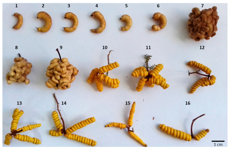
Fresh leaves of six species from the genus Prosopis were collected between April 2018 and March 2019; three species from the Algarobia section and three species from the Strombocarpa section. The species P. chilensis was collected by the Forest Institute of Chile (INFOR) in the Metropolitan Region, while CRIDESAT collected species of P. tamarugo, P. strombulifera and P. burkartii in the Pampa del Tamarugal, and P. alba and P. flexuosa in the Copiapó Valley. In order to compare with another species in the Strombocarpa section, we include a seventh species, P. reptans, which has only been found in the Atacama Region [50], and was collected at the same location as first found by Zöllner and Olivares [50] (Km 837 of the Pan-American Highway 5 north). Additionally, a genotype of P. burkartii from San Pedro de Atacama (Antofagasta Region, collected by CONAF) was used, which was grown from seed to seedling stage. According to CONAF, there are records of four adult individuals of P. burkartii in San Pedro de Atacama. The species of Geoffroea decorticans (Gd; 27°20′39.3 “S 70°21′46.0 “W), belonging to the same family as Prosopis (Fabaceae or Leguminosae), was included as an external (out-) group. Taxonomic identification of the species was done using keys and descriptions [2,51]. The fruits of the sampled species of the Strombocarpa section are shown in Figure 12, and Table 2 gives an overview of the collected samples, their geographical location and the registration number (Index Herbarium Code) given by the EIF herbarium of the Department of Forestry and Nature Conservation (Faculty of Forestry Sciences, University of Chile).
Figure 12.
Fruits of Prosopis tamarugo (Pt1 (1), Pt2 (2), Pt3 (3), Pt4 (4), Pt5 (5), Pt6 (6)), P. burkartii (Pb1 (7), Pb2 (8), Pb3 (9)), P. strombulifera (Ps1 (10), Ps2 (11)) and P. reptans (Pr1 (12), Pr2 (13), Pr3 (14), Pr4 (15), Pr5 (16)).
Table 2.
Species, geographical coordinates and herbarium where the samples were deposited.
| Species | Name | Province | Latitud (S) | Longitud (W) | Herbarium 1 |
|---|---|---|---|---|---|
| P. tamarugo | Pt1 | El Tamarugal | 20°19′46.1″ | 69°42′27.2″ | EIF 13338 |
| P. tamarugo | Pt2 | El Tamarugal | 20°20′37.1″ | 69°39′53.2″ | EIF 13337 |
| P. tamarugo | Pt3 | El Tamarugal | 20°20′57.5″ | 69°39′51.1″ | EIF 13336 |
| P. tamarugo | Pt4 | El Tamarugal | 20°21′03.6″ | 69°39′48.1″ | EIF 13335 |
| P. tamarugo | Pt5 | El Tamarugal | 20°21′03.6″ | 69°39′47.9″ | EIF 13334 |
| P. tamarugo | Pt6 | El Tamarugal | 20°21′22.5″ | 69°39′12.9″ | EIF 13333 |
| P. burkartii | Pb1 | El Tamarugal | 20°23′11.5″ | 69°35′57.3″ | EIF 13344 |
| P. burkartii | Pb2 | El Tamarugal | 20°27′59.2″ | 69°33′23.2″ | EIF 13347 |
| P. burkartii | Pb3 | El Loa | 22°59′3.29″ | 68°9′19.23″ | EIF 13824 |
| P. burkartii | Pb4 | El Tamarugal | 20°28′00.1″ | 69°33′23.2″ | EIF 13348 |
| P. burkartii | Pb5 | El Tamarugal | 20°24′45.0″ | 69°41′29.7″ | EIF 13355 |
| P. strombulifera | Ps1 | El Tamarugal | 20°27′59.8″ | 69°33′23.5″ | EIF 13332 |
| P. strombulifera | Ps2 | El Tamarugal | 20°28′00.1″ | 69°33′23.3″ | EIF 13351 |
| P. strombulifera | Ps3 | El Tamarugal | 20°27′59.9″ | 69°33′23.5″ | EIF 13350 |
| P. strombulifera | Ps4 | El Tamarugal | 20°28′00.2″“ | 69°33′23.3″ | EIF 13352 |
| P. strombulifera | Ps5 | El Tamarugal | 20°30′09.7″“ | 69°22′54.1″ | EIF 13822 |
| P. reptans | Pr1 | Copiapó | 27°20′10.1″ | 70°35′42.8″ | EIF 13324 |
| P. reptans | Pr2 | Copiapó | 27°20′10.9″ | 70°35′42.7″ | EIF 13331 |
| P. reptans | Pr3 | Copiapó | 27°20′10.7″ | 70°35′42.6″ | EIF 13325 |
| P. reptans | Pr4 | Copiapó | 27°20′06.3″ | 70°35′45.1″ | ------------- |
| P. reptans | Pr5 | Copiapó | 27°20′08.6″ | 70°35′42.8″ | EIF 13326 |
| P. flexuosa | Pf | Copiapó | 27°21′21.8″ | 70°39′54.7″ | EIF 13330 |
| P. chilensis | Pc | Chacabuco | 33°05′24.9″ | 70°39′07.4″ | EIF 13328 |
| P. alba | Pa | Copiapó | 27°21′39.3″ | 70°20′33.8″ | EIF 13329 |
1 Index Herbariorum code = EIF.
4.2. DNA Extraction
DNA was extracted from fresh leaves, using a modified CTAB (cetyl trimethylammonium bromide) method described by Contreras et al. [52]. Fine fresh leaf powder (100 mg) was obtained after grinding the tissue with liquid nitrogen. The powder was placed in 2 mL tubes and mixed with 700 µL of extraction buffer [Tris-HCl 100 mM, pH 8.0; NaCl 1.4 M; EDTA 20 mM; 2% (w/v) CTAB], supplemented with 1% of β-mercaptoethanol, 14 µL of 5% Sarkosyl, 0.045 g of D-sorbitol (MW 182.17 g/mol), 2% PVP-40 and 4 µL proteinase K (10 mg/mL), and preheated to 65 °C. The samples were vigorously shaken for 10 s and incubated for 1 h at 65 °C, this step was repeated three times to improve the mixture. The water phase was recovered by centrifugation at 14,000 rpm for 10 min, and then mixed with an equal volume of chloroform-isoamyl alcohol (24:1). The mixture was gently swirled for 2 min and centrifuged at 14,000 rpm for 10 min. The upper phase was transferred to a new tube and treated with 5 µL RNase A (100 µg/mL) and incubated at 37 °C for 30 min. The extraction was mixed with two thirds volume of isopropanol at −20 °C and gently swirled 30 times. A volume of 600 µL of the mixture was transferred to a HiBind DNA® minicolumn with a collecting tube (Omega Bio-Tek) and incubated for 2 min at room temperature. The minicolumns were centrifuged for 1 min at 10,000 rpm and the purified liquid was discarded. A precipitation solution of 600 µL 70% ethanol and ammonium acetate (10 mM) was added to the minicolumns and centrifuged again for 1 min at 10,000 rpm. The liquid was discarded and the precipitation step was repeated. To ensure that all the ethanol was removed, the sample was centrifuged for 2 min at 14,000 rpm, and the collection tube was replaced with a new 1.5 mL tube. To elute the DNA, 40 µL of Tris-EDTA buffer (TE) was added to the minicolumns, which were then incubated at 37 °C for 20 min, followed by a 2-min centrifugation at 14,000 rpm; the DNA eluate was then stored at −20 °C for further analysis. The quality and concentration of the total DNA was checked with a Colibri micro volume spectrophotometer (Titertek Berthold, Germany) at 260, 280 and 230 nm; in addition, the integrity of the genomic DNA was checked on a 0.7% agarose gel.
4.3. ISSR Amplification
We used 18 UBC (University of British Columbia) markers for the ISSR amplification: UBC802, UBC807, UBC808, UBC809, UBC810, UBC811, UBC813, UBC815, UBC816, UBC818, UBC821, UBC823, UBC825, UBC834, UBC850, UBC880, UBC856 and ISSR001 [53,54], of which 12 were selected that provided higher numbers of fragments and better reproducibility. The 20 µL PCR solution consisted of: 10 µL Master Mix SapphireAmp 2× (Takara, Clontech, USA), 5 µL ISSR primer (5 µM), 1 µL genomic DNA (5 ng/µL) and 4 µL nuclease-free water. Amplifications were performed in a MultiGene Optimax thermal cycler (Labnet) using the following protocol: an initial step of 5 min at 94 °C, 45 cycles of 30 s at 94 °C, 45 s at 52 °C and 2 min at 72 °C, followed by a final extension step of 6 min at 72 °C. The amplification products were separated by agarose gel electrophoresis (1.2%) and colored with GelRedTM (10,000×) at the ratio of 10 µL/100 mL of gel (with Tris-borate-EDTA at 0.5×). The electrophoresis run was performed at 100 V for 110 min. The gel was then placed in a UV trans illuminator (Vilber Lourmat, France) and photographed with a digital camera (Canon, SX160 IS. USA).
4.4. TrnL Amplification and Sequencing
For the amplification of the trnL intron (UAA) the primers forward A49325 5′-CGAAATCGGTAGACGCTACG-3′ and reverse B49863 5′-GGGATAGGGACTTGAAC-3′ described by Taberlet et al. [21] were used. The 24 µL PCR solution was prepared as follows: 12 µL Master Mix SapphireAmp 2× (Takara, Clontech), 1.5 µL forward (5 µM), 1.5 µL reverse (5 µM), 6 µL genomic DNA (5 ng/µL) and 3 µL nuclease-free water. Amplification was performed in a MultiGene Optimax thermal cycler (Labnet) using the following conditions: an initial step of 5 min at 95 °C, 35 cycles of 45 s at 95 °C, 45 s at 55 °C and 2 min at 72 °C, followed by a final step of extension of 6 min at 72 °C. The amplified products were separated and visualized as described in the “ISSR Amplification” section. The PCR amplicons were cut from the gel and purified with the Wizard® SV Gel and PCR Clean-Up System (Promega) kit, according to the manufacturer’s recommendations. The purified PCR product was then shipped to Macrogen Inc. for sequencing using ABI3730XL equipment. (Seoul, South Korea).
4.5. ISSR Analysis
In each individual (21), the ISSR-PCR fragments were considered as an independent locus, which were analyzed as presence (1) or absence (0) of bands. We obtained the total number of ISSR bands (TNB), percentage of polymorphic bands (P%) at 99%, the number of different genotypes (NG), resolving power (Rp) and the number of private bands (NPB). The resolving power (Rp) of a primer was calculated as Rp = Σ Ib, where Ib (band information) takes the value of 1 − [2 × (0.5 − p)], with p being the frequency of the lines the band contains [55]. The polymorphic information content (PIC) was estimated as PIC = 2p (1 − p) [56].
The similarity index was calculated from presence-absence matrix of the bands. This matrix was used for the construction of a dendrogram, applying unweighted pair group method with arithmetic average (UPGMA). The bootstrap values with 1000 repetitions were obtained with the Winboot program [57] to test the robustness of the groups. The dendrograms were built with Phylip 3.6 software [58] and edited using FigTree 1.4.0 [59]. Additionally, a multivariate analysis by Multidimensional Scaling (MDS) was performed using the PAST program [60].
The genetic structure was determined by a Bayesian analysis, using STRUCTURE v.2.3 software [61]. The optimal number of subpopulations (K) was identified after five independent runs for each K value oscillating between 1 to 5, with a burn-in period of 10,000 replicates followed by 20,000 replicates of Markov Monte Carlo Chains (CMMC). We examined the delta K criteria (ΔK) to find the optimal K values [62], which were entered into the Structure Harvester program [63].
4.6. Barcode Analysis
The forward and reverse sequences were edited with the software Chromas Pro v1 (Technelysium Pty, Ltd.). The DNA sequences of both ends were assembled, using the DNA Baser Sequence Assembler v4.10 software (Biosoft 2012). Sequence editing was done, using the automatic analyses for editing “contig” with default program parameters. To characterize the sequences and observe differences in SNPs (Single Nucleotide Polymorphisms) and indels, multiple sequence alignments were performed using MEGA 6 software [64] and the ClustalW program [65]. The sequences were deposited in Genbank with the following codes: P. flexuosa (MK450531), P. chilensis (MK450532), P. alba (MK450533), P. tamarugo (MK450534), P. burkartii (MK450535), P. strombulifera (MK450536) and G. decorticans (MK450537). Cluster analyses were performed using the MEGA 6 program in order to illustrate the differences between species and clusters. Trees were generated using maximum parsimony (MP) and maximum likelihood (ML) Bayesian methods. All cluster analyses were performed with a bootstrap support of 1000 replicates.
4.7. Specific Primer Design and PCR Optimization
Specific primers were designed, using the polymorphic information of trnL sequences obtained from P. tamarugo (MK450534), P. burkartii (MK450535) and P. strombulifera (MK450536). The sequences were aligned with MEGA 6 and “Clustal W” software. Using the Oligo® software, primer pairs were designed with a mismatch in the 3′ SNPs of P. tamarugo and P. strombulifera, in order to identify the chloroplast DNA (cpDNA) of the specific individuals. The primer pairs to identify chloroplast DNA of P. tamarugo species were PTAM-1F (5′-AAATGGAGTTGACGACATC-3′)/PTAM-1R (5′-TCAACTTGGAATCGATTCA-3′) with a 208 bp product and PTAM-1F/PTAM-2R (5′-GTCGACGATTTCCTCTT-3′) with a 367 bp product. The primer pair for identifying cpDNA of P. strombulifera species was PSTROM-1F (5′-ATGGAGTTGACGACATT-3′)/PSTROM-2R (5′-CCATCTTTTATTGTCATATA-3′) with a product of 188 bp. The PCR solution contained: 8 µL Master Mix SapphireAmp 2× (Takara, Clontech), 0.8 µL forward (5 µM), 0.8 µL reverse (5 µM), 3.2 µL genomic DNA (5 ng/µL) and 3.2 µL nuclease-free water. Amplification was performed in a MultiGene Optimax thermal cycler (Labnet) with the following protocol: an initial step of 4 min at 94 °C, 30 cycles of 45 s at 94 °C, 45 s at annealing temperature (Ta, see below) and 1 min at 72 °C, followed by a final extension step of 5 min at 72 °C. The amplification products were separated and visualized as described in the “ISSR Amplification” section. To determine the optimal Ta, a PCR gradient ranging from 50 °C to 65 °C was used with DNA from Pt6, Pb3, Ps1 and Pr1 samples. With the specific primer pairs already optimized, PCRs were then performed on the rest of the samples from the Strombocarpa section.
4.8. Fingerprinting by SSR–PCR Reaction
Two SSR markers develop from P. Tamarugo described by Contreras et al. [66] were selected. The SSR primer pairs SSRTA9179 (F: TGAATTGTATGGAAATACGACTCTG and R: TCATTGGCCCTTGTAGTTGA; motif TTTC; accession MT136897) and SSRTA21110 (F: TGGTTGGCTCAAAAGTGAAA and R: TGTGAGAAGCAAGTCCTCGTT; motif TG; accession MT136909) were used. PCRs were carried out in a total volume of 16 µL that contained 8 µL of SapphireAmp Fast PCR 2× Master Mix (Takara-Clontech, USA), 3.2 µL of genomic DNA (5 ng/µL), 0.8 µL of each primer (forward and reverse, at a 5 µM concentration), and 3.2 µL of nuclease-free water. PCR amplification was conducted in a Labnet MultiGene OptiMax Thermal Cycler under the following conditions: DNA was denatured at 94 °C for 3 min, followed by 45 cycles of 98 °C for 5 s, Ta of 59 °C for 5 s, 72 °C for 40 s and a final extension at 72 °C for 4 min. The PCR products were analyzed by electrophoresis on 2% agarose gels stained with GelRed DNA stain (10,000×, Biotium). The band sizes were approximated based on 100 bp DNA ladder (Thermo Fisher, Waltham, MA, USA)
5. Conclusions
In conclusion, we could confirm the natural hybrid status of P. burkartii, from the MDS, UPGMA and STRUCTURE analyses based on ISSR fragment data, SSR-PCR, and the results of trnL intron-specific PCR tests. We could also deduce that P. tamarugo and P. strombulifera are likely to be involved as source species. This is the first time that ISSR, SSR and trnL sequence data are provided for P. burkartii, to clarify its hybrid status and possible parental status. There is a high diversity of individuals with different maternal components, distributed in the Pampa del Tamarugal (Tarapacá Region) and in San Pedro de Atacama (Antofagasta Region). Unambiguously, this study indicates the large commitment that government institutions should provide to this species, which requires better legal protection and urgent measures for its conservation.
Acknowledgments
We thank Vincenzo Porcile and Fernanda Aguayo. We sincerely thank the Corporación Nacional Forestal (CONAF), Tarapacá Region, for the sampling authorization (N°00024/08-11-2019 (JBH/FAP/JVO)).
Author Contributions
Conceptualization, R.C. and L.v.d.B.; Methodology, R.C.; Software, R.C.; Validation, R.C. and L.v.d.B.; Formal analysis, R.C.; Investigation, R.C., M.G., B.B. and S.G.; Resources, R.C.; Data curation, R.C.; Writing—Original draft preparation, R.C., L.v.d.B., B.B. and M.G.; Writing—Review and editing, R.C., L.v.d.B., S.G. and B.B.; Supervision, R.C.; Project administration, R.C.; Funding acquisition, R.C. All authors have read and agreed to the published version of the manuscript.
Funding
This research was funded by CRIDESAT of the University of Atacama grant number DIUDA 22380 and co-funded by FIC project “ADN Vegetal de Atacama” of the Regional Government of Atacama, grant number BIP 30432984-0.
Conflicts of Interest
The authors declare no conflict of interest.
References
- 1.Barros S. El género Prosopis, valioso recurso forestal de las zonas áridas y semiáridas de América, Asia y Africa. Cienc. Invest. 2010;16:91–128. [Google Scholar]
- 2.Burkart A. A monograph of the genus Prosopis (leguminosae subfam. mimosoideae) J. Arnold Arbor. 1976;57:219–249. [Google Scholar]
- 3.Barros S., Wrann J. El Género Prosopis en Chile. Cienc. Invest. 1992;6:295–334. [Google Scholar]
- 4.Ministerio de Medio Ambiente de Chile. [(accessed on 5 May 2020)]; Available online: http://www.mma.gob.cl/clasificacionespecies/index2.htm.
- 5.Muñoz P. Una nueva especie de Prosopis para el norte de Chile. Bol. Mus. Nac. Hist. Nat. Chile. 1971;32:363–370. [Google Scholar]
- 6.Ministerio de Medio Ambiente de Chile. [(accessed on 5 May 2020)]; Available online: http://www.mma.gob.cl/clasificacionespecies/fichas8proceso/fichas_finales/Prosopis_burkartii_P08_propuesta.
- 7.Palacios R.A., Brizuela M.M., Burghardt A.D., Zallocchi E.M., Mom M.P. Prosopis burkartii and its possible hybrid origin. Bull. Int. Group Study Mimosoideae. 1991;19:146–161. [Google Scholar]
- 8.Burghardt A.D. Estudio electroforético de proteínas de semilla en Prosopis (Leguminosae) II: Sección Strombocarpa. Bol. Soc. Argent. Bot. 2000;35:149–156. [Google Scholar]
- 9.Baack E.J., Rieseberg L.H. A genomic view of introgression and hybrid speciation. Curr. Opin. Genet. Dev. 2007;17:513–518. doi: 10.1016/j.gde.2007.09.001. [DOI] [PMC free article] [PubMed] [Google Scholar]
- 10.Wolfe A.D., Xiang Q.Y., Kephart S.R. Diploid hybrid speciation in Penstemon (Scrophulariaceae) Proc. Natl. Acad. Sci. USA. 1998;95:5112–5115. doi: 10.1073/pnas.95.9.5112. [DOI] [PMC free article] [PubMed] [Google Scholar]
- 11.Nadeem M.A., Nawaz M.A., Shahid M.Q., Doğan Y., Comertpay G., Yıldız M., Hatipoğlu R., Ahmad F., Alsaleh A., Labhane N. DNA molecular markers in plant breeding: Current status and recent advancements in genomic selection and genome editing. Biotechnol. Biotechnol. Equip. 2017;32:1–25. doi: 10.1080/13102818.2017.1400401. [DOI] [Google Scholar]
- 12.Hegarty M.J., Hiscock J.S. Hybrid speciation in plants: New insights from molecular studies. New Phytol. 2004;165:411–423. doi: 10.1111/j.1469-8137.2004.01253.x. [DOI] [PubMed] [Google Scholar]
- 13.Zhang R., Gong X., Ryan F. Evidence for continual hybridization rather than hybrid speciation between Ligularia duciformis and l. paradoxa (asteraceae) PeerJ. 2017;5:3884. doi: 10.7717/peerj.3884. [DOI] [PMC free article] [PubMed] [Google Scholar]
- 14.Zietkiewicz E., Rafalski A., Labuda D. Genome fingerprinting by simple sequence repeat (SSR-Anchored) polymerase chain reaction amplification. Genomics. 1994;20:176–183. doi: 10.1006/geno.1994.1151. [DOI] [PubMed] [Google Scholar]
- 15.Mogensen H.L. The hows and whys of cytoplasmic inheritance in seed plants. Am. J. Bot. 1996;83:383–404. doi: 10.1002/j.1537-2197.1996.tb12718.x. [DOI] [Google Scholar]
- 16.McCauley D.E., Sunby A.K., Bailey M.F., Welch M.E. Inheritance of chloroplast DNA is not strictly maternal in Silene vulgaris (Caryophyllaceae): Evidence from experimental crosses and natural populations. Am. J. Bot. 2007;94:1333–1337. doi: 10.3732/ajb.94.8.1333. [DOI] [PubMed] [Google Scholar]
- 17.Hebert P.D.N., Cywinska A., Ball S.L., Dewaard J.R. Biological identifications through DNA barcodes. Proc. Biol. Sci. 2003;270:313–321. doi: 10.1098/rspb.2002.2218. [DOI] [PMC free article] [PubMed] [Google Scholar]
- 18.Pei N., Chen B., Kress W.J. Advances of community-level plant DNA barcoding in China. Front. Plant. Sci. 2017;8:225. doi: 10.3389/fpls.2017.00225. [DOI] [PMC free article] [PubMed] [Google Scholar]
- 19.Santos C., Pereira F. Identification of plant species using variable length chloroplast DNA sequences. Forensic Sci. Int. Genet. 2018;36:1–12. doi: 10.1016/j.fsigen.2018.05.009. [DOI] [PubMed] [Google Scholar]
- 20.Taberlet P., Gielly L., Pautou G., Bouvet J. Universal primers for amplification of three noncoding regions of chloroplast DNA. Plant Mol. Biol. 1991;17:1105–1109. doi: 10.1007/BF00037152. [DOI] [PubMed] [Google Scholar]
- 21.Taberlet P., Coissac E., Pompanon F., Gielly L., Miquel C., Valentini A., Vermat T., Corthier G., Brochmann C., Willerslev E. Power and limitations of the chloroplast trnL (UAA) intron for plant DNA barcoding. Nucleic Acids Res. 2006;35:e14. doi: 10.1093/nar/gkl938. [DOI] [PMC free article] [PubMed] [Google Scholar]
- 22.Quandt D., Müller K., Stech M., Frahm J.P., Frey W., Hilu K.W., Borsch T. Molecular evolution of the chloroplast trnL-F region in land plants. Monogr. Syst. Bot. Missouri Botanic Garden. 2004;98:13–37. [Google Scholar]
- 23.Bessega C., Vilardi J.C., Saidman B.O. Genetic relationships among American species of the genus Prosopis (Mimosoideae, Leguminosae) inferred from ITS sequences: Evidence for long-distance dispersal. J. Biogeogr. 2006;33:1905–1915. doi: 10.1111/j.1365-2699.2006.01561.x. [DOI] [Google Scholar]
- 24.Catalano S.A., Vilardi J.C., Tosto D., Saidman B.O. Molecular phylogeny and diversification history of Prosopis (Fabaceae: Mimososideae) Biol. J. Linn. Soc. 2008;93:621–640. doi: 10.1111/j.1095-8312.2007.00907.x. [DOI] [Google Scholar]
- 25.Maloukh L., Kumarappan A., Jarrar M., Salehi J., El-Wakil H., Lakshmi T.R. Discriminatory power of rbcL barcode locus for authentication of some of United Arab Emirates (UAE) native plants. 3 Biotech. 2017;7:144. doi: 10.1007/s13205-017-0746-1. [DOI] [PMC free article] [PubMed] [Google Scholar]
- 26.Hunziker J.H., Saidman B.O., Naranjo C.A., Palacios R.A., Poggio L., Burghardt A.D. Hybridization and genetic variation of Argentine species of Prosopis. For. Ecol. Manag. 1986;16:301–315. doi: 10.1016/0378-1127(86)90030-7. [DOI] [Google Scholar]
- 27.Saidman B.O., Vilardi J.C., Pocovi M.I., Acreche N. Genetic divergence among species of the section Strombocarpa genus Prosopis (Leguminosae) J. Genet. 1996;75:139–149. doi: 10.1007/BF02931757. [DOI] [Google Scholar]
- 28.Burghardt A.D., Espert S.M. Phylogeny of Prosopis (Leguminosae) as shown by morphological and biochemical evidence. Austral. Syst. Bot. 2007;20:332–339. doi: 10.1071/SB06043. [DOI] [Google Scholar]
- 29.Ramírez L., De La Vega A., Razkin N., Luna V., Harris P.J.C. Analysis of the relationship between species of the Genus Prosopis revealed by the use of molecular markers. Agronomie. 1999;19:31–43. doi: 10.1051/agro:19990104. [DOI] [Google Scholar]
- 30.Ruas P.M., Ruas C.F., Rampim L., Carvalho V.P., Ruas E.A., Sera T. Genetic relationship in Coffea species and parentage determination of inter-specific hybrids using ISSR (inter-simple sequence repeat) markers. Genet. Mol. Biol. 2003;26:319–327. doi: 10.1590/S1415-47572003000300017. [DOI] [Google Scholar]
- 31.Sun M., Lo E. Genomic markers reveal introgressive hybridization in the indo-west pacific mangroves: A case study. PLoS ONE. 2011;6:e19671. doi: 10.1371/journal.pone.0019671. [DOI] [PMC free article] [PubMed] [Google Scholar]
- 32.Sutkowska A., Pasierbinski A., Baba W., Warzecha T., Mitka J. Additivity of ISSR markers in natural hybrids of related forest species Bromus benekenii and B. ramosus (Poaceae) Acta Biol. Cracov. Bot. 2015;57:82–94. doi: 10.1515/abcsb-2015-0015. [DOI] [Google Scholar]
- 33.Burghardt A.D., Prosopis L. Ph.D. Thesis. Universidad de Buenos Aires; Buenos Aires, Argentina: 1992. Caracterización Electroforética de sus Especies. [Google Scholar]
- 34.Saidman B.O. Ph.D. Thesis. Universidad de Buenos Aires; Buenos Aires, Argentina: 1985. Estudio de la Variación Alozímica en el Género Prosopis. [Google Scholar]
- 35.Carevic F., Delatorre-Herrera J., Carrasco H. Plant water variables and reproductive traits are influenced by seasonal climatic variables in Prosopis burkartii (Fabaceae) at Northern Chile. Flora. 2017;233:7–11. doi: 10.1016/j.flora.2017.04.012. [DOI] [Google Scholar]
- 36.Schwarzacher T., Anamthawat-Jensson K., Harrison G.E. Genomic in situ hybridization to identify alien chromosomes and chromosome segments in wheat. Theor. Appl. Genet. 1992;84:778–786. doi: 10.1007/BF00227384. [DOI] [PubMed] [Google Scholar]
- 37.Younis A., Ramzan F., Hwang Y.J., Lim K.B. FISH and GISH: Molecular cytogenetic tools and their applications in ornamental plants. Plant Cell Rep. 2015;34:1477–1488. doi: 10.1007/s00299-015-1828-3. [DOI] [PubMed] [Google Scholar]
- 38.Lo E.Y.Y. Testing hybridization hypotheses and evaluating the evolutionary potential of hybrids in mangrove plant species. J. Evol. Biol. 2010;23:2249–2261. doi: 10.1111/j.1420-9101.2010.02087.x. [DOI] [PubMed] [Google Scholar]
- 39.Silva J.N., Bezerra Da Costa A., Silva J.S., Almeida C. DNA barcoding and phylogeny in neotropical species of the genus spondias. Biochem. Syst. Ecol. 2015;61:240–243. doi: 10.1016/j.bse.2015.06.005. [DOI] [Google Scholar]
- 40.Larranaga N., Hormaza J.I. DNA barcoding of perennial fruit tree species of agronomic interest in the genus Annona (Annonaceae) Front. Plant Sci. 2015;6:589. doi: 10.3389/fpls.2015.00589. [DOI] [PMC free article] [PubMed] [Google Scholar]
- 41.Balasaravanan T., Chezhian P., Kamalakannan R., Yasodha R., Varghese R., Gurumurthi K., Ghosh M. Identification of species-diagnostic ISSR markers for six Eucalyptus species. Silvae Genet. 2006;55:119–122. doi: 10.1515/sg-2006-0017. [DOI] [Google Scholar]
- 42.Dong W., Xu C., Cheng T., Lin K., Zhou S. Sequencing angiosperm plastid genomes made easy: A complete set of universal primers and a case study on the phylogeny of saxifragales. Genome Biol. Evol. 2013;5:989–997. doi: 10.1093/gbe/evt063. [DOI] [PMC free article] [PubMed] [Google Scholar]
- 43.Cbol Plant Working Group A DNA barcode for land plants. Proc. Natl. Acad. Sci. USA. 2009;106:12794–12797. doi: 10.1073/pnas.0905845106. [DOI] [PMC free article] [PubMed] [Google Scholar]
- 44.Besnard G., Rubio De Casas R., Christin P., Vargas P. Phylogenetics of Olea (Oleaceae) based on plastid and nuclear ribosomal DNA sequences: Tertiary climatic shifts and lineage differentiation times. Ann. Bot. 2009;104:143–160. doi: 10.1093/aob/mcp105. [DOI] [PMC free article] [PubMed] [Google Scholar]
- 45.Kwok P.Y. Methods for genotyping single nucleotide polymorphisms. Annu. Rev. Genom. Hum. Genet. 2001;2:235–258. doi: 10.1146/annurev.genom.2.1.235. [DOI] [PubMed] [Google Scholar]
- 46.Liu J., Huang S., Sun M., Liu S., Liu Y., Wang W., Zhang X., Wang H., Hua W. An improved allele-specific PCR primer design method for SNP marker analysis and its application. Plant Methods. 2012;8:34. doi: 10.1186/1746-4811-8-34. [DOI] [PMC free article] [PubMed] [Google Scholar]
- 47.Contreras R., Sepúlveda B., Aguayo F., Porcile V. Rapid diagnostic PCR method for identification of the genera Sarcocornia and Salicornia. Idesia. 2018;36:95–106. doi: 10.4067/S0718-34292018005001501. [DOI] [Google Scholar]
- 48.Birky C.W. Uniparental inheritance of organelle genes. Curr. Biol. 2008;18:692–695. doi: 10.1016/j.cub.2008.06.049. [DOI] [PubMed] [Google Scholar]
- 49.Zimmer E.A., Wen J. Using nuclear gene data for plant phylogenetics: Progress and prospects. Mol. Phylogenet. Evol. 2013;66:539–550. doi: 10.1016/j.ympev.2013.01.005. [DOI] [PubMed] [Google Scholar]
- 50.Zöllner O., Olivares M.A. Prosopis reptans Benth. var. chilensis (Mimosaceae), una nueva variedad para Chile. Noticiario Mensual del Museo Nacional de Historia Natural (Chile) 2001;344:9–14. [Google Scholar]
- 51.Burkart A. A monograph of the genus prosopis (Leguminosae, subfam. Mimosoideae). Cataloque of the recognized species of Prosopis. J. Arnold Arbor. 1977;57:450–525. [Google Scholar]
- 52.Contreras R., Aguayo F., Gugiana-Nilo D., Porcile V. An efficient protocol to perform genetic traceability in Geoffroea decorticans foods. Chil. J. Agric. Anim. Sci. 2019;35:224–237. doi: 10.4067/S0719-38902019005000402. [DOI] [Google Scholar]
- 53.Contreras R., Tapia F. Identificación genética de la variedad de olivo (Olea europaea L.) Sevillana y su relación con variedades productivas existentes en la provincia del Huasco. Idesia. 2016;34:15–22. doi: 10.4067/S0718-34292016000300003. [DOI] [Google Scholar]
- 54.Contreras R., Porcile V., Aguayo F. Genetic diversity of Geoffroea decorticans, a native woody leguminous species from Atacama Desert in Chile. Bosque. 2018;39:321–332. doi: 10.4067/S0717-92002018000200321. [DOI] [Google Scholar]
- 55.Prevost A., Wilkinson M.J. A new system of comparing PCR primers applied to ISSR fingerprinting of potato cultivars. Theor. Appl. Genet. 1999;98:107–112. doi: 10.1007/s001220051046. [DOI] [Google Scholar]
- 56.Roldan-Ruiz I., Dendauw J., Vanbockstaele E., Depicker A., De Loose M. AFLP markers reveal high polymorphic rates in ryegrasses (Lolium spp.) Mol. Breed. 2000;6:125–134. doi: 10.1023/A:1009680614564. [DOI] [Google Scholar]
- 57.Yap I.V., Nelson R.J. Winboot: A Program for Performing Bootstrap Analysis of Binary Data to Determine the Confidence Limits of UPGMA-Based Dendrograms. International Rice Research Institute; Manila, Philippines: 1996. 22p [Google Scholar]
- 58.Felsenstein J. PHYLIP-Phylogeny inference package (version 3.2) Cladistics. 1989;5:164–166. [Google Scholar]
- 59.Figtree: Tree Figure Drawing Tool Version 1.4. [(accessed on 6 May 2020)]; Available online: http://tree.bio.ed.ac.uk/software/figtree.
- 60.PAST: Paleontological Statistics Software Package for Education and Data Analysis. [(accessed on 6 May 2020)]; Available online: http://palaeo-electronica.org/2001_1/past/issue1_01.htm.
- 61.Pritchard J.K., Stephens M., Donnelly P. Inference of population structure using multilocus genotype data. Genetics. 2000;155:945–959. doi: 10.1111/j.1471-8286.2007.01758.x. [DOI] [PMC free article] [PubMed] [Google Scholar]
- 62.Evanno G., Regnaut S., Goudet J. Detecting the number of clusters of individuals using the software STRUCTURE: A simulation study. Mol. Ecol. 2005;14:2611–2620. doi: 10.1111/j.1365-294X.2005.02553.x. [DOI] [PubMed] [Google Scholar]
- 63.Earl D.A. STRUCTURE HARVESTER: A website and program for visualizing STRUCTURE output and implementing the Evanno method. Conserv. Genet. Resour. 2012;4:359–361. doi: 10.1007/s12686-011-9548-7. [DOI] [Google Scholar]
- 64.Tamura K., Stecher G., Peterson D., Filipski A., Kumar S. MEGA6: Molecular evolutionary genetics analysis version 6.0. Mol. Biol. Evol. 2013;30:2725–2729. doi: 10.1093/molbev/mst197. [DOI] [PMC free article] [PubMed] [Google Scholar]
- 65.Chenna R., Sugawara H., Koike T., Lopez R., Gibson T.J., Higgins D.G., Thompson J.D. Multiple sequence alignment with the clustal series of programs. Nucleic Acids Res. 2003;31:3497–3500. doi: 10.1093/nar/gkg500. [DOI] [PMC free article] [PubMed] [Google Scholar]
- 66.Contreras R. Development and characterization of microsatellites in Prosopis tamarugo Phil using next-generation sequencing (NGS); Proceedings of the VII Congreso Chileno de Ciencias Forestales; Concepción, Chile. 8–10 October 2019; Abstract Number 24 (in topic 4: Innovations in Forest Biotechnology) [Google Scholar]


