Abstract
Transmission of Francisella tularensis, the etiologic agent of tularemia, has been associated with various water sources. Survival of many waterborne pathogens within free-living amoeba (FLA) is well documented; however, the role of amoebae in the environmental persistence of F. tularensis is unclear. In this study, axenic FLA cultures of Acanthamoeba castellanii, Acanthamoeba polyphaga, and Vermamoeba vermiformis were each inoculated with virulent strains of F. tularensis (Types A and B), the attenuated live vaccine strain, and Francisella novicida. Experimental parameters included low and high multiplicity of infection and incubation temperatures of 25 and 30 °C for 0–10 days. Francisella spp. survival was enhanced by the presence of FLA; however, bacterial growth and protozoa infectivity were not observed. In contrast, co-infections of A. polyphaga and Legionella pneumophila, used as an amoeba pathogen control, resulted in bacterial proliferation, cytopathic effects, and amoebal lysis. Collectively, even though short-term incubation with FLA was beneficial, the long-term effects on Francisella survival are unknown, especially given the expenditure of available amoebal derived nutrients and the fastidious nature of Francisella spp. These factors have clear implications for the role of FLA in Francisella environmental persistence.
Keywords: host, parasite, endosymbiont, environmental persistence, co-cultures, Francisella, Legionella, Acanthamoeba, Vermamoeba
Introduction
Francisella tularensis is the causative agent of the bacterial zoonotic disease, tularemia, and is designated as a Tier 1 select agent due to its low infectious dose, aerosol transmission as a route of infection, and previous bioweapon development [1]. F. tularensis (Ft) is divided into three subspecies: tularensis (Type A), holarctica (Type B), and mediasiatica with the classification of Francisella novicida (Fn), as either a fourth subspecies or separate species, currently debated [2, 3]. Each Ft subspecies and Fn vary in both pathogenicity and geographic distribution with the major routes of transmission being inhalation of contaminated dust and aerosols, bites by infected vectors, contact with infected animals, or ingestion of contaminated food and water (reviewed in [4]).
The environmental reservoir for Francisella spp. has not been established; however, persistence within aquatic environments is likely given that Francisella contamination of water has been associated with subsequent human infections. Contaminated surface, well, and domestic rural water, as well as community water supplies with unchlorinated or inadequate treatment processes, have all been implicated as the sources of Francisella outbreaks ([5–7] and reviewed in [8]). More recently, two separate cases of Ft Type B and Fn bacteremia resulted from near drowning in seawater and an outbreak of Fn bacteremia within a correctional facility was associated with the use of contaminated ice [9–11]. Collectively, these suggest that the persistence in aquatic environments may be important in Francisella ecology.
The ability of Francisella spp. to survive within natural water sources is further supported by the isolation of Ft Type A, Fn, and Francisella philomiragia from brackish water, natural spring water, and sediment [12, 13], and the molecular identification of Ft Type B in surface water and sediment [14] during non-outbreak periods. Furthermore, Ft Type B survival in water environments has been attributed to its transstadial maintenance within adult mosquitoes resulting from acquisition of Ft Type B during the aquatic larval stage [15, 16]. In tularemia endemic areas such as Sweden and Finland, evidence suggests that mosquitoes are the main arthropod vectors in the transmission of Ft [17, 18]; thus, interactions between Ft and mosquitoes in environmental waters play an important role in Ft epidemiology and environmental maintenance.
Free-living amoebae (FLAs), such as those in the genera Acanthamoeba and Vermamoeba, are ubiquitous in the environment and have been isolated from surface water, hot springs, drinking water and within treatment plants, and in extreme environments, such as the ocean floor/sediment and Antarctic waters [19–23]. Presence of mosquito larvae is reported to have a significant predatory impact on protozoan (Amoeba, Ciliophora, Rotifers, and Zoomastigophora) populations, with predation increasing protozoan prey densities and richness [24, 25]. Given the localization and feeding behavior of both mosquito larvae and amoebae at the water–air interface within the water column [26, 27], the putative mosquito-mediated interactions between FLA and Francisella in these ecological niches could be likely. The interactions between FLA and Francisella spp. may have significant implications for Francisella environmental persistence since many genera of FLA support growth and survival of other water-based human pathogens, such as Legionella pneumophila and Mycobacterium spp. [19, 28].
In this study, four strains of Francisella spp. representing F. tularensis subsp. tularensis (Type A), F. tularensis subsp. holarctica (Type B), and F. novicida were tested for their ability to infect and amplify within four axenically grown FLA strains, representing Acanthamoeba spp. and Vermamoeba vermiformis, at various temperatures and multiplicities of infection. The aim of this study was to examine the possible role of FLA in the survival of Francisella in the environment.
Materials and Methods
Bacterial and amoeba culture preparation
Bacteria and FLA strains used in this study are listed in Table I. Stock cultures of Francisella spp. were stored in brain heart infusion (BHI) broth (BD Biosciences, USA) with 15% (vol/vol) glycerol at −80 °C. A 500 μL volume of stock cultures were inoculated into 25 mL of either tryptic soy broth (BD Biosciences, USA) or BHI broth containing 2% (vol/vol) IsoVitaleX™ Enrichment (BD Biosciences, USA) and incubated statically at 37 °C for 48 h. Stock cultures of L. pneumophila were stored in buffered yeast extract (BYE) broth (10 g ACES, 10 g yeast extract, 0.4 g L-cysteine, and 0.135 g ferric nitrate per L) with 15%–25% (vol/vol) glycerol at −80 °C. A 10 μL volume of stock cultures were streaked onto buffered charcoal yeast extract (BCYE) agar plates (BD Biosciences, USA) and incubated at 37 °C for 72 h. A single colony of L. pneumophila (Lp) stain Lp02 was inoculated into 5 mL BYE broth and grown overnight at 37 °C with shaking. Amoebae were grown as monolayers at 25 °C in either American Type Culture Collection (ATCC) 712 medium for Acanthamoeba polyphaga (Ap) and Acanthamoeba castellanii (Ac) or ATCC 1034 medium for V. vermiformis (Vv).
Table I.
Microbial strains used in this study
| Type | Name and strain | Strain | Description | Abbreviation | Source/Origin |
|---|---|---|---|---|---|
| Bacteria | L. pneumophila | Lp02 | Clinical isolate, derivative of Philadelphia-1 strain | Lp | Michele Swansona |
| F. tularensis subsp. tularensis | Schu4 | Clinical isolate, Type A strain | Schu4 | Ohio/Laura Roseb | |
| F. tularensis subsp. holarctica | LVS | Attenuated live vaccine strain, Type B strain | LVS | Russia/Laura Roseb | |
| NY98 | Clinical isolate, Type B strain | NY98 | New York/Laura Roseb | ||
| F. novicida | Utah 112 | Environmental isolate, saltwater | Fn | BEI resources | |
| Amoebae | A. castellanii | Neff | Environmental isolate, soil | Ac30010 | ATCC 30010 |
| A. castellanii | n/a | Derivative of ATCC 30011 strain, isolated from yeast culture | Ac30234 | ATCC 30234 | |
| A. polyphaga | Puschkarew | Clinical isolate, human corneal scrapings | Ap | ATCC 30461 | |
| V. vermiformis | CDC-19 | Environmental isolate, hospital cooling tower drain | Vv | ATCC 50237 |
Note: ATCC = American Type Culture Collection, USA; BEI Resources = Biodefense and Emerging Infections Research Resources Repository, USA.
University of Michigan.
Center for Disease Control and Prevention, USA.
For the use in experiments, broth grown cultures of bacteria were washed twice by centrifugation (3,000g for 10 min under ambient conditions) and diluted to the desired concentration in amoeba buffer [(AB): 4 mM MgSO4·7H2O, 0.5 mM CaCl2, 3 mM HOC(COONa)(CH2COONa)2 · 2H2O, 5 µM Fe(NH4)2(SO4)2·6H2O, 2.5 mM Na2HPO4·7H2O, and 2.5 mM KH2PO4]. Amoeba cells were harvested on the day of the experiment, washed twice with AB after centrifugation (250gfor 5 min under ambient conditions), enumerated with the aid of a hemocytometer and diluted to the desired concentration in AB. Growth of bacteria and amoeba cells did not occur in this medium.
Colony-forming unit (CFU) enumeration
To determine Lp population densities [as measured by colony-forming unit (CFU)], an aliquot of the bacterial suspension was serially diluted and plated on BCYE agar plates and incubated for 48 h at 37 °C. To determine Francisella spp. population densities, an aliquot of the bacterial suspension was serially diluted and plated on chocolate agar plates (BD Biosciences, USA) and incubated for 24 h (for Fn) or 48 h (for Ft) at 37 °C. The limit of detection (LOD) was 1.95 log10 CFU mL−1.
Bacteria and amoeba co-inoculation experiments
Amoeba cells were harvested as described above and then seeded into 24 well plates at a density of 105 cells per well. AB-diluted Legionella or Francisella cells were added in each well to yield the desired multiplicity of infection (MOI) of 25, 50, or 100 (i.e., 25, 50, or 100 bacteria per amoeba cell) with a total well volume of 1 mL. Cultures were incubated at 25 or 30 °C, which are associated with environmental temperatures rather than within mammalian hosts. Sampling time points occurred at 0, 1, 2, 3, 7, and 10 days post incubation. At each time point, phase contrast microscopic images were taken of the wells containing amoeba only, bacteria only, and co-inoculated wells under 200× magnification using an Axio Observer inverted microscope (ZEISS International, Germany). CFU enumeration was performed at each time point, as described above, and amoeba densities were determined using either a Neubauer or Nageotte hemocytometer (Hausser Scientific, USA). The LOD for the Neubauer and Nageotte hemocytometer was calculated to be 3.4 and 1.7 log10 cells mL−1, respectively.
Experiments with Ft Schu4 and NY98 strains were conducted under biosafety level 3 conditions at the University of Cincinnati College of Medicine with protocols approved by the university’s Institutional Biosafety Committee and the Select Agent Program.
Statistical analysis
Percent lysis was calculated using the formula, [(Tc− Tn)/Tc × 100], where Tn is the amoeba cell density in the presence of bacteria at each time point and Tc is the amoeba cell number in the control wells at the corresponding time point. Statistical significance was determined using two-way analysis of variance (ANOVA) or repeated measures ANOVA with either the Bonferroni or Dunnett post hoc adjustment of P-values to account for multiple comparisons. Statistical analyses were performed using Instat 3 and Prism 6 (GraphPad Software, USA) and graphs were generated using Prism 6 (GraphPad Software, USA).
Results
Prolonged survival of Francisella in the presence of FLA
Infectivity of F. tularensis subsp. holarctica attenuated live vaccine strain (LVS) was examined for each FLA with the initial parameters of 25 °C incubation temperature and low MOI of 50 (i.e., 50 bacteria per amoeba cell). A significant decrease of LVS culturability was observed in control wells from 6.4 ± 0.31 at day 0 to 4.0 ± 1.3 log10 CFU mL−1 at day 10 (Figure 1A, P < 0.001) suggesting that exposure to AB is not amenable to LVS survival. However, the presence of each FLA significantly prolonged LVS culturability (P < 0.01); yet decreases in CFU for LVS cultured with Ac30234 and Ap were observed as early as day 1 post inoculation. A MOI increase to 100 also showed the same trend for prolonged survival of LVS in the presence of Ac30010 or Vv cells (P < 0.001); however, the same decrease in LVS CFU levels in the presence of Ac30234 and Ap cells were observed and those levels were not significantly different than the control at day 10 (P > 0.05, Figure 1B).
Figure 1.
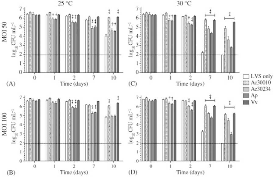
CFU densities of F. tularensis LVS in the presence of various FLAs. LVS cells were incubated at (A, C) 25 °C or (B, D) 30 °C with each FLA strain at an MOI of (A, B) 50 or (C, D) 100. Data were generated from two independent experiments with three replicates each (n = 6). Error bars shown represent the standard error mean. The dotted line indicates the LOD of 1.95 log10 CFU mL−1. *P < 0.05; †P < 0.01; ‡P < 0.001
To determine if LVS infectivity of FLA cells is temperature dependent, the incubation temperature was increased to 30 °C and the same MOIs of 50 and 100 were tested respectively (Figure 1C and D). At 30 °C, the enhanced survival of LVS was also observed in the presence of FLA but was more pronounced due to the temperature sensitivity of LVS. Specifically, control CFU levels decreased from between 6.4 ± 0.3 and 6.7 ± 0.3 at day 0 to below the LOD of 2.0 log10CFU mL−1 at day 10 during incubation at 30 °C, indicating LVS culturability is temperature sensitive. Although enhanced survival of LVS was observed in the presence of each FLA, culture with Ac30234 and Ap at 30 °C also resulted in LVS CFU decreases from day 1 to 10 compared to incubation at 25 °C.
Levels of Fn during culture with each FLA strain were also assessed to determine if enhanced survival in the presence of FLA was LVS specific. Similar to LVS, control levels of Fn log10 CFU mL−1 decreased from 6.7 ± 0.1 (MOI 50) and 7.0 ± 0.2 (MOI 100) at day 0 to 4.4 ± 0.6 (MOI 50) and 5.6 ± 0.7 (MOI 100) at day 10 under 25 °C incubation (Figure 2A and B, P < 0.001). At 30 °C, control levels of Fn log10 CFU mL−1 decreased from 6.7 ± 0.1 (MOI 50) and 7.0 ± 0.2 (MOI 100) at day 0 to 2.3 ± 0.6 (MOI 50) and 3.5 ± 1.2 (MOI 100) at day 10 (Figure 2C and D, P < 0.001). Interestingly, the negative effect of Ac30234 and Ap on LVS CFU levels was not observed for Fn cells cultured with either FLA; thus, the effect of enhanced bacterial survival was more pronounced for Fn in the presence of each FLA.
Figure 2.
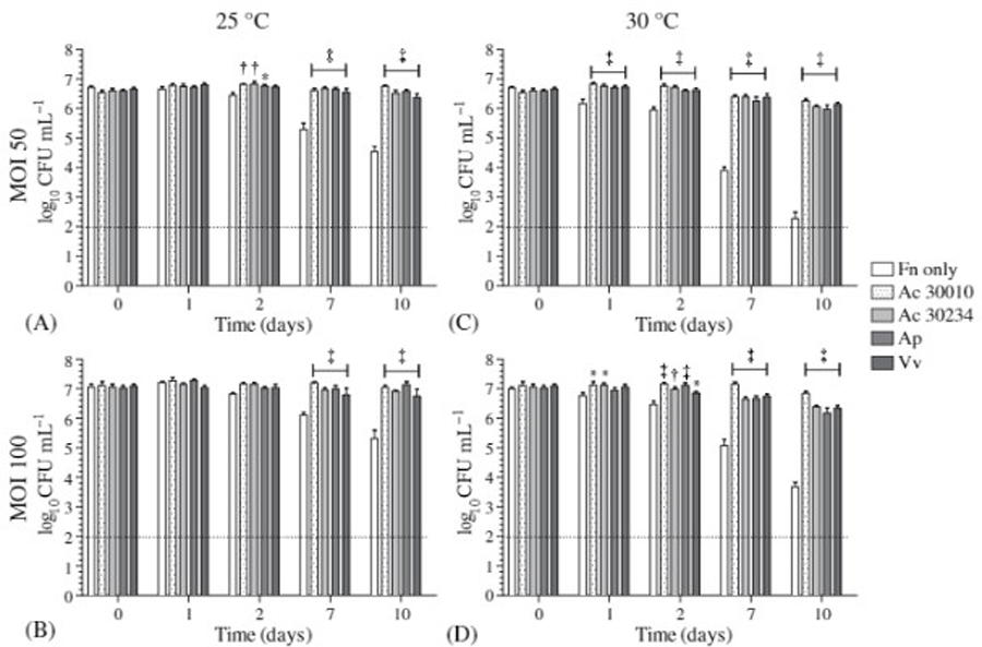
CFU densities of F. novicida in the presence of various FLAs. Fn cells were incubated at (A, C) 25 °C or (B, D) 30 °C with each FLA strain at an MOI of (A, B) 50 or (C, D) 100. Data were generated from two independent experiments with three replicates each (n = 6). Error bars shown represent the standard error mean. The dotted line indicates the LOD of 1.95 log10 CFU mL−1. *P < 0.05; †P < 0.01; ‡P < 0.001
To determine if virulent, clinical isolates of F. tularensis are infectious for FLA, F. tularensis Type A (Schu4) and Type B (NY98) strains were cultured with each FLA at the high MOI of 100 and at 30 °C. Previous studies have reported the temperature-dependent bacterial infectivity of FLA. Depending on the bacteria species, at low temperatures (<20–30 °C), bacteria were actively digested, displayed low amplification rates, and/or eliminated from the FLA, A. polyphagaor A. castellanii; however, at higher temperatures (25–37 °C), the amoeba was parasitized by the same strain of bacteria [29–31]. Thus, in this study, the higher MOI and temperature were used for the evaluation of the putative infectivity of virulent Schu4 and NY98 strains for the FLA cells. Figure 3 presents the data demonstrating the same enhanced survival and maintenance of bacterial culturability in the presence of each FLA. In the presence of FLA, Schu4 and NY98 log10 CFU mL−1 levels ranged from 7.0–7.1 and 6.0–6.6 at day 0 to 4.9–5.7 and 4.2–5.0 at day 7, respectively, and were significantly higher than those of the control (Figure 3, P < 0.01). Interestingly, by day 7, control CFU levels for Schu4 and NY98 were at or below the LOD of 2.0 log10 CFU mL−1 which, when compared to day 7 control CFU levels of LVS (3.45 ± 0.8, Figure 1D) and Fn (4.8 ± 0.7, Figure 2D), indicated that culturability of the virulent Type A and B strains is more temperature sensitive than LVS and Fn at 30 °C.
Figure 3.
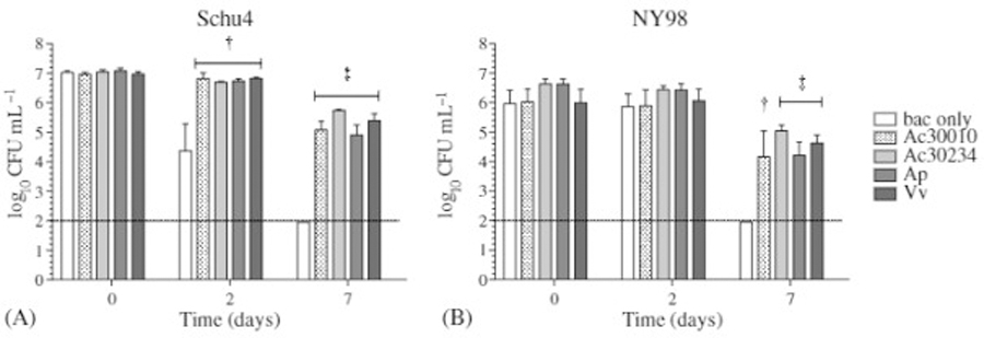
CFU densities of virulent F. tularensis Type A and B strains in the presence of various FLAs. (A) Schu4 and (B) NY98 cells were incubated at 30 °C with each FLA strain at an MOI of 100. Data were generated from two independent experiments with three replicates each (n = 6). Error bars shown represent the standard error mean. White bars represent the data from control, bacteria (bac) only wells. The dotted line indicates the LOD of 1.95 log10 CFU mL−1. †P < 0.01; ‡P < 0.001
Absence of amoebal cytopathic effects during culture with Francisella spp.
During Francisella spp. incubation with each FLA, amoebal densities were monitored to determine if amoebal lysis was a result of the enhanced Francisella survival and maintenance of Francisella culturability. Percent amoebal lysis, during culture with LVS for 0–10 days at 25 and 30 °C and MOIs of 50 and 100, ranged between 0%–0.8% for Ac30010, 0%–1.3% for Ac30234, 0%–0.2% for Ap, and 0%–6.0% for Vv (Figure 4A). Similarly, culture with Fn resulted in percent amoebal lysis of 0%–1.0% for Ac30010, 0%–2.0% for Ac30234, 0%–0.6% for Ap, and 0%–1.4% for Vv (Figure 4B).
Figure 4.
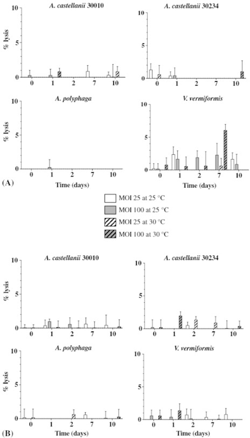
Absence of amoebal lysis in the presence of F. tularensis LVS and F. novicida under various conditions. (A) LVS and (B) Fn were cultured with four FLA strains at an MOI of 25 (white bars) and 100 (gray bars) and incubated at 25 °C (unhatched bars) and 30 °C (hatched bars). All negative values (between −4 and 0 for all samples) were set at 0%. Data were generated from two independent experiments with three replicates each (n = 6). Error bars shown represent the standard error mean
As a positive control for amoeba infectivity, Lp was cultured with Ap. Lp CFU levels and amoebal lysis were monitored for 0–7 days at 25 and 30 °C and at MOIs of 25 and 100. For all culture conditions tested, percent amoebal lysis ranged between 1%–5% at day 1, 4%–34% at day 2, 14%–39% at day 3, and 24%–44% at day 7 (Figure 5A), which is dramatically higher than those levels observed for LVS and Fn incubation with Ap (Figure 4). Furthermore, in contrast to all four Francisella strains, significant amplification of Lp cells resulted from culture with Ap cells (Figure 5B and C). At 25 °C, control Lp levels were between 6.2 and 6.7 log10 CFU mL−1; while in the presence of Ap cells, Lp log10 CFU mL−1levels peaked to 9.0 at day 2 and decreased to 8.0 at day 7 (Figure 5B, P < 0.001). Moreover, at a 100 MOI, significant Lp CFU increases were observed as early as day 1 at 30 °C incubation, compared to day 2 levels at 25 °C (Figure 4C, P < 0.001) confirming the observations from previous studies that Legionella infectivity of FLA is temperature dependent [29, 32].
Figure 5.

Infection of A. polyphaga with L. pneumophila. Ap cells were infected with Lp at either a low MOI of 25 (white bar/symbols) or high MOI of 100 (gray bars/symbols). (A) The percent lysis of amoeba cells during the course of Lp infection was calculated as described above. Bacterial density data are presented as log10 CFU mL−1 and derived from cultures incubated at (B) 25 °C and (C) 30 °C. The dotted and solid lines show control (con, bacteria only) and bacteria cultured with amoeba (+Ap) data, respectively. Data were generated from two independent experiments with three replicates each (n = 6). Error bars shown represent the standard error mean. ‡, P < 0.001 Lp only versus Lp + Ap
Phase contrast microscopic images were also collected during LVS, Fn, and Lp culture with Ap at 30°C and MOI 100 (Figure 6). Control wells displayed the expected amorphous trophozoite (AT), actively feeding form of Ap at days 0–7 with eventual encystment of amoeba cells due to the absence of a rich carbon source, like glucose (indicated as C for cysts). In concurrence with the lack of LVS and Fn amplification and amoebal lysis during Ap culture (Figures 1, 2, and 4), Ap cells displayed the same morphology in the presence of LVS and Fn as those in the control wells (Figure 6). In contrast, Lp incubation with Ap cells resulted in the lysis of trophozoites, indicated by white arrows, as well as the presence of infected Ap cells as early as day 1 post inoculation (black arrow, Figure 6).
Figure 6.
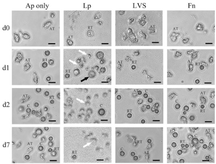
Phase contrast microscopic images illustrating the differences in the cytopathic effects of Lp, LVS, and Fn on A. polyphaga cells. Amoebae were infected with an MOI of 100 with Lp, LVS, or Fn cells and incubated at 30 °C. Images were taken at each time point (days 0, 1, 2, and 7) and are representative of two independent experiments with three replicates each (n = 6). Black scale bars represent 20 μm in length. AT, amorphous trophozoite; RT, rounded trophozoite; C, cyst. The black arrow indicates an infected cell. White arrows show lysed amoebae
Cytopathic effects were also not observed in the cultures of LVS and Fn with Ac30010, Ac30234, and Vv cells, which displayed the same cellular morphology as control wells at each time point (supplementary figure). LVS and Fn cultures with each FLA for the other three conditions, 25 °C MOI 50 and 100 and 30 °C MOI 50, displayed the same absence of amoeba cytopathic effect (data not shown).
Discussion
A mechanism for the persistence of Francisella spp. in water sources is likely given the existence of waterborne cases of tularemia [5–7, 9–11] and the observed acquisition of F. tularensis subsp. holarctica (Type B) during the aquatic life cycle of the mosquito vector [15, 16]. Moreover, within these aquatic environments, interactions can occur between Francisella and ubiquitous FLA, well-documented mediators of environmental survival and amplification for various human water-based pathogens. Thus, in this study, growth of virulent Ft Type A (Schu4) and Type B (NY98), attenuated Type B (LVS), and Fn was evaluated during culture with various axenically grown FLA (Ac strain 30010 and 30234, Ap, and Vv).
Culturability of all four Francisella strains in AB alone decreased by days 7–10 post incubation; however, in the presence of each FLA strain, Francisella CFU levels were significantly higher (Figures 1–3) with the exception being LVS cultured with Ac30234 and Ap at 25 °C and MOI 100 (Figure 1B). Despite the similarities in co-culture conditions evaluated and the rigorous testing of numerous Francisella and FLA strains in this study, amplification of each Francisella spp. strain in the presence of FLA was not observed (Figures 1–3), which is contrary to the previous studies reporting increases in Francisella CFU densities following co-culture with FLA [33–36]. Specifically, 3–4 log10 CFU mL−1increases of LVS and Fn were observed by days 6–15 in the presence of Ac30010, Ac30234, and Vv at 25–30 °C incubation and MOI of 10 [33, 35, 36]. However, in all of those studies, the assay buffer used was ATCC 712 medium, or peptone yeast extract (PYG) broth, which is the nutrient rich growth medium for propagation of Ap, Ac30010, and Ac30234 cells. El-Etr et al. [34] demonstrated that PYG broth alone supported LVS and Fn U112 growth; thus, amplification of LVS and Fn observed in the previous reports may not have been solely FLA mediated. The AB, used in this study, was shown not to support Francisella growth (Figures 1–3), which eliminated the possibility of false positives, i.e., detection of non-FLA mediated Francisella amplification.
LVS levels, in the presence of Ac30234 and Ap, were generally lower than during incubation with Ac30010 and Vv (Figure 1), which indicated either amoebal digestion of LVS cells or inhibition of LVS growth attributable to Ac30234 and Ap-derived culture products. However, the former explanation seems likely since, by day 7, there was a larger proportion of Ap and Ac30234 ATs (actively feeding form) during incubation with LVS than Ac30010 and Vv cells, where microscopy images showed mostly rounded trophozoites/pre-cysts and cysts (C, dormant form) (Figure 6 and Supplementary Figure 1). With various Gram-positive and Gram-negative bacteria that are commonly found in soil and water as the sole food source, Ap, Ac, and Vv cells exhibited an increased growth and ammonium production rate, in that order, indicating select Ap and Ac strains are more adept at utilizing bacteria as a food source than Vv [37]. However, the ability to degrade microbial cell wall components, or bacteriolytic activity, of Vv culture lysates were greater than Ac and Ap cultures when evaluating lysis of similar environmental Gram-negative and Gram-positive bacteria [38], indicating that the feeding behavior assessment of FLA may be assay and strain dependent. Pathogenic isolates of Acanthamoeba spp., Ac, Ap, Acanthamoeba palestinensis, Acanthamoeba astronyxis, and Acanthamoeba griffini can produce cytopathic effects on corneal epithelial cells and can tolerate high osmolarity growth media compared to their non-pathogenic counterparts [39]. Thus, the differences between CFU titres of each Francisella strain in the presence of FLA, observed in this study, may be due to the phenotypic differences in FLA behavior between species, strains, and isolates.
Results from this study also suggest the feeding behavior of FLA is Francisella strain dependent as LVS, Schu4, and NY98 bacteria were fed upon more heavily by Ap cells than Fn bacteria (Figures 1–3). The preferential selection of bacteria by amoebae has been previously reported with large amoebae observed to more readily digest the soil bacteria, Rhizobium and Agrobacterium, which were inedible to smaller amoebae [40]. From this study, 30 ATs of each FLA strain were measured from the microscopic images obtained from the two independent experiments and were found to be 30 ± 5 µm for Ap, 27 ± 7 µm for Ac30010, 24 ± 5 µm for Ac30234, and 20 ± 3 µm for Vv cells. The larger average size of Ap cells could account for the lower CFU titres of LVS at day 10 and of Schu4 and NY98 at day 7 in the presence of Ap cells, if amoeba size is indicative of bacterial-feeding potential (Figures 1 and 3). Moreover, genomic analysis of LVS, Schu4, and Fn revealed: (1) repeated genomic rearrangements in LVS and Schu4, but not in Fn; (2) absence of 40+ genes in the Fn genome that is present in LVS and Schu4; and (3) presence of approximately 10 genes and pseudogenes unique to Schu4 that presumably confers greater virulence observed with tularensis strains compared with the holarctica strains [41] indicating genetic variations between Ft and Fn may also have contributed to the differences in uptake by each FLA observed in this study.
Although survival of Francisella spp. was enhanced in the presence of each FLA, no amplification of bacteria or amoebae, significant amoebal lysis, or differences in amoeba morphology was observed during Francisella and FLA culture. This is in contrast to the water-based pathogen, Lp, which exhibits high infectivity potential for Acanthamoebaspp. and V. vermiformis resulting in rapid intracellular amplification and release of Lp and significant amoebal lysis (Figure 5) [32, 42–45]. The isolation and identification of amoebae-resisting bacteria (ARB) using the well-established co-culture method involves inoculation of samples onto a monolayer of axenic amoebae and continuous monitoring of cultures for amoebal lysis, resulting from infection by ARBs (reviewed in [46, 47]). From soil, drinking water, ground and surface water, and clinical samples, the amoebal co-culture method, with amoebal lysis as an assay endpoint, has been used by numerous groups to identify human pathogens such as those belonging to the class Chlamydiae and the genera Aeromonas, Bacillus, Enterobacter, Legionella, Mycobacterium, Pseudomonas, Rhodococcus, and Streptococcus as well as novel bacteria belonging to the genera Afipia and Bosea[48–56]. Thus, the absence of amoebal lysis observed in this study between each FLA and Francisella spp. strain was strongly indicative of a lack of bacterial infection. The exclusion of microscopic analysis, to determine the intracellular localization of Francisella spp., was a limitation in this study. However, because no amplification of Francisella spp. was observed during co-culture, visual confirmation of cellular localization would not have altered the observation that no Francisella spp. amplification occurred in the presence of each FLA species. Future work will aim to determine whether Francisella spp. cells are digested after FLA uptake or if each FLA is able to support intracellular Francisella spp. without bacterial amplification. The latter mechanism could demonstrate a potential commensal relationship with FLA providing an intracellular reservoir for Francisella survival in the environment.
The Lp mammalian lifecycle entails: (1) entry into host cells via coiling phagocytosis; (2) formation of the Legionella-containing vacuole (LCV); (3) conversion of the LCV into a rough endoplasmic reticulum (RER)-like compartment to avoid lysosome fusion; (4) replication of non-flagellated Lp within the RER-like LCV; and (5) extracellular release of flagellated Lp [57, 58], all of which is dependent on the Lp factors: Icm/Dot T4SS, SidC, Mip, PmiA, FliA, LetA/GacA, Type IV pili, and TatBC [59]. These Lp factors have also been shown to be important for Ap, Ac, and Vv infectivity except SidC [59]. In contrast, the LVS and Schu4 mammalian lifecycle entails: (1) entry into host cells via asymmetric spacious pseudopod loops; (2) formation of the Francisella-containing phagosome (FCP); (3) recruitment of early and late endosomal markers to the FCP to avoid lysosome fusion; (4) degradation of the FCP membrane and egress into the host cytosol; and (5) rapid cytosolic growth and depletion of host cell nutrients leading to cellular death and extracellular release of Francisella bacteria (reviewed in [60, 61]) which is dependent on the Francisella factors, IglC, MglA, and FTT1103 [62], with IglC and MglA also shown to be important for Ac and Vv intra-amoebae survival [36, 63]. These observed differences in the mammalian lifecycles of Lp and Francisella spp., and genetic factors required, could explain the differences in amoeba infectivity between Lp and the Francisella spp. strains observed in this study.
Collectively, the results from this study indicated that Francisella infectivity of FLA and intracellular amplification did not occur. However, each FLA strain enhanced the survival of Francisella in low nutrient environments especially at higher temperatures; thus, the possible role of environmental commensalism in Francisella persistence is not excluded. In general, the genomes of intracellular microbes are smaller and more genetically stable than their free-living counterparts, whose larger genomes exhibit a higher frequency of rearrangements and greater degree of variability in genetic content between species [64]. Previously reported genomic analysis of Francisella spp. revealed a small size of <2 Mb, limited genetic variation within the three subspecies and Fn, a high proportion of disrupted biosynthetic pathways, and an enrichment of eukaryotic protein domains also found in other intracellular pathogens; collectively suggesting a host-dependent phase in the lifecycle of Francisella spp. [65–67]. However, given the: (1) rapid loss of Francisella culturability in low nutrient buffer; (2) lack of significant amplification within FLA; (3) conflicting observations between studies; and (4) unreported identification of Francisella spp. within FLA environmental water isolates to date, it is still unclear if FLA is a significant reservoir of Francisella spp. in the environment.
Supplementary Material
Acknowledgements
The authors would like to thank Worth Calfee and Alan Lindquist for their critical review of this manuscript. The U.S. Environmental Protection Agency through its Office of Research and Development funded and managed the research described herein under contract # EP-C-11-006 to Pegasus Technical Services, Inc. It has been reviewed by the Agency but does not necessarily reflect the Agency’s views. No official endorsement should be inferred. EPA does not endorse the purchase or sale of any commercial products or services.
Footnotes
Conflict of Interest
The authors declare that they have no conflict of interest.
References
- 1.Federal Select Agents Program: Select Agents and Toxins–Public health, Code of Federal Regulations, 42.CFR, Part 73, 2015, Available at http://www.selectagents.gov/SelectAgentsandToxinsList.html. [Google Scholar]
- 2.Johansson A, Celli J, Conlan W, Elkins KL, Forsman M, Keim PS, Larsson P, Manoil C, Nano FE,Petersen JM, Sjöstedt A: Objections to the transfer of Francisella novicida to the subspecies rank of Francisella tularensis. Int J Syst Evol Microbiol 60, 1717–1718 (2010). [DOI] [PMC free article] [PubMed] [Google Scholar]
- 3.Busse HJ, Huber B, Anda P, Escudero R, Scholz HC, Seibold E, Splettstoesser WD, Kämpfer P:Objections to the transfer of Francisella novicida to the subspecies rank of Francisella tularensis – Response to Johansson et al. Int J Syst Evol Microbiol 60, 1718–1720 (2010). [DOI] [PubMed] [Google Scholar]
- 4.Carvalho CL, Lopes de Carvalho I, Zé-Zé L, Núncio MS, Duarte EL: Tularaemia: A challenging zoonosis.Comp Immunol Microbiol Infect Dis 37, 85–96 (2014). [DOI] [PMC free article] [PubMed] [Google Scholar]
- 5.Jellison WL, Epler DC, Kuhns E, Kohls GM: Tularemia in man from a domestic rural water supply. Public Health Rep 65, 1219–1226 (1950). [PubMed] [Google Scholar]
- 6.Karpoff SP, Antonoff NI: The spread of tularemia through water, as a new factor in its epidemiology. J Bacteriol 32, 243–258 (1936). [DOI] [PMC free article] [PubMed] [Google Scholar]
- 7.Greco D, Allegrini G, Tizzi T, Ninu E, Lamanna A, Luzi S: A waterborne tularemia outbreak. Eur J Epidemiol 3,35–38 (1987). [DOI] [PubMed] [Google Scholar]
- 8.Rice EW: Occurrence and control of tularemia in drinking water. J Am Water Works Assoc 107, E486–E496(2015). [Google Scholar]
- 9.Brett M, Doppalapudi A, Respicio-Kingry LB, Myers D, Husband B, Pollard K, Mead P, Petersen JM,Whitener CJ: Francisella novicida bacteremia after a near-drowning accident. J Clin Microbiol 50, 2826–2829(2012). [DOI] [PMC free article] [PubMed] [Google Scholar]
- 10.Brett ME, Respicio-Kingry LB, Yendell S, Ratard R, Hand J, Balsamo G, Scott-Waldron C, O’Neal C,Kidwell D, Yockey B, Singh P, Carpenter J, Hill V, Petersen JM, Mead P: Outbreak of Francisella novicidabacteremia among inmates at a Louisiana correctional facility. Clin Infect Dis 59, 826–833 (2014). [DOI] [PubMed] [Google Scholar]
- 11.Ughetto E, Héry-Arnaud G, Cariou ME, Pelloux I, Maurin M, Caillon J, Moreau P, Ygout JF, Corvec S:An original case of Francisella tularensis subsp. holarctica bacteremia after a near-drowning accident. Infect Dis (Lond) 47, 588–590 (2015). [DOI] [PubMed] [Google Scholar]
- 12.Berrada Z, Telford Iii S: Survival of Francisella tularensis Type A in brackish-water. Arch Microbiol 193, 223–226 (2011). [DOI] [PMC free article] [PubMed] [Google Scholar]
- 13.Whitehouse CA, Kesterson KE, Duncan DD, Eshoo MW, Wolcott M: Identification and characterization of Francisella species from natural warm springs in Utah, USA. Lett Appl Microbiol 54, 313–324 (2012). [DOI] [PubMed] [Google Scholar]
- 14.Broman T, Thelaus J, Andersson A-C, Bäckman S, Wikström P, Larsson E, Granberg M, Karlsson L,Bäck E, Eliasson H, Mattsson R, Sjöstedt A, Forsman M: Molecular detection of persistent Francisella tularensis subspecies holarctica in natural waters. Int J Microbiol 2011, 10 (2011). [DOI] [PMC free article] [PubMed] [Google Scholar]
- 15.Thelaus J, Andersson A, Broman T, Bäckman S, Granberg M, Karlsson L, Kuoppa K, Larsson E,Lundmark E, Lundström JO, Mathisen P, Näslund J, Schäfer M, Wahab T, Forsman M: Francisella tularensissubspecies holarctica occurs in Swedish mosquitoes, persists through the developmental stages of laboratory-infected mosquitoes and is transmissible during blood feeding. Microb Ecol 67, 96–107 (2014). [DOI] [PMC free article] [PubMed] [Google Scholar]
- 16.Bäckman S, Näslund J, Forsman M, Thelaus J: Transmission of tularemia from a water source by transstadial maintenance in a mosquito vector. Sci Rep 5, 7793 (2015). [DOI] [PMC free article] [PubMed] [Google Scholar]
- 17.Eliasson H, Bäck E: Tularaemia in an emergent area in Sweden: An analysis of 234 cases in five years. Scand J Infect Dis 39, 880–889 (2007). [DOI] [PubMed] [Google Scholar]
- 18.Rossow H, Ollgren J, Klemets P, Pietarinen I, Saikku J, Pekkanen E, Nikkari S, Syrjälä H, Kuusi M, Nuorti JP: Risk factors for pneumonic and ulceroglandular tularaemia in Finland: A population-based case-control study.Epidemiol Infect 142, 2207–2216 (2014). [DOI] [PMC free article] [PubMed] [Google Scholar]
- 19.Declerck P, Behets J, van Hoef V, Ollevier F: Detection of Legionella spp. and some of their amoeba hosts in floating biofilms from anthropogenic and natural aquatic environments. Water Res 41, 3159–3167 (2007). [DOI] [PubMed] [Google Scholar]
- 20.Hsu BM, Lin CL, Shih FC: Survey of pathogenic free-living amoebae and Legionella spp. in mud spring recreation area. Water Res 43, 2817–2828 (2009). [DOI] [PubMed] [Google Scholar]
- 21.Kuiper MW, Valster RM, Wullings BA, Boonstra H, Smidt H, van der Kooij D: Quantitative detection of the free-living amoeba Hartmannella vermiformis in surface water by using real-time PCR. Appl Environ Microbiol 72, 5750–5756 (2006). [DOI] [PMC free article] [PubMed] [Google Scholar]
- 22.Khan NA: Acanthamoeba: Biology and Pathogenesis, Caister Academic Press, Norfolk, UK, 2009, 290 pp. [Google Scholar]
- 23.Buse HY, Lu J, Struewing IT, Ashbolt NJ: Eukaryotic diversity in premise drinking water using 18S rDNA sequencing: Implications for health risks. Environ Sci Pollut Res Int 20, 6351–6366 (2013). [DOI] [PubMed] [Google Scholar]
- 24.Östman Ö, Lundström J, Persson Vinnersten T: Effects of mosquito larvae removal with Bacillus thuringiensis israelensis (Bti) on natural protozoan communities. Hydrobiologia 607, 231–235 (2008). [Google Scholar]
- 25.Addicott JF: Predation and prey community structure: An experimental study of the effect of mosquito larvae on the protozoan communities of pitcher plants. Ecology 55, 475–492 (1974). [Google Scholar]
- 26.Preston TM, Richards H, Wotton RS: Locomotion and feeding of Acanthamoeba at the water-air interface of ponds. FEMS Microbiol Lett 194, 143–147 (2001). [DOI] [PubMed] [Google Scholar]
- 27.Merritt RW, Dadd RH, Walker ED: Feeding behavior, natural food, and nutritional relationships of larval mosquitoes. Annu Rev Entomol 37, 349–376 (1992). [DOI] [PubMed] [Google Scholar]
- 28.Delafont V, Mougari F, Cambau E, Joyeux M, Bouchon D, Héchard Y, Moulin L: First evidence of amoebae-mycobacteria association in drinking water network. Environ Sci Technol 48, 11872–11882 (2014). [DOI] [PubMed] [Google Scholar]
- 29.Ohno A, Kato N, Sakamoto R, Kimura S, Yamaguchi K: Temperature-dependent parasitic relationship between Legionella pneumophila and a free-living amoeba (Acanthamoeba castellanii). Appl Environ Microbiol 74,4585–4588 (2008). [DOI] [PMC free article] [PubMed] [Google Scholar]
- 30.Cirillo JD, Falkow S, Tompkins LS, Bermudez LE: Interaction of Mycobacterium avium with environmental amoebae enhances virulence. Infect Immun 65, 3759–3767 (1997). [DOI] [PMC free article] [PubMed] [Google Scholar]
- 31.Saeed A, Johansson D, Sandström G, Abd H: Temperature depended role of Shigella flexneri invasion plasmid on the interaction with Acanthamoeba castellanii. Int J Microbiol 2012, 917031 (2012). [DOI] [PMC free article] [PubMed] [Google Scholar]
- 32.Buse HY, Ashbolt NJ: Differential growth of Legionella pneumophila strains within a range of amoebae at various temperatures associated with in-premise plumbing. Lett Appl Microbiol 53, 217–224 (2011). [DOI] [PubMed] [Google Scholar]
- 33.Abd H, Johansson T, Golovliov I, Sandström G, Forsman M: Survival and growth of Francisella tularensisin Acanthamoeba castellanii. Appl Environ Microbiol 69, 600–606 (2003). [DOI] [PMC free article] [PubMed] [Google Scholar]
- 34.El-Etr SH, Margolis JJ, Monack D, Robison RA, Cohen M, Moore E, Rasley A: Francisella tularensistype A strains cause the rapid encystment of Acanthamoeba castellanii and survive in amoebal cysts for three weeks postinfection. Appl Environ Microbiol 75, 7488–7500 (2009). [DOI] [PMC free article] [PubMed] [Google Scholar]
- 35.Verhoeven AB, Durham-Colleran MW, Pierson T, Boswell WT, Van Hoek ML: Francisella philomiragiabiofilm formation and interaction with the aquatic protist Acanthamoeba castellanii. Biol Bull 219, 178–188 (2010). [DOI] [PubMed] [Google Scholar]
- 36.Santic M, Ozanic M, Semic V, Pavokovic G, Mrvcic V, Kwaik YA: Intra-vacuolar proliferation of F. novicidawithin H. vermiformis. Front Microbiol 2, 78 (2011). [DOI] [PMC free article] [PubMed] [Google Scholar]
- 37.Weekers PH, Bodelier PL, Wijen JP, Vogels GD: Effects of grazing by the free-living soil amoebaeAcanthamoeba castellanii, Acanthamoeba polyphaga, and Hartmannella vermiformis on various bacteria. Appl Environ Microbiol 59, 2317–2319 (1993). [DOI] [PMC free article] [PubMed] [Google Scholar]
- 38.Weekers PH, Engelberts AM, Vogels GD: Bacteriolytic activities of the free-living soil amoebae,Acanthamoeba castellanii, Acanthamoeba polyphaga and Hartmannella vermiformis. Antonie Van Leeuwenhoek 68, 237–243 (1995). [DOI] [PubMed] [Google Scholar]
- 39.Khan NA, Jarroll EL, Paget TA: Acanthamoeba can be differentiated by the polymerase chain reaction and simple plating assays. Curr Microbiol 43, 204–208 (2001). [DOI] [PubMed] [Google Scholar]
- 40.Singh BN: Selection of bacterial food by soil flagellates and amoebae. Ann Appl Biol 29, 18–22 (1942). [Google Scholar]
- 41.Rohmer L, Fong C, Abmayr S, Wasnick M, Larson Freeman TJ, Radey M, Guina T, Svensson K, Hayden HS, Jacobs M, Gallagher LA, Manoil C, Ernst RK, Drees B, Buckley D, Haugen E, Bovee D, Zhou Y,Chang J, Levy R, Lim R, Gillett W, Guenthener D, Kang A, Shaffer SA, Taylor G, Chen J, Gallis B, D’Argenio DA, Forsman M, Olson MV, Goodlett DR, Kaul R, Miller SI, Brittnacher MJ: Comparison of Francisella tularensis genomes reveals evolutionary events associated with the emergence of human pathogenic strains.Genome Biol 8, R102 (2007). [DOI] [PMC free article] [PubMed] [Google Scholar]
- 42.Seno M, Sakaki M, Ogawa H, Matsuda H, Takeda Y: Effective proliferation of low level Legionella pneumophila serogroup 1 cells using coculture procedure with Acanthamoeba castellanii. J Microbiol Methods 66,564–567 (2006). [DOI] [PubMed] [Google Scholar]
- 43.Gao LY, Kwaik YA: The mechanism of killing and exiting the protozoan host Acanthamoeba polyphaga byLegionella pneumophila. Environ Microbiol 2, 79–90 (2000). [DOI] [PubMed] [Google Scholar]
- 44.Moffat JF, Tompkins LS: A quantitative model of intracellular growth of Legionella pneumophila inAcanthamoeba castellanii. Infect Immun 60, 296–301 (1992). [DOI] [PMC free article] [PubMed] [Google Scholar]
- 45.Wadowsky RM, Wilson TM, Kapp NJ, West AJ, Kuchta JM, States SJ, Dowling JN, Yee RB:Multiplication of Legionella spp. in tap water containing Hartmannella vermiformis. Appl Environ Microbiol 57,1950–1955 (1991). [DOI] [PMC free article] [PubMed] [Google Scholar]
- 46.Jacquier N, Aeby S, Lienard J, Greub G: Discovery of new intracellular pathogens by amoebal coculture and amoebal enrichment approaches. J Vis Exp 80, e51055 (2013). [DOI] [PMC free article] [PubMed] [Google Scholar]
- 47.Tosetti N, Croxatto A, Greub G: Amoebae as a tool to isolate new bacterial species, to discover new virulence factors and to study the host-pathogen interactions. Microb Pathog 77, 125–130 (2014). [DOI] [PubMed] [Google Scholar]
- 48.Corsaro D, Feroldi V, Saucedo G, Ribas F, Loret JF, Greub G: Novel Chlamydiales strains isolated from a water treatment plant. Environ Microbiol 11, 188–200 (2009). [DOI] [PubMed] [Google Scholar]
- 49.Corsaro D, Pages GS, Catalan V, Loret JF, Greub G: Biodiversity of amoebae and amoeba-associated bacteria in water treatment plants. Int J Hyg Environ Health 213, 158–166 (2010). [DOI] [PubMed] [Google Scholar]
- 50.Evstigneeva A, Raoult D, Karpachevskiy L, La Scola B: Amoeba co-culture of soil specimens recovered 33 different bacteria, including four new species and Streptococcus pneumoniae. Microbiology 155, 657–664 (2009). [DOI] [PubMed] [Google Scholar]
- 51.La Scola B, Mezi L, Weiller PJ, Raoult D: Isolation of Legionella anisa using an amoebic coculture procedure. J Clin Microbiol 39, 365–366 (2001). [DOI] [PMC free article] [PubMed] [Google Scholar]
- 52.Loret JF, Jousset M, Robert S, Saucedo G, Ribas F, Thomas V, Greub G: Amoebae-resisting bacteria in drinking water: Risk assessment and management. Water Sci Technol 58, 571–577 (2008). [DOI] [PubMed] [Google Scholar]
- 53.Pagnier I, Raoult D, La Scola B: Isolation and identification of amoeba-resisting bacteria from water in human environment by using an Acanthamoeba polyphaga co-culture procedure. Environ Microbiol 10, 1135–1144 (2008). [DOI] [PubMed] [Google Scholar]
- 54.Thomas V, Casson N, Greub G: New Afipia and Bosea strains isolated from various water sources by amoebal co-culture. Syst Appl Microbiol 30, 572–579 (2007). [DOI] [PubMed] [Google Scholar]
- 55.Thomas V, Herrera-Rimann K, Blanc DS, Greub G: Biodiversity of amoebae and amoeba-resisting bacteria in a hospital water network. Appl Environ Microbiol 72, 2428–2438 (2006). [DOI] [PMC free article] [PubMed] [Google Scholar]
- 56.Thomas V, Loret JF, Jousset M, Greub G: Biodiversity of amoebae and amoebae-resisting bacteria in a drinking water treatment plant. Environ Microbiol 10, 2728–2745 (2008). [DOI] [PubMed] [Google Scholar]
- 57.Dietrich C, Heuner K, Brand BC, Hacker J, Steinert M: Flagellum of Legionella pneumophila positively affects the early phase of infection of eukaryotic host cells. Infect Immun 69, 2116–2122 (2001). [DOI] [PMC free article] [PubMed] [Google Scholar]
- 58.Cazalet C, Rusniok C, Bruggemann H, Zidane N, Magnier A, Ma L, Tichit M, Jarraud S, Bouchier C,Vandenesch F, Kunst F, Etienne J, Glaser P, Buchrieser C: Evidence in the Legionella pneumophila genome for exploitation of host cell functions and high genome plasticity. Nat Genet 36, 1165–1173 (2004). [DOI] [PubMed] [Google Scholar]
- 59.Hilbi H, Weber SS, Ragaz C, Nyfeler Y, Urwyler S: Environmental predators as models for bacterial pathogenesis. Environ Microbiol 9, 563–575 (2007). [DOI] [PubMed] [Google Scholar]
- 60.Barel M, Charbit A: Francisella tularensis intracellular survival: To eat or to die. Microbes Infect 15, 989–997(2013). [DOI] [PubMed] [Google Scholar]
- 61.Jones BD, Faron M, Rasmussen JA, Fletcher JR: Uncovering the components of the Francisella tularensis virulence stealth strategy. Front Cell Infect Microbiol 4, 32 (2014). [DOI] [PMC free article] [PubMed] [Google Scholar]
- 62.Ray K, Marteyn B, Sansonetti PJ, Tang CM: Life on the inside: The intracellular lifestyle of cytosolic bacteria. Nat Rev Microbiol 7, 333–340 (2009). [DOI] [PubMed] [Google Scholar]
- 63.Lauriano CM, Barker JR, Yoon SS, Nano FE, Arulanandam BP, Hassett DJ, Klose KE: MglA regulates transcription of virulence factors necessary for Francisella tularensis intraamoebae and intramacrophage survival. Proc Natl Acad Sci U S A 101, 4246–4249 (2004). [DOI] [PMC free article] [PubMed] [Google Scholar]
- 64.Mira A, Klasson L, Andersson SG: Microbial genome evolution: Sources of variability. Curr Opin Microbiol 5, 506–512 (2002). [DOI] [PubMed] [Google Scholar]
- 65.Schmitz-Esser S, Tischler P, Arnold R, Montanaro J, Wagner M, Rattei T, Horn M: The genome of the amoeba symbiont “Candidatus Amoebophilus asiaticus” reveals common mechanisms for host cell interaction among amoeba-associated bacteria. J Bacteriol 192, 1045–1057 (2010). [DOI] [PMC free article] [PubMed] [Google Scholar]
- 66.Monroe D: Looking for chinks in the armor of bacterial biofilms. PLoS Biol 5, e307 (2007). [DOI] [PMC free article] [PubMed] [Google Scholar]
- 67.Larsson P, Oyston PCF, Chain P, Chu MC, Duffield M, Fuxelius H-H, Garcia E, Halltorp G, Johansson D, Isherwood KE, Karp PD, Larsson E, Liu Y, Michell S, Prior J, Prior R, Malfatti S, Sjöstedt A, Svensson K, Thompson N, Vergez L, Wagg JK, Wren BW, Lindler LE, Andersson SGE, Forsman M, Titball RW:The complete genome sequence of Francisella tularensis, the causative agent of tularemia. Nat Genet 37, 153–159 (2005). [DOI] [PubMed] [Google Scholar]
Associated Data
This section collects any data citations, data availability statements, or supplementary materials included in this article.


