

Abstract
Recently, our group identified that harmine is able to induce β-cell proliferation both in vitro and in vivo, mediated via the DYRK1A-NFAT pathway. Since, harmine suffers from lack of selectivity, both against other kinases and CNS off-targets, therefore, we sought to expand structure-activity relationships for harmine’s DYRK1A activity, to enhance selectivity for off-targets, while retaining human β-cell proliferation activity. We carried out optimization of the 9-N-position of harmine to synthesize 29 harmine-based analogs. Several novel inhibitors showed excellent DYRK1A inhibition and human β-cell proliferation capability. An optimized DYRK1A inhibitor, 2-2c, was identified as a novel, efficacious in vivo lead candidate. 2-2c also demonstrates improved selectivity for kinases and CNS off-targets, as well as in vivo efficacy for β-cell proliferation and regeneration at lower doses than harmine. Collectively, these findings demonstrate that 2-2c is a much-improved in vivo lead candidate as compared to harmine, for the treatment of diabetes.
Keywords: Dual-specificity Tyrosine-Regulated Kinases (DYRKs), Harmine, DYRK1A inhibitor, Structure Activity Relationship Study, β-Cell Proliferation, Diabetes
Graphical Abstract

INTRODUCTION
The Dual-specificity Tyrosine-phosphorylation-Regulated Kinase A (DYRK1A) belongs to the CMGC (CDK, MAPK, CDC-Like Kinases, GSK3 kinase) family of eukaryotic protein kinases that has been shown to play important roles in neurodegenerative diseases 1, 2, tumorigenesis and apoptosis 3, 4. More recently, DYRK1A was identified as a regulator of regenerative pathways relevant to human insulin-producing pancreatic β-cells 5–8. Numerous studies have explored development of DYRK1A inhibitor scaffolds in view of the involvement of DYRK1A in these diseases 2–6, 8–10. Several DYRK1A inhibitors, from natural sources like harmine, as well as from small molecule drug discovery programs, have been identified and characterized 8, 11–33. Among all the DYRK1A inhibitors, harmine and its analogues (β-carbolines) are the most commonly studied, and remain one of the most potent and orally bioavailable class of inhibitors known 2, 10. In addition to its kinase inhibitory activity, harmine has been suggested to be a hallucinogen, due to its presence in the hallucinogenic infusion, ayahuasca, and its affinity for the serotonin, tryptamine and other receptors in the central nervous system (CNS) 34–36. Harmine and several related analogues have also been found to inhibit DYRK1A-mediated phosphorylation of tau protein in the CNS 37 and to have anti-proliferative cancer activity, including inhibition of topoisomerase I, 38, 39 inhibition of CDKs 40, induction of cell apoptosis 41, and DNA intercalation 42.
Recently, our group found that harmine is able to induce human β-cell proliferation using a luciferase reporter phenotypic high-throughput small-molecule Screen (HTS). Subsequently, we identified DYRK1A-NFAT pathway as being the major pathway for this cell proliferation 5. These results have been confirmed in other labs with DYRK1A inhibitors unrelated to the harmine scaffold, including from our own lab 6, 8, 43–45. DYRK1A is expressed ubiquitously; accordingly DYRK1A inhibitors including harmine, developed for other therapeutic purposes, predicted to have adverse effects on multiple tissues, thereby limiting their therapeutic potential. Thus, there is an urgent need to develop strategies to generate more selective harmine analogs as pharmacological lead candidates with limited CNS activities, yet retaining human β-cell proliferative effects, with improved pharmacological profile for therapeutic development 46. In addition, optimized, tissue-specific β-cell targeted harmine analogs may also be useful for treatment of diabetes. One such prototype technology, for example reported by DiMarchi and colleagues, conjugated estrogen derivatives to GLP-1 analog peptides to effectively target cells that highly express GLP1-receptor, including β-cells 47, 48.
With knowledge of this previous work, we developed a systematic medicinal chemistry strategy to investigate structure-activity relationships of harmine analogs for both DYRK1A activity and β-cell proliferation, with intention of identifying a kinase and CNS off-target selective, as well as linkable, harmine analogs for targeted therapy (Fig. 1). In our previous work, we recently reported the optimization of the 1-position of harmine which led to two compounds which exhibited robust human β-cell proliferation at doses of 3-30 μM with one compound I (R = CH2OH) showing improved kinase selectivity as compared to harmine 49. Herein, we report our subsequent efforts for medicinal chemistry optimization of harmine 9-N-position. We describe the identification of an improved, harmine-based in vivo DYRK1A inhibitor molecular probe with excellent in vitro human β-cell proliferation potency, improved kinase and CNS receptor selectivity and enhanced efficacy in three in vivo models of β-cell proliferation and regeneration.
Fig. 1.
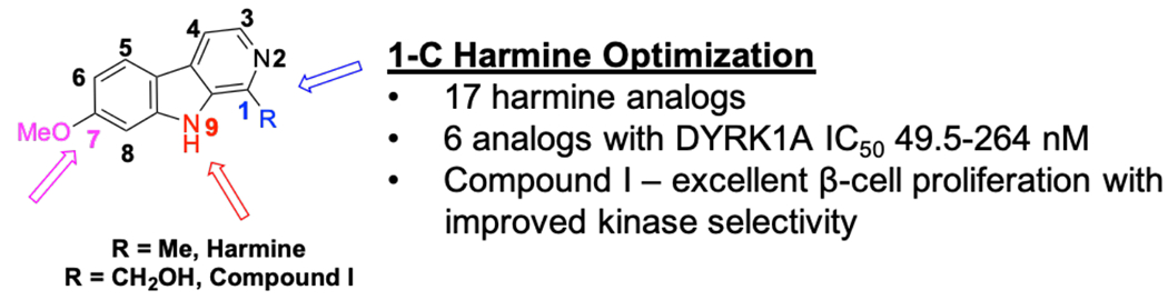
Optimization of the 1-position of harmine.
RESULTS AND DISCUSSIONS:
Chemistry:
Synthesis of 9-N-substituted harmine analogs:
Modification at the 9-N-position of harmine was carried out by the reaction sequences outlined in Scheme 1. Alkylation of harmine with various alkyl bromides in the presence of sodium hydride as base generated harmine analogs 2-1a -2-1j, 2-1k and 2-1l having terminal methyl ester or t-butyl ester group with carbon chain lengths of 1-5 carbons in 40-68% yield 50, 51. Harmine analogs 2-1a-2-1e and 2-1k with terminal methyl esters were refluxed in excess methanolic ammonia solution to provide the corresponding terminal carboxamide analogs 2-2a-2-2e and 2-2f in 84-95% yield. t-Butyl ester harmine analogs 2-1f-2-1j were treated with hydrochloric acid at room temperature to give corresponding acids 2-3a-2-3e in excellent yield51. Harmine alpha-methyl carboxylic acid analogs 2-3f and 2-3g were synthesized from their corresponding benzyl esters by hydrogenolysis using palladium on carbon and triethyl silane 52. 9-N substituted analogs with terminal amino groups 2-5a and 2-5b were synthesized in 34-36% yield respectively, by phthalimide deprotection of intermediates 1-2m and 1-2n using hydrazine monohydrate 51. Acylation of amines 2-5a and 2-5b with acetic anhydride and trimethylamine afforded harmine acetamide analogs 2-6a and 2-6b in 44 and 63% yield, respectively. N-Methylations of compounds 2-5a,b were carried out by first, formylation using ethyl formate followed by LiAlH4 reduction to generate mono N-methylamino analogs 2-7a and 2-7b in 27% and 41% yield, respectively. Acetylation of these N-methylated harmine analogs using acetic anhydride and trimethylamine similarly afforded acetamides 2-8a and 2-8b.
Scheme 1. Synthesis of 9-N Substituted Harmine Analog.


Reagents and conditions: (a) NaH (2 eq.), Br(CH2)nCO2R (2 eq.), DMF, 50 °C, 12 h, 21-82%; (b) 7 N NH3 in MeOH (20 eq.), 90 °C, 12 h, 40-100%; (c) 4N HCl in dioxane, rt, 12 h, 60-100%; (d) Et3SiH (8 eq.), Pd-C, MeOH, rt, 1h, 6-64% (2 steps); (e) NaH (2 eq.), Br(CH2)nNHPhth (2 eq.) (for 2-1m, 2-1n) and Br(CH2)3NHBoc (2 eq.) (for 2-1o), DMF, 50 °C, 12 h, 60-84%; (f) hydrazine monohydrate (20 eq.), MeOH, reflux, 3 h, 34-36%; (g) Acetic anhydride (1 eq), Et3N (2.2eq.), DCM, rt, 12 h, 38-63%; (h) (i) ethylformate (0.5 eq.), EtOH, Microwave, 150 °C, 30 min; (ii) LiAlH4 (3 eq.), THF, reflux, 4 h, 27-41%.
Structure-Activity Relationship Studies of Harmine Analogs:
Harmine is considered a type I DYRK1A inhibitor that binds to the canonical ATP-binding pocket. The compound forms two hydrogen bonding interactions with both the side chain of Lys188 and backbone Leu241 (PDB ID 3ANR, Fig. 2)29. Based on the known binding pose of harmine, we surmised that there are three positions, 1-methyl, 7-methoxy and 9-N indole, where rational modifications of harmine can be carried out (Fig 2). We sought to identify novel harmine-based DYRK1A inhibitors with equal or improved DYRK1A activity, human β-cell proliferation while also increasing kinase and other off-target selectivity as compared to harmine. Such optimized harmine analogs are expected to serve as more selective in vivo chemical probe DYRK1A inhibitors for β-cell proliferation. A DYRK1A inhibitor optimized for this type of diabetes application would also have potential for conjugation to prototype carriers, such as GLP1 analogs, for β-cell targeted therapy. In our earlier work,49 modifications of the 1-position in harmine was explored (Fig. 1) to provide compounds with improved β-cell proliferation ability, However opportunities for further improvements to provide more advanced in vivo chemical probes were limited. Similarly, 7-position optimization of harmine was carried out but majority of 7-substituted analogs demonstrate that these harmine modifications do not result in DYRK1A inhibitors superior in potency as compared to harmine53 (unpublished work). Subsequently, we continued the harmine SAR studies for optimization through additional systematic structural modifications at its 9-N-positions, while retaining the 1-position methyl and 7-position methoxy groups as shown in Fig. 2.
Fig. 2.

Substituents explored for optimization of 9-N position of harmine
SAR Studies of 9-N Harmine Analogs:
Modifications of the 9-N position of the harmine were studied by introducing substituents bearing terminal methyl ester (2-1a to 2-1l), carboxamide (2-2a to 2-2f), carboxylic acid (2-3a to 2-3g) and amino/substituted amino (2-1q, 2-5, 2-6, 2-7 and 2-8) functional groups with varying chain lengths of 1-5 carbons (Fig 2 and Scheme 1). Gratifyingly, modifications at the 9-N-position nitrogen were more generally well tolerated to retain potent DYRK1A inhibition as compared to changes at harmine’s 7-position.53 Several analogs were found to show similar or occasionally better DYRK1A inhibitory activity as compared to harmine (Table 1). Among all the analogs with terminal methyl esters (2-1a to 2-1e, 2-1k and 2-1l), compounds 2-1c, 2-1k and 2-1l containing 3-carbon chain length exhibited the most potent DYRK1A inhibition with IC50 in the range of 13-27 nM. Carbon chain length significantly impacted DYRK1A binding; decreasing to 1 or 2 carbon atom linkers or increasing to 4 and 5 carbon atom linkers, reduced the DYRK1A inhibitory activity. These data suggest that 3-carbon atom chain length is optimal for added pharmacophores to retain DYRK1A inhibition activity at the 9-N position. Compounds bearing terminal carboxylic acids (2-3a to 2-3g) at the 9-N position were detrimental for the DYRK1A activity regardless of the carbon chain length. The docking studies of these novel harmine analogs 2-3a to 2-3g to the crystal structure of DYRK1A bound to harmine analog 2-2c (vide infra) (PDB ID 6UWY) suggest that carboxylic acid functional group is disfavored because of the highly negative electrostatic environment in the ATP-binding site (See Supplementary Material, Fig S1B). In contrast, harmine analogs with carboxamide functional groups, behaved similar to those analogs bearing terminal methyl esters. For example, compounds 2-2c and 2-2f with three carbon chain length to this pharmacophore, exhibited potent DYRK1A inhibitory activity with IC50’s in the range of 14-25 nM, comparable or better than that of parent harmine. This further confirmed that three carbon chain length to new pharmacophores at the 9-N position is optimal for the DYRK1A binding activity. With the knowledge that three carbon chain length is critical for DYRK1A activity, we synthesized several harmine analogs with terminal amino/substituted amino moieties to investigate the effect of these groups on DYRK1A activity. Comparison of the two harmine analogs 2-5a and 2-5b bearing a terminal amino group, 2-5b with three carbon chain length demonstrated better IC50 = 152 nM (as compared to 2-5a), though ~5 fold less active than harmine. When the basic amino group was replaced with a N-BOC amino group (2-1o), the activity improved ~4 fold, comparable to that of harmine, in spite of the sterically demanding t-butyl group. A similar trend in activity was observed, wherein methylamino substituted harmine analogs 2-7a and 2-7b had poor activity as compared to those bearing acetamido group (2-6a and 2-6b). The DYRK1A inhibitory activity of these compounds (2-7a and 2-7b) is partially or fully recovered for the related acetylated analogs 2-8a and 2-8b. These results indicate that non-basic substituted N-BOC-amino or acetamido groups have improved DYRK1A binding activity as compared to their basic amino analogs. For example, primary amine and methylamine substituted harmine analogs (with the exception of 2-1o) were inferior to analogs 2-1c, 2-1k, 2-11 (3-carbon methyl ester) and 2-2c, 2-2f, 2-2g (3-carbon carboxamide) harmine analogs. Based on the docking studies of these 9-N harmine analogs and later confirmed by the cocrystal structure of 2-2c bound to DYRK1A (Fig 4), we can safely conclude that the activity of these compounds is due to their ability to effectively hydrogen bond to the side chain of Asn 244 and the backbone of Glu 291 (Fig S1A). It appears the hydrogen bonding to Asn 244 is the stronger interaction
Table 1.
DYRK1A inhibition of 9-N harmine analogsa
 |
= IC50 values are determined using ten serial three fold dilutions (N=2 independent experiments)
Fig. 4.

Crystal structure of compound 2-2c bound to DYRK1A (PDB code 6UWY). A. Overlay of harmine (cyan) with the crystal structure of 2-2c (yellow) bound to DYRK1A. The previously published harmine structure (3ANR) was superimposed on the new 2-2c structure . B. Contacts of 2-2c are shown, including the new contacts of the amide of 2-2c to the Glu291 backbone and Asn244 sidechain of chain D, along with the hinge binding and Lys188 previously seen in harmine. C. Overlay of 2-2c structure and ATP. Using the structure of ATP bound to PKA (4DH3)55, the ATP position was superimposed onto DYRK1A, showing the overlapping areas.
These results demonstrate that modifications at the indole nitrogen are well tolerated for non-ionizable, H-bonding pharmacophores and consistent with the following considerations: 1) the available space as indicated by the crystal structure of harmine bound to DYRK1A (PDB 3ANR) and 2) the new hydrogen bond(s) made by several analogs (i.e. 2-2c) to the side chain of Asn 244 and/or the backbone of Glu 291. In addition, harmine analogs bearing terminal methyl ester or carboxamide with 3-carbon chain exhibit the best DYRK1A inhibitory activity of these 9-N- harmine analogs with several analogs comparable to that of harmine. We next explored the structure-activity relationships of identified potent DYRK1A inhibitor harmine analogs for in vitro β-cell proliferation activity. Those with significant beta cell proliferation were then studied for kinase selectivity and at known harmine CNS off-targets.
Effects of harmine analogs on human β-cell proliferation:
We next explored the structure-activity relationships of the most potent 9-N DYRK1A inhibitor harmine analogs for in vitro β-cell proliferation activity. Twenty compounds with DYRK1A inhibitory activity (IC 50 < 200 nM) were studied for their ability to induce human β-cell proliferation in vitro as assessed by Ki-67-insulin co-immunolabeling54 (Fig 3A). Among these new harmine analogues, 2-2c, 2-2d, 2-2f, 2-1k and 2-11 exhibited in vitro human β-cell proliferation at the single screening concentration of 10 μM (Fig. 3A,3B). Dose-response experiments for these active analogs indicated that 2-2c, 2-2f and 2-1k, were found to be more potent than harmine, exhibiting maximal proliferation rates at 3 μM. This is ~3-fold lower than that of harmine’s maximal proliferative dose (10 μM)5. However these analogs inhibited proliferation at 30 μM. It should be noted that the harmine dose-response curve is identical to that of 2-2d5. Compounds 2-2d and 2-1k behaved identically to harmine, with maximal proliferation at 10 μM, and, like harmine, inhibited proliferation at higher doses5. Since DYRK1A inhibitors mediate β-cell proliferation in part via induction of cytoplasmic-to-nuclear translocation of the NFAT transcription factor family 5, 49, we also explored NFaT translocation for this new series of compounds. Notably, compounds 2-2c, 2-2d, 2-2f, 2-1k and 2-1l induced NFAT2-GFP nuclear translocation in the mouse R7T1 insulinoma cell line, as had been observed for harmine (Fig. 3D,3E).5
Fig. 3.


Effects of harmine and new analogues on human β-cell proliferation. A. Initial screening of harmine analogues on human β-cell proliferation at 10 μM. DMSO was used as a negative control, and harmine was used as a positive control (N = 3 independent experiments with different human islet donors for each compound). B. A representative example from (A) of Ki67-insulin double-positive cells induced by 2-2c. C. Dose-response curves for human β-cell proliferation for 1-2b, 2-2c, 2-2d, 2-2f, 2-1k and 2-11 in human β cells N = 3 independent experiments with different human islet donors for each dose; harmine (10 μM) is the positive control. Dosage 1 μM, 30 μM, 10 μM and 30 μM. Abbreviation H=Harmine, D=DMSO. D. Quantitation of nuclear frequency of NFATC1-GFP in the R7T1 mouse insulinoma cell line treated with the compounds that showed proliferation in human islets. DMSO was used as a negative control, and harmine was used as a positive control (10 μM, 24 h; N = 3 independent experiments for each compound). E. A representative example of 2-2c (10 μM, 24 h) increasing the nuclear frequency of NFATC1-GFP in R7T1 rodent β-cell. F. Effects in human islets of DMSO or 2-2c treatment as in A on mRNA expression of four canonical markers of β-cell differentiation, INS, PDX1, NKX6.1 and MAFA, in three different human islet preparations ( N=3 independent experiments) Error bars indicate SEM and * indicates P <0.05. A minimum of 1000 β-cells was counted for each graph. In all relevant panels, error bars indicate SEM and, * indicates P <0.05. A minimum of 1000 β-cells was counted for each graph.
Harmine analog 2-2c enhances β-cell proliferation as well as differentiation:
Analog 2-2c, which contains an optimal 3-carbon linker to a carboxamide, was selected for detailed study, since it displayed the best combination of potent DYRK1A kinase inhibition and cell-based bioactivity (Table 2). 2-2c induced proliferation at ~2-3 fold lower concentration (3-5 μM) in both rat and human β-cells, achieving proliferation rates similar to that of harmine at 10 μM (Fig. S2A–D).
Table 2.
| Strain: | CD-1 |
| Age: | 7-9 Weeks |
| Sex: | Male |
| Source of Animals: | Lingchang |
| Initial Body Weight: | 25-27 g |
| Experimental Unit: | Individual animal |
| Replicates per Timepoint: | N=3 per timepoint |
| Inclusion Criteria: | Animals will be healthy at the start of the trial |
| Exclusion Criteria: | Any of the above inclusion criteria out of specification |
| Randomization: | Animals will be assigned randomly to treatment groups |
To address the theoretical concern that the activation of mitogenic pathways might lead to dedifferentiation of β-cells, we next examined expression of four canonical markers of β-cell differentiation: the insulin gene (INS) and three essential β-cell transcription factors, PDX1, NKX6.1 and MAFA (Fig. 3F). Reassuringly, 2-2c not only failed to induce de-differentiation at 5μM, it actually enhanced expression of all four key β-cell differentiation markers, as had been observed for harmine itself at 10 μM5. Collectively, these findings indicate 2-2c is a potent DYRK1A inhibitor with Kd comparable to harmine for this kinase, that can induce human β-cell proliferation at 3-fold lower concentrations, while maintaining or improving β-cell differentiation.
Harmine analog 2-2c extends into the far side of the active site where the ribose binds:
In order to understand the binding mode of 2-2c to DYRK1A, we obtained an X-ray crystal structure of 2-2c bound to DYRK1A (Fig 4A, Table S1). The compound co-crystallized with DYRK1A yielding clear density for the inhibitor upon refinement. The refined structure contains four copies of DYRK1A in the asymmetric unit. As seen in Fig S3, the electron density from the fo-fc omit map shows unambiguously the alkyl chain of 2-2c extending out from the harmine scaffold. The compound makes several polar contacts to the kinase, as seen in Fig. 4B. Compound 2-2c makes the previously described contacts for the harmine scaffold to the Leu241 backbone in the hinge and the Lys188 sidechain. In addition, it was observed that the carboxamide moiety of 2-2c makes two new additional contacts at the end of the alkyl chain with the DYRK1A protein. In chain D (Fig. 4B), the density suggests bidentate contacts of the 2-2c carboxamide to the Glu291 backbone and Asn244 sidechain not previously observed for harmine. In chain A, it is likely contacting the Asn244 sidechain alone, suggesting some flexibility in the 2-2c carboxamide moiety. Interestingly, this part of the pocket is normally unoccupied in the harmine structure, but overlays with the ATP binding pocket. When the structure of ATP bound to PKA is overlaid (Fig. 4C), it is clear that the displaced water and the alkyl chain line up with the ribose of ATP. Therefore, this binding mode indicates that the carboxamide could make similar contacts as the ATP ribose, which is stabilized by a contact to the sidechain and backbone carbonyl of Glu291 in the PKA structure. This suggests that 2-2c is taking advantage of the ribose binding pocket that is previously occupied by water molecules when harmine is bound. Based on the X-ray crystal structure of 2-2c bound to DYRK1A, one can conclude that some of the greater DYRK1A inhibitory activity of this and related compounds is due to their ability to make new hydrogen bonds to the side chain of Asn244 and the backbone of Glu291.
Kinome scan profile:
Since harmine is known to exhibit varying degrees of inhibition for kinases other than DYRK1A 33, we explored the full kinome profile (468 kinases) of compounds 2-2c, harmine and compound I49 at 10 μM to understand their kinome selectivity profiles (Fig 5). Note that for Fig 5, results are shown only for kinases for which activities <20% remains of the 468 kinases screened (full data sets in Supplementary Data Table S2). Harmine inhibited 17 kinases at 10uM (<20% activity remaining) in addition to DYRK1A, similar to earlier reports 33. In comparison, compound 2-2c exhibited a cleaner kinome profile, with <20% activity remaining against only 12 kinases in addition to DYRK1A. Importantly, compound 2-2c showed reduced inhibition against CSNK1A1, CSNK1D, CSNK1E, CSNK1G2, CSNK2A1, IRAKI and VPS3 as compared to harmine, and showed inhibition greater than harmine only for PIK3CG (13 vs. 49% for 2-2c and harmine, respectively). Interestingly, the kinome profile of compound 2-2c showed similar degree of selectivity when compared to the previously reported 1-C harmine analog I 49, albeit against different kinases. Furthermore, unlike harmine and 2-2c, compound I showed lower activity against DYRK1B at 10 μM, supporting the notion that DYRK1B is less important than DYRK1A for human β-cell proliferation. Similarly, in contrast to harmine and compound I, 2-2c was a poor inhibitor of the casein kinase family, suggesting that casein kinases are not required for induction of β-cell proliferation by harmine analogues. Collectively, the kinome profile data indicate that compound 2-2c, has ~20% improved selectivity (12 vs 17 hits), as compared to harmine, equivalent DYRK1A inhibition, and 3-fold greater potency in vitro for human β-cell proliferation activity. For these reasons, we selected 2-2c as the lead compound for further characterization.
Fig 5.

Kinome scan data of compound 2-2c, harmine and compound I. A. Compounds were screened 10 μM against 468 kinases using Discoverx Kinome Scan (N=2 independent experiments). Results for primary screen binding interactions are reported as ‘% DMSO Control’, and the backbone of Glu291. where lower values indicate stronger affinity (see the Supporting Information for details). B. TreeSpot™ interaction map for harmine and compound 2-2c.
Improved in vitro CNS Off-Target Activity:
As noted above, harmine-containing plant extracts are psychoactive, effects that have been attributed to interactions with CNS receptors for serotonin, tryptamine and related targets. These effects limit the utility of harmine for therapeutic use in chronic diseases such as diabetes. Therefore, we next explored the CNS off-target profile of 2-2c at 10 μM against a panel of known harmine and closely related CNS off-targets (Fig 6A, Table S3). In comparison to harmine, 2-2c was far more selective, showing no binding activity against any of these receptors, with activity observed at only against monoamine oxidase A (MAO-A) at the testing concentration. Collectively, these data, together with the kinome scan data, illustrate that systematic modification of harmine permits retention of its positive attributes and separation from its translational liabilities. Compound 2-2c displays both improved kinase and superior CNS off-target profiles as compared to harmine, while maintaining potent DYRK1A inhibition and β-cell proliferative activity.
Fig. 6.

In vitro and in vivo CNS off target activity of Compound 2-2c. A. Percent inhibition of a selected panel of CNS off-targets (known for harmine) at 10 μM of compound 2-2c and harmine. N=2 independent experiments. B. Brain plasma ratio of compound 2-2c in CD-I mice after oral dosing at 30 mg/Kg. C. Effects of compound 2-2c and harmine in the induction of tremor in mice. Representative of Snapshot from the video of 12 week old C57BL6/N male mice that received ip injections of either saline (a), harmine 10 mg/Kg (b) and compound 2-2c 30 mg/Kg (c). Mice were analyzed for 15 minutes after injection (Video attached as supplementary material).
In vivo CNS effects of harmine and 2-2c:
Tremor and hyperactivity are hallmarks of CNS hyperexcitability in murine models. Therefore, we next analyzed whether tremor and mouse movement were different following intraperitoneal (i.p.) administration of saline, harmine and compound 2-2c in 12 week-old male C57BL6N mice. As shown in Fig 6C, Panel A, and Supplementary Movie S1, mice injected with saline showed normal exploratory and grooming activity, and frequent normal alterations in cage position. As previously reported 56, ip administration of harmine at 10 mg/Kg produced tremor and hyperactivity that lasted 5-15 min (Fig. 6C Panel B, Supplementary Material Movie S2). The tremor was present both at rest and during movement, and was accompanied by the Straub tail phenomenon, and hunched-back posture, both CNS excitatory signs, 56 but mice were able to ambulate normally (Fig. 6C Panel B, Supplementary Material Movie S2). Gratifyingly, despite i.p. administration of large doses, 10 or even 30 mg/Kg, 2-2c did not produce tremor, motor alterations or Straub tail phenomenon and their overall behavior was indistinguishable from the saline-treated mice during the 15 min. observation period (Fig. 6C Panel C, Supplementary Material Movie S3). Thus, compound 2-2c eliminated off-target CNS behavioral effects as compared to harmine, both at equal and a three-fold higher dose.
Compound 2-2c was next tested for brain penetration in CD-1 mice after oral dosing at 30 mg/Kg. Compound 2-2c displayed a low brain-plasma ratio of 0.2-0.4 (at 15-30 minutes, respectively) (Fig. 6B, S4, Table S4,S5 and S6), far lower than the brain-plasma ratio of 9 to 10 for harmine, at the same 30 mg/kg i.p. dose 57 Thus, 2-2c avoids the off-target CNS activity exhibited by harmine in two ways: by reducing activity at relevant CNS targets, and by its decreased brain penetration in vivo.
In vivo administration of compound 2-2c enhances mouse and human β-cell replication and β-cell mass expansion.
The in vitro Ki67 assays described thus far may or may not translate into actual increases in β-cell numbers. To assess the ability of compound 2-2c to unequivocally enhance mouse and human β-cell proliferation and augment β-cell mass, we used three different in vivo animal models 5, 58.
In the first model, we explored whether compound 2-2c increases β-cell Ki67 immunolabeling in vivo. We used progressively increasing daily ip doses of compound 2-2c or harmine over seven days in C57BL6N mice, and measured Ki67 labeling of pancreatic β-cells. As shown in Fig. 7A–B, increasing doses of compound 2-2c robustly enhanced Ki-67 β-cell labeling, with maximally effective doses at 1 and 3 mg/Kg. In the same experiments, as reported previously 5, harmine also increased mouse β-cell replication comparably, but required higher doses of 6 and 10 mg/Kg i.p., respectively (Fig. 7C). These results indicate that compound 2-2c is more potent than harmine in inducing mouse β-cell replication in vivo.
Fig. 7.


Effects of compound 2-2c in three in vivo models of β-cell replication and regeneration. A. Representative images (n=10-20 images per animal) of Ki-67 immunolabeling of β-cells in 12-week old C57BL6N male mice receiving daily ip injections of either saline or different doses of compound 2-2c and harmine for 7 days. B. Quantification of Ki-67 labeling of β-cells in saline and compound 2-2c treated mice, with six to twelve mice in each group. *P < 0.05 vs saline. C. Quantification of Ki-67 labeling of β-cells in saline and harmine treated mice, with four mice in each group. *P < 0.05 vs saline. D. Quantification of Ki-67 labeling of β-cells in 12-week old C57BL6N male mice receiving sham operation or partial pancreatectomy (PPx) and treated daily ip injections of either saline or compound 2-2c for 7 days, with five to six mice in each group. *P < 0.05. E. Quantification of β-cell mass in saline and compound 2-2c treated mice receiving sham operation or PPx and either saline of compound 2-2c for 7 days, with five to six mice in each group. *P < 0.05. F. Representative images of Ki-67 immunolabeling of human β-cells in islet grafts transplanted under the kidney capsule of immunodeficient mice receiving daily injections of either saline, 2-2c or harmine for 7 days. G. Quantification of Ki-67 labeling of human β-cells in saline, 2-2c and harmine treated mice, with four different human islet preparations in four mice per treatment. *P < 0.05.
The studies in the first model do not unequivocally prove that increases in Ki-67 labeling translate into increases in actual β-cell numbers. Accordingly, we turned to a second in vivo model, a standard partial pancreatectomy (PPX) mouse model, which permits assessment not only of β-cell proliferation markers, but also quantifies actual changes in β-cell mass. In this model, mice undergo a 60% PPX or a sham-PPX, and are then treated with a drug of interest or control vehicle for seven days, at which point the pancreas remnant in the PPX mice is harvested and total β-cell mass measured. As shown in Fig 7D, treatment with compound 2-2c (1 mg/kg) again induced β-cell Ki-67 labeling in C57BL6N mice, in both sham-operated mice and mice subjected to PPX. The most robust proliferation occurred in the β-cells of compound 2-2c-treated PPX mice. Most importantly, restoration of β-cell mass following PPX was substantially more rapid in compound 2-2c-treated mice than in the controls, reaching near-normal values in only seven days (Fig. 7D). It should also be noted that, as expected and reported previously,58, 59 lower rates of proliferation occurred in the Sham-PPX animals, but did not translate into an increase in β-cell mass, reflecting the fact that mouse beta cell mass only increases in the presence of PPX, hyperglycemia or insulin resistance, none of which occurred in the sham animals. 60 β-cell mass and glucose homeostasis were normal at baseline. Taken together, these studies indicate that compound 2-2c unequivocally increases mammalian β-cell mass in vivo. Moreover, at the 1 mg/Kg dose, it is substantially more effective than the 10-fold higher dose required for efficacy of harmine in this animal model.
The first two models used murine β-cells as their endpoint, but do not address whether compound 2-2c can increase human β-cell proliferation in vivo. Thus, we turned to a third in vivo model in which human islets are transplanted into the renal capsule of immunodeficient mice so that they cannot be rejected. This model permits in vivo assessment of human β-cell proliferation within the human islet grafts in response to drugs of interest. We used the standard euglycemic non-obese diabetic, severe combined immunodeficient (NOD-SCID) mouse model 5,58. As observed previously 5,58 harmine (10 mg/Kg) administered i.p. daily for seven days increased human β-cell Ki67 labeling in the grafts by two- to three-fold compared to saline treated mice (Fig. 7F,G). Importantly, administration of a ten-fold lower dose of compound 2-2c (1 mg/Kg) produced an identical increase in human β-cell Ki67 labeling (Fig. 7F–G).
Collectively, these results indicate that compound 2-2c can induce both human and mouse β-cell proliferation in vivo, increases not only markers of β-cell proliferation, but also actual β-cell mass, and is three- to ten-fold more potent than harmine in vivo.
CONCLUSION:
While harmine analogues offer promise in β-cell regenerative approaches to Type 1 and Type 2 diabetes, they confront two major translational hurdles: 1) sub-optimal pharmacological properties, most notably poor selectivity both against other kinases and CNS off-targets; and, 2) the widespread tissue distribution of DYRK1A, predicting adverse effects in other tissues of next-generation DYRK1A inhibitors. In the current studies, we have addressed both of these challenges. First, we have overcome kinase and off-target selectivity issues through optimization of the 9-N-position, synthesizing compound 2-2c, a novel harmine-based DYRK1A inhibitor with improved kinase and CNS receptor selectivity, and enhanced human β-cell mitogenic efficacy. We note that 2-2c likely remains a MAO-A inhibitor. Since MAOA is present in peripheral tissues, it remains possible in theory that 2-2c may elicit undesirable effects outside the CNS via inhibition of MAO-A in peripheral tissues. We also note that although harmine and 2-2c display similar DYRK1A IC50’s, this cannot explain the superior efficacy of 2-2c on human β-cell proliferation. We speculate that this may reflect improvement in the off-target profile against uncertain anti-kinases or other molecules, superior β-cell permeability, longer on-time in DYRK1A or other as yet uncertain beneficial features.
Second, selective harmine analogs such as 2-2c demonstrate linkers that may permit attachment to β-cell-specific delivery molecules which can be chemically linked to the harmine scaffold without diminishing - or even enhancing - human β-cell mitogenic activity and other druglike properties.
With regard to the first challenge, we extensively studied structure-activity relationships of the 9-N-position of harmine. Modifications at the harmine 9-N position were generally well tolerated. Several 9-N-harmine analogs showed comparable or better DYRK1A inhibitory activity than harmine. Among the 62 compounds synthesized, compound 2-2c was the best compound identified in this series. It induced human β-cell proliferation at a 2- to 3-fold lower dosage than harmine in both rat and human islets, while maintaining or improving β-cell differentiation. This bodes well for both Type 1 and Type 2 diabetes therapy, where β-cell de-differentiation is known to occur 61,62, 63. Further, selectivity profiles of 2-2c, for both the full kinome panel indicate that compound 2-2c has ~20% improved kinase selectivity over harmine (with activity at only 12 other kinases in addition to DYRK1A) while retaining similar in vitro β-cell proliferation activity and DYRK1A inhibition, and improved β-cell mitogenic potency. Furthermore, the kinome scan selectivity data also support the notion that inhibition of casein kinases is not required for induction of human β-cell proliferation by harmine analogues. Since silencing DYRK1A in human beta cells mimics the proliferative effect of harmine and 5-IT 5,8,64 it is presumed that DYRK1A is the principal mitogenic target of harmine and other DYR1A inhibitors. The kinome scan for compound I also suggests that inhibition of DYRK1B is less important than DYRK1A, but it remains uncertain whether harmine- or 2-2c-mediated inhibition of DYRK1B and/or DYRK2 may also contribute to human beta cell proliferation of this class of molecules.
When tested in CNS off-target assays, compound 2-2c was more far selective than harmine, showing no binding activity to 8 of the 9 CNS-targets for which harmine shows activity, with the sole exception being monoamine oxidase A. This enhanced in vitro CNS selectivity was recapitulated in vivo: supra-efficacious doses of 2-2c at 10 or even 30 mg/Kg in 12 week-old male C57BL6N mice did not produce tremor, motor alterations or other signs of psychoactivity as observed for harmine at 10 mg/Kg, further confirming the improved CNS tolerability in vivo. In addition, compound 2-2c also showed low brain penetration in mice which may have been influenced by the addition of the polar carboxamide N-pharmacophore. These data indicate improved CNS safety and tolerability for compound 2-2c as compared to harmine due to both reduced activity on CNS targets and higher peripheral vs. brain exposure of the compound.
Three different in vivo animal models were used to assess the ability of compound 2-2c to enhance mouse and human β-cell proliferation and augment β-cell mass. In each model, compound 2-2c was significantly superior to harmine. In the first model, this was demonstrated by enhanced Ki-67 mouse and human β-cell labeling despite lower, maximally effective doses of 1 and 3 mg/kg for 2-2c. In comparison, harmine achieved similar rates of mouse β-cell replication, though required three- to ten-fold higher doses: 6 and 10mg/kg. In the second model, a partial pancreatectomy (PPX) model, regeneration of β-cell mass was substantially more rapid in the 2-2c-treated mice, and occurred at a 10-fold lower dose than harmine. In the third model, a standard NOD-SCID immunodeficient mouse model transplanted with human islets, compound 2-2c (1 mg/Kg) yielded significant human β-cell Ki67 labeling at a ten-fold lower dose than required for harmine. These data collectively point to an approximate 30-fold safety margin (no CNS effects observed at 30 mg/Kg) for behavioral effects in mice as compared to harmine, where these are seen at efficacious doses for β-cell proliferation.
In summary, we describe a novel potential lead candidate for in vivo rodent and human β-cell proliferation with improved properties as compared to harmine. Harmine suffers from sub-optimal kinase selectivity and adverse CNS effects in vivo, limiting the ability for long-term dosing regimen in animals and for chronic therapeutic indications in humans with diabetes. We have demonstrated that compound 2-2c, as compared to harmine, has similar in vitro DYRK1A inhibition activity, with enhanced in vitro selectivity both at CNS targets as well as a full 468 member kinome panel. In addition and importantly, compound 2-2c has potent in vitro human β-cell proliferation activity and in vivo efficacy for β-cell proliferation in three different models of diabetes at 10-fold lower doses than harmine. These occur without any observed in vivo behavioral adverse effects, even at 30-fold higher doses than harmine. In a recent study 64, we showed that a combination of harmine with TGF-β inhibitors not only increases Ki-67 indices, but also increases actual β-cell numbers, after only four or seven days of treatment. Like harmine, we expect that this new, non-CNS active compound 2-2c will provide an additional advantage for increasing β-cell number and β-cell mass for long term treatment. In conclusion, through extensive SAR studies and biological characterization, we have identified promising in vivo lead candidate 2-2c, which retains the positive attributes of harmine, potent DYRK1A inhibition, in vitro and in vivo beta cell proliferation, while separating its negative liabilities of poor kinase selectivity and untoward in vitro and in vivo CNS effects.
EXPERIMENTAL
Synthetic procedure
Materials and Methods
1H and 13C NMR spectra were acquired on a Bruker DRX-600 spectrometer at 600 MHz for 1H and 150 MHz for 13C. TLC was performed on silica coated aluminum sheets (thickness 200 μm) or alumina coated (thickness 200 μm) aluminum sheets supplied by Sorbent Technologies and column chromatography was carried out on Teledyne ISCO combiflash equipped with a variable wavelength detector and a fraction collector using a RediSep Rf high performance silica flash columns by Teledyne ISCO.
Purity Analysis:
LCMS/HPLC analysis for purity and HRMS was conducted on an Agilent Technologies G1969A high-resolution API-TOF mass spectrometer attached to an Agilent Technologies 1200 HPLC system. Samples were ionized by electrospray ionization (ESI) in positive mode. Chromatography was performed on a 2.1 × 150 mm Zorbax 300SB-C18 5-pm column with water containing 0.1% formic acid as solvent A and acetonitrile containing 0.1% formic acid as solvent B at a flow rate of 0.4 mL/min. The gradient program was as follows: 1% B (0-1 min), 1-99% B (1-4 min), and 99% B (4-8 min). The temperature of the column was held at 50 °C for the entire analysis. Purity of the compounds was determined to be >95%.
The chemicals and reagents were purchased from Aldrich Co., Alfa Aesar, Enamine, TCI USA. All solvents were purchased in anhydrous from Acros Organics and used without further purification.
General procedure for the synthesis of 2-1
To a solution of harmine (1 mmol) in DMF (7 mL) was added NaH (2 eq.) and stirred at room temperature for 1 hour. To this solution was added alkyl bromide (1.5 eq.) at 50 oC and stirred at that temperature for 12 hours. After completion of the reaction confirmed by TLC, the reaction mixture was diluted with water, transferred to separatory funnel and extracted with ethyl acetate (50 mL X 2). The organic layer was washed with water, dried over magnesium sulfate, filtered, evaporated and purified by flash column chromatography using DCM/MeOH (9/1) as eluent to yield the desired product 2-1 as white solid.
2-(7-Methoxy-1-methyl-β-carbolin-9-yl)acetic acid methyl ester (2-1a)
White solid. Yield 67%. 1H-NMR (600 MHz, CD3OD): δ 8.12 (d, J = 4.8 Hz, 1H), 8.01 (d, J = 8.4 Hz, 1H), 7.86 (d, J = 5.4 Hz, 1H), 7.03 (d, J = 1.2 Hz, 1H), 6.91 (m, 1H), 5.41 (s, 2H), 3.91 (s, 3H), 3.77 (s, 3H), 2.86 (s, 3H); HRMS (ESI): m/z [M + H]+ calcd for C16H17N2O3+: 285.1234, found: 285.1251; Purity >95%
3-(7-Methoxy-1-methyl-β-carbolin-9-yl)propionic acid methyl ester (2-1b)
White solid. Yield 81%. 1H-NMR (600 MHz, CD3OD): δ 8.12 (d, J = 5.4 Hz, 1H), 8.01 (d, J = 8.4 Hz, 1H), 7.84 (d, J = 4.2 Hz, 1H), 7.10 (s, 1H), 6.89 (d, J = 8.4 Hz, 1H), 4.91 (m, 2H), 3.94 (s, 3H), 3.55 (s, 3H), 2.99 (s, 3H), 2.82 (t, J = 7.2 Hz, 2H); HRMS (ESI): m/z [M + H]+ calcd for C17H19N2O3+: 299.1390, found: 299.1384; Purity >95%
4-(7-Methoxy-1-methyl-β-carbolin-9-yl)butyric acid methyl ester (2-1c)
White solid. Yield 74%. 1H-NMR (600 MHz, CD3OD): δ 8.11 (d, J = 5.4 Hz, 1H), 8.02 (d, J = 8.4 Hz, 1H), 7.84 (d, J = 5.4 Hz, 1H), 7.18 (s, 1H), 6.90 (m, 1H), 4.61 (t, J = 7.8 Hz, 2H), 3.95 (s, 3H), 3.64 (s, 3H), 3.00 (s, 3H), 2.47 (t, J = 6.6 Hz, 2H), 2.10 (m, 2H); HRMS (ESI): m/z [M + H]+ calcd for C18H21N2O3+: 313.1547, found: 313.1556; Purity >95%
5-(7-Methoxy-1-methyl-β-carbolin-9-yl)pentanoic acid methyl ester (2-1d)
White solid. Yield 79%. 1H-NMR (600 MHz, CD3OD): δ 8.08 (d, J = 5.4 Hz, 1H), 7.98 (d, J = 8.4 Hz, 1H), 7.81 (d, J = 5.4 Hz, 1H), 7.03 (d, J = 1.8 Hz, 1H), 6.85 (m, 1H), 4.52 (t, J = 8.4 Hz, 2H), 3.93 (s, 3H), 3.60 (s, 3H), 2.96 (s, 3H), 2.37 (t, J = 7.2 Hz, 2H), 1.80 (m, 2H), 1.70 (m, 2H); HRMS (ESI): m/z [M + H]+ calcd for C19H23N2O3+: 327.1703, found: 327.1715; Purity >95%
6-(7-Methoxy-1-methyl-β-carbolin-9-yl)hexanoic acid methyl ester (2-1e)
White solid. Yield 82%. 1H-NMR (600 MHz, CD3OD): δ 8.08 (d, J = 5.4 Hz, 1H), 7.98 (d, J = 8.4 Hz, 1H), 7.81 (d, J = 5.4 Hz, 1H), 7.00 (d, J = 1.8 Hz, 1H), 6.87 (m, 1H), 4.50 (t, J = 7.8 Hz, 2H), 3.92 (s, 3H), 3.59 (s, 3H), 2.94 (s, 3H), 2.29 (t, J = 7.2 Hz, 2H), 1.78 (m, 2H), 1.64 (m, 2H), 1.40 (m, 2H); HRMS (ESI): m/z [M + H]+ calcd for C20H25N2O3+: 341.1860, found: 341.1841; Purity >95%
2-(7-Methoxy-1-methyl-β-carbolin-9-yl)acetic acid f-butyl ester (2-1f)
White solid. Yield 100%. 1H-NMR (600 MHz, CD3OD): δ 8.13 (d, J = 5.4 Hz, 1H), 8.02 (d, J = 8.4 Hz, 1H), 7.86 (d, J = 4.8 Hz, 1H), 7.03 (bs, 1H), 6.92 (d, J = 8.4 Hz, 1H), 5.30 (s, 2H), 3.92 (s, 3H), 2.89 (s, 3H), 1.42 (s, 9H); HRMS (ESI): m/z [M + H]+ calcd for C19H23N2O3+: 327.1703, found: 327.1715; Purity >95%
3-(7-Methoxy-1-methyl-β-carbolin-9-yl)propionic acid t-butyl ester (2-1g)
White solid. Yield 21%. 1H-NMR (600 MHz, d6-DMSO): δ 8.17 (d, J = 4.8 Hz, 1H), 8.09 (d, J = 9 Hz, 1H), 7.88 (d, J = 5.4 Hz, 1H), 7.23 (d, J = 2.4 Hz, 1H), 6.88 (m, 1H), 4.84 (t, J = 7.8 Hz, 2H), 3.90 (s, 3H), 2.95 (s, 3H), 2.71 (t, J = 7.2 Hz, 2H), 1.23 (s, 9H); HRMS (ESI): m/z [M + H]+ calcd for C20H25N2O3+: 341.1860, found: 341.1886; Purity >95%
4-(7-Methoxy-1-methyl-β-carbolin-9-yl)butyric acid f-butyl ester (2-1h)
White solid. Yield 82%. Ή-NMR (600 MHz, d6-DMSO): δ 8.16 (d, J = 5.4 Hz, 1H), 8.09 (d, J = 8.4 Hz, 1H), 7.88 (d, J = 5.4 Hz, 1H), 7.24 (d, J = 2.4 Hz, 1H), 6.88 (m, 1H), 4.55 (t, J = 7.8 Hz, 2H), 3.91 (s, 3H), 2.94 (s, 3H), 2.35 (t, J = 6.6 Hz, 2H), 1.94 (m, 2H), 1.38 (s, 9H); HRMS (ESI): m/z [M + H]+ calcd for C21H27N2O3+: 355.2016, found: 355.2016; Purity >95%
5-(7-Methoxy-1-methyl-β-carbolin-9-yl)pentanoic acid f-butyl ester (2-1i)
White solid. Yield 80%. 1H-NMR (600 MHz, CDCl3): δ 8.28 (d, J = 5.4 Hz, 1H), 7.98 (d, J = 8.4 Hz, 1H), 7.74 (d, J = 5.4 Hz, 1H), 6.89 (d, J = 1.8 Hz, 1H), 6.87 (m, 1H), 4.48 (t, J = 7.8 Hz, 2H), 3.96 (s, 3H), 3.02 (s, 3H), 2.28 (t, J = 7.2 Hz, 2H), 1.86 (m, 2H), 1.72 (m, 2H), 1.41 (s, 9H); HRMS (ESI): m/z [M + H]+ calcd for C22H29N2O3+: 369.2173, found: 369.2173; Purity >95%
6-(7-Methoxy-1-methyl-β-carbolin-9-yl)hexanoic acid f-butyl ester (2-1j)
White solid. Yield 76%. 1H-NMR (600 MHz, CDCl3): δ 8.28 (d, J = 5.4 Hz, 1H), 7.98 (d, J = 8.4 Hz, 1H), 7.74 (d, J = 5.4 Hz, 1H), 6.89 (d, J = 8.4 Hz, 1H), 6.84 (d, J = 1.8 Hz, 1H), 4.46 (t, J = 7.8 Hz, 2H), 3.95 (s, 3H), 3.01 (s, 3H), 2.21 (t, J = 7.2 Hz, 2H), 1.84 (m, 2H), 1.64 (m, 2H), 1.41 (m, 11H); HRMS (ESI): m/z [M + H]+ calcd for C23H31N2O3+: 383.2329, found: 383.2329; Purity >95%
2-methyl-4-(7-Methoxy-1-methyl-β-carbolin-9-yl)butyric acid methyl ester (2-1k)
White solid. Yield 72%. 1H-NMR (600 MHz, CD3OD): δ 8.09 (d, J = 4.8 Hz, 1H), 7.99 (d, J = 8.4 Hz, 1H), 7.81 (d, J = 5.4 Hz, 1H), 7.06 (d, J = 1.8 Hz, 1H), 6.88 (m, 1H), 4.50 (m, 2H), 3.94 (s, 3H), 3.66 (s, 3H), 2.95 (s, 3H), 2.65 (m, 1H), 2.12 (m, 1H), 1.87 (m, 1H), 1.23 (d, J = 7.2 Hz, 3H); HRMS (ESI): m/z [M + H]+ calcd for C19H23N2O3+: 327.1703, found: 327.1703; Purity >95%
2,2-dimethyl-4-(7-Methoxy-1-methyl-β-carbolin-9-yl)butyric acid methyl ester (2-11)
White solid. Yield 68%. Ή-NMR (600 MHz, d6-DMSO): δ 8.16 (d, J = 4.8 Hz, 1H), 8.11 (d, J = 9 Hz, 1H), 7.88 (d, J = 4.8 Hz, 1H), 7.04 (s, 1H), 6.90 (d, J = 9 Hz, 1H), 4.49 (t, J = 8.4 Hz, 2H), 3.92 (s, 3H), 3.72 (s, 3H), 2.95 (s, 3H), 1.92 (t, J = 8.4 Hz, 1H), 1.30 (s, 6H); HRMS (ESI): m/z [M + H]+ calcd for C20H25N2O3+: 341.1860, found: 341.1860; Purity >95%
N-(3-(7-Methoxy-1-methyl-β-carbolin-9-yl)propyl)phthalimide (2-1m)
White solid. Yield 60%. 1H-NMR (600 MHz, CD3OD): δ 8.08 (d, J = 5.4 Hz, 1H), 7.95 (d, J = 8.4 Hz, 1H), 7.83-7.78 (m, 5H), 7.02 (d, J = 2.4 Hz, 1H), 6.85 (m, 1H), 4.64 (t, J = 7.8 Hz, 2H), 3.88 (s, 3H), 3.84 (t, J = 7.8 Hz, 2H), 2.92 (s, 3H), 2.24 (m, 2H); MS (ESI) m/z 400.71 (M+H)+.
N-(4-(7-Methoxy-1-methyl-β-carbolin-9-yl)butyl)phthalimide (2-1n)
White solid. Yield 84%. 1H-NMR (600 MHz, CD3OD): δ 8.09 (d, J = 5.4 Hz, 1H), 7.99 (d, J = 8.4 Hz, 1H), 7. 83 (m, 6H), 7.11 (s, 1H), 6.86 (d, J = 9.6 Hz, 1H), 4.64 (m, 2H), 3.95 (s, 3H), 3.73 (m, 2H), 2.97 (s, 3H), 1.87 (m, 4H); MS (ESI) m/z 414.23 (M+H)+.
N-(3-(7-Methoxy-1-methyl-β-carbolin-9-yl)propyl tert-butyl carbamate (2-1o)
White solid. Yield 60%. 1H-NMR (600 MHz, CDCl3): δ 8.28 (d, J = 5.4 Hz, 1H), 7.96 (d, J = 8.4 Hz, 1H), 7.75 (d, J = 4.8 Hz, 1H), 6.90 (m, 2H), 4.54 (t, J = 7.8 Hz, 2H), 3.95 (s, 3H), 3.22 (m, 2H), 3.04 (s, 3H), 2.02 (t, J = 7.8 Hz, 2H), 1.44 (s, 9H); HRMS (ESI): m/z [M + H]+ calcd for C21H28N3O3+: 370.2125, found: 370.2130; Purity >95%
General procedure for the synthesis of 2-2
A solution of 2-1a to 2-1e, 2-1k (0.175 mmol) and 7 N ammonia in methanol (4 mL) in a sealed pressure vessel was stirred at 90 oC for 12 hours. After the completion of the reaction, the mixture was evaporated and purified by flash column chromatography using DCM/MeOH/Ammonia (90/9/1) as eluent to give the desired final compound 2-2 as white solid.
2-(7-Methoxy-1-methyl-β-carbolin-9-yl)acetamide (2-2a)
White solid. Yield 100%. 1H-NMR (600 MHz, d6-DMSO): δ 8.15 (d, J = 5.4 Hz, 1H), 8.09 (d, J = 8.4 Hz, 1H), 7.87 (d, J = 4.8 Hz, 1H), 7.71 (s, 1H), 7.35 (s, 1H), 7.15 (d, J = 1.8 Hz, 1H), 6.87 (m, 1H), 5.18 (s, 2H), 3.88 (s, 3H), 2.85 (s, 3H); HRMS (ESI): m/z [M + H]+ calcd for C15H16N3O2+: 270.1237, found: 270.1246; Purity >95%
3-(7-Methoxy-1-methyl-β-carbolin-9-yl)propionamide (2-2b)
White solid. Yield 100%. 1H-NMR (600 MHz, d6-DMSO): δ 8.16 (d, J = 4.8 Hz, 1H), 8.08 (d, J = 8.4 Hz, 1H), 7.87 (d, J = 5.4 Hz, 1H), 7.41 (s, 1H), 7.23 (d, J = 1.8 Hz, 1H), 6.96 (s, 1H), 6.87 (m, 1H), 4.78 (t, J = 7.2 Hz, 2H), 3.90 (s, 3H), 2.97 (s, 3H), 2.57 (t, J = 7.8 Hz, 2H); HRMS (ESI): m/z [M + H]+ calcd for C16H18N3O2+: 284.1394, found: 284.1403; Purity >95%
4-(7-Methoxy-1-methyl-β-carbolin-9-yl)butanamide (2-2c)
White solid. Yield 99%. 1H-NMR (600 MHz, d6-DMSO): δ 8.16 (d, J = 4.8 Hz, 1H), 8.09 (d, J = 8.4 Hz, 1H), 7.87 (d, J = 4.8 Hz, 1H), 7.36 (s, 1H), 7.28 (d, J = 1.8 Hz, 1H), 6.88 (s, 1H), 6.86 (m, 1H), 4.53 (t, J = 7.8 Hz, 2H), 3.91 (s, 3H), 2.95 (s, 3H), 2.19 (t, J = 7.2 Hz, 2H), 1.94 (m, 2H); HRMS (ESI): m/z [M + H]+ calcd for C17H20N3O2+: 298.1550, found: 298.1559; Purity >95%
5-(7-Methoxy-1-methyl-β-carbolin-9-yl)pentanamide (2-2d)
White solid. Yield 99%. 1H-NMR (600 MHz, d6-DMSO): δ 8.16 (d, J = 4.8 Hz, 1H), 8.09 (d, J = 8.4 Hz, 1H), 7.87 (d, J = 4.8 Hz, 1H), 7.26 (s, 1H), 7.19 (d, J = 1.8 Hz, 1H), 6.87 (m, 1H), 6.73 (s, 1H), 4.54 (t, J = 7.8 Hz, 2H), 3.90 (s, 3H), 2.94 (s, 3H), 2.10 (t, J = 7.2 Hz, 2H), 1.70 (m, 2H), 1.59 (m, 2H); HRMS (ESI): m/z [M + H]+ calcd for C18H22N3O2+: 312.1707, found: 312.1709; Purity >95%
6-(7-Methoxy-1-methyl-β-carbolin-9-yl)hexanamide (2-2e)
White solid. Yield 80%. 1H-NMR (600 MHz, d6-DMSO): δ 8.16 (d, J = 5.4 Hz, 1H), 8.09 (d, J = 8.4 Hz, 1H), 7.87 (d, J = 4.8 Hz, 1H), 7.22 (s, 1H), 7.17 (d, J = 1.8 Hz, 1H), 6.87 (m, 1H), 6.70 (s, 1H), 4.53 (t, J = 7.8 Hz, 2H), 3.90 (s, 3H), 2.94 (s, 3H), 2.02 (t, J = 7.2 Hz, 2H), 1.72 (m, 2H), 1.53 (m, 2H), 1.36 (m, 2H); HRMS (ESI): m/z [M + H]+ calcd for C19H24N3O2+: 326.1863, found: 326.1867; Purity >95%
2-methyl-4-(7-Methoxy-1-methyl-β-carbolin-9-yl)butanamide (2-2f)
White solid. Yield 40%. 1H-NMR (600 MHz, d6-DMSO): δ 8.17 (d, J = 4.8 Hz, 1H), 8.10 (d, J = Hz, 1H), 7.91 (bs, 1H), 7.46 (s, 1H), 7.19 (s, 1H), 6.99 (s, 1H), 6.89 (d, J = 7.8 Hz, 1H), 4.52 (m, 1H), 4.40 (m, 1H), 3.91 (s, 3H), 2.96 (s, 3H), 2.46 (m, 1H), 1.96 (m, 1H), 1.72 (m, 1H), 1.11 (d, J = 7.2 Hz, 3H); HRMS (ESI): m/z [M + H]+ calcd for C18H22N3O2+: 312.1707, found: 312.1710; Purity >95%
General procedure for the synthesis of 2-3
A solution of 2-1f to 20-1j (0.18 mmol) and 4 N HCl in dioxane (4 mL) was stirred at room temperature for 24 hours. The reaction mixture was evaporated and triturated with diethyl ether to get the desired acid 2-3 as white solid.
2-(7-Methoxy-1-methyl-p-carbolin-9-yl)acetic acid (2-3a)
White solid. Yield 91%. Ή-NMR (600 MHz, d6-DMSO): δ 8.54 (d, J = 6.6 Hz, 1H), 8.41 (d, J = 6.6 Hz, 1H), 8.38 (d, J = 9 Hz, 1H), 7.50 (d, J = 2.4 Hz, 1H), 7.07 (m, 1H), 5.60 (s, 2H), 3.94 (s, 3H), 3.10 (s, 3H); HRMS (ESI): m/z [M + H]+ calcd for C15H15N2O3+: 271.1077, found: 271.1082; Purity >95%
3-(7-Methoxy-1-methyl-β-carbolin-9-yl)propionic acid hydrochloride(2-3b)
White solid. Yield 60%. 1H-NMR (600 MHz, d6-DMSO): δ 8.31 (d, J = 6 Hz, 1H), 8.25 (d, J = 9 Hz, 2H), 7.37 (s, 1H), 6.99 (m, 1H), 4.88 (t, J = 7.2 Hz, 2H), 3.95 (s, 3H), 3.10 (s, 3H), 2.82 (t, J = 7.8 Hz, 2H); HRMS (ESI): m/z [M + H]+ calcd for C16H17N2O3+: 285.1234, found: 285.1240; Purity >95%
4-(7-Methoxy-1-methyl-β-carbolin-9-yl)butyric acid hydrochloride (20-3c)
White solid. Yield 100%. 1H-NMR (600 MHz, d6-DMSO): δ 8.53 (d, J = 6.6 Hz, 1H), 8.44 (d, J = 6.6 Hz, 1H), 8.39 (d, J = 8.4 Hz, 1H), 7.45 (s, 1H), 7.09 (d, J = 8.4 Hz, 1H), 4.66 (t, J = 7.8 Hz, 2H), 3.98 (s, 3H), 3.18 (s, 3H), 2.44 (t, J = 6.6 Hz, 2H), 2.00 (m, 2H); HRMS (ESI): m/z [M + H]+ calcd for C17H19N2O3+: 299.1390, found: 299.1392; Purity >95%
5-(7-Methoxy-1-methyl-β-carbolin-9-yl)pentanoic acid hydrochloride (2-3d)
White solid. Yield 99%. 1H-NMR (600 MHz, d6-DMSO): δ 8.53 (d, J = 6 Hz, 1H), 8.43 (d, J = 6 Hz, 1H), 8.39 (d, J = 8.4 Hz, 1H), 7.40 (s, 1H), 7.08 (d, J = 7.8 Hz, 1H), 4.68 (t, J = 7.8 Hz, 2H), 3.97 (s, 3H), 3.16 (s, 3H), 2.30 (t, J = 7.2 Hz, 2H), 1.81 (m, 2H), 1.63 (m, 2H); HRMS (ESI): m/z [M + H]+ calcd for C18H21N2O3+: 313.1547, found: 313.1559; Purity >95%
6-(7-Methoxy-1-methyl-β-carbolin-9-yl)hexanoic acid hydrochloride (2-3e)
White solid. Yield 100%. 1H-NMR (600 MHz, d6-DMSO): δ 8.53 (d, J = 6 Hz, 1H), 8.43 (d, J = 6.6 Hz, 1H), 8.39 (d, J = 8.4 Hz, 1H), 7.39 (s, 1H), 7.08 (d, J = 9 Hz, 1H), 4.66 (t, J = 7.8 Hz, 2H), 3.97 (s, 3H), 3.16 (s, 3H), 2.20 (t, J = 7.2 Hz, 2H), 1.79 (m, 2H), 1.56 (m, 2H), 1.41 (m, 2H); HRMS (ESI): m/z [M + H]+ calcd for C19H23N2O3+: 327.1703, found: 327.1714; Purity >95%
2-methyl-4-(7-Methoxy-1-methyl-β-carbolin-9-yl)butyric acid (2-3f)
To a solution of harmine (666 mg, 3.14 mmol) in DMF (10 mL) was added NaH (188 mg, 4.71 mmol) and stirred at room temperature for 1 hour. To this solution was added benzyl 2-methyl-4-bromobutyrate (1.343 g, 4.71 mmol) at 50 oC and stirred at that temperature for 12 hours. After completion of the reaction confirmed by TLC, the reaction mixture was diluted with water, transferred to separatory funnel and extracted with ethyl acetate (50 mL X 2). The organic layer was washed with water, dried over magnesium sulfate, filtered, evaporated to get off white solid. To this compound was added 10 % palladium on carbon and the mixture was suspended in methanol (10 mL). Slowly triethylsilane (10 eq.) was added to the above solution at room temperature. After the addition, reaction was stirred at room temperature for 1 hour. Upon completion of reaction, catalyst was filtered over celite, the filtrate was rotary evaporated and the crude was purified by flash column chromatography using DCM/MeOH (9/1) as eluent to get the desired acid 2-3f as white solid. Yield 64%. 1H-NMR (600 MHz, d6-DMSO): δ 8.17 (d, J = 4.8 Hz, 1H), 8.11 (d, J = 8.4 Hz, 1H), 7.89 (bs, 1H), 7.19 (s, 1H), 6.89 (d, J = 8.4 Hz, 1H), 4.55 (t, J = 4.8 Hz, 2H), 3.90 (s, 3H), 2.95 (s, 3H), 2.59 (m, 1H), 2.00 (m, 1H), 1.79 (m, 1H), 1.19 (d, J = 7.2 Hz, 3H); HRMS (ESI): m/z [M + H]+ calcd for C18H21N2O3+: 313.1547, found: 313.1572; Purity >95%
2,2-dimethyl-4-(7-Methoxy-1-methyl-β-carbolin-9-yl)butyric acid (2-3g)
Similar to compound 2-3f, 2,2-dimethyl-4-(7-Methoxy-1-methyl-β-carbolin-9-yl)butyric acid (2-3g) was obtained as white solid. Yield 60%. 1H-NMR (600 MHz, d6-DMSO): δ 8.16 (d, J = 5.4 Hz, 1H), 8.10 (d, J = 8.4 Hz, 1H), 7.88 (d, J = 4.8 Hz, 1H), 7.11 (d, J = 1.2 Hz, 1H), 6.89 (m, 1H), 4.51 (t, J = 8.4 Hz, 2H), 3.90 (s, 3H), 2.96 (s, 3H), 1.90 (m, 2H), 1.27 (s, 6H); HRMS (ESI): m/z [M + H]+ calcd for C19H23N2O3+: 327.1703, found: 327.1732; Purity >95%
General Procedure for the synthesis of 2-5
A solution of 2-1m or 2-1n (0.12 mmol) and hydrazine monohydrate (20 eq.) in methanol (4 mL) was refluxed for 4 hours. After completion of the reaction, solvent was evaporated and the crude was purified by flash column chromatography using DCM/MeOH/Et3N (9:1:0.1) as eluent to get the desired product 2-5 as white solid.
3-(7-Methoxy-1-methyl-β-carbolin-9-yl)propylamine (2-5a)
White solid. Yield 36%. 1H-NMR (600 MHz, CD3OD): δ 8.11 (d, J = 5.4 Hz, 1H), 8.02 (d, J = 8.4 Hz, 1H), 7.84 (d, J = 5.4 Hz, 1H), 7.10 (d, J = 1.8 Hz, 1H), 6.88 (m, 1H), 4.61 (t, J = 7.8 Hz, 2H), 3.95 (s, 3H), 3.00 (s, 3H), 2.75 (m, 2H), 1.97 (m, 2H); HRMS (ESI): m/z [M + H]+ calcd for C16H20N3O+: 270.1601, found: 270.1604; Purity >95%
4-(7-Methoxy-1-methyl-β-carbolin-9-yl)butylamine (2-5b)
White solid. Yield 34%. 1H-NMR (600 MHz, CD3OD): δ 8.11 (d, J = 5.4 Hz, 1H), 8.03 (d, J = 8.4 Hz, 1H), 7.86 (d, J = 5.4 Hz, 1H), 7.08 (d, J = 1.8 Hz, 1H), 6.90 (m, 1H), 4.60 (t, J = 7.8 Hz, 2H), 3.95 (s, 3H), 3.00 (s, 3H), 2.67 (t, J = 7.8 Hz, 2H), 1.86 (m, 2H), 1.56 (m, 2H); HRMS (ESI): m/z [M + H]+ calcd for C17H22N3O+: 284.1757, found: 284.1778; Purity >95%
General Procedure for the synthesis of 2-6
To a solution of 2-5 (0.21 mmol) and DIPEA (1.2 eq.) in methylene chloride (2 mL) was added acetic anhydride (1.1 eq.) at 0 oC and stirred at room temperature for 1hour. The reaction mixture was evaporated and purified by column chromatography using DCM/MeOH (9:1) as eluent to get the desired product 20-6 as white solid.
N-acetyl-3-(7-Methoxy-1-methyl-β-carbolin-9-yl)propylamine (2-6a)
White solid. Yield 47%. 1H-NMR (600 MHz,CDCl3): δ 8.20 (t, J = 6 Hz, 1H), 8.06 (m, 2H), 7.40 (m, 1H), 7.06 (d, J = 8.4 Hz, 1H), 6.96 (s, 1H), 4.67 (t, J = 8.4 Hz, 2H), 4.01 (s, 3H), 3.52 (m, 5H), 2.16 (m, 2H), 2.08 (s, 3H); HRMS (ESI): m/z [M + H]+ calcd for C18H22N3O2+: 312.1707, found: 312.1711; Purity >95%
N-acetyl-4-(7-Methoxy-1-methyl-β-carbolin-9-yl)butylamine (2-6b)
White solid. Yield 63%. 1H-NMR (600 MHz, CD3OD): δ 8.12 (d, J = 5.4 Hz, 1H), 8.05 (d, J = 9 Hz, 1H), 7.91 (d, J = 5.4 Hz, 1H), 7.09 (m, 1H), 6.92 (s, 1H), 4.61 (m, 2H), 3.95 (s, 3H), 3.19 (t, J = 7.2 Hz, 2H), 3.00 (s, 3H), 1.95 (s, 3H), 1.84 (m, 2H), 1.58 (m, 2H); HRMS (ESI): m/z [M + H]+ calcd for C19H24N3O2+: 326.1863, found: 326.1881; Purity >95%
General Procedure for the synthesis of 2-7
A solution of 2-5 (0.35 mmol) and ethyl formate (0.5 eq.) in ethanol (1 mL) was heated in CEM microwave at 150 oC for 30 minutes. After the completion of the reaction, solvent was evaporated and the crude reaction mixture was dissolved in THF (2 mL) and LiAlH4 (3 eq.) was added in portion at room temperature. After the addition, reaction mixture was refluxed for 4 hours. Solvent was evaporated and the crude reaction mixture was purified by flash column chromatography using DCM/MeOH (9:1) as eluent to get desired product 2-7 as white solid
N-methyl-3-(7-Methoxy-1-methyl-β-carbolin-9-yl)propylamine (2-7a)
White solid. Yield 41%. 1H-NMR (600 MHz, d6-DMSO): δ 8.16 (d, J = 6.6 Hz, 1H), 8.09 (d, J = 8.4 Hz, 1H), 7.88 (d, J = 5.4 Hz, 1H), 7.24 (d, J = 2.4 Hz, 1H), 6.86 (m, 1H), 4.62 (t, J = 7.8 Hz, 2H), 3.90 (s, 3H), 2.96 (s, 3H), 2.62 (t, J = 7.2 Hz, 2H), 2.34 (s, 3H), 1.90 (t, J = 7.2 Hz, 2H),; HRMS (ESI): m/z [M + H]+ calcd for C17H22N3O+: 284.1757, found: 284.1760; Purity >95%
N-methyl-4-(7-Methoxy-1-methyl-β-carbolin-9-yl)butylamine (2-7b)
White solid. Yield 27%. 1H-NMR (600 MHz, CD3OD): δ 8.07 (d, J = 5.4 Hz, 1H), 7.96 (d, J = 9 Hz, 1H), 7.78 (d, J = 5.4 Hz, 1H), 6.98 (m, 1H), 6.86 (m, 1H), 4.46 (t, J = 7.8 Hz, 2H), 3.91 (s, 3H), 2.92 (s, 3H), 2.53 (t, J = 7.2 Hz, 2H), 2.31 (s, 3H), 1.78 (s, 2H), 1.54 (m, 2H); HRMS (ESI): m/z [M + H]+ calcd for C18H24N3O+: 298.1914, found: 298.1912; Purity >95%
General Procedure for the synthesis of 2-8
To a solution of 2-7 (0.03 mmol) and DIPEA (1.2 eq.) in methylene chloride (1 mL) was added acetic anhydride (1.1 eq.) at 0 °C and stirred at room temperature for 1hour. The reaction mixture was evaporated and purified by column chromatography using DCM/MeOH (9:1) as eluent to get the desired product 2-8 as white solid.
N-acetyl-N-methyl-3-(7-Methoxy-1-methyl-β-carbolin-9-yl)propylamine (2-8a)
White solid. Yield 41%. 1H-NMR (600 MHz,CDCl3): δ 8.27 (d, J = 5.4 Hz, 1H), 7.97 (d, J = 9 Hz, 1H), 7.77 (d, J = 5.4 Hz, 1H), 6.91 (m, 1H), 6.86 (m, 1H), 4.51 (t, J = 7.8 Hz, 2H), 3.95 (s, 3H), 3.52 (t, J = 6.6 Hz, 2H), 3.05 (s, 3H), 2.97 (s, 3H), 2.09 (s, 3H), 2.02 (m, 2H); HRMS (ESI): m/z [M + H]+ calcd for C19H24N3O2+: 326.1863, found: 326.1857; Purity >95%
N-acetyl-N-methyl-4-(7-Methoxy-1-methyl-β-carbolin-9-yl)butylamine (2-8b)
White solid. Yield 38%. 1H-NMR (600 MHz, CDCl3): δ 8.27 (d, J = 5.4 Hz, 1H), 7.98 (d, J = 8.4 Hz, 1H), 7.78 (d, J = 4.8 Hz, 1H), 6.97 (m, 1H), 6.89 (m, 1H), 4.54 (t, J = 7.8 Hz, 2H), 3.98 (s, 3H), 3.43 (t, J = 7.2 Hz, 2H), 3.06 (s, 3H), 2.91 (s, 3H), 2.06 (s, 3H), 1.79 (m, 2H), 1.62 (m, 2H); HRMS (ESI): m/z [M + H]+ calcd for C19H24N3O2+: 340.2020, found: 340.2013; Purity >95%
DYRK1A Binding Assays:
Compounds were tested for DYRK1A binding activity by a commercial kinase profiling services, Life Technologies which uses the FRET-based LanthaScreen® Eu Kinase Binding Assay 49. Compounds were screened for DYRK1A activity at concentrations of 1000 nM and 300 nM. The IC50 was determined by 10 point LanthaScreen® Eu Kinase Binding Assay49 with N=2 independent experiments.
β-cell proliferation assay:
Human pancreatic islets were obtained from the NIH/NIDDK-supported Integrated Islet Distribution Program (IIDP) (https://iidp.coh.org) and studied using protocols that have been published in detail 5. Briefly, islets were first dispersed with Accutase (Sigma, St. Louis, MO) onto coverslips, and treated with vehicle (0.1% DMSO) or with harmine-related compounds in RPMI1640 complete medium for 96 hours. Then the cells were then, fixed with formaldehyde and immunolabeled for insulin and Ki67 as described below. Total insulinpositive cells and cells double positive Ki67 and insulin positive cells were imaged and counted. Islets from 3-5 different adult donors were tested as described in Figure 3. At least 1000 β-cells were counted for human islet donor. Rat islets were isolated from 8- to 10-week-old male Wistar rats (Charles River Laboratories, Wilmington, MA) as described previously65 and treated with harmine-related compounds in the same manner described for human islets. N = 3 independent experiments with different human islet donors for each dose.
NFAT2 translocation assay:
The immortalized R7T1 mouse insulinoma cell line was infected with an adenovirus expressing NFAT2-GFP in 50μl serum free DMEM medium in poly-D-Lysine thin glass bottom 96 well plate (BioCoat, Cat#356640) for 2 hours, after two hours infection, add 150 μl complete medium to stop the virus infection for 24 hours, then the medium was replaced with medium contained with different compounds at 10 μM for another 24 hours, all as described previously 5. The cells were imaged and counted for the NFAT2-GFP nuclear translocation. At least 1000 cells were counted for each data point. N =3 independent experiments for each compound.
Immunocytochemistry:
Cells on coverslips were fixed in fresh 4% paraformaldehyde for 15 min at 25 °C, washed with PBS and incubated in blocking buffer (1.0% BSA, 0.5% Triton, and 5% normal goat serum (NGS) in PBS) for 1 h at 25 °C. Cells on coverslips were incubated with primary antisera overnight at 4 °C in blocking buffer. Then the cells were washed three times with PBST. Secondary antisera were added for 1 h at 25 °C in secondary buffer (1% BSA, 0.5% Triton in PBS, 5% NGS). Primary antisera were as follows: Ki-67 (RM-9106-s1, Thermo Scientific, 1:300), Insulin (DAKO A0564). Secondary antisera were as follows: Goat anti-guinea pig Alexa Fluor 488 (Lifetechnologies A11073), Goat anti-rabbit Alexa Fluor 594 (Lifetechnologies A11037).
Gene expression studies:
RNA was isolated from three different islet sources and quantitative RT-PCR was performed as described previously 5. CYPA was used as the reference gene to normalize the gene expression. Gene expression in dispersed islets or cell lines was analyzed by real-time PCR performed on an ABI 7500 System. Primer sequences are the same as described earlier 5 and sequence are as follows. N = 3 human islet donors for each dose.
PDX1: Forward, TGATGTGTCTCTCGGTCAAGTT; Reverse, ACCAAAGCTCACGCGTGGAAA
MAFA: Forward, GAGCGGCTACCAGCATCAC; Reverse, CTCTGGAGTTGGCACTTCTCG
INS: Forward, TCACACCTGGTGGAAGCTCTCTA; Reverse, ACAATGCCACGCTTCTGCAGGGAC
NKX6.1: Forward, ACACGAGACCCACTTTTTCCG; Reverse, TGCTGGACTTGTGCTTCTTCAAC
CYPA: Forward, CACCGTGTTCTTCGACATTG; Reverse, TGAAGTCACCACCCTGACAC
Kinome Scan Profile:
Compounds were screened against 468 kinases at single concentration of 10 μM at DiscoverX using their proprietary KINOMEscan® Assay (N=2 independent experiments) 66. The results for primary screen binding interactions are reported as ‘% DMSO Ctrl’, where lower values indicate stronger affinity. See supplementary information table S3 for full kinome profile.
Protein purification and x-ray crystallography:
DYRK1A was a gift from Nicola Burgess-Brown (Addgene plasmid # 38913 ; http://n2t.net/addgene:38913 ; RRID:Addgene_38913). The plasmid was transformed into BL21 LOBSTR cells and expressed and purified according to previous SGC conditions 67 After gel filtration, the protein was concentrated to about 18 mg/ml and frozen in aliquots. The protein was mixed with inhibitor to a final concentration of 1 mM. Crystals were then obtained by hanging drop method by mixing the protein inhibitor complex with reservoir containing 30-40% PEG 400, 0.2 M Lithium Sulfate, and Tris pH 8.5-9.0 at 4 degrees C. Crystals appeared after 3 days and were flash frozen in liquid nitrogen. Data was collected at NSLS II on the AMX beamline (17-ID-1), using a wavelength of 0.920126 A. The data was then processed using autoPROC68 and scaled with Aimless69 in CCP4 70. The structure was solved by molecular replacement in Phaser71 using 4YLJ as a search model 72. The structures was refined using Phenix73, 74 and BUSTER75, 76 with positional, ADP, and TLS refinements (obtained from the TLSMD server77, 78), along with interspersed manual adjustments in Coot.79 The ligand restraints were generated using Phenix eLBOW 80. Figures from the structure were generated with PyMOL. The structure has been deposited to PDB (PDB ID 6UWY) and the coordinates will be released upon publication of the manuscript.
In vitro CNS Off-Target Activity.
Compound 2-2c and harmine were screened against a selected panel of 9 CNS off-targets at 10 μM concentration (N=2 independent experiments) in Eurofins Panlabs biochemical radiometric enzymatic assay. Please see the supplementary information table S3 for the detailed protocol.
Brain penetration determination of compound 2-2c:
Brain plasma exposure of compound 2-2c in male CD-1 mice was carried out at BioDuro following oral dosage of 30mg/Kg. A total of 21 male CD-1 mice were randomly divided into 7 groups (3 mice per timepoint) and dosed with 2-2c following single oral administration in formulations. The in-life phases were conducted at BioDuro (Shanghai) Co., Ltd. under BioDuro study no. MTI-FFS-PK-20181117-03 with IACUC protocol no. BD-201812274.
Blood were collected from cardiac puncture into EDTA-K2 tubes, plasm and brain with the time-points were 0.25, 0.5, 1, 2, 4, 8, and 24 hr post-dose. Immediately following blood collection, the samples were inverted several times and put on wet ice pending centrifugation. Within 30 minutes of collection, plasma was separated by centrifugation at 4600 rpm for 5 minutes at 4°C. A 20 pL aliquot of plasma sample and 2 pL MeOH were mixed with 200 pL methanol/acetonitrile (1:1, v/v) containing an internal standard (50 ng/mL of terfenadine). The samples were mixed and vortexed for 1 minute and centrifuged at 4000 rpm at 4°C for 15 minutes, and then transferred. The supernatant was diluted (10* dilution) with water (with 0.1%FA) prior to injection. A 50 pL aliquot of brain homogenate sample and 5 pL MeOH were mixed with 200 pL Methanol/acetonitrile (1:1, v/v) containing an internal standard (50 ng/mL of terfenadine). The samples were mixed and vortexed for 1 minute and centrifuged at 4000 rpm at 4°C for 15 minutes, and then transferred. The supernatant was diluted (10* dilution) with water (with 0.1%FA) prior to injection. For 2-2c, all chromatographic separations were performed on a Shimadzu LC-30AD at room temperature with a flow rate of 0.7 mL/min. column: Kinetex 2.6u C18 100A column (50 mm * 3.00 mm). Mobile phase A consisted of 5 mM NH4OAC in Water with 0.05% formic acid and mobile phase B consisted of acetonitrile with 0.1% formic acid. The chromatographic time program started maintaining 15% mobile phase B for 0.4 minute, 15 to 95% mobile phase B over 2.3 minutes, followed by a 95% mobile phase B wash for 0.1 minute, and then down to 35% mobile phase B within 0.01 min and a re-equilibration for 0.69 minute. The mass spectrometer (API-5500+, Applied Biosystems/MDS SCIEX Instruments, Foster City, CA) was operated in positive ion multiple reaction monitoring mode (MRM) and the column temperature was room temperature during all the run times. The MRM channels of 2-2c and internal standard (terfenadine) were 298.21/213.20 and m/z 472.40/436.40, respectively. Data collection was performed using Analyst Software version 1.6.3 including linear regression with weighting (1/X2) was performed using the same software. The retention time for 2-2c was 1.35 min. The retention time for the internal standard was 2.0 min. Individual and mean Plasma/brain concentrations (μM) of 2-2c is outlined in table S4 and S5. Brain Plasma ratio of 2-2c is outlined in table S6.
Animal Studies
All studies were approved in advance by, and performed in compliance with, the Icahn School of Medicine at Mount Sinai Institutional Animal Care and Use Committee. the mouse protocol number that covers the animal studies done at Mount Sinai is IACUC-2015-0107. Mice used in these studies were purchased from Charles River (C57BL6/N) and Jackson Labs (NOD.SCID).
Mouse Pancreas Studies
Male C57BL/6N mice (12 week-old) received vehicle (saline), harmine HCl, or compound 2-2c by intraperitoneal injection daily for 7 days. Mice were sacrificed on day 7, pancreata harvested, fixed in 10% neutral buffered formalin, paraffin embedded and sectioned. Sections were stained for Ki67 and insulin as previously reported 5,58. A minimum of 2,000 β-cells per pancreas was counted. Mouse partial pancreatectomy (PPX) studies. This model has been described in detail 58. Briefly, 2- to 3-month-old male C57BL/6 mice were randomized into two groups (PPX or sham PPX), and a 60% PPX (splenic portion) or sham PPX (laparotomy only) was performed. Following PPX, mice were allowed to recover for 24 h, and then further randomized to receive vehicle (saline), 10 mg/Kg harmine HCl, or 1 mg/Kg compound 202c by intraperitoneal injection daily for 7 d. They were killed on day 7 for Ki-67 labelling and β-cell mass determination. β-cell mass and islet number were measured in four insulin-stained pancreas sections per mouse using ImageJ software (National Institutes of Health, Bethesda, MD).58 Sections were also stained for Ki-67 and insulin. A minimum of 2,000 β-cells per pancreas were counted. Investigators were blinded as to group assignments. NOD-SCID Mouse Studies. Male NOD- diabetic mice (12 week old) were transplanted with human cadaveric islets in the left renal subcapsular space as described previously 81, 82. On postoperative day 7, they were randomized to receive vehicle (saline), 10 mg/kg harmine HCl or 1 mg/kg compound 2-2c by intraperitoneal injection daily for seven days. The renal grafts then were harvested, fixed, sectioned, immunolabelled for insulin and Ki67, and counted as described above and as reported 5. Four human islet donors were used in each of four sets of three NOD-SCID mice. A minimum of 2000 human β-cells were counted per graft. Investigators were blinded as to group assignments in all studies.
Docking:
All docking calculations and molecule preparations were carried out using tools in the Schrodinger Small Molecule Drug Discovery suite release 2018-4. The crystal structure of DYRK1A in complex with compound 2-2c was used as the target for all docking calculation and was prepared using the Protein Preparation wizard in Maestro to add missing atoms, optimize hydrogen bonds, and minimize the structure (heavy atom convergence to RMSD 0.3 A), with default parameters. Structures of ligands were prepared using ligprep with the OPLS3e force field and Epik ionization. The docking grid was generated centered on the compound 2-2c location in the prepared DYRK1A crystal structure. Compounds were docked into the docking grid using Glide with XP precision, using a core constraint restricting the Harmine core to its reference position in the 2-2c complex with a tolerance of 0.1 A 83–85. In each case the top pose (best docking score) was selected as the docking solution.
Electrostatic Potential:
Electrostatic potential calculation was carried out on the DYRK1A crystal structure in complex with compound 2-2c using the Poisson-Boltzmann method as implemented in APBS within Maestro (Schrodinger, LLC) with default parameters. The potential was mapped on the protein surface 86, 87.
Supplementary Material
Acknowledgments
FUNDING SOURCES
This research was supported by grant DK015015, DK116904 (R.J.D., K.K., P.W., A.F.S., E.A.S.), UC4 DK104211, P-30 DK020541, JDRF 2-SRA-2017 514-S-B (A.F.S., P.W., E.A.S.), DK020541, DK105015 and DK113079 (A.G.O., J.W., V.Z., C.B.), GM124838 (M.L., C.S., S.K.). This work was supported in part through the computational resources and staff expertise provided by Scientific Computing at the Icahn School of Medicine at Mount Sinai, the Human Islet and Adenoviral Core of the Einstein-Sinai Diabetes Research Center, and the NIDDK Human Islet Research Network (HIRN). This research used beamline 17-ID-1 (AMX) of the National Synchrotron Light Source II, a U.S. Department of Energy (DOE) Office of Science User Facility operated for the DOE Office of Science by Brookhaven National Laboratory under Contract No. DE-SC0012704.
ABBREVIATIONS
- DYRK1A
The Dual-Specificity Tyrosine Phosphorylation-Regulated Kinase 1A
- NFAT
nuclear factor of activated T cells
- CNS
central nervous system
- FRET
Fluorescence resonance energy transfer
- SAR
structure activity relationship
- TLC
thin layer chromatography
Footnotes
Supporting Information
All data associated with this study are present in the paper or the Supplementary Materials. β-cell proliferation assay of harmine analogs, crystal structure data of 2-2c bound to DYRK1A, molecular modeling of harmine analogs, full kinomescan data, in vitro CNS off-target assay procedure, brain penetration determination of 2-2c, video of 12 week old C57BL6/N male mice that received ip injections of saline, harmine and 2-2c, Chemical synthesis procedure and NMR spectral data.
Accession Codes
Atomic coordinates for the X-ray structures of compound 2-2c (PDB code 6UWY) bound to DYRK1A are available from the RCSB Protein Data Bank (www.rcsb.org). Authors will release the atomic coordinates upon article publication.
REFERENCES
- 1.Wegiel J; Gong C-X; Hwang Y-W, The role of DYRK1A in neurodegenerative diseases. FEBS J. 2011, 278 (2), 236–245. [DOI] [PMC free article] [PubMed] [Google Scholar]
- 2.Smith B; Medda F; Gokhale V; Dunckley T; Hulme C, Recent advances in the design, synthesis, and biological evaluation of selective DYRK1A inhibitors: A new avenue for a disease modifying treatment of alzheimer’s? ACS Chem. Neurosci 2012, 3 (11), 857–872. [DOI] [PMC free article] [PubMed] [Google Scholar]
- 3.Ionescu A; Dufrasne F; Gelbcke M; Jabin I; Kiss R; Lamoral-Theys D, DYRK1A kinase inhibitors with emphasis on cancer. Mini-Rev. Med. Chem 2012,12 (13), 1315–1329. [DOI] [PubMed] [Google Scholar]
- 4.Fernandez-Martinez P; Zahonero C; Sanchez-Gomez P, DYRK1A: the double-edged kinase as a protagonist in cell growth and tumorigenesis. Mol. Cell. Oncol 2016, 2, e970048. [DOI] [PMC free article] [PubMed] [Google Scholar]
- 5.Wang P; Alvarez-Perez J-C; Felsenfeld DP; Liu H; Sivendran S; Bender A; Kumar A; Sanchez R; Scott DK; Garcia-Ocana A; Stewart AF, A high-throughput chemical screen reveals that harmine-mediated inhibition of DYRK1A increases human pancreatic beta cell replication. Nat. Med. (N. Y., NY, U. S.) 2015, 21 (4), 383–388. [DOI] [PMC free article] [PubMed] [Google Scholar]
- 6.Shen W; Taylor B; Jin Q; Nguyen-Tran V; Meeusen S; Zhang Y-Q; Kamireddy A; Swafford A; Powers AF; Walker J; Lamb J; Bursalaya B; DiDonato M; Harb G; Qiu M; Filippi CM; Deaton L; Turk CN; Suarez-Pinzon WL; Liu Y; Hao X; Mo T; Yan S; Li J; Herman AE; Hering BJ; Wu T; Seidel HM; McNamara P; Glynne R; Laffitte B, Inhibition of DYRK1A and GSK3B induces human β-cell proliferation. Nat. Commun 2015, 6, 8372. [DOI] [PMC free article] [PubMed] [Google Scholar]
- 7.Rachdi L; Kariyawasam D; Aiello V; Herault Y; Janel N; Delabar J-M; Polak M; Scharfmann R, Dyrk1A induces pancreatic β cell mass expansion and improves glucose tolerance. Cell Cycle 2014, 13 (14), 2221–2229. [DOI] [PMC free article] [PubMed] [Google Scholar]
- 8.Dirice E; Walpita D; Vetere A; Meier BC; Kahraman S; Hu J; Dancik V; Burns SM; Gilbert TJ; Olson DE; Clemons PA; Kulkarni RN; Wagner BK, Inhibition of DYRK1A stimulates human beta-cell proliferation. Diabetes 2016, 65 (6), 1660–1671. [DOI] [PMC free article] [PubMed] [Google Scholar]
- 9.Becker W; Soppa U; Tejedor FJ, DYRK1A: A potential drug target for multiple down syndrome neuropathologies. CNSNeurol. Disord.: Drug Targets 2014, 13 (1), 26–33. [DOI] [PubMed] [Google Scholar]
- 10.Becker W; Sippl W, Activation, regulation, and inhibition of DYRK1A. FEBS J. 2011,278 (2), 246–256. [DOI] [PubMed] [Google Scholar]
- 11.Ahmadu A; Abdulkarim A; Grougnet R; Myrianthopoulos V; Tillequin F; Magiatis P; Skaltsounis A-L, Two new peltogynoids from Acacia nilotica Delile with kinase inhibitory activity. Planta Med. 2010, 76 (5), 458–460. [DOI] [PubMed] [Google Scholar]
- 12.Akue-Gedu R; Debiton E; Ferandin Y; Meijer L; Prudhomme M; Anizon F; Moreau P, Synthesis and biological activities of aminopyrimidyl-indoles structurally related to meridianins. Bioorg. Med. Chem 2009, 17 (13), 4420–4424. [DOI] [PubMed] [Google Scholar]
- 13.Annes JP; Ryu JH; Lam K; Carolan PJ; Utz K; Hollister-Lock J; Arvanites AC; Rubin LL; Weir G; Melton DA, Adenosine kinase inhibition selectively promotes rodent and porcine islet β-cell replication. Proc. Natl. Acad. Sci. U. S. A 2012, 109 (10), 3915–3920. [DOI] [PMC free article] [PubMed] [Google Scholar]
- 14.Bain J; McLauchlan H; Elliott M; Cohen P, The specificities of protein kinase inhibitors: an update. Biochem. J 2003, 371 (1), 199–204. [DOI] [PMC free article] [PubMed] [Google Scholar]
- 15.Coutadeur S; Benyamine H; Delalonde L; de Oliveira C; Leblond B; Foucourt A; Besson T; Casagrande A-S; Taverne T; Girard A; Pando MP; Desire L, A novel DYRK1A (Dual specificity tyrosine phosphorylation-regulated kinase 1A) inhibitor for the treatment of alzheimer’s disease: effect on tau and amyloid pathologies in vitro. J. Neurochem 2015, 133 (3), 440–451. [DOI] [PubMed] [Google Scholar]
- 16.Cozza G; Mazzorana M; Papinutto E; Bain J; Elliott M; di Maira G; Gianoncelli A; Pagano MA; Sarno S; Ruzzene M; Battistutta R; Meggio F; Moro S; Zagotto G; Pinna LA, Quinalizarin as a potent, selective and cell-permeable inhibitor of protein kinase CK2. Biochem. J 2009, 421 (3), 387–395. [DOI] [PubMed] [Google Scholar]
- 17.Echalier A; Bettayeb K; Ferandin Y; Lozach O; Clelment M; Valette A; Liger F; Marquet B; Morris JC; Endicott JA; Joseph B; Meijer L, Meriolins (3-(pyrimidin-4-yl)-7-azaindoles): Synthesis, kinase inhibitory activity, cellular effects, and structure of a CDK2/Cyclin A/meriolin complex. J. Med. Chem 2008, 51 (4), 737–751. [DOI] [PubMed] [Google Scholar]
- 18.Falke H; Chaikuad A; Becker A; Loaec N; Lozach O; Abu Jhaisha S; Becker W; Jones PG; Preu L; Baumann K; Knapp S; Meijer L; Kunick C, 10-Iodo-11H-indolo[3,2-c]quinoline-6-carboxylic acids are selective inhibitors of DYRK1A. J. Med. Chem 2015, 58 (7), 3131–3143. [DOI] [PMC free article] [PubMed] [Google Scholar]
- 19.Foucourt A; Hedou D; Dubouilh-Benard C; Girard A; Taverne T; Casagrande A-S; Desire L; Leblond B; Besson T, Design and synthesis of thiazolo[5,4-f]quinazolines as DYRK1A inhibitors, part II. Molecules 2014, 19 (10), 15411–15439. [DOI] [PMC free article] [PubMed] [Google Scholar]
- 20.Giraud F; Alves G; Debiton E; Nauton L; Thery V; Durieu E; Ferandin Y; Lozach O; Meijer L; Anizon F; Pereira E; Moreau P, Synthesis, protein kinase inhibitory potencies, and in vitro antiproliferative activities of meridianin derivatives. J. Med. Chem 2011, 54 (13), 4474–4489. [DOI] [PubMed] [Google Scholar]
- 21.Gourdain S; Dairou J; Denhez C; Bui LC; Rodrigues-Lima F; Janel N; Delabar JM; Cariou K; Dodd RH, Development of DANDYs, new 3,5-diaryl-7-azaindoles demonstrating potent DYRK1A kinase inhibitory activity. J. Med. Chem 2013, 56 (23), 9569–9585. [DOI] [PubMed] [Google Scholar]
- 22.Guedj F; Sebrie C; Rivals I; Ledru A; Paly E; Bizot JC; Smith D; Rubin E; Gillet B; Arbones M; Delabar JM, Green tea polyphenols rescue of brain defects induced by overexpression of DYRK1A. PLoS One 2009, 4 (2), e4606. [DOI] [PMC free article] [PubMed] [Google Scholar]
- 23.Kassis P; Brzeszcz J; Beneteau V; Lozach O; Meijer L; Le Guevel R; Guillouzo C; Lewinski K; Bourg S; Colliandre L; Routier S; Merour J-Y, Synthesis and biological evaluation of new 3-(6-hydroxyindol-2-yl)-5-(phenyl) pyridine or pyrazine V-shaped molecules as kinase inhibitors and cytotoxic agents. Eur. J. Med. Chem 2011, 46 (11), 5416–5434. [DOI] [PubMed] [Google Scholar]
- 24.Kii I; Sumida Y; Goto T; Sonamoto R; Okuno Y; Yoshida S; Kato-Sumida T; Koike Y; Abe M; Nonaka Y; Ikura T; Ito N; Shibuya H; Hosoya T; Hagiwara M, Selective inhibition of the kinase DYRK1A by targeting its folding process. Nat. Commun 2016, 7, 11391. [DOI] [PMC free article] [PubMed] [Google Scholar]
- 25.Kim ND; Yoon J; Kim JH; Lee JT; Chon YS; Hwang M-K; Ha I; Song W-J, Putative therapeutic agents for the learning and memory deficits of people with Down syndrome. Bioorg. Med. Chem. Lett 2006, 16 (14), 3772–3776. [DOI] [PubMed] [Google Scholar]
- 26.Koo KA; Kim ND; Chon YS; Jung M-S, Lee B-J; Kim JH; Song W-J, QSAR analysis of pyrazolidine-3,5-diones derivatives as Dyrk1A inhibitors. Bioorg. Med. Chem. Lett 2009, 19 (8), 2324–2328. [DOI] [PubMed] [Google Scholar]
- 27.Naert G; Ferre V; Meunier J; Keller E; Malmstrom S; Givalois L; Carreaux F; Bazureau J-P; Maurice T, Leucettine L41, a DYRK1A-preferential DYRKs/CLKs inhibitor, prevents memory impairments and neurotoxicity induced by oligomeric Aβ25–35 peptide administration in mice. Eur. Neuropsychopharmacol. 2015, 25 (11), 2170–2182. [DOI] [PubMed] [Google Scholar]
- 28.Neagoie C; Vedrenne E; Buron F; Merour J-Y; Rosca S; Bourg S; Lozach O; Meijer L; Baldeyrou B; Lansiaux A; Routier S, Synthesis of chromeno[3,4-b]indoles as Lamellarin D analogues: a novel DYRK1A inhibitor class. Eur J Med Chem 2012, 49, 379–396. [DOI] [PubMed] [Google Scholar]
- 29.Ogawa Y; Nonaka Y; Goto T; Ohnishi E; Hiramatsu T; Kii I; Yoshida M; Ikura T; Onogi H; Shibuya H; Hosoya T; Ito N; Hagiwara M, Development of a novel selective inhibitor of the down syndrome-related kinase Dyrk1A. Nat. Commun 2010, 1 86. [DOI] [PubMed] [Google Scholar]
- 30.Rosenthal AS; Tanega C; Shen M; Mott BT; Bougie JM; Nguyen D-T; Misteli T ; Auld DS; Maloney DJ; Thomas CJ, Potent and selective small molecule inhibitors of specific isoforms of Cdc2-like kinases (Clk) and dual specificity tyrosine-phosphorylation-regulated kinases (Dyrk). Bioorg. Med. Chem. Lett 2011, 21 (10), 3152–3158. [DOI] [PMC free article] [PubMed] [Google Scholar]
- 31.Sanchez C; Salas AP; Brana AF; Palomino M; Pineda-Lucena A; Carbajo RJ; Mendez C; Moris F; Salas JA, Generation of potent and selective kinase inhibitors by combinatorial biosynthesis of glycosylated indolocarbazoles. Chem. Commun. (Cambridge, U. K.) 2009, (27), 4118–4120. [DOI] [PubMed] [Google Scholar]
- 32.Sarno S; Mazzorana M; Traynor R; Ruzzene M; Cozza G; Pagano MA; Meggio F; Zagotto G; Battistutta R; Pinna LA, Structural features underlying the selectivity of the kinase inhibitors NBC and dNBC: role of a nitro group that discriminates between CK2 and DYRK1A. Cell. Mol. Life Sci 2012, 69 (3), 449–460. [DOI] [PMC free article] [PubMed] [Google Scholar]
- 33.Tahtouh T; Elkins JM; Filippakopoulos P; Soundararajan M; Burgy G; Durieu E; Cochet C; Schmid RS; Lo DC; Delhommel F; Oberholzer AE; Pearl LH; Carreaux F; Bazureau J-P; Knapp S; Meijer L, Selectivity, cocrystal structures, and neuroprotective properties of leucettines, a family of protein kinase inhibitors derived from the marine sponge alkaloid leucettamine B. J. Med. Chem 2012, 55 (21), 9312–9330. [DOI] [PubMed] [Google Scholar]
- 34.Brierley DI; Davidson C, Developments in harmine pharmacology - Implications for ayahuasca use and drug-dependence treatment. Prog. Neuro-Psychopharmacol. Biol. Psychiatry 2012, 39 (2), 263–272. [DOI] [PubMed] [Google Scholar]
- 35.Airaksinen MM; Lecklin A; Saano V; Tuomisto L; Gynther J, Tremorigenic effect and inhibition of tryptamine and serotonin receptor binding by β-carbolines. Pharmacol. Toxicol. (Copenhagen) 1987, 60 (1), 5–8. [DOI] [PubMed] [Google Scholar]
- 36.Neuner A , T. H., Oberdle wirkung der alkaloide von peganiro ilarroals insbesondere dea Harmallns. Arch Exp Pathol Phannakol 1895, 35. [Google Scholar]
- 37.Frost D; Meechoovet B; Wang T; Gately S; Giorgetti M; Shcherbakova I; Dunckley T, β-carboline compounds, including harmine, inhibit DYRK1A and tau phosphorylation at multiple Alzheimer’s disease-related sites. PLoS One 2011, 6 (5), e19264. [DOI] [PMC free article] [PubMed] [Google Scholar]
- 38.Cao R; Peng W; Chen H; Ma Y; Liu X; Hou X; Guan H; Xu A, DNA binding properties of 9-substituted harmine derivatives. Biochem. Biophys. Res. Commun 2005, 338 (3), 1557–1563. [DOI] [PubMed] [Google Scholar]
- 39.Sobhani AM; Ebrahimi S-A; Mahmoudian M, An in vitro evaluation of human DNA topoisomerase I inhibition by Peganum harmala L. seeds extract and its β-carboline alkaloids. J. Pharm. Pharm. Sci 2002, 5 (1), 18–22. [PubMed] [Google Scholar]
- 40.Song Y; Kesuma D; Wang J; Deng Y; Duan J; Wang JH; Qi RZ, Specific inhibition of cyclin-dependent kinases and cell proliferation by harmine. Biochem. Biophys. Res. Commun 2004, 317 (1), 128–132. [DOI] [PubMed] [Google Scholar]
- 41.Cao M-R; Li Q; Liu Z-L; Liu H-H; Wang W; Liao X-L; Pan Y-L; Jiang J-W, Harmine induces apoptosis in HepG2 cells via mitochondrial signaling pathway. Hepatobiliary Pancreatic Dis. Int 2011, 10 (6), 599–604. [DOI] [PubMed] [Google Scholar]
- 42.Taira Z; Kanzawa S; Dohara C; Ishida S; Matsumoto M; Sakiya Y, Intercalation of six β-carboline derivatives into DNA. Jpn. J. Toxicol. Environ. Health 1997, 43 (2), 83–91. [Google Scholar]
- 43.Abdolazimi Y; Lee S; Xu H; Allegretti P; Horton TM; Yeh B; Moeller HP; McCutcheon D; Armstrong NA; Annes JP; Allegretti P; Horton TM; McCutcheon D; Smith M; Annes JP; Horton TM; Nichols RJ; Shalizi A; Smith M, CC-401 promotes β-cell replication via pleiotropic consequences of DYRK1A/B inhibition. Endocrinology 2018, 59 (9), 3143–3157 [DOI] [PMC free article] [PubMed] [Google Scholar]
- 44.Kumar K; Man-Un Ung P; Wang P; Wang H; Li H; Andrews MK; Stewart AF; Schlessinger A; DeVita RJ, Novel selective thiadiazine DYRK1A inhibitor lead scaffold with human pancreatic β-cell proliferation activity. Eur. J. Med. Chem 2018, 157, 1005–1016. [DOI] [PMC free article] [PubMed] [Google Scholar]
- 45.Aamodt KI; Powers AC; Aramandla R; Brissova M; Powers AC; Brown JJ; Fiaschi-Taesch N; Wang P; Stewart AF; Powers AC, Development of a reliable automated screening system to identify small molecules and biologics that promote human β-cell regeneration. Am JPhysiol Endocrinol Metab 2016, 311 (5), E859–E868. [DOI] [PMC free article] [PubMed] [Google Scholar]
- 46.Arrowsmith CH; Audia JE; Austin C; Baell J; Bennett J; Blagg J; Bountra C; Brennan PE; Brown PJ; Bunnage ME; Buser-Doepner C; Campbell RM; Carter AJ; Cohen P; Copeland RA; Cravatt B; Dahlin JL; Dhanak D; Edwards AM; Frederiksen M; Frye SV; Gray N; Grimshaw CE; Hepworth D; Howe T; Huber KVM; Jin J; Knapp S; Kotz JD; Kruger RG; Lowe D; Mader MM; Marsden B ; Mueller-Fahrnow A; Muller S; O’Hagan RC; Overington JP; Owen DR; Rosenberg SH; Ross R; Roth B; Schapira M; Schreiber SL; Shoichet B; Sundstrom M; Superti-Furga G; Taunton J; Toledo-Sherman L; Walpole C; Walters MA; Willson TM; Workman P; Young RN; Zuercher WJ, The promise and peril of chemical probes. Nature Chem. Biol 2015, 11, 536. [DOI] [PMC free article] [PubMed] [Google Scholar]
- 47.Finan B; Yang B; Ottaway N; Stemmer K; Mueller TD; Yi C-X; Habegger K; Schriever SC; Garcia-Caceres C; Kabra DG; Hembree J; Holland J; Raver C; Seeley RJ; Hans W; Irmler M; Beckers J; Hrabe de Angelis M; Tiano JP; Mauvais-Jarvis F; Perez-Tilve D; Pfluger P; Zhang L; Gelfanov V; DiMarchi RD; Tschoep MH, Targeted estrogen delivery reverses the metabolic syndrome. Nat. Med. (N. Y, NY, U. S.) 2012,18 (12), 1847–1856. [DOI] [PMC free article] [PubMed] [Google Scholar]
- 48.Tiano JP; Tate CR; Yang BS; DiMarchi R; Mauvais-Jarvis F, Effect of targeted estrogen delivery using glucagon-like peptide-1 on insulin secretion, insulin sensitivity and glucose homeostasis. Sci. Rep 2015, 5, 10211. [DOI] [PMC free article] [PubMed] [Google Scholar]
- 49.Kumar K; Wang P; Sanchez R; Swartz EA; Stewart AF; DeVita RJ, Development of kinase-selective, harmine-based DYRK1A inhibitors that induce pancreatic human β-cell proliferation. J. Med. Chem 2018, 61 (17), 7687–7699. [DOI] [PMC free article] [PubMed] [Google Scholar]
- 50.Reniers J; Robert S; Frederick R; Masereel B; Vincent S; Wouters J, Synthesis and evaluation of β-carboline derivatives as potential monoamine oxidase inhibitors. Bioorg. Med. Chem 2011, 19 (1), 134–144. [DOI] [PubMed] [Google Scholar]
- 51.Cuny GD; Ulyanova NP; Patnaik D; Liu J-F; Lin X; Auerbach K; Ray SS; Xian J; Glicksman MA; Stein RL; Higgins JMG, Structure-activity relationship study of beta-carboline derivatives as haspin kinase inhibitors. Bioorg. Med. Chem. Lett 2012, 22 (5), 2015–2019. [DOI] [PMC free article] [PubMed] [Google Scholar]
- 52.Mandal PK; McMurray JS, Pd-C-induced catalytic transfer hydrogenation with triethylsilane. J. Org. Chem 2007, 72 (17), 6599–6601. [DOI] [PubMed] [Google Scholar]
- 53.Kumar KW, P.; Swartz EA; Sanchez R; Stewart AF; DeVita RJ ,unpublished [Google Scholar]
- 54.Badura L; Swanson T; Adamowicz W; Adams J; Cianfrogna J; Fisher K; Holland J; Kleiman R; Nelson F; Reynolds L; Germain K; Schaeffer E; Tate B; Sprouse J, An inhibitor of casein kinase Ιε induces phase delays in circadian rhythms under free-running and entrained conditions. J. Pharmacol. Exp. Ther 2007, 322 (2), 730–738. [DOI] [PubMed] [Google Scholar]
- 55.Kovalevsky AY; Johnson H; Hanson BL; Waltman MJ; Fisher SZ; Taylor S; Langan P, Low- and room-temperature X-ray structures of protein kinase A ternary complexes shed new light on its activity. Acta Crystallogr D Biol Crystallogr 2012, 68 (Pt 7), 854–860. [DOI] [PMC free article] [PubMed] [Google Scholar]
- 56.Ahmed A; Taylor NR, The analysis of drug-induced tremor in mice. Br J Pharmacol Chemother 1959, 14, 350–354. [DOI] [PMC free article] [PubMed] [Google Scholar]
- 57.Li Y; Sattler R; Yang E-J; Nunes A; Ayukawa Y; Akhtar S; Ji G; Zhang P-W; Rothstein JD, Harmine, a natural beta-carboline alkaloid, upregulates astroglial glutamate transporter expression. Neuropharmacology 2011, 60 (7–8), 1168–1175. [DOI] [PMC free article] [PubMed] [Google Scholar]
- 58.Alvarez-Perez JC; Ernst S; Demirci C; Casinelli GP; Mellado-Gil JMD; Rausell-Palamos F; Vasavada RC; Garcia-Ocana A, Hepatocyte growth factor/c-Met signaling is required for β-cell regeneration. Diabetes 2014, 63 (1), 216–223. [DOI] [PMC free article] [PubMed] [Google Scholar]
- 59.De Le0n DD; Deng S; Madani R; Ahima RS; Drucker DJ; Stoffers DA, Role of endogenous glucagon-like peptide-1 in islet regeneration after partial pancreatectomy. Diabetes 2003, 52 (2), 365–371. [DOI] [PubMed] [Google Scholar]
- 60.De Leon ER; Brinkman JA; Fenske RJ; Gregg T; Schmidt BA; Sherman DS; Cummings NE; Peter DC; Kimple ME; Lamming DW; Merrins MJ, Age-dependent protection of insulin secretion in diet induced obese mice. Scientific Reports 2018, 8 (1), 17814. [DOI] [PMC free article] [PubMed] [Google Scholar]
- 61.Talchai C; Xuan S; Lin HV; Sussel L; Accili D, Pancreatic β cell dedifferentiation as a mechanism of diabetic β cell failure. Cell (Cambridge, MA, U. S.) 2012, 150 (6), 1223–1234. [DOI] [PMC free article] [PubMed] [Google Scholar]
- 62.Cinti F; Bouchi R; Kim-Muller JY; Ohmura Y; Sandoval PR; Masini M; Marselli L; Suleiman M; Ratner LE; Marchetti P; Accili D, Evidence of β-cell dedifferentiation in human type 2 diabetes. J. Clin. Endocrinol. Metab 2016, 101 (3), 1044–1054. [DOI] [PMC free article] [PubMed] [Google Scholar]
- 63.Rui J; Deng S; Arazi A; Perdigoto AL; Liu Z; Herold KC, β cells that resist immunological attack develop during progression of autoimmune diabetes in NOD mice. Cell Metab. 2017, 25 (3), 727–738. [DOI] [PMC free article] [PubMed] [Google Scholar]
- 64.Wang P; Karakose E; Liu H; Swartz E; Ackeifi C; Zlatanic V; Wilson J; Gonzalez BJ; Bender A; Takane KK; Ye L; Harb G; Pagliuca F; Homann D; Egli D; Argmann C; Scott DK; Garcia-Ocana A; Stewart AF, Combined inhibition of DYRK1A, SMAD, and trithorax pathways synergizes to induce robust replication in adult human beta cells. Cell Metab. 2019, 29 (3), 638–652. [DOI] [PMC free article] [PubMed] [Google Scholar]
- 65.Cozar-Castellano I; Harb G; Selk K; Takane K; Vasavada R; Sicari B; Law B; Zhang P; Scott DK; Fiaschi-Taesch N; Stewart AF, Lessons from the first comprehensive molecular characterization of cell cycle control in rodent insulinoma cell lines. Diabetes 2008, 57 (11), 3056–3068. [DOI] [PMC free article] [PubMed] [Google Scholar]
- 66.Fabian MA; Biggs WH; Treiber DK; Atteridge CE; Azimioara MD; Benedetti MG; Carter TA; Ciceri P; Edeen PT; Floyd M; Ford JM; Galvin M; Gerlach JL; Grotzfeld RM; Herrgard S; Insko DE; Insko MA; Lai AG; Lelias J-M; Mehta SA; Milanov ZV; Velasco AM; Wodicka LM; Patel HK; Zarrinkar PP; Lockhart DJ, A small molecule-kinase interaction map for clinical kinase inhibitors. Nat. Biotechnol 2005, 23 (3), 329–336. [DOI] [PubMed] [Google Scholar]
- 67.Soundararajan M; Roos AK; Savitsky P; Filippakopoulos P; Kettenbach AN; Olsen JV; Gerber SA; Eswaran J; Knapp S; Elkins JM, Structures of down syndrome kinases, DYRKs, reveal mechanisms of kinase activation and substrate recognition. Structure 2013, 21 (6), 986–996. [DOI] [PMC free article] [PubMed] [Google Scholar]
- 68.Vonrhein C; Flensburg C; Keller P; Sharff A; Smart O; Paciorek W; Womack T; Bricogne G, Data processing and analysis with the autoPROC toolbox. Acta Crystallogr D Biol Crystallogr 2011, 67 (Pt 4), 293–302. [DOI] [PMC free article] [PubMed] [Google Scholar]
- 69.Evans PR; Murshudov GN, How good are my data and what is the resolution? Acta Crystallogr D Biol Crystallogr 2013, 69 (Pt 7), 1204–1214. [DOI] [PMC free article] [PubMed] [Google Scholar]
- 70.Winn MD; Ballard CC; Cowtan KD; Dodson EJ; Emsley P; Evans PR; Keegan RM; Krissinel EB; Leslie AG; McCoy A; McNicholas SJ; Murshudov GN; Pannu NS; Potterton EA; Powell HR; Read RJ; Vagin A; Wilson KS, Overview of the CCP4 suite and current developments. Acta Crystallogr D Biol Crystallogr 2011, 67 (Pt 4), 235–242. [DOI] [PMC free article] [PubMed] [Google Scholar]
- 71.McCoy AJ; Grosse-Kunstleve RW; Adams PD; Winn MD; Storoni LC; Read RJ, Phaser crystallographic software. J Appl Crystallogr 2007, 40 (Pt 4), 658–674. [DOI] [PMC free article] [PubMed] [Google Scholar]
- 72.Falke H; Chaikuad A; Becker A; Loaec N; Lozach O; Abu Jhaisha S; Becker W; Jones PG; Preu L; Baumann K; Knapp S; Meijer L; Kunick C, 10-iodo-11H-indolo[3,2-c]quinoline-6-carboxylic acids are selective inhibitors of DYRK1A. J Med Chem 2015, 58 (7), 3131–3143. [DOI] [PMC free article] [PubMed] [Google Scholar]
- 73.Adams PD; Afonine PV; Bunkoczi G; Chen VB; Davis IW; Echols N; Headd JJ; Hung LW; Kapral GJ; Grosse-Kunstleve RW; McCoy AJ; Moriarty NW; Oeffner R; Read RJ; Richardson DC; Richardson JS; Terwilliger TC; Zwart PH , PHENIX: a comprehensive python-based system for macromolecular structure solution. Acta Crystallogr D Biol Crystallogr 2010, 66 (Pt 2), 213–221. [DOI] [PMC free article] [PubMed] [Google Scholar]
- 74.Adams PD; Grosse-Kunstleve RW; Hung LW; Ioerger TR; McCoy AJ; Moriarty NW; Read RJ; Sacchettini JC; Sauter NK; Terwilliger TC, PHENIX: building new software for automated crystallographic structure determination. Acta Crystallogr D Biol Crystallogr 2002, 58 (Pt 11), 1948–1954. [DOI] [PubMed] [Google Scholar]
- 75.Bricogne GBE, Brandl M, Flensburg C, Keller P, Paciorek W,; Roversi PSA, Smart OS, Vonrhein C, Womack TO BUSTER version 2.10.3, Global Phasing Ltd.: Cambridge, United Kingdom, 2017. [Google Scholar]
- 76.Smart OS; Womack TO; Flensburg C; Keller P; Paciorek W; Sharff A; Vonrhein C; Bricogne G, Exploiting structure similarity in refinement: automated NCS and target-structure restraints in BUSTER. Acta Crystallogr D Biol Crystallogr 2012, 68 (Pt 4), 368–380. [DOI] [PMC free article] [PubMed] [Google Scholar]
- 77.Painter J; Merritt EA, Optimal description of a protein structure in terms of multiple groups undergoing TLS motion. Acta Crystallogr D Biol Crystallogr 2006, 62 (Pt 4), 439–450. [DOI] [PubMed] [Google Scholar]
- 78.Painter J; Merritt EA, TLSMD web server for the generation of multi-group TLS models. J. Appld. Crystallogr 2006, 39, 109–111. [Google Scholar]
- 79.Emsley P; Lohkamp B; Scott WG; Cowtan K, Features and development of Coot. Acta Crystallogr D Biol Crystallogr 2010, 66 (Pt 4), 486–501. [DOI] [PMC free article] [PubMed] [Google Scholar]
- 80.Moriarty NW; Grosse-Kunstleve RW; Adams PD, electronic Ligand Builder and Optimization Workbench (eLBOW): a tool for ligand coordinate and restraint generation. Acta Crystallogr D Biol Crystallogr 2009, 65 (Pt 10), 1074–80. [DOI] [PMC free article] [PubMed] [Google Scholar]
- 81.Rao P; Roccisana J; Takane KK; Bottino R; Zhao A; Trucco M; Garcia-Ocana A, Gene transfer of constitutively active Akt markedly improves human islet transplant outcomes in diabetic severe combined immunodeficient mice. Diabetes 2005, 54 (6), 1664–1675. [DOI] [PubMed] [Google Scholar]
- 82.Fiaschi-Taesch NM; Berman DM; Sicari BM; Takane KK; Garcia-Ocana A; Ricordi C; Kenyon NS; Stewart AF, Hepatocyte growth factor (HGF) enhances engraftment and function of non-human primate islets. Diabetes 2008, 57(10), 2745–2754 [DOI] [PMC free article] [PubMed] [Google Scholar]
- 83.Halgren TA; Murphy RB; Friesner RA; Beard HS; Frye LL; Pollard WT; Banks JL, Glide: a new approach for rapid, accurate docking and scoring. 2. Enrichment factors in database screening. J Med Chem 2004, 47 (7), 1750–1759. [DOI] [PubMed] [Google Scholar]
- 84.Friesner RA; Murphy RB; Repasky MP; Frye LL; Greenwood JR; Halgren TA; Sanschagrin PC; Mainz DT, Extra precision glide: docking and scoring incorporating a model of hydrophobic enclosure for protein-ligand complexes. J Med Chem 2006, 49 (21), 6177–6196. [DOI] [PubMed] [Google Scholar]
- 85.Friesner RA; Banks JL; Murphy RB; Halgren TA; Klicic JJ; Mainz DT; Repasky MP; Knoll EH; Shelley M; Perry JK; Shaw DE; Francis P; Shenkin PS, Glide: a new approach for rapid, accurate docking and scoring. 1. Method and assessment of docking accuracy. J Med Chem 2004, 47 (7), 1739–1749. [DOI] [PubMed] [Google Scholar]
- 86.Unni S; Huang Y; Hanson RM; Tobias M; Krishnan S; Li WW; Nielsen JE; Baker NA, Web servers and services for electrostatics calculations with APBS and PDB2PQR. J Comput Chem 2011, 32 (7), 1488–1491. [DOI] [PMC free article] [PubMed] [Google Scholar]
- 87.Jurrus E; Engel D; Star K; Monson K; Brandi J; Felberg LE; Brookes DH; Wilson L; Chen J; Liles K; Chun M; Li P; Gohara DW; Dolinsky T; Konecny R; Koes DR; Nielsen JE; Head-Gordon T; Geng W; Krasny R; Wei GW; Holst MJ; McCammon JA; Baker NA, Improvements to the APBS biomolecular solvation software suite. Protein Sci 2018, 27 (1), 112–128. [DOI] [PMC free article] [PubMed] [Google Scholar]
Associated Data
This section collects any data citations, data availability statements, or supplementary materials included in this article.