Abstract
Although strength imbalances using isokinetic dynamometer have been examined for injury risk screening in soccer players, it is very expensive and time-consuming, making the evaluation of new methods appealing. The aim of the study was to analyze the agreement between muscular strength imbalances and skin temperature bilateral asymmetries as well as skin temperature differences in the hamstrings and quadriceps. The skin temperature of the anterior and posterior thigh of 59 healthy male soccer athletes was assessed at baseline using infrared thermography for the identification of hamstrings-quadriceps skin temperature differences and thermal asymmetries (>0.5 °C). Subsequently, concentric and eccentric peak torque of the quadriceps and hamstrings were considered in the determination of the ratios, as well as muscular asymmetries (>15%). When considering the torque parameters, 37.3% (n = 22) of the players would be classified as high risk for injuries. The percentage of those presenting skin temperature imbalances superior to 0.5 °C was 52.5% (n = 31). The skin temperature assessment showed sensitivity (22%) and specificity (32.2%) to identify torque asymmetries, demonstrating the inability to identify false negatives (15.3%) and false positives (30.5%) from all soccer athletes. In conclusion, skin temperature differences between hamstrings and quadriceps could be more related to thermoregulatory factors than strength imbalances.
Keywords: injury, prevention, thermal image, strength imbalance, football, thermography
1. Introduction
Soccer presents a high rate of skeletal-muscle injuries in comparison with other sports, which can result in elevated medical leave rates during matches and training [1,2]. Among them, hamstring injuries caused by indirect mechanism (i.e., no contact injury) [3,4] have been a problem in male soccer players [3,5,6]. In general, these injuries commonly occur at the time of eccentric action to the hamstrings (deceleration of the lower limb in the late swing phase) and quick changes from eccentric to concentric action, mainly when the hamstrings become active extensors of the hip joint [5,7,8]. Therefore, strength imbalances have been assessed for injury risk screening in soccer players [3,4,5,6,9].
For these athletes, the diagnostics of side-to-side strength asymmetries and muscle imbalance between the flexor and extensor were assessed by an isokinetic dynamometer [4,10,11], which was proven valid and reliable [12]. Croisier et al. [5] evaluated 462 soccer players during pre-season, where the authors observed that untreated athletes with muscular deficits (asymmetry and muscle imbalances) presented more than fourfold the risk of hamstring injuries compared to players without any preseason muscular deficits. Similarly, Lehance et al. [6] noticed that 64% of the previously injured soccer players continued to exhibit muscle imbalances, indicating a high risk of another injury. These findings reinforce that the preseason isokinetic evaluation in soccer players is important to identify muscle deficits. On the other hand, isokinetic evaluations are very expensive and time-consuming, diminishing the sports training time [5,6]. Thus, innovative strategies that are reliable and expeditious in the identification of muscular deficits can be useful for team sports.
Recently, several studies have used infrared thermography (IRT) in the medical diagnosis for different pathologies [13,14,15]. IRT is a non-invasive technique that is low-cost (i.e., compared to isokinetic dynamometer), painless, contactless, non-ionizing radiation and innocuous; besides, it allows the evaluation of skin temperature (Tskin) in real-time [13,16,17,18]. Studies have suggested that bilateral differences greater than 0.5 or 0.7 °C have been associated with physiological abnormalities [19,20,21]. In the sports scenario, IRT assessed at rest has been frequently used to pinpoint the injury location through skin temperature asymmetries [19,22,23], which could be mainly associated with inflammation or alteration of blood perfusion processes [17,24].
The aims of this study were: (a) to identify bilateral strength asymmetries and muscular imbalances of the hamstring and quadricep muscles of soccer players; (b) to identify skin temperature bilateral asymmetries of the aforementioned muscles and hamstrings and quadriceps skin temperature differences; and (c) to assess the agreement between strength imbalances and skin temperature parameters. It was hypothesized that athletes classified as high risk for hamstring injuries by isokinetic parameters could present high bilateral and hamstrings-quadriceps asymmetries in skin temperature.
2. Materials and Methods
2.1. Participants
Fifty-nine healthy male soccer athletes (19.7 ± 3.3 years, 68.8 ± 9.0 kg and 10.3 ± 4.4% body fat) participated in the present study. Lower-limb preference was determined by asking them, “If you were to shoot a ball at a target, which leg would you use?” [25]. Forty-five players preferred the right lower limb. All participants gave their written informed consent before participation, and the study was approved by the ethics committee of the university in agreement with the Declaration of Helsinki (CAAE: 56226716.7.0000.5020) on 25 July 2016.
To reduce skin temperature variability among the participants and to obtain reliable measurements, they were instructed to [26]: (a) avoid high-intensity exercise the day before the test; (b) not drink alcohol, coffee, stimulant drinks or smoke 12 h before the test; (c) not sunbathe or be exposed to UV rays 24 h before the test; (d) avoid body lotions and cream; and (e) eat at least 2 h before the test and refrain from having a heavy meal.
2.2. Thermography Data Collection and Analysis
The protocol involved the acquisition of infrared thermography images, after participants arrived at the laboratory, under controlled conditions of temperature (23.9 ± 1.4 °C) and relative humidity (49.8 ± 2.25%). The Tskin was measured by a thermography camera with an infrared resolution of 320 × 240 pixels and a thermal sensitivity of 0.045 °C (T420, FLIR, Wilsonville, OR, USA). The Thermal Imaging in Sports and Exercise Medicine (TISEM) checklist was used to ensure that all the important aspects related to thermographic measurements were verified [14]. Before the acquisition of the thermal images, participants remained standing at rest wearing only underwear for 10 min of their thermal adaption to the room [27]. Moreover, the thermal camera was turned on 10 min before measurements to ensure its stabilization [28]. Afterward, thermal images of the thighs were recorded. The thermal images were taken perpendicular to the thighs while participants were standing wearing only underwear, at a distance of 1 m from the camera. To ensure the quality and reproducibility of the thermal image, it was taken with the lights off, with only the thermography technician and the participant in the measurement room. No electronic equipment was located within a 5 m of the measurement space, and an anti-reflective panel was placed behind the participant to avoid the effects of radiation reflected by the wall [13,14,17].
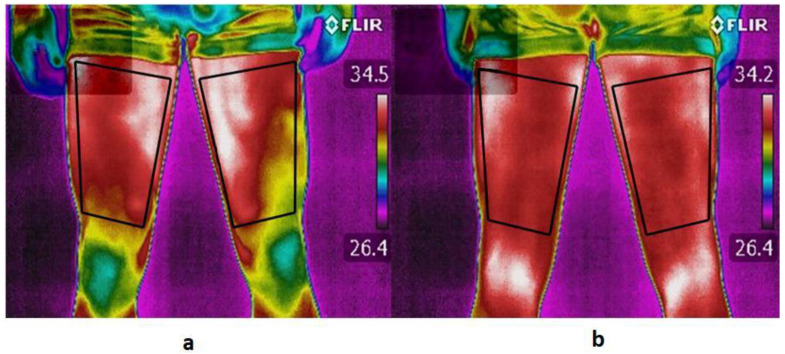
Air temperature, relative humidity and reflected temperature were measured and included in the camera settings. Two regions of interest (ROIs) were defined in the thermal images of the anterior (quadriceps) and posterior thighs (hamstrings), in both lower limbs, preferred (PL) and non-preferred lower limb (NPL) (Figure 1). Mean skin temperature was obtained using thermography software (Thermacam Researcher Pro 2.10, FLIR, Wilsonville, USA). All analyses were performed with a skin emissivity of 0.98 [29]. Regarding skin temperature differences, at least one of the following parameters was used to identify imbalances: bilateral (PL-NPL) and hamstrings–quadriceps differences greater than 0.5 °C. In a previous study, 0.4 °C was determined as the maximum temperature symmetry difference in healthy subjects, thus 0.5 °C was considered the cut-off for temperature imbalance [20].
Figure 1.
Determination of the ROIs of the anterior thigh (a) and posterior thigh (b) in both lower limbs. ROI was determined to cover the entire thigh, being the upper definition of the crotch, and the lower definition of the knee or popliteus.
2.3. Isokinetic Data Collection and Analysis
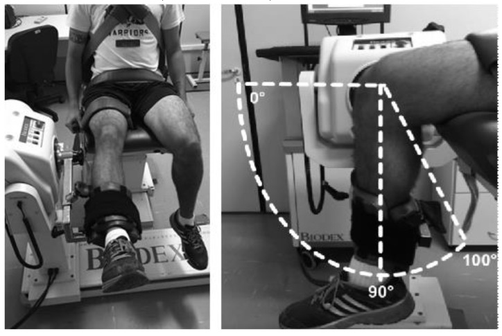
Isokinetic collection and analysis were performed after the acquisition of the thermal images according to the methods and recommendations described by Croiser et al. [5]. An isokinetic dynamometer, Biodex System 4.0 (Biodex Medical Systems, Shirley, NY, USA) was used to test the concentric and eccentric torque of the knee extensor and flexor muscles. Prior to the test, the device was calibrated following the manufacturer’s recommendations. A warm-up was performed in a cycle-ergometer with a load of 75–100 W for 5 min. Subsequently, the evaluator explained the testing procedures in detail and placed the volunteer on the equipment seat, at an angle allowing the hip joint to be at 105° flexion, with the body stabilized by straps around the thigh, waist and chest to avoid compensation. The range of knee motion was fixed at 100° flexion from the active maximum extension (Figure 2).
Figure 2.
Participant positioning for the isokinetic dynamometry test.
The rotational axis of the dynamometer arm was aligned with the lateral epicondyle of the femur of the right and left lower limb. The site of force application was positioned approximately 2 cm from the medial malleolus. Belts were fixed to the trunk, pelvis and thigh to prevent compensatory movements. The concentric torque of the quadriceps and hamstrings were evaluated in 3 repetitions at 60°/s and in 5 repetitions at 240°/s. The eccentric torque of the hamstrings was evaluated in 3 repetitions at 30°/s and in 4 repetitions at 120°/s. The recovery interval time between sets was 1 min. The same procedure was performed for the left lower limb. The volunteers were instructed to perform maximal strength. During the test, the participants were strongly encouraged, verbally, to exert “harder” and “stronger” efforts.
The results analyses were expressed in absolute (N.m) concentric and eccentric Peak Torque (PT) of knee extensors and flexors, as well as the bilateral comparison (preferred and non-preferred limbs), led to the determination of asymmetries [30]. The concentric H/Q peak torque ratio of flexors and extensors was established (at 60°/s or 240°/s) and the mixed Hecc/Qconc ratio was associated with the eccentric performance of the hamstrings and the concentric action of the quadriceps muscles (hamstrings at 30°/s versus quadriceps at 240°/s). Regarding the strength imbalance profile, this study followed the procedures described by Crosier et al. [5]: bilateral differences above 15% in concentric and/or eccentric in the hamstrings; concentric ratio (in at least 1 leg) of less than 0.45; and a mixed ratio of less than 0.89 on Biodex. Thus, at least 2 of the following parameters were used to identify the injury risk: concentric (at 60°/s or 240°/s) and eccentric (at 30°/s or 120°/s) bilateral asymmetries (>15%); conventional Hconc/Qconc (at 60°/s or 240°/s); and mixed Hecc/Qconc ratio.
2.4. Statistical Analyses
Data are presented as means, standard deviation and frequencies. The Kolmogorov–Smirnov test was used to check the normality of the data distribution. Torque and temperature ratios for preferred and non-preferred lower limbs were compared implementing independent t-tests. Two-way analysis of variance (group factor (high and low risk of strength imbalances) and limb factor (preferred and non-preferred)) was applied to compare muscular group (quadriceps and hamstrings) temperature. The Bonferroni post hoc was used for all analyses. All tests were performed using SPSS Statistics for Windows, version 21.0 (SPSS Inc., Chicago, IL, USA). The significance level was set at 0.05 for all comparisons.
3. Results
NPL hamstrings presented lower values (Table 1) for PT in three of the four velocities evaluated (30°/s and 120°/s eccentric and 240°/s concentric). In addition, the NPL exhibited statistically lower values for the conventional ratio (Conc/Conc 240°/s) and mixed ratio (Ecc 30°/s/Conc 240°/s).
Table 1.
Mean ± standard deviation PT and the hamstrings-to-quadriceps ratio (H/Q ratio) in preferred (PL) and non-preferred (NPL) limbs and asymmetry index (AI).
| Isokinetic Variables | PL (N.m) | NPL (N.m) | AI (%) | p |
|---|---|---|---|---|
| Quadriceps | ||||
| 60°/s Conc. | 206 ± 36 | 196 ± 36 | 3.99 ± 13.7 | 0.15 |
| 240°/s Conc. | 127 ± 21 | 128 ± 26 | −1.04 ± 18.0 | 0.93 |
| Hamstrings | ||||
| 30°/s Ecc. | 172 ± 31 | 160 ± 31 | 6.62 ± 14.3 | 0.03 * |
| 60°/s Con. | 115 ± 23 | 114 ± 28 | 0.81 ± 26.1 | 0.85 |
| 120°/s Ecc. | 173 ± 33 | 162 ± 27 | 4.66 ± 15.5 | 0.04 * |
| 240°/s Con. | 84 ± 15 | 78 ± 14 | 6.77 ± 11.7 | 0.02 * |
| H/Q Ratio | ||||
| Conc/Conc 60°/s | 0.56 ± 0.09 | 0.59 ± 0.16 | - | 0.22 |
| Conc/Conc 240°/s | 0.66 ± 0.08 | 0.62 ± 0.09 | - | 0.006 * |
| Ecc30°/s/Conc240°/s | 1.37 ± 0.24 | 1.27 ± 0.24 | - | 0.03 * |
* Significant differences between PL and NPL.
No significant differences were found between PL and NPL for the skin temperature of the quadricep and hamstring ROIs between the high and low risk groups of muscular imbalances (Table 2).
Table 2.
Skin temperature in the quadriceps and hamstrings for preferred (PL) and non-preferred limb (NPL) for the groups of high-risk torque imbalances (n = 22) and low-risk torque imbalances (n = 37).
| Variables | Imbalance Risk | PL (°C) | NPL (°C) | F 1 | p |
|---|---|---|---|---|---|
| Quadriceps | High | 32.51 ± 0.81 | 32.49 ± 0.76 | 0.002 | 0.964 |
| Low | 32.38 ± 0.82 | 32.37 ± 0.79 | |||
| Hamstrings | High | 32.32 ± 0.77 | 32.35 ± 0.75 | 0.002 | 0.962 |
| Low | 32.29 ± 0.68 | 32.30 ± 0.69 | |||
| ∆Temp (H-Q) | High | −0.19 ± 0.51 | −0.13 ± 0.49 | 0.022 | 0.882 |
| Low | −0.10 ± 0.46 | −0.07 ± 0.43 |
1 Anova interaction.
The highest rates of players with bilateral differences in PT were observed at velocities of 30°/s eccentric (30.5%), 60°/s concentric (23.7%) and 120°/s eccentric (22.0%) (Table 3). Regarding tskin differences higher than 0.5 °C, higher rates of players were observed when considering the difference between hamstrings and quadriceps (28.8% for PL and 44.1% for NPL) than for the difference between the preferred and non-preferred limb (3.9% for quadriceps and 0% for hamstrings). In terms of torque parameters, 37.3% (n = 22) of those evaluated would be classified as having high risk of injuries. On the other hand, 52.5% (n = 31) of those assessed presented thermal imbalances above 0.5 °C.
Table 3.
Rate of soccer players with torque and temperature imbalances criteria.
| Variables | Rate of Players (%) |
|---|---|
| Bilateral Difference | |
| Conc 60°/s | 14/59 (23.7) |
| Conc 240°/s | 6/59 (10.2) |
| Ecc 30°/s | 18/59 (30.5) |
| Ecc 120°/s | 13/59 (22.0) |
| ∆ Temp. (Quadriceps PL–Quadriceps NPL) | 2/59 (3.9) |
| ∆ Temp. (Hamstring PL–Hamstring NPL) | 0/59 (0.0) |
| Preferred Limb | |
| Conc 60°/s/Conc 60°/s | 5/59 (8.5) |
| Conc 240°/s/Conc 240°/s | 0/59 (0.0) |
| Mixed Ecc 30°/s/Conc2 40°/s | 1/59 (1.7) |
| ∆Temp. (Hamstrings–Quadriceps) | 17/59 (28.8) |
| Non-preferred Limb | |
| Conc 60°/s/Conc 60°/s | 6/59 (10.2) |
| Conc 240°/s/Conc 240°/s | 2/59 (3.4) |
| Mixed Ecc3 0°/s/Conc 240°/s | 4/59 (6.8) |
| ∆ Temp. (Hamstrings–Quadriceps) | 26/59 (44.1) |
| Injury Criteria | |
| Deficiency at least 2 parameters | 22/59 (37.3) |
| Total difference of temperature | 31/59 (52.5) |
Conc, concentric; Ecc, eccentric; H/Q, hamstring/quadriceps ratio; P, preferred limb; NP, non-preferred limb.
The IRT showed sensitivity (capacity to identify strength imbalance) and specificity (strength balance) of 22% and 32.2%, respectively, demonstrating the inability to identify false negatives (tskin balance and strength imbalance) and false positives (tskin imbalance and strength balance) of 15.3% and 30.5%, respectively (Table 4).
Table 4.
Prevalence of torque and skin temperature asymmetries, differences, sensitivity, specificity, false-positive and false-negative rates of infrared thermography to identify torque asymmetries observed by the isokinetic method.
| Frequency Analysis | Overall |
|---|---|
| Strength imbalance (%) | 37.3 |
| Skin temperature imbalance (%) | 52.5 |
| Difference (%) | 15.2 |
| Sensitivity (%) | 22.0 |
| Specificity (%) | 32.2 |
| Negative-false (%) | 15.3 |
| Positive-false (%) | 30.5 |
4. Discussion
This study aimed to identify asymmetries between limbs and muscular imbalances of soccer players and to associate tskin asymmetries through IRT. We hypothesized that athletes classified in the hamstring injury risk group by isokinetic evaluation would also present asymmetries in skin temperature parameters. However, the results indicate that the tskin asymmetry was not related to the strength imbalances indicated in the isokinetic test with cut-off points and normative data [5]. To the best of the authors’ knowledge, this is the first study to associate dynamometric parameters that are indicative of a high risk of hamstring injuries in soccer players with tskin through IRT.
Considering torque asymmetries, significant differences were observed between preferred and non-preferred limbs for hamstring concentric contractions at 240°/s and eccentric contractions at 30°/s. Similarly, bilateral asymmetries for soccer and futsal players were observed by other studies as well [3,4,5,6,31,32]. Ruas et al. [31] showed that the preferred limb of soccer players presented higher eccentric hamstring strength than the non-preferred limb. In addition, Nunes et al. [32] demonstrated that futsal players had greater concentric (at 240°/s) and eccentric (at 30°/s and 120°/s) hamstring strength for the preferred limb in comparison to the non-preferred limb. Despite these findings in the current study, the group average asymmetry did not exceed 15% (Table 1); however, across all torque categories (Conc at 60°/s and 240°/s; Ecc at 30°/s and 120°/s), only 10.2–30.5% of players exhibited asymmetries above 15% (Table 3). As a possible explanation, the non-preferred limb plays an important role in supportive strength to coordinate dominant knee actions [31].
In the present study, according to Croisier et al. [5], strength imbalances assessed by conventional and functional ratios presented low incidence (Table 3). In soccer players, the hamstrings muscle group is the most affected by injuries [33]. The hamstring muscles have a significant function in decelerating the extension of the lower limb in the thigh during ball striking, which can harm or damage the muscle–tendon unit [34,35]. In addition, hamstring muscles can be vulnerable to injury usually during quick changes from eccentric to concentric action, especially when the hamstrings become hip joint extensors [5,36]. Therefore, the factors responsible for the high incidence of hamstring injuries are muscle weakness, strength imbalance and previous injuries [5,6]. In an attempt to reduce the number of injuries in this muscle, recent studies have shown positive results with the inclusion of eccentric training sessions in the players’ routine [37,38].
Regarding tskin evaluated by IRT, no significant differences were found between the quadricep and hamstring ROIs, regardless of the injury risk or limb preference (Table 2). Bilateral tskin asymmetries in the quadricep and hamstring ROIs presented low incidence. Similarly, Bouzas Marins et al. [39] also reported the absence of bilateral tskin asymmetries for soccer players. These results are in agreement with other studies that observed the differences in the application of forces between lower limbs did not result in tskin asymmetries [40,41]. Hence, bilateral tskin asymmetries could be more related to the effect of an injury (e.g., inflammatory process or alteration of blood perfusion) [17,22] than with muscular strength imbalance.
On the other hand, a high incidence of thermal imbalances (>0.5 °C) was observed for hamstrings–quadriceps tskin of PL (28%) and NPL (44%) (Table 3). In this context, some investigations showed contradictory results. While Chudecka and Lubkowska [42] reported superior tskin in the quadriceps compared to hamstring ROIs, these findings were not confirmed by Bouzas-Marins et al. [39]. The causes for the presence of a higher local tskin are generally linked to the occurrence of hyperemia due to the inflammation process caused by the increase in blood flow to the injured region [43]; nonetheless, in the current study, no athlete reported injury at the time of evaluation. This data also indicate that the assessment of tskin by IRT when resting does not seem to be able to screen strength imbalances (positive-false = 30.5% and negative-false = 15.3%), resulting in hamstring injury risk. Therefore, it can be assumed that thermal differences between quadriceps and hamstrings could be more of a result of differences in tissue proportion (e.g., body fat), blood perfusion and capacity of heat dissipation than muscular strength imbalance.
Some limitations could be associated to the study. A higher sample with female players and a larger age range would have allowed the analysis of gender and age effects. Moreover, the measurement of other physiological parameters, such as muscle damage, neuromuscular activation or muscle oxygenation, would assist in interpreting the results as well. However, this study pioneered the investigation of possible relationships among the parameters commonly used for the identification of injury risk factors for the thigh muscles (dynamometric parameters) of soccer players and tskin by IRT of hamstrings and quadriceps. Considering previous reports about the importance of a daily IRT asymmetry assessment in injury prevention [17,19,21,22,23], long-term follow-up studies of athletes using IRT evaluation are encouraged. Finally, based on a recent study [44], further investigations assessing the skin temperature recovery after a cold stress test could add valuable information to the vascularization status of the region analyzed by infrared thermography.
5. Conclusions
The evaluation of skin temperature asymmetries by IRT at rest was unable to identify strength imbalances, and, consequently, hamstring injury risk in soccer players. Thermal differences between hamstrings and quadriceps could be more related to thermoregulatory factors than with strength imbalances.
Author Contributions
Conceptualization, R.M.T. and M.R.; methodology, J.I.P.-Q.; software, R.A.D. and T.M.P.d.R.; validation, J.F.d.S. and J.C.B.P.M.; formal analysis, R.M.T.; investigation, R.M.T. and T.M.P.d.R.; resources, M.R.; data curation, R.A.D. and T.M.P.d.R.; writing—original draft preparation, R.M.T.; and M.R.; and R.A.D.; writing—review and editing, J.I.P.-Q.; visualization, R.M.T.; R.A.D.; J.I.P.-Q.; J.C.B.P.M.; J.F.d.S.; T.M.P.d.R.; and M.R.; supervision, M.R.; project administration, R.M.T.; funding acquisition, M.R. All authors have read and agreed to the published version of the manuscript.
Funding
This research received no external funding.
Conflicts of Interest
The authors declare no conflict of interest.
References
- 1.Ekstrand J., Hägglund M., Waldén M. Epidemiology of muscle injuries in professional football (soccer) Am. J. Sports Med. 2011;39:1226–1232. doi: 10.1177/0363546510395879. [DOI] [PubMed] [Google Scholar]
- 2.Pfirrmann D., Herbst M., Ingelfinger P., Simon P., Tug S. Analysis of injury incidences in male professional adult and elite youth soccer players: A systematic review. J. Athl. Train. 2016;51:410–424. doi: 10.4085/1062-6050-51.6.03. [DOI] [PMC free article] [PubMed] [Google Scholar]
- 3.Fousekis K., Tsepis E., Vagenas G. Lower limb strength in professional soccer players: Profile, asymmetry, and training age. J. Sports Sci. Med. 2010;9:364–373. [PMC free article] [PubMed] [Google Scholar]
- 4.Boccia G., Brustio P.R., Buttacchio G., Calabrese M., Bruzzone M., Casale R., Rainoldi A. Interlimb asymmetries identified using the rate of torque development in ballistic contraction targeting submaximal torques. Front. Physiol. 2018;9:1–10. doi: 10.3389/fphys.2018.01701. [DOI] [PMC free article] [PubMed] [Google Scholar]
- 5.Croisier J.-L., Ganteaume S., Binet J., Genty M., Ferret J.-M. Strength imbalances and prevention of hamstring injury in professional soccer players: A prospective study. Am. J. Sports Med. 2008;36:1469–1475. doi: 10.1177/0363546508316764. [DOI] [PubMed] [Google Scholar]
- 6.Lehance C., Binet J., Bury T., Croisier J.L. Muscular strength, functional performances and injury risk in professional and junior elite soccer players. Scand. J. Med. Sci. Sports. 2009;19:243–251. doi: 10.1111/j.1600-0838.2008.00780.x. [DOI] [PubMed] [Google Scholar]
- 7.Ribeiro-Alvares J.B., Dornelles M.P., Fritsch C.G., de Lima-e-Silva F.X., Medeiros T.M., Severo-Silveira L., Marques V.B., Baroni B.M. Prevalence of hamstring strain injury risk factors in professional and under-20 male football (soccer) players. J. Sport Rehabil. 2019:1–7. doi: 10.1123/jsr.2018-0084. [DOI] [PubMed] [Google Scholar]
- 8.Petersen J., Thorborg K., Nielsen M.B., Budtz-Jørgensen E., Hölmich P. Preventive effect of eccentric training on acute hamstring injuries in men’s soccer. Am. J. Sports Med. 2011;39:2296–2303. doi: 10.1177/0363546511419277. [DOI] [PubMed] [Google Scholar]
- 9.Ekstrand J., Hagglund M., Walden M. Injury incidence and injury patterns in professional football: The UEFA injury study. Br. J. Sports Med. 2011;45:553–558. doi: 10.1136/bjsm.2009.060582. [DOI] [PubMed] [Google Scholar]
- 10.Anne-Marie van Beijsterveldt A.M.C., Stubbe J.H., Schmikli S.L., Van De Port I.G.L., Backx F.J.G. Differences in injury risk and characteristics between Dutch amateur and professional soccer players. J. Sci. Med. Sport. 2015;18:145–149. doi: 10.1016/j.jsams.2014.02.004. [DOI] [PubMed] [Google Scholar]
- 11.Denadai B.S., de Oliveira F.B.D., de Abreu Camarda S.R., Ribeiro L., Greco C.C. Hamstrings-to-quadriceps strength and size ratios of male professional soccer players with muscle imbalance. Clin. Physiol. Funct. Imaging. 2016;36:159–164. doi: 10.1111/cpf.12209. [DOI] [PubMed] [Google Scholar]
- 12.Grygorowicz M., Kubacki J., Pilis W., Gieremek K., Rzapka R. Selected isokinetic tests in knee injury prevention. Biol. Sport. 2010;27:47–51. doi: 10.5604/20831862.907793. [DOI] [Google Scholar]
- 13.Carpes F.P., Mello-Carpes P.B., Priego Quesada J.I., Pérez-Soriano P., Salvador Palmer R., Ortiz de Anda R.M.C. Insights on the use of thermography in human physiology practical classes. Adv. Physiol. Educ. 2018;42:521–525. doi: 10.1152/advan.00118.2018. [DOI] [PubMed] [Google Scholar]
- 14.Moreira D.G., Costello J.T., Brito C.J., Adamczyk J.G., Ammer K., Bach A.J.E., Costa C.M.A., Eglin C., Fernandes A.A., Fernández-Cuevas I., et al. Thermographic imaging in sports and exercise medicine: A Delphi study and consensus statement on the measurement of human skin temperature. J. Therm. Biol. 2017;69:155–162. doi: 10.1016/j.jtherbio.2017.07.006. [DOI] [PubMed] [Google Scholar]
- 15.Lahiri B.B., Bagavathiappan S., Jayakumar T., Philip J. Medical applications of infrared thermography: A review. Infrared Phys. Technol. 2012;55:221–235. doi: 10.1016/j.infrared.2012.03.007. [DOI] [PMC free article] [PubMed] [Google Scholar]
- 16.Fernandes A.A., Dos Santos Amorim P.R., Brito C.J., De Moura A.G., Moreira D.G., Costa C.M.A., Sillero-Quintana M., Marins J.C.B. Measuring skin temperature before, during and after exercise: A comparison of thermocouples and infrared thermography. Physiol. Meas. 2014;35:189–203. doi: 10.1088/0967-3334/35/2/189. [DOI] [PubMed] [Google Scholar]
- 17.Hildebrandt C., Raschner C., Ammer K. An overview of recent application of medical infrared thermography in sports medicine in Austria. Sensors. 2010;10:4700–4715. doi: 10.3390/s100504700. [DOI] [PMC free article] [PubMed] [Google Scholar]
- 18.Colim A., Arezes P., Flores P., Vardasca R., Braga A.C. Thermographic differences due to dynamic work tasks on individuals with different obesity levels: A preliminary study. Comput. Methods Biomech. Biomed. Eng. Imaging Vis. 2020;8:323–333. doi: 10.1080/21681163.2019.1697757. [DOI] [Google Scholar]
- 19.Fernández-Cuevas I., Lastras J.A., Galindo V.E., Carmona P.G. Infrared thermography for the detection of injury in sports medicine. In: Priego Quesada J.I., editor. Application of Infrared Thermography in Sports Science. Springer International Publishing; Cham, Switzerland: 2017. pp. 81–109. Biological and Medical Physics, Biomedical Engineering. [Google Scholar]
- 20.Vardasca R., Ring E., Plassmann P. Thermal symmetry of the upper and lower extremities in healthy subjects. Thermol. Int. 2012;22:53–60. [Google Scholar]
- 21.Sanchis-Sánchez E., Vergara-Hernández C., Cibrián R.M., Salvador R., Sanchis E., Codoñer-Franch P. Infrared thermal imaging in the diagnosis of musculoskeletal injuries: A systematic review and meta-analysis. Am. J. Roentgenol. 2014;203:875–882. doi: 10.2214/AJR.13.11716. [DOI] [PubMed] [Google Scholar]
- 22.Côrte A.C., Pedrinelli A., Marttos A., Souza I.F.G., Grava J., José Hernandez A. Infrared thermography study as a complementary method of screening and prevention of muscle injuries: Pilot study. BMJ Open Sport Exerc. Med. 2019;5:1–5. doi: 10.1136/bmjsem-2018-000431. [DOI] [PMC free article] [PubMed] [Google Scholar]
- 23.Gómez-Carmona P., Fernández-Cuevas I., Sillero-Quintana M., Arnaiz-Lastras J., Navandar A. Infrared thermography protocol on reducing the incidence of soccer injuries. J. Sport Rehabil. 2020:1–6. doi: 10.1123/jsr.2019-0056. [DOI] [PubMed] [Google Scholar]
- 24.Gatt A., Formosa C., Cassar K., Camilleri K.P., De Raffaele C., Mizzi A., Azzopardi C., Mizzi S., Falzon O., Cristina S., et al. Thermographic patterns of the upper and lower limbs: Baseline data. Int. J. Vasc. Med. 2015;2015:1–9. doi: 10.1155/2015/831369. [DOI] [PMC free article] [PubMed] [Google Scholar]
- 25.van Melick N., Meddeler B.M., Hoogeboom T.J., Nijhuis-van der Sanden M.W.G., van Cingel R.E.H. How to determine leg dominance: The agreement between self-reported and observed performance in healthy adults. PLoS ONE. 2017;12 doi: 10.1371/journal.pone.0189876. [DOI] [PMC free article] [PubMed] [Google Scholar]
- 26.Priego Quesada J.I., Lucas-Cuevas A.G., Salvador Palmer R., Pérez-Soriano P., Cibrián Ortiz de Anda R.M. Definition of the thermographic regions of interest in cycling by using a factor analysis. Infrared Phys. Technol. 2016;75:180–186. doi: 10.1016/j.infrared.2016.01.014. [DOI] [Google Scholar]
- 27.Marins J.C.B., Moreira D.G., Cano S.P., Quintana M.S., Soares D.D., de Andrade Fernandes A., da Silva F.S., Costa C.M.A., dos Santos Amorim P.R. Time required to stabilize thermographic images at rest. Infrared Phys. Technol. 2014;65:30–35. doi: 10.1016/j.infrared.2014.02.008. [DOI] [Google Scholar]
- 28.Priego Quesada J.I., Kunzler M.R., Carpes F.P. Application of Infrared Thermography in Sports Science. Springer International Publishing; Cham, Switzerland: 2017. Methodological aspects of infrared thermography in human assessment; pp. 49–79. [Google Scholar]
- 29.Steketee J. Spectral emissivity of skin and pericardium. Phys. Med. Biol. 1973;18:307. doi: 10.1088/0031-9155/18/5/307. [DOI] [PubMed] [Google Scholar]
- 30.Chavet P., Lafortune M., Gray J.R. Asymmetry of lower extremity responses to external impact loading. Hum. Mov. Sci. 1997;16:391–406. doi: 10.1016/S0167-9457(96)00046-2. [DOI] [Google Scholar]
- 31.Ruas C.V., Minozzo F., Pinto M.D., Brown L.E., Pinto R.S. Lower-extremity strength ratios of professional soccer players according to field position. J. Strength Cond. Res. 2015;29:1220–1226. doi: 10.1519/JSC.0000000000000766. [DOI] [PubMed] [Google Scholar]
- 32.Nunes R.F.H., Dellagrana R.A., Nakamura F.Y., Buzzachera C.F., Almeida F.A.M., Flores L.J.F., Guglielmo L.G.A., da Silva S.G. Isokinetic assessment of muscular strength and balance in brazilian elite futsal players. Int. J. Sports Phys. Ther. 2018;13:94–103. doi: 10.26603/ijspt20180094. [DOI] [PMC free article] [PubMed] [Google Scholar]
- 33.Ekstrand J., Healy J.C., Waldén M., Lee J.C., English B., Hägglund M. Hamstring muscle injuries in professional football: The correlation of MRI findings with return to play. Br. J. Sports Med. 2012;46:112–117. doi: 10.1136/bjsports-2011-090155. [DOI] [PubMed] [Google Scholar]
- 34.Aicale R., Tarantino D., Maffulli N. Overuse injuries in sport: A comprehensive overview. J. Orthop. Surg. 2018;13:309. doi: 10.1186/s13018-018-1017-5. [DOI] [PMC free article] [PubMed] [Google Scholar]
- 35.Garrett W.E. Muscle strain injuries: Clinical and basic aspects. Med. Sci. Sports Exerc. 1990;22:436–443. doi: 10.1249/00005768-199008000-00003. [DOI] [PubMed] [Google Scholar]
- 36.Lee J.W.Y., Mok K.-M., Chan H.C.K., Yung P.S.H., Chan K.-M. Eccentric hamstring strength deficit and poor hamstring-to-quadriceps ratio are risk factors for hamstring strain injury in football: A prospective study of 146 professional players. J. Sci. Med. Sport. 2018;21:789–793. doi: 10.1016/j.jsams.2017.11.017. [DOI] [PubMed] [Google Scholar]
- 37.Shadle I.B., Cacolice P.A. Eccentric exercises reduce hamstring strains in elite adult male soccer players: A critically appraised topic. J. Sport Rehabil. 2016;26:573–577. doi: 10.1123/jsr.2015-0196. [DOI] [PubMed] [Google Scholar]
- 38.Van Der Horst N., Smits D.W., Petersen J., Goedhart E.A., Backx F.J.G. The preventive effect of the nordic hamstring exercise on hamstring injuries in amateur soccer players: A randomized controlled trial. Am. J. Sports Med. 2015;43:1316–1323. doi: 10.1177/0363546515574057. [DOI] [PubMed] [Google Scholar]
- 39.Bouzas Marins J.C., de Andrade Fernandes A., Gomes Moreira D., Souza Silva F., Costa C.M.A., Pimenta E.M., Sillero-Quintana M. Thermographic profile of soccer players’ lower limbs. Rev. Andal. Med. Deporte. 2014;7:1–6. doi: 10.1016/S1888-7546(14)70053-X. [DOI] [Google Scholar]
- 40.Arfaoui A., Bertucci W., Letellier T., Polidori G. Thermoregulation during incremental exercise in masters cycling. J. Sci. Cycl. 2014;3:33–41. [Google Scholar]
- 41.Trecroci A., Formenti D., Ludwig N., Gargano M., Bosio A., Rampinini E., Alberti G. Bilateral asymmetry of skin temperature is not related to bilateral asymmetry of crank torque during an incremental cycling exercise to exhaustion. PeerJ. 2018;6:e4438. doi: 10.7717/peerj.4438. [DOI] [PMC free article] [PubMed] [Google Scholar]
- 42.Chudecka M., Lubkowska A. Thermal maps of young women and men. Infrared Phys. Technol. 2015;69:81–87. doi: 10.1016/j.infrared.2015.01.012. [DOI] [Google Scholar]
- 43.Herborn K.A., Graves J.L., Jerem P., Evans N.P., Nager R., McCafferty D.J., McKeegan D.E.F. Skin temperature reveals the intensity of acute stress. Physiol. Behav. 2015;152:225–230. doi: 10.1016/j.physbeh.2015.09.032. [DOI] [PMC free article] [PubMed] [Google Scholar]
- 44.Priego-Quesada J.I., Pérez-Guarner A., Gandia-Soriano A., Oficial-Casado F., Galindo C., Anda R.M.C.O.D., Piñeiro-Ramos J.D., Sánchez-Illana Á., Kuligowski J., Barbosa M.A.G., et al. Effect of a marathon on skin temperature response after a cold-stress test and its relationship with perceptive, performance, and oxidative-stress biomarkers. Int. J. Sports Physiol. Perform. 2020;1:1–9. doi: 10.1123/ijspp.2019-0963. [DOI] [PubMed] [Google Scholar]


