

Abstract
Propolis is a natural resinous material produced by bees and has been used in folk medicines since ancient times. Due to it possessing a broad spectrum of biological activities, it has gained significant scientific and commercial interest over the last two decades. As a result of searching 122 publications reported up to the end of 2019, we assembled a unique compound database consisting of 578 components isolated from both honey bee propolis and stingless bee propolis, and analyzed the chemical space and chemical diversity of these compounds. The results demonstrated that both honey bee propolis and stingless bee propolis are valuable sources for pharmaceutical and nutraceutical development.
Keywords: honey bee propolis, stingless bee propolis, natural products, phenolics, terpenoids, chemoinformatics, chemical space, chemical diversity
1. Introduction
The emergence of new infectious and chronic diseases makes the need for new drugs paramount [1]. Although the search for new drugs can begin from different sources, natural products have proven to be one of the richest sources of bioactive ingredients and molecules with privileged scaffolds for the discovery and development of new and novel drugs [2,3,4,5,6]. They were historically the sources of all folk medicines [7]. Having evolved over millions of years, structures of natural products have been fine-tuned by nature for optimal bioactivity [5]. Modern studies revealed natural products possess an advantageous structural foundation and cover a wide range of biologically relevant chemical space that cannot be efficiently explored by synthetic compounds [8,9,10]. These features positively influence the probability of the clinical success of natural product-based drug candidates [11]. A detailed analysis of 1394 new small molecule drugs approved by the US Food and Drug Administration (FDA) between 1981 and 2019 [6] revealed that 32% of those drugs were natural products or direct derivatives of natural products.
Propolis, which is a product of bees, has been used in the folk medicine of many cultures to treat microbial infections since the year 300 B.C. [12]. The name “propolis” originally came from the Greek words meaning “defence of the city” (“pro” meaning “to defend” and “polis” meaning the city) [13]. Historically, the Greeks and the Romans used propolis for treating bruises and suppurating sores; the Egyptians applied propolis for embalming cadavers and preventing infections; the Arabians utilised propolis as an antiseptic, a wound healing agent, and a mouth disinfectant; the Incas described propolis as an antipyretic agent [14]. Owing to its antibacterial characteristics, propolis was approved as an official drug in the London pharmacopoeia in the 17th century and, since then, has become more popular [15]. Propolis was also used to treat wounds during World War II (1939–1945) [14]. In 1969, propolis was approved as human and veterinary drugs with several applications, including the treatment of tuberculosis in the Union of Soviet Socialist Republics [14].
Since the early 21st century, there has been a significant increase in scientific publications on propolis (Figure 1). Studies validated the antimicrobial property of propolis extracts and discovered additional therapeutic properties, including antioxidant, anti-inflammatory, antidiabetic, dermatoprotective, antiallergic, laxative, immunomodulatory, and anticancer activities [16]. Nowadays, propolis is used in pharmaceutical and cosmetic industries as a unique natural constituent in cough syrups, dietary supplement tablets, antiacne creams, facial and body creams, ointments, lotions, toothpastes, and mouthwash products [17]. It has also been used in some foods and beverages as an alternate preservative agent or food supplement [13]. The first patent referring to propolis was described in 1904 with a claim of using propolis as one of the compositions to treat piano pins and strings [18]. Propolis-related patents numbered about 500 by the end of the 20th century and increased dramatically by almost three-fold and nine-fold in the first and second decades of the 21st century, respectively. The number of patents referring to propolis from 2011 to 2019 accounted for 50% of its total publications in the same period (Figure 1). Medicinal and nutraceutical products were observed in high frequency in these patent applications.
Figure 1.

Number of scientific outputs containing the word “propolis” per decade (publications include books, clinical trials, commentaries, conferences, dissertations, editorials, journals, letters, patents, preprints, reports, and reviews—searched on SciFinder database (Chemical Abstract Service) on 2nd Jan 2020).
Over the last two decades, the relationships between the pharmacological properties of propolis and its components have attracted the attention of the scientific community. It is known that raw propolis, in general, consists of about 50% resin, 30% wax, 10% essential oils, 5% pollen, and 5% others (including amino acids, peptides, dead bees, and soil) [19]. By employing different chromatography and spectroscopic techniques, such as thin layer chromatography, gas chromatography (GC), high-performance liquid chromatography (HPLC), mass spectroscopy (MS), and nuclear magnetic resonance spectroscopy (NMR), over 300 volatile and non-volatile components have been identified in propolis [20]. Among them, phenolics and terpenoids have been confirmed to play important roles in the biological activities of propolis [17,21,22,23].
Several comprehensive reviews have reported the natural compositions found in propolis [13,17,20,24,25,26,27,28,29,30] and their biological activities [13,14,16,22,23,24,31,32,33,34,35]. However, the chemical space and the chemical diversity of propolis components have been underexplored. In this article, we review all compounds isolated from both honey bee propolis (HBP) and stingless bee propolis (SBP), which have been fully characterized and reported in the literature up to the end of 2019. Compounds identified from GC-MS and LC-MS were excluded in this study. As a result of the search, we assembled a database with 578 unique compounds. The chemical space and chemical diversity of the propolis components were characterized to assess their potential for future developments as pharmaceuticals and nutraceuticals.
2. Propolis Components: Chemistry and Geographical Distributions
2.1. Propolis Classification
2.1.1. Honey Bee Propolis
The honey bee genus Apis is the only genus of the tribe Apini in the Apidae family [36]. This genus consists of 11 species, including A. andreniformis, A. binghami, A. breviligula, A. cerana, A. dorsata, A. florea, A. koschevnikovi, A. laboriosa, A. mellifera, A. nigrocincta, and A. nuluensis [36]. These bees are well known for their production of honey, as well as being the pollinator of the majority of the worlds commercial fruit crops [36]. Apis mellifera, which is the most common species of honey bee, is indigenous to Europe, Africa, and the Middle East, but nowadays has been found in almost all regions of the world [28]. It has been known that A. mellifera produces a high yield of propolis, while other honey bee species provide relatively small or no propolis [21,36].
Honey bee propolis (HBP) is produced mainly from the exudates of plant tissues, such as flower buds, bark and fruit, mixed with saliva and beeswax by bees [24]. The bees gather plant exudates, often referred to as resin, which contain substances involving chemical defense systems to protect plants against their herbivores, bacteria, fungi, moulds and viruses, during the warm part of the day when resin is soft [36]. The bees pack resins on their hind legs and transport them back to the hive to fill hive cracks, reducing the size of the hive entrance to prevent the invasion of other insects and to seal up the inside of the hive by mixing it with wax to maintain an antiseptic environment for the colony and larvae [37,38]. Physically, propolis is soft, pliable, and very sticky when warm, but becomes hard and brittle when cold. Its melting point is around 65 °C, but in some samples it is as high as 100 °C [17]. It has a pleasant aromatic smell and varies in colour depending on its plant sources and age [24]. On average, one bee can bring 10 mg propolis per flight to its hive, and one colony collects about 50–150 g propolis annually [39]. With the application of specialised collection procedures, the sub-species of the European honey bee, A. mellifera causasica, can produce 250–1000 g of propolis annually, per hive [21,40].
2.1.2. Stingless Bee Propolis (Cerumen or Geopropolis)
Stingless bees belonging to the tribe Meliponini, in the Apidae family, are the largest group of eusocial bees on Earth, and are closely related to the common honey bee, A. mellifera [41]. About 619 stingless bee species in 61 genera have been found in tropical regions of America (South and Central Americas), Africa, Southeast Asia, and Northern Oceania [41]. It is estimated that 40–90% of native or cultivated plant species in the tropics are pollinated by stingless bees [33]. Compared to honey bees, stingless bees have many different features, including colony size, nesting biology, brood comb composure, bee queen production, stocking strategy, and bee recruitment mechanisms [41]. The most significant difference is that they are ‘stingless’, which refers to the fact that their sting is highly reduced, and they do not use it for defense. Instead, some stingless bees develop other methods to protect themselves, such as a strong bite or increasing the pain of the bite by producing formic acid through their mandibular glands [29].
Both honey bees and stingless bees are able to produce propolis (Figure 2). While the honey bee’s nests are structurally double-sided hexagonal combs built primarily from wax and their hives are sealed by propolis resin, the nests of stingless bees are more complex with a great variety of forms and size, and are made primarily from a propolis-based substance called cerumen [42]. The terms cerumen and propolis are used interchangeably in the literature with respect to stingless bees. Propolis from stingless bees is sometimes found as a mixture of resin and clay or soil. Therefore, this product is also called geopropolis [29].
Figure 2.

Propolis of the honey bee A. mellifera (A) and the Australian stingless bee Tetragonula carbonaria (B).
2.2. Chemical Components of Propolis
Chemical investigations of HBP have been undertaken since the mid-20th century. However, the literature reports of the discovery of HBP compositions were relatively small prior to 1996, with a significant increase since 2010 (Figure 3). Potentially, this increase in interest was stimulated by the scientific validation of the pharmacological properties of HBP during the late 1990s and early 2000s [30]. Up to December 2019, there were 502 different natural products isolated and characterised from materials collected in 40 countries (Figure 4 and Figure 5C, and Supporting Information 2). In contrast, propolis produced by stingless bees has only relatively recently been studied with the first isolation of three diterpenes from the Brazilian Melipona quadrifasciata anthidioides SBP in 2000 [43]. In the early 2000s, most studies were dedicated to Brazilian SBP. However, more recently the number of publications on SBP from Southeast Asia and Australia has grown significantly. A total of 100 compounds have been identified from SBP from 2000 to 2019 (Figure 4, and Supporting Information 2). A total of 24 of the 100 compounds have been previously identified in HBP.
Figure 3.

Publications reporting compounds discovered from propolis (n = 122) [25,42,43,44,45,46,47,48,49,50,51,52,53,54,55,56,57,58,59,60,61,62,63,64,65,66,67,68,69,70,71,72,73,74,75,76,77,78,79,80,81,82,83,84,85,86,87,88,89,90,91,92,93,94,95,96,97,98,99,100,101,102,103,104,105,106,107,108,109,110,111,112,113,114,115,116,117,118,119,120,121,122,123,124,125,126,127,128,129,130,131,132,133,134,135,136,137,138,139,140,141,142,143,144,145,146,147,148,149,150,151,152,153,154,155,156,157,158,159,160,161,162].
Figure 4.

Number of compounds isolated from propolis (n = 578) (Blue: HBP (n = 502); orange: SBP (n = 100)) (overlapped compounds were removed).
Figure 5.

Geographic distribution of compounds isolated from HBP (A,C) and SBP (B,D) based on continents (A,B) and countries (C,D).
America, particularly Central and South America, is a continent where the most HBP compounds (352 compounds) have been identified and reported, followed by Asia (166 compounds), Africa (100 compounds), Europe (72 compounds), and Oceania (68 compounds) (Figure 5A). Among the 40 countries where compounds have been isolated and identified from HBP, Brazil is a leader with 158 compounds discovered, followed by Mexico (69 compounds), Nepal (37 compounds), Australia (36 compounds), and Greece (35 compounds) (Figure 5C).
In term of SBP, most compounds have been reported from Asian SBP (Figure 5B). Only seven countries, including Brazil (Melipona interrupta [126], M. quadrifasciata anthidioides [43], M. seminigra [126], M. scutellaris [139], M. subnitida [122], and Tetragonula (Trigona) spinipes [88] bees), Indonesia (Tetragonula aff. biroi [42], T. sapiens [160], and T. incisa [136] bees), Malaysia (Heterotrigona itama [149] bee), Philippines (Tetragonula biroi [137] bee), Thailand (Tetragonula laeviceps [138], T. pagdeni [151], and Tetrigona melanoleuca [138] bees), Vietnam (Lisotrigona cacciae [157], L. furva [153], and Tetragonula minor [146,152] bees), and Australia (Tetragonula carbonaria [131,140] bee) have published their SBP studies (Figure 5D). Vietnam is leading the numbers of compounds isolated from SBP, with 34 compounds, followed by Brazil (29 compounds) and Thailand (19 compounds). Australia is the only representative of Oceania reporting eight compounds identified from SBP. Interestingly, there are no reports of isolated compounds from African SBP, although the extracts of Kenyan SBP Dactylurina schimidti [163] and Nigerian SBP Dactylurina studingeri [164] were reported to have an antimicrobial activity (Figure 5B).
Collation and analysis of the compounds isolated from HBP and SBP revealed that phenolics and terpenoids were the two compound classes that were most often found in propolis. Figure 6A and Figure 7A highlighted that phenolic compounds were dominant, with 79.5% and 63.0% of compounds isolated from HBP and SBP, respectively. Following the ways of the phenolic sub-class classification utilized in previous propolis reviews [20,26,165], nearly 30 sub-classes of phenolics were found in HBP but only half of them were identified in SBP (Figure 6B and Figure 7B). Phenylpropanoids (20.1%) and flavanone (12.5%) were commonly present in HBP (Figure 6B), while flavanone (20.6%) and xanthone (20.6%) were often found in SBP (Figure 7B).
Figure 6.

(A) Class of compounds isolated from HBP (n = 502) (phenolics and terpenoids include their glycosides); (B) sub-class of phenolics; (C) sub-class of terpenoids (overlapped compounds were removed).
Figure 7.
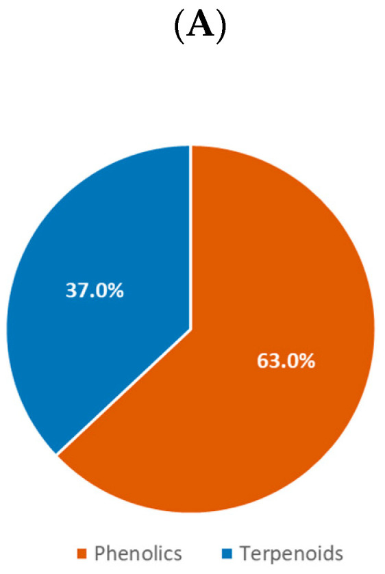

(A) Class of compounds isolated from SBP (n = 100) (phenolics include their glycosides); (B) sub-class of phenolics; (C) sub-class of terpenoids (overlapped compounds were removed).
The terpenoids accounted for 18.9% of all compounds found in HBP and 37.0% in SBP (Figure 6C and Figure 7C). They consisted of triterpenoids, diterpenoids, sesquiterpenoids, and monoterpenoids. The HBP diterpenes and triterpenes were similarly represented, with 46.3% and 45.3%, respectively. However, triterpenes occupied the highest proportion of compounds identified in SBP, with 86.5%. Approximately 6.0% of terpenoids identified in both types of propolis were sesquiterpenes. Only two monoterpenes, tschimgin and tschimganin, have been reported so far [107]. These two compounds were isolated from Iranian HBP of which a plant Ferula spp. is their botanical source [107]. Interestingly, only 5 out of 578 propolis compounds were identified as glycoside compounds including isorhamnetin-3-O-rutinoside from Cretan (Greek) A. mellifera HBP [96], ent-8(17)-labden-15-O-α-l-rhamnopyranoside, and ent-8(17)-labden-15-O-(3′-O-acetyl)-α-l-rhamnopyranoside from Salvadorian A. mellifera HBP [64], and naringenin-4′-O-β-d-glucopyranoside and myricetin-3-O-β-d-glucopyranoside from Brazilian Melipona interrupta and M. seminigra SBP [126].
2.3. Characteristic Chemical Class of Propolis
According to the chemo-geographic data, Bankova [165] classified six main HBP types, consisting of (a) Poplar propolis from Europe, North America, and the non-tropical regions of Asia, containing flavones, flavanones, and phenylpropanoids; (b) Birch propolis from Russia containing flavones and flavonols; (c) green propolis from Brazil containing prenylated phenylpropanoids; (d) red propolis from Cuba and Venezuela containing polyprenylated acylphloroglucinols; (e) Pacific propolis from Okinawa and Taiwan containing prenylated flavanones; and (f) Canarian propolis from Canary Islands containing furofuran lignans. More recently, Salatino and his co-workers [26] suggested five HBP types based on climate zones, including (a) temperate poplar propolis derived from Populus spp. with flavonoids, esters of phenylpropanoids; (b) Brazilian tropical green propolis with prenylated phenylpropanoids and caffeoylquinic acids; (c) Brazilian tropical brown propolis derived from Clusia spp. with polyprenylated acylphloroglucinols; (d) sub-tropical and tropical Pacific propolis derived from Macaranga spp., with geranyl flavonoids; and (e) Greek, Cretan, and Turkish propolis (Mediterranean region) with either diterpenoids or anthraquinones. Several reviews of SBP reported the chemical compositions and their biological activities [29,30,166]. However, most of the compounds reviewed were identified by HPLC, GC-MS, and LC-MS. In this review, we only included fully characterized compounds from HBP and SBP and categorized them based on their chemical classes (Figure 8).
Figure 8.


Characteristic chemical constituents of propolis (black: compound name; blue: compound class; purple: continental distribution). (A) HBP; (B) SBP.
Flavanone, flavone and phenylpropanoid, particularly phenylpropanoid esters, are often found from temperate HBP in Africa, America, Asia, Europe, and Oceania (Figure 8A). These compounds were likely foraged from Populus spp. (Algeria [124,154], Mexico [101], Uruguay [68], China [120], Bulgaria [45], and the Netherlands [65]), Zuccagnia punctate (Argentina [98]), Liquidambar styraciflua (Honduras [119]), Pinus halepensis (Jordan [113]), Styrax spp. (Thailand [123]), Betula verrucosa (Russia [25]), or Xanthorrhoea spp. (Australia [44]) (Table 1). Pinocembrin, chrysin, and caffeic acid phenyl ester (CAPE or phenethyl caffeate) are three common compounds present in these types of propolis. They showed a wide range of biological activities such as antioxidation, anticancer, antimicrobes, anti-inflammation, neuroprotection, and hepatoprotection (Table 2) [167,168,169].
Table 1.
Botanical sources of propolis categorized by chemical class.
| Plant Species | Plant Family | Characteristic Chemical Class | Bee Species | Country |
|---|---|---|---|---|
| Acacia paradoxa | Fabaceae | Chalcone Flavanonol |
A. mellifera | Australia [121] |
| Anacardium occidentale | Anacardiaceae | Cycloartane-type triterpene | A. mellifera | Brazil [90] |
| Araucaria heterophylla | Araucariaceae | Labdane-type diterpene | A. mellifera | Brazil [48] |
| Azadirachta indica | Meliaceae | Prenylated flavanone | A. mellifera | Oman [125] |
| Baccharis spp. | Asteraceae | Flavanone/Flavanonol Flavone/Flavonol Phenylpropanoid ester Prenylated phenylpropanoid Labdane-type diterpene |
A. mellifera | Brazil [53,59] |
| Betula verrucosa | Betulaceae | Flavone/Flavonol | A. mellifera | Russia [25] |
| Bursera simaruba | Burseraceae | Cycloartane-type triterpene | A. mellifera | Mexico [158] |
| Cistus spp. | Cistaceae | Labdane-type diterpene | A. mellifera | Algeria [124] |
| Clusia spp. | Clusiaceae | Polyprenylated acylphloroglucinol | A. mellifera | Cuba [66] and Venezuela [77] |
| Corymbia torelliana | Myrtaceae | Flavanone/Flavanonol | T. carbonaria | Australia [131] |
| Dalbergia spp. | Fabaceae | Pterocarpan Isoflavone Isoflavane Dalbergione |
A. mellifera | Brazil [89], Cuba [81,129], Mexico [103], Nepal [78,86,87], and Nigeria [141,154] |
| Garcinia mangostana | Guttiferae | Xanthone |
T. laeviceps
T. pagdeni L. cacciae |
Thai [138,151] and Vietnamese [157] |
| Kielmeyera sp. | Calophyllaceae | Coumarin | M. scutellaris | Brazil [139] |
| Lepidosperma spp. | Cyperaceae | Stilbene | A. mellifera | Australia [121,145] |
| Liquidambar styraciflua | Altingiaceae | Flavanone Phenylpropanoid ester |
A. mellifera | Honduras [119] |
| Macaranga spp. | Euphorbiaceae | Prenylated flavanone | A. mellifera | Japan [75,85], Taiwan [70,84], Fiji [143], Solomon Island [106,117,118], Egypt [92,100] and Nigeria [141] |
| Mangifera indica | Anacardiaceae | Cycloartane-type triterpene |
A. mellifera
Tetragonula sapiens T. minor |
Brazil [79], Indonesia [114,160], Myanmar [93], Thailand [148], Vietnam [146] |
| Pinus halepensis | Pinaceae | Flavanone/Flavanonol Flavone/Flavonol |
A. mellifera | Jordan [113] |
| Populus spp. | Salicaceae | Flavanone/Flavone Phenylpropanoid ester |
A. mellifera | Algeria [124,154], Mexico [101], Uruguay [68], China [120], Bulgaria [45], Netherland [65] |
| Styrax spp. | Styracaceae | Flavanone/Flavanonol Flavone/Flavonol Phenylpropanoid ester |
A. mellifera | Thailand [123] |
| Xanthorrhoea spp. | Xanthorrhoeaceae | Flavanone | A. mellifera | Australia [44] |
| Zuccagnia punctate | Caesalpinieae | Flavanone/Flavonol | A. mellifera | Argentina [98] |
Table 2.
Representative compounds in propolis with known biological activities.
| Compound | Chemical Class | Phenotypic Activity | Molecular Target Activity |
|---|---|---|---|
| Artepillin C | Prenylated phenylpropanoids | Antibacteria (inhibition of B. cereus, B. Subtilis, M. lysodeikticus, P. aeruginosa, E. aerogenes, M. smegmatis, S. faecalis, E. coli, C. equi, and S. aureus [177]) Antifungi (inhibition of C. albicans, C. tropicalis, C. neoformans, S. cerevisiae, A. fumigatus, A. flavus, A. niger, M.canis, M. gypseum, E. floccosum, T. rubrum, and T. mentagrophytes [177]) Antitrypanosome (inhibition of trypomastigote forms of T. cruzi [184]) Antioxidation (in vivo inhibition of lipid peroxidation [185]) Anticancer (inhibition of human cancer cell lines [186,187,188]) |
Anti-inflammation (in vitro and in vivo inhibition of NO through NF-κB [178]) |
| Caffeic acid phenyl ester—CAPE (Phenethyl caffeate) | Phenylpropanoid ester | Antibacteria (inhibition of S. aureus, B. subtilis, and P. aeruginosa [189]) Antivirus (inhibition of AH1N1 [189] and hepatitis C virus [190]) |
Antioxidation (inhibition of 5-lipoxygenase [191]) Antivirus (inhibition of HIV-1 integrase [192]) Anti-inflammation (in vivo inhibition of COX- 2 [193], inhibition of NF-κB [194], in vitro and in vivo scavenging of NO and modulation of iNOS expression [195]) Anticancer (inhibition of protein kinase C [196], in vitro and in vivo inhibition of MMP-2, MMP-9 and VEGF [197]) Neuroprotection (scavenging ROS [198]) Hepatoprotection (in vivo inhibition of CYP2E1 [199]) |
| Chrysin | Flavone | Neuroprotection (in vitro and in vivo inhibition of acrylamide-induced toxicity [200]) Antivirus (inhibition of enterovirus 71 [201]) |
Anticancer (in vitro and in vivo activation of Notch1 signalling [202], regulating MMP-10 and epithelial-mesenchymal transition [203], inhibition of HIF-1a [204]) Anti-inflammation (in vivo inhibition of COX-2 and iNOS [205]) Neuroprotection (inhibition of NF-κB and iNOS [206]) Antidiabetes (inhibition of AGE-RAGE mediated oxidative stress and inflammation [207]) |
| Cinnamoyloxy-mammeisin | Coumarin | Antibacteria (inhibition of methicillin-resistant S. aureus adherence to host cells and disruption of biofilm development [183]) Toxicity (low acute toxicity on Gallleria mellonella larvae model [183]) |
Anti-inflammation (in vivo reduction of neutrophil migration by inhibiting the release of TNF-α and CXCL2/MIP-2 associated with inhibition of ERK 1/2, JNK, and p38 MAPK phosphorylation, AP-1, and NF-κB [182]) |
| 5,4′-Dihydroxy-3,3′-dimethoxy-2-prenyl-(E)-stilbene | Stilbene | Antioxidation (scavenging DPPH radical [116]) Anticancer (inhibiting the growth of NCI-60 cancer cell lines growth [145]) |
|
| Isocupressic acid | Diterpene | Antibacteria (inhibition of S. aureus [48,73]) Antitrypanosome (inhibition of T. brucei [161]) |
|
| Mangiferonic acid | Triterpene | Antitrypanosome (inhibition of T. brucei [147,161]) Antimalaria (inhibition of P. falciparum [161]) |
Antidiabetes (in vitro inhibition of α-glucosidase [208]) |
| α-Mangostin | Xanthone | Antibacteria (inhibition of S. epidermidis [209], and S. aureus biofilm formation [210]) Antimalaria (inhibition of P. falciparum [211]) Antivirus (inhibition of severe dengue virus [212]) |
Anticancer (inhibition of fatty acid synthase [213], PERK [214]) Anti-inflammation (inhibition of p65 acetylation, COX-2 and iNOS [215]) Neuroprotection (inhibition of self-induced β-amyloid aggregation [216]) Anti-obesity (inhibition of PPARγ [217]) |
| Medicarpin | Pterocarpan | Antibacteria (inhibition of P. aeruginosa and B. cereus [172]) Antifungi (inhibition of T. versicolor [218]) |
Bone healing (in vivo bone generation by activating Wnt and notch signalling in pre-osteoblasts [174], in vitro downregulation of GRP78 [219]) Anticancer (Sensitizing human myeloid leukemia cells to TRAIL-induced apoptosis [220], enhancing cytotoxicity of chemotherapy drugs by modulating P-gp-mediated efflux [221]) |
| (S)-4-Methoxydalbergione | Dalbergione (Neoflavonoid) | Anti-inflammation (inhibition of the release of β-glucuronidase and superoxide formation induced by phorbol myristate acetate [180]) Anticancer (in vitro and in vivo suppression of osteosarcoma cells through downregulation of JAK2/STAT3 pathway [180]) |
|
| Nemorosone | Polyprenylated acylphloroglucinol | Antioxidation (scavenging DPPH radical [66]) Anticancer (inhibition of cancer cell lines [66]) Antibacteria (inhibition of P. larvae, P. alvei and S. aureus [222,223]) Antimalaria (inhibition of P. falciparum [223]) Antitrypanosome (inhibition of T. brucei and T. cruzi [223]) Antileishmania (inhibition of L. amazonensis and L. infantum [223]) |
Anticancer (activation of p300 histone acetyltransferase [224]) |
| Pinocembrin | Flavanone | Antibacteria (inhibition of S. aureus [225]) Antimalaria (inhibition of P. berghei [226]) |
Neuroprotection (inhibition of MAPK, IκB, NF-κB p65 [167]) Anti-inflammation (inhibition of Th2 cytokines, IL-4, IL-5, IL-13, IκBα, NF-κB p65 phosphorylation, MMP-1, MMP-3, and MMP-13 [167]) Hepatoprotection (inhibition of ROS, PI3K/Akt and SMAD [167]) |
| Propolin G | Prenylated flavanone | Antioxidation (scavenging DPPH radical) [84] | Hepatoprotection (disruption of TGF-β-Smad2/3 signalling by reducing Smad2/3 formation) [170] Neuroprotection (prevention of neuronal death against oxidative stress challenges) [84] |
| Vestitol | Isoflavane | Antibacteria (inhibition of S. aureus, S. mutans, S. sobrinus and A. naeslundii growth) [171,175] Anti-inflammation (in vivo inhibition of neutrophil migration) [171] |
Prenylated flavanone-type compounds, which were previously classified as a chemical marker of Pacific HBP, have been found not only in Asia (Japan [75,85], Oman [125], and Taiwan [70,84]), and Oceania (Fiji [143] and Solomon Island [106,117,118]), but also in Africa (Egypt [92,100] and Nigeria [141]). These compounds originated from Macaranga spp. (predominantly M. tanarius) and Azadirachta indica (Table 1). A representative of this compound class is propolin G, which has been found to have strong antioxidant, neuroprotective, and hepatoprotective properties (Table 2) [37,170].
Two sub-classes of isoflavanoids, pterocarpan and isoflavane, have been found from HBP in America (Brazil [89], Cuba [81,129], and Mexico [103]), Asia (Nepal [78,86,87]) and Africa (Nigeria [141,154]). Dalbergia spp. has been known as a botanical source of these specific propolis (Table 1). Two compounds, medicarpin and vestitol, that were frequently isolated in these HBP, both exhibited antibacterial activity [171,172]. Moreover, medicarpin was found as a potential anticancer and bone healing agent [173,174], while vestitol showed potent antioxidant and anti-inflammatory properties [171,175,176] (Table 2).
Labdane-type diterpene compounds, which were previously classified as major chemical components of Mediterranean HBP, have been found from HBP not only in the Mediterranean area (Greece [96,105], Italy [73], Algeria [124], and Libya [133,161]) but also in America (Brazil [48,53] and Colombia [95]). Botanical sources of these compounds were determined from Araucaria heterophylla (Brazil [48]), Baccharis spp. (Brazil [53]) and Cistus spp (Algeria [124]) (Table 1). The labdane-type diterpenes in propolis, particularly isocupressic acid, showed strong antibacterial and antitrypanosomal activities (Table 2) [48,73,161].
Cycloartane-type triterpenes have been identified from African (Cameroon [130,132], Libya [161] and Nigeria [147]), American (Brazil [67,79,90] and Mexico [158]) and Asian (Indonesia [114], Myanmar [93], and Thailand [148]) HBP. Plant sources of these triterpenes were identified from Anacardium occidentale (Brazil [90]), Bursera simaruba (Mexico [158]) and Mangifera indica (Brazil [79], Indonesia [114], Myanmar [93], and Thailand [148]) (Table 1). Mangiferonic acid, which is a common compound in these propolis, exhibited antidiabetic, antitrypanosomal, and antimalarial properties (Table 2) [37,147,161].
Whilst finding similar components in propolis is relatively common, propolis of different continents also has their characteristic chemical classes. The Brazilian green propolis from Baccharis spp. is a source of a prenylated phenylpropanoid, artepillin C, which exhibits a wide spectrum of biological activities including antioxidation, anticancer, antibacteria, antifungi, antitrypanosome, and anti-inflammation (Table 2) [177,178]. The South American brown propolis (mainly in Cuba and Venezuela) from Clusia spp. is famous for its high content of polyprenylated acylphloroglucinols. Nemorosone in this propolis showed potent antioxidant, anticancer, antileishmanial, antitrypanosomal, and antiviral properties (Table 2) [66,179]. The Nepalese propolis from Dalbergia spp. is characterized by the presence of the open-chain neoflavonoids dalbergione. The compound, 4-methoxydalbergione, and its analogues, are known to contribute to the anticancer and anti-inflammatory activities of this propolis (Table 2) [180]. In Australia, HBP collected in Kangaroo Island, South Australia, is unique with a large number of stilbenes accumulated from the exudates of the Australian native sedge plant Lepidosperma spp. [121,145]. The Kangaroo Island propolis displayed four times stronger antioxidant activity than the Brazilian green propolis [116]. The compound, 5,4′-dihydroxy-3,3′-dimethoxy-2-prenyl-(E)-stilbene, present in this propolis, inhibited the growth of cancer cell lines more potently than the anticancer agent tamoxifen (Table 2) [145].
With regards to SBP components (Figure 8B), flavanone-rich propolis are common in Asia (Indonesia [42] and Philippines [137]), America (Brazil [88,122]) and Oceania (Australia [131]). In addition to flavanone, Thai [138,151], and Vietnamese [157] SBP are particularly rich in xanthones. Studies indicated Garcinia mangostana, which is a common plant in both countries, is a botanical source of these propolis [138,151,157]. A major xanthone component of Thai and Vietnamese SBP, α-mangostin, has antioxidant, anticancer, anti-inflammatory, antibacterial, antimalarial, antiviral, anti-obesity, and neuroprotective activities [181]. One type of Brazilian SBP originating from the plant Kielmeyera sp. contained coumarin-type compounds as chemical markers [139]. Cinnamoyloxy-mammeisin present in this Brazilian SBP exhibited anti-inflammatory and antibacterial activities (Table 2) [182,183]. Similarly to honey bees in Brazil [79], Myanmar [93], and Thailand [148], stingless bees in Indonesia [160] and Vietnam [146] also collect resin from Mangifera indica to produce propolis containing mainly cycloartane-type triterpenes.
3. Physicochemical Property Profiles and Chemical Diversity Analysis of Propolis Components
The chemical space and diversity coverage of HBP and SBP components reviewed in this work were analysed using well-established descriptors and chemoinformatic methods. In order to assess the potential of compounds isolated from HBP and SBP for the development of pharmaceuticals and nutraceuticals based on the chemical structure perspective, the HBP and SBP molecular databases were compared to two public repositories including a large collection of food chemicals (FC) (http://foodb.ca/) and FDA-approved small molecule drugs obtained from Drugbank (DB) [227] (https://www.drugbank.ca/) (Table 3).
Table 3.
Summary of the datasets used for comparison.
| Dataset | Initial Compounds | Unique Compounds b | Source |
|---|---|---|---|
| HBP | 502 a | 471 | This review |
| SBP | 100 a | 94 | This review |
| FC | 28,771 | 18,556 | http://foodb.ca/ |
| DB | 2413 | 2077 | https://www.drugbank.ca/ |
a Overlapped compounds were removed. b Compounds were obtained after being filtered with criteria defined in Supporting Information 1.
Chemoinformatic analysis of the four databases (Figure 9A) indicated that 77% of HBP and 48% of SBP compounds were unique, with 21% of HBP compounds and 40% of SBP compounds being present in the FC database. Of the 24 compounds that were found in both HBP and SBP, 13 compounds were also in the FC database. Four HBP compounds were found in the DB database whereas none of SBP compounds were identified in DB. The four compounds shared between HBP, FC, and DB included a fungistatic agent—benzoic acid [228]; an anaesthetic and antimicrobial agent—benzyl alcohol [228]; a support agent in the diagnosis of allergic contact dermatitis—cinnamyl alcohol [228]; and an antineoplastic agent—nordihydroguaiaretic acid (masoprocol) [228] (Figure 9B).
Figure 9.

(A) Overlapping compounds in four datasets HBP, SBP, FC and DB; (B) Chemical structures of the four HBP compounds present in both FC and DB.
3.1. Physicochemical Property Profiles
From the analysis of approximately 2,500 drugs and candidate drugs reaching phase II clinical trials, Lipinski and his co-workers [229] defined four simple physicochemical parameter ranges (molecular weight ≤ 500, logP ≤ 5, hydrogen bond donor (HBD) ≤ 5, and hydrogen bond acceptor (HBA) ≤ 10) as an empirical rule or guide to assess the potential cellular permeability of the molecule. According to Lipinski’s rule, there is a high probability that bioactivity of the molecule via the oral route of administration will be low if it has more than one violation of the four criteria. However, meeting Lipinski’s rule (often referred to as the Rule of Five) is no guarantee that a compound is drug-like [230,231]. By measuring the oral bioavailability of 1100 drug candidates in rats, Veber and co-workers [230] found that the number of rotatable bonds (RB) and topological polar surface area (tPSA) of a molecule link with its oral bioavailability. An RB of 10 or fewer and a tPSA of 140 Å2 or less support the oral bioavailability. These two parameters became additional features to assess the oral bioavailability property of potential drug-like molecules [230]. Therefore, the chemical space of the HBP and SBP and two reference databases (FC and DB) was analysed based on the six physicochemical properties (molecular weight, logP, HBD, HBA, rotatable bond, and tPSA) (Figure 10).
Figure 10.

Comparisons of the physicochemical properties (Lipinski and Veber descriptors) of propolis components, food chemicals and approved drugs. (A) Molecular weight; (B) LogP; (C) hydrogen bond donors; (D) hydrogen bond acceptors; (E) rotatable bonds; (F) topological polar surface area; (G) Lipinski compliance; and (H) Veber compliance.
The molecular weight profile (Figure 10A) shows both HBP and SBP compounds are in a range from 100 Da up to 700 Da (108.14 Da–709.20 Da for HBP compounds, and 256.26 Da–552.62 Da for SBP compounds). Approximately 94% of HBP and SBP compounds have molecular weights below 500 Da, while 60% of compounds in the FC and 83% in the DB are in this range. Most HBP compounds distribute between 300 Da and 400 Da, while SBP compounds distribute relatively higher from 400 Da to 500 Da. The logP histogram (Figure 10B) shows a logP distribution of HBP compounds ranging from 3 to 5, which is similar to compounds in the FC and DB databases, whereas SBP compounds have logP mainly distributing higher than 5. This result indicates that compounds identified from SBP are less polar than those from HBP. This is consistent with the fact that a relatively large proportion of SBP compounds (37.0%) are terpenoids, as compared to HBP (18.9%). It was found that an increasing number of HBD and HBA hinders the permeability of a compound across a lipid bilayer membrane resulting in the decrease in its oral bioavailability [229]. The distribution of the calculated HBD (Figure 10C) is similar for both HBP and SBP compounds, with HBD being 5 or less. Of the HBP and SBP compounds, 98% are Lipinski-compliant and most of the compounds possess 1–2 HBD. The HBA of HBP compounds range from 3 to 5, while the HBA of SBP compounds reach a maximum at 6 (Figure 10D). Generally, the HBA profile of HBP is relatively close to the HBA profile of the DB compounds but is different to that of SBP compounds and food chemicals. Interestingly, 99% of HBP and SBP compounds have HBA of 10 or less. The rotatable bond profiles of both HBP and SBP compounds (Figure 10E) show a similar pattern to that of compounds in the DB database with approximately 95% of compounds falling within the Veber-compliant rotatable bond region, while 88% and 64% of compounds in the DB and FC databases, respectively, are in this region. The tPSA of HBP compounds peaks between 60–80 Å2, whereas tPSA of SBP compounds is higher between 80–100 Å2 (Figure 10F). However, 97% of HBP and SBP compounds have a tPSA of 140 Å2 or less, which is significantly more than compounds in the FC (74%) and DB (85%) databases.
Overall, approximately 93% of both HBP and SBP components follow the Lipinski’s rule of five, which is significantly greater than compounds of both the FC (59%) and DB (87%) databases (Figure 10G). Taking Veber’s criteria into account (Figure 10H), about 91% of HBP and SBP compounds follow the rule while only 50% of food chemicals and 79% of approved drugs were compliant. This analysis of the physicochemical properties based on Lipinski and Veber descriptors indicates that there is relatively high chance (about 90%) to find drug-like potential compounds with oral bioavailability in propolis sources. When comparing physicochemical properties of HBP and SBP compounds with those of drugs derived from natural products [232], HBP and SBP compounds are close to those of oral, topical and inhalant drugs, and significantly different from injectable drugs (Supporting Information 1, Figure S1).
3.2. Structural Diversity
3.2.1. Fingerprint-Based Diversity
Despite the fact that physicochemical properties represent an intuitive manner to describe compound databases, they do not provide information of the atom connectivity and information of the topology. For instance, it might happen that two molecules with different chemical structures share similar or even identical drug-like properties based on the Lipinski and Veber descriptors. Therefore, in addition to using physicochemical properties to characterize the HBP and SBP, the datasets were further characterized by molecular fingerprints to describe rapidly, but efficiently, the molecular structures based on their atom connectivity and topology, and complement the diversity analysis of compound collections [233]. Molecular diversity analysis based on molecular fingerprint representations provides information on the diversity of the entire molecule by comparing the presence or absence of fragment fingerprint features within the molecule [234]. In this work, the molecular diversity was computed using the well-known fingerprint representation Molecular ACCess System (MACCS) keys (166-bits) and the Tanimoto coefficient [235]. High value of the Tanimoto coefficient (close to one) indicates high structure similarity (based on that particular fingerprint), hence, a low diversity. The cumulative distribution function of the pairwise MACCS keys fingerprints/Tanimoto similarity values for each dataset (Figure 11A) indicated that SBP was less diverse than HBP. The relative order of diversity was further confirmed by the median Tanimoto similarity values (Table 4) with 0.545 for SBP versus 0.479 for HBP. Having the median similarity values of 0.302 and 0.323, DB and FC were the first and second diverse databases, respectively. The results of the fingerprint diversity for the reference collections (FC and DB) are consistent with previous reports [236].
Figure 11.

Structural diversity of HBP, SBP and reference compound datasets. (A) Fingerprint-based diversity; (B) scaffold diversity—CSR curve; (C) scaffold overlap; (D) four overlapped scaffolds present in all four datasets.
Table 4.
Summary of structural diversity of HBP, SBP, and reference datasets.
| Dataset | Size | Chemotype | Median Similarity | Scaffold Diversity (AUC) | Scaffold Diversity (F50) |
|---|---|---|---|---|---|
| HBP | 471 | 115 | 0.479 | 0.809 | 0.078 |
| SBP | 94 | 38 | 0.545 | 0.737 | 0.158 |
| FC | 3772 | 0.323 | 0.878 | 0.004 | |
| DB | 2077 | 1164 | 0.302 | 0.707 | 0.144 |
3.2.2. Scaffold Diversity
To further characterize the diversity of compound datasets, molecular scaffolds are commonly used in chemoinformatic analysis as they provide direct information of the molecular structure and are intuitive to interpret [236,237]. A scaffold is defined as the core structure of the compound consisting of all of its rings and connecting linkers [238]. A scaffold with a privileged substructure character associated with specific biological activities can be used as a template for target-directed compound development or compound library design [239]. In this analysis, the scaffold diversity of the four databases was quantified using cyclic system recovery (CSR) curves, which represents a way to capture the distribution of compounds in the cyclic systems of a compound collection [240]. The lower the area under the CSR curve (AUC), the larger the scaffold diversity [241]. The graph (Figure 11B) indicated that the DB database, being the closest to a diagonal, was the most diverse (AUC = 0.707). With an AUC value of 0.737, SBP was the second most diverse dataset, followed by HBP (0.809) and FC (0.878). The high scaffold diversity of the approved drug dataset was expected, not only because of the dataset size but also because of the nature of the molecules (directed to a broad range of molecular targets and therapeutic indications). However, it was remarkable that the SBP dataset had high scaffold diversity regardless its relatively small size (94 molecules). As for the FC dataset, it has been shown that the low scaffold diversity (despite the large size with 18,556 molecules) is due to the high number (32%) of acyclic compounds [236].
The scaffold diversity can also be assessed from the CSR curves by the fraction of cyclic systems required to retrieve 50% (F50) of the molecules of the dataset. Thus, larger F50 values indicate higher diversity [241]. Based on this metric (Table 4), the diversity of the four databases decreased in the following order: SBP > DB > HBP > FC. In general, both AUC and F50 values obtained from CSR curves indicated that SBP had quantitatively higher scaffold diversity than HBP even though SBP displayed less fingerprint-based diversity than HBP.
The comparisons of the scaffolds in HBP and SBP with the scaffolds in the FC and DB databases (Figure 11C) indicated that HBP shared 14 scaffolds with both FC and DB compounds while SBP shared five scaffolds with both FC and DB. Four scaffolds were identified to be present in all four datasets including benzene, coumarin, flavane, and flavone scaffolds (Figure 11D). The analysis also revealed 56 unique scaffolds in 89 compounds of HBP and 10 unique scaffolds in 13 compounds of SBP (Supporting Information 2). Approximately 50% of the unique scaffolds from HBP were found in tropical regions and only 10% were found in temperate areas. In terms of SBP, only one unique scaffold was identified in a sub-tropical area of Australia (South East Queensland) whereas the other nine unique scaffolds were found from tropical regions. Several representative compounds containing the unique scaffolds of HBP and SBP such as (2R,4R,6R)-4-hydroxy-2-methoxy-6-((S)-1-phenylallyl)cyclohexan-1-one [78], hyperibone A [91], moronic acid [61], and cinnamoyloxy-mammeisin [182] exhibited potent anti-bacterial, anti-HIV, and anti-inflammatory activities (Figure 12). About 30% of unique HBP scaffolds and 60% of unique SBP scaffolds have not been assessed for their biological activities.
Figure 12.

Examples of unique scaffolds and their representative compounds identified in HBP (blue) and SBP (orange).
4. Conclusions and Perspectives
It is generally accepted that the chemistry of propolis depends on the bee species and the flora of the region inhabited by the bees. However, this study has shown that bees in different regions harvest similar compounds from different plant families, such as chrysin, pinocembrin, mangiferonic acid, and isocupressic acid. We also found that both honey bees and stingless bees are attracted by similar flavanone and cycloartane-type triterpenes. Although the current literature does not identify the mechanisms that drive bees to recognize the compounds, the coincidences in chemical components of propolis indicate that bees actively and selectively forage plant resins containing bioactive compounds, particularly antimicrobial compounds (antibacterial, antifungal, antiparasitic, and antiviral properties), to protect themselves against pathogens and predators.
A unique compound database consisting of 502 compounds from HBP and 100 compounds from SBP (of which 24 compounds overlapped between the two) was assembled in this work and is freely accessible in the Supporting Information. Although HBP and SBP components are mainly phenolics and terpenoids originally from plant resins, new and novel compounds in propolis continue being identified. This study showed that over 90% of the compounds found from HBP and SBP have oral bioavailability property and fit in the chemical space of drug-like molecules as defined by Lipinski’s and Veber’s rules [229,230], which is a greater proportion than is observed in the food chemical and approved drug databases.
Chemical diversity analysis provided quantitative evidence that HBP had higher structural diversity based on molecular fingerprints, but lower scaffold diversity than SBP. However, the larger number of HBP compounds, as compared to SBP compounds (502 compounds versus 100 compounds), could significantly affect the structural diversity analysis. Therefore, we may find that the SBP database has higher structural diversity when additional SBP compounds are discovered. Despite the relatively small number of compounds identified from HBP and SBP, they have provided access to 66 novel scaffolds, which are not currently represented in food chemicals and approved drugs. Interestingly, 31 novel scaffolds from HBP and 9 novel scaffolds from SBP were from the compounds identified in tropical regions where bees can access a wide range of floral sources, due to the high biodiversity in the tropical zone. Although we remain largely unaware of their therapeutic benefits, research has revealed that over 50% of compounds containing these unique scaffolds showed at least one biological activity including anti-microbial, anti-inflammatory, and anticancer properties. The identification of these novel scaffolds may be valuable starting points for future drug design and development to treat infectious and chronic diseases.
Acknowledgments
T.D.T. acknowledges the STEM+ Business Fellowship from the Science and Industry Endowment Fund (SIEF). N.S.-C is thankful to the Consejo Nacional de Ciencia y Tecnología (CONACyT) for the granted scholarship number 335997.
Abbreviations
| AGE | Advanced glycation endproducts |
| AH1N1 | Influenza A virus subtype H1N1 |
| AP-1 | Activator protein 1 |
| AUC | Area under the cyclic system recovery curve |
| CAPE | Caffeic acid phenyl ester |
| COX-2 | Cyclooxygenase-2 |
| CSR | Cyclic system recovery |
| CXCL2 | Chemokine ligand 2 |
| CYP2E1 | Cytochrome P450 Family 2 Subfamily E Member 1 |
| DB | Drug bank |
| DPPH | 2,2-Diphenyl-1-picrylhydrazyl |
| ERK 1/2 | Extracellular signal-regulated protein kinase 1/2 |
| FC | Food chemicals |
| FDA | Food and Drug Administration |
| GC | Gas chromatography |
| GC-MS | Gas chromatography—Mass spectrometry |
| GRP78 | Glucose Regulated Protein 78 |
| HBA | Hydrogen bond acceptor |
| HBD | Hydrogen bond donor |
| HBP | Honey bee propolis |
| HIF-1a | Hypoxia-inducible factor 1-alpha |
| HIV-1 | Human immunodeficiency virus 1 |
| HPLC | High-performance liquid chromatography |
| IL-4 | Interleukin 4 |
| IL-5 | Interleukin 5 |
| IL-13 | Interleukin 13 |
| iNOS | Inducible nitric oxide synthase |
| JAK2 | Janus Kinase 2 |
| JNK | Jun N-terminal kinases |
| LC-MS | Liquid chromatography—Mass spectrometry |
| LogP | Partition coefficient between octanol and water |
| MACCS | Molecular ACCess System |
| MIP-2 | Macrophage inflammatory protein 2 |
| MMP-1 | Matrix metalloproteinase-1 |
| MMP-2 | Matrix metalloproteinase-2 |
| MMP-3 | Matrix metalloproteinase-3 |
| MMP-9 | Matrix metalloproteinase-9 |
| MMP-10 | Matrix metalloproteinase-10 |
| MMP-13 | Matrix metalloproteinase-13 |
| MS | Mass spectrometry |
| NF-κB | Nuclear factor kappa B |
| NMR | Nuclear magnetic resonance |
| NO | Nitric oxide |
| p38 MAPK | p38 mitogen-activated protein kinases |
| PERK | Protein kinase RNA-like endoplasmic reticulum kinase |
| PI3K | Phosphoinositide 3-kinases |
| P-gp | Permeability glycoprotein |
| PPARγ | Peroxisome proliferator-activated receptor gamma |
| RAGE | Receptor for advanced glycation endproducts |
| RB | Rotatable bond |
| ROS | Reactive oxygen species |
| SBP | Stingless bee propolis |
| STAT3 | Signal transducer and activator of transcription 3 |
| TGF-β | Transforming growth factor beta |
| TNF-α | Tumor necrosis factor alpha |
| tPSA | Topological polar surface area |
| TRAIL | Tumor necrosis factor-related apoptosis-inducing ligand |
| VEGF | Vascular endothelial growth factor |
| Wnt | Wingless-related integration site |
Supplementary Materials
Supplementary materials can be found at https://www.mdpi.com/1422-0067/21/14/4988/s1.
Author Contributions
T.D.T. and R.J.Q. conceived and designed the project; T.D.T. assembled the database and prepared the original draft manuscript; S.M.O. and P.R.B. wrote the propolis classification; N.S.-C. and J.L.M.-F. conducted the chemoinformatic analysis. All authors reviewed, edited and prepared the final manuscript. All authors have read and agreed to the published version of the manuscript.
Funding
This research received no external funding.
Conflicts of Interest
The authors declare no conflict of interest.
References
- 1.Ribeiro da Cunha B., Fonseca L.P., Calado C.R.C. Antibiotic discovery: Where have we come from, where do we go? Antibiotics. 2019;8:45. doi: 10.3390/antibiotics8020045. [DOI] [PMC free article] [PubMed] [Google Scholar]
- 2.Newman D.J., Cragg G.M. Natural products as sources of new drugs from 1981 to 2014. J. Nat. Prod. 2016;79:629–661. doi: 10.1021/acs.jnatprod.5b01055. [DOI] [PubMed] [Google Scholar]
- 3.Cragg G.M., Newman D.J. Natural products: A continuing source of novel drug leads. Biochim. Biophys. Acta. 2013;1830:3670–3695. doi: 10.1016/j.bbagen.2013.02.008. [DOI] [PMC free article] [PubMed] [Google Scholar]
- 4.Ogbourne S.M., Parsons P.G. The value of nature’s natural product library for the discovery of new chemical entities: The discovery of ingenol mebutate. Fitoterapia. 2014;98:36–44. doi: 10.1016/j.fitote.2014.07.002. [DOI] [PubMed] [Google Scholar]
- 5.Davison E.K., Brimble M.A. Natural product derived privileged scaffolds in drug discovery. Curr. Opin. Chem. Biol. 2019;52:1–8. doi: 10.1016/j.cbpa.2018.12.007. [DOI] [PubMed] [Google Scholar]
- 6.Newman D.J., Cragg G.M. Natural products as sources of new drugs over the nearly four decades from 01/1981 to 09/2019. J. Nat. Prod. 2020;83:770–803. doi: 10.1021/acs.jnatprod.9b01285. [DOI] [PubMed] [Google Scholar]
- 7.Harvey A.L., Edrada-Ebel R., Quinn R.J. The re-emergence of natural products for drug discovery in the genomics era. Nat. Rev. Drug Discov. 2015;14:111–129. doi: 10.1038/nrd4510. [DOI] [PubMed] [Google Scholar]
- 8.Lachance H., Wetzel S., Kumar K., Waldmann H. Charting, navigating, and populating natural product chemical space for drug discovery. J. Med. Chem. 2012;55:5989–6001. doi: 10.1021/jm300288g. [DOI] [PubMed] [Google Scholar]
- 9.Gerry C.J., Schreiber S.L. Chemical probes and drug leads from advances in synthetic planning and methodology. Nat. Rev. Drug Discov. 2018;17:333–352. doi: 10.1038/nrd.2018.53. [DOI] [PMC free article] [PubMed] [Google Scholar]
- 10.Chávez-Hernández A.L., Sánchez-Cruz N., Medina-Franco J.L. A fragment library of natural products and its comparative chemoinformatic characterization. Mol. Inf. 2020;39:2000050. doi: 10.1002/minf.202000050. [DOI] [PubMed] [Google Scholar]
- 11.Lovering F., Bikker J., Humblet C. Escape from Flatland: Increasing Saturation as an Approach to Improving Clinical Success. J. Med. Chem. 2009;52:6752–6756. doi: 10.1021/jm901241e. [DOI] [PubMed] [Google Scholar]
- 12.Ghisalberti E.L. Propolis: A review. Bee World. 1979;60:59–84. doi: 10.1080/0005772X.1979.11097738. [DOI] [Google Scholar]
- 13.Toreti V.C., Sato H.H., Pastore G.M., Park Y.K. Recent progress of propolis for its biological and chemical compositions and its botanical origin. Evid. Based Complement. Altern. Med. 2013;2013:697390. doi: 10.1155/2013/697390. [DOI] [PMC free article] [PubMed] [Google Scholar]
- 14.Silva-Carvalho R., Baltazar F., Almeida-Aguiar C. Propolis: A complex natural product with a plethora of biological activities that can be explored for drug development. Evid. Based Complement. Altern. Med. 2015;2015:206439. doi: 10.1155/2015/206439. [DOI] [PMC free article] [PubMed] [Google Scholar]
- 15.Castaldo S., Capasso F. Propolis, an old remedy used in modern medicine. Fitoterapia. 2002;73:S1–S6. doi: 10.1016/S0367-326X(02)00185-5. [DOI] [PubMed] [Google Scholar]
- 16.Patel S. Emerging adjuvant therapy for cancer: Propolis and its constituents. J. Diet. Suppl. 2016;13:245–268. doi: 10.3109/19390211.2015.1008614. [DOI] [PubMed] [Google Scholar]
- 17.Aminimoghadamfarouj N., Nematollahi A. Propolis diterpenes as a remarkable bio-source for drug discovery development: A review. Int. J. Mol. Sci. 2017;18:1290. doi: 10.3390/ijms18061290. [DOI] [PMC free article] [PubMed] [Google Scholar]
- 18.Pierce C.H. Composition for Treating Piano Pins and Strings. No. 767,499. U.S Patent. 1904 Aug 16;
- 19.Wagh V.D. Propolis: A wonder bees product and its pharmacological potentials. Adv. Pharmacol. Sci. 2013:308249. doi: 10.1155/2013/308249. [DOI] [PMC free article] [PubMed] [Google Scholar]
- 20.Huang S., Zhang C.P., Wang K., Li G.Q., Hu F.L. Recent advances in the chemical composition of propolis. Molecules. 2014;19:19610–19632. doi: 10.3390/molecules191219610. [DOI] [PMC free article] [PubMed] [Google Scholar]
- 21.Bogdanov S., Bankova V. The Propolis Book, Chapter 1—Propolis: Origin, Production, Composition. Bee Product Science; Muehlethurnen, Switzerland: 2016. [Google Scholar]
- 22.Freires I.A., de Alencar S.M., Rosalen P.L. A pharmacological perspective on the use of Brazilian red propolis and its isolated compounds against human diseases. Eur. J. Med. Chem. 2016;110:267–279. doi: 10.1016/j.ejmech.2016.01.033. [DOI] [PubMed] [Google Scholar]
- 23.Franchin M., Freires I.A., Lazarini J.G., Nani B.D., da Cunha M.G., Colón D.F., de Alencar S.M., Rosalen P.L. The use of Brazilian propolis for discovery and development of novel anti-inflammatory drugs. Eur. J. Med. Chem. 2018;153:49–55. doi: 10.1016/j.ejmech.2017.06.050. [DOI] [PubMed] [Google Scholar]
- 24.Marcucci M.C. Propolis: Chemical composition, biological properties and therapeutic activity. Apidologie. 1995;26:83–99. doi: 10.1051/apido:19950202. [DOI] [Google Scholar]
- 25.Bankova V.S., De Castro S.L., Marcucci M.C. Propolis: Recent advances in chemistry and plant origin. Apidologie. 2000;31:3–15. doi: 10.1051/apido:2000102. [DOI] [Google Scholar]
- 26.Salatino A., Fernandes-Silva C.C., Righi A.A., Salatino M.L.F. Propolis research and the chemistry of plant products. Nat. Prod. Rep. 2011;28:925–936. doi: 10.1039/c0np00072h. [DOI] [PubMed] [Google Scholar]
- 27.Miguel M.G., Antunes M.D. Is propolis safe as an alternative medicine? J. Pharm. Bioall. Sci. 2011;3:479–495. doi: 10.4103/0975-7406.90101. [DOI] [PMC free article] [PubMed] [Google Scholar]
- 28.Bankova V., Popova M., Trusheva B. The phytochemistry of the honeybee. Phytochemistry. 2018;155:1–11. doi: 10.1016/j.phytochem.2018.07.007. [DOI] [PubMed] [Google Scholar]
- 29.Lavinas F.C., Macedo E.H.B.C., Sá G.B.L., Amaral A.C.F., Silva J.R.A., Azevedo M., Vieira B.A., Domingos T.F.S., Vermelho A.B., Carneiro C.S. Brazilian stingless bee propolis and geopropolis: Promising sources of biologically active compounds. Rev. Bras. Farmacogn. 2019;29:389–399. doi: 10.1016/j.bjp.2018.11.007. [DOI] [Google Scholar]
- 30.Popova M., Trusheva B., Bankova V. Propolis of stingless bees: A phytochemist’s guide through the jungle of tropical biodiversity. Phytomedicine. 2019 doi: 10.1016/j.phymed.2019.153098. in press. [DOI] [PubMed] [Google Scholar]
- 31.Martinotti S., Ranzato E. Propolis: A new frontier for wound healing? Burn. Trauma. 2015;3:9. doi: 10.1186/s41038-015-0010-z. [DOI] [PMC free article] [PubMed] [Google Scholar]
- 32.Oryan A., Alemzadeh E., Moshiri A. Potential role of propolis in wound healing: Biological properties and therapeutic activities. Biomed. Pharmacother. 2018;98:469–483. doi: 10.1016/j.biopha.2017.12.069. [DOI] [PubMed] [Google Scholar]
- 33.Sanches M.A., Pereira A.M.S., Serrão J.E. Pharmacological actions of extracts of propolis of stingless bees (Meliponini) J. Apic. Res. 2017;56:50–57. doi: 10.1080/00218839.2016.1260856. [DOI] [Google Scholar]
- 34.Khurshid Z., Naseem M., Zafar M.S., Najeeb S., Zohaib S. Propolis: A natural biomaterial for dental and oral healthcare. J. Dent. Res. Dent. Clin. Dent. Prospect. 2017;11:265–274. doi: 10.15171/joddd.2017.046. [DOI] [PMC free article] [PubMed] [Google Scholar]
- 35.Silva L.M.d., Souza P.d., Jaouni S.K.A., Harakeh S., Golbabapour S., De Andrade S.F. Propolis and its potential to treat gastrointestinal disorders. Evid. Based Complement. Altern. Med. 2018;2018:2035820. doi: 10.1155/2018/2035820. [DOI] [PMC free article] [PubMed] [Google Scholar]
- 36.Bradbear N. Bees and Their Role in Forest Livelihoods: A Guide to the Services Provided by Bees and the Sustainable Harvesting, Processing and Marketing of Their Products. Food and Agriculture Organization of the United Nations (FAO); Rome, Italy: 2009. [Google Scholar]
- 37.Meyer W., Ulrich W. ‘Propolis Bees’ and Their Activities. Bee World. 1956;37:25–36. doi: 10.1080/0005772X.1956.11094916. [DOI] [Google Scholar]
- 38.Sahinler N., Kaftanoglu O. Natural product propolis: Chemical composition. Nat. Prod. Res. 2005;19:183–188. doi: 10.1080/14786410410001704877. [DOI] [PubMed] [Google Scholar]
- 39.Shkenderov S., Produkti I.T.P. The bee products, in Bulgarian. Zemizdat. 1983:1–238. [Google Scholar]
- 40.Clarke M. Australian Propolis Market and Production Potential. AgriFutures Australia; Canberra, Australia: 2019. pp. 1–46. [Google Scholar]
- 41.Hrncir M., Jarau S., Barth F.G. Stingless bees (Meliponini): Senses and behavior. J. Comp. Physiol. A. 2016;202:597–601. doi: 10.1007/s00359-016-1117-9. [DOI] [PubMed] [Google Scholar]
- 42.Miyata R., Sahlan M., Ishikawa Y., Hashimoto H., Honda S., Kumazawa S. Propolis components from stingless bees collected on south sulawesi, Indonesia, and their xanthine oxidase inhibitory activity. J. Nat. Prod. 2019;82:205–210. doi: 10.1021/acs.jnatprod.8b00541. [DOI] [PubMed] [Google Scholar]
- 43.Velikova M., Bankova V., Tsvetkova I., Kujumgiev A., Marcucci M.C. Antibacterial ent-kaurene from Brazilian propolis of native stingless bees. Fitoterapia. 2000;71:693–696. doi: 10.1016/S0367-326X(00)00213-6. [DOI] [PubMed] [Google Scholar]
- 44.Ghisalberti E.L., Jefferies P.R., Lanteri R., Matisons J. Constituents of propolis. Experientia. 1978;34:157–158. doi: 10.1007/BF01944648. [DOI] [Google Scholar]
- 45.Bankova V.S., Popov S.S., Marekov N.L. A study on flavonoids of propolis. J. Nat. Prod. 1983;46:471–474. doi: 10.1021/np50028a007. [DOI] [Google Scholar]
- 46.Yamauchi R., Kato K., Oida S., Kanaeda J., Ueno Y. Benzyl caffeate, an antioxidative compound isolated from propolis. Biosci. Biotechnol. Biochem. 1992;56:1321–1322. doi: 10.1271/bbb.56.1321. [DOI] [Google Scholar]
- 47.Aga H., Shibuya T., Sugimoto T., Kurimoto M., Nakajima S. Isolation and identification of antimicrobial compounds in Brazilian propolis. Biosci. Biotechnol. Biochem. 1994;58:945–946. doi: 10.1271/bbb.58.945. [DOI] [Google Scholar]
- 48.Bankova V., Marcucci M.C., Simova S., Nikolova N., Kujumgiev A., Popov S. Antibacterial diterpenic acids from Brazilian propolis. Z. Naturforsch. C. 1996;51:277–280. doi: 10.1515/znc-1996-5-602. [DOI] [PubMed] [Google Scholar]
- 49.Bankova V., Nikolova N., Marcucci M. A new lignan from Brazilian propolis. Z. Naturforsch. C. 1996;51:735–737. doi: 10.1515/znc-1996-9-1019. [DOI] [PubMed] [Google Scholar]
- 50.Basnet P., Matsushige K., Hase K., Kadota S., Namba T. Four di-O-caffeoyl quinic acid derivatives from propolis. Potent hepatoprotective activity in experimental liver injury models. Biol. Pharm. Bull. 1996;19:1479–1484. doi: 10.1248/bpb.19.1479. [DOI] [PubMed] [Google Scholar]
- 51.Tatefuji T., Izumi N., Ohta T., Arai S., Ikeda M., Kurimoto M. Isolation and identification of compounds from Brazilian propolis which enhance macrophage spreading and mobility. Biol. Pharm. Bull. 1996;19:966–970. doi: 10.1248/bpb.19.966. [DOI] [PubMed] [Google Scholar]
- 52.Matsuno T., Matsumoto Y., Saito M., Morikawa J. Isolation and characterization of cytotoxic diterpenoid isomers from propolis. Z. Naturforsch. C. 1997;52:702–704. doi: 10.1515/znc-1997-9-1020. [DOI] [PubMed] [Google Scholar]
- 53.Banskota A.H., Tezuka Y., Prasain J.K., Matsushige K., Saiki I., Kadota S. Chemical constituents of Brazilian propolis and their cytotoxic activities. J. Nat. Prod. 1998;61:896–900. doi: 10.1021/np980028c. [DOI] [PubMed] [Google Scholar]
- 54.Tazawa S., Warashina T., Noro T., Miyase T. Studies on the constituents of Brazilian propolis. Chem. Pharm. Bull. 1998;46:1477–1479. doi: 10.1248/cpb.46.1477. [DOI] [Google Scholar]
- 55.Valcic S., Montenegro G., Timmermann B.N. Lignans from Chilean propolis. J. Nat. Prod. 1998;61:771–775. doi: 10.1021/np980017j. [DOI] [PubMed] [Google Scholar]
- 56.Christov R., Bankova V., Tsvetkova I., Kujumgiev A., Tejera A.D. Antibacterial furofuran lignans from Canary Islands propolis. Fitoterapia. 1999;70:89–92. doi: 10.1016/S0367-326X(98)00044-6. [DOI] [Google Scholar]
- 57.Hayashi K., Komura S., Isaji N., Ohishi N., Yagi K. Isolation of antioxidative compounds from Brazilian propolis: 3, 4-dihydroxy-5-prenylcinnamic acid, a novel potent antioxidant. Chem. Pharm. Bull. 1999;47:1521–1524. doi: 10.1248/cpb.47.1521. [DOI] [Google Scholar]
- 58.Rubio O.C., Cuellar A.C., Rojas N., Castro H.V., Rastrelli L., Aquino R. A polyisoprenylated benzophenone from Cuban propolis. J. Nat. Prod. 1999;62:1013–1015. doi: 10.1021/np980339n. [DOI] [PubMed] [Google Scholar]
- 59.Tazawa S., Warashina T., Noro T. Studies on the constituents of Brazilian propolis. II. Chem. Pharm. Bull. 1999;47:1388–1392. doi: 10.1248/cpb.47.1388. [DOI] [Google Scholar]
- 60.Banskota A.H., Tezuka Y., Midorikawa K., Matsushige K., Kadota S. Two novel cytotoxic benzofuran derivatives from Brazilian propolis. J. Nat. Prod. 2000;63:1277–1279. doi: 10.1021/np000143z. [DOI] [PubMed] [Google Scholar]
- 61.Ito J., Chang F.-R., Wang H.-K., Park Y.K., Ikegaki M., Kilgore N., Lee K.-H. Anti-AIDS agents. 48. Anti-HIV activity of moronic acid derivatives and the new melliferone-related triterpenoid isolated from Brazilian propolis. J. Nat. Prod. 2001;64:1278–1281. doi: 10.1021/np010211x. [DOI] [PubMed] [Google Scholar]
- 62.Kusumoto T., Miyamoto T., Higuchi R., Doi S., Sugimoto H., Yamada H. Isolation and structures of two new compounds from the essential oil of Brazilian propolis. Chem. Pharm. Bull. 2001;49:1207–1209. doi: 10.1248/cpb.49.1207. [DOI] [PubMed] [Google Scholar]
- 63.Popova M., Bankova V., Spassov S., Tsvetkova I., Silva M.V., Tsartsarova M., Naydenski C. New bioactive chalcones in propolis from El Salvador. Z. Naturfosch. C. 2001;56:593–596. doi: 10.1515/znc-2001-7-819. [DOI] [PubMed] [Google Scholar]
- 64.Popova M., Bankova V., Tsvetkova I., Naydenski C., Silva M.V. The first glycosides isolated from propolis: Diterpene rhamnosides. Z. Naturfosch. C. 2001;56:1108–1111. doi: 10.1515/znc-2001-11-1230. [DOI] [PubMed] [Google Scholar]
- 65.Banskota A.H., Nagaoka T., Sumioka L.Y., Tezuka Y., Awale S., Midorikawa K., Matsushige K., Kadota S. Antiproliferative activity of the Netherlands propolis and its active principles in cancer cell lines. J. Ethnopharmacol. 2002;80:67–73. doi: 10.1016/S0378-8741(02)00022-3. [DOI] [PubMed] [Google Scholar]
- 66.Cuesta-Rubio O., Frontana-Uribe B.A., Ramírez-Apan T., Cárdenas J. Polyisoprenylated benzophenones In Cuban propolis; biological activity of nemorosone. Z. Nat. C. 2002;57:372–378. doi: 10.1515/znc-2002-3-429. [DOI] [PubMed] [Google Scholar]
- 67.Furukawa S., Takagi N., Ikeda T., Ono M., Nafady A.M., Nohara T., Sugimoto H., Doi S., Yamada H. Two novel long-chain alkanoic acid esters of lupeol from Alecrim-propolis. Chem. Pharm. Bull. 2002;50:439–440. doi: 10.1248/cpb.50.439. [DOI] [PubMed] [Google Scholar]
- 68.Kumazawa S., Hayashi K., Kajiya K., Ishii T., Hamasaka T., Nakayama T. Studies of the constituents of Uruguayan propolis. J. Agric. Food Chem. 2002;50:4777–4782. doi: 10.1021/jf020279y. [DOI] [PubMed] [Google Scholar]
- 69.Usia T., Banskota A.H., Tezuka Y., Midorikawa K., Matsushige K., Kadota S. Constituents of Chinese propolis and their antiproliferative activities. J. Nat. Prod. 2002;65:673–676. doi: 10.1021/np010486c. [DOI] [PubMed] [Google Scholar]
- 70.Chen C.-N., Wu C.-L., Shy H.-S., Lin J.-K. Cytotoxic prenylflavanones from Taiwanese propolis. J. Nat. Prod. 2003;66:503–506. doi: 10.1021/np0203180. [DOI] [PubMed] [Google Scholar]
- 71.Nafady A.M., El-Shanawany M.A., Mohamed M.H., Hassanean H.A.-H., Nohara T., Yoshimitsu H., Ono M., Sugimoto H., Doi S., Sasaki K. Cyclodextrin-enclosed substances of Brazilian propolis. Chem. Pharm. Bull. 2003;51:984–985. doi: 10.1248/cpb.51.984. [DOI] [PubMed] [Google Scholar]
- 72.Negri G., Salatino M.L.F., Salatino A. ‘Green propolis’: Unreported constituents and a novel compound from chloroform extracts. J. Apic. Res. 2003;42:39–41. doi: 10.1080/00218839.2003.11101087. [DOI] [Google Scholar]
- 73.Trusheva B., Popova M., Bankova V., Tsvetkova I., Naydenski C., Sabatini A.G. A new type of European propolis, containing bioactive labdanes. Riv. Ital. Eppos. 2003;13:3–8. [Google Scholar]
- 74.Chen C.-N., Wu C.-L., Lin J.-K. Propolin C from propolis induces apoptosis through activating caspases, Bid and cytochrome c release in human melanoma cells. Biochem. Pharmacol. 2004;67:53–66. doi: 10.1016/j.bcp.2003.07.020. [DOI] [PubMed] [Google Scholar]
- 75.Kumazawa S., Goto H., Hamasaka T., Fukumoto S., Fujimoto T., Nakayama T. A new prenylated flavonoid from propolis collected in Okinawa, Japan. Biosci. Biotechnol. Biochem. 2004;68:260–262. doi: 10.1271/bbb.68.260. [DOI] [PubMed] [Google Scholar]
- 76.Melliou E., Chinou I. Chemical analysis and antimicrobial activity of Greek propolis. Planta Med. 2004;70:515–519. doi: 10.1055/s-2004-827150. [DOI] [PubMed] [Google Scholar]
- 77.Trusheva B., Popova M., Naydenski H., Tsvetkova I., Rodriguez J.G., Bankova V. New polyisoprenylated benzophenones from Venezuelan propolis. Fitoterapia. 2004;75:683–689. doi: 10.1016/j.fitote.2004.08.001. [DOI] [PubMed] [Google Scholar]
- 78.Awale S., Shrestha S.P., Tezuka Y., Ueda J.-y., Matsushige K., Kadota S. Neoflavonoids and related constituents from Nepalese propolis and their nitric oxide production inhibitory activity. J. Nat. Prod. 2005;68:858–864. doi: 10.1021/np050009k. [DOI] [PubMed] [Google Scholar]
- 79.Da Silva M.D.S.S., Citó M.G., Chaves M.H., Lopes J.A. Triterpenóides tipo cicloartano de própolis de Teresina-PI. Química Nova. 2005;28:801–804. doi: 10.1590/S0100-40422005000500013. [DOI] [Google Scholar]
- 80.Hernández I.M., Fernandez M.C., Cuesta-Rubio O., Piccinelli A.L., Rastrelli L. Polyprenylated benzophenone derivatives from Cuban propolis. J. Nat. Prod. 2005;68:931–934. doi: 10.1021/np0495884. [DOI] [PubMed] [Google Scholar]
- 81.Piccinelli A.L., Campo Fernandez M., Cuesta-Rubio O., Marquez Hernandez I., De Simone F., Rastrelli L. Isoflavonoids isolated from Cuban propolis. J. Agric. Food Chem. 2005;53:9010–9016. doi: 10.1021/jf0518756. [DOI] [PubMed] [Google Scholar]
- 82.Kumazawa S., Suzuki S., Ahn M.-R., Kamihira M., Udagawa Y., Bang K.-S., Nakayama T. A new chalcone from propolis collected on Jeju Island, Korea. Food Sci. Technol. Res. 2006;12:67–69. doi: 10.3136/fstr.12.67. [DOI] [Google Scholar]
- 83.Trusheva B., Popova M., Bankova V., Simova S., Marcucci M.C., Miorin P.L., Pasin F.d.R., Tsvetkova I. Bioactive constituents of Brazilian red propolis. Evid. Based Complement. Altern. Med. 2006;3:249–254. doi: 10.1093/ecam/nel006. [DOI] [PMC free article] [PubMed] [Google Scholar]
- 84.Huang W.-J., Huang C.-H., Wu C.-L., Lin J.-K., Chen Y.-W., Lin C.-L., Chuang S.-E., Huang C.-Y., Chen C.-N. Propolin G, a prenylflavanone, isolated from Taiwanese propolis, induces caspase-dependent apoptosis in brain cancer cells. J. Agric. Food Chem. 2007;55:7366–7376. doi: 10.1021/jf0710579. [DOI] [PubMed] [Google Scholar]
- 85.Kumazawa S., Ueda R., Hamasaka T., Fukumoto S., Fujimoto T., Nakayama T. Antioxidant prenylated flavonoids from propolis collected in Okinawa, Japan. J. Agric. Food Chem. 2007;55:7722–7725. doi: 10.1021/jf071187h. [DOI] [PubMed] [Google Scholar]
- 86.Shrestha S.P., Narukawa Y., Takeda T. Chemical constituents of Nepalese propolis: Isolation of new dalbergiones and related compounds. J. Nat. Med. 2007;61:73–76. doi: 10.1007/s11418-006-0024-8. [DOI] [Google Scholar]
- 87.Shrestha S.P., Narukawa Y., Takeda T. Chemical constituents of Nepalese propolis (II) Chem. Pharm. Bull. 2007;55:926–929. doi: 10.1248/cpb.55.926. [DOI] [PubMed] [Google Scholar]
- 88.Freitas M.O., Ponte F.A.F., Lima M.A.S., Silveira E.R. Flavonoids and triterpenes from the nest of the stingless bee Trigona spinipes. J. Braz. Chem. Soc. 2008;19:532–535. doi: 10.1590/S0103-50532008000300022. [DOI] [Google Scholar]
- 89.Li F., Awale S., Tezuka Y., Kadota S. Cytotoxic constituents from Brazilian red propolis and their structure–activity relationship. Bioorg. Med. Chem. 2008;16:5434–5440. doi: 10.1016/j.bmc.2008.04.016. [DOI] [PubMed] [Google Scholar]
- 90.Silva M.S.S., De Lima S.G., Oliveira E.H., Lopes J.A.D., Chaves M.H., Reis F.A.M., Citó A.M.G.L. Anacardic acid derivatives from Brazilian propolis and their antibacterial activity. Eclet. Quím. 2008;33:53–58. doi: 10.1590/S0100-46702008000300008. [DOI] [Google Scholar]
- 91.Castro M.L., do Nascimento A.M., Ikegaki M., Costa-Neto C.M., Alencar S.M., Rosalen P.L. Identification of a bioactive compound isolated from Brazilian propolis type 6. Bioorg. Med. Chem. 2009;17:5332–5335. doi: 10.1016/j.bmc.2009.04.066. [DOI] [PubMed] [Google Scholar]
- 92.El-Bassuony A.A. New prenilated compound from Egyptian propolis with antimicrobial activity. Rev. Latinoam. Quim. 2009;37:85–90. [Google Scholar]
- 93.Li F., Awale S., Zhang H., Tezuka Y., Esumi H., Kadota S. Chemical constituents of propolis from Myanmar and their preferential cytotoxicity against a human pancreatic cancer cell line. J. Nat. Prod. 2009;72:1283–1287. doi: 10.1021/np9002433. [DOI] [PubMed] [Google Scholar]
- 94.Lima B., Tapia A., Luna L., Fabani M.P., Schmeda-Hirschmann G., Podio N.S., Wunderlin D.A., Feresin G.E. Main flavonoids, DPPH activity, and metal content allow determination of the geographical origin of propolis from the province of San Juan (Argentina) J. Agric. Food Chem. 2009;57:2691–2698. doi: 10.1021/jf803866t. [DOI] [PubMed] [Google Scholar]
- 95.Meneses E.A., Durango D.L., García C.M. Antifungal activity against postharvest fungi by extracts from Colombian propolis. Quím. Nova. 2009;32:2011–2017. doi: 10.1590/S0100-40422009000800006. [DOI] [Google Scholar]
- 96.Popova M.P., Chinou I.B., Marekov I.N., Bankova V.S. Terpenes with antimicrobial activity from Cretan propolis. Phytochemistry. 2009;70:1262–1271. doi: 10.1016/j.phytochem.2009.07.025. [DOI] [PubMed] [Google Scholar]
- 97.Sha N., Guan S.-H., Lu Z.-Q., Chen G.-T., Huang H.-L., Xie F.-B., Yue Q.-X., Liu X., Guo D.-A. Cytotoxic constituents of Chinese propolis. J. Nat. Prod. 2009;72:799–801. doi: 10.1021/np900118z. [DOI] [PubMed] [Google Scholar]
- 98.Agüero M.A.B.N., Gonzalez M., Lima B., Svetaz L., Sanchez M., Zacchino S., Feresin G.E., Schmeda-Hirschmann G., Palermo J., Wunderlin D. Argentinean propolis from Zuccagnia punctata Cav. (Caesalpinieae) exudates: Phytochemical characterization and antifungal activity. J. Agric. Food Chem. 2010;58:194–201. doi: 10.1021/jf902991t. [DOI] [PubMed] [Google Scholar]
- 99.Díaz-Carballo D., Ueberla K., Kleff V., Ergun S., Malak S., Freistuehler M., Somogyi S., Kücherer C., Bardenheuer W., Strumberg D. Antiretroviral activity of two polyisoprenylated acylphloroglucinols, 7-epi-nemorosone and plukenetione A, isolated from Caribbean propolis. Int. J. Clin. Pharmacol. Ther. 2010;48:670. doi: 10.5414/CPP48670. [DOI] [PubMed] [Google Scholar]
- 100.El-Bassuony A., AbouZid S. A new prenylated flavanoid with antibacterial activity from propolis collected in Egypt. Nat. Prod. Commun. 2010;5:43–45. doi: 10.1177/1934578X1000500111. [DOI] [PubMed] [Google Scholar]
- 101.Li F., Awale S., Tezuka Y., Esumi H., Kadota S. Study on the constituents of Mexican propolis and their cytotoxic activity against PANC-1 human pancreatic cancer cells. J. Nat. Prod. 2010;73:623–627. doi: 10.1021/np900772m. [DOI] [PubMed] [Google Scholar]
- 102.Li F., Awale S., Tezuka Y., Kadota S. Cytotoxicity of constituents from Mexican propolis against a panel of six different cancer cell lines. Nat. Prod. Commun. 2010;5:1601–1606. doi: 10.1177/1934578X1000501018. [DOI] [PubMed] [Google Scholar]
- 103.Lotti C., Campo Fernandez M., Piccinelli A.L., Cuesta-Rubio O., Marquez Hernandez I., Rastrelli L. Chemical constituents of red Mexican propolis. J. Agric. Food Chem. 2010;58:2209–2213. doi: 10.1021/jf100070w. [DOI] [PubMed] [Google Scholar]
- 104.Petrova A., Popova M., Kuzmanova C., Tsvetkova I., Naydenski H., Muli E., Bankova V. New biologically active compounds from Kenyan propolis. Fitoterapia. 2010;81:509–514. doi: 10.1016/j.fitote.2010.01.007. [DOI] [PubMed] [Google Scholar]
- 105.Pratsinis H., Kletsas D., Melliou E., Chinou I. Antiproliferative activity of Greek propolis. J. Med. Food. 2010;13:286–290. doi: 10.1089/jmf.2009.0071. [DOI] [PubMed] [Google Scholar]
- 106.Raghukumar R., Vali L., Watson D., Fearnley J., Seidel V. Antimethicillin-resistant Staphylococcus aureus (MRSA) activity of ‘pacific propolis’ and isolated prenylflavanones. Phytother. Res. 2010;24:1181–1187. doi: 10.1002/ptr.3096. [DOI] [PubMed] [Google Scholar]
- 107.Trusheva B., Todorov I., Ninova M., Najdenski H., Daneshmand A., Bankova V. Antibacterial mono-and sesquiterpene esters of benzoic acids from Iranian propolis. Chem. Cent. J. 2010;4:8. doi: 10.1186/1752-153X-4-8. [DOI] [PMC free article] [PubMed] [Google Scholar]
- 108.Agüero M.B., Svetaz L., Sánchez M., Luna L., Lima B., López M.L., Zacchino S., Palermo J., Wunderlin D., Feresin G.E. Argentinean Andean propolis associated with the medicinal plant Larrea nitida Cav. (Zygophyllaceae). HPLC–MS and GC–MS characterization and antifungal activity. Food Chem. Toxicol. 2011;49:1970–1978. doi: 10.1016/j.fct.2011.05.008. [DOI] [PubMed] [Google Scholar]
- 109.Li F., He Y.-M., Awale S., Kadota S., Tezuka Y. Two new cytotoxic phenylallylflavanones from Mexican propolis. Chem. Pharm. Bull. 2011;59:1194–1196. doi: 10.1248/cpb.59.1194. [DOI] [PubMed] [Google Scholar]
- 110.Piccinelli A.L., Lotti C., Campone L., Cuesta-Rubio O., Campo Fernandez M., Rastrelli L. Cuban and Brazilian red propolis: Botanical origin and comparative analysis by high-performance liquid chromatography–photodiode array detection/electrospray ionization tandem mass spectrometry. J. Agric. Food Chem. 2011;59:6484–6491. doi: 10.1021/jf201280z. [DOI] [PubMed] [Google Scholar]
- 111.Popova M., Trusheva B., Antonova D., Cutajar S., Mifsud D., Farrugia C., Tsvetkova I., Najdenski H., Bankova V. The specific chemical profile of Mediterranean propolis from Malta. Food Chem. 2011;126:1431–1435. doi: 10.1016/j.foodchem.2010.11.130. [DOI] [Google Scholar]
- 112.Segueni N., Magid A.A., Decarme M., Rhouati S., Lahouel M., Antonicelli F., Lavaud C., Hornebeck W. Inhibition of stromelysin-1 by caffeic acid derivatives from a propolis sample from Algeria. Planta Med. 2011;77:999–1004. doi: 10.1055/s-0030-1270713. [DOI] [PubMed] [Google Scholar]
- 113.Shaheen S.A., Zarga M.H.A., Nazer I.K., Darwish R.M., Al-Jaber H.I. Chemical constituents of Jordanian propolis. Nat. Prod. Res. 2011;25:1312–1318. doi: 10.1080/14786419.2010.509060. [DOI] [PubMed] [Google Scholar]
- 114.Trusheva B., Popova M., Koendhori E.B., Tsvetkova I., Naydenski C., Bankova V. Indonesian propolis: Chemical composition, biological activity and botanical origin. Nat. Prod. Res. 2011;25:606–613. doi: 10.1080/14786419.2010.488235. [DOI] [PubMed] [Google Scholar]
- 115.Vera N., Solorzano E., Ordoñez R., Maldonado L., Bedascarrasbure E., Isla M.I. Chemical composition of Argentinean propolis collected in extreme regions and its relation with antimicrobial and antioxidant activities. Nat. Prod. Commun. 2011;6:823–827. doi: 10.1177/1934578X1100600618. [DOI] [PubMed] [Google Scholar]
- 116.Abu-Mellal A., Koolaji N., Duke R.K., Tran V.H., Duke C.C. Prenylated cinnamate and stilbenes from Kangaroo island propolis and their antioxidant activity. Phytochemistry. 2012;77:251–259. doi: 10.1016/j.phytochem.2012.01.012. [DOI] [PubMed] [Google Scholar]
- 117.Inui S., Hosoya T., Shimamura Y., Masuda S., Ogawa T., Kobayashi H., Shirafuji K., Moli R.T., Kozone I., Shin-ya K. Solophenols B–D and solomonin: New prenylated polyphenols isolated from propolis collected from the Solomon Islands and their antibacterial activity. J. Agric. Food Chem. 2012;60:11765–11770. doi: 10.1021/jf303516w. [DOI] [PubMed] [Google Scholar]
- 118.Inui S., Shimamura Y., Masuda S., Shirafuji K., Moli R.T., Kumazawa S. A new prenylflavonoid isolated from propolis collected in the Solomon Islands. Biosci. Biotechnol. Biochem. 2012;76:1038–1040. doi: 10.1271/bbb.120021. [DOI] [PubMed] [Google Scholar]
- 119.Lotti C., Piccinelli A.L., Arevalo C., Ruiz I., Migliani De Castro G.M., Figueira Reis De Sá L., Tessis A.C., Ferreira-Pereira A., Rastrelli L. Constituents of Hondurian propolis with inhibitory effects on Saccharomyces cerevisiae multidrug resistance protein Pdr5p. J. Agric. Food Chem. 2012;60:10540–10545. doi: 10.1021/jf302578r. [DOI] [PubMed] [Google Scholar]
- 120.Shi H., Yang H., Zhang X., Sheng Y., Huang H., Yu L. Isolation and characterization of five glycerol esters from Wuhan propolis and their potential anti-inflammatory properties. J. Agric. Food Chem. 2012;60:10041–10047. doi: 10.1021/jf302601m. [DOI] [PubMed] [Google Scholar]
- 121.Tran V.H., Duke R.K., Abu-Mellal A., Duke C.C. Propolis with high flavonoid content collected by honey bees from Acacia paradoxa. Phytochemistry. 2012;81:126–132. doi: 10.1016/j.phytochem.2012.06.002. [DOI] [PubMed] [Google Scholar]
- 122.Alves de Souza S., Camara C.A., Monica Sarmento Da Silva E., Silva T.M.S. Composition and antioxidant activity of geopropolis collected by Melipona subnitida (Jandaíra) bees. Evid. Based Complement. Altern. Med. 2013:801383. doi: 10.1155/2013/801383. [DOI] [PMC free article] [PubMed] [Google Scholar]
- 123.Athikomkulchai S., Awale S., Ruangrungsi N., Ruchirawat S., Kadota S. Chemical constituents of Thai propolis. Fitoterapia. 2013;88:96–100. doi: 10.1016/j.fitote.2013.04.008. [DOI] [PubMed] [Google Scholar]
- 124.Piccinelli A.L., Mencherini T., Celano R., Mouhoubi Z., Tamendjari A., Aquino R.P., Rastrelli L. Chemical composition and antioxidant activity of Algerian propolis. J. Agric. Food Chem. 2013;61:5080–5088. doi: 10.1021/jf400779w. [DOI] [PubMed] [Google Scholar]
- 125.Popova M., Dimitrova R., Al-Lawati H.T., Tsvetkova I., Najdenski H., Bankova V. Omani propolis: Chemical profiling, antibacterial activity and new propolis plant sources. Chem. Cent. J. 2013;7:158. doi: 10.1186/1752-153X-7-158. [DOI] [PMC free article] [PubMed] [Google Scholar]
- 126.Silva E.C.C.D., Muniz M.P., Nunomura R.D.C.S., Nunomura S.M., Zilse G.A.C. Constituintes fenólicos e atividade antioxidante da geoprópolis de duas espécies de abelhas sem ferrão amazônicas. Quím. Nova. 2013;36:628–633. doi: 10.1590/S0100-40422013000500003. [DOI] [Google Scholar]
- 127.Almutairi S., Eapen B., Chundi S.M., Akhalil A., Siheri W., Clements C., Fearnley J., Watson D.G., Edrada-Ebel R. New anti-trypanosomal active prenylated compounds from African propolis. Phytochem. Lett. 2014;10:35–39. doi: 10.1016/j.phytol.2014.07.002. [DOI] [Google Scholar]
- 128.Almutairi S., Edrada-Ebel R., Fearnley J., Igoli J.O., Alotaibi W., Clements C.J., Gray A.I., Watson D.G. Isolation of diterpenes and flavonoids from a new type of propolis from Saudi Arabia. Phytochem. Lett. 2014;10:160–163. doi: 10.1016/j.phytol.2014.08.022. [DOI] [Google Scholar]
- 129.Díaz-Carballo D., Acikelli A.H., Bardenheuer W., Gustmann S., Malak S., Stoll R., Kedziorski T., Nazif M.A., Jastrow H., Wennemuth G. Identification of compounds that selectively target highly chemotherapy refractory neuroblastoma cancer stem cells. Int. J. Clin. Pharmacol. Ther. 2014;52:787–801. doi: 10.5414/CP202154. [DOI] [PubMed] [Google Scholar]
- 130.Kardar M.N., Zhang T., Coxon G.D., Watson D.G., Fearnley J., Seidel V. Characterisation of triterpenes and new phenolic lipids in Cameroonian propolis. Phytochemistry. 2014;106:156–163. doi: 10.1016/j.phytochem.2014.07.016. [DOI] [PubMed] [Google Scholar]
- 131.Massaro C.F., Katouli M., Grkovic T., Vu H., Quinn R.J., Heard T.A., Carvalho C., Manley-Harris M., Wallace H., Brooks P. Anti-staphylococcal activity of C-methyl flavanones from propolis of Australian stingless bees (Tetragonula carbonaria) and fruit resins of Corymbia torelliana (Myrtaceae) Fitoterapia. 2014;95:247–257. doi: 10.1016/j.fitote.2014.03.024. [DOI] [PubMed] [Google Scholar]
- 132.Paul S., Emmanuel T., Matchawe C., Alembert T.T., Sophie L., Maurice T.F., Joel Y.G.A., De T.A. Pentacyclic triterpenes and crude extracts with antimicrobial activity from Cameroonian brown propolis samples. J. Appl. Pharm. Sci. 2014;4:1. [Google Scholar]
- 133.Siheri W., Igoli J.O., Gray A.I., Nasciemento T.G., Zhang T., Fearnley J., Clements C.J., Carter K.C., Carruthers J., Edrada-Ebel R. The isolation of antiprotozoal compounds from Libyan propolis. Phytother. Res. 2014;28:1756–1760. doi: 10.1002/ptr.5194. [DOI] [PubMed] [Google Scholar]
- 134.Alday E., Valencia D., Carreño A.L., Picerno P., Piccinelli A.L., Rastrelli L., Robles-Zepeda R., Hernandez J., Velazquez C. Apoptotic induction by pinobanksin and some of its ester derivatives from Sonoran propolis in a B-cell lymphoma cell line. Chem. Biol. Interact. 2015;242:35–44. doi: 10.1016/j.cbi.2015.09.013. [DOI] [PubMed] [Google Scholar]
- 135.Funakoshi-Tago M., Okamoto K., Izumi R., Tago K., Yanagisawa K., Narukawa Y., Kiuchi F., Kasahara T., Tamura H. Anti-inflammatory activity of flavonoids in Nepalese propolis is attributed to inhibition of the IL-33 signaling pathway. Int. Immunopharmacol. 2015;25:189–198. doi: 10.1016/j.intimp.2015.01.012. [DOI] [PubMed] [Google Scholar]
- 136.Kustiawan P.M., Phuwapraisirisan P., Puthong S., Palaga T., Arung E.T., Chanchao C. Propolis from the stingless bee Trigona incisa from East Kalimantan, Indonesia, induces in vitro cytotoxicity and apoptosis in cancer cell lines. Asian Pac. J. Cancer Prev. 2015;16:6581–6589. doi: 10.7314/APJCP.2015.16.15.6581. [DOI] [PubMed] [Google Scholar]
- 137.Ragasa C.Y., Galian R.A.F., Ebajo V.D., Jr., Aguda R.M., Cervancia C.R., Shen C.-C. Propolins and glyasperin A from stingless bee nests. Rev. Bras. Farmacogn. 2015;25:177–179. doi: 10.1016/j.bjp.2015.03.006. [DOI] [Google Scholar]
- 138.Sanpa S., Popova M., Bankova V., Tunkasiri T., Eitssayeam S., Chantawannakul P. Antibacterial compounds from propolis of Tetragonula laeviceps and Tetrigona melanoleuca (Hymenoptera: Apidae) from Thailand. PLoS ONE. 2015;10:e0126886. doi: 10.1371/journal.pone.0126886. [DOI] [PMC free article] [PubMed] [Google Scholar]
- 139.Da Cunha M.G., Rosalen P.L., Franchin M., De Alencar S.M., Ikegaki M., Ransom T., Beutler J.A. Antiproliferative constituents of geopropolis from the bee Melipona scutellaris. Planta Med. 2016;82:190–194. doi: 10.1055/s-0035-1558142. [DOI] [PMC free article] [PubMed] [Google Scholar]
- 140.Nishimura E., Murakami S., Suzuki K., Amano K., Tanaka R., Shinada T. Structure determination of monomeric phloroglucinol derivatives with a cinnamoyl group isolated from propolis of the stingless bee, Tetragonula carbonaria. Asian J. Org. Chem. 2016;5:855–859. doi: 10.1002/ajoc.201600106. [DOI] [Google Scholar]
- 141.Omar R.M., Igoli J., Gray A.I., Ebiloma G.U., Clements C., Fearnley J., Edrada Ebel R.A., Zhang T., De Koning H.P., Watson D.G. Chemical characterisation of Nigerian red propolis and its biological activity against Trypanosoma brucei. Phytochem. Anal. 2016;27:107–115. doi: 10.1002/pca.2605. [DOI] [PubMed] [Google Scholar]
- 142.Segueni N., Zellagui A., Moussaoui F., Lahouel M., Rhouati S. Flavonoids from Algerian propolis. Arab. J. Chem. 2016;9:S425–S428. doi: 10.1016/j.arabjc.2011.05.013. [DOI] [Google Scholar]
- 143.Trusheva B., Stancheva K., Gajbhiye N., Dimitrova R., Popova M., Saraf R., Bankova V. Two new prenylated stilbenes with an irregular sesquiterpenyl side chain from propolis from Fiji Islands. Rec. Nat. Prod. 2016;10:465–471. [Google Scholar]
- 144.Aminimoghadamfarouj N., Nematollahi A. Structure elucidation and botanical characterization of diterpenes from a specific type of bee glue. Molecules. 2017;22:1185. doi: 10.3390/molecules22071185. [DOI] [PMC free article] [PubMed] [Google Scholar]
- 145.Duke C.C., Tran V.H., Duke R.K., Abu-Mellal A., Plunkett G.T., King D.I., Hamid K., Wilson K.L., Barrett R.L., Bruhl J.J. A sedge plant as the source of Kangaroo Island propolis rich in prenylated p-coumarate ester and stilbenes. Phytochemistry. 2017;134:87–97. doi: 10.1016/j.phytochem.2016.11.005. [DOI] [PubMed] [Google Scholar]
- 146.Nguyen H.X., Nguyen M.T., Nguyen N.T., Awale S. Chemical constituents of propolis from Vietnamese trigona minor and their antiausterity activity against the panc-1 human pancreatic cancer cell line. J. Nat. Prod. 2017;80:2345–2352. doi: 10.1021/acs.jnatprod.7b00375. [DOI] [PubMed] [Google Scholar]
- 147.Omar R., Igoli J.O., Zhang T., Gray A.I., Ebiloma G.U., Clements C.J., Fearnley J., Ebel R.E., Paget T., De Koning H.P. The chemical characterization of Nigerian propolis samples and their activity against Trypanosoma brucei. Sci. Rep. 2017;7:923. doi: 10.1038/s41598-017-01038-2. [DOI] [PMC free article] [PubMed] [Google Scholar]
- 148.Sanpa S., Popova M., Tunkasiri T., Eitssayeam S., Bankova V., Chantawannakul P. Chemical profiles and antimicrobial activities of Thai propolis collected from Apis mellifera. Chiang Mai J. Sci. 2017;44:438–448. [Google Scholar]
- 149.Zhao L., Yu M., Sun M., Xue X., Wang T., Cao W., Sun L. Rapid determination of major compounds in the ethanol extract of geopropolis from Malaysian stingless bees, Heterotrigona itama, by UHPLC-Q-TOF/MS and NMR. Molecules. 2017;22:1935. doi: 10.3390/molecules22111935. [DOI] [PMC free article] [PubMed] [Google Scholar]
- 150.Guzmán-Gutiérrez S., Nieto-Camacho A., Castillo-Arellano J., Huerta-Salazar E., Hernández-Pasteur G., Silva-Miranda M., Argüello-Nájera O., Sepúlveda-Robles O., Espitia C., Reyes-Chilpa R. Mexican propolis: A source of antioxidants and anti-inflammatory compounds, and isolation of a novel chalcone and ε-caprolactone derivative. Molecules. 2018;23:334. doi: 10.3390/molecules23020334. [DOI] [PMC free article] [PubMed] [Google Scholar]
- 151.Ishizu E., Honda S., Vongsak B., Kumazawa S. Identification of plant origin of propolis from Thailand stingless bees by comparative analysis. Nat. Prod. Commun. 2018;13:973–975. doi: 10.1177/1934578X1801300813. [DOI] [Google Scholar]
- 152.Nguyen H.X., Van Do T.N., Nguyen M.T.T., Dang P.H., Tho L.H., Awale S., Nguyen N.T. A new alkenylphenol from the propolis of stingless bee Trigona minor. Nat. Prod. Commun. 2018;13:69–70. doi: 10.1177/1934578X1801300121. [DOI] [Google Scholar]
- 153.Thanh L.N., Oanh V.T.K., Thoa H.T., Phuong D.T.L., Lien N.T.P., Giap T.H., Hang N.T.M., Hung N.V., Bankova V. Isolated triterpenes from stingless bee Lisotrigona furva propolis in Vietnam. J. Apither. Nat. 2018;1:73. [Google Scholar]
- 154.Alaribe C.S., Esposito T., Sansone F., Sunday A., Pagano I., Piccinelli A.L., Celano R., Cuesta Rubio O., Coker H.A., Nabavi S.M. Nigerian propolis: Chemical composition, antioxidant activity and α-amylase and α-glucosidase inhibition. Nat. Prod. Res. 2019:1–5. doi: 10.1080/14786419.2019.1682576. [DOI] [PubMed] [Google Scholar]
- 155.Bloor S., Catchpole O., Mitchell K., Webby R., Davis P. Antiproliferative acylated glycerols from New Zealand Propolis. J. Nat. Prod. 2019;82:2359–2367. doi: 10.1021/acs.jnatprod.8b00562. [DOI] [PubMed] [Google Scholar]
- 156.Bouaroura A., Segueni N., Diaz J.G., Bensouici C., Akkal S., Rhouati S. Preliminary analysis of the chemical composition, antioxidant and anticholinesterase activities of Algerian propolis. Nat. Prod. Res. 2019:1–5. doi: 10.1080/14786419.2018.1556658. [DOI] [PubMed] [Google Scholar]
- 157.Georgieva K., Popova M., Dimitrova L., Trusheva B., Phuong D.T.L., Lien N.T.P., Najdenski H., Bankova V. Phytochemical analysis of Vietnamese propolis produced by the stingless bee Lisotrigona cacciae. PLoS ONE. 2019;14:e0216074. doi: 10.1371/journal.pone.0216074. [DOI] [PMC free article] [PubMed] [Google Scholar]
- 158.Herrera-López M.G., Rubio-Hernández E.I., Leyte-Lugo M.A., Schinkovitz A., Richomme P., Calvo-Irabién L.M., Peña-Rodríguez L.M. Botanical origin of triterpenoids from Yucatecan propolis. Phytochem. Lett. 2019;29:25–29. doi: 10.1016/j.phytol.2018.10.015. [DOI] [Google Scholar]
- 159.Popova M.P., Trusheva B.S., Nedialkov P.T., Tsvetkova I., Pardo-Mora D.P., Najdenski H., Torres-García O.A., Sforcin J.M., Bankova V.S. New Δ-tocotrienol derivatives from Colombian propolis. Nat. Prod. Res. 2019:1–8. doi: 10.1080/14786419.2019.1590710. [DOI] [PubMed] [Google Scholar]
- 160.Pujirahayu N., Suzuki T., Katayama T. Cycloartane-type triterpenes and botanical origin of propolis of stingless Indonesian bee Tetragonula sapiens. Plants. 2019;8:57. doi: 10.3390/plants8030057. [DOI] [PMC free article] [PubMed] [Google Scholar]
- 161.Siheri W., Ebiloma G.U., Igoli J.O., Gray A.I., Biddau M., Akrachalanont P., Alenezi S., Alwashih M.A., Edrada-Ebel R., Muller S. Isolation of a novel flavanonol and an alkylresorcinol with highly potent anti-trypanosomal activity from Libyan propolis. Molecules. 2019;24:1041. doi: 10.3390/molecules24061041. [DOI] [PMC free article] [PubMed] [Google Scholar]
- 162.Tani H., Hikami S., Takahashi S., Kimura Y., Matsuura N., Nakamura T., Yamaga M., Koshino H. Isolation, identification, and synthesis of a new prenylated cinnamic acid derivative from Brazilian green propolis and simultaneous quantification of bioactive components by LC-MS/MS. J. Agric. Food Chem. 2019;67:12303–12312. doi: 10.1021/acs.jafc.9b04835. [DOI] [PubMed] [Google Scholar]
- 163.Muli E., Maingi J.M., Macharia J. Antimicrobial properties of propolis and honey from the Kenyan stingless bee, Dactylurina schimidti. Apiacta. 2008;43:49–61. [Google Scholar]
- 164.Anibijuwon I., Gbala I., Adeyemi J., Abioye J. Antibacterial activity of stingless bee (Dactylurina studingeri) propolis on bacteria isolated from wound. SMU Med. J. 2017;4:43–50. [Google Scholar]
- 165.Bankova V. Chemical diversity of propolis and the problem of standardization. J. Ethnopharmacol. 2005;100:114–117. doi: 10.1016/j.jep.2005.05.004. [DOI] [PubMed] [Google Scholar]
- 166.Bankova V., Popova M. Propolis of stingless bees: A promising source of biologically active compounds. Pharmacogn. Rev. 2007;1:88–92. [Google Scholar]
- 167.Shen X., Liu Y., Luo X., Yang Z. Advances in biosynthesis, pharmacology, and pharmacokinetics of pinocembrin, a promising natural small-molecule drug. Molecules. 2019;24:2323. doi: 10.3390/molecules24122323. [DOI] [PMC free article] [PubMed] [Google Scholar]
- 168.Mani R., Natesan V. Chrysin: Sources, beneficial pharmacological activities, and molecular mechanism of action. Phytochemistry. 2018;145:187–196. doi: 10.1016/j.phytochem.2017.09.016. [DOI] [PubMed] [Google Scholar]
- 169.Murtaza G., Karim S., Akram M.R., Khan S.A., Azhar S., Mumtaz A., Asad B., Hassan M.H. Caffeic acid phenethyl ester and therapeutic potentials. Biomed. Res. Int. 2014;2014:145342. doi: 10.1155/2014/145342. [DOI] [PMC free article] [PubMed] [Google Scholar]
- 170.Su K.-Y., Hsieh C.-Y., Chen Y.-W., Chuang C.-T., Chen C.-T., Chen Y.-L.S. Taiwanese green propolis and propolin G protect the liver from the pathogenesis of fibrosis via eliminating TGF-β-induced smad2/3 phosphorylation. J. Agric. Food Chem. 2014;62:3192–3201. doi: 10.1021/jf500096c. [DOI] [PubMed] [Google Scholar]
- 171.Bueno-Silva B., Alencar S.M., Koo H., Ikegaki M., Silva G.V.J., Napimoga M.H., Rosalen P.L. Anti-inflammatory and antimicrobial evaluation of neovestitol and vestitol isolated from Brazilian red propolis. J. Agric. Food Chem. 2013;61:4546–4550. doi: 10.1021/jf305468f. [DOI] [PubMed] [Google Scholar]
- 172.Promchai T., Janhom P., Maneerat W., Rattanajak R., Kamchonwongpaisan S., Pyne S.G., Limtharakul T. Antibacterial and cytotoxic activities of phenolic constituents from the stem extracts of Spatholobus parviflorus. Nat. Prod. Res. 2020;34:1394–1398. doi: 10.1080/14786419.2018.1512990. [DOI] [PubMed] [Google Scholar]
- 173.Yang X., Zhao Y., Hsieh M.-T., Xin G., Wu R.-T., Hsu P.-L., Horng L.-Y., Sung H.-C., Cheng C.-H., Lee K.-H. Total synthesis of (+)-medicarpin. J. Nat. Prod. 2017;80:3284–3288. doi: 10.1021/acs.jnatprod.7b00741. [DOI] [PubMed] [Google Scholar]
- 174.Dixit M., Raghuvanshi A., Gupta C.P., Kureel J., Mansoori M.N., Shukla P., John A.A., Singh K., Purohit D., Awasthi P. Medicarpin, a natural pterocarpan, heals cortical bone defect by activation of notch and wnt canonical signaling pathways. PLoS ONE. 2015;10:e0144541. doi: 10.1371/journal.pone.0144541. [DOI] [PMC free article] [PubMed] [Google Scholar]
- 175.Oldoni T.L.C., Cabral I.S.R., D’Arce M.A.B.R., Rosalen P.L., Ikegaki M., Nascimento A.M., Alencar S.M. Isolation and analysis of bioactive isoflavonoids and chalcone from a new type of Brazilian propolis. Sep. Purif. Technol. 2011;77:208–213. doi: 10.1016/j.seppur.2010.12.007. [DOI] [Google Scholar]
- 176.Franchin M., Cólon D.F., Castanheira F.V.S., da Cunha M.G., Bueno-Silva B., Alencar S.M., Cunha T.M., Rosalen P.L. Vestitol isolated from Brazilian red propolis inhibits neutrophils migration in the inflammatory process: Elucidation of the mechanism of action. J. Nat. Prod. 2016;79:954–960. doi: 10.1021/acs.jnatprod.5b00938. [DOI] [PubMed] [Google Scholar]
- 177.Duque Estrada G.O., Mendes da Silva J.F., Ceva Antunes O.A. Artepillin C: A review. Lett. Drug Des. Discov. 2008;5:88–92. [Google Scholar]
- 178.Paulino N., Abreu S.R.L., Uto Y., Koyama D., Nagasawa H., Hori H., Dirsch V.M., Vollmar A.M., Scremin A., Bretz W.A. Anti-inflammatory effects of a bioavailable compound, Artepillin C, in Brazilian propolis. Eur. J. Pharmacol. 2008;587:296–301. doi: 10.1016/j.ejphar.2008.02.067. [DOI] [PubMed] [Google Scholar]
- 179.Sparling B.A., Tucker J.K., Moebius D.C., Shair M.D. Total synthesis of (−)-nemorosone and (+)-secohyperforin. Org. Lett. 2015;17:3398–3401. doi: 10.1021/acs.orglett.5b01121. [DOI] [PubMed] [Google Scholar]
- 180.Ninh The S. A review on the medicinal plant Dalbergia odorifera species: Phytochemistry and biological activity. Evid. Based Complement. Altern. Med. 2017;2017:7142370. doi: 10.1155/2017/7142370. [DOI] [PMC free article] [PubMed] [Google Scholar]
- 181.Chen G., Li Y., Wang W., Deng L. Bioactivity and pharmacological properties of α-mangostin from the mangosteen fruit: A review. Expert Opin. Ther. Pat. 2018;28:415–427. doi: 10.1080/13543776.2018.1455829. [DOI] [PubMed] [Google Scholar]
- 182.Franchin M., Rosalen P.L., da Cunha M.G., Silva R.L., Colon D.F., Bassi G.S., de Alencar S.M., Ikegaki M., Alves-Filho J.C., Cunha F.Q. Cinnamoyloxy-mammeisin isolated from geopropolis attenuates inflammatory process by inhibiting cytokine production: Involvement of MAPK, AP-1, and NF-κB. J. Nat. Prod. 2016;79:1828–1833. doi: 10.1021/acs.jnatprod.6b00263. [DOI] [PMC free article] [PubMed] [Google Scholar]
- 183.Da Cunha M.G., Sardi J.D.C.O., Freires I.A., Franchin M., Rosalen P.L. Antimicrobial, anti-adherence and antibiofilm activity against Staphylococcus aureus of a 4-phenyl coumarin derivative isolated from Brazilian geopropolis. Microb. Pathog. 2020;139:103855. doi: 10.1016/j.micpath.2019.103855. [DOI] [PubMed] [Google Scholar]
- 184.Marcucci M.C., Ferreres F., Garc;ía-Viguera C., Bankova V.S., De Castro S.L., Dantas A.P., Valente P.H.M., Paulino N. Phenolic compounds from Brazilian propolis with pharmacological activities. J. Ethnopharmacol. 2001;74:105–112. doi: 10.1016/S0378-8741(00)00326-3. [DOI] [PubMed] [Google Scholar]
- 185.Uto Y., Ae S., Koyama D., Sakakibara M., Otomo N., Otsuki M., Nagasawa H., Kirk K.L., Hori H. Artepillin C isoprenomics: Design and synthesis of artepillin C isoprene analogues as lipid peroxidation inhibitor having low mitochondrial toxicity. Bioorg. Med. Chem. 2006;14:5721–5728. doi: 10.1016/j.bmc.2006.04.015. [DOI] [PubMed] [Google Scholar]
- 186.Kimoto T., Aga M., Hino K., Koya-Miyata S., Yamamoto Y., Micallef M.J., Hanaya T., Arai S., Ikeda M., Kurimoto M. Apoptosis of human leukemia cells induced by artepillin C, an active ingredient of Brazilian propolis. Anticancer Res. 2001;21:221–228. [PubMed] [Google Scholar]
- 187.Akao Y., Maruyama H., Matsumoto K., Ohguchi K., Nishizawa K., Sakamoto T., Araki Y., Mishima S., Nozawa Y. Cell growth inhibitory effect of cinnamic acid derivatives from propolis on human tumor cell lines. Biol. Pharm. Bull. 2003;26:1057–1059. doi: 10.1248/bpb.26.1057. [DOI] [PubMed] [Google Scholar]
- 188.Ahn M.-R., Kunimasa K., Ohta T., Kumazawa S., Kamihira M., Kaji K., Uto Y., Hori H., Nagasawa H., Nakayama T. Suppression of tumor-induced angiogenesis by Brazilian propolis: Major component artepillin C inhibits in vitro tube formation and endothelial cell proliferation. Cancer Lett. 2007;252:235–243. doi: 10.1016/j.canlet.2006.12.039. [DOI] [PubMed] [Google Scholar]
- 189.Kishimoto N., Kakino Y., Iwai K., Mochida K.Y.O., Fujita T. In vitro antibacterial, antimutagenic and anti-Influenza virus activity of caffeic acid phenethyl esters. Biocontrol Sci. 2005;10:155–161. doi: 10.4265/bio.10.155. [DOI] [Google Scholar]
- 190.Shen H., Yamashita A., Nakakoshi M., Yokoe H., Sudo M., Kasai H., Tanaka T., Fujimoto Y., Ikeda M., Kato N. Inhibitory effects of caffeic acid phenethyl ester derivatives on replication of hepatitis C virus. PLoS ONE. 2013;8:e82299. doi: 10.1371/journal.pone.0082299. [DOI] [PMC free article] [PubMed] [Google Scholar]
- 191.Sud’ina G.F., Mirzoeva O.K., Pushkareva M.A., Korshunova G.A., Sumbatyan N.V., Varfolomeev S.D. Caffeic acid phenethyl ester as a lipoxygenase inhibitor with antioxidant properties. FEBS Lett. 1993;329:21–24. doi: 10.1016/0014-5793(93)80184-V. [DOI] [PubMed] [Google Scholar]
- 192.Burke T.R., Jr., Fesen M., Mazumder A., Yung J., Wang J., Carothers A.M., Grunberger D., Driscoll J., Pommier Y., Kohn K. Hydroxylated aromatic inhibitors of HIV-1 integrase. J. Med. Chem. 1995;38:4171–4178. doi: 10.1021/jm00021a006. [DOI] [PubMed] [Google Scholar]
- 193.Michaluart P., Masferrer J.L., Carothers A.M., Subbaramaiah K., Zweifel B.S., Koboldt C., Mestre J.R., Grunberger D., Sacks P.G., Tanabe T., et al. Inhibitory effects of caffeic acid phenethyl ester on the activity and expression of cyclooxygenase-2 in human oral epithelial cells and in a rat model of inflammation. Cancer Res. 1999;59:2347–2352. [PubMed] [Google Scholar]
- 194.Natarajan K., Singh S., Burke T.R., Grunberger D., Aggarwal B.B. Caffeic acid phenethyl ester is a potent and specific inhibitor of activation of nuclear transcription factor NF-kappa B. Proc. Natl. Acad. Sci. USA. 1996;93:9090–9095. doi: 10.1073/pnas.93.17.9090. [DOI] [PMC free article] [PubMed] [Google Scholar]
- 195.Da Cunha F.M., Duma D., Assreuy J., Buzzi F.C., Niero R., Campos M.M., Calixto J.B. Caffeic acid derivatives: In vitro and in vivo anti-inflammatory properties. Free Radic. Res. 2004;38:1241–1253. doi: 10.1080/10715760400016139. [DOI] [PubMed] [Google Scholar]
- 196.Zheng Z.S., Xue G.Z., Grunberger D., Prystowsky J.H. Caffeic acid phenethyl ester inhibits proliferation of human keratinocytes and interferes with the EGF regulation of ornithine decarboxylase. Oncol. Res. 1995;7:445–452. [PubMed] [Google Scholar]
- 197.Liao H.-F., Chen Y.-Y., Liu J.-J., Hsu M.-L., Shieh H.-J., Liao H.-J., Shieh C.-J., Shiao M.-S., Chen Y.-J. Inhibitory effect of caffeic acid phenethyl ester on angiogenesis, tumor invasion, and metastasis. J. Agric. Food Chem. 2003;51:7907–7912. doi: 10.1021/jf034729d. [DOI] [PubMed] [Google Scholar]
- 198.Ilhan A., Iraz M., Gurel A., Armutcu F., Akyol O. Caffeic acid phenethyl ester exerts a neuroprotective effect on CNS against pentylenetetrazol-induced seizures in mice. Neurochem. Res. 2004;29:2287–2292. doi: 10.1007/s11064-004-7038-y. [DOI] [PubMed] [Google Scholar]
- 199.Lee K.J., Choi J.H., Khanal T., Hwang Y.P., Chung Y.C., Jeong H.G. Protective effect of caffeic acid phenethyl ester against carbon tetrachloride-induced hepatotoxicity in mice. Toxicology. 2008;248:18–24. doi: 10.1016/j.tox.2008.03.009. [DOI] [PubMed] [Google Scholar]
- 200.Mehri S., Karami H.V., Hassani F.V., Hosseinzadeh H. Chrysin reduced acrylamide-induced neurotoxicity in both in vitro and in vivo assessments. Iran. Biomed. J. 2014;18:101. doi: 10.6091/ibj.1291.2013. [DOI] [PMC free article] [PubMed] [Google Scholar]
- 201.Wang J., Zhang T., Du J., Cui S., Yang F., Jin Q. Anti-enterovirus 71 effects of chrysin and its phosphate ester. PLoS ONE. 2014;9:e89668. doi: 10.1371/journal.pone.0089668. [DOI] [PMC free article] [PubMed] [Google Scholar]
- 202.Yu X.-M., Phan T.A., Patel P.N., Jaskula-Sztul R., Chen H. Chrysin activates Notch1 signaling and suppresses tumor growth of anaplastic thyroid carcinoma in vitro and in vivo. Cancer. 2013;119:774–781. doi: 10.1002/cncr.27742. [DOI] [PMC free article] [PubMed] [Google Scholar]
- 203.Yang B., Huang J., Xiang T., Yin X., Luo X., Huang J., Luo F., Li H., Li H., Ren G. Chrysin inhibits metastatic potential of human triple-negative breast cancer cells by modulating matrix metalloproteinase-10, epithelial to mesenchymal transition, and PI3K/Akt signaling pathway. J. Appl. Toxicol. 2014;34:105–112. doi: 10.1002/jat.2941. [DOI] [PubMed] [Google Scholar]
- 204.Fu B., Xue J., Li Z., Shi X., Jiang B.-H., Fang J. Chrysin inhibits expression of hypoxia-inducible factor-1α through reducing hypoxia-inducible factor-1α stability and inhibiting its protein synthesis. Mol. Cancer Ther. 2007;6:220–226. doi: 10.1158/1535-7163.MCT-06-0526. [DOI] [PubMed] [Google Scholar]
- 205.Rehman M.U., Ali N., Rashid S., Jain T., Nafees S., Tahir M., Khan A.Q., Lateef A., Khan R., Hamiza O.O., et al. Alleviation of hepatic injury by chrysin in cisplatin administered rats: Probable role of oxidative and inflammatory markers. Pharmacol. Rep. 2014;66:1050–1059. doi: 10.1016/j.pharep.2014.06.004. [DOI] [PubMed] [Google Scholar]
- 206.Zhang Z., Li G., Szeto S.S.W., Chong C.M., Quan Q., Huang C., Cui W., Guo B., Wang Y., Han Y., et al. Examining the neuroprotective effects of protocatechuic acid and chrysin on in vitro and in vivo models of Parkinson disease. Free Radic. Biol. Med. 2015;84:331–343. doi: 10.1016/j.freeradbiomed.2015.02.030. [DOI] [PubMed] [Google Scholar]
- 207.Rani N., Bharti S., Bhatia J., Nag T., Ray R., Arya D.S.J.C.-B.I. Chrysin, a PPAR-γ agonist improves myocardial injury in diabetic rats through inhibiting AGE-RAGE mediated oxidative stress and inflammation. Chem. Biol. Interact. 2016;250:59–67. doi: 10.1016/j.cbi.2016.03.015. [DOI] [PubMed] [Google Scholar]
- 208.Pujirahayu N., Bhattacharjya D.K., Suzuki T., Katayama T. α-Glucosidase inhibitory activity of cycloartane-type triterpenes isolated from Indonesian stingless bee Propolis and their structure–activity relationship. Pharmaceuticals. 2019;12:102. doi: 10.3390/ph12030102. [DOI] [PMC free article] [PubMed] [Google Scholar]
- 209.Sivaranjani M., Prakash M., Gowrishankar S., Rathna J., Pandian S.K., Ravi A.V. In vitro activity of alpha-mangostin in killing and eradicating Staphylococcus epidermidis RP62A biofilms. Appl. Microbiol. Biotechnol. 2017;101:3349–3359. doi: 10.1007/s00253-017-8231-7. [DOI] [PubMed] [Google Scholar]
- 210.Phuong N.T.M., Van Quang N., Mai T.T., Anh N.V., Kuhakarn C., Reutrakul V., Bolhuis A. Antibiofilm activity of α-mangostin extracted from Garcinia mangostana L. against Staphylococcus aureus. Asian Pac. J. Trop. Med. 2017;10:1154–1160. doi: 10.1016/j.apjtm.2017.10.022. [DOI] [PubMed] [Google Scholar]
- 211.Upegui Y., Robledo S.M., Gil Romero J.F., Quiñones W., Archbold R., Torres F., Escobar G., Nariño B., Echeverri F. In vivo antimalarial activity of α-mangostin and the new xanthone δ-mangostin. Phytother. Res. 2015;29:1195–1201. doi: 10.1002/ptr.5362. [DOI] [PubMed] [Google Scholar]
- 212.Tarasuk M., Songprakhon P., Chimma P., Sratongno P., Na-Bangchang K., Yenchitsomanus P.-T. Alpha-mangostin inhibits both dengue virus production and cytokine/chemokine expression. Virus Res. 2017;240:180–189. doi: 10.1016/j.virusres.2017.08.011. [DOI] [PubMed] [Google Scholar]
- 213.Li P., Tian W., Ma X. Alpha-mangostin inhibits intracellular fatty acid synthase and induces apoptosis in breast cancer cells. Mol. Cancer. 2014;13:138. doi: 10.1186/1476-4598-13-138. [DOI] [PMC free article] [PubMed] [Google Scholar]
- 214.Li G., Petiwala S.M., Nonn L., Johnson J.J. Inhibition of CHOP accentuates the apoptotic effect of α-mangostin from the mangosteen fruit (Garcinia mangostana) in 22Rv1 prostate cancer cells. Biochem. Biophys. Res. Commun. 2014;453:75–80. doi: 10.1016/j.bbrc.2014.09.054. [DOI] [PubMed] [Google Scholar]
- 215.Franceschelli S., Pesce M., Ferrone A., Patruno A., Pasqualone L., Carlucci G., Ferrone V., Carlucci M., de Lutiis M.A., Grilli A., et al. A novel biological role of α-mangostin in modulating inflammatory response through the activation of SIRT-1 signaling pathway. J. Cell. Physiol. 2016;231:2439–2451. doi: 10.1002/jcp.25348. [DOI] [PubMed] [Google Scholar]
- 216.Zhao L.X., Wang Y., Liu T., Wang Y.X., Chen H.Z., Xu J.R., Qiu Y. α-Mangostin decreases β-amyloid peptides production via modulation of amyloidogenic pathway. CNS Neurosci. Ther. 2017;23:526–534. doi: 10.1111/cns.12699. [DOI] [PMC free article] [PubMed] [Google Scholar]
- 217.Taher M., Amiroudine M., Ali M.Z., Zakaria T., Syafiq T.M.F., Susanti D., Ichwan S.J., Kaderi M.A., Ahmed Q.U., Zakaria Z.A. α-Mangostin improves glucose uptake and inhibits adipocytes differentiation in 3T3-L1 cells via PPARγ, GLUT4, and leptin expressions. Evid. Based Complement. Altern. Med. 2015:740238. doi: 10.1155/2015/740238. [DOI] [PMC free article] [PubMed] [Google Scholar]
- 218.Martínez-Sotres C., López-Albarrán P., Cruz-de-León J., García-Moreno T., Rutiaga-Quiñones J.G., Vázquez-Marrufo G., Tamariz-Mascarúa J., Herrera-Bucio R. Medicarpin, an antifungal compound identified in hexane extract of Dalbergia congestiflora Pittier heartwood. Int. Biodeterior. Biodegrad. 2012;69:38–40. doi: 10.1016/j.ibiod.2011.11.016. [DOI] [Google Scholar]
- 219.Kureel J., John A.A., Raghuvanshi A., Awasthi P., Goel A., Singh D. Identification of GRP78 as a molecular target of medicarpin in osteoblast cells by proteomics. Mol. Cell Biochem. 2016;418:71–80. doi: 10.1007/s11010-016-2734-x. [DOI] [PubMed] [Google Scholar]
- 220.Trivedi R., Maurya R., Mishra D.P. Medicarpin, a legume phytoalexin sensitizes myeloid leukemia cells to TRAIL-induced apoptosis through the induction of DR5 and activation of the ROS-JNK-CHOP pathway. Cell Death Dis. 2014;5:e1465. doi: 10.1038/cddis.2014.429. [DOI] [PMC free article] [PubMed] [Google Scholar]
- 221.Gatouillat G., Magid A.A., Bertin E., El btaouri H., Morjani H., Lavaud C., Madoulet C. Medicarpin and millepurpan, two flavonoids isolated from Medicago sativa, induce apoptosis and overcome multidrug resistance in leukemia P388 cells. Phytomedicine. 2015;22:1186–1194. doi: 10.1016/j.phymed.2015.09.005. [DOI] [PubMed] [Google Scholar]
- 222.Lokvam J., Braddock J.F., Reichardt P.B., Clausen T.P. Two polyisoprenylated benzophenones from the trunk latex of Clusia grandiflora (Clusiaceae) Phytochemistry. 2000;55:29–34. doi: 10.1016/S0031-9422(00)00193-X. [DOI] [PubMed] [Google Scholar]
- 223.Monzote L., Cuesta-Rubio O., Matheeussen A., Van Assche T., Maes L., Cos P. Antimicrobial evaluation of the polyisoprenylated benzophenones nemorosone and guttiferone A. Phytother. Res. 2011;25:458–462. doi: 10.1002/ptr.3401. [DOI] [PubMed] [Google Scholar]
- 224.Dal Piaz F., Tosco A., Eletto D., Piccinelli A.L., Moltedo O., Franceschelli S., Sbardella G., Remondelli P., Rastrelli L., Vesci L., et al. The Identification of a novel natural activator of p300 histone acetyltranferase provides new insights into the modulation mechanism of this enzyme. ChemBioChem. 2010;11:818–827. doi: 10.1002/cbic.200900721. [DOI] [PubMed] [Google Scholar]
- 225.Soromou L.W., Zhang Y., Cui Y., Wei M., Chen N., Yang X., Huo M., Baldé A., Guan S., Deng X., et al. Subinhibitory concentrations of pinocembrin exert anti-Staphylococcus aureus activity by reducing α-toxin expression. J. Appl. Microbiol. 2013;115:41–49. doi: 10.1111/jam.12221. [DOI] [PubMed] [Google Scholar]
- 226.Melaku Y., Worku T., Tadesse Y., Mekonnen Y., Schmidt J., Arnold N., Dagne E. Antiplasmodial compounds from leaves of Dodonaea angustifolia. Curr. Bioact. Compd. 2017;13:268–273. doi: 10.2174/1573407213666170403121222. [DOI] [PMC free article] [PubMed] [Google Scholar]
- 227.Wishart D.S., Feunang Y.D., Guo A.C., Lo E.J., Marcu A., Grant J.R., Sajed T., Johnson D., Li C., Sayeeda Z. DrugBank 5.0: A major update to the DrugBank database for 2018. Nucleic Acids Res. 2018;46:D1074–D1082. doi: 10.1093/nar/gkx1037. [DOI] [PMC free article] [PubMed] [Google Scholar]
- 228. [(accessed on 29 April 2020)]; Available online: https://www.drugbank.ca/drugs.
- 229.Lipinski C.A., Lombardo F., Dominy B.W., Feeney P.J. Experimental and computational approaches to estimate solubility and permeability in drug discovery and development settings. Adv. Drug Deliv. Rev. 1997;23:3–25. doi: 10.1016/S0169-409X(96)00423-1. [DOI] [PubMed] [Google Scholar]
- 230.Veber D.F., Johnson S.R., Cheng H.-Y., Smith B.R., Ward K.W., Kopple K.D. Molecular properties that influence the oral bioavailability of drug candidates. J. Med. Chem. 2002;45:2615–2623. doi: 10.1021/jm020017n. [DOI] [PubMed] [Google Scholar]
- 231.Lipinski C.A. Lead-and drug-like compounds: The rule-of-five revolution. Drug Discov. Today Technol. 2004;1:337–341. doi: 10.1016/j.ddtec.2004.11.007. [DOI] [PubMed] [Google Scholar]
- 232.Camp D., Garavelas A., Campitelli M. Analysis of Physicochemical Properties for Drugs of Natural Origin. J. Nat. Prod. 2015;78:1370–1382. doi: 10.1021/acs.jnatprod.5b00255. [DOI] [PubMed] [Google Scholar]
- 233.Singh N., Guha R., Giulianotti M.A., Pinilla C., Houghten R.A., Medina-Franco J.L. Chemoinformatic analysis of combinatorial libraries, drugs, natural products, and molecular libraries small molecule repository. J. Chem. Inf. Model. 2009;49:1010–1024. doi: 10.1021/ci800426u. [DOI] [PMC free article] [PubMed] [Google Scholar]
- 234.Koutsoukas A., Paricharak S., Galloway W.R.J.D., Spring D.R., IJzerman A.P., Glen R.C., Marcus D., Bender A. How diverse are diversity assessment methods? A comparative analysis and benchmarking of molecular descriptor space. J. Chem. Inf. Model. 2014;54:230–242. doi: 10.1021/ci400469u. [DOI] [PubMed] [Google Scholar]
- 235.González-Medina M., Prieto-Martínez F.D., Owen J.R., Medina-Franco J.L. Consensus diversity plots: A global diversity analysis of chemical libraries. J. Cheminform. 2016;8:63. doi: 10.1186/s13321-016-0176-9. [DOI] [PMC free article] [PubMed] [Google Scholar]
- 236.Naveja J.J., Rico-Hidalgo M.P., Medina-Franco J.L. Analysis of a large food chemical database: Chemical space, diversity, and complexity. F1000Research. 2018;7:993. doi: 10.12688/f1000research.15440.2. [DOI] [PMC free article] [PubMed] [Google Scholar]
- 237.González-Medina M., Medina-Franco J.L. Chemical diversity of cyanobacterial compounds: A chemoinformatics analysis. ACS Omega. 2019;4:6229–6237. doi: 10.1021/acsomega.9b00532. [DOI] [Google Scholar]
- 238.Bajorath J. Computational scaffold hopping: Cornerstone for the future of drug design? Future Med. Chem. 2017;9:629–631. doi: 10.4155/fmc-2017-0043. [DOI] [PubMed] [Google Scholar]
- 239.Dimova D., Stumpfe D., Hu Y., Bajorath J. Analog series-based scaffolds: Computational design and exploration of a new type of molecular scaffolds for medicinal chemistry. Future Sci. OA. 2016;2:FSO149. doi: 10.4155/fsoa-2016-0058. [DOI] [PMC free article] [PubMed] [Google Scholar]
- 240.Lipkus A.H., Yuan Q., Lucas K.A., Funk S.A., Bartelt W.F., Schenck R.J., Trippe A.J. Structural diversity of organic chemistry. A scaffold analysis of the CAS registry. J. Org. Chem. 2008;73:4443–4451. doi: 10.1021/jo8001276. [DOI] [PubMed] [Google Scholar]
- 241.Medina-Franco J.L., Martínez-Mayorga K., Bender A., Scior T. Scaffold diversity analysis of compound data sets using an entropy-based measure. QSAR Comb. Sci. 2009;28:1551–1560. doi: 10.1002/qsar.200960069. [DOI] [Google Scholar]
Associated Data
This section collects any data citations, data availability statements, or supplementary materials included in this article.