Abstract
We hereby present two case reports of moderate coronavirus disease patients, suffering from profound hypoxaemia, further deteriorating later on. A schedule pre‐planned awake prone position manoeuvres were executed during their hospital stay. Following this, the patients' saturation improved, later to be weaned from oxygen support. Paucity of evidence and data regarding this topic led us to review the concept of awake prone position.
Keywords: pandemic, coronavirus, awake, prone position, hypoxaemia respiratory failure
Prone position is a well known technique for treating severe hypoxaemia in acute respiratory distress syndrome (ARDS) patients. 1 Randomised controlled trials have demonstrated that prone position in ARDS patients reduces mortality. 2 Patients suffering from coronavirus disease (COVID‐19) infection might progress to severe hypoxaemia, respiratory failure and ARDS typically within 7–10 days from the disease onset (classified as moderate to severe disease). These patients require, first, high‐flow oxygen support and later on ventilation support, either non‐invasive or invasive mechanical ventilation. The prognosis then is usually poor with mortality rates reaching as high as 65%. 3 In 2015, Scaravilli et al. demonstrated that a combination of non‐invasive ventilation with awake prone position in patients with hypoxaemic acute respiratory failure improved their oxygenation. 4 Therefore, we hypothesised that early recognition and treatment of mild to moderate, hypoxic, COVID‐19 patients by awake prone position may prevent further deterioration and may render mechanical ventilation unnecessary.
Paucity of evidence and data regarding this topic have led us to review the concept of awake prone position. After a waiver from informed consent was given by the Chaim Sheba Medical Center institutional review board, we collected and present two case reports of moderate COVID‐19 patients (diagnosed by both nasal and throat polymerase chain reaction) who were treated in our department during this pandemic. Their clinical results are later discussed in light of the current available literature.
Implementation of awake (self‐) prone positioning in our department was based on the following: (i) all patients with hypoxaemia (Spo 2 < 90% in room air) were considered eligible. All were encouraged to self‐prone and stay in prone position at least 12 h daily and for longer durations as tolerated; (ii) old and obese patients who were intolerable of prone positioning were encouraged to lie on their sides; (iii) prone positioning was considered to fail when saturation did not increase in at least 5% (never happened); (iv) patient classified as ‘severe ARDS’ were considered ineligible for awake prone positioning and were considered for early invasive ventilation; and (v) combination of prone positioning with non‐invasive ventilation was considered inappropriate.
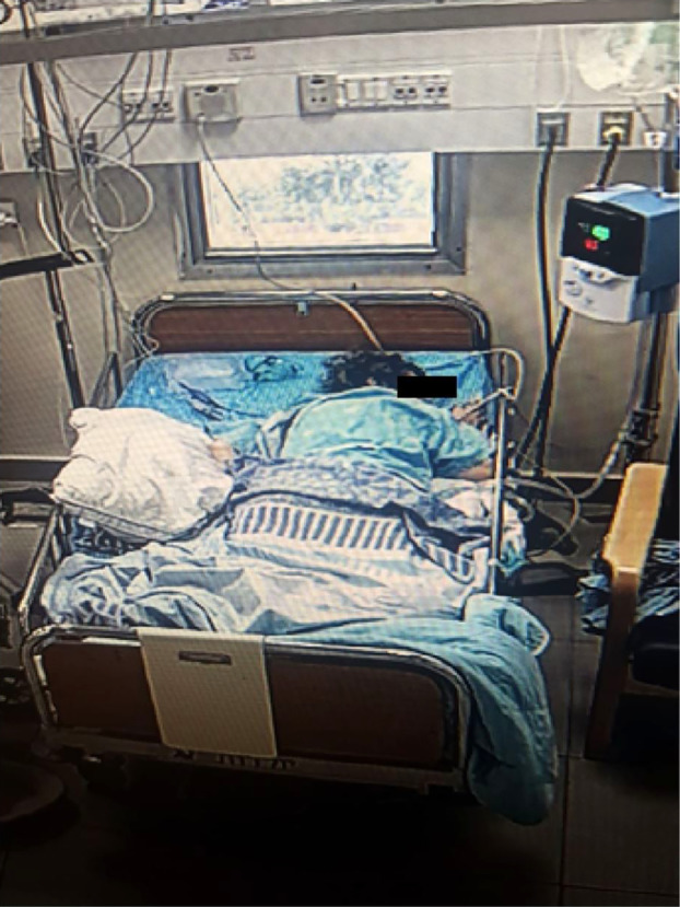
A 52‐year‐old woman with a history of type 2 diabetes mellitus and hyperlipidaemia was admitted to our department with moderate COVID‐19 infection. Her vital signs on admission were as follows: fever 39.2°, blood pressure 142/92 mmHg, heart rate 102 b.p.m., oxygen saturation in room air was 86%, while on 2 L per min (LPM) oxygenation with nasal cannula the saturation increased to 93%. On physical examination, she was tachypnoeic with a respiratory rate of 25 b.p.m. All other physical findings were unremarkable. The chest X‐ray demonstrated bilateral, peripheral consolidations. The electrocardiogram (ECG) showed normal sinus rhythm, 90 b.p.m. The venous blood gas equation on admission showed the following: pH = 7.48, = 29.3 and Pco 2 = 42 mmHg. All other laboratory findings are shown in Table 1. She was initially treated with 2 LPM oxygenation via nasal cannula, levofloxacin and hydroxychloroquine. Despite this treatment, after 48 h she further deteriorated: the respiratory rate went up to 45–50 b.p.m., the saturation decreased to 85% with 10 LPM. Repeated venous blood gas sampling showed: pH = 7.48, Pco 2 = 42 mmHg and = 26.03. High‐flow nasal, humidified oxygen cannula support (HFNC, 40 LPM; 33°C; 100% O2) was initiated and the peripheral saturation improved to 90%. At that point of time, the patient was asked to self‐prone position for 5 h (Fig. 1). Shortly after (less than 30 min) this manoeuvre, the saturation increased to 100% (combined with HFNC) and the respiratory rate decreased to 25 b.p.m. On the following days, she continued to self‐prone position (for a mean duration of 4 h per day) and gradually improved her oxygenation, eventually weaned off any respiratory support (stayed on ambient air) and transferred to a step down department for further care.
Table 1.
Laboratory parameters of two selected patients
| Laboratory findings on admission | Patient 1 | Patient 2 |
|---|---|---|
| WBC (K/mcL) | 6.33 | 5.51 |
| Haemoglobin (g/dL) | 10.3 | 13.08 |
| PLT (K/mcL) | 580 | 190 |
| Lymphocyte (K/mcL) | 800 | 650 |
| AST (IU/L) | 72 | 31 |
| ALT (IU/L) | 56 | 27 |
| LDH (IU/L) | 420 | 412 |
| Troponin‐I HS (ng/L) | 11 | 4.4 |
| CRP (mg/L) | 178 | 140.9 |
| D‐dimer (ng/mL) | 10 367 | 725 |
| Ferritin (ng/mL) | 591 | 913 |
| Procalcitonin (ng/mL) | 0.24 | <0.2 |
ALT, alanine aminotransferase; AST, aspartate aminotransferase; CRP, C‐reactive protein; LDH, lactate dehydrogenase; PLT, platelet count.
Figure 1.

Awake prone position of a COVID‐19 patient.
A 40‐year‐old male patient with no significant, past medical history was admitted to our department with moderate COVID‐19 infection. The vital signs on admission were as follows: fever 38.0°C, blood pressure of 122/77 mmHg and heart rate of 85 b.p.m. The oxygen saturation was 88% in room air and increased up to 92% with oxygenation (4 LPM via nasal cannula). On physical examination, he appeared lethargic and was tachypnoeic (respiratory rate of 30 b.p.m.). His chest X‐ray demonstrated mild bilateral peripheral consolidations. The ECG showed normal sinus rhythm (85 b.p.m.). The venous blood gas equation on admission showed pH of 7.401, Pco 2 of 42.7 mmHg and of 26.0. Other relevant laboratory findings are detailed in Table 1. The patient was initially treated with levofloxacin, hydroxychloroquine and low‐dose molecular weight heparin (due to low mobilisation). On the second day after admission, he began a daily session of respiratory physiotherapy, which included approximately 30 min staying in awake prone position. He was instructed by the physiotherapist to continue self‐proning for at least 2 h per day. While in prone position, within 10 min of proning, a significant increase in saturation up to values of 96–98% (combined with 2 LPM oxygenation) was noticed. After 6 days of hospitalisation, the peripheral oxygen saturation was 95% in room air without oxygen support and the patient was discharged.
Discussion
Prone positioning is indicated in ARDS patients who fulfil the Berlin criteria.5, 6 The suggested mechanisms by which prone positioning improves these patients' oxygenation include recruitment of the non‐aerated dorsal lung territories, and resultant improvement in ventilation–perfusion mismatches. Prone position may also improve CO2 clearance due to an increase in the number of open and ventilated alveoli. It should be emphasised that among all our COVID‐19 patients, hypercapnia was not evident as a dominant characteristic of disease. Regarding potential benefits among mechanically ventilated patients, typical ventilator lung injury (VILI) arises due to repeated mechanical force on the alveoli. The prone position produces a more homogeneous stress on the lung parenchyma, therefore reducing the risk of VILI.1, 5
The newly described COVID‐19 course is divided into three stages: the first early stage includes viral replication and is associated with either mild, flu‐like symptoms such as fever, malaise, myalgia and dry cough or no signs and symptoms at all. The second stage includes viral pneumonia and exacerbated inflammation in lung parenchyma, in this stage patients experience hypoxaemia along with fever and dry cough. In the third stage, there is a transition to hyperinflammation state (cytokine storm) and possibly haemodynamic collapse. 7 As it is regarding all potential treatment modalities, lack of evidence in the literature exists regarding awake prone position as part of COVID‐19 patients' management. In 2003, Valter et al. published three case reports of patients who presented with hypoxaemia (mean Pao 2 = 51) and tachypnoea who improved dramatically right after short awake prone position manoeuvres. 8 Scaravilli et al. further demonstrated this phenomenon in their cohort study: 15 non‐intubated patients (5 females and 10 males) most of whom presented with pneumonia (one presented with fasciitis and the others with sepsis) and Pao 2/Fio 2 < 300 were treated with prone position together with different respiratory devices (oxygen mask, high flow nasal cannula, helmet continuous positive airway pressure (CPAP), non‐invasive ventilation (NIV) mask). During the study, 43 prone position procedures were performed with an average of two procedures per subject with a median of 3 h per session. Among their study population, mean Pao 2/Fio 2 ratios were significantly higher during prone position (186 ± 72 mmHg), as compared to pre‐prone position (127 ± 49 mmHg). The largest improvement was demonstrated in patients who were treated with non‐invasive ventilation support, mean Pao 2/Fio 2 was significantly higher during the prone position (214 ± 71 mmHg), as compared with pre‐prone position (157 ± 44 mmHg). 4
The fact that early application of awake prone positioning together with HFNC or non‐invasive ventilation that may enable avoidance of endotracheal intubation was demonstrated by Ding et al. Their patient selection included 20 patients with moderate ARDS (Pao 2/Fio 2 < 200 mmHg), who were admitted to the respiratory intensive care unit. This study revealed that early awake prone positioning together with NIV/HFNC made endotracheal intubation redundant in more than 50% of patients (11/20). In face of the fact that endotracheal intubation rate among ARDS patients is 75%, based on previous publications, the investigators were able to decrease the intubation rate by a substantial percentage. 9
Emerging evidence for early recognition and treatment for COVID‐19 patients with ARDS and pneumonia using high flow nasal cannula together with awake prone position and restrictive fluid resuscitation showed a decrease in invasive mechanical ventilation rate also in the Jiangsu Province, China. 10 Prone positions applied on awake patients require the patients' cooperation and depend on their tolerance and compliance. There are several contraindications to the awake prone positioning, which include recent abdominal surgery, increased intra‐abdominal pressure, facial injuries and unstable fractures. 8 Severe ARDS patients (Pao 2/Fio 2 < 100 mmHg) are not appropriate candidates for awake prone position, this might delay unavoidable intubation and subsequent treatment failure. 9
Early application of awake prone position in mild to moderate COVID‐19 patients improves oxygenation and may avoid intubation and deterioration to severe disease. Based on our experience, exemplified in the cases herein, we encourage further study and application for future COVID‐19 patients.
Funding: None.
Conflict of interest: None.
References
- 1. Kallet RH. A comprehensive review of prone position in ARDS. Respir Care 2015; 60: 1660–87. [DOI] [PubMed] [Google Scholar]
- 2. Guérin C, Reignier J, Richard JC, Beuret P, Gacouin A, Boulain T et al. Prone positioning in severe acute respiratory distress syndrome. N Engl J Med 2013; 368: 2159–68. [DOI] [PubMed] [Google Scholar]
- 3. Wu C, Chen X, Cai Y, Xia J, Zhou X, Xu S et al. Risk factors associated with acute respiratory distress syndrome and death in patients with coronavirus disease 2019 Pneumonia in Wuhan, China. JAMA Intern Med 2020. 10.1001/jamainternmed.2020.0994. [DOI] [PMC free article] [PubMed] [Google Scholar]
- 4. Scaravilli V, Grasselli G, Castagna L, Zanella A, Isgrò S, Lucchini A et al. Prone positioning improves oxygenation in spontaneously breathing nonintubated patients with hypoxemic acute respiratory failure: a retrospective study. J Crit Care 2015; 30: 1390–4. [DOI] [PubMed] [Google Scholar]
- 5. Gattinoni L, Taccone P, Carlesso E, Marini JJ. Prone position in acute respiratory distress syndrome. Rationale, indications, and limits. Am J Respir Crit Care Med 2013; 188: 1286–93. [DOI] [PubMed] [Google Scholar]
- 6. Ranieri VM, Rubenfeld GD, Thompson BT, Ferguson ND, Caldwell E, Fan E et al. Acute respiratory distress syndrome: the Berlin definition. J Am Med Assoc 2012; 307: 2526–33. [DOI] [PubMed] [Google Scholar]
- 7. Siddiqi Hasan K, Mehra Mandeep R. COVID‐19 illness in native and immunosuppressed states: a clinical–therapeutic staging proposal. J Heart Lung Transpl 2020; 39: 405–7. [DOI] [PMC free article] [PubMed] [Google Scholar]
- 8. Valter C, Christensen AM, Tollund C, Schønemann NK. Response to the prone position in spontaneously breathing patients with hypoxemic respiratory failure. Acta Anaesthesiol Scand 2003; 47: 416–8. [DOI] [PubMed] [Google Scholar]
- 9. Ding L, Wang L, Ma W, He H. Efficacy and safety of early prone positioning combined with HFNC or NIV in moderate to severe ARDS: a multi‐center prospective cohort study. Crit Care 2020; 24: 28. [DOI] [PMC free article] [PubMed] [Google Scholar]
- 10. Sun Q, Qiu H, Huang M, Yang Y. Lower mortality of COVID‐19 by early recognition and intervention: experience from Jiangsu Province. Ann Intensive Care 2020; 10: 33. [DOI] [PMC free article] [PubMed] [Google Scholar]


