

Abstract
Alzheimer’s disease (AD) is a neurodegenerative and neuroinflammatory disease characterized by the presence of extracellular amyloid plaques (APs) and intracellular neurofibrillary tangles (NFTs) in the brain. There is no disease modifying therapeutic options currently available for this disease. Hippocampus, entorhinal cortex (Broadmann area 28), perirhinal cortex (Broadmann area 35) and insular cortices are areas within the brain that are first ones to be severely affected in AD. Neuroinflammation is an important factor that induces neurodegeneration in AD. Glia maturation factor (GMF), a proinflammatory factor plays a crucial role in AD through activation of microglia and astrocytes to release proinflammatory mediators in the brain. Through immunohistochemical studies, we have previously shown that GMF is highly expressed in the vicinity of APs and NFTs in AD brains. Glial fibrillary acidic protein (GFAP), reactive astrocytes, ionized calcium binding adaptor molecule-1 (Iba-1) labelled activated microglia and GMF immunoreactive glial cells are increased in the entorhinal cortical layers especially at the sites of APs and Tau containing NFTs indicating a role for GMF. Overexpression of GMF in glial cells leads to neuroinflammation and neurodegeneration. Inhibition of GMF expression reduces neurodegeneration. Therefore, we suggest that GMF is a novel therapeutic target not only for AD but also for various other neurodegenerative diseases.
Keywords: Alzheimer’s disease, amyloid plaques, neurofibrillary tangles, glia maturation factor
Introduction
Alzheimer’s disease (AD) is a chronic progressive neurological disorder affecting 5.8 million Americans and 35 million individuals worldwide (Alzheimer’s disease Association, Chicago, IL). Extracellular amyloid plaques (APs) and intracellular neurofibrillary tangles (NFTs) are the hallmarks of AD. NFTs are made up of abnormally phosphorylated Tau proteins and its number is closely associated with clinical symptoms in AD patients (1–10). Entorhinal cortex is involved in memory formation, retrieval, and extinction. Hippocampus is important for learning and memory functions. Together with hippocampal formation, the entorhinal cortex forms the major part of medial temporal lobe that is involved in AD and has dense NFTs formation and amyloid deposits. Entorhinal cortex and hippocampus are severely affected, atrophied and inflamed in AD patients. Memory loss is one of the earliest symptoms in AD patients due to the destruction of entorhinal cortex projections and the perforant pathways to the hippocampal formation (2, 3). In this mini review, we discuss on the role of glia maturation factor (GMF) in the pathogenesis of AD.
Glia Maturation Factor and Alzheimer’s disease
Recent studies suggest that inflammation plays a critical role in the onset and progression of neuroinflammatory and neurodegenerative diseases including AD (11–15). GMF, a brain protein was previously isolated and characterized in our laboratory is mainly found to be localized in glial cells and in some neurons in the central nervous system (CNS) (16–18). GMF in excess leads to the death of neurons by inducing neuroinflammation in neurodegenerative diseases including AD, Parkinson’s disease (PD), Multiple Sclerosis (MS) and its animal model Experimental Autoimmune Encephalomyelitis (EAE) (Figure 1) (19–28). There is specific upregulation of GMF expression in glial cells associated with APs and NFTs in the entorhinal cortical layers and hippocampus of AD brains (2, 3). We believe that GMF is a novel therapeutic target and therefore inhibition of GMF expression can inhibit the onset and progression of neurodegenerative diseases including AD (29–33).
Figure 1.
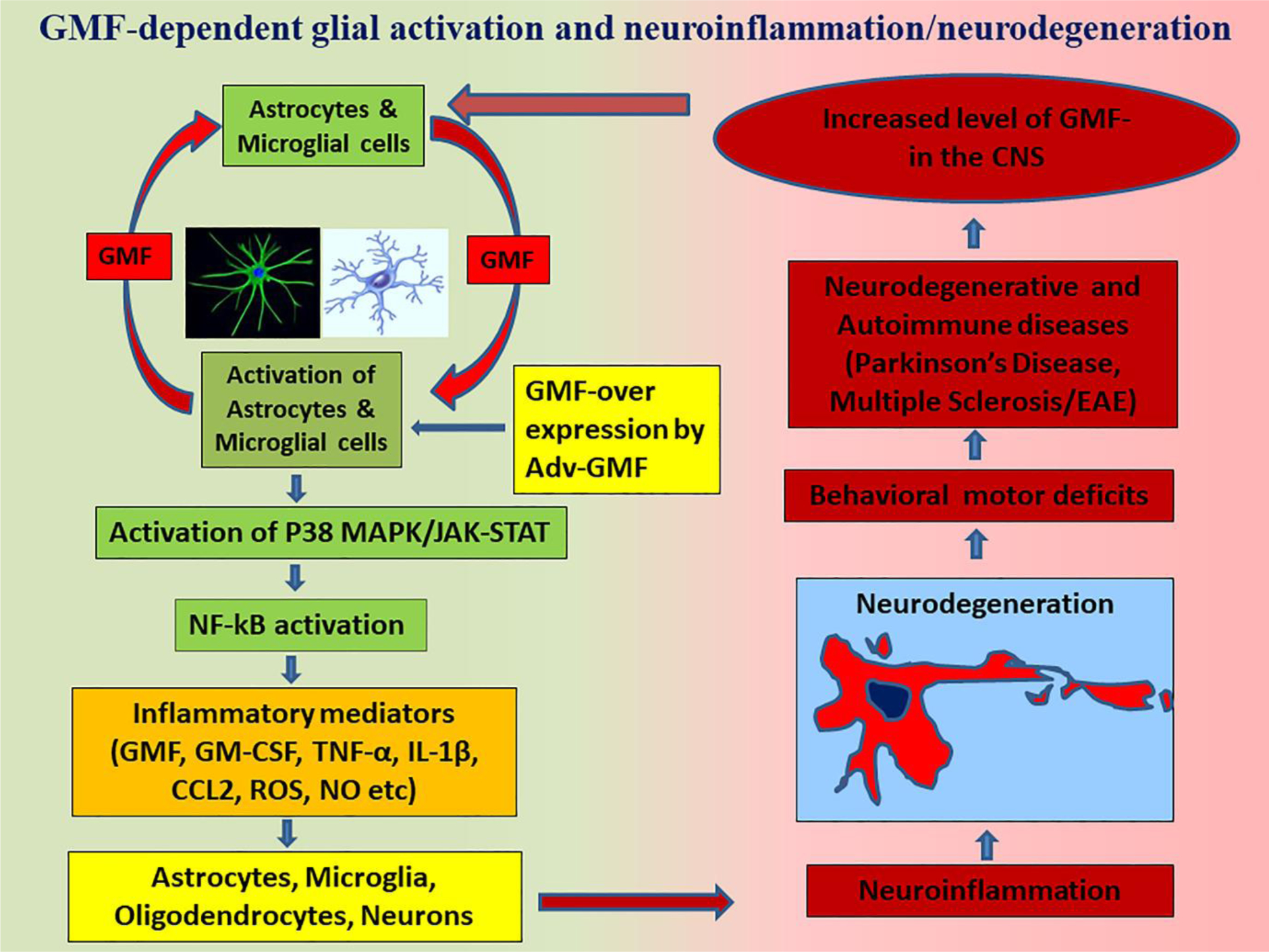
Schematic diagram showing GMF-β-dependent glial cells activation-mediated neuroinflammation and neurodegeneration. Activated astrocytes and microglial cells release GMF-β that in turn acts on glial cells/neurons. Increased expression of GMF-β in neurodegenerative diseases or GMF-β overexpression in animas by adenoviral vector-mediated GMF gene transfection activates MAPKs/JAK-STAT and NF-kB pathways and induces the release of several neuroinflammatory mediators such as cytokines, chemokines, ROS, nitric oxide and GMF-β. These mediators induce neuroinflammation and neurodegeneration leading to dementia and motor behavioral disorders in neurodegenerative diseases/EAE.
GMF and glial fibrillary acidic protein (GFAP) are increasingly expressed in the hippocampus and entorhinal cortex of AD brain especially at the sites of APs and NFTs (2–6). During normal aging process hippocampus and entorhinal cortex are subjected to widespread oxidative stress, decreased antioxidant function and enhanced expression of GFAP. GMF accelerates and potentiates these processes and makes the neuronal cells more susceptible to degeneration in neuroinflammatory conditions (3–7, 17, 34, 35). Neuronal death in AD is mediated by APs and NFTs. Other factors such as local inflammatory infiltrates including glial cells (microglia/astrocytes) and release of inflammatory molecules contribute to neuroinflammation and disease severity (4, 16, 36–38). AD pathogenesis involves sustained neuroinflammation by glial activation that produces proinflammatory cytokines, chemokines, free radicals such as reactive oxygen species/ reactive nitrogen species, and activation of transcription factor nuclear factor-kappa B (NF-κB) and mitogen activated protein kinases (MAPKs) (4, 7, 16, 36–38). Mitochondrial dysfunction plays a crucial role in the development and progression of AD. Mitochondria are the main sites for reactive oxygen species (ROS) production. Increased expression of inducible nitric oxide synthase (iNOS) around the plaques has been shown to contribute to the oxidative stress in AD brains. Uncoupling proteins (UCPs) are inner mitochondrial proteins that protect neurons by reducing the production of free radicals (7). UCP2 and UCP4 are down regulated in AD brains with upregulation of GMF expression in the glial cells along with increased iNOS and NF-κB activities thereby indicating that GMF plays a proinflammatory role in the pathogenesis of AD probably by promoting mitochondrial dysfunction through down regulation of the UCPs. Mitochondrial dynamics in AD may be regulated on one hand by UCPs through their action on FASN/fatty acid synthase and on other hand by GMF through its action on the NLRP3 inflammasome (7).
Recently, we have shown that in human AD brains, GMF co-localizes with the NLRP3 inflammasome and the autophagosome markers lysosome associated membrane protein1 (LAMP1) and autophagic protein sequestosome1 (SQSTM1)/p62, clearly pointing to the role of GMF in increased Aβ level in AD. Analysis of human AD brain tissue sections from the temporal cortex showed besides GMF increased expression of the inflammasome components NACHT, LRR and PYD domains-containing protein 3 (NLRP3) and caspase-1 along with the products interleukin-1beta (IL-1β) and IL-18 (39). The co-localization of inflammasome components and pro-inflammatory cytokines with GMF was found in the vicinity and periphery of APs and NFTs.
GMF and apolipoprotein E4 (ApoE4) are strongly expressed and co-associated in the APs and reactive astrocytes surrounding APs in AD brains (40). These results show that GMF and ApoE4 should together be contributing to the neuropathological changes associated with AD. GMF enhances astrocyte activation through secretion of granulocyte-macrophage-colony stimulating factor (GM-CSF) (19). High level expression of GMF in activated glial cells further augments chemo attraction, proliferation, activation and release of inflammatory mediators IL-1, IL-33, tumour necrosis factor-alpha (TNF-α), macrophage inflammatory proteins-1 beta (MIP-1β), complement protein C1q, class II major histocompatibility complex (MHC) proteins, 12-lipoxygenase and chemokine CX3C and exacerbating the pathogenesis of AD. This reflects the paracrine and/or autocrine signalling by GMF (1–4, 16, 41, 42). Increased expression of GMF in association with β-amyloid (Aβ) has been shown to amplify the deleterious inflammation propagated by the NLRP3 inflammasome causing mitochondrial dysfunction, and further that GMF and Aβ synergize in bringing upon this dysfunction by changing mitochondrial dynamics through alterations in fission and fusion proteins (43).
Conclusion
Increased expression of GMF by glial cells in the temporal cortex of AD brain suggests GMF’s proinflammatory role of neurodegeneration in the pathogenesis of AD. Furthermore, we suggest that GMF is a novel therapeutic target not only for AD but also for various other neurodegenerative diseases.
Acknowledgements
This work was supported by the National Institutes of Health (NIH) Grant AG048205 and Veterans Affairs Research Career Scientist Award to Asgar Zaheer.
References
- 1.Thangavel R, Stolmeier D, Yang X, Anantharam P, Zaheer A. 2012. Expression of glia maturation factor in neuropathological lesions of Alzheimer’s disease. Neuropathol Appl Neurobiol 38:572–81. [DOI] [PMC free article] [PubMed] [Google Scholar]
- 2.Stolmeier D, Thangavel R, Anantharam P, Khan MM, Kempuraj D, Zaheer A. 2013. Glia maturation factor expression in hippocampus of human Alzheimer’s disease. Neurochem Res 38:1580–9. [DOI] [PMC free article] [PubMed] [Google Scholar]
- 3.Thangavel R, Kempuraj D, Stolmeier D, Anantharam P, Khan M, Zaheer A. 2013. Glia maturation factor expression in entorhinal cortex of Alzheimer’s disease brain. Neurochem Res 38:1777–84. [DOI] [PMC free article] [PubMed] [Google Scholar]
- 4.Thangavel R, Van Hoesen GW, Zaheer A. 2009. The abnormally phosphorylated tau lesion of early Alzheimer’s disease. Neurochem Res 34:118–23. [DOI] [PubMed] [Google Scholar]
- 5.Xiong Z, Thangavel R, Kempuraj D, Yang E, Zaheer S, Zaheer A. 2014. Alzheimer’s Disease: Evidence for the Expression of Interleukin-33 and Its Receptor ST2 in the Brain. J Alzheimers Dis 40(2):297–308. [DOI] [PMC free article] [PubMed] [Google Scholar]
- 6.Zaheer S, Thangavel R, Sahu SK, Zaheer A. 2011. Augmented expression of glia maturation factor in Alzheimer’s disease. Neuroscience 194:227–33. [DOI] [PMC free article] [PubMed] [Google Scholar]
- 7.Thangavel R, Kempuraj D, Zaheer S, Raikwar S, Ahmed ME, Selvakumar GP, Iyer SS, Zaheer A. 2017. Glia Maturation Factor and Mitochondrial Uncoupling Proteins 2 and 4 Expression in the Temporal Cortex of Alzheimer’s Disease Brain. Front Aging Neurosci 9:150. [DOI] [PMC free article] [PubMed] [Google Scholar]
- 8.Thangavel R, Sahu SK, Van Hoesen GW, Zaheer A. 2009. Loss of nonphosphorylated neurofilament immunoreactivity in temporal cortical areas in Alzheimer’s disease. Neuroscience 160:427–33. [DOI] [PMC free article] [PubMed] [Google Scholar]
- 9.Thangavel R, Sahu SK, Van Hoesen GW, Zaheer A. 2008. Modular and laminar pathology of Brodmann’s area 37 in Alzheimer’s disease. Neuroscience 152:50–5. [DOI] [PMC free article] [PubMed] [Google Scholar]
- 10.Thangavel R, Van Hoesen GW, Zaheer A. 2008. Posterior parahippocampal gyrus pathology in Alzheimer’s disease. Neuroscience 154:667–76. [DOI] [PMC free article] [PubMed] [Google Scholar]
- 11.Kempuraj D, Thangavel R, Natteru PA, Selvakumar GP, Saeed D, Zahoor H, Zaheer S, Iyer SS, Zaheer A. 2016. Neuroinflammation Induces Neurodegeneration. J Neurol Neurosurg Spine 1(1) pii: 1003. [PMC free article] [PubMed] [Google Scholar]
- 12.Kempuraj D, Ahmed ME, Selvakumar GP, Thangavel R, Dhaliwal AS, Dubova I, Mentor S, Premkumar K, Saeed D, Zahoor H, Raikwar SP, Zaheer S, Iyer SS, Zaheer A. 2019. Brain Injury-Mediated Neuroinflammatory Response and Alzheimer’s Disease. Neuroscientist doi: 10.1177/1073858419848293:1073858419848293. [DOI] [PMC free article] [PubMed] [Google Scholar]
- 13.Kempuraj D, Mentor S, Thangavel R, Ahmed ME, Selvakumar GP, Raikwar SP, Dubova I, Zaheer S, Iyer SS, Zaheer A. 2019. Mast Cells in Stress, Pain, Blood-Brain Barrier, Neuroinflammation and Alzheimer’s Disease. Front Cell Neurosci 13:54. [DOI] [PMC free article] [PubMed] [Google Scholar]
- 14.Kempuraj D, Selvakumar GP, Thangavel R, Ahmed ME, Zaheer S, Raikwar SP, Iyer SS, Bhagavan SM, Beladakere-Ramaswamy S, Zaheer A. 2017. Mast Cell Activation in Brain Injury, Stress, and Post-traumatic Stress Disorder and Alzheimer’s Disease Pathogenesis. Front Neurosci 11:703. [DOI] [PMC free article] [PubMed] [Google Scholar]
- 15.Kempuraj D, Thangavel R, Selvakumar GP, Zaheer S, Ahmed ME, Raikwar SP, Zahoor H, Saeed D, Natteru PA, Iyer S, Zaheer A. 2017. Brain and Peripheral Atypical Inflammatory Mediators Potentiate Neuroinflammation and Neurodegeneration. Front Cell Neurosci 11:216. [DOI] [PMC free article] [PubMed] [Google Scholar]
- 16.Lim R, Miller JF, Zaheer A. 1989. Purification and characterization of glia maturation factor beta: a growth regulator for neurons and glia. Proc Natl Acad Sci U S A 86:3901–5. [DOI] [PMC free article] [PubMed] [Google Scholar]
- 17.Khan MM, Zaheer S, Thangavel R, Patel M, Kempuraj D, Zaheer A. 2015. Absence of Glia Maturation Factor Protects Dopaminergic Neurons and Improves Motor Behavior in Mouse Model of Parkinsonism. Neurochem Res 40(5):980–990. [DOI] [PMC free article] [PubMed] [Google Scholar]
- 18.Lim R, Zaheer A, Lane WS. 1990. Complete amino acid sequence of bovine glia maturation factor beta. Proc Natl Acad Sci U S A 87:5233–7. [DOI] [PMC free article] [PubMed] [Google Scholar]
- 19.Zaheer A, Yorek MA, Lim R. 2001. Effects of glia maturation factor overexpression in primary astrocytes on MAP kinase activation, transcription factor activation, and neurotrophin secretion. Neurochem Res 26:1293–9. [DOI] [PubMed] [Google Scholar]
- 20.Zaheer A, Zaheer S, Sahu SK, Knight S, Khosravi H, Mathur SN, Lim R. 2007. A novel role of glia maturation factor: induction of granulocyte-macrophage colony-stimulating factor and pro-inflammatory cytokines. J Neurochem 101:364–76. [DOI] [PubMed] [Google Scholar]
- 21.Zaheer A, Knight S, Zaheer A, Ahrens M, Sahu SK, Yang B. 2008. Glia maturation factor overexpression in neuroblastoma cells activates glycogen synthase kinase-3beta and caspase-3. Brain Res 1190:206–14. [DOI] [PMC free article] [PubMed] [Google Scholar]
- 22.Zaheer S, Wu Y, Yang X, Ahrens M, Sahu SK, Zaheer A. 2012. Clinical course of myelin oligodendrocyte glycoprotein 35–55 induced experimental autoimmune encephalomyelitis is aggravated by glia maturation factor. Neurochem Int 60:215–9. [DOI] [PMC free article] [PubMed] [Google Scholar]
- 23.Zaheer S, Wu Y, Bassett J, Yang B, Zaheer A. 2007. Glia maturation factor regulation of STAT expression: a novel mechanism in experimental autoimmune encephalomyelitis. Neurochem Res 32:2123–31. [DOI] [PubMed] [Google Scholar]
- 24.Khan MM, Kempuraj D, Thangavel R, Zaheer A. 2013. Protection of MPTP-induced neuroinflammation and neurodegeneration by Pycnogenol. Neurochem Int 62:379–88. [DOI] [PMC free article] [PubMed] [Google Scholar]
- 25.Kempuraj D, Thangavel R, Selvakumar GP, Ahmed ME, Zaheer S, Raikwar SP, Zahoor H, Saeed D, Dubova I, Giler G, Herr S, Iyer SS, Zaheer A. 2018. Mast Cell Proteases Activate Astrocytes and Glia-Neurons and Release Interleukin-33 by Activating p38 and ERK1/2 MAPKs and NF-kappaB. Mol Neurobiol 56(3):1681–1693. [DOI] [PMC free article] [PubMed] [Google Scholar]
- 26.Selvakumar GP, Iyer SS, Kempuraj D, Ahmed ME, Thangavel R, Dubova I, Raikwar SP, Zaheer S, Zaheer A. 2018. Molecular Association of Glia Maturation Factor with the Autophagic Machinery in Rat Dopaminergic Neurons: a Role for Endoplasmic Reticulum Stress and MAPK Activation. Mol Neurobiol 56(6):3865–3381. [DOI] [PMC free article] [PubMed] [Google Scholar]
- 27.Selvakumar GP, Iyer SS, Kempuraj D, Raju M, Thangavel R, Saeed D, Ahmed ME, Zahoor H, Raikwar SP, Zaheer S, Zaheer A. 2018. Glia Maturation Factor Dependent Inhibition of Mitochondrial PGC-1alpha Triggers Oxidative Stress-Mediated Apoptosis in N27 Rat Dopaminergic Neuronal Cells. Mol Neurobiol 55(9):7132–7152. [DOI] [PMC free article] [PubMed] [Google Scholar]
- 28.Kempuraj D, Thangavel R, Yang E, Pattani S, Zaheer S, Santillan DA, Santillan MK, Zaheer A. 2015. Dopaminergic Toxin 1-Methyl-4-Phenylpyridinium, Proteins alpha-Synuclein and Glia Maturation Factor Activate Mast Cells and Release Inflammatory Mediators. PLoS One 10:e0135776. [DOI] [PMC free article] [PubMed] [Google Scholar]
- 29.Raikwar SP, Thangavel R, Dubova I, Selvakumar GP, Ahmed ME, Kempuraj D, Zaheer SA, Iyer SS, Zaheer A. 2018. Targeted Gene Editing of Glia Maturation Factor in Microglia: a Novel Alzheimer’s Disease Therapeutic Target. Mol Neurobiol 56(1):378–393. [DOI] [PMC free article] [PubMed] [Google Scholar]
- 30.Raikwar SP, Kikkeri NS, Sakuru R, Saeed D, Zahoor H, Premkumar K, Mentor S, Thangavel R, Dubova I, Ahmed ME, Selvakumar GP, Kempuraj D, Zaheer S, Iyer SS, Zaheer A. 2019. Next Generation Precision Medicine: CRISPR-mediated Genome Editing for the Treatment of Neurodegenerative Disorders. J Neuroimmune Pharmacol doi: 10.1007/s11481-019-09849-y. [DOI] [PMC free article] [PubMed] [Google Scholar]
- 31.Raikwar SP, Thangavel R, Dubova I, Ahmed ME, Selvakumar PG, Kempuraj D, Zaheer S, Iyer S, Zaheer A. 2018. Neuro-Immuno-Gene- and Genome-Editing-Therapy for Alzheimer’s Disease: Are We There Yet? J Alzheimers Dis 65:321–344. [DOI] [PMC free article] [PubMed] [Google Scholar]
- 32.Selvakumar GP, Ahmed ME, Raikwar SP, Thangavel R, Kempuraj D, Dubova I, Saeed D, Zahoor H, Premkumar K, Zaheer S, Iyer S, Zaheer A. 2019. CRISPR/Cas9 Editing of Glia Maturation Factor Regulates Mitochondrial Dynamics by Attenuation of the NRF2/HO-1 Dependent Ferritin Activation in Glial Cells. J Neuroimmune Pharmacol doi: 10.1007/s11481-019-09833-6. [DOI] [PMC free article] [PubMed] [Google Scholar]
- 33.Fan J, Fong T, Chen X, Chen C, Luo P, Xie H. 2018. Glia maturation factor-beta: a potential therapeutic target in neurodegeneration and neuroinflammation. Neuropsychiatr Dis Treat 14:495–504. [DOI] [PMC free article] [PubMed] [Google Scholar]
- 34.Zaheer S, Wu Y, Yang X, Thangavel R, Sahu SK, Zaheer A. 2012. Efficient down-regulation of glia maturation factor expression in mouse brain and spinal cord. Neurochem Res 37:1578–83. [DOI] [PMC free article] [PubMed] [Google Scholar]
- 35.Zaheer S, Thangavel R, Wu Y, Khan MM, Kempuraj D, Zaheer A. 2013. Enhanced expression of glia maturation factor correlates with glial activation in the brain of triple transgenic Alzheimer’s disease mice. Neurochem Res 38:218–25. [DOI] [PMC free article] [PubMed] [Google Scholar]
- 36.Luan K, Rosales JL, Lee KY. 2013. Viewpoint: Crosstalks between neurofibrillary tangles and amyloid plaque formation. Ageing Res Rev 12:174–81. [DOI] [PubMed] [Google Scholar]
- 37.Shoghi-Jadid K, Small GW, Agdeppa ED, Kepe V, Ercoli LM, Siddarth P, Read S, Satyamurthy N, Petric A, Huang SC, Barrio JR. 2002. Localization of neurofibrillary tangles and beta-amyloid plaques in the brains of living patients with Alzheimer disease. Am J Geriatr Psychiatry 10:24–35. [PubMed] [Google Scholar]
- 38.Dickerson BC, Goncharova I, Sullivan MP, Forchetti C, Wilson RS, Bennett DA, Beckett LA, deToledo-Morrell L. 2001. MRI-derived entorhinal and hippocampal atrophy in incipient and very mild Alzheimer’s disease. Neurobiol Aging 22:747–54. [DOI] [PubMed] [Google Scholar]
- 39.Ahmed ME, Iyer S, Thangavel R, Kempuraj D, Selvakumar GP, Raikwar SP, Zaheer S, Zaheer A. 2017. Co-Localization of Glia Maturation Factor with NLRP3 Inflammasome and Autophagosome Markers in Human Alzheimer’s Disease Brain. J Alzheimers Dis 60:1143–1160. [DOI] [PMC free article] [PubMed] [Google Scholar]
- 40.Thangavel R, Bhagavan SM, Ramaswamy SB, Surpur S, Govindarajan R, Kempuraj D, Zaheer S, Raikwar S, Ahmed ME, Selvakumar GP, Iyer SS, Zaheer A. 2018. Co-Expression of Glia Maturation Factor and Apolipoprotein E4 in Alzheimer’s Disease Brain. J Alzheimers Dis 61:553–560. [DOI] [PMC free article] [PubMed] [Google Scholar]
- 41.Zaheer A, Zaheer S, Thangavel R, Wu Y, Sahu SK, Yang B. 2008. Glia maturation factor modulates beta-amyloid-induced glial activation, inflammatory cytokine/chemokine production and neuronal damage. Brain Res 1208:192–203. [DOI] [PMC free article] [PubMed] [Google Scholar]
- 42.Kempuraj D, Khan MM, Thangavel R, Xiong Z, Yang E, Zaheer A. 2013. Glia maturation factor induces interleukin-33 release from astrocytes: implications for neurodegenerative diseases. J Neuroimmune Pharmacol 8:643–50. [DOI] [PMC free article] [PubMed] [Google Scholar]
- 43.Ahmed ME, Selvakumar GP, Kempuraj D, Thangavel R, Mentor S, Dubova I, Raikwar SP, Zaheer S, Iyer S, Zaheer A. 2019. Synergy in Disruption of Mitochondrial Dynamics by Abeta (1–42) and Glia Maturation Factor (GMF) in SH-SY5Y Cells Is Mediated Through Alterations in Fission and Fusion Proteins. Mol Neurobiol 56:6964–6975. [DOI] [PMC free article] [PubMed] [Google Scholar]