Historically, live attenuated vaccines produced by blind passage usually undergo adaptation in cell cultures or nonsusceptible hosts and attenuation in natural hosts, with a classical example being the classical swine fever virus (CSFV) lapinized vaccine C-strain, which was developed by hundreds of passages in rabbits. However, the mechanism of viral adaptation to nonsusceptible hosts and the molecular basis for viral adaptation and attenuation remain largely unknown. In this study, we demonstrated that P108 and T109 on the E2 glycoprotein together with the Erns glycoprotein of the rabbit-adaptive C-strain confer adaptation to rabbits on the highly virulent CSFV Shimen strain by affecting viral entry during infection but do not attenuate the Shimen strain in pigs. Our results provide vital information on the different molecular bases of CSFV adaptation to rabbits and attenuation in pigs.
KEYWORDS: adaptation, classical swine fever virus, entry, virulence
ABSTRACT
The classical swine fever virus (CSFV) live attenuated vaccine C-strain is adaptive to rabbits and attenuated in pigs, in contrast with the highly virulent CSFV Shimen strain. Previously, we demonstrated that P108 and T109 on the E2 glycoprotein (E2P108-T109) in domain I (E2DomainI) rather than R132, S133, and D191 in domain II (E2DomainII) determine C-strain’s adaptation to rabbits (ATR) (Y. Li, L. Xie, L. Zhang, X. Wang, C. Li, et al., Virology 519:197–206, 2018). However, it remains elusive whether these critical amino acids affect the ATR of the Shimen strain and virulence in pigs. In this study, three chimeric viruses harboring E2P108-T109, E2DomainI, or E2DomainII of C-strain based on the non-rabbit-adaptive Shimen mutant vSM-HCLVErns carrying the Erns glycoprotein of C-strain were generated and evaluated. We found that E2P108-T109 or E2DomainI but not E2DomainII of C-strain renders vSM-HCLVErns adaptive to rabbits, suggesting that E2P108-T109 in combination with the Erns glycoprotein (E2P108-T109-Erns) confers ATR on the Shimen strain, creating new rabbit-adaptive CSFVs. Mechanistically, E2P108-T109-Erns of C-strain mediates viral entry during infection in rabbit spleen lymphocytes, which are target cells of C-strain. Notably, pig experiments showed that E2P108-T109-Erns of C-strain does not affect virulence compared with the Shimen strain. Conversely, the substitution of E2DomainII and Erns of C-strain attenuates the Shimen strain in pigs, indicating that the molecular basis of the CSFV ATR and that of virulence in pigs do not overlap. Our findings provide new insights into the mechanism of adaptation of CSFV to rabbits and the molecular basis of CSFV adaptation and attenuation.
IMPORTANCE Historically, live attenuated vaccines produced by blind passage usually undergo adaptation in cell cultures or nonsusceptible hosts and attenuation in natural hosts, with a classical example being the classical swine fever virus (CSFV) lapinized vaccine C-strain, which was developed by hundreds of passages in rabbits. However, the mechanism of viral adaptation to nonsusceptible hosts and the molecular basis for viral adaptation and attenuation remain largely unknown. In this study, we demonstrated that P108 and T109 on the E2 glycoprotein together with the Erns glycoprotein of the rabbit-adaptive C-strain confer adaptation to rabbits on the highly virulent CSFV Shimen strain by affecting viral entry during infection but do not attenuate the Shimen strain in pigs. Our results provide vital information on the different molecular bases of CSFV adaptation to rabbits and attenuation in pigs.
INTRODUCTION
Viruses have a predefined host spectrum (1). However, viruses can breach the interspecies barrier through serial passaging in a nonsusceptible host, leading to viral adaptation to nonsusceptible hosts and attenuation in primary hosts. Well-known examples of this are the Chinese hog cholera lapinized virus (HCLV, also known as C-strain) and the lapinized rinderpest virus (2, 3). Remarkably, the viral infection is limited if one stage of the life cycle is blocked in nonsusceptible hosts, especially if the virus is unable to utilize a factor(s) which is necessary for infection or to evade a restriction factor(s). Hence, mechanisms of viral adaptation to nonsusceptible hosts by serial passaging are as follows: (i) improving entry efficiency (4, 5), (ii) enhancing the ability to antagonize or evade the nonsusceptible host's restriction factor(s) (6–8), and (iii) promoting the assembly and release of virus particles (9). Entry is the first and vital step during viral infection, which is mediated by the interaction between viral envelope proteins and cellular receptors (10).
Classical swine fever (CSF) is a devastating infectious disease of pigs caused by classical swine fever virus (CSFV), which often leads to huge economic losses to the pork industry (11). CSFV is a small, enveloped virus with a single-stranded positive RNA molecule encoding four structural proteins (C, Erns, E1, and E2) and eight nonstructural proteins (Npro, p7, NS2, NS3, NS4A, NS4B, NS5A, and NS5B) (12). The Erns glycoprotein mediates viral attachment by interacting with LamR (13). The E2 glycoprotein, which forms homodimers and heterodimers with E1, is essential for viral entry into cells (14, 15) and also plays a pivotal role in viral tropism (16). Meanwhile, Erns and E2 glycoproteins of CSFV are closely associated with virulence and pathogenicity in pigs (17–22).
In general, CSFV infects only domestic pigs and wild boar (23). However, the species barrier of CSFV between pigs and rabbits was crossed by hundreds of passages of a highly virulent strain in rabbits. C-strain, a live attenuated vaccine against CSF, was developed by passaging a highly virulent CSFV strain in rabbits in the 1950s and is characterized by adaptation to rabbits (ATR) and attenuation in pigs (3). Until now, however, the mechanism of the C-strain ATR and the molecular basis for ATR and attenuation in pigs have remained elusive.
Our previous study found that vSM-HCLVErns carrying the Erns of C-strain in the background of the highly virulent CSFV Shimen strain is not adaptive to rabbits, indicating that the Erns of C-strain alone could not render the Shimen strain adaptive to rabbits (24). In contrast, the E2 and Erns glycoproteins of C-strain together are sufficient for enabling the adaptation of the Shimen strain to rabbits. Furthermore, we observed that the C-strain backbone chimeras harboring E2 domain I (E2DomainI) or crucial mutation sites (E2P108L-T109I) on E2DomainI of the Shimen strain do not adapt to rabbits, while the chimera harboring E2 domain II (E2DomainII) of the Shimen strain is adaptive to rabbits (24). Therefore, P108 and T109 on E2DomainI (E2P108-T109) of C-strain are responsible for its ATR, whereas E2DomainII containing three different residues (R132, S133, and D191) of C-strain are not determinants of its ATR. Because of the above findings, we further investigated the effects of these critical amino acids of C-strain on the Shimen strain ATR and the virulence in pigs.
In the present study, we demonstrated that E2P108-T109 in combination with the Erns glycoprotein (E2P108-T109-Erns) of C-strain confers adaptation to rabbits on the Shimen strain. Remarkably, our data revealed that E2P108-T109-Erns of C-strain promotes viral entry during infection in the target cells using a series of pseudotyped viruses. Finally, we demonstrated that E2P108-T109-Erns of C-strain, responsible for the CSFV ATR, does not affect viral virulence in pigs, whereas E2DomainII-Erns, which is not associated with the adaptation, attenuates the Shimen strain in pigs.
RESULTS
In vitro rescue and evaluation of chimeric viruses in the background of the Shimen strain.
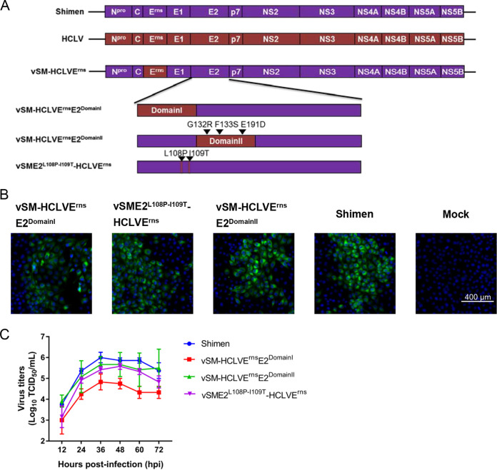
Based on previous results (24), we speculated that E2P108-T109 or E2DomainI but not E2DomainII of C-strain is necessary for conferring adaptation to rabbits on the non-rabbit-adaptive Shimen mutant vSM-HCLVErns. Since vSM-HCLVErnsE2, harboring the Erns and E2 glycoproteins of C-strain, showed better adaptation to rabbits than vSM-HCLVE1E2, containing the E1 and E2 glycoproteins of C-strain, according to the reproductive animal experiments in our previous study (24), three infectious clones (pSME2L108P-I109T-HCLVErns, pSM-HCLVErnsE2DomainI, and pSM-HCLVErnsE2DomainII) were constructed based on the infectious clone pSM-HCLVErns containing Erns of C-strain in the backbone of the Shimen strain (Fig. 1A). Chimeric viruses were rescued by transfecting individual infectious clones into swine kidney (SK6) cells, and transfected cells were serially passaged. Viral genomic sequencing results demonstrated that the sequences of the rescued chimeric viruses were as expected. The results of indirect immunofluorescence assay (IFA) demonstrated that the rescued viruses were infectious in the cells (Fig. 1B). The growth characteristics of the chimeras were determined in porcine kidney (PK-15) cells. Compared with the parental virus (Shimen strain), vSM-HCLVErnsE2DomainI had lower viral titers at different time points, while viral titers of vSM-HCLVErnsE2DomainII and vSME2L108P-I109T-HCLVErns were not significantly different (P > 0.05) (Fig. 1C). Collectively, the expected chimeric viruses were generated.
FIG 1.
Generation and characterization of chimeric CSFVs. (A) Schematic diagram of the genomic organization of parental and chimeric viruses. Purple boxes indicate genes from the highly virulent Shimen strain, while red boxes indicate genes derived from the lapinized vaccine C-strain (also known as HCLV). The infectious clones of chimeric viruses (vSM-HCLVErnsE2DomainI, vSM-HCLVErnsE2DomainII, and vSME2L108P-I109T-HCLVErns) were developed based on the infectious clone of the non-rabbit-adaptive chimeric virus vSM-HCLVErns carrying the Erns glycoprotein of C-strain. (B) Indirect immunofluorescence staining of the PK-15 cells infected by chimeric viruses and parental virus. The nuclei were stained with 4′,6-diamidino-2-phenylindole (DAPI). Bar, 400 μm. (C) Growth kinetics of parental and chimeric viruses in PK-15 cells. The error bars represent the standard deviations for three replicates.
E2P108-T109-Erns of C-strain confers adaptation to rabbits on the Shimen strain.
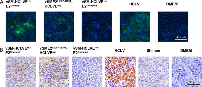
To investigate whether E2P108-T109-Erns of C-strain adapts the Shimen strain to rabbits, five groups of 24 rabbits were inoculated with different chimeric viruses (Table 1). Rectal temperature was recorded 24 h before and after inoculation and then every 6 h until 72 h postinoculation (hpi). According to the fever response standard described previously (24), vSME2L108P-I109T-HCLVErns and vSM-HCLVErnsE2DomainII did not induce a fever response in rabbits, in contrast to C-strain (Table 1). Four rabbits from each group were randomly selected to be euthanized for determining copy numbers of the viral genome in the spleens by real-time reverse transcription-quantitative PCR (RT-qPCR). The viral genome was detected in the rabbits inoculated with vSME2L108P-I109T-HCLVErns, vSM-HCLVErnsE2DomainI, or C-strain but not vSM-HCLVErnsE2DomainII (Table 1). To further determine whether the progeny viruses were present in rabbit spleens, virus isolation was performed in SK6 cells. The IFA results demonstrated that the viruses were isolated from the spleens of rabbits inoculated with vSME2L108P-I109T-HCLVErns, vSM-HCLVErnsE2DomainI, or C-strain but not with vSM-HCLVErnsE2DomainII or Dulbecco’s modified Eagle’s medium (DMEM), and no mutation occurred, as confirmed by sequencing (Fig. 2A). The immunohistochemistry results showed that the E2 glycoprotein was expressed in the spleen lymphocytes of the rabbits inoculated with C-strain, vSM-HCLVErnsE2DomainI, or vSME2L108P-I109T-HCLVErns but not vSM-HCLVErnsE2DomainII or the Shimen strain (Fig. 2B). These results indicated that chimeric viruses carrying E2P108-T109-Erns or E2DomainI-Erns of C-strain are adaptive to rabbits. Additionally, at 10 days postinoculation (dpi), anti-E2 antibodies were detected in the remaining two rabbits of each group (Table 1). As expected, we demonstrated that E2P108-T109 or E2DomainI but not E2DomainII of C-strain could render vSM-HCLVErns adaptive to rabbits, suggesting that E2P108-T109-Erns of C-strain could confer adaptation to rabbits on the Shimen strain, generating new rabbit-adaptive CSFVs.
TABLE 1.
Viral replication in the spleens of the rabbits inoculated with the Shimen-based chimeric viruses and HCLV
| Inoculum | Dose (TCID50) | No. with fever/total | No. with viral replication/total | Mean viral RNA copies in the spleens (copies/μl) | No. with seroconversion at 10 dpi/total |
|---|---|---|---|---|---|
| vSME2L108P-I109T-HCLVErns | 104 | 0/6 | 2/4 | 2.52 × 10² | 2/2 |
| vSM-HCLVErnsE2DomainI | 104 | 5/6 | 2/4 | 1.08 × 10² | 2/2 |
| vSM-HCLVErnsE2DomainII | 104 | 0/6 | 0/4 | No count | 2/2 |
| HCLV | 104 | 4/4 | 2/2 | 6.30 × 10³ | 2/2 |
| DMEM | 1 ml | 0/2 | 0/1 | No count | 0/1 |
FIG 2.
Rabbit-adaptive CSFV mutants were detected in rabbit spleens. (A) Indirect immunofluorescence staining of the SK6 cells infected with viruses isolated from the rabbit spleens and the nuclei stained with DAPI. Bar, 400 μm. (B) Viral antigens in the spleen samples detected by immunohistochemistry. The E2 glycoprotein was detected by anti-E2 antibody in the spleen lymphocytes from the rabbits inoculated with HCLV, vSM-HCLVErnsE2DomainI, or vSME2L108P-I109T-HCLVErns but not the Shimen strain, vSM-HCLVErnsE2DomainII, or DMEM. Bar, 50 μm.
Growth curves of rabbit-adaptive CSFV mutants in primary rabbit spleen lymphocytes and swine macrophages.
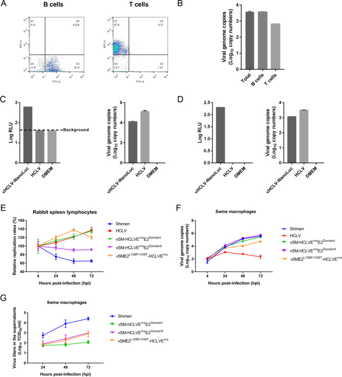
To examine the growth property of rabbit-adaptive CSFV mutants in rabbit cells, spleen lymphocytes were identified as target cells in the rabbits infected with C-strain based on the results of immunohistochemistry assay (Fig. 2B). To determine the exact cell type (T cells or B cells) in the spleens of the rabbits inoculated with C-strain, T cells and B cells were isolated from total spleen cells using the FACSAria cell-sorting system from rabbits inoculated with C-strain at 3 dpi (Fig. 3A). The viral genome copy numbers of C-strain in T cells and B cells were determined by RT-qPCR (Fig. 3B). Furthermore, viral genome copy numbers and NanoLuc activities were determined in spleen lymphocytes isolated from the rabbits inoculated with C-strain or the reporter virus vHCLV-NanoLuc (Fig. 3C). Importantly, luciferase activities were detected in the spleen lymphocytes infected with vHCLV-NanoLuc (Fig. 3D). These results indicated that C-strain infects the spleen lymphocytes (both T cells and B cells) of the rabbits.
FIG 3.
Growth curves of CSFV mutants in primary rabbit spleen lymphocytes and swine macrophages. (A) T cells and B cells isolated by flow cytometry. T cells and B cells from the spleen lymphocytes of rabbits inoculated with HCLV were isolated by flow cytometry. The cells in the Q1 region are T cells, and cells in the Q3 region are B cells. (B) Viral genome copy numbers in T cells and B cells isolated from rabbits inoculated with C-strain. The viral genome copy numbers in both T cells and B cells isolated by flow cytometry were determined using RT-qPCR. Total cells before sorting served as a control. (C) NanoLuc activities or viral genome copy numbers were tested in the spleen lymphocytes isolated from rabbits inoculated with vHCLV-NanoLuc, HCLV, or DMEM. (D) NanoLuc activities or viral genome copy numbers were tested in the spleen lymphocytes infected with vHCLV-NanoLuc or HCLV or treated with DMEM only. (E) Spleen lymphocytes isolated from healthy rabbits were infected with HCLV, the Shimen strain, vSM-HCLVErnsE2DomainI, vSM-HCLVErnsE2DomainII, or vSME2L108P-I109T-HCLVErns at an MOI of 0.01. Viral genome copy numbers were measured at 4, 24, 48, and 72 h postinoculation (hpi). Relative replication rates were analyzed based on the viral genome copy numbers at 4 hpi. (F) Viral genome copy numbers in primary swine macrophages infected with parental or chimeric viruses. (G) Titers of progeny viruses in the supernatants of primary swine macrophage cell cultures. The viral titers were determined in SK6 cells.
Next, primary rabbit spleen lymphocytes were isolated and infected with the Shimen strain, C-strain, vSME2L108P-I109T-HCLVErns, vSM-HCLVErnsE2DomainI, or vSM-HCLVErnsE2DomainII. The viral genome copy numbers in the cells were analyzed at different time points. The results showed that viral genome copy numbers of rabbit-adaptive CSFV mutants (vSME2L108P-I109T-HCLVErns and vSM-HCLVErnsE2DomainI) or the parental virus C-strain increased over time. In contrast, the viral genome copy numbers of non-rabbit-adaptive CSFVs (vSM-HCLVErnsE2DomainII and the Shimen strain) decreased over time (Fig. 3E). Unfortunately, the progeny viruses in the supernatants were undetectable using IFA in SK6 cells due to low-level replication, which is consistent with observations in rabbits. These results further demonstrated that chimeric viruses vSME2L108P-I109T-HCLVErns and vSM-HCLVErnsE2DomainI but not vSM-HCLVErnsE2DomainII are adaptive to primary rabbit spleen lymphocytes.
Furthermore, primary swine macrophages were inoculated with a series of chimeric viruses. The viral genome copy numbers in macrophages or the titers of progeny viruses in the supernatants were determined at different time points. The viral genome copy numbers of the Shimen strain and three chimeric viruses were indistinguishable, except those of C-strain in primary swine macrophages (Fig. 3F). However, the viral titers in the supernatants of the macrophages infected with the Shimen strain were higher than those in cells infected with chimeric viruses (Fig. 3G).
E2P108-T109-Erns of C-strain affects viral entry in the target cells.
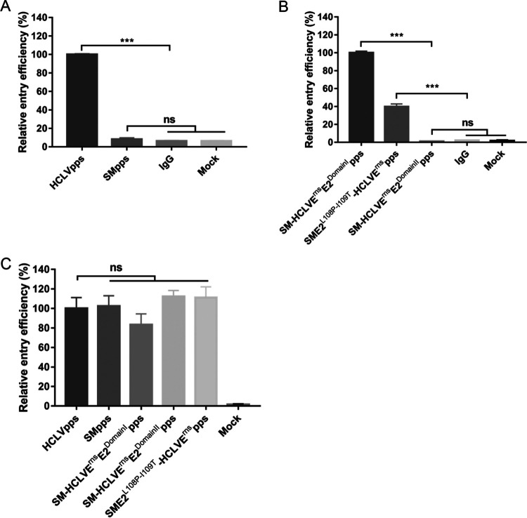
The E2 glycoprotein has been reported to be responsible for viral entry and tropism (14–16), while RNA replication is mediated by nonstructural proteins (NS3, NS4A-4B, and NS5A-5B) (25). Therefore, we speculated that the distinct adaptation potential of these chimeric viruses in rabbits may be due to different viral entry efficiency. A series of pseudotyped viruses (pps) bearing the Erns, E1, and E2 glycoproteins from the Shimen strain (SMpps) or C-strain (HCLVpps) were generated to investigate whether their differences occur in viral entry. We infected rabbits with SMpps or HCLVpps and isolated the spleen lymphocytes at 48 hpi. To amplify the fluorescence signals, an anti-EGFP (enhanced green fluorescent protein) antibody was used to measure EGFP expression. Meanwhile, mouse IgG was used to exclude nonspecific signals. We detected EGFP in the spleen lymphocytes from the rabbits inoculated with HCLVpps but not SMpps (Fig. 4A), suggesting that the C-strain ATR results from improved entry efficiency.
FIG 4.
Entry was affected by the E2P108-T109-Erns of C-strain. (A) HCLVpps but not SMpps could enter spleen lymphocytes of rabbits. Spleen lymphocytes were isolated from rabbits inoculated with 107 transducing units (TU) of HCLVpps or SMpps or with 1 ml of DMEM. EGFP expression was detected in the spleen lymphocytes of the rabbits inoculated with HCLVpps but not SMpps or DMEM. (B) E2P108-T109-Erns of C-strain affected viral entry into lymphocytes. Spleen lymphocytes were isolated from rabbits inoculated with 107 TU of SME1-HCLVErnsE2DomainIpps, SME1-HCLVErnsE2DomainIIpps, or SME1E2L108P-I109T-HCLVErnspps or with 1 ml of DMEM. EGFP expression was detected in the spleen lymphocytes of the rabbits inoculated with SME1-HCLVErnsE2DomainIpps or SME1E2L108P-I109T-HCLVErnspps but not those inoculated with SME1-HCLVErnsE2DomainIIpps or DMEM. EGFP expression was detected using anti-EGFP antibody, and an irrelevant mouse IgG served as negative control. (C) The E2P108-T109-Erns of C-strain did not affect viral entry into swine SK6 cells. EGFP expression was measured in SK6 cells infected with parental and chimeric pseudotyped viruses at an MOI of 2 at 48 hpi.
We further investigated the role of the E2P108-T109-Erns of C-strain in viral entry using the chimeric pseudotyped viruses SME1-HCLVErnsE2DomainIpps, SME1-HCLVErnsE2DomainIIpps, and SME1E2L108P-I109T-HCLVErnspps. The results showed that EGFP was detected in the spleen lymphocytes from rabbits inoculated with SME1-HCLVErnsE2DomainIpps or SME1E2L108P-I109T-HCLVErnspps but not in those from SME1-HCLVErnsE2DomainIIpps-inoculated animals (Fig. 4B), indicating that E2P108-T109-Erns of C-strain plays decisive roles in viral entry. Meanwhile, these pseudotyped viruses could enter swine SK6 cells with similar efficiency (Fig. 4C).
E2P108-T109-Erns of C-strain does not attenuate the Shimen strain in pigs.
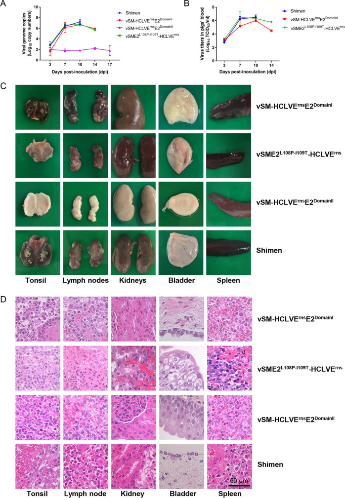
To examine whether the key amino acids contributing to viral ATR affect virulence in pigs, four groups of pigs were inoculated intramuscularly (i.m.) with vSME2L108P-I109T-HCLVErns, vSM-HCLVErnsE2DomainI, vSM-HCLVErnsE2DomainII, and the Shimen strain, using 105 50% tissue culture infective doses (TCID50). Clinical signs and rectal temperatures were monitored. All the pigs showed typical clinical signs (26) of CSF starting at 3 to 5 dpi, and the pigs inoculated with vSME2L108P-I109T-HCLVErns, vSM-HCLVErnsE2DomainI, or the Shimen strain died at 11 to 16 dpi (Table 2). However, the temperatures of the pigs inoculated with vSM-HCLVErnsE2DomainII normalized at 11 dpi, and the pigs survived until the end of this experiment. Viremia kinetics in animals inoculated with vSME2L108P-I109T-HCLVErns or vSM-HCLVErnsE2DomainI were indistinguishable from those induced by the parental Shimen strain (Fig. 5A and B). In contrast, the pigs inoculated with vSM-HCLVErnsE2DomainII showed lower viremia than the parental virus (Fig. 5A), while virus was undetectable in the blood samples from the pigs inoculated with vSM-HCLVErnsE2DomainII using IFA in SK6 cells (Fig. 5B).
TABLE 2.
Swine survival and fever response following inoculation with chimeric viruses and the parental Shimen strain
| Viruses | No. of survivors/total | Mean time to death (days)a | Fever responsea
|
||
|---|---|---|---|---|---|
| No. of days to onset | Duration (days) | Maximum avg temp (°C) | |||
| vSME2L108P-I109T-HCLVErns | 0/3 | 13 (1.63) | 3 (0) | 9.3 (0.94) | 41.7 (0.16) |
| vSM-HCLVErnsE2DomainI | 0/3 | 13 (2.16) | 3 (0) | 10 (1.41) | 41.8 (0.08) |
| vSM-HCLVErnsE2DomainII | 3/3 | 2.67 (0.47) | 6.7 (0.47) | 41.1 (0.08) | |
| Shimen | 0/2 | 12.5 (1.5) | 3 (0) | 9.5 (1.5) | 41.7 (0.05) |
Values are means (standard deviations).
FIG 5.
Viremia and representative pathological and histopathological changes of the pigs inoculated with chimeric viruses. (A) Viral genome copy numbers in the blood samples of the pigs inoculated with vSME2L108P-I109T-HCLVErns, vSM-HCLVErnsE2DomainI, vSM-HCLVErnsE2DomainII, or the Shimen strain. (B) Viral titers in the blood samples from the pigs inoculated with vSM-HCLVErnsE2DomainI, vSME2L108P-I109T-HCLVErns, or the Shimen strain in SK6 cells. (C) Representative pathological changes of various organs from the inoculated pigs. (D) Histopathological changes of various organs from the inoculated pigs. Bar, 50 μm.
The pigs inoculated with vSME2L108P-I109T-HCLVErns, vSM-HCLVErnsE2DomainI, or the Shimen strain displayed CSF-specific pathological changes (including enlargement and hemorrhages in the lymph nodes, petechiae in the kidneys, and infarcts in the spleen) (25). In contrast, no obvious or slight pathological lesions were observed in the pigs inoculated with vSM-HCLVErnsE2DomainII (Fig. 5C).
Moreover, histopathological examination of the various organs from the pigs inoculated with vSM-HCLVErnsE2DomainII showed slight histopathological lesions, including depletion in lymph nodes and reactive hyperplasia in spleens. However, the pigs inoculated with vSM-HCLVErnsE2DomainI or vSME2L108P-I109T-HCLVErns showed histopathological lesions similar to those in pigs inoculated with the Shimen strain in tonsils (lymphopenia), kidneys (denaturalization and necrosis in some tubular epithelia), and bladder (epithelial degeneration) (Fig. 5D).
Taken together, these observations indicate that E2P108-T109-Erns of C-strain contributes to the ATR but does not alter the virulence of the Shimen strain in pigs, whereas E2DomainII-Erns of C-strain attenuates the Shimen strain, suggesting that there are different molecular determinants of CSFV ATR and virulence in pigs.
DISCUSSION
C-strain, which was developed by passaging a highly virulent CSFV strain in rabbits, is adaptive to rabbits and attenuated in pigs. It has been established that for the Shimen strain, acquiring the adaptation to rabbits depends on E2 together with Erns or E1 of C-strain. Furthermore, P108 and T109 in E2DomainI are crucial amino acids for C-strain to be adaptive to rabbits (24). In the present study, three chimeric viruses containing E2P108-T109, E2DomainI, or E2DomainII of C-strain in the backbone of the non-rabbit-adaptive Shimen mutant vSM-HCLVErns were generated and evaluated for CSFV ATR and virulence in pigs. Our study shows for the first time that E2P108-T109 or E2DomainI but not E2DomainII of C-strain could render vSM-HCLVErns adaptive to rabbits, suggesting that E2P108-T109-Erns of C-strain confers adaptation to rabbits on the Shimen strain. Importantly, we demonstrated that E2P108-T109-Erns of C-strain mediated the adaptation of CSFV by prompting viral entry during infection in rabbit spleen lymphocytes. However, pathogenicity analysis in pigs showed that E2P108-T109-Erns of C-strain did not alter the virulence of the Shimen strain in pigs.
It has been demonstrated that the E2 glycoproteins of pestiviruses are associated with viral tropism. A chimeric pestivirus containing border disease virus or CSFV E2 glycoprotein in the background of bovine viral diarrhea virus (BVDV) alters the tropism to different cells in contrast to the parental BVDV (13, 27). Recombinant E2 glycoproteins derived from three different pestiviruses have different abilities to modify BVDV and CSFV to be able to infect permissive cells, suggesting that the E2 glycoprotein is involved in host tropism of pestiviruses at the entry stage (28). Our findings demonstrated that E2P108-T109-Erns of C-strain is responsible for the CSFV ATR, which further indicates the important roles of E2 and Erns in the tropism of pestiviruses. However, the amount of replication that takes place in primary rabbit spleen lymphocytes is lower than that in primary swine macrophages. The distinct replication ability of CSFV in rabbits and pigs was observed. The possible explanations are as follows: the rabbit is not the natural host for CSFV, the adaptive ability of CSFV is low in primary rabbit spleen lymphocytes, and there is no cell-cell junction in suspended primary rabbit spleen lymphocytes.
The mechanism of viral adaptation to a heterogeneous host by passaging in nonsusceptible hosts can be associated with mutations in the viral genome, which can improve entry efficiency and promote viral genome replication and virion assembly and release (5, 29). Among these possible mechanisms, entry is the first and most important step for viral infection, and it is determined by the viral surface proteins. It has been reported that the highly conserved residue Q226 is associated with H2N2 and H3N2 viral adaptation to human receptors (4, 30). Three mutations in the viral glycoproteins E1 (L216R) and E2 (V388G and M405T) improve the efficiency of hepatitis C virus (HCV) entry into Lunet N mCD81 cells, which are likely to promote exposure of the CD81-binding site (5). Mutation of A281 was observed during HIV adaptation in macaques and affected the ability of the HIV-1 Env to use macaque CD4 (31). For pestiviruses, increasing evidence has proved that entry is mediated by several glycoproteins. The Erns glycoprotein of pestiviruses attaches to the virion envelope by directly interacting with the E2 glycoprotein to form Erns-E2 heterodimers and is indispensable for virus attachment and infection of target cells (32). BVDV and CSFV belong to the genus Pestivirus, sharing similar properties (33), and E1-E2 heterodimers are essential for BVDV entry (34). In our study, E2P108-T109-Erns of C-strain was demonstrated to be associated with the adaption of CSFV to rabbits by affecting viral entry during infection, which expands our knowledge of the entry of pestiviruses.
Typically, live attenuated vaccines developed by blind passage in cell cultures or nonsusceptible hosts are adaptive to the nonsusceptible host while being attenuated in the primary cell or host. For example, the adaptation of African swine fever virus to Vero cells leads to a gradual attenuation of virulence in pigs (35). Attenuation of the strain PC22A of porcine epidemic diarrhea virus was achieved by cell culture passage (36). Live attenuated vaccines such as C-strain and the lapinized rinderpest virus were developed by passaging a highly virulent strain in rabbits. However, the molecular basis for adaptation and attenuation remains largely unclear. Mutations in the surface glycoprotein E increase the adaptation of tick-borne encephalitis virus to BHK-21 cells and significantly attenuate neuroinvasiveness in adult mice (37). A single mutation in VP2 (A221T) confers the adaptation of infectious pancreatic necrosis virus to CHSE cells and attenuates virulence in Atlantic salmon fry (38). In contrast, D279N and A284T mutations can confer infectious bursal disease virus adaptation in cell culture but do not lead to virus attenuation (39). It was reported that the E2 glycoprotein of the CS vaccine strain derived from the LK VNIIVIM parental vaccine strain (40) could markedly attenuate the CSFV Brescia strain (18). Furthermore, many residues on the E2 were identified as being associated with CSFV virulence, including a discrete epitope (TAVSPTTLR) (19), W871T, W875D, and V878T in the internal fusion peptide (17), T830A (41), and T745I and M979K (22). The N269A/Q substitution, which removed the putative glycosylation site in the Erns glycoprotein, decreased virulence of the Brescia strain (21). Notably, our results showed that E2P108-T109-Erns of C-strain, which is associated with CSFV ATR, did not affect CSFV virulence in pigs. The Erns glycoprotein of C-strain does not affect the virulence of the chimeric viruses, which may be due to the N269 present on both C-strain and the Shimen strain (21). However, E2 glycoprotein domain II, which is irrelevant to adaptation, alters virulence, further demonstrating that the molecular determinants of the ATR are different from those of attenuation of CSFV in pigs.
In summary, we demonstrated that E2P108-T109-Erns of C-strain determines the adaptation of CSFV by facilitating viral entry during infection of rabbits. Furthermore, the molecular determinants of CSFV ATR do not affect virulence in pigs. Our findings contribute to our understanding of the molecular basis for adaptation and attenuation of live attenuated vaccines developed by blind passage in cell cultures or hosts. This study also implies that novel live attenuated vaccines against CSF may be developed by targeting genetic modifications instead of random evolution through blind cell passage.
MATERIALS AND METHODS
Cells and viruses.
SK6 and PK-15 cells were cultured with Dulbecco’s modified Eagle’s medium (DMEM) (catalog no. C11995500BT; Gibco) supplemented with 5% heat-inactivated fetal bovine serum (FBS) (catalog no. 10099-141C; Gibco) in a 37°C incubator with 5% CO2. HEK293T cells were cultured with DMEM supplemented with 10% FBS. The spleen lymphocytes of rabbits were maintained in Roswell Park Memorial Institute (RPMI) 1640 medium (catalog no. C11875500BT; Gibco) supplemented with 10% FBS, 1% antibiotics-antimycotics (catalog no. 15240-062; Gibco), 1% l-glutamine, and 0.20 ng/ml interleukin 2 (IL-2) (catalog no. ab119439; Abcam) (42). Primary swine macrophages were prepared as described previously (43). The CSFV C-strain (GenBank no. AY805221) and Shimen strain (GenBank no. AF092448.2) were used for the construction of infectious cDNA clones.
Generation of chimeric viruses.
Based on the infectious cDNA clone pSM-HCLVErns, which harbors Erns of C-strain in the background of the Shimen strain (24), we took advantage of XhoI and BamHI restriction sites to construct pSM-HCLVErnsE2DomainI, pSM-HCLVErnsE2DomainII, and pSME2L108P-I109T-HCLVErns using fusion PCR with the primers listed in Table 3. The E2 domains I and II were amplified from pCSFV-HCLV using primers pSM-HCLVErnsE2DomainI-2F/2R and pSM-HCLVErnsE2DomainII-2F/2R, respectively. The PCR products were fused with products from pSM-HCLVErnsE2DomainI-1F/1R and pSM-HCLVErnsE2DomainI-3F/3R or from pSM-HCLVErnsE2DomainII-1F/1R and pSM-HCLVErnsE2DomainII-3F/3R. The PCR products obtained with the pSM-HCLVErns template using pSME2L108P-I109T-HCLVErns-1F/1R and pSME2L108P-I109T-HCLVErns-2F/2R primers designed for site-specific mutagenesis were fused with pSME2L108P-I109T-HCLVErns-1F/2R. Both fusion PCR products and pSM-HCLVErns were digested with XhoI and BamHI and then linked with T4 DNA ligase (catalog no. M0202S; New England BioLabs). All these constructed infectious cDNA clones were identified by PCR, enzyme digestion, and sequencing.
TABLE 3.
Primers used in this study
| Primers | Sequences (5′–3′) |
|---|---|
| pSM-HCLVErnsE2DomainI-1F | CCACCTCGAGATGCTATGTGG |
| pSM-HCLVErnsE2DomainI-1R | GGAATGCAATGGTTGATGCGCTATTCCAGACCCTGGTTAA |
| pSM-HCLVErnsE2DomainI-2F | TTAACCAGGGTCTGGAATAGCGCATCAACCATTGCATTCC |
| pSM-HCLVErnsE2DomainI-2R | CTTCGGTTGATGGGTTGGTCCCGTCGAACAGGAGCTCGAATG |
| pSM-HCLVErnsE2DomainI-3F | CATTCGAGCTCCTGTTCGACGGGACCAACCCATCAACCGAAG |
| pSM-HCLVErnsE2DomainI-3R | TAGATGGATCCTCTCCACTAT |
| pSM-HCLVErnsE2DomainII-1F | CACCTCGAGATGCTATGTGGACG |
| pSM-HCLVErnsE2DomainII-1R | CCTCAGTTGATGGGTTGGTCCCGTCGAACAGGAGCTCGAATGTCACG |
| pSM-HCLVErnsE2DomainII-2F | CGTGACATTCGAGCTCCTGTTCGACGGGACCAACCCATCAACTGAGG |
| pSM-HCLVErnsE2DomainII-2R | CTTCATTTTCCACTGTGGTGGTCACACAATCCATTCTGTGCGG |
| pSM-HCLVErnsE2DomainII-3F | CCGCACAGAATGGATTGTGTGACCACCACAGTGGAAAATGAAG |
| pSM-HCLVErnsE2DomainII-3R | GATGGATCCTCTCCACTATAATAG |
| pSME2L108P-I109T-HCLVErns-1F | CCACCTCGAGATGCTATGTGG |
| pSME2 L108P-I109T-HCLVErns-1R | CTCGAATGTCACGGAAGTGGGTAAAGCCCCCTTATGC |
| pSME2 L108P-I109T-HCLVErns-2F | GCATAAGGGGGCTTTACCCACTTCCGTGACATTCGAG |
| pSME2 L108P-I109T-HCLVErns-2R | TAGATGGATCCTCTCCACTAT |
Three chimeric viruses were rescued as described previously with a slight modification (24). Six micrograms of each plasmid mixed with 6 μl of X-tremeGENE HP DNA transfection reagent (catalog no. 6366546001; Roche) was transfected into SK6 cells cultured in a 6-well plate. The transfected cells were passaged several times, and the supernatants were subjected to detection of the Erns glycoprotein by a CSFV antigen test kit (catalog no. 99-40939; IDEXX). The positive samples were identified by RT-PCR, sequencing, and IFA.
IFA and virus titration.
The viral titers in the rabbit spleens or the pig blood samples at different time points were determined as previously described (44). SK6 or PK-15 cells were inoculated with serial 10-fold dilutions of the samples and cultured in 37°C for 48 h. Cold absolute ethanol was used for fixing cells at −20°C for 20 min. The fixed cells were washed three times with phosphate-buffered saline (PBS) and incubated with an anti-E2 polyclonal antibody at 37°C for 2 h (45). After five washes with PBS, cells were incubated with Alexa Fluor 488 goat anti-rabbit IgG (catalog no. A11034; Roche) at 37°C for 1 h and then washed five times with PBS. Then, the cells were stained with 0.5 μg/ml 4′,6-diamidino-2-phenylindole (DAPI; catalog no. C0060; Solarbio) for 15 min and washed three times with PBS. The cells were analyzed for green fluorescence using an inverted fluorescence microscope (EVOS FL; Life Technologies). The viral titers were calculated according to the Reed-Muench method and expressed as median tissue culture infective doses (TCID50) per milliliter (46).
Growth curves of the rescued viruses.
To determine the multistep growth curves of the rescued viruses, PK-15 cells cultured in 24-well plates were infected with these rescued viruses at a multiplicity of infection (MOI) of 0.1. Two hours later, the supernatants were removed, and the cells were washed three times with PBS. Then, fresh DMEM supplemented with 2% FBS was added to each well, and the cells were cultured at 37°C and 5% CO2. The cells were harvested at 12-h intervals and used to determine viral titers.
RNA extraction and RT-qPCR.
Total RNA from tissues or cells was extracted using RNAiso Plus (catalog no. 9109; TaKaRa) according to the manufacturer’s protocol. cDNA synthesis was processed in a 20-μl volume with avian myeloblastosis virus (AMV) reverse transcriptase XL (catalog no. 2621; TaKaRa). The copy numbers of the CSFV genome were determined using a previously described RT-qPCR assay (47).
Luciferase assay.
The reporter virus vHCLV-NanoLuc expressing the NanoLuc protein fused with the Npro protein (between the amino acids 13 and 14) based on C-strain was generated. Rabbit spleen lymphocytes infected with vHCLV-NanoLuc or C-strain or treated with DMEM only were lysed with 100 μl of passive lysis buffer (catalog no. N1150; Promega) and incubated on a shaker for 1 h at 4°C. The supernatants were collected by centrifuging at 12,000 × g for 10 min at 4°C. NanoLuc activities were measured with EnVision multilabel plate readers (PerkinElmer).
Flow cytometry.
The spleens from the rabbits inoculated with C-strain were collected after rabbits were euthanized at 3 dpi, and spleen cells were obtained by smearing the organs on 200-mesh copper wire mesh with RPMI 1640 medium. The red blood cells were lysed using red blood cell lysis buffer (catalog no. R1010; Solarbio), and the lymphocyte suspensions were washed with PBS. The following antibodies were used for flow cytometry, according to the manufacturers’ instructions: goat anti-rabbit IgM μ chain preadsorbed to secondary antibody (DyLight 488) (catalog no. ab98454; Abcam) for isolating B cells, mouse anti-rabbit T lymphocytes (catalog no. MCA800GA; Bio-Rad) for isolating T cells, and Alexa Fluor 633 goat anti-mouse IgG (heavy plus light chain [H+L]) (catalog no. A21052; Invitrogen) as the secondary antibody. Stained mononuclear cells from the spleens of the rabbits infected with C-strain were added to cytometry tubes and sorted with a high-speed cell sorter (MoFlo XDP; Beckman Coulter).
Infection of primary cells from rabbits or pigs with chimeric viruses.
Primary rabbit spleen lymphocytes or swine macrophages were isolated from the animals and infected with parental or chimeric viruses with an MOI of 0.01. After 2 h of incubation in a CO2 incubator, the infected cells were washed three times with PBS and further incubated for 4, 24, 48, or 72 h (17). The viral genome copy numbers or the viral titers at different time points were measured.
Preparation of pseudotyped viruses.
The DNA fragments encoding the last 60 amino acids of the C, Erns, E1, and E2 proteins from the infectious clones of C-strain, the Shimen strain, pSME1-HCLVErnsE2DomainI, pSME1-HCLVErnsE2DomainII, and pSME1E2L108P-I109T-HCLVErns were amplified and inserted into the pCAGGS vector (15, 48). Pseudotyped viruses were packaged by cotransfection into HEK293T cells with pNL4.3-GFP-ΔEnv and pCAGGS-HCLVErnsE1E2, pCAGGS-SMErnsE1E2, pCAGGS-SME1-HCLVErnsE2DomainI, pCAGGS-SME1-HCLVErnsE2DomainII, or pCAGGS-SME1E2L108P-I109T-HCLVErns (49). At 48 h posttransfection (hpt), the supernatants were collected and centrifuged for concentration using Amicon Ultra centrifugal filters (catalog no. UFC901096; Amicon). HIV p24 antigen content was assessed by enzyme-linked immunoassay (ELISA) (catalog no. BF06203; Biodragon Immunotechnologies).
Experimental infection of rabbits with chimeric viruses.
Twenty-four 14-week-old New Zealand White rabbits were divided into 5 groups and inoculated intravenously (i.v.) via the marginal ear vein with the viruses indicated in Table 1. The rectal temperature of all rabbits was monitored every 6 h from 24 to 72 hpi as described previously (24). Four rabbits were selected randomly from each group and euthanized at 3 hpi. The viral genome copy numbers in the spleens of the rabbits were determined and virus isolation was performed as described previously (24). The anti-E2 antibodies of the remainder rabbits were tested at 10 hpi using the classical swine fever virus antibody test kit (catalog no. 99-43220; IDEXX) according to the manufacturer's manuals.
Experimental infection of rabbits with pseudotyped viruses.
The rabbits were inoculated i.v. with different chimeric pseudotyped viruses. Spleen lymphocytes were isolated at 48 hpi. Mouse anti-coral green fluorescent protein (cGFP)-tagged monoclonal antibody (MAb) (catalog no. A00185; GenScript) was used to detect the expression of EGFP in lymphocytes, and irrelevant mouse IgG (catalog no. A7028; Beyotime) was used as a negative control. As the secondary immunoreagent, fluorescein isothiocyanate-labeled goat anti-mouse IgG (catalog no. A11029; Invitrogen) was used. All antibodies mentioned were diluted at 1:200 in PBS. For flow cytometry analysis, 106 cells in each sample were permeabilized with 0.15% Triton X-100. The cells were further incubated with primary antibody for 1 h and washed three times for 5 min with PBS. Then the cells were incubated with secondary antibody for 45 min and washed three times for 5 min with PBS. The fluorescence signal was analyzed with an Accuri C6 Plus flow cytometer (BD Biosciences).
Experimental infection of pigs with chimeric viruses.
To assess the virulence of chimeric viruses relative to the Shimen strain, 5-week-old healthy pigs were randomly divided into 4 groups (groups 1 to 3, n = 3; group 4, n = 2), and each group was housed in an individual room. Group 1 was inoculated i.m. with vSM-HCLVErnsE2DomainI, group 2 was inoculated i.m. with vSM-E2L108P-I109T-HCLVErns, group 3 was inoculated i.m. with vSM-HCLVErnsE2DomainII, and group 4 was inoculated i.m. with the Shimen strain. The inoculation dose of the virus for each group was 105 TCID50 (17). Clinical signs and rectal temperature were monitored daily, and anticoagulated blood samples of pigs were collected every 3 or 4 days. CSFV RNA was determined in anticoagulated blood samples by RT-qPCR. The chimeric viruses in the blood samples were titrated in SK6 cells using IFA.
Pathological examinations.
Macroscopic and microscopic pathological changes of the pig tissues were examined as described previously (50). The tonsils, lymph nodes, kidneys, bladders, and spleens were fixed with 10% formalin and then embedded in paraffin wax. For histopathological examinations, prepared tissue sections were stained with hematoxylin and eosin (H&E).
Immunohistochemistry.
The spleens of rabbits inoculated with the Shimen strain, C-strain, vSME2L108P-I109T-HCLVErns, vSM-HCLVErnsE2DomainI, or vSM-HCLVErnsE2DomainII were subjected to immunohistochemistry examinations using an anti-CSFV E2 antibody as described previously (51).
Animal ethics.
All animal experiments were carried out in strict accordance with the recommendations in the Guide for the Care and Use of Laboratory Animals of Heilongjiang Province of the People's Republic of China. The protocols were approved by the Committee on the Ethics of Animal Experiments of Harbin Veterinary Research Institute (HVRI) of the Chinese Academy of Agricultural Sciences (CAAS) (approval numbers SY-2018-Ra-002-01, SY-2018-Ra-02, and SY-2019-RA-004 for rabbit experiments and SY-2019-SW-033 for pig experiments).
Statistical analysis.
SPSS 22.0 software was used to analyze all data. An unadjusted P value of <0.05 was considered significant.
ACKNOWLEDGMENTS
We thank Yonghui Zheng (Harbin Veterinary Research Institute, Chinese Academy of Agricultural Sciences) for providing the plasmid pNL4.3-GFP-ΔEnv. We thank Ryan H. Gumpper (Department of Pharmacology, School of Medicine, University of North Carolina at Chapel Hill, USA) for improving the writing.
This study was supported by the National Natural Science Foundation of China (no. 31772774, 31972673, and 31630080).
REFERENCES
- 1.Koutsoudakis G, Herrmann E, Kallis S, Bartenschlager R, Pietschmann T. 2007. The level of CD81 cell surface expression is a key determinant for productive entry of hepatitis C virus into host cells. J Virol 81:588–598. doi: 10.1128/JVI.01534-06. [DOI] [PMC free article] [PubMed] [Google Scholar]
- 2.Walker RV. 1947. Rinderpest studies: attenuation of the rabbit adapted strain of rinderpest virus. Can J Comp Med Vet Sci 11:11–16. [PMC free article] [PubMed] [Google Scholar]
- 3.Qiu HJ, Shen RX, Tong GZ. 2006. The lapinized Chinese strain vaccine against classical swine fever virus: a retrospective review spanning half a century. J Integr Agric 5:1–14. doi: 10.1016/S1671-2927(06)60013-8. [DOI] [Google Scholar]
- 4.Gao R, Cao B, Hu Y, Feng Z, Wang D, Hu W, Chen J, Jie Z, Qiu H, Xu K, Xu X, Lu H, Zhu W, Gao Z, Xiang N, Shen Y, He Z, Gu Y, Zhang Z, Yang Y, Zhao X, Zhou L, Li X, Zou S, Zhang Y, Li X, Yang L, Guo J, Dong J, Li Q, Dong L, Zhu Y, Bai T, Wang S, Hao P, Yang W, Zhang Y, Han J, Yu H, Li D, Gao GF, Wu G, Wang Y, Yuan Z, Shu Y. 2013. Human infection with a novel avian-origin influenza A (H7N9) virus. N Engl J Med 368:1888–1897. doi: 10.1056/NEJMoa1304459. [DOI] [PubMed] [Google Scholar]
- 5.Bitzegeio J, Bankwitz D, Hueging K, Haid S, Brohm C, Zeisel MB, Herrmann E, Iken M, Ott M, Baumert TF, Pietschmann T. 2010. Adaptation of hepatitis C virus to mouse CD81 permits infection of mouse cells in the absence of human entry factors. PLoS Pathog 6:e1000978. doi: 10.1371/journal.ppat.1000978. [DOI] [PMC free article] [PubMed] [Google Scholar]
- 6.Frentzen A, Anggakusuma, Gurlevik E, Hueging K, Knocke S, Ginkel C, Brown RJ, Heim M, Dill MT, Kroger A, Kalinke U, Kaderali L, Kuehnel F, Pietschmann T. 2014. Cell entry, efficient RNA replication, and production of infectious hepatitis C virus progeny in mouse liver-derived cells. Hepatology 59:78–88. doi: 10.1002/hep.26626. [DOI] [PubMed] [Google Scholar]
- 7.Zhu Y, Zhang R, Zhang B, Zhao T, Wang P, Liang G, Cheng G. 2017. Blood meal acquisition enhances arbovirus replication in mosquitoes through activation of the GABAergic system. Nat Commun 8:1262. doi: 10.1038/s41467-017-01244-6. [DOI] [PMC free article] [PubMed] [Google Scholar]
- 8.Stremlau M, Owens CM, Perron MJ, Kiessling M, Autissier P, Sodroski J. 2004. The cytoplasmic body component TRIM5alpha restricts HIV-1 infection in Old World monkeys. Nature 427:848–853. doi: 10.1038/nature02343. [DOI] [PubMed] [Google Scholar]
- 9.Hueging K, Doepke M, Vieyres G, Bankwitz D, Frentzen A, Doerrbecker J, Gumz F, Haid S, Wolk B, Kaderali L, Pietschmann T. 2014. Apolipoprotein E codetermines tissue tropism of hepatitis C virus and is crucial for viral cell-to-cell transmission by contributing to a postenvelopment step of assembly. J Virol 88:1433–1446. doi: 10.1128/JVI.01815-13. [DOI] [PMC free article] [PubMed] [Google Scholar]
- 10.Smit JM, Moesker B, Rodenhuis-Zybert I, Wilschut J. 2011. Flavivirus cell entry and membrane fusion. Viruses 3:160–171. doi: 10.3390/v3020160. [DOI] [PMC free article] [PubMed] [Google Scholar]
- 11.Blome S, Staubach C, Henke J, Carlson J, Beer M. 2017. Classical swine fever-an updated review. Viruses 9:86. doi: 10.3390/v9040086. [DOI] [PMC free article] [PubMed] [Google Scholar]
- 12.Rümenapf T, Unger G, Strauss JH, Thiel HJ. 1993. Processing of the envelope glycoproteins of pestiviruses. J Virol 67:3288–3294. doi: 10.1128/JVI.67.6.3288-3294.1993. [DOI] [PMC free article] [PubMed] [Google Scholar]
- 13.Chen J, He WR, Shen L, Dong H, Yu J, Wang X, Yu S, Li Y, Li S, Luo Y, Sun Y, Qiu HJ. 2015. The laminin receptor is a cellular attachment receptor for classical swine fever virus. J Virol 89:4894–4906. doi: 10.1128/JVI.00019-15. [DOI] [PMC free article] [PubMed] [Google Scholar]
- 14.Hulst MM, Moormann RJ. 1997. Inhibition of pestivirus infection in cell culture by envelope proteins E(rns) and E2 of classical swine fever virus: E(rns) and E2 interact with different receptors. J Gen Virol 78:2779–2787. doi: 10.1099/0022-1317-78-11-2779. [DOI] [PubMed] [Google Scholar]
- 15.Wang Z, Nie Y, Wang P, Ding M, Deng H. 2004. Characterization of classical swine fever virus entry by using pseudotyped viruses: E1 and E2 are sufficient to mediate viral entry. Virology 330:332–341. doi: 10.1016/j.virol.2004.09.023. [DOI] [PubMed] [Google Scholar]
- 16.Liang D, Sainz IF, Ansari IH, Gil LH, Vassilev V, Donis RO. 2003. The envelope glycoprotein E2 is a determinant of cell culture tropism in ruminant pestiviruses. J Gen Virol 84:1269–1274. doi: 10.1099/vir.0.18557-0. [DOI] [PubMed] [Google Scholar]
- 17.Holinka LG, Largo E, Gladue DP, O'Donnell V, Risatti GR, Nieva JL, Borca MV. 2016. Alteration of a second putative fusion peptide of structural glycoprotein E2 of classical swine fever virus alters virus replication and virulence in swine. J Virol 90:10299–10308. doi: 10.1128/JVI.01530-16. [DOI] [PMC free article] [PubMed] [Google Scholar]
- 18.Risatti GR, Borca MV, Kutish GF, Lu Z, Holinka LG, French RA, Tulman ER, Rock DL. 2005. The E2 glycoprotein of classical swine fever virus is a virulence determinant in swine. J Virol 79:3787–3796. doi: 10.1128/JVI.79.6.3787-3796.2005. [DOI] [PMC free article] [PubMed] [Google Scholar]
- 19.Risatti GR, Holinka LG, Carrillo C, Kutish GF, Lu Z, Tulman ER, Sainz IF, Borca MV. 2006. Identification of a novel virulence determinant within the E2 structural glycoprotein of classical swine fever virus. Virology 355:94–101. doi: 10.1016/j.virol.2006.07.005. [DOI] [PubMed] [Google Scholar]
- 20.Fernandez-Sainz I, Holinka LG, Gladue D, O'Donnell V, Lu Z, Gavrilov BK, Risatti GR, Borca MV. 2011. Substitution of specific cysteine residues in the E1 glycoprotein of classical swine fever virus strain Brescia affects formation of E1-E2 heterodimers and alters virulence in swine. J Virol 85:7264–7272. doi: 10.1128/JVI.00186-11. [DOI] [PMC free article] [PubMed] [Google Scholar]
- 21.Sainz IF, Holinka LG, Lu Z, Risatti GR, Borca MV. 2008. Removal of a N-linked glycosylation site of classical swine fever virus strain Brescia Erns glycoprotein affects virulence in swine. Virology 370:122–129. doi: 10.1016/j.virol.2007.08.028. [DOI] [PubMed] [Google Scholar]
- 22.Wu R, Li L, Zhao Y, Tu J, Pan Z. 2016. Identification of two amino acids within E2 important for the pathogenicity of chimeric classical swine fever virus. Virus Res 211:79–85. doi: 10.1016/j.virusres.2015.10.006. [DOI] [PubMed] [Google Scholar]
- 23.Vilcek S, Nettleton PF. 2006. Pestiviruses in wild animals. Vet Microbiol 116:1–12. doi: 10.1016/j.vetmic.2006.06.003. [DOI] [PubMed] [Google Scholar]
- 24.Li Y, Xie L, Zhang L, Wang X, Li C, Han Y, Hu S, Sun Y, Li S, Luo Y, Liu L, Munir M, Qiu HJ. 2018. The E2 glycoprotein is necessary but not sufficient for the adaptation of classical swine fever virus lapinized vaccine C-strain to the rabbit. Virology 519:197–206. doi: 10.1016/j.virol.2018.04.016. [DOI] [PubMed] [Google Scholar]
- 25.Chen Y, Xiao J, Xiao J, Sheng C, Wang J, Jia L, Zhi Y, Li G, Chen J, Xiao M. 2012. Classical swine fever virus NS5A regulates viral RNA replication through binding to NS5B and 3′UTR. Virology 432:376–388. doi: 10.1016/j.virol.2012.04.014. [DOI] [PubMed] [Google Scholar]
- 26.Luo Y, Ji S, Lei JL, Xiang GT, Liu Y, Gao Y, Meng XY, Zheng G, Zhang EY, Wang Y, Du ML, Li Y, Li S, He XJ, Sun Y, Qiu HJ. 2017. Efficacy evaluation of the C-strain-based vaccines against the subgenotype 2.1d classical swine fever virus emerging in China. Vet Microbiol 201:154–161. doi: 10.1016/j.vetmic.2017.01.012. [DOI] [PubMed] [Google Scholar]
- 27.Reimann I, Depner K, Trapp S, Beer M. 2004. An avirulent chimeric pestivirus with altered cell tropism protects pigs against lethal infection with classical swine fever virus. Virology 322:143–157. doi: 10.1016/j.virol.2004.01.028. [DOI] [PubMed] [Google Scholar]
- 28.Asfor AS, Wakeley PR, Drew TW, Paton DJ. 2014. Recombinant pestivirus E2 glycoproteins prevent viral attachment to permissive and non permissive cells with different efficiency. Virus Res 189:147–157. doi: 10.1016/j.virusres.2014.05.016. [DOI] [PubMed] [Google Scholar]
- 29.Ostermann E, Pawletko K, Indenbirken D, Schumacher U, Brune W. 2015. Stepwise adaptation of murine cytomegalovirus to cells of a foreign host for identification of host range determinants. Med Microbiol Immunol 204:461–469. doi: 10.1007/s00430-015-0400-7. [DOI] [PubMed] [Google Scholar]
- 30.Rogers GN, Daniels RS, Skehel JJ, Wiley DC, Wang XF, Higa HH, Paulson JC. 1985. Host-mediated selection of influenza virus receptor variants. Sialic acid-alpha 2,6Gal-specific clones of A/duck/Ukraine/1/63 revert to sialic acid-alpha 2,3Gal-specific wild type in ovo. J Biol Chem 260:7362–7327. [PubMed] [Google Scholar]
- 31.Del Prete GQ, Keele BF, Fode J, Thummar K, Swanstrom AE, Rodriguez A, Raymond A, Estes JD, LaBranche CC, Montefiori DC, KewalRamani VN, Lifson JD, Bieniasz PD, Hatziioannou T. 2017. A single gp120 residue can affect HIV-1 tropism in macaques. PLoS Pathog 13:e1006572. doi: 10.1371/journal.ppat.1006572. [DOI] [PMC free article] [PubMed] [Google Scholar]
- 32.Lazar C, Zitzmann N, Dwek RA, Branza-Nichita N. 2003. The pestivirus Erns glycoprotein interacts with E2 in both infected cells and mature virions. Virology 314:696–705. doi: 10.1016/S0042-6822(03)00510-5. [DOI] [PubMed] [Google Scholar]
- 33.Wang FI, Deng MC, Huang YL, Chang CY. 2015. Structures and functions of pestivirus glycoproteins: not simply surface matters. Viruses 7:3506–3529. doi: 10.3390/v7072783. [DOI] [PMC free article] [PubMed] [Google Scholar]
- 34.Ronecker S, Zimmer G, Herrler G, Greiser-Wilke I, Grummer B. 2008. Formation of bovine viral diarrhea virus E1-E2 heterodimers is essential for virus entry and depends on charged residues in the transmembrane domains. J Gen Virol 89:2114–2121. doi: 10.1099/vir.0.2008/001792-0. [DOI] [PubMed] [Google Scholar]
- 35.Krug PW, Holinka LG, O'Donnell V, Reese B, Sanford B, Fernandez-Sainz I, Gladue DP, Arzt J, Rodriguez L, Risatti GR, Borca MV. 2015. The progressive adaptation of a Georgian isolate of African swine fever virus to Vero cells leads to a gradual attenuation of virulence in swine corresponding to major modifications of the viral genome. J Virol 89:2324–2332. doi: 10.1128/JVI.03250-14. [DOI] [PMC free article] [PubMed] [Google Scholar]
- 36.Lin CM, Hou Y, Marthaler DG, Gao X, Liu X, Zheng L, Saif LJ, Wang Q. 2017. Attenuation of an original US porcine epidemic diarrhea virus strain PC22A via serial cell culture passage. Vet Microbiol 201:62–71. doi: 10.1016/j.vetmic.2017.01.015. [DOI] [PMC free article] [PubMed] [Google Scholar]
- 37.Mandl CW, Kroschewski H, Allison SL, Kofler R, Holzmann H, Meixner T, Heinz FX. 2001. Adaptation of tick-borne encephalitis virus to BHK-21 cells results in the formation of multiple heparan sulfate binding sites in the envelope protein and attenuation in vivo. J Virol 75:5627–5637. doi: 10.1128/JVI.75.12.5627-5637.2001. [DOI] [PMC free article] [PubMed] [Google Scholar]
- 38.Song H, Santi N, Evensen O, Vakharia VN. 2005. Molecular determinants of infectious pancreatic necrosis virus virulence and cell culture adaptation. J Virol 79:10289–10299. doi: 10.1128/JVI.79.16.10289-10299.2005. [DOI] [PMC free article] [PubMed] [Google Scholar]
- 39.Ben Abdeljelil N, Khabouchi N, Kassar S, Miled K, Boubaker S, Ghram A, Mardassi H. 2014. Simultaneous alteration of residues 279 and 284 of the VP2 major capsid protein of a very virulent infectious bursal disease virus (vvIBDV) strain did not lead to attenuation in chickens. Virol J 11:199–199. doi: 10.1186/s12985-014-0199-7. [DOI] [PMC free article] [PubMed] [Google Scholar]
- 40.Zaberezhny AD, Grebennikova TV, Kurinnov VV, Tsybanov SG, Vishnyakov IF, Biketov SF, Aliper TI, Nepoklonov EA. 1999. Differentiation between vaccine strain and field isolates of classical swine fever virus using polymerase chain reaction and restriction test. Dtsch Tierarztl Wochenschr 106:394–397. [PubMed] [Google Scholar]
- 41.Tamura T, Sakoda Y, Yoshino F, Nomura T, Yamamoto N, Sato Y, Okamatsu M, Ruggli N, Kida H. 2012. Selection of classical swine fever virus with enhanced pathogenicity reveals synergistic virulence determinants in E2 and NS4B. J Virol 86:8602–8613. doi: 10.1128/JVI.00551-12. [DOI] [PMC free article] [PubMed] [Google Scholar]
- 42.Uddin MJ, Suen WW, Prow NA, Hall RA, Bielefeldt-Ohmann H. 2015. West Nile virus challenge alters the transcription profiles of innate immune genes in rabbit peripheral blood mononuclear cells. Front Vet Sci 2:76. doi: 10.3389/fvets.2015.00076. [DOI] [PMC free article] [PubMed] [Google Scholar]
- 43.Genovesi EV, Villinger F, Gerstner DJ, Whyard TC, Knudsen RC. 1990. Effect of macrophage-specific colony-stimulating factor (CSF-1) on swine monocyte/macrophage susceptibility to in vitro infection by African swine fever virus. Vet Microbiol 25:153–176. doi: 10.1016/0378-1135(90)90074-6. [DOI] [PubMed] [Google Scholar]
- 44.Zsak L, Lu Z, Kutish GF, Neilan JG, Rock DL. 1996. An African swine fever virus virulence-associated gene NL-S with similarity to the herpes simplex virus ICP34.5 gene. J Virol 70:8865–8871. doi: 10.1128/JVI.70.12.8865-8871.1996. [DOI] [PMC free article] [PubMed] [Google Scholar]
- 45.Sun Y, Liu DF, Wang YF, Liang BB, Cheng D, Li N, Qi QF, Zhu QH, Qiu HJ. 2010. Generation and efficacy evaluation of a recombinant adenovirus expressing the E2 protein of classical swine fever virus. Res Vet Sci 88:77–82. doi: 10.1016/j.rvsc.2009.06.005. [DOI] [PubMed] [Google Scholar]
- 46.Reed LJ, Muench HR. 1938. A simple method of estimating 50 per cent end points. Am J Hyg 27:493–497. doi: 10.1093/oxfordjournals.aje.a118408. [DOI] [Google Scholar]
- 47.Zhao JJ, Cheng D, Li N, Sun Y, Shi Z, Zhu QH, Tu C, Tong GZ, Qiu HJ. 2008. Evaluation of a multiplex real-time RT-PCR for quantitative and differential detection of wild-type viruses and C-strain vaccine of classical swine fever virus. Vet Microbiol 126:1–10. doi: 10.1016/j.vetmic.2007.04.046. [DOI] [PubMed] [Google Scholar]
- 48.Yu S, Yin C, Song K, Li S, Zheng GL, Li LF, Wang J, Li Y, Luo Y, Sun Y, Qiu HJ. 2019. Engagement of cellular cholesterol in the life cycle of classical swine fever virus: its potential as an antiviral target. J Gen Virol 100:156–165. doi: 10.1099/jgv.0.001178. [DOI] [PubMed] [Google Scholar]
- 49.Connor RI, Chen BK, Choe S, Landau NR. 1995. Vpr is required for efficient replication of human immunodeficiency virus type-1 in mononuclear phagocytes. Virology 206:935–944. doi: 10.1006/viro.1995.1016. [DOI] [PubMed] [Google Scholar]
- 50.Wang Y, Xia SL, Lei JL, Cong X, Xiang GT, Luo Y, Sun Y, Qiu HJ. 2015. Dose-dependent pathogenicity of a pseudorabies virus variant in pigs inoculated via intranasal route. Vet Immunol Immunopathol 168:147–152. doi: 10.1016/j.vetimm.2015.10.011. [DOI] [PubMed] [Google Scholar]
- 51.Ferrari M, Gualandi GL, Corradi A, Monaci C, Romanelli MG, Tosi G, Cantoni AM. 1998. Experimental infection of pigs with a thymidine kinase negative strain of pseudorabies virus. Comp Immunol Microbiol Infect Dis 21:291–303. doi: 10.1016/s0147-9571(98)00012-5. [DOI] [PubMed] [Google Scholar]


