Abstract
Stress is a normal part of life for fungi, which can survive in environments considered inhospitable or hostile for other organisms. Due to the ability of fungi to respond to, survive in, and transform the environment, even under severe stresses, many researchers are exploring the mechanisms that enable fungi to adapt to stress. The International Symposium on Fungal Stress (ISFUS) brings together leading scientists from around the world who research fungal stress. This article discusses presentations given at the third ISFUS, held in São José dos Campos, São Paulo, Brazil in 2019, thereby summarizing the state-of-the-art knowledge on fungal stress, a field that includes microbiology, agriculture, ecology, biotechnology, medicine, and astrobiology.
Keywords: Agricultural mycology, Fungal stress mechanisms and responses, Industrial mycology, Medical mycology
1. Introduction
Fungi play an essential role in many industrial, agricultural, and medical processes (Hyde et al., 2019; Rangel et al., 2018), and yet the importance and impact that these microorganisms have on humans and the environment is often underappreciated. Fungi can be a source of food and are essential for fermentation, including the production of bread, wine, beer, and other consumables. Fungi produce medicine, enzymes for industrial use, recombinant proteins, bioethanol, and biodiesel. Fungi serve as bioremediators, bioinsecticides, and can inhibit other plant-pathogenic microbes. Fungi can balance ecosystems via their roles as decomposers and by forming mechanical/physiological networks between other living systems. However, fungi can inflict diseases on humans, animals, and plants; degrade habitats or items of value; contaminate buildings; and act as a primary agent to spoil foods and feeds (Hyde et al., 2019; Rangel et al., 2018).
Fungi can survive in inhospitable and hostile environments. For instance, pathogenic fungi can survive in the interior of other organisms, despite the potential perils presented by anoxia and the host’s immune system (Brown et al., 2014). They can also withstand thermal stress, radiation, osmotic stress, desiccation, nutrient deprivation, and the presence of chaotropes, hydrophobes, and other aggressive compounds (Araújo et al., 2018, 2019; Dias et al., 2018; Hassett et al., 2015; Rangel, 2011; Rangel et al., 2005; Yakimov et al., 2015). Moreover, enduring stress during growth can allow fungi to withstand other stresses (Rangel, 2011). While psychology considers stress a negative force that disturbs well-being, for organisms like most fungi, the exposure to stress is a normal part of theirlives (Hallsworth, 2018). In general, stress can enhance vitality of the system by stimulating energy generation and other adaptations. This is consistent with the observation of German philosopher Frie-drich Nietzsche “What does not kill me, makes me stronger” (Was mich nicht umbringt, macht mich stärker) (Nietzsche, 1888).
Their ability to respond to, survive in, and transform the environment, even in the face of severe stress(es), is one of the reasons scientists seek to discover, understand, and utilize the biochemical and molecular mechanisms that enable fungi to adapt to stress. For some fungi, resistance to stress is a desirable characteristic; however, for other fungi, their resistance to stress poses a problem for humans. Knowledge about the stress mechanisms of fungi may help scientists to develop methods that modulate their ability to adapt to a specific environment and, by doing so, benefit the interests of society.
Further understanding about how stress affects fungi and how they circumvent potential constraints is the focus of the International Symposium on Fungal Stress (ISFUS). This Symposium takes an interdisciplinary approach attracting researchers with degrees in Microbiology, Mycology, Biology, Biochemistry, Molecular Biology, Genetics, Chemistry, Biotechnology, Microbial Physiology, Biomedical Sciences, Plant Pathology, and Ecology. Leading scientists from around the world have gathered in Brazil to present and discuss their research about fungal stress. ISFUS is the brainchild of Drauzio E. N. Rangel, who dreamed about bringing together scientists that focused specifically on the many stresses that fungi must endure (Rangel and Alder-Rangel, 2020). In 2014, Rangel invited senior scientists to the first ISFUS and acquired funding from the São Paulo Research Foundation (FAPESP), to bring them to Brazil. Rangel was assisted by Alene Alder-Rangel and other members of the Organizing Committee. The first ISFUS took place in October 2014 in São Jose dos Campos, São Paulo, Brazil, at the Universidade do Vale do Paraiba. The second ISFUS occurred in May 2017 in Goiania, Goiás, Brazil, at the Universidade Federal de Goiás, and received funding from the Coordenação de Aperfeiçoamento de Pessoal de Nível Superior (CAPES) and the Fundação de Amparo à Pesquisa do Estado de Goiás (FAPEG).
The third International Symposium on Fungal Stress (ISFUS-2019) returned to São José dos Campos, São Paulo, Brazil, and occurred on May 20 to 23, 2019 at the Hotel Nacional Inn. This Symposium was supported by grants from FAPESP and CAPES, and the Universidade Brasil acted as the host institution. ISFUS-2019 was larger than the previous ISFUS meetings, with 39 featured speakers from 16 countries (Figs. 1 and 2), 58 posters presentations, and around 125 participants. Elsevier (Amsterdam, Netherlands) and the Journal of Fungi (Basel, Switzerland) provided the student awards. Corporate sponsors were Biocontrol (Sertãozinho, SP, Brazil), Meter (São José dos Campos, SP, Brazil), and Alder’s English Services (São Jose dos Campos, SP, Brazil). The Organizing Committee included Drauzio E. N. Rangel, Alene Alder-Rangel, Claudia B. L. Campos, Ekaterina Dadachova, Gustavo H. Goldman, Gilberto U. L. Braga, Luis M. Corrochano, and John E. Hallsworth. The logo of the symposium features one of the most-studied ascomycetes, Aspergillus nidulans, and illustrates several key stress parameters that fungi must cope with to survive (Fig. 3). The Annals of the third International Symposium on Fungal Stress, which feature abstracts from the presentations and posters, is available in the Electronic Supplementary Material 1.
Fig. 1.
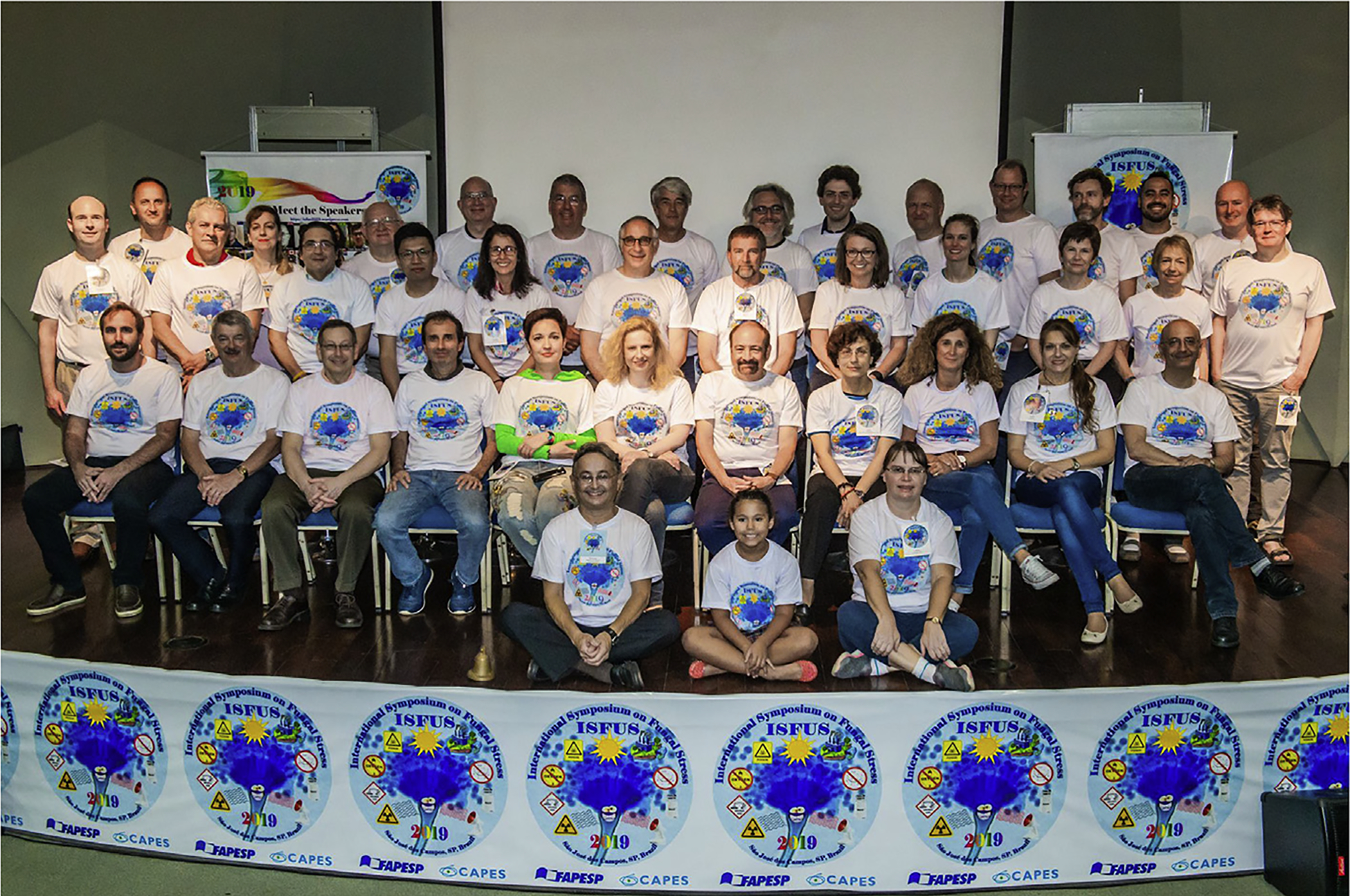
Speakers of the third ISFUS in 2019 held in São José dos Campos, SP, Brazil. Front row from left to right: Drauzio E. N. Rangel, Amanda E. A. Rangel, Alene Alder-Rangel. Second row from left to right: Thiago Olitta Basso (Brazil), Graeme M. Walker (UK), David E. Levin (USA), Gilberto U.L. Braga (Brazil), Irina Druzhinina (Russia/China), Julia Schumacher (Germany), Rocco L. Mancinelli (USA), Anna Gorbushina (Russia/Germany), Natalia Requena (Spain/Germany), Laura Selbmann (Italy), and Luis Corrochano (Spain). Third row from left to right: Alexander Idnurm (Australia), Jesús Aguirre (Mexico), Gustavo H. Goldman (Brazil), Chris Koon Ho Wong (Macau), Claudia B. L. Campos (Brazil), Oded Yarden (Israel), Martin Kupiec (Israel), Deborah Bell-Pedersen (USA), Christina M. Kelliher (USA), Michelle Momany (USA), Alexandra C. Brand (UK), and Jan Dijksterhuis (The Netherlands). Fourth row from left to right: Tamás Emri (Hungary), Ekaterina Dadachova (Russia/Canada), István Pócsi (Hungary), Alistair J. P. Brown (UK), Geoffrey M. Gadd (UK), Reinhard Fischer (Germany), Luis Larrondo (Chile), Guilherme T. P. Brancini (Brazil), Gerhard Braus (Germany), Florian F. Bauer (South Africa), Mikael Molin (Sweden), Radamés J.B. Cordero (USA), and John E. Hallsworth (UK).
Fig. 2.

Meet the speakers banner. This banner was printed on a poster and placed in the auditorium so everyone could remember their preferred speaker’s names for future scientific discussion. Below the speakers’ pictures are the logos of the grant agencies and sponsors.
Fig. 3.
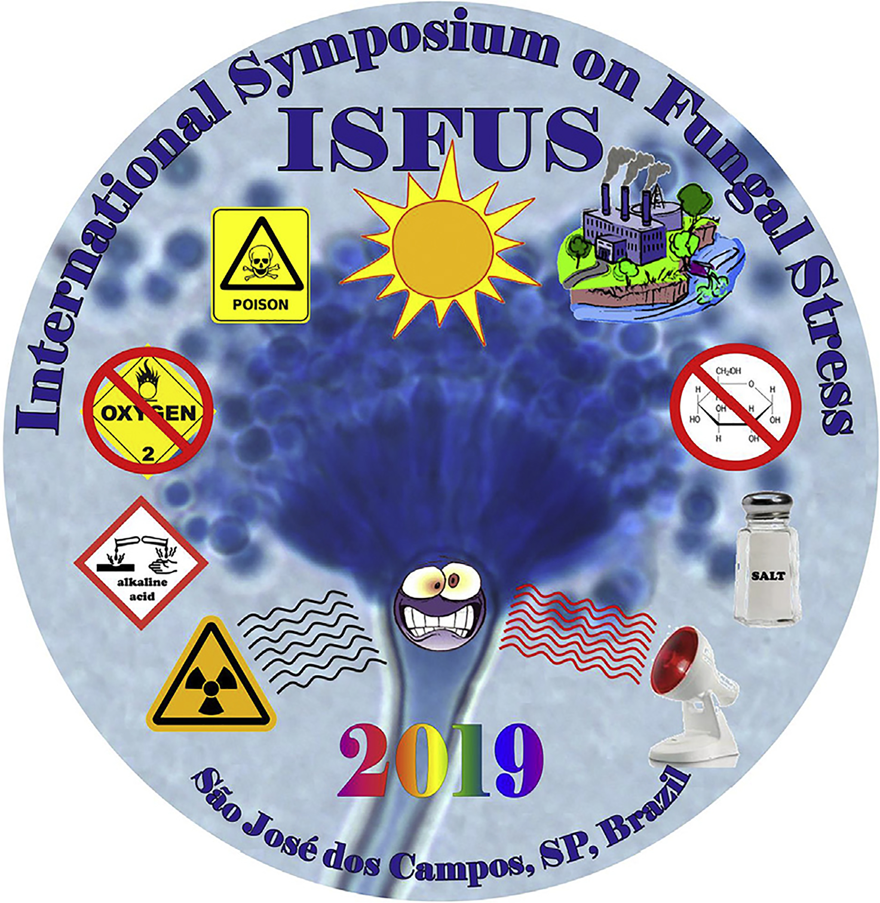
Logo of the third International Symposium on Fungal Stress (ISFUS-2019). This figure illustrates some of the stress parameters that fungi are subjected to such as ionizing radiation, acidic and alkaline environments, hypoxic or anoxic conditions, poisons in general such as genotoxic and oxidative products, UV radiation from the sun, pollution from industry and agriculture, salt stress, nutritive stress, and heat from solar radiation and other sources.
Each ISFUS has represented a major step in bringing together the community of fungal biologists interested in the mechanisms that fungi use to cope with stress. The first ISFUS as the initial meeting set the basic format of the symposium with a small size, a program touching different aspects of fungal stress biology, and activities in addition to the scientific program to increase scientific interactions among participants. The main role of ISFUS as an international forum for the exchange of ideas and to foster scientific interactions and international collaborations on fungal stress was clearly defined in the first ISFUS. The second and third ISFUS have grown upon these themes, expanding the number of topics covered and providing lecture time to students and young postdocs in the community, while keeping the number of participants both international and Brazilian to a level that allows easy and frequent interactions during lectures and free time. We anticipate that topics covered by future ISFUS will highlight the role of fungal stress biology in understanding how fungi contribute and adapt to global changes in the climate and to provide alternative resources for food, feed, and bioenergy.
A special issue has been published after each ISFUS that featured articles related to fungal stress primarily from researchers who presented at that ISFUS: for ISFUS-2014 in Current Genetics (Rangel et al., 2015a, 2015b), and for ISFUS-2017 in Fungal Biology, by Elsevier on behalf of the British Mycological Society (Alder-Rangel et al., 2018). After the success of that special issue, Fungal Biology agreed to publish this special issue arising from ISFUS-2019, which is titled “Fungal Adaptation to Hostile Challenges” focused on cellular biology, ecology, photobiology, environment, agricultural, industrial, and medical mycology in the context of fungal stress (Acheampong et al., 2019; Antal et al., 2019; Araújo et al., 2019; Brown et al., 2020; Dias et al., 2018; Fomina et al., 2019; Harari et al., 2019; Kelliher et al., 2019; Király et al., 2019; Laz et al., 2019; Malo et al., 2019; Medina et al., 2020; Mendoza-Martínez et al., 2019; Rodrigues et al., 2019; Schumacher and Gorbushina, 2020; Sethiya et al., 2019; Tagua et al., 2019; Walker and Basso, 2019; Yu et al., 2020; Yuan et al., 2019), and several other manuscripts under review, for a total of 31 articles in this special edition.
2. The third International Symposium on Fungal Stress - a synopsis
Although the Symposium started Monday, May 20, most international speakers arrived in Brazil on Saturday, May 18 to have time to recuperate from long flights. They took the opportunity to become better acquainted with each other and São Jose dos Campos with a tour of Vicentina Aranha Park, which features live music, a craft fair, and farmers market on Sunday morning. Amanda Estella Alder Rangel, the organizer’s six-year-old daughter, helped lead the tour and even translate when needed for our foreign guests helping them interact with locals, make purchases, etc (Fig. 4).
Fig. 4.
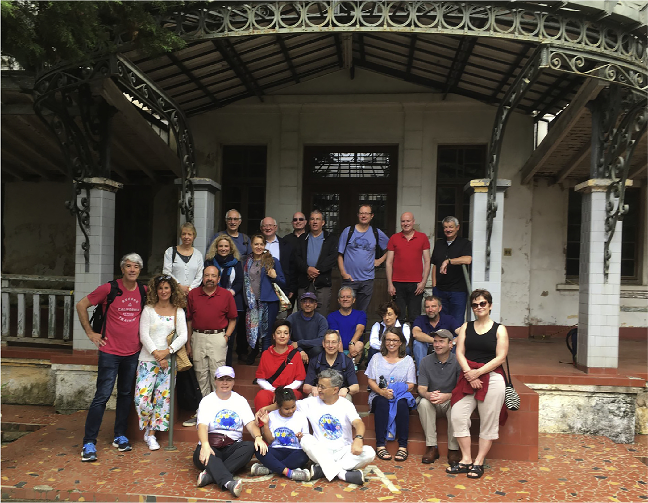
Speakers at the Vicentina Aranha Park, São José dos Campos, SP, Brazil.
The Symposium officially began Monday morning with a welcome presentation by Drauzio E.N. Rangel. He explained how the ISFUS series originated and that the motivation for the meetings has been driven throughout by the enthusiasm and hard work of his family. He welcomed the delegation by discussing the joyful nature of science. He talked about happiness with examples from his own life. After requesting that everyone recall their happiest memories, he asked them to stand up and join hands in a circle, reminiscent of a mushroom fairy ring, around the auditorium. Rangel went on to talk about intuition involved in scientific discovery and having an “open heart” during the research process (Fig. 5).
Fig. 5.
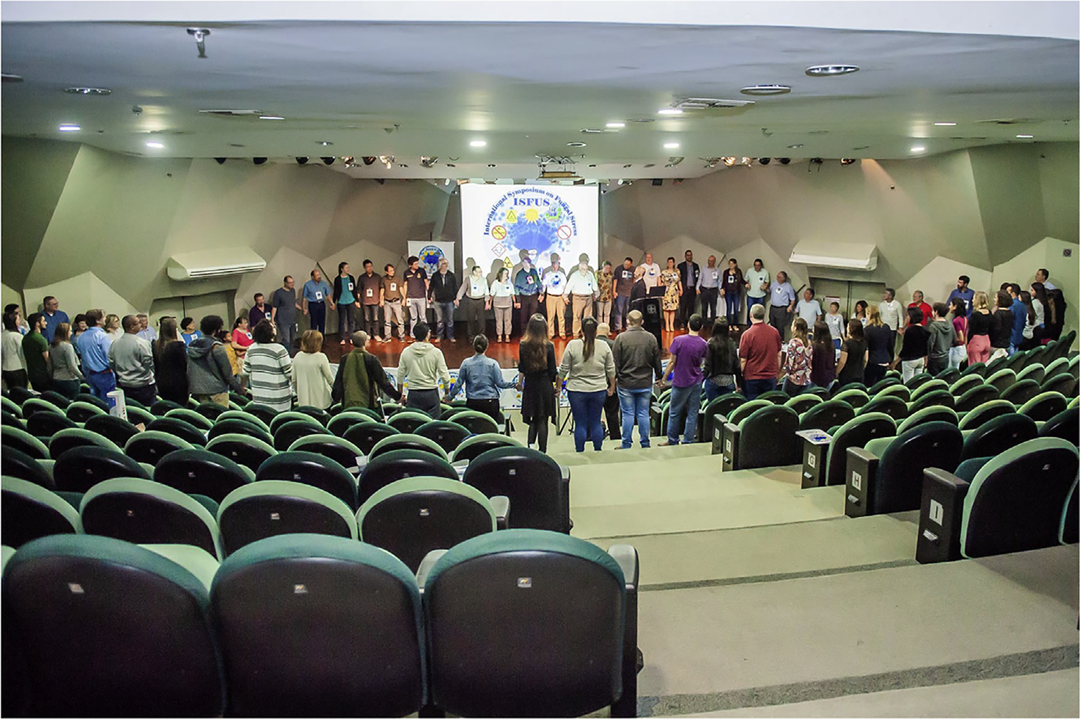
Speakers and participants holding hands and sharing happy moments.
The Symposium was organized around seven general topics related to fungal stress.
Stress mechanisms and responses in fungi: molecular biology, biochemistry, and cellular biology;
Fungal photobiology, clock regulation, and stress;
Fungal stress in industry;
Fungal biology in extreme environments;
Ionizing radiation, heat, and other stresses in fungal biology;
Stress in populations, fungal communities, and symbiotic interactions;
Stress in fungal pathogenesis.
The following text provides a synopsis of each topic, arranged in the order presented during the Symposium.
2.1. Stress mechanisms and responses in fungi: molecular biology, biochemistry, and cellular biology
Representatives of the fungal kingdom occupy almost every conceivable niche on Earth which is a testament to their versatility and evolutionary adaptation to their environment. A broad understanding of how fungi have adapted to diverse environments can come from genetic screening approaches that identify genes responsible for conferring tolerance. Researchers can then drill down for a deeper understanding of how these systems work at the molecular and evolutionary levels to explain the adaptation process. A consequence of this is an appreciation of how environmental fluctuation might challenge the viability of susceptible fungal species. The mechanisms involved in coping/adapting to stress are as diverse as the array of fungal species studied. Regardless of the stress/organism studied, rarely are the identified signaling and other biochemical and physiological pathways and elements unique to one organism. In addition, the function of stress-related pathways often spans growth, developmental, and reproductive networks, which have functions in non-stress conditions (Brown et al., 2017; Rangel et al., 2018).
Martin Kupiec gave the first presentation at ISFUS-2019. He focused on telomeres, which are the ends of the linear eukaryotic chromosomes. Telomeres are essential for maintaining the integrity of the genome and play important roles in aging and cancer (Mersaoui and Wellinger, 2019). A systematic analysis identified ~500 genes that regulate telomere length in the yeast Saccharomyces cerevisiae (Askree et al., 2004; Ungar et al., 2009). Kupiec’s group also found that small molecules, such as ethanol, caffeine, and acetic acid, can affect telomere length. Having a full list of genes and physiological actuators enabled research about the interface between the genome and the environment (to address the contributions of nature vs. nurture on physiological outcomes). Kupiec reported finding genes that mediate the environmental signal transduction to the telomere-regulating genes (Harari et al., 2019; Harari and Kupiec, 2018; Mersaoui and Wellinger, 2019; Romano et al., 2013).
István Pócsi talked about the Fungal Stress Response Database (FSRD) (de Vries et al., 2017; Karányi et al., 2013) and Fungal Stress Database (FSD) (de Vries et al., 2017; Orosz et al., 2018). The FSRD accommodates 43,725 stress protein orthologs identified in 41 fully sequenced genomes of 39 fungal species (de Vries et al., 2017). The FSD is a repository of 1412 photos taken on agar plate colonies of 17 Aspergillus species, exposed to oxidative, high-osmolarity, heavy metal, and cell wall integrity stress (de Vries et al., 2017; Orosz et al., 2018). Data in the FSRD were used to identify stress response protein orthologs in Drechmeria coniospora (Zhang et al., 2016b) and several Aspergillus spp. (de Vries et al., 2017; Emri et al., 2018). Data in the FSRD and FSD were used (i) in evolutionary biological studies in the aspergilli (Emri et al., 2018), and (ii) to shed light on cadmium tolerance of Aspergillus fumigatus (Antal et al., 2019; Bakti et al., 2018; Kurucz et al., 2018a).
David E. Levin discussed how various stresses activate the yeast SAPK Hog1 and how the cell mobilizes stress-specific outputs from activated Hog1. In response to hyper-osmotic shock, Hog1 induces the production of glycerol and its accumulation through closure of glycerol channel Fps1. Hog1 activated by the toxic metalloid arsenite similarly induces closure of Fps1, the main entry port for this toxin. However, under conditions of arsenite stress, cells do not accumulate glycerol. This is because S. cerevisiae uses a methylated metabolite of arsenite to inhibit the first enzymatic step in glycerol biosynthesis. Levin’s work provides insight into the mechanisms by which Hog1, as stimulated by two different stresses, can evoke physiologically coherent, but opposite, outputs (Laz et al., 2019; Lee and Levin, 2018, 2019; Lee et al., 2013, 2019).
Oded Yarden talked about how the Nuclear DBF-related (NDR) kinase colonial temperature sensitive-1 (cot-1) plays a role in the regulation of polar growth and development in Neurospora crassa and other fungi (Ziv et al., 2009). COT−1 is a kinase in the RAM pathway that is widely conserved in cell wall maintenance in eukaryotes (Osherov and Yarden, 2010; Saputo et al., 2012). Osmotic, oxidative, and other stresses result in partial phenotypic suppression of the cot-1 mutant defects (Gorovits and Yarden, 2003). Some of the phenotypic responses involve type 2A phosphatases and the translational regulator GUL-1 (Herold et al., 2019; Herold and Yarden, 2017; Shomin-Levi and Yarden, 2017).
Michelle Momany explained that many fungal infections start with the inhalation of spores from the environment. Despite the importance of spores to infection, little is known about how the environment when sporulation occurs impacts fungal spores. Momany’s group used RNAseq to examine A. fumigatus conidia (asexual spores) produced under several conditions including low Zn, high temperature, and high salt. They found that conidial transcriptomes from differing conditions contain a large set of common transcripts and a much smaller set of condition-enhanced transcripts. Generally, the condition-enhanced transcripts do not appear to be unique, rather they appear to differ mostly in level of expression.
Jesús Aguirre’s presentation addressed the signaling role of reactive oxygen species (ROS) in the regulation of cell differentiation in A. nidulans and other fungi. He showed that in A. nidulans NapA, a redox-regulated transcription factor, which is homologous to yeast Yap1, is involved not only in the antioxidant response, but also in the regulation of genes involved in nutrient assimilation, secondary metabolism, and development, and how this is related to peroxiredoxin function (Mendoza-Martínez et al., 2017, 2019).
Gustavo H. Goldman discussed how the CrzA and ZipD transcription factors are involved in calcium metabolism and the caspofungin paradoxical effect in the human pathogenic species A. fumigatus (Ries et al., 2017). At low concentrations of the drug, inhibition occurs, whereas that inhibition is lost at higher concentrations.
John E. Hallsworth began by explaining that we do not have any term or concept to identify a stress-free state in microorganisms (Hallsworth, 2018). The talk focused on what cellular stress actually is, taking a lucid tour around the logical geography of an otherwise complex topic. The distinction between toxicity and stress was discussed (Hallsworth, 2018), and data were presented relating to the water activity limit-for-life for halophilic bacteria and Archaea (Lee et al., 2018; Stevenson et al., 2015) and the extreme xerophile/halophile Aspergillus penicillioides (Stevenson et al., 2017). Hallsworth concluded by summarizing the 20 y of work which led to a new limit-for-life on Earth (Stevenson et al., 2017); this fascinating story revolved around a wooden owl which was the source of the most xerophilic microbe thus-far discovered: a strain of A. penicillioides (Hallsworth, 2019).
2.2. Fungal photobiology, clock regulation, and stress
The second day of the Symposium was devoted to fungal photobiology. Fungi use light as an environmental signal to regulate developmental transitions, modulate their direction of growth, and modify their metabolism. Fungi often synthesize protective pigments, such as melanins and carotenoids, in response to illumination because an excess of light can produce reactive oxygen species and UV radiation can damage DNA (Brancini et al., 2018, 2019; Corrochano, 2019; Yu and Fischer, 2019). In addition, the presence of light during fungal growth is known to up-regulate a variety of stress genes that induce higher conidial tolerance to UV radiation, heat, and osmotic stress (Dias et al., 2019; Rangel et al., 2011, 2015c). Many organisms, including fungi, have circadian clocks to anticipate daily changes in illumination, temperature, and water availability/humidity, as well as several environmental signals, including light, that regulate the activity of circadian clocks (Dunlap and Loros, 2017).
Deborah Bell-Pedersen stated that evidence supporting circadian clock regulation of mRNA translation exists in several organisms (Caster et al., 2016; Jouffe et al., 2013; Robles et al., 2014); however, the underlying mechanisms for translational control are largely unknown. Bell-Pedersen’s group discovered that the clock regulates the activity of the N. crassa eIF2a kinase CPC-3. Daytime active CPC-3 promotes phosphorylation and inactivation of the conserved translation initiation factor eIF2a, leading to reduced translation of specific mRNAs during the day and likely coordinating mRNA translation with increased energy availability and reduced stress at night.
Luis Larrondo’s thought-provoking talk was about light as a source of information, stress, biotechnological applications, and art. He described how the model species N. crassa can be used as a highly sensitive light sensor to record its environment, effectively acting as a photocopier of information or the film in a pinhole camera. The overlap between science and art was reflected in the gift of a N. crassa derived image, which was presented to Pope Francis during his visit to Chile.
Reinhard Fischer focused on A. nidulans and Alternaria alternata, which are two ascomycetes that are able to adapt to many different environments. Light is a reliable indicator for potential stressful conditions, and light sensing is tightly coupled to stress responses at the molecular level. For instance, the red-light sensor phytochrome uses the HOG pathway for signal transduction. In addition, both fungi use a flavin-containing protein as a blue-light receptor, and A. alternata an opsin for green light sensing. Fischer’s work focuses on the analysis of the interplay of the different light-sensing systems and their link to stress adaptation (Igbalajobi et al., 2019; Yu and Fischer, 2019; Yu et al., 2016), particularly the link between red light and temperature sensing via the phytochrome FphA (Yu et al., 2019).
Christina M. Kelliher introduced compensation, a core principle of all circadian clocks where the period of approximately 24 h is maintained across a range of physiologically relevant environmental conditions (Pittendrigh and Caldarola, 1973). A handful of genes involved in transcriptional regulation are required for the N. crassa clock to compensate at both high levels of glucose and in starvation conditions — an RNA helicase period-1 (Emerson et al., 2015), a co-repressor rco-1 (Olivares-Yañez et al., 2016), and a transcription factor repressor csp-1 (Sancar et al., 2012). The full mechanism of nutritional compensation, including upstream signaling pathways and downstream regulation on core circadian clock factors, is not characterized in any eukaryotic model. Kelliher and colleagues leveraged the whole genome knockout collection of N. crassa (Colot et al., 2006) in a screen to identify genes that are required for clock compensation under starvation, beginning with canonical carbon source signaling pathways, kinases, and transcriptional regulators. Currently, two kinases and two novel RNA-binding proteins have been identified as effectors required for normal nutritional compensation of the clock at high and no glucose levels in Neurospora. Future work will molecularly characterize screen hits in both solid medium and liquid assays (Kelliher et al., 2019) for circadian clock function.
Mikael Molin highlighted the ability of S. cerevisiae to respond to light despite lacking genes homologous to dedicated light receptors. Light sensing in this yeast is intimately connected to oxidative stress resistance and a group of peroxidases and peroxide receptors, peroxiredoxins, which seem to regulate stress-related kinases in a unique manner involving hydrogen peroxide signaling. Utilizing a genome-wide genetic screen, his group has also explored which parts of the cellular network that growth of S. cerevisiae in the presence of light engages. The data may form a framework to understand connections between light exposure, protein synthesis, and stress-related kinases such as the MAPKs and PKA in fungi and higher organisms (Bodvard et al., 2011, 2013, 2017; Nystrom et al., 2012).
Julia Schumacher explained that fungi sharing light-flooded habitats with phototrophic organisms suffer from light-induced stresses and experience altered light spectra (‘green gap’) enriched for green and far-red light. The plant pathogenic Leotiomycete Botrytis cinerea responds to light qualities covering the entire visible spectrum and beyond and uses light to coordinate stress responses, growth, reproduction, and host infection (Schumacher, 2017). The equally high number of photoreceptors in the rock-inhabiting black Eurotiomycete Knufia petricola suggests that photoregulation is equally important in mutualistic interactions of fungi with microbial phototrophs.
Gerhard Braus described how the coordination of the control of fungal reactions to light is impaired if cellular protein degradation is disturbed. The COP9 signalosome multiprotein complex is necessary for light regulation, stress responses, and development. It also coordinates secondary metabolism in A. nidulans (Busch et al., 2007) and controls, together with the protein substrate receptor exchange factor CandA/Cand1, the covalent labeling of substrates with chains of the small modifier ubiquitin for proteasome-mediated protein degradation (Braus et al., 2010). CandA of A. nidulans is required for light regulation of development and secondary metabolite formation (Köhler et al., 2019). The COP9 signalosome serves as the platform that interacts with numerous additional proteins, such as the deubiquitinase UspA, which is required to fine-tune light controlled development and secondary metabolism (Meister et al., 2019). Together, the specific control of protein homeostasis plays an important role for light induced stress with consequences in fungal development and secondary metabolism.
Luis Corrochano discussed how light regulates developmental pathways in most fungi. As the roles of light in the ecophysiology of plants and other primary producers, or the neurology and physiology of animal systems, is widely appreciated, it may seem counter-intuitive that heterotrophic fungi are controlled by this environmental signal. Development and secondary metabolism are often coordinated through the activity of the velvet protein complex (Bayram and Braus, 2012). In N. crassa, the velvet protein VE-1 interacts in vegetative hyphae with the velvet protein VE-2 and the methyltransferase LAE-1. This velvet complex regulates the growth of aerial hyphae and the accumulation of carotenoids after light exposure in vegetative mycelia (Bayram et al., 2019). Corrochano’s group observed that VE-1 is unstable and proposed that the regulation of VE-1 degradation is a relevant aspect of conidiation and its regulation by light in N. crassa.
Guilherme T. P. Brancini’s talk focused on how transcriptomics and proteomics can be combined to elucidate light responses in the entomopathogenic fungus Metarhizium acridum. Exposing M. acridum mycelium to light resulted in changes at the mRNA level for 1128 genes or 11.3 % of the genome (Brancini et al., 2019). High-throughput proteomics revealed that the abundance of only 57 proteins changed significantly under the same conditions. Light downregulated proteins involved in translation, including subunits of the eukaryotic translation initiation factor 3, the eIF5A-activating enzyme deoxyhypusine hydroxylase, and ribosomal proteins. As reducing and reprogramming translational activity are known cellular responses to stress (Crawford and Pavitt, 2019; Spriggs et al., 2010; Yamasaki and Anderson, 2008), this result indicates that light acts as both a signal and a source of stress in M. acridum. The reduced translational activity is thus a potential explanation for the small number of light-regulated proteins. Therefore, measuring protein levels is essential to fully understand light responses in fungi (Brancini et al., 2019).
Gilberto U. L. Braga examined how recent increases in consumer awareness about and legislation regarding environmental and human health, as well as the urgent need to improve food security, are driving increased demand for safer antimicrobials. A step-change is needed in the approaches for controlling pre- and post-harvest diseases and food-borne human pathogens. The use of light-activated antimicrobial substances for the so-called photodynamic treatment of diseases is known to be effective in a clinical context (Brancini et al., 2016; Tonani et al., 2018). They could be equally effective for use in agriculture to control plant-pathogenic fungi and bacteria, and to eliminate food-borne human pathogens from seeds, sprouted seeds, fruits, and vegetables (Fracarolli et al., 2016; Gonzales et al., 2017). Braga took a holistic approach in reviewing recent findings on (i) the ecology of naturally-occurring, (ii) photodynamic processes including the light-activated antimicrobial activities of some plant metabolites, and (iii) fungus-induced photosensitization of plants, against the backdrop of existing knowledge. The inhibitory mechanisms of both natural and synthetic light-activated substances, known as photosensitizers, were discussed in the contexts of microbial stress biology and agricultural biotechnology.
2.3. Fungal stress in industry
Wednesday began with presentations linking fungi, industrial applications, and stress in several ways. Notably, fungi are a continuous concern in the food industry as they spoil numerous products. To discourage fungi from proliferating on nutrient-rich food stuffs, several strategies are employed including pre-treatments, storage conditions, and preservatives. However, fungi can circumvent many of the obstacles used in food production to prevent this. A small subset of fungi, “spoil” food, but others can enhance the properties of food, make it more digestible, add vitamins, and protect against other fungi that can form toxic compounds. Alternatively, fungi are used widely in industry to produce metabolites, such as antibiotics and other drugs, organic acids, vitamins, and enzymes. This can be by either liquid fermentation or solid-state fermentation, in which the fungi are grown on grain or other solid material(s). Because of heat production, low water activity, drying, cold-storage, freezing, and anoxia, fungi encounter several stresses when present in food or during fermentation. As fungal strains used in biotechnology are selected for their ability to potentially synthesize commercial amounts of product, metabolic routings inside the cells make the desired product heavily burdened, far above the “normal” level. This might lead to very specific stresses due to accumulation of intermediates inside the cell. Further, expression of heterologous protein in a fungus may result in the “unfolded protein response” (Guillemette et al., 2011). The following contributions deal with these stresses with yeast cells, that have long been used as a microbial workhorse for fermentation and other applications.
Graeme M. Walker discussed how during industrial yeast fermentation processes, cells of S. cerevisiae are subjected to several physical, chemical, and biological stress factors that can detrimentally affect ethanol yields and overall efficiency of production. These stresses include ethanol toxicity, osmostress, pH extemes, low water activity, and temperature shock, as well as biotic stress due to contaminating microorganisms. Several physiological cell engineering approaches to mitigate stress during industrial fermentations are available with beneficial impact not only for yeast, but more generally for industrial fungal bioprocesses (Birch and Walker, 2000; Trofimova et al., 2010; Walker, 1998; Walker and Basso, 2019; Walker and Walker, 2018).
Thiago Olitta Basso stated that during industrial fermentations, yeasts face a myriad of stress factors (Della-Bianca et al., 2013). Additional obstacles arise in the second-generation ethanol production process, where lignocellulosic residues are the substrates for fermentation (Klinke et al., 2004). He discussed the effects of major lignocellulosic compounds on important quantitative physiological parameters of S. cerevisiae strains, the organism of choice for ethanol production. Basso’s group has also investigated how the growth of S. cerevisiae under full anaerobiosis depends on the widely used anaerobic growth factors, ergosterol and oleic acid (da Costa et al., 2018). For that purpose, a continuous cultivation setup was employed. The lipid (fatty acid and sterol) composition dramatically altered when cells were grown anaerobically without anaerobic growth factors. These lipid alterations are probably related to the decreased fitness of cells when exposed to typical stresses encountered in industry, e.g. low pH and chaotropicity caused by high ethanol concentration (Walker and Basso, 2019).
2.4. Fungal biology in extreme environments
The next session of ISFUS-2019 focused on fungi in extreme environments. Very few microbes, given the dynamic nature of their habitats and environmental events, experience biophysically stable conditions or avoid hostile environmental challenges. Stress and events that are biophysically or physicochemically extreme (or, at least, challenging) are the norm for living systems (Araújo et al., 2018; Araújo et al., 2019; Hallsworth, 2018; Lovett and St. Leger, 2015). However, some microbes seem to thrive under conditions that are more extreme than those tolerated by most taxa. These include the fungi that inhabit niches within the cryosphere, and those on rock surfaces or the walls of artificial structures such as buildings and space craft.
Laura Selbmann works with Friedmanniomyces endolithicus, which is the most widespread black fungus from the endolithic communities of the ice-free areas of Victoria Land, Antarctica (Selbmann et al., 2005), accounted as the closest Martian analogue on Earth (Nienow and Friedmann, 1993; Onofri et al., 2004), indicating the highest degree of adaptation and stress tolerance (Pacelli et al., 2018). Selbmann presented the first comparative genomic study to highlight the peculiar traits of this fungus to elucidate the genetic base of its success under extreme conditions. More than 60 % of its genes were duplicated in F. endolithicus, and among the other extremophiles used as comparison, it had the highest number of unique protein-encoding genes, not shared with others. Many of these over expressed genes were involved in meristematic growth and cold adaptation, both characteristics fundamental for the success in a hyper-stressing and hyper-cold environment.
Anna Gorbushina studies the interface between the atmosphere and mineral substrates, which is the oldest terrestrial habitat (Gorbushina, 2007). Gorbushina and colleagues isolated novel black fungi from desert rock surfaces (Nai et al., 2013) and anthropogenic habitats such as building materials and solar panels (Martin-Sanchez et al., 2018). Their studies revealed that microbial biofilms on solid subaerial surfaces are dominated by highly stress-resistant microcolonial black fungi. Using one of them (K. petricola strain A95) as a model (Nai et al., 2013; Noack-Schönmann et al., 2014), Gorbushina’s group conduct experiments to clarify interactions of black fungi with inorganic substrates. Available mutants were used to determine the functional consequences of changes in the outer cell wall – from excreted extracellular polymeric substances (EPS) (Breitenbach et al., 2018) to layers of protective pigments. A genetic toolbox to manipulate this representative of Chaetothyriales is in further development. Gorbushina’s long-term goal is to understand the fundamental mechanisms of how black fungi are able (i) to adhere to dry atmosphere-exposed surfaces, (ii) to survive multiple stresses, and (iii) to change the underlying substrates including rocks.
Rocco L. Mancinelli explained that Earth’s biosphere has evolved for more than 3 billion years shielded by the atmosphere and magnetosphere that has protected terrestrial life from the hostile outer space environment. Within the last 50 years, space technology has provided tools for transporting terrestrial life beyond this protective shield to study, in situ, their responses to selected conditions of space. Microbes have flown in space since the early 1960s and nearly all organisms exposed to the space environment were killed except Bacillus subtilis spores. Recent studies show that UV radiation and not space vacuum is the primary cause of cell death in the short term. Within a spacecraft, the immediate and primary physical factor organisms need to contend with is microgravity. Data from the International Space Station and Mir illustrate that space station habitats are conducive to fungal growth, especially Aspergillus and Penicillium spp. Data gathered from space experiments provide a better understanding of the physiology of organisms and their stress responses (De Middeleer et al., 2019; Horneck et al., 2010; Mancinelli, 2015; Nicholson et al., 2011; Onofri et al., 2012).
2.5. Ionizing radiation, heat, and other stresses in fungal biology
Confronting multiple stresses simultaneously is the norm for any living organism and fungi are no exception (Rangel et al., 2018). Survival and pathogenesis depend on the ability of fungi to overcome environmentally imposed stress factors or host defenses, while successful fungal cultivation in industry depends on optimal conditions for growth, physiology, and metabolite production. For simplicity, fungal stress factors are often dealt with in isolation, but this often obscures the complexity of the different stresses that can be experienced simultaneously and possible differences and/or similarities between them and the stress responses involved. More attention should be paid to the mechanisms involved in mitigating against multiple simultaneous stresses. Furthermore, stress factors can induce specific or general cellular responses, while intrinsic structural properties of fungi may also be effective against a range of stress factors. A good example is fungal melanin which can play an important protective role against irradiation, desiccation, toxic metals, and others (Cordero et al., 2017; Gorbushina, 2007). Multiple mechanisms exist for toxic metal tolerance, both intrinsic and specific, with some leading to metal immobilization within and outside cells, and external deposition as mineral forms (Gadd, 2017b). Such mechanisms have a key significance in geomycology (Gadd, 2007). Several speakers discussed how the ability of fungi to react to single and multiple stresses under a wide range of conditions is key to their survival and participation in a range of important environmental and applied processes.
The goal of Ekaterina Dadachova’s study was to develop radiation adaptive fungal strains through a protracted exposure to 225Actinium - a mixed α-, β-, and γ-emitter. Dadachova’s group aimed to develop strains that would be more sensitive to low levels of radiation, and possibly develop the ability to discern between qualitatively different forms of radiation. Their results demonstrated that a radio-stimulatory response in fungus is due not only to direct interaction with ionizing radiation but is also a result of interaction with some by-product of the ionizing radiation and the environment (Turick et al., 2011). This response suggests that the adaptation positions the fungus to sense radiation in its environment even in the absence of direct contact and respond to it in a melanin-dependent fashion. Melanin pigment could be acting as a signaling molecule through its redox capacity (Turick et al., 2011), and possibly like chlorophyll, it could harness the energy generated by ionizing radiation if it is sensing and adjusting fungal growth response (Malo and Dadachova, 2019).
Geoffrey M. Gadd described the impact of fungi on geological processes in the context of geomycology. Fungi are important geoactive agents in soil, rock, and mineral surface layers, whether free-living or in symbioses with phototrophs, and significant bio-deteriogens of rock and mineral-based substrates in the built environment, all these processes involving metal and mineral transformations (Gadd, 2016, 2017a, 2017b). The abilities of fungi to mediate changes in metal mobility underpin a variety of tolerance mechanisms and are also important in rock and mineral dissolution and bioweathering, element cycling, and biomineralization (Gadd, 2016, 2017a, 2017b). Metal and mineral transformations by fungi are also of applied potential for bioremediation, element biorecovery, and the production of useful micro- and nanoscale biomineral products (Gadd, 2010; Liang and Gadd, 2017).
Radamés J. B. Cordero explained that melanins are polymeric pigments capable of trapping much of the sunlight that reaches the Earth’s surface. The absorbed radiation energy is translated in the form of heat, and many organisms rely on pigments like melanin to maintain comfortable body temperatures in cold environments. This mechanism of pigment-mediated thermoregulation is also known as thermal melanism and is observed in ectothermic animals, including arthropods and reptiles (Clusella Trullas et al., 2007). Cordero discussed the first evidence that thermal melanism is also relevant in microbiology (Cordero et al., 2018). A database of yeast isolates around the globe revealed that, on average, dark-colored species are common at high latitudes. A comparison between melanized and non-melanized clones of the yeast Cryptococcus neoformans demonstrated that fungal melanin increases heat capture from sunlight and provides a growth advantage under cold stress. A recent study on mushroom assemblages confirmed the relevance of thermal melanism in microbiology (Krah et al., 2019). These studies suggest that melanization is an ancient mechanism for harvesting energy and introduce fungi as a new eukaryotic model system to study thermal biology.
Tamás Emri stated that the survival of fungi in an environment such as the human body depends on how they can cope with the combination of stresses occurring there rather than on how efficiently they can respond to a single stress. Combined stress experiments demonstrated that even a relatively modest level of stress, which has no detectable effect on cultures, can significantly modify the behavior of fungi concomitantly suffering from another stress (Brown et al., 2014; Kurucz et al., 2018b). Hence, the stress tolerance attributes determined in vitro in single stress experiments, drug susceptibility values, and even the Achilles’ heels of the fungal stress response systems can change markedly when fungi grow in vivo under combined stress conditions. Revealing and understanding the interplays and cross-talks between the responses to various types of environmental stress may help us to set up new in vitro experimental systems mimicking better in vivo conditions for fungi. Such experimental arrangements would help us to understand the behavior and adaptation of fungi in their natural habitats and, hence, to control their growths more effectively.
Drauzio E. N. Rangel stated that exposure of Metarhizium robertsii during mycelial growth to one type of abiotic stress (e.g. nutritive stress, osmotic stress, heat shock stress, or oxidative stress) induces higher conidial tolerance to many other stress conditions (Rangel et al., 2006, 2008), a phenomenon called cross-protection (Rangel, 2011). The higher tolerance of conidia produced under abiotic stress is due to high trehalose and mannitol accumulation inside conidia (Rangel et al., 2008; Rangel and Roberts, 2018). However, there is a paucity of information about whether growth under biotic stress can confer cross-protection against abiotic stresses. Rangel’s presentation focused on the implications of biotic stress caused by Trichoderma atroviride in M. robertsii. T. atroviride causes nutritive, osmotic, and oxidative stresses in its fungal opponents (Delgado-Jarana et al., 2006; Druzhinina et al., 2011). Therefore, his research analyzed the stress tolerance of M. robertsii conidia produced under dual culture with T. atroviride (Medina et al., 2020).
2.6. Stress in populations, fungal communities, and symbiotic interactions
Competition for limited resources is the most common mode of interaction in fungal communities. Consequently, fungi have evolved a multitude of defense mechanisms that allow them to protect their habitat from aggressive invaders. Above this, the obligate (mycoparasitism) and facultative fungivory appear to be essentially more widespread than previously considered. The increasing numbers of genome-wide studies evidence the long evolutionary history of interfungal relations (Druzhinina et al., 2011; Ujor et al., 2018).
Irina Druzhinina presented her investigation about the competitive interaction between two environmentally opportunistic biotrophic hypocrealean fungi. Contrary to numerous cases of a ‘deadlock’ reaction when the growth of contacted fungi remains arrested, fungi such as Trichoderma guizhouense can overgrow Fusarium oxysporum, cause sporadic cell death, and inhibit its growth (Zhang et al., 2016a). Transcriptomic analysis of this interaction found that T. guizhouense underwent a succession of metabolic stresses while F. oxysporum responded relatively neutrally but used the constitutive expression of several toxin-encoding genes as a protective strategy. Because of these toxins, T. guizhouense could not approach this competitor on the substrate surface and attacked F. oxysporum from above. The success of T. guizhouense was secured by excessive production of hydrogen peroxide (H2O2), which was stored in microscopic bag-like guttation droplets hanging on the contacting hyphae. The deletion of NADPH oxidase nox1 and its regulator, nor1, in T. guizhouense led to a substantial decrease in H2O2 formation with concomitant loss of antagonistic activity (Zhang et al., 2019).
Florian F. Bauer explained that stress responses in microorganisms have primarily been investigated with regards to physical or chemical factors, and impressive data sets have been accumulated. In S. cerevisiae, these data provide one of the most systematic and widest evaluation of stress responses of any biological system. Yet, it can be argued that the evolutionary relevance of stresses imposed by environmental sources is less significant than stresses that are due to the presence of competing microorganisms. An integrated approach, including the analysis of multispecies consortia (Bagheri et al., 2018), laboratory-based evolution with biotic selection pressures, synthetic ecology (Naidoo et al., 2019), genome sequencing, and transcriptome analysis (Shekhawat et al., 2019), suggested several mechanisms by which yeast respond to biotic stresses and challenges in multispecies systems, including metabolic adaptations to optimize resource utilization (Bagheri et al., 2018), modulation of cell wall composition and properties (Rossouw et al., 2018), and the importance of direct physical contact (Rossouw et al., 2018) between cells in regulating the response to the presence of other species.
Natalia Requena explained that microorganisms are permanently challenged with hazardous environmental conditions that restrict their potential for survival and reproduction. To overcome this, many of them evolutionarily opted for a life in symbiosis. Fungi from the Glomeromycotina engage in mutualistic interaction with plant roots starting more than 450 million years ago. Since then, plants have provided fungi with carbohydrates and lipids in return for improved water uptake, drought tolerance, and inorganic nutrition, especially phosphate. The arbuscular mycorrhizal (AM) symbiosis is a fine-tuned regulated process where fungal colonization is limited to the root cortex, contrasting with fungal parasitic interactions that usually invade the vascular cylinder. This is remarkable considering that AM fungi are obligate symbionts and need to feed on photoassimilates during their in planta growth to complete their life cycle. To do that, AM fungi must first sort out the defense barriers of the host during colonization and then use carbon resources allocated to the root without provoking a parasitic invasion. Uncovering the molecular mechanisms of how plants and AM fungi recognize each other to achieve an almost perfect relationship is the focus of this work (Heck et al., 2016; Helber et al., 2011; Kloppholz et al., 2011; Tisserant et al., 2013).
Jan Dijksterhuis explained that spores are excellent structures for distribution of fungi, and are omnipresent in air, water, soil, and on surfaces. Their shape, mode of formation, dormancy, and stress resistance are highly variable between species. Airborne spores encounter different types of stress including those caused by transient dehydration, UV radiation, and heat. These spores often contaminate and spoil food, and knowledge of variation in the stress resistance between strains of the same fungal species is important for risk assessment. The causes of heterogeneity in stress resistance of spores include age of the colony or the spore and the conditions during spore formation. Furthermore, such variation occurs even within one colony. His group used the biobank of the Westerdijk Institute, one of the world’s largest culture collections of fungi, to select over a hundred strains of the food spoilage fungus Paecilomyces variotii. The fungal strains were cultivated on a standard malt extract medium, asexual spores (conidia) were harvested, and the heterogeneity of heat resistance evaluated. The results found that D60 values (time needed to kill 90 % of the spores at a temperature of 60 °C) vary approximately seven-fold. Some of these strains produce conidia with the highest heat resistance ever reported for conidia. Other characteristics such as cell size, conidia formation, and compatible solute levels vary within and between the fungal strains (Teertstra et al., 2017; van den Brule et al., 2019).
2.7. Stress in fungal pathogenesis
The final topic focused on how stress responses play critical roles in fungal pathogenesis. In general, whether they are animals or plants, hosts impose stresses on fungal invaders in an effort to prevent colonization or fight an established infection. Therefore, to thrive, fungal pathogens must acclimate to, circumvent, and/or detoxify these host-imposed stresses. At the same time, pathogenic fungi must tune their metabolism to the available nutrients in their immediate microenvironment. This nutrient adaptation is tightly linked with stress adaptation, partly because growth control is intimately linked with stress adaptation, and partly because metabolism provides the requisite energy for stress adaptation and detoxification mechanisms for some stressors. These links were illustrated by several speakers who described signaling pathways that coordinate stress and nutrient responses in evolutionarily divergent fungal pathogens.
Alexander Idnurm outlined how fungi are subjected to high levels of stress when exposed to antifungal chemicals, restricting their growth or, in severe cases, killing them. Fungicides are used widely as therapies against human mycoses and in agriculture against plant diseases, but a number of molecular mechanisms can alter fungi to confer resistance to fungicides and therefore reduce the stress (Fisher et al., 2018). A commonly-used class of fungicide, the azoles, target the ergosterol biosynthesis enzyme Erg11 (also known as Cyp51). Mutations can occur within the coding region of the gene to change protein structure. Another system for increasing resistance is to change the promoter region of erg11. Isolation of azole-resistant mutants of the plant pathogenic fungus Leptosphaeria maculans was achieved using a screen of plants exposed to fungicides (Van de Wouw et al., 2017). This revealed a number of potential changes in the genome of the fungus, including in the erg11 promoter, which are linked to altered responses to agricultural fungicides. The research extends beyond L. maculans in two ways. First, a current limitation to testing for the efficacy of antifungal agents is that the assays use growth under in vitro conditions, which may not reflect what occurs during disease. Second, the property of large AT-rich DNA regions in the L. maculans genome may contribute to the evolution of resistance, and such structures are found in many filamentous ascomycete species (Testa et al., 2016).
Alistair J. P. Brown explained that some fungi have evolved anticipatory responses that enhance their fitness by protecting them against impending environmental challenges (Brown et al., 2019; Mitchell et al., 2009). The major fungal pathogen Candida albicans exploits specific host signals to activate defenses against our innate immune defenses. Glucose enhances oxidative stress resistance and protects the fungus against phagocytic killing (Rodaki et al., 2009). Meanwhile, lactate and hypoxia trigger the masking of b-glucan (a major pathogen-associated molecular pattern at the fungal cell surface), thereby reducing phagocytic recognition and engulfment (Ballou et al., 2016; Pradhan et al., 2018). Therefore, as C. albicans adapts to the nutrients and stresses in host niches, the fungus triggers anticipatory responses that promote immune evasion as well as its fitness in vivo (Brown et al., 2019).
Alexandra C. Brand discussed how opportunistic fungal pathogens generally rely on mechanisms that otherwise underpin normal cell homeostasis to persist and cause disease in immune-deficient patient groups. Calcium-calmodulin signaling, which acts via calcineurin and its transcription factor, Crz1, is one such pathway (Brand et al., 2007; Chen et al., 2014; Karababa et al., 2006; Kraus and Heitman, 2003; Pianalto et al., 2019). Laboratory methods for studying stress responses employ commonly-used compounds, including hydrogen peroxide, NaCl, and the surfactant sodium dodecyl sulfate (SDS), to generate oxidative, osmotic, and membrane stress, respectively. To understand the link between cell stress and calcium-flux, Brand’s group has adapted a genetically-encoded, intracellular calcium reporter in C. albicans and tested its output in the presence of compounds that induce well-characterized cell responses. A key finding was that each stress condition induced a unique calcium-flux response and recovery signature, which distinguish between short and longer-term stress adaptation mechanisms. This new work paves the way for a better understanding of calcium flux and its interaction with stress signaling pathways in C. albicans.
Koon Ho Wong studies the opportunistic fungal pathogen Candida glabrata (Fidel et al., 1999), which can survive and multiply inside macrophage (Kaur et al., 2007; Otto and Howard, 1976; Roetzer et al., 2010; Seider et al., 2011). This ability is essential for its virulence. Details on the immediate C. glabrata response to macrophage phagocytosis and how it survives and multiplies within macrophage are not well understood. He presented a systematic analysis on genome-wide transcription changes of C. glabrata in high temporal resolution upon macrophage phagocytosis and the regulatory mechanisms underlying specific transcription responses to macrophage.
Elis C. A. Eleutherio examined the use of S. cerevisiae to investigate the molecular mechanisms of human diseases. A considerable number of yeast and human genes perform the same roles in both organisms, meaning that the expression of a human gene can be replaced for that of the yeast. One of those conserved genes is SOD1, which codes for Cu, Zn superoxide dismutase. Around 20 % of familial Amyotrophic Lateral Sclerosis (fALS) cases are attributed to heterozygotic mutations in the SOD1 gene. Consequently, the S. cerevisiae cell has long served as an effective research model for studies of oxidative stress response. Exponential-phase glucose-grow yeast cells only ferment and, consequently, show low levels of reactive oxygen species (ROS), which increase in chronologically-aged cells. This study sheds light into the effects of fALS Sod1 mutations on inclusion formation, dynamics, and antioxidant response, opening novel avenues for investigating the role of fALS Sod1 mutations in pathogenesis.
Renata C. Pascon emphasized that fungal infections can be life threatening and difficult to treat. Only a few antifungal options exist for treatment. Cryptococosis is one of these invasive fungal infections caused by C. neoformans, a fungal pathogen of clinical importance and used as a biological model for virulence and pathogenesis studies. Her research is about the regulatory circuit that governs sulfate uptake and sulfur amino acid biosynthesis aiming to identify a novel target for antifungal development. Pas-con’s group deleted a major transcription factor (Cys3) that governs sulfur amino acid biosynthesis and found it to be essential for virulence (Calvete et al., 2019; de Melo et al., 2019; Fernandes et al., 2015; Martho et al., 2016, 2019).
Claudia B. L. Campos was the final speaker at the Symposium. She works with Paracoccidioides spp., which are the agents of par-acoccidioidomycosis, a systemic mycosis found in Brazil and other South American countries. Calcineurin, a Ca2+-calmodulin-dependent phosphatase, regulates processes related to cell dimorphism and proliferation in Paracoccidioides brasiliensis through a yet unknown mechanism. Campos’ group found that calcineurin inhibition in yeast cells induces enlargement of lipid bodies, which prevents cells from uptaking or oxidizing glucose. The proteomic profile of yeast cells revealed that inhibition of calcineurin for 24 h leads to an overall reprograming of metabolism, with an increase in protein degradation while protein synthesis is resting, alteration in beta-oxidation, and synthesis of lipids, an apparent stimulation of gluconeogenesis and glyoxylate cycle, followed by an extensive change in mitochondrial function. Their work aims to understand how calcineurin regulates fundamental process that are behind its role on cell fittingness to environmental changes in Paracoccidioides spp. (Matos et al., 2013; Ribeiro et al., 2018).
3. Awards
3.1. Elsevier student awards
To apply for the Elsevier awards at ISFUS-2019, students had to submit a manuscript about their research. Two students were selected based on their articles, receiving certificates in the categories: Silver (US$ 300) and Bronze (US$ 200). The Silver Award was given to Vitor Martins de Andrade, a PhD student advised by Katia Conceição from the Universidade Federal de São Paulo in São José dos Campos, SP, Brazil. Vitor was selected based on his manuscript “Antifungal and anti-biofilm activity of designed derivatives from Kyotorphin” (Martins de Andrade et al., 2019). The Bronze Award was given to Brigida de Almeida Amorim Spagnol for her work titled “Maturity favors longevity and downregulation of aging genes in S. cerevisiae submitted to high hydrostatic pressure” (Spagnol et al., 2019). Brigida is doing her PhD with Patricia M.B. Fernandes at the Universidade Federal do Espírito Santo, Vitória, ES, Brazil (Fig. 6).
Fig. 6.
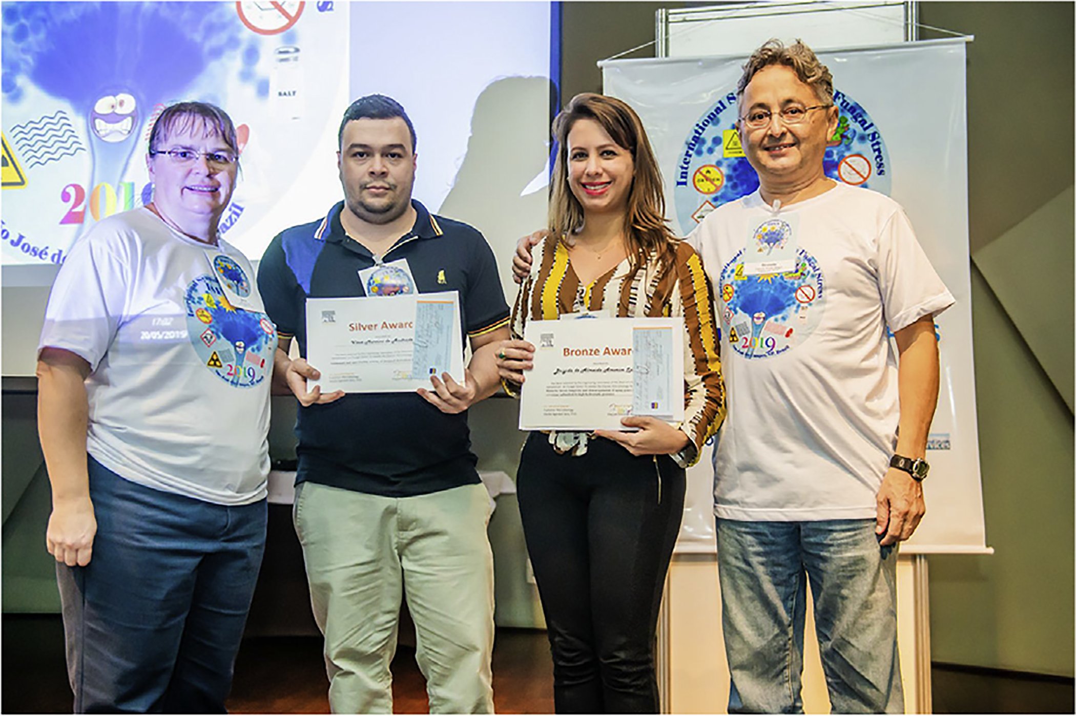
Elsevier Student Awards. From left to right: Alene Alder-Rangel, Vitor Martins de Andrade, Brigida de Almeida Amorim Spagnol, and Drauzio E. N. Rangel.
3.2. Journal of Fungi student award
The winner of the Journal of Fungi Award for the best poster was Marlene Henríquez Urrutia from Pontificia Universidad Católica de Chile, Santiago, Chile. She is a PhD student of Dr. Luis Larrondo and presented a poster titled “Circadian regulation of a mycoparasitic interaction between B. cinerea and T. atroviride” (Fig. 7).
Fig. 7.
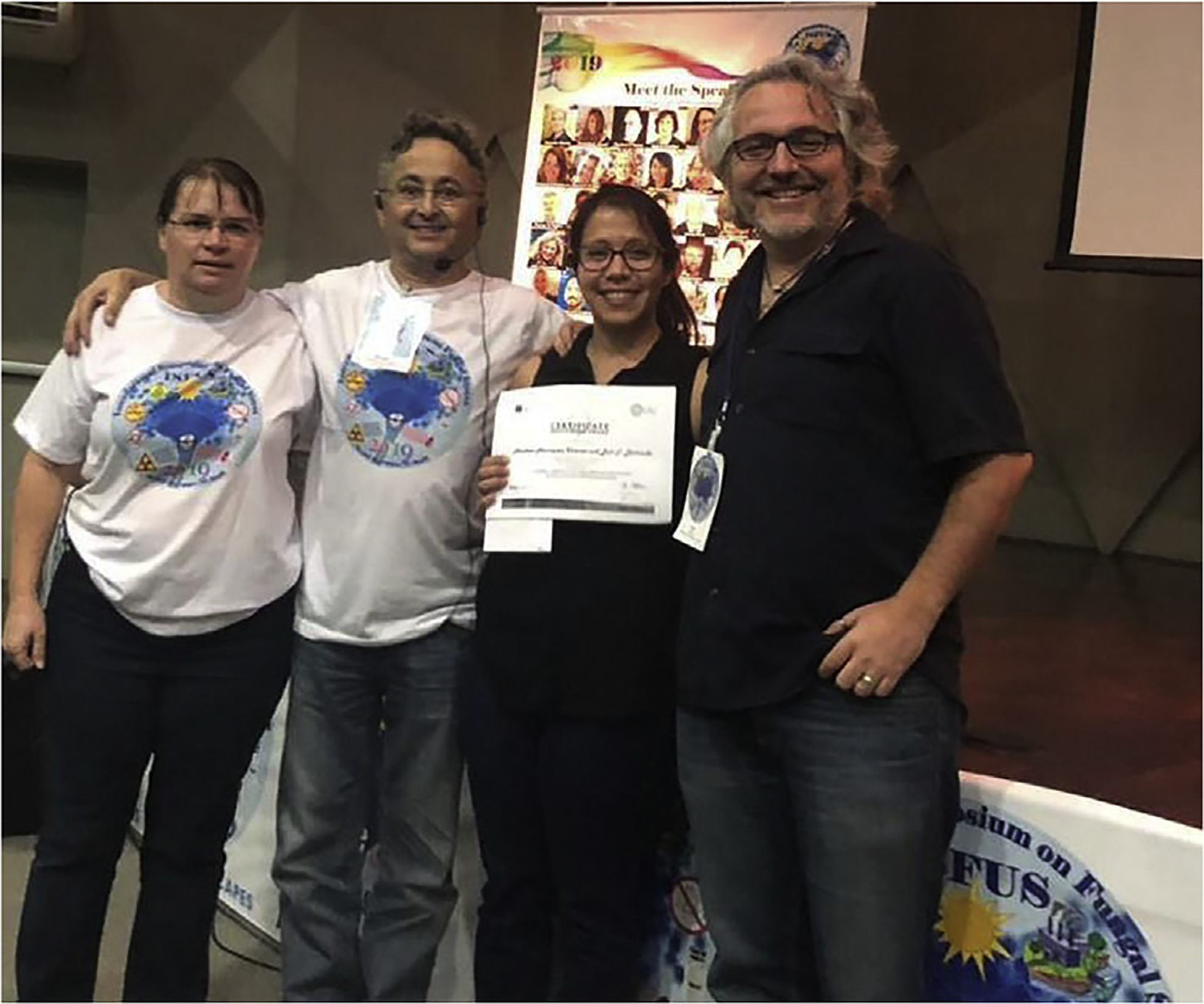
Journal of Fungi Student Award. From left to right: Alene Alder-Rangel, Drauzio E. N. Rangel, Marlene Henríquez Urrutia, and Luis Larrondo.
3.3. Award to Drauzio Eduardo Naretto Rangel
At the closing ceremony of ISFUS-2019, and on behalf of the Organizing Committee, John E. Hallsworth and Luis M. Corrochano gave an overview of the ISFUS series. These meetings have been convivial gatherings, bringing together international and Brazilian scientists for a shared scientific (as well as cultural and social) experience. Thus far, there have been 95 ISFUS speakers, coming from 22 countries. Hallsworth highlighted the world-leading mycological research endeavors of Brazilian science in relation to entomopathogens (biological control), biodiversity, trehalose metabolism, UV stress, and bioethanol by explaining how important it is for international delegates to interact with Brazilian students, academics, and industry. He detailed how the ISFUS special issues of 2015 (Current Genetics) and 2018 (Fungal Biology) have been successful. For example, ISFUS special-issue papers make up 9 out of 10 most-cited papers in Current Genetics for 2015, and all 10 of the most-cited papers in Fungal Biology for 2018 (Web of Science, on 20 May 2019). ISFUS has also generated new collaborations between participants, new funding streams, new lines of scientific inquiry, joint publications, and exchange of students between participants to support joint research projects. This exemplifies how the fungal stress meetings can generate impacts beyond the immediate field. Furthermore, these impacts can be as varied as they are indeterminate. Hallsworth also explained that each ISFUS appears to be even more convivial and scientifically stimulating than the last.
Drauzio E. N. Rangel, he went on to say, has acted as an ambassador for Brazil and for Brazilian mycology, through the ISFUS series of symposia. Rangel also has his own innovative way of doing science, is scholastic in his research style, is highly collaborative, and has a series of unique research outputs that stimulate new lines of experimentation in other research groups. Hallsworth stated that Rangel has made a consistent, unique, and profound contribution to field of fungal stress. On behalf of the Committee, Corrochano and Hallsworth then surprised Rangel by presenting him with an award, in the form of a glass globe, inscribed with the words: “Awarded for Outstanding Contribution to Mycology to Professor Drauzio E. N. Rangel at III International Symposium on Fungal Stress & conferred by the Organizing Committee, May 2019, (São José dos Campos, SP, Brazil).” Rangel responded to the award with gratitude and tears (Fig. 8).
Fig. 8.
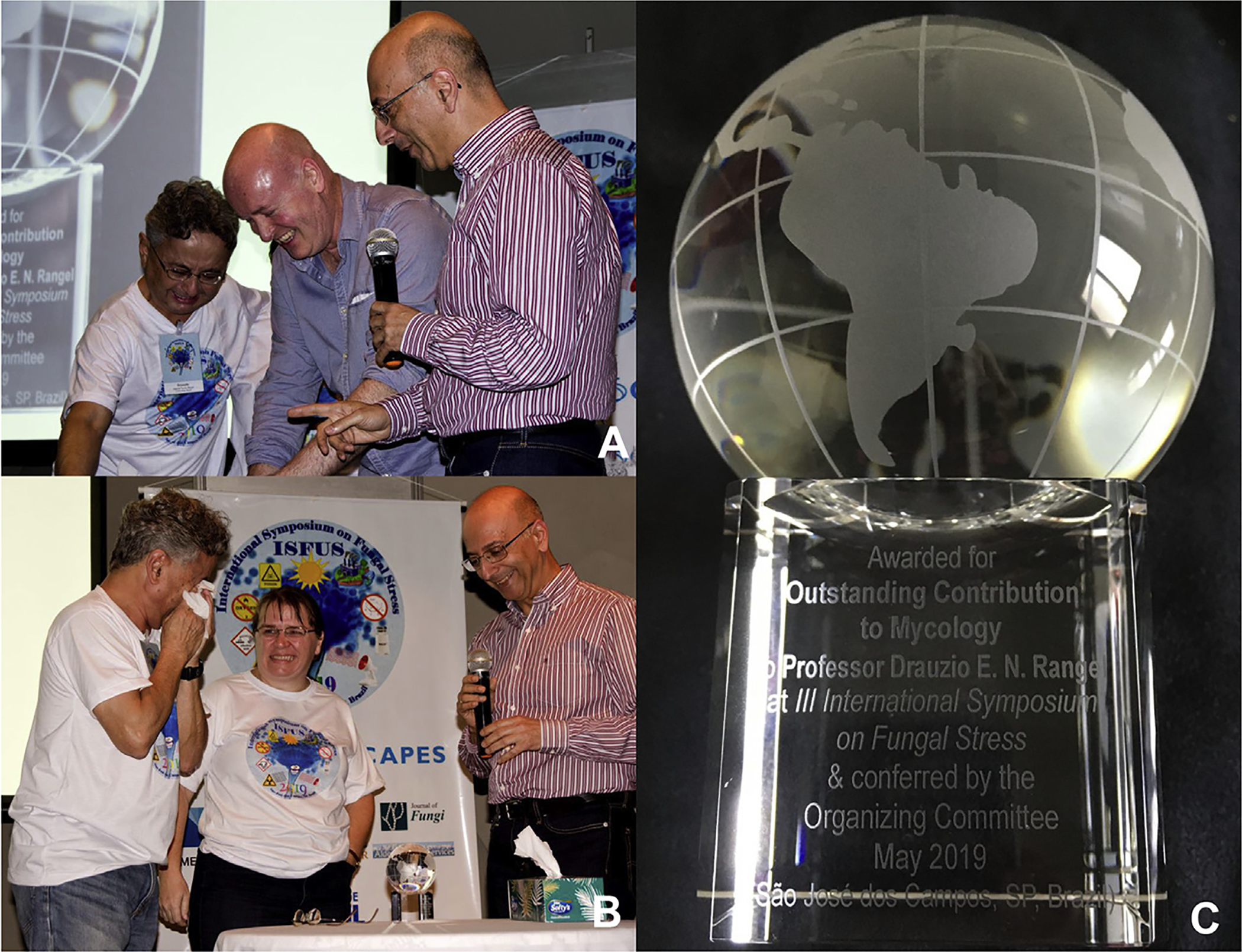
Award given to Drauzio E. N. Rangel during the closing ceremony of the Third International Symposium on Fungal Stress. A) Drauzio E. N. Rangel, John Hallsworth, and Luis Corrochano. B) Drauzio E. N. Rangel, Alene Alder-Rangel, and Luis Corrochano. C) Glass globe inscribed with the words: “Awarded for Outstanding Contribution to Mycology to Professor Drauzio E. N. Rangel at III International Symposium on Fungal Stress & conferred by the Organizing Committee, May 2019, (São José dos Campos, SP, Brazil)”.
4. Excursion
The weekend after ISFUS-2019 most of the speakers traveled to São Sebastião for a scientific retreat at the beach. On Saturday, they partook of a traditional Brazilian barbeque on a chartered boat. This was an exquisite opportunity for the participants to become better acquainted with each other, form new collaborations and friendships while thoroughly enjoying another aspect of Brazilian hospitality (Fig. 9).
Fig. 9.
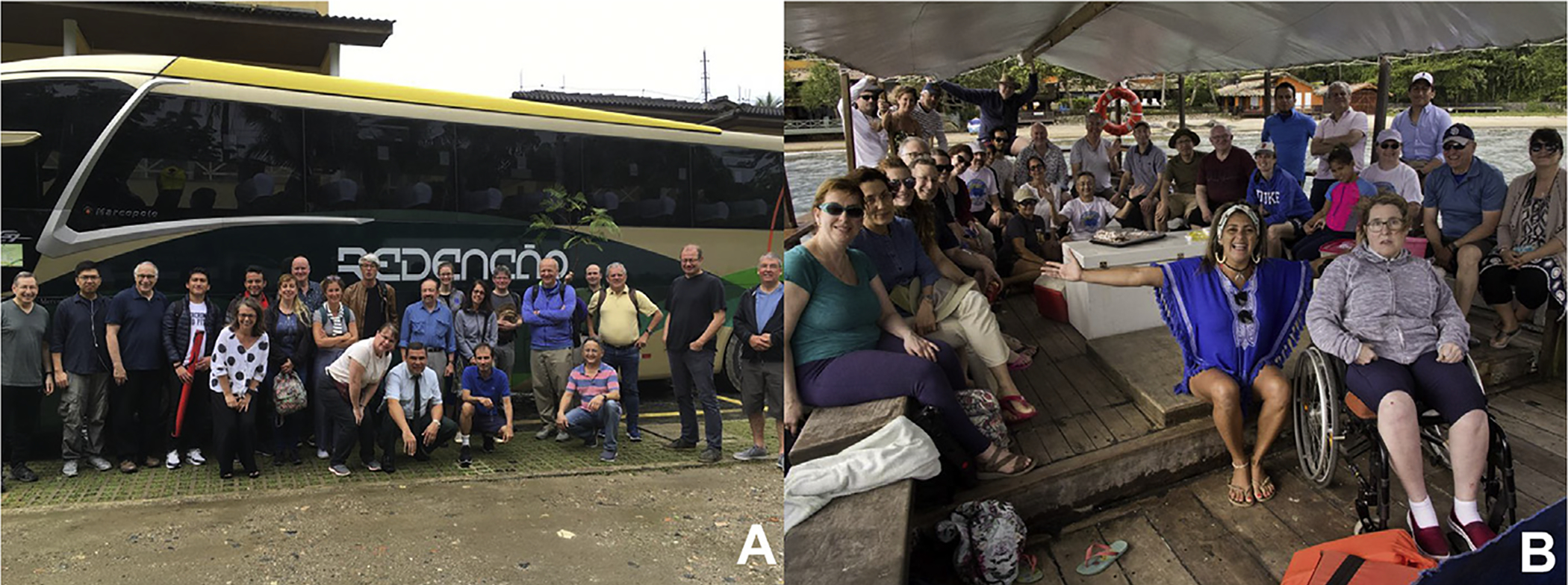
Participants and speakers of the Third ISFUS in the excursion to the beach in São Sebastião, São Paulo, Brazil: A) outside the excursion bus and B) on the boat.
5. The next ISFUS in 2021
Rangel already began planning the fourth ISFUS, even before the third ISFUS was completed and eleven speakers have already confirmed their presence https://isfus2021.wordpress.com/. During ISFUS-2019, Jesús Aguirre proposed a joint meeting combining ISFUS-2021 with the International Fungal Biology Conference (IFBC). This international conference began in 1965 and has taken place in several different countries: UK 1965, USA 1973, Switzerland 1980, UK 1987, USA 1991, Germany 1996, The Netherlands 1999, Mexico 2002, France 2006, Mexico 2009, Germany 2013, and South Korea 2017, but this will be the first edition in South America. Therefore, we cordially invite you to São José dos Campos, Brazil, for the IV ISFUS and XIII IFBC in June of 2021. We are confident that a joint ISFUS-IFBC meeting will bring together complementary and exciting cutting-edge fields of fungal biology that should be attractive to many researchers young and old, from all over the world.
6. Conclusions
The presentations at ISFUS-2019, which covered approximately 30 fungal species, collectively highlight the diversity of responses that fungi can trigger to protect themselves. What general themes emerged? The first was the challenge in providing a clear definition of what stress would mean to a species. The second was the extensive use of genomic-level methods to analyze the impact of stress on fungi. The third was how fungi relate to time, and fourth about interactions with the lithosphere. Fifth, novel stresses and stress responses were identified. Finally, it is clear that there is substantially more to uncover about how fungi sense and respond to stress in their environment.
Supplementary Material
Acknowledgments
This manuscript is part of the special issue Fungal Adaptation to Hostile Challenges arising from the third International Symposium on Fungal Stress - ISFUS, which is supported by FAPESP 2018/20571-6 and CAPES 88881.289327/2018-01.
This work was supported by grants from the following funders identified by the author’s initials. AI - The Australian Research Council [LP170100548] and Australian Grains Research and Development Corporation [UM00051]; ACB & AJPB - The Wellcome Trust [206412/Z/17/Z] and [097377], Royal Society [UF130579], UK Medical Research Council [MR/N006364/1], The MRC Centre for Medical Mycology [MR/N006364/2]; AG - The Deutsche Forschungsgemeinschaft (DFG) [GO 897/13-1] and The German Federal Ministry of Economics and Energy (BMWi) [0325716F]; CMK - The U.S. National Institutes of Health (NIH) [F32 GM128252], [R35 GM118021], [R35 GM118022]; CBC - The Brazilian National Council for Scientific and Technological Development (CNPq) and The Coordenação de Aperfeiçoamento de Pessoal de Nível Superior (CAPES); DEL - The NIH [R01 GM48533-25]; DBP - The NIH [GM R35 GM126966], [GM R01 GM058529]; ED - The Defense Threat Reduction Agency, USA [HDTRA-17-1-0020]; FFB - The National Research Foundation of South Africa (NRF) [SARChI Grant UID 83471]; GMG - The UK Natural Environment Research Council [NE/M010910/1 (TeaSe)] and [NE/M011275/1 (COG3)]; GHB - The Deutsche Forschungsgemeinschaft (DFG); GTPB - Fundação de Amparo à Pesquisa do Estado de São Paulo (FAPESP) [2016/11386-5], [2015/24305-0]; GULB - The FAPESP [2016/11386-5] and The CNPq [425998/2018-5], [307738/2018-3]; ID - The Chinese Ministry of Science and Technology [973 Programme, 2015CB150500], The Natural, Science Foundation of Jiangsu Province [BK20160726], and The Austrian Science Foundation (FWF) [P25613-B20]; IP - The European Union and the European Social Fund [EFOP-3.6.1-16-2016-00022], The National Research, Development and Innovation Office of Hungary [K100464], [K112181], [K119494], and The Higher Education Institutional Excellence Programme [NKFIH-1150-6/2019] of the Ministry of Innovation and Technology in Hungary, within the framework of the Biotechnology thematic programme of the University of Debrecen; TE - The National Research, Development and Innovation Office of Hungary [K112181], [NN 125671]; JD - Organized by and executed under the auspices of TiFN in The Netherlands, a public - private partnership on precompetitive research in food and nutrition; JA - JA e CON-ACYT grants Investigacion en Fronteras de la Ciencia [2015-I-319], CONACYT-DFG [277869], and PAPIIT-UNAM grants [IN200719], [IV200519]; JEH - The UK Biotechnology and Biological Sciences Research Council (BBSRC) [BBF003471]; The Department of Education and Learning (DEL, Northern Ireland), and the Department of Agriculture, Environment and Rural Affairs (DAERA, Northern Ireland; KHW - The Science and Technology Development Fund, Macau SAR [FDCT.085.2014.A2] and the Research and Development Administration Office of the University of Macau [MYRG2016-00211-FHS]; LS - The Italian National Program for Antarctic Research (PNRA) and the Italian Antarctic National Museum (MNA); LMC - The European Regional Development Fund and the Spanish Ministerio de Ciencia, Innovación y Universidades [BIO2015-67148-R and RTI2018-098636-B-I00]; MK -The Israel Cancer Research Fund, the Minerva Center for in-lab evolution, and the Israel Science Foundation; MMomany - “Regulation of spore dormancy in pathogenic fungi” on Experiment.com and University of Georgia President’s Interdisciplinary Seed Grant Program; MMolin - The Swedish Research Council VR, Cancerfonden, the Foundation Olle Engkvist byggmästare, and the Foundation Carl Tryggers stiftelse; NR - The DFG [DFG Re1556/7-1]; OY - The Israel Science Foundation; RF - The DFG [DFG Fi459/19-1]; RCP - The FAPESP [2011/51298-4], [2016/14542-8]; RLM - The NASA Co-operative agreement [NNX12AD05A] and NASA Grant [80NSSC18K0751]; TOB - The FAPESP [2018/17172-2]; DENR - The FAPESP [2010/06374-1], [2013/50518-6], [2014/01229-4], [2017/22669-0], and the CNPq [PQ1D 308436/2014-8], [PQ1D 302100/2018-0], [PQ2 304816/2017-5].
We are also thankful to Sheba Agarwal-Jans, Microbiology and Mycology Publisher, at Elsevier by providing the Student Awards, and to Gowri Vasanthkumar, Publishing Content Specialist, Strategy and Journal Services at Elsevier and Hayley Tyrrell, Journal Manager, Elsevier Research Content Operations for the editorial assistance of this special issue.
Footnotes
Appendix A. Supplementary data
Supplementary data to this article can be found online at https://doi.org/10.1016/j.funbio.2020.02.007.
References
- Acheampong MA, Hill MP, Moore SD, Coombes CA, 2019. UV sensitivity of Beauveria bassiana and Metarhizium anisopliae isolates under investigation as potential biological control agents in South African citrus orchards. Fungal Biol-Uk 10.1016/j.funbio.2019.08.009. [DOI] [PubMed] [Google Scholar]
- Alder-Rangel A, Bailão AM, da Cunha AF, Soares CMA, Wang C, Bonatto D, Dadachova E, Hakalehto E, Eleutherio ECA, Fernandes EKK, Braus GH, Braga GUL, Goldman GH, Malavazi I, Hallsworth JE, Takemoto JY, Fuller K, Selbmann L, Corrochano LM, Bertolini MC, Schmoll M, Pedrini N, Loera O, Finlay RD, Peralta RM, Rangel DEN, 2018. The second international symposium on fungal stress: ISFUS. Fungal Biol 122, 386–399. 10.1016/j.funbio.2017.10.011. [DOI] [PubMed] [Google Scholar]
- Antal K, Gila BC, Pócsi I, Emri T, 2019. General stress response or adaptation to rapid growth in Aspergillus nidulans? Fungal Biol 10.1016/j.funbio.2019.10.009. [DOI] [PubMed] [Google Scholar]
- Araújo CAS, Dias LP, Ferreira PC, Mittmann J, Pupin B, Brancini GTP, Braga GÚL, Rangel DEN, 2018. Responses of entomopathogenic fungi to the mutagen 4-nitroquinoline 1-oxide. Fungal Biol 122, 621–628. 10.1016/j.funbio.2018.03.007. [DOI] [PubMed] [Google Scholar]
- Araújo CAS, Ferreira PC, Pupin B, Dias LP, Avalos J, Edwards J, Hallsworth JE, Rangel DEN, 2019. Osmotolerance as a determinant of microbial ecology: a study of phylogenetically diverse fungi. Fungal Biol 10.1016/j.funbio.2019.09.001. [DOI] [PubMed] [Google Scholar]
- Askree SH, Yehuda T, Smolikov S, Gurevich R, Hawk J, Coker C, Krauskopf A, Kupiec M, McEachern MJ, 2004. A genome-wide screen for Saccharomyces cerevisiae deletion mutants that affect telomere length. Proc. Natl. Acad. Sci. U.S.A 101, 8658–8663. 10.1073/pnas.0401263101. [DOI] [PMC free article] [PubMed] [Google Scholar]
- Bagheri B, Zambelli P, Vigentini I, Bauer FF, Setati ME, 2018. Investigating the effect of selected non-Saccharomyces species on wine ecosystem function and major volatiles. Front. Bioeng. Biotechnol 6 10.3389/fbioe.2018.00169, 169–169. [DOI] [PMC free article] [PubMed] [Google Scholar]
- Bakti F, Sasse C, Heinekamp T, Pócsi I, Braus GH, 2018. Heavy metal-induced expression of PcaA provides cadmium tolerance to Aspergillus fumigatus and supports its virulence in the Galleria mellonella model. Front. Microbiol 9 10.3389/fmicb.2018.00744, 744–744. [DOI] [PMC free article] [PubMed] [Google Scholar]
- Ballou ER, Avelar GM, Childers DS, Mackie J, Bain JM, Wagener J, Kastora SL, Panea MD, Hardison SE, Walker LA, Erwig LP, Munro CA, Gow NAR, Brown GD, MacCallum DM, Brown AJP, 2016. Lactate signalling regulates fungal b-glucan masking and immune evasion. Nat. Microbiol 2, 16238 10.1038/nmicrobiol.2016.238. [DOI] [PMC free article] [PubMed] [Google Scholar]
- Bayram Ö, Braus GH, 2012. Coordination of secondary metabolism and development in fungi: the velvet family of regulatory proteins. FEMS Microbiol. Rev 36, 1–24. 10.1111/j.1574-6976.2011.00285.x. [DOI] [PubMed] [Google Scholar]
- Bayram ÖS, Dettmann A, Karahoda B, Moloney NM, Ormsby T, McGowan J, Cea-S anchez S, Miralles-Durán A, Brancini GTP, Luque EM, Fitzpatrick DA, Cánovas D, Corrochano LM, Doyle S, Selker EU, Seiler S, Bayram Ö, 2019. Control of development, secondary metabolism and light-dependent carotenoid biosynthesis by the velvet complex of Neurospora crassa. Genetics 212, 691–710. 10.1534/genetics.119.302277. [DOI] [PMC free article] [PubMed] [Google Scholar]
- Birch RM, Walker GM, 2000. Influence of magnesium ions on heat shock and ethanol stress responses of Saccharomyces cerevisiae. Enzym. Microb. Technol 26, 678–687. 10.1016/S0141-0229(00)00159-9. [DOI] [PubMed] [Google Scholar]
- Bodvard K, Jörhov A, Blomberg A, Molin M, Käll M, 2013. The yeast transcription factor Crz1 is activated by light in a Ca2+/calcineurin-dependent and PKA-independent manner. PloS One 8, e53404. [DOI] [PMC free article] [PubMed] [Google Scholar]
- Bodvard K, Peeters K, Roger F, Romanov N, Igbaria A, Welkenhuysen N, Palais G, Reiter W, Toledano MB, Käll M, Molin M, 2017. Light-sensing via hydrogen peroxide and a peroxiredoxin. Nat. Commun 8, 14791 10.1038/ncomms14791. [DOI] [PMC free article] [PubMed] [Google Scholar]
- Bodvard K, Wrangborg D, Tapani S, Logg K, Sliwa P, Blomberg A, Kvarnström M, Käll M, 2011. Continuous light exposure causes cumulative stress that affects the localization oscillation dynamics of the transcription factor Msn2p. Biochim. Biophys. Acta Mol. Cell Res 1813, 358–366. 10.1016/j.bbamcr.2010.12.004. [DOI] [PubMed] [Google Scholar]
- Brancini GTP, Bachmann L, Ferreira M.E.d.S., Rangel DEN, Braga GÚL, 2018. Exposing Metarhizium acridum mycelium to visible light up-regulates a photolyase gene and increases photoreactivating ability. J. Invertebr. Pathol 152, 35–37. 10.1016/j.jip.2018.01.007. [DOI] [PubMed] [Google Scholar]
- Brancini GTP, Ferreira MES, Rangel DEN, Braga GÚL, 2019. Combining transcriptomics and proteomics reveals potential post-transcriptional control of gene expression after light exposure in Metarhizium acridum. G3: Genes Genome Genet 9, 2951–2961. 10.1534/g3.119.400430. [DOI] [PMC free article] [PubMed] [Google Scholar]
- Brancini GTP, Rodrigues GB, Rambaldi M.d.S.L., Izumi C, Yatsuda AP, Wainwright M, Rosa JC, Braga GÚL, 2016. The effects of photodynamic treatment with new methylene blue N on the Candida albicans proteome. Photochem. Photobiol. Sci 15, 1503–1513. 10.1039/c6pp00257a. [DOI] [PubMed] [Google Scholar]
- Brand A, Shanks S, Duncan VMS, Yang M, Mackenzie K, Gow NAR, 2007. Hyphal orientation of Candida albicans is regulated by a calcium-dependent mechanism. Curr. Biol 17, 347–352. 10.1016/j.cub.2006.12.043. [DOI] [PMC free article] [PubMed] [Google Scholar]
- Braus GH, Irniger S, Bayram Ö, 2010. Fungal development and the COP9 signalosome. Curr. Opin. Microbiol 13, 672–676. 10.1016/j.mib.2010.09.011. [DOI] [PubMed] [Google Scholar]
- Breitenbach R, Silbernagl D, Toepel J, Sturm H, Broughton WJ, Sassaki GL, Gorbushina AA, 2018. Corrosive extracellular polysaccharides of the rock-inhabiting model fungus Knufia petricola. Extremophiles 22, 165–175. 10.1007/s00792-017-0984-5. [DOI] [PMC free article] [PubMed] [Google Scholar]
- Brown AJ, Budge S, Kaloriti D, Tillmann A, Jacobsen MD, Yin Z, Ene IV, Bohovych I, Sandai D, Kastora S, Potrykus J, Ballou ER, Childers DS, Shahana S, Leach MD, 2014. Stress adaptation in a pathogenic fungus. J. Exp. Biol 217, 144–155. 10.1242/jeb.088930. [DOI] [PMC free article] [PubMed] [Google Scholar]
- Brown AJP, Cowen LE, di Pietro A, Quinn J, 2017. Stress adaptation In: Heitman J, Howlett BJ, Crous PW, Stukenbrock EH, James TY, Gow NAR (Eds.), The Fungal Kingdom American Society of Microbiology, Washington, DC, pp. 463–485. [Google Scholar]
- Brown AJP, Gow NAR, Warris A, Brown GD, 2019. Memory in fungal pathogens promotes immune evasion, colonisation, and infection. Trends Microbiol 27, 219–230. 10.1016/j.tim.2018.11.001. [DOI] [PubMed] [Google Scholar]
- Brown AJP, Larcombe DE, Pradhan A, 2020. Thoughts on the evolution of core environmental responses in yeasts. Fungal Biol 10.1016/j.funbio.2020.01.003. [DOI] [PMC free article] [PubMed] [Google Scholar]
- Busch S, Schwier EU, Nahlik K, Bayram O, Helmstaedt K, Draht OW, Krappmann S, Valerius O, Lipscomb WN, Braus GH, 2007. An eight-subunit COP9 signalosome with an intact JAMM motif is required for fungal fruit body formation. Proc. Natl. Acad. Sci. U.S.A 104, 8089–8094. 10.1073/pnas.0702108104. [DOI] [PMC free article] [PubMed] [Google Scholar]
- Calvete CL, Martho KF, Felizardo G, Paes A, Nunes JM, Ferreira CO, Vallim MA, Pascon RC, 2019. Amino acid permeases in Cryptococcus neoformans are required for high temperature growth and virulence; and are regulated by Ras signaling. PloS One 14, e0211393 10.1371/journal.pone.0211393. [DOI] [PMC free article] [PubMed] [Google Scholar]
- Caster SZ, Castillo K, Sachs MS, Bell-Pedersen D, 2016. Circadian clock regulation of mRNA translation through eukaryotic elongation factor eEF-2. Proc. Natl. Acad. Sci. U.S.A 113, 9605–9610. 10.1073/pnas.1525268113. [DOI] [PMC free article] [PubMed] [Google Scholar]
- Chen Y-L, Yu S-J, Huang H-Y, Chang Y-L, Lehman VN, Silao FGS, Bigol UG, Bungay AAC, Averette A, Heitman J, 2014. Calcineurin controls hyphal growth, virulence, and drug tolerance of Candida tropicalis. Eukaryot. Cell 13, 844–854. 10.1128/ec.00302-13. [DOI] [PMC free article] [PubMed] [Google Scholar]
- Clusella Trullas S, van Wyk JH, Spotila JR, 2007. Thermal melanism in ectotherms. J. Therm. Biol 32, 235–245. 10.1016/j.jtherbio.2007.01.013. [DOI] [Google Scholar]
- Colot HV, Park G, Turner GE, Ringelberg C, Crew CM, Litvinkova L, Weiss RL, Borkovich KA, Dunlap JC, 2006. A high-throughput gene knockout procedure for Neurospora reveals functions for multiple transcription factors. Proc. Natl. Acad. Sci. U.S.A 103, 10352–10357. 10.1073/pnas.0601456103. [DOI] [PMC free article] [PubMed] [Google Scholar]
- Cordero RJB, Robert V, Cardinali G, Arinze ES, Thon SM, Casadevall A, 2018. Impact of yeast pigmentation on heat capture and latitudinal distribution. Curr. Biol 28, 2657–2664. 10.1016/j.cub.2018.06.034. e2653 [DOI] [PMC free article] [PubMed] [Google Scholar]
- Cordero RJB, Vij R, Casadevall A, 2017. Microbial melanins for radioprotection and bioremediation. Microb. Biotechnol 10, 1186–1190. 10.1111/1751-7915.12807. [DOI] [PMC free article] [PubMed] [Google Scholar]
- Corrochano LM, 2019. Light in the fungal world: from photoreception to gene transcription and beyond. Annu. Rev. Genet 53, 149–170. 10.1146/annurev-genet-120417-031415. [DOI] [PubMed] [Google Scholar]
- Crawford RA, Pavitt GD, 2019. Translational regulation in response to stress in Saccharomyces cerevisiae. Yeast 36, 5–21. 10.1002/yea.3349. [DOI] [PMC free article] [PubMed] [Google Scholar]
- da Costa BLV, Basso TO, Raghavendran V, Gombert AK, 2018. Anaerobiosis revisited: growth of Saccharomyces cerevisiae under extremely low oxygen availability. Appl. Microbiol. Biotechnol 102, 2101–2116. 10.1007/s00253-017-8732-4. [DOI] [PubMed] [Google Scholar]
- de Melo AT, Martho KF, Roberto TN, Nishiduka ES, Machado J Jr., Brustolini OJB, Tashima AK, Vasconcelos AT, Vallim MA, Pascon RC, 2019. The regulation of the sulfur amino acid biosynthetic pathway in Cryptococcus neoformans: the relationship of Cys3, Calcineurin, and Gpp2 phosphatases. Sci. Rep 9, 11923 10.1038/s41598-019-48433-5. [DOI] [PMC free article] [PubMed] [Google Scholar]
- De Middeleer G, Leys N, Sas B, De Saeger S, 2019. Fungi and mycotoxins in space—a review. Astrobiology 19, 915–926. 10.1089/ast.2018.1854. [DOI] [PubMed] [Google Scholar]
- de Vries RP, Riley R, Wiebenga A, Aguilar-Osorio G, Amillis S, Uchima CA, Anderluh G, Asadollahi M, Askin M, Barry K, Battaglia E, Bayram Ö, Benocci T, Braus-Stromeyer SA, Caldana C, Cánovas D, Cerqueira GC, Chen F, Chen W, Choi C, Clum A, Dos Santos RAC, Damásio ARL, Diallinas G, Emri T, Fekete E, Flipphi M, Freyberg S, Gallo A, Gournas C, Habgood R, Hainaut M, Harispe ML, Henrissat B, Hildén KS, Hope R, Hossain A, Karabika E, Karaffa L, Karányi Z, Kraševec N, Kuo A, Kusch H, LaButti K, Lagendijk EL, Lapidus A, Levasseur A, Lindquist E, Lipzen A, Logrieco AF, MacCabe A, Mäkelä MR, Malavazi I, Melin P, Meyer V, Mielnichuk N, Miskei M, Molnár ÁP, Mule G, Ngan CY, Orejas M, Orosz E, Ouedraogo JP, Overkamp KM, Park H-S, Perrone G, Piumi F, Punt PJ, Ram AFJ, Ramón A, Rauscher S, Record E, Riaño-Pachón DM, Robert V, Röhrig J, Ruller R, Salamov A, Salih NS, Samson RA, Sándor E, Sanguinetti M, Schütze T, Sepčić K, Shelest E, Sherlock G, Sophianopoulou V, Squina FM, Sun H, Susca A, Todd RB, Tsang A, Unkles SE, van de Wiele N, van Rossen-Uffink D, Oliveira J.V.d.C., Vesth TC, Visser J, Yu J-H, Zhou M, Andersen MR, Archer DB, Baker SE, Benoit I, Brakhage AA, Braus GH, Fischer R, Frisvad JC, Goldman GH, Houbraken J, Oakley B, Pócsi I, Scazzocchio C, Seiboth B, vanKuyk PA, Wortman J, Dyer PS, Grigoriev IV, 2017. Comparative genomics reveals high biological diversity and specific adaptations in the industrially and medically important fungal genus Aspergillus. Genome Biol 18 10.1186/s13059-017-1151-0, 28–28. [DOI] [PMC free article] [PubMed] [Google Scholar]
- Delgado-Jarana J, Sousa S, González F, Rey M, Llobell A, 2006. ThHog1 controls the hyperosmotic stress response in Trichoderma harzianum. Microbiology 152, 1687–1700. 10.1099/mic.0.28729-0. [DOI] [PubMed] [Google Scholar]
- Della-Bianca BE, Basso TO, Stambuk BU, Basso LC, Gombert AK, 2013. What do we know about the yeast strains from the Brazilian fuel ethanol industry? Appl. Microbiol. Biotechnol 97, 979–991. 10.1007/s00253-012-4631-x. [DOI] [PubMed] [Google Scholar]
- Dias LP, Araújo CAS, Pupin B, Ferreira PC, Braga GÚL, Rangel DEN, 2018. The Xenon Test Chamber Q-SUN® for testing realistic tolerances of fungi exposed to simulated full spectrum solar radiation. Fungal Biol 122, 592–601. 10.1016/j.funbio.2018.01.003. [DOI] [PubMed] [Google Scholar]
- Dias LP, Pedrini N, Braga GUL, Ferreira PC, Pupin B, Araújo CAS, Corrochano LM, Rangel DEN, 2019. Outcome of blue, green, red, and white light on Metarhizium robertsii during mycelial growth on conidial stress tolerance and gene expression. Fungal Biol 10.1016/j.funbio.2019.04.007. [DOI] [PubMed] [Google Scholar]
- Druzhinina IS, Seidl-Seiboth V, Herrera-Estrella A, Horwitz BA, Kenerley CM, Monte E, Mukherjee PK, Zeilinger S, Grigoriev IV, Kubicek CP, 2011. Trichoderma: the genomics of opportunistic success. Nat. Rev. Microbiol 9, 749–759. [DOI] [PubMed] [Google Scholar]
- Dunlap JC, Loros JJ, 2017. Making time: conservation of biological clocks from fungi to animals. Microbiol. Spectr 5 10.1128/microbiolspec. FUNK-0039–2016. [DOI] [PMC free article] [PubMed] [Google Scholar]
- Emerson JM, Bartholomai BM, Ringelberg CS, Baker SE, Loros JJ, Dunlap JC, 2015. period-1 encodes an ATP-dependent RNA helicase that influences nutritional compensation of the Neurospora circadian clock. Proc. Natl. Acad. Sci. U.S.A 112, 15707–15712. 10.1073/pnas.1521918112. [DOI] [PMC free article] [PubMed] [Google Scholar]
- Emri T, Antal K, Riley R, Karányi Z, Miskei M, Orosz E, Baker SE, Wiebenga A, de Vries RP, Pócsi I, 2018. Duplications and losses of genes encoding known elements of the stress defence system of the Aspergilli contribute to the evolution of these filamentous fungi but do not directly influence their environmental stress tolerance. Stud. Mycol 91, 23–36. 10.1016/j.simyco.2018.10.003. [DOI] [PMC free article] [PubMed] [Google Scholar]
- Fernandes JD, Martho K, Tofik V, Vallim MA, Pascon RC, 2015. The role of amino acid permeases and tryptophan biosynthesis in Cryptococcus neoformans survival. PloS One 10, e0132369 10.1371/journal.pone.0132369. [DOI] [PMC free article] [PubMed] [Google Scholar]
- Fidel PL, Vazquez JA, Sobel JD, 1999. Candida glabrata: review of epidemiology, pathogenesis, and clinical disease with comparison to C. albicans. Clin. Microbiol. Rev 12, 80 10.1128/cmr.12.1.80. [DOI] [PMC free article] [PubMed] [Google Scholar]
- Fisher MC, Hawkins NJ, Sanglard D, Gurr SJ, 2018. Worldwide emergence of resistance to antifungal drugs challenges human health and food security. Science 360, 739–742. 10.1126/science.aap7999. [DOI] [PubMed] [Google Scholar]
- Fomina M, Hong JW, Gadd GM, 2019. Effect of depleted uranium on a soil microcosm fungal community and influence of a plant-ectomycorrhizal association. Fungal Biol 10.1016/j.funbio.2019.08.001. [DOI] [PubMed] [Google Scholar]
- Fracarolli L, Rodrigues GB, Pereira AC, Massola Junior NS, Silva-Junior GJ, Bachmann L, Wainwright M, Bastos JK, Braga GUL, 2016. Inactivation of plant-pathogenic fungus Colletotrichum acutatum with natural plant-produced photosensitizers under solar radiation. J. Photochem. Photobiol., B 162, 402–411. 10.1016/j.jphotobiol.2016.07.009. [DOI] [PubMed] [Google Scholar]
- Gadd GM, 2007. Geomycology: biogeochemical transformations of rocks, minerals, metals and radionuclides by fungi, bioweathering and bioremediation. Mycol. Res 111, 3–49. 10.1016/j.mycres.2006.12.001. [DOI] [PubMed] [Google Scholar]
- Gadd GM, 2010. Metals, minerals and microbes: geomicrobiology and bioremediation. Microbiology 156, 609–643. 10.1099/mic.0.037143-0. [DOI] [PubMed] [Google Scholar]
- Gadd GM, 2016. Geomycology In: Purchase D (Ed.), Fungal Applications in Sustainable Environmental Biotechnology Springer International Publishing, Switzerland. [Google Scholar]
- Gadd GM, 2017a. Geomicrobiology of the built environment. Nat. Microbiol 2, 16275 10.1038/nmicrobiol.2016.275. [DOI] [PubMed] [Google Scholar]
- Gadd GM, 2017b. Fungi, rocks and minerals. Elements 13, 171–176. 10.2113/gselements.13.3.171. [DOI] [Google Scholar]
- Gonzales JC, Brancini GTP, Rodrigues GB, Silva-Junior GJ, Bachmann L, Wainwright M, Braga GÚL, 2017. Photodynamic inactivation of conidia of the fungus Colletotrichum abscissum on Citrus sinensis plants with methylene blue under solar radiation. J. Photochem. Photobiol., B 176, 54–61. 10.1016/j.jphotobiol.2017.09.008. [DOI] [PubMed] [Google Scholar]
- Gorbushina AA, 2007. Life on the rocks. Environ. Microbiol 9, 1613–1631. 10.1111/j.1462-2920.2007.01301.x. [DOI] [PubMed] [Google Scholar]
- Gorovits R, Yarden O, 2003. Environmental suppression of Neurospora crassa cot-1 hyperbranching: a link between COT1 kinase and stress sensing. Eukaryot. Cell 2, 699–707. 10.1128/ec.2.4.699-707.2003. [DOI] [PMC free article] [PubMed] [Google Scholar]
- Guillemette T, Ram AF, Carvalho ND, Joubert A, Simoneau P, Archer DB, 2011. Methods for investigating the UPR in filamentous fungi. Methods Enzymol 490, 1–29. 10.1016/B978-0-12-385114-7.00001-5. [DOI] [PubMed] [Google Scholar]
- Hallsworth JE, 2018. Stress-free microbes lack vitality. Fungal Biol 122, 379–385. 10.1016/j.funbio.2018.04.003. [DOI] [PubMed] [Google Scholar]
- Hallsworth JE, 2019. Wooden owl that redefines Earth’s biosphere may yet catapult a fungus into space. Environ. Microbiol 21, 2202–2211. 10.1111/1462-2920.14510. [DOI] [PMC free article] [PubMed] [Google Scholar]
- Harari Y, Gershon L, Alonso-Perez E, Klein S, Berneman Y, Choudhari K, Singh P, Sau S, Liefshitz B, Kupiec M, 2019. Telomeres and stress in yeast cells: when genes and environment interact. Fungal Biol 10.1016/j.funbio.2019.09.003. [DOI] [PubMed] [Google Scholar]
- Harari Y, Kupiec M, 2018. Do long telomeres affect cellular fitness? Curr. Genet 64, 173–176. 10.1007/s00294-017-0746-z. [DOI] [PubMed] [Google Scholar]
- Hassett MO, Fischer MWF, Money NP, 2015. Mushrooms as rainmakers: how spores act as nuclei for raindrops. PloS One 10, e0140407. [DOI] [PMC free article] [PubMed] [Google Scholar]
- Heck C, Kuhn H, Heidt S, Walter S, Rieger N, Requena N, 2016. Symbiotic fungi control plant root cortex development through the novel GRAS transcription factor MIG1. Curr. Biol 26, 2770–2778. 10.1016/j.cub.2016.07.059. [DOI] [PubMed] [Google Scholar]
- Helber N, Wippel K, Sauer N, Schaarschmidt S, Hause B, Requena N, 2011. A versatile monosaccharide transporter that operates in the arbuscular mycorrhizal fungus Glomus sp is crucial for the symbiotic relationship with plants. Plant Cell 23, 3812–3823. 10.1105/tpc.111.089813. [DOI] [PMC free article] [PubMed] [Google Scholar]
- Herold I, Kowbel D, Delgado-Álvarez DL, Garduño-Rosales M, Mouriño-Pérez RR, Yarden O, 2019. Transcriptional profiling and localization of GUL-1, a COT-1 pathway component, in Neurospora crassa. Fungal Genet. Biol 126, 1–11. 10.1016/j.fgb.2019.01.010. [DOI] [PubMed] [Google Scholar]
- Herold I, Yarden O, 2017. Regulation of Neurospora crassa cell wall remodeling via the cot-1 pathway is mediated by gul-1. Curr. Genet 63, 145–159. 10.1007/s00294-016-0625-z. [DOI] [PubMed] [Google Scholar]
- Horneck G, Klaus DM, Mancinelli RL, 2010. Space microbiology. Microbiol. Mol. Biol. Rev 74, 121–156. 10.1128/MMBR.00016-09. [DOI] [PMC free article] [PubMed] [Google Scholar]
- Hyde KD, Xu J, Rapior S, Jeewon R, Lumyong S, Niego AGT, Abeywickrama PD, Aluthmuhandiram JVS, Brahamanage RS, Brooks S, Chaiyasen A, Chethana KWT, Chomnunti P, Chepkirui C, Chuankid B, de Silva NI, Doilom M, Faulds C, Gentekaki E, Gopalan V, Kakumyan P, Harishchandra D, Hemachandran H, Hongsanan S, Karunarathna A, Karunarathna SC, Khan S, Kumla J, Jayawardena RS, Liu J-K, Liu N, Luangharn T, Macabeo APG, Marasinghe DS, Meeks D, Mortimer PE, Mueller P, Nadir S, Nataraja KN, Nontachaiyapoom S, O’Brien M, Penkhrue W, Phukhamsakda C, Ramanan US, Rathnayaka AR, Sadaba RB, Sandargo B, Samarakoon BC, Tennakoon DS, Siva R, Sriprom W, Suryanarayanan TS, Sujarit K, Suwannarach N, Suwunwong T, Thongbai B, Thongklang N, Wei D, Wijesinghe SN, Winiski J, Yan J, Yasanthika E, Stadler M, 2019. The amazing potential of fungi: 50 ways we can exploit fungi industrially. Fungal Divers 97, 1–136. 10.1007/s13225-019-00430-9. [DOI] [Google Scholar]
- Igbalajobi O, Yu Z, Fischer R, 2019. Red- and blue-light sensing in the plant pathogen Alternaria alternata depends on phytochrome and the white-collar protein LreA. mBio 10, e00371 10.1128/mBio.00371-19, 00319. [DOI] [PMC free article] [PubMed] [Google Scholar]
- Jouffe C, Cretenet G, Symul L, Martin E, Atger F, Naef F, Gachon F, 2013. The circadian clock coordinates ribosome biogenesis. PLoS Biol 11, e1001455. [DOI] [PMC free article] [PubMed] [Google Scholar]
- Karababa M, Valentino E, Pardini G, Coste AT, Bille J, Sanglard D, 2006. CRZ1, a target of the calcineurin pathway in Candida albicans. Mol. Microbiol 59, 1429–1451. 10.1111/j.1365-2958.2005.05037.x. [DOI] [PubMed] [Google Scholar]
- Kar anyi Z, Holb I, Hornok L, Pócsi I, Miskei M, 2013. FSRD: fungal stress response database. Database (Oxford) 2013 10.1093/database/bat037 bat037. [DOI] [PMC free article] [PubMed] [Google Scholar]
- Kaur R, Ma B, Cormack BP, 2007. A family of glycosylphosphatidylinositol-linked aspartyl proteases is required for virulence of Candida glabrata. Proc. Natl. Acad. Sci. U.S.A 104, 7628 10.1073/pnas.0611195104. [DOI] [PMC free article] [PubMed] [Google Scholar]
- Kelliher CM, Loros JJ, Dunlap JC, 2019. Evaluating the circadian rhythm and response to glucose addition in dispersed growth cultures of Neurospora crassa. Fungal Biol 10.1016/j.funbio.2019.11.004. [DOI] [PMC free article] [PubMed] [Google Scholar]
- Király A, Hámori C, Gyémánt G, Kövér KE, Pócsi I, Leiter É, 2019. Characterization of gfdb, putatively encoding a glycerol 3-phosphate dehydrogenase in Aspergillus nidulans. Fungal Biol 10.1016/j.funbio.2019.09.011. [DOI] [PubMed] [Google Scholar]
- Klinke HB, Thomsen AB, Ahring BK, 2004. Inhibition of ethanol-producing yeast and bacteria by degradation products produced during pre-treatment of biomass. Appl. Microbiol. Biotechnol 66, 10–26. 10.1007/s00253-004-1642-2. [DOI] [PubMed] [Google Scholar]
- Kloppholz S, Kuhn H, Requena N, 2011. A secreted fungal effector of Glomus intraradices promotes symbiotic biotrophy. Curr. Biol 21, 1204–1209. 10.1016/j.cub.2011.06.044. [DOI] [PubMed] [Google Scholar]
- Köhler AM, Harting R, Langeneckert AE, Valerius O, Gerke J, Meister C, Strohdiek A, Braus GH, 2019. Integration of fungus-specific CandA-C1 into a trimeric CandA complex allowed splitting of the gene for the conserved receptor exchange factor of CullinA E3 ubiquitin ligases in Aspergilli. mBio 10 10.1128/mBio.01094-19 e01094–01019. [DOI] [PMC free article] [PubMed] [Google Scholar]
- Krah FS, Buntgen U, Schaefer H, Muller J, Andrew C, Boddy L, Diez J, Egli S, Freckleton R, Gange AC, Halvorsen R, Heegaard E, Heideroth A, Heibl C, Heilmann-Clausen J, Hoiland K, Kar R, Kauserud H, Kirk PM, Kuyper TW, Krisai-Greilhuber I, Norden J, Papastefanou P, Senn-Irlet B, Bassler C, 2019. European mushroom assemblages are darker in cold climates. Nat. Commun 10, 2890 10.1038/s41467-019-10767-z. [DOI] [PMC free article] [PubMed] [Google Scholar]
- Kraus PR, Heitman J, 2003. Coping with stress: calmodulin and calcineurin in model and pathogenic fungi. Biochem. Biophys. Res. Commun 311, 1151–1157. 10.1016/s0006-291x(03)01528-6. [DOI] [PubMed] [Google Scholar]
- Kurucz V, Kiss B, Szigeti ZM, Nagy G, Orosz E, Hargitai Z, Harangi S, Wiebenga A, de Vries RP, Pócsi I, Emri T, 2018a. Physiological background of the remarkably high Cd2+ tolerance of the Aspergillus fumigatus Af293 strain. J. Basic Microbiol 58, 957–967. 10.1002/jobm.201800200. [DOI] [PubMed] [Google Scholar]
- Kurucz V, Krüger T, Antal K, Dietl AM, Haas H, Pócsi I, Kniemeyer O, Emri T, 2018b. Additional oxidative stress reroutes the global response of Aspergillus fumigatus to iron depletion. BMC Genom 19, 357 10.1186/s12864-018-4730-x. [DOI] [PMC free article] [PubMed] [Google Scholar]
- Laz EV, Lee J, Levin DE, 2019. Crosstalk between Saccharomyces cerevisiae SAPKs Hog1 and Mpk1 is mediated by glycerol accumulation. Fungal Biol 10.1016/j.funbio.2019.10.002. [DOI] [PMC free article] [PubMed] [Google Scholar]
- Lee CJD, McMullan PE, O’Kane CJ, Stevenson A, Santos IC, Roy C, Ghosh W, Mancinelli RL, Mormile MR, McMullan G, Banciu HL, Fares MA, Benison KC, Oren A, Dyall-Smith ML, Hallsworth JE, 2018. NaCl-saturated brines are thermodynamically moderate, rather than extreme, microbial habitats. FEMS Microbiol. Rev 42, 672–693. 10.1093/femsre/fuy026. [DOI] [PubMed] [Google Scholar]
- Lee J, Levin DE, 2018. Intracellular mechanism by which arsenite activates the yeast stress MAPK Hog1. Mol. Biol. Cell 29, 1904–1915. 10.1091/mbc.E18-03-0185. [DOI] [PMC free article] [PubMed] [Google Scholar]
- Lee J, Levin DE, 2019. Methylated metabolite of arsenite blocks glycerol production in yeast by inhibition of glycerol-3-phosphate dehydrogenase. Mol. Biol. Cell 30, 2134–2140. 10.1091/mbc.E19-04-0228. [DOI] [PMC free article] [PubMed] [Google Scholar]
- Lee J, Liu L, Levin DE, 2019. Stressing out or stressing in: intracellular pathways for SAPK activation. Curr. Genet 65, 417–421. 10.1007/s00294-018-0898-5. [DOI] [PMC free article] [PubMed] [Google Scholar]
- Lee J, Reiter W, Dohnal I, Gregori C, Beese-Sims S, Kuchler K, Ammerer G, Levin DE, 2013. MAPK Hog1 closes the S. cerevisiae glycerol channel Fps1 by phosphorylating and displacing its positive regulators. Genes Dev 27, 2590–2601. 10.1101/gad.229310.113. [DOI] [PMC free article] [PubMed] [Google Scholar]
- Liang X, Gadd GM, 2017. Metal and metalloid biorecovery using fungi. Microbial. Biotechnol 10, 1199–1205. 10.1111/1751-7915.12767. [DOI] [PMC free article] [PubMed] [Google Scholar]
- Lovett B, St Leger RJ, 2015. Stress is the rule rather than the exception for Metarhizium. Curr. Genet 61, 253–261. 10.1007/s00294-014-0447-9. [DOI] [PubMed] [Google Scholar]
- Malo ME, Dadachova E, 2019. Melanin as an energy transducer and a radio-protector in black fungi In: Tiquia-Arashiro SM, Grube M (Eds.), Fungi in Extreme Environments: Ecological Role and Biotechnological Significance Springer International Publishing, Cham, Switzerland, pp. 175–182. [Google Scholar]
- Malo ME, Frank C, Dadachova E, 2019. Radioadapted Wangiella dermatitidis senses radiation in its environment in a melanin-dependent fashion. Fungal Biol 10.1016/j.funbio.2019.10.011. [DOI] [PubMed] [Google Scholar]
- Mancinelli RL, 2015. The affect of the space environment on the survival of Halorubrum chaoviator and Synechococcus (Nägeli): data from the Space Experiment OSMO on EXPOSE-R. Int. J. Astrobiol 14, 123–128. 10.1017/s147355041400055x. [DOI] [Google Scholar]
- Martho KF, Brustolini OJB, Vasconcelos AT, Vallim MA, Pascon RC, 2019. The glycerol phosphatase Gpp2: a link to osmotic stress, sulfur assimilation and virulence in Cryptococcus neoformans. Front. Microbiol 10, 2728 10.3389/fmicb.2019.02728. [DOI] [PMC free article] [PubMed] [Google Scholar]
- Martho KF, de Melo AT, Takahashi JP, Guerra JM, Santos DC, Purisco SU, Melhem MS, Fazioli RD, Phanord C, Sartorelli P, Vallim MA, Pascon RC, 2016. Amino acid permeases and virulence in Cryptococcus neoformans. PloS One 11, e0163919 10.1371/journal.pone.0163919. [DOI] [PMC free article] [PubMed] [Google Scholar]
- Martin-Sanchez PM, Gebhardt C, Toepel J, Barry J, Munzke N, Günster J, Gorbushina AA, 2018. Monitoring microbial soiling in photovoltaic systems: a qPCR-based approach. Int. Biodeterior. Biodegrad 129, 13–22. 10.1016/j.ibiod.2017.12.008. [DOI] [Google Scholar]
- Martins de Andrade V, Bardaji E, Heras M, Ramu VG, Junqueira JC, Diane dos Santos J, Castanho MARB, Conceição K, 2019. Antifungal and anti-biofilm activity of designed derivatives from Kyotorphin. Fungal Biol 10.1016/j.funbio.2019.12.002. [DOI] [PubMed] [Google Scholar]
- Matos TG, Morais FV, Campos CB, 2013. Hsp90 regulates Paracoccidioides brasiliensis proliferation and ROS levels under thermal stress and cooperates with calcineurin to control yeast to mycelium dimorphism. Med. Mycol 51, 413–421. 10.3109/13693786.2012.725481. [DOI] [PubMed] [Google Scholar]
- Medina EQA, Oliveira AS, Medina HR, Rangel DEN, 2020. Serendipity in the wrestle between Trichoderma and Metarhizium. Fungal Biol 10.1016/j.funbio.2020.01.002. [DOI] [PubMed] [Google Scholar]
- Meister C, Thieme KG, Thieme S, Kohler AM, Schmitt K, Valerius O, Braus GH, 2019. COP9 signalosome interaction with UspA/Usp15 deubiquitinase controls VeA-mediated fungal multicellular development. Biomolecules 9 10.3390/biom9060238. [DOI] [PMC free article] [PubMed] [Google Scholar]
- Mendoza-Martínez AE, Cano-Domínguez N, Aguirre J, 2019. Yap1 homologs mediate more than the redox regulation of the antioxidant response in filamentous fungi. Fungal Biol 10.1016/j.funbio.2019.04.001. [DOI] [PubMed] [Google Scholar]
- Mendoza-Martínez AE, Lara-Rojas F, S anchez O, Aguirre J, 2017. NapA mediates a redox regulation of the antioxidant response, carbon utilization and development in Aspergillus nidulans. Front. Microbiol 8 10.3389/fmicb.2017.00516. [DOI] [PMC free article] [PubMed] [Google Scholar]
- Mersaoui SY, Wellinger RJ, 2019. Fine tuning the level of the Cdc13 telomere-capping protein for maximal chromosome stability performance. Curr. Genet 65, 109–118. 10.1007/s00294-018-0871-3. [DOI] [PubMed] [Google Scholar]
- Mitchell A, Romano GH, Groisman B, Yona A, Dekel E, Kupiec M, Dahan O, Pilpel Y, 2009. Adaptive prediction of environmental changes by microorganisms. Nature 460, 220–224. 10.1038/nature08112. [DOI] [PubMed] [Google Scholar]
- Nai C, Wong HY, Pannenbecker A, Broughton WJ, Benoit I, de Vries RP, Gueidan C, Gorbushina AA, 2013. Nutritional physiology of a rock-inhabiting, model microcolonial fungus from an ancestral lineage of the Chaetothyriales (Ascomycetes). Fungal Genet. Biol 56, 54–66. 10.1016/j.fgb.2013.04.001. [DOI] [PubMed] [Google Scholar]
- Naidoo RK, Simpson ZF, Oosthuizen JR, Bauer FF, 2019. Nutrient exchange of carbon and nitrogen promotes the formation of stable mutualisms between Chlorella sorokiniana and Saccharomyces cerevisiae under engineered synthetic growth conditions. Front. Microbiol 10 10.3389/fmicb.2019.00609, 609–609. [DOI] [PMC free article] [PubMed] [Google Scholar]
- Nicholson WL, Ricco AJ, Agasid E, Beasley C, Diaz-Aguado M, Ehrenfreund P, Friedericks C, Ghassemieh S, Henschke M, Hines JW, Kitts C, Luzzi E, Ly D, Mai N, Mancinelli R, McIntyre M, Minelli G, Neumann M, Parra M, Piccini M, Rasay RM, Ricks R, Santos O, Schooley A, Squires D, Timucin L, Yost B, Young A, 2011. The O/OREOS mission: first science data from the Space Environment Survivability of Living Organisms (SESLO) payload. Astrobiology 11, 951–958. 10.1089/ast.2011.0714. [DOI] [PubMed] [Google Scholar]
- Nienow JA, Friedmann EI, 1993. Terrestrial lithophytic (rock) communities In: Friedmann EI (Ed.). Antarctic Microbiology Wiley-Liss, New York, pp. 343–412. [Google Scholar]
- Nietzsche FW, 1888. Twilight of the Idols, or, How to Philosophize with a Hammer Oxford University Press, New York. [Google Scholar]
- Noack-Schönmann S, Bus T, Banasiak R, Knabe N, Broughton WJ, Den Dulk-Ras H, Hooykaas PJJ, Gorbushina AA, 2014. Genetic transformation of Knufia petricola A95 - a model organism for biofilm-material interactions. AMB Express 4 10.1186/s13568-014-0080-5, 80–80. [DOI] [PMC free article] [PubMed] [Google Scholar]
- Nystrom T, Yang J, Molin M, 2012. Peroxiredoxins, gerontogenes linking aging to genome instability and cancer. Genes Dev 26, 2001–2008. 10.1101/gad.200006.112. [DOI] [PMC free article] [PubMed] [Google Scholar]
- Olivares-Yañez C, Emerson J, Kettenbach A, Loros JJ, Dunlap JC, Larrondo LF, 2016. Modulation of circadian gene expression and metabolic compensation by the RCO-1 corepressor of Neurospora crassa. Genetics 204, 163–176. 10.1534/genetics.116.191064. [DOI] [PMC free article] [PubMed] [Google Scholar]
- Onofri S, de la Torre R, de Vera JP, Ott S, Zucconi L, Selbmann L, Scalzi G, Venkateswaran KJ, Rabbow E, Sanchez Inigo FJ, Horneck G, 2012. Survival of rock-colonizing organisms after 1.5 years in outer space. Astrobiology 12, 508–516. 10.1089/ast.2011.0736. [DOI] [PubMed] [Google Scholar]
- Onofri S, Selbmann L, Zucconi L, Pagano S, 2004. Antarctic microfungi as models for exobiology. Planet. Space Sci 52, 229–237. 10.1016/j.pss.2003.08.019. [DOI] [Google Scholar]
- Orosz E, van de Wiele N, Emri T, Zhou M, Robert V, de Vries RP, Pócsi I, 2018. Fungal Stress Database (FSD) d a repository of fungal stress physiological data. Database: J. Biol. Databases Cur. 2018 10.1093/database/bay009 bay009. [DOI] [PMC free article] [PubMed] [Google Scholar]
- Osherov N, Yarden O, 2010. The cell wall of filamentous fungi In: Borkovich KA, E. D (Ed.), Cellular and Molecular Biology of Filamentous Fungi; ASM, Washington, DC, pp. 224–237. [Google Scholar]
- Otto V, Howard DH, 1976. Further studies on the intracellular behavior of Torulopsis glabrata. Infect. Immun 14, 433. [DOI] [PMC free article] [PubMed] [Google Scholar]
- Pacelli C, Bryan RA, Onofri S, Selbmann L, Zucconi L, Shuryak I, Dadachova E, 2018. Survival and redox activity of Friedmanniomyces endolithicus, an Antarctic endemic black meristematic fungus, after gamma rays exposure. Fungal Biol 122, 1222–1227. 10.1016/j.funbio.2018.10.002. [DOI] [PubMed] [Google Scholar]
- Pianalto KM, Billmyre RB, Telzrow CL, Alspaugh JA, 2019. Roles for stress response and cell wall biosynthesis pathways in caspofungin tolerance in Cryptococcus neoformans. Genetics 213, 213–227. 10.1534/genetics.119.302290. [DOI] [PMC free article] [PubMed] [Google Scholar]
- Pittendrigh CS, Caldarola PC, 1973. General homeostasis of the frequency of circadian oscillations. Proc. Natl. Acad. Sci. U.S.A 70, 2697–2701. 10.1073/pnas.70.9.2697. [DOI] [PMC free article] [PubMed] [Google Scholar]
- Pradhan A, Avelar GM, Bain JM, Childers DS, Larcombe DE, Netea MG, Shekhova E, Munro CA, Brown GD, Erwig LP, Gow NAR, Brown AJP, 2018. Hypoxia promotes immune evasion by triggering β-glucan masking on the Candida albicans cell surface via mitochondrial and cAMP-protein kinase A signaling. mBio 9 10.1128/mBio.01318-18. [DOI] [PMC free article] [PubMed] [Google Scholar]
- Rangel DEN, 2011. Stress induced cross-protection against environmental challenges on prokaryotic and eukaryotic microbes. World J. Microbiol. Biotechnol 27, 1281–1296. 10.1007/s11274-010-0584-3. [DOI] [PubMed] [Google Scholar]
- Rangel DEN, Alder-Rangel A, 2020. History of the International Symposium on Fungal Stress e ISFUS, a dream come true! Fungal Biol. UK 10.1016/j.funbio.2020.02.004. [DOI] [PubMed] [Google Scholar]
- Rangel DEN, Alder-Rangel A, Dadachova E, Finlay RD, Dijksterhuis J, Braga GUL, Corrochano LM, Hallsworth JE, 2015a. The international symposium on fungal stress: ISFUS. Curr. Genet 61, 479–487. 10.1007/s00294-015-0501-2. [DOI] [PubMed] [Google Scholar]
- Rangel DEN, Alder-Rangel A, Dadachova E, Finlay RD, Kupiec M, Dijksterhuis J, Braga GUL, Corrochano LM, Hallsworth JE, 2015b. Fungal stress biology: a preface to the Fungal Stress Responses special edition. Curr. Genet 61, 231–238. 10.1007/s00294-015-0500-3. [DOI] [PubMed] [Google Scholar]
- Rangel DEN, Anderson AJ, Roberts DW, 2006. Growth of Metarhizium anisopliae on non-preferred carbon sources yields conidia with increased UV-B tolerance. J. Invertebr. Pathol 93, 127–134. 10.1016/j.jip.2006.05.011. [DOI] [PubMed] [Google Scholar]
- Rangel DEN, Anderson AJ, Roberts DW, 2008. Evaluating physical and nutritional stress during mycelial growth as inducers of tolerance to heat and UV-B radiation in Metarhizium anisopliae conidia. Mycol. Res 112, 1362–1372. 10.1016/j.mycres.2008.04.013. [DOI] [PubMed] [Google Scholar]
- Rangel DEN, Braga GUL, Anderson AJ, Roberts DW, 2005. Variability in conidial thermotolerance of Metarhizium anisopliae isolates from different geographic origins. J. Invertebr. Pathol 88, 116–125. 10.1016/j.jip.2004.11.007. [DOI] [PubMed] [Google Scholar]
- Rangel DEN, Braga GUL, Fernandes ÉKK, Keyser CA, Hallsworth JE, Roberts DW, 2015c. Stress tolerance and virulence of insect-pathogenic fungi are determined by environmental conditions during conidial formation. Curr. Genet 61, 383–404. 10.1007/s00294-015-0477-y. [DOI] [PubMed] [Google Scholar]
- Rangel DEN, Fernandes EKK, Braga GUL, Roberts DW, 2011. Visible light during mycelial growth and conidiation of Metarhizium robertsii produces conidia with increased stress tolerance. FEMS Microbiol. Lett 315, 81–86. 10.1111/j.1574-6968.2010.02168.x. [DOI] [PubMed] [Google Scholar]
- Rangel DEN, Finlay RD, Hallsworth JE, Dadachova E, Gadd GM, 2018. Fungal strategies for dealing with environmental and agricultural stress. Fungal Biol 122, 602–612. 10.1016/j.funbio.2018.02.002. [DOI] [PubMed] [Google Scholar]
- Rangel DEN, Roberts DW, 2018. Possible source of the high UV-B and heat tolerance of Metarhizium acridum (isolate ARSEF 324). J. Invertebr. Pathol 157, 32–35. 10.1016/j.jip.2018.07.011. [DOI] [PubMed] [Google Scholar]
- Ribeiro GF, Goes CG, Onorio DS, Campos CBL, Morais FV, 2018. Autophagy in Paracoccidioides brasiliensis under normal mycelia to yeast transition and under selective nutrient deprivation. PloS One 13, e0202529 10.1371/journal.pone.0202529. [DOI] [PMC free article] [PubMed] [Google Scholar]
- Ries LNA, Rocha MC, de Castro PA, Silva-Rocha R, Silva RN, Freitas FZ, de Assis LJ, Bertolini MC, Malavazi I, Goldman GH, 2017. The Aspergillus fumigatus CrzA transcription factor activates chitin synthase gene expression during the caspofungin paradoxical effect. mBio 8, e00705 10.1128/mBio.00705-17, 00717. [DOI] [PMC free article] [PubMed] [Google Scholar]
- Robles MS, Cox J, Mann M, 2014. In-vivo quantitative proteomics reveals a key contribution of post-transcriptional mechanisms to the circadian regulation of liver metabolism. PLoS Genet 10, e1004047. [DOI] [PMC free article] [PubMed] [Google Scholar]
- Rodaki A, Bohovych IM, Enjalbert B, Young T, Odds FC, Gow NAR, Brown AJP, 2009. Glucose promotes stress resistance in the fungal pathogen Candida albicans. Mol. Biol. Cell 20, 4845–4855. 10.1091/mbc.e09-01-0002. [DOI] [PMC free article] [PubMed] [Google Scholar]
- Rodrigues GB, Brancini GTP, Pinto MR, Primo FL, Wainwright M, Tedesco AC, Braga GÚL, 2019. Photodynamic inactivation of Candida albicans and Candida tropicalis with aluminum phthalocyanine chloride nanoemulsion. Fungal Biol-Uk 10.1016/j.funbio.2019.08.004. [DOI] [PubMed] [Google Scholar]
- Roetzer A, Gratz N, Kovarik P, Schüller C, 2010. Autophagy supports Candida glabrata survival during phagocytosis. Cell Microbiol 12, 199–216. 10.1111/j.1462-5822.2009.01391.x. [DOI] [PMC free article] [PubMed] [Google Scholar]
- Romano GH, Harari Y, Yehuda T, Podhorzer A, Rubinstein L, Shamir R, Gottlieb A, Silberberg Y, Pe’er D, Ruppin E, Sharan R, Kupiec M, 2013. Environmental stresses disrupt telomere length homeostasis. PLoS Genet 9 10.1371/journal.pgen.1003721 e1003721–e1003721. [DOI] [PMC free article] [PubMed] [Google Scholar]
- Rossouw D, Meiring SP, Bauer FF, 2018. Modifying Saccharomyces cerevisiae adhesion properties regulates yeast ecosystem dynamics. mSphere 3 10.1128/mSphere.00383-18. [DOI] [PMC free article] [PubMed] [Google Scholar]
- Sancar G, Sancar C, Brunner M, 2012. Metabolic compensation of the Neurospora clock by a glucose-dependent feedback of the circadian repressor CSP1 on the core oscillator. Genes Dev 26, 2435–2442. 10.1101/gad.199547.112. [DOI] [PMC free article] [PubMed] [Google Scholar]
- Saputo S, Chabrier-Rosello Y, Luca FC, Kumar A, Krysan DJ, 2012. The RAM network in pathogenic fungi. Eukaryot. Cell 11, 708–717. 10.1128/EC.00044-12. [DOI] [PMC free article] [PubMed] [Google Scholar]
- Schumacher J, 2017. How light affects the life of Botrytis. Fungal Genet. Biol 106, 26–41. 10.1016/j.fgb.2017.06.002. [DOI] [PubMed] [Google Scholar]
- Schumacher J, Gorbushina A, 2020. Light sensing in plant- and rock-associated black fungi. Fungal Biol 10.1016/j.funbio.2020.01.004. [DOI] [PubMed] [Google Scholar]
- Seider K, Brunke S, Schild L, Jablonowski N, Wilson D, Majer O, Barz D, Haas A, Kuchler K, Schaller M, Hube B, 2011. The facultative intracellular pathogen Candida glabrata subverts macrophage cytokine production and phagolysosome maturation. J. Immunol 187, 3072–3086. 10.4049/jimmunol.1003730. [DOI] [PubMed] [Google Scholar]
- Selbmann L, de Hoog GS, Mazzaglia A, Friedmann EI, Onofri S, 2005. Fungi at the edge of life: cryptoendolithic black fungi from Antarctic deserts. Stud. Mycol 51, 1–32. [Google Scholar]
- Sethiya P, Rai MN, Rai R, Parsania C, Tan K, Wong KH, 2019. Transcriptomic analysis reveals global and temporal transcription changes during Candida glabrata adaptation to an oxidative environment . Fungal Biol 10.1016/j.funbio.2019.12.005. [DOI] [PubMed] [Google Scholar]
- Shekhawat K, Patterton H, Bauer FF, Setati ME, 2019. RNA-seq based transcriptional analysis of Saccharomyces cerevisiae and Lachancea thermotolerans in mixed-culture fermentations under anaerobic conditions. BMC Genom 20, 145 10.1186/s12864-019-5511-x. [DOI] [PMC free article] [PubMed] [Google Scholar]
- Shomin-Levi H, Yarden O, 2017. The Neurospora crassa PP2A regulatory subunits RGB1 and B56 are required for proper growth and development and interact with the NDR kinase COT1. Front. Microbiol 8 10.3389/fmicb.2017.01694, 1694–1694. [DOI] [PMC free article] [PubMed] [Google Scholar]
- Spagnol BAA, Antunes TFS, Quadros OF, Fernandes AAR, Fernandes PMB, 2019. Differences in gene modulation in Saccharomyces cerevisiae indicate that maturity plays an important role in the high hydrostatic pressure stress response and resistance. Fungal Biol 10.1016/j.funbio.2019.11.010. [DOI] [PubMed] [Google Scholar]
- Spriggs KA, Bushell M, Willis AE, 2010. Translational regulation of gene expression during conditions of cell stress. Mol. Cell 40, 228–237. 10.1016/j.molcel.2010.09.028. [DOI] [PubMed] [Google Scholar]
- Stevenson A, Cray JA, Williams JP, Santos R, Sahay R, Neuenkirchen N, McClure CD, Grant IR, Houghton JDR, Quinn JP, Timson DJ, Patil SV, Singhal RS, Anton J, Dijksterhuis J, Hocking AD, Lievens B, Rangel DEN, Voytek MA, Gunde-Cimerman N, Oren A, Timmis KN, McGenity TJ, Hallsworth JE, 2015. Is there a common water-activity limit for the three domains of life? ISME J 9, 1333–1351. 10.1038/ismej.2014.219. [DOI] [PMC free article] [PubMed] [Google Scholar]
- Stevenson A, Hamill PG, O’Kane CJ, Kminek G, Rummel JD, Voytek MA, Dijksterhuis J, Hallsworth JE, 2017. Aspergillus penicillioides differentiation and cell division at 0.585 water activity. Environ. Microbiol 19, 687–697. 10.1111/1462-2920.13597. [DOI] [PubMed] [Google Scholar]
- Tagua VG, Navarro E, Gutiérrez G, Garre V, Corrochano LM, 2019. Light regulates a Phycomyces blakesleeanus gene family similar to the carotenogenic repressor gene of Mucor circinelloides. Fungal Biol 10.1016/j.funbio.2019.10.007. [DOI] [PubMed] [Google Scholar]
- Teertstra WR, Tegelaar M, Dijksterhuis J, Golovina EA, Ohm RA, Wösten HAB, 2017. Maturation of conidia on conidiophores of Aspergillus niger. Fungal Genet. Biol 98, 61–70. 10.1016/j.fgb.2016.12.005. [DOI] [PubMed] [Google Scholar]
- Testa AC, Oliver RP, Hane JK, 2016. OcculterCut: a comprehensive survey of AT-rich regions in fungal genomes. Genome Biol. Evol 8, 2044–2064. 10.1093/gbe/evw121. [DOI] [PMC free article] [PubMed] [Google Scholar]
- Tisserant E, Malbreil M, Kuo A, Kohler A, Symeonidi A, Balestrini R, Charron P, Duensing N, Frei dit Frey N, Gianinazzi-Pearson V, Gilbert LB, Handa Y, Herr JR, Hijri M, Koul R, Kawaguchi M, Krajinski F, Lammers PJ, Masclaux FG, Murat C, Morin E, Ndikumana S, Pagni M, Petitpierre D, Requena N, Rosikiewicz P, Riley R, Saito K, San Clemente H, Shapiro H, van Tuinen D, Bécard G, Bonfante P, Paszkowski U, Shachar-Hill YY, Tuskan GA, Young JPW, Sanders IR, Henrissat B, Rensing SA, Grigoriev IV, Corradi N, Roux C, Martin F, 2013. Genome of an arbuscular mycorrhizal fungus provides insight into the oldest plant symbiosis. Proc. Natl. Acad. Sci. U.S.A 110, 20117–20122. 10.1073/pnas.1313452110. [DOI] [PMC free article] [PubMed] [Google Scholar]
- Tonani L, Morosini NS, de Menezes HD, da Silva MENB, Wainwright M, Braga GÚL, Kress M.R.v.Z., 2018. In vitro susceptibilities of Neoscytalidium spp. sequence types to antifungal agents and antimicrobial photodynamic treatment with phenothiazinium photosensitizers. Fungal Biol 122, 436–448. 10.1016/j.funbio.2017.08.009. [DOI] [PubMed] [Google Scholar]
- Trofimova Y, Walker G, Rapoport A, 2010. Anhydrobiosis in yeast: influence of calcium and magnesium ions on yeast resistance to dehydration–rehydration. FEMS Microbiol. Lett 308, 55–61. 10.1111/j.1574-6968.2010.01989.x. [DOI] [PubMed] [Google Scholar]
- Turick CE, Ekechukwu AA, Milliken CE, Casadevall A, Dadachova E, 2011. Gamma radiation interacts with melanin to alter its oxidation-reduction potential and results in electric current production. Bioelectrochemistry 82, 69–73. 10.1016/j.bioelechem.2011.04.009. [DOI] [PubMed] [Google Scholar]
- Ujor VC, Adukwu EC, Okonkwo CC, 2018. Fungal wars: the underlying molecular repertoires of combating mycelia. Fungal Biol 122, 191–202. 10.1016/j.funbio.2018.01.001. [DOI] [PubMed] [Google Scholar]
- Ungar L, Yosef N, Sela Y, Sharan R, Ruppin E, Kupiec M, 2009. A genome-wide screen for essential yeast genes that affect telomere length maintenance. Nucleic Acids Res 37, 3840–3849. 10.1093/nar/gkp259. [DOI] [PMC free article] [PubMed] [Google Scholar]
- Van de Wouw AP, Elliott VL, Chang S, López-Ruiz FJ, Marcroft SJ, Idnurm A, 2017. Identification of isolates of the plant pathogen Leptosphaeria maculans with resistance to the triazole fungicide fluquinconazole using a novel in planta assay. PloS One 12, e0188106 10.1371/journal.pone.0188106. [DOI] [PMC free article] [PubMed] [Google Scholar]
- van den Brule T, Punt M, Teertstra W, Houbraken J, Wösten H, Dijksterhuis J, 2019. The most heat-resistant conidia observed to date are formed by distinct strains of Paecilomyces variotii. Environ. Microbiol 10.1111/1462-2920.14791. [DOI] [PMC free article] [PubMed] [Google Scholar]
- Walker GM, 1998. Magnesium as a stress-protectant for industrial strains of Saccharomyces cerevisiae. J. Am. Soc. Brew. Chem 56, 109–113. 10.1094/asbcj-56-0109. [DOI] [Google Scholar]
- Walker GM, Basso TO, 2019. Mitigating stress in industrial yeasts. Fungal Biol 10.1016/j.funbio.2019.10.010. [DOI] [PubMed] [Google Scholar]
- Walker GM, Walker RSK, 2018. Enhancing yeast alcoholic fermentations In: Gadd GM, Sariaslani S (Eds.), Advances in Applied Microbiology Academic Press, pp. 87–129. [DOI] [PubMed] [Google Scholar]
- Yakimov MM, La Cono V, Spada GL, Bortoluzzi G, Messina E, Smedile F, Arcadi E, Borghini M, Ferrer M, Schmitt-Kopplin P, Hertkorn N, Cray JA, Hallsworth JE, Golyshin PN, Giuliano L, 2015. Microbial community of the deep-sea brine Lake Kryos seawaterebrine interface is active below the chaotropicity limit of life as revealed by recovery of mRNA. Environ. Microbiol 17, 364–382. 10.1111/1462-2920.12587. [DOI] [PubMed] [Google Scholar]
- Yamasaki S, Anderson P, 2008. Reprogramming mRNA translation during stress. Curr. Opin. Cell Biol 20, 222–226. 10.1016/j.ceb.2008.01.013. [DOI] [PMC free article] [PubMed] [Google Scholar]
- Yu Z, Ali A, Igbalajobi OA, Streng C, Leister K, Krauss N, Lamparter T, Fischer R, 2019. Two hybrid histidine kinases, TcsB and the phytochrome FphA, are involved in temperature sensing in Aspergillus nidulans. Mol. Microbiol 112, 1814–1830. 10.1111/mmi.14395. [DOI] [PubMed] [Google Scholar]
- Yu Z, Fischer R, 2019. Light sensing and responses in fungi. Nat. Rev. Microbiol 17, 25–36. 10.1038/s41579-018-0109-x. [DOI] [PubMed] [Google Scholar]
- Yu Z, Huebner J, Herrero S, Gourain V, Fischer R, 2020. On the role of the global regulator RlcA in red-light sensing in Aspergillus nidulans. Fungal Biol 10.1016/j.funbio.2019.12.009. In this issue. [DOI] [PubMed] [Google Scholar]
- Yu ZZ, Armant O, Fischer R, 2016. Fungi use the SakA (HogA) pathway for phytochrome-dependent light signalling. Nat. Microbiol 1, 16019 10.1038/nmicrobiol.2016.19. [DOI] [PubMed] [Google Scholar]
- Yuan Z, Druzhinina IS, Wang X, Zhang X, Peng L, Labbé J, 2019. Insight into a highly polymorphic endophyte isolated from the roots of the halophytic seepweed Suaeda salsa: Laburnicola rhizohalophila sp. nov. (Didymosphaeriaceae, Pleosporales). Fungal Biol 10.1016/j.funbio.2019.10.001. [DOI] [PubMed] [Google Scholar]
- Zhang J, Bayram Akcapinar G, Atanasova L, Rahimi MJ, Przylucka A, Yang D, Kubicek CP, Zhang R, Shen Q, Druzhinina IS, 2016a. The neutral metal-lopeptidase NMP1 of Trichoderma guizhouense is required for mycotrophy and self-defence. Environ. Microbiol 18, 580–597. 10.1111/1462-2920.12966. [DOI] [PubMed] [Google Scholar]
- Zhang J, Miao Y, Rahimi MJ, Zhu H, Steindorff A, Schiessler S, Cai F, Pang G, Chenthamara K, Xu Y, Kubicek CP, Shen Q, Druzhinina IS, 2019. Guttation capsules containing hydrogen peroxide: an evolutionarily conserved NADPH oxidase gains a role in wars between related fungi. Environ. Microbiol 21, 2644–2658. 10.1111/1462-2920.14575. [DOI] [PMC free article] [PubMed] [Google Scholar]
- Zhang L, Zhou Z, Guo Q, Fokkens L, Miskei M, Pócsi I, Zhang W, Chen M, Wang L, Sun Y, Donzelli BGG, Gibson DM, Nelson DR, Luo J-G, Rep M, Liu H, Yang S, Wang J, Krasnoff SB, Xu Y, Molnár I, Lin M, 2016b. Insights into adaptations to a near-obligate nematode endoparasitic lifestyle from the finished genome of Drechmeria coniospora. Sci. Rep 6, 23122 10.1038/srep23122. [DOI] [PMC free article] [PubMed] [Google Scholar]
- Ziv C, Kra-Oz G, Gorovits R, März S, Seiler S, Yarden O, 2009. Cell elongation and branching are regulated by differential phosphorylation states of the nuclear Dbf2-related kinase COT1 in Neurospora crassa. Mol. Microbiol 74, 974–989. 10.1111/j.1365-2958.2009.06911.x. [DOI] [PubMed] [Google Scholar]
Associated Data
This section collects any data citations, data availability statements, or supplementary materials included in this article.


