Pediatric obesity remains a major public health problem in the United States, where 17% of children and adolescents are obese, and rates of pediatric “severe obesity” are increasing. Children and adolescents with obesity face higher health risks, and noninvasive therapies for pediatric obesity often have limited success. The human gut microbiome has been implicated in adult obesity, and microbiota-directed therapies can aid weight loss in adults with obesity. However, less is known about the microbiome in pediatric obesity, and microbiota-directed therapies are understudied in children and adolescents. Our research has two important findings: (i) dietary prebiotics (fiber) result in the microbiota from adolescents with obesity producing more SCFA, and (ii) the effectiveness of each prebiotic is donor dependent. Together, these findings suggest that prebiotic supplements could help children and adolescents with obesity, but that these therapies may not be “one size fits all.”
KEYWORDS: fermentation, microbiome, pediatric obesity, prebiotics, short-chain fatty acids
ABSTRACT
Pediatric obesity remains a public health burden and continues to increase in prevalence. The gut microbiota plays a causal role in obesity and is a promising therapeutic target. Specifically, the microbial production of short-chain fatty acids (SCFA) from the fermentation of otherwise indigestible dietary carbohydrates may protect against pediatric obesity and metabolic syndrome. Still, it has not been demonstrated that therapies involving microbiota-targeting carbohydrates, known as prebiotics, will enhance gut bacterial SCFA production in children and adolescents with obesity (age, 10 to 18 years old). Here, we used an in vitro system to examine the SCFA production by fecal microbiota from 17 children with obesity when exposed to five different commercially available over-the-counter (OTC) prebiotic supplements. We found microbiota from all 17 patients actively metabolized most prebiotics. Still, supplements varied in their acidogenic potential. Significant interdonor variation also existed in SCFA production, which 16S rRNA sequencing supported as being associated with differences in the host microbiota composition. Last, we found that neither fecal SCFA concentration, microbiota SCFA production capacity, nor markers of obesity positively correlated with one another. Together, these in vitro findings suggest the hypothesis that OTC prebiotic supplements may be unequal in their ability to stimulate SCFA production in children and adolescents with obesity and that the most acidogenic prebiotic may differ across individuals.
INTRODUCTION
Approximately 17% of children in the United States have obesity, and the prevalence continues to increase among all ages and populations (1). The prevalence of pediatric obesity is even higher in Hispanic and African American populations in the United States, where rates of severe obesity continue to increase (1). Children with obesity have an increased risk of adverse health events and incur higher health care costs (2–4). Despite the severity of the pediatric obesity epidemic, current common treatment strategies centered around lifestyle changes, including behavioral, dietary, and exercise interventions, often fail or have limited success (5). The high prevalence of pediatric obesity, coupled with the low success rate of common interventions, highlights the need for more efficacious, safe strategies to lower the body mass index (BMI) in children and adolescents.
The human gut microbiome has emerged as a promising therapeutic target in pediatric obesity. Over the past decade, differences in gut microbial community composition and metabolic activity between obese and lean individuals have been observed (6–8). Causal links have also been established; fecal transplantation can transfer the obesity phenotype from obese donors to lean recipients and recapitulate some key metabolic changes in human obesity (9). Multiple mechanisms for this link have been proposed, including increased energy harvest by obese microbiota (10), activation of enteroendocrine signaling pathways by short-chain fatty acids (SCFAs) (11–13), modulation of glucose and energy homeostasis through bile acid signaling (14), and increased local and systemic inflammation caused by a variety of microbial metabolites (15).
Recent attention in obesity research has been specifically drawn to the role of microbially derived SCFAs. SCFAs—primarily acetate, propionate, and butyrate—are produced by enteric microbes as end products of anaerobic fermentation of undigested, microbially accessible dietary carbohydrates, and serve a variety of important roles in the gut. Of particular interest is the SCFA butyrate, which serves as the primary nutrient source for colonocytes (16) and functions as a histone deacetylase inhibitor (17, 18). Through its inhibition of NF-κB signaling in colonocytes, butyrate contributes to barrier integrity maintenance and reduces levels of intestinal inflammation markers (19–22). Acetate, propionate, and butyrate also each activate G-protein-coupled receptors (GPRs) that modulate key metabolic hormones, including peptide YY (PYY) and GLP-1 (12, 23). Consistent with these mechanistic findings, mouse studies have shown that supplementation with acetate, propionate, butyrate, or some mixture of these can protect against weight gain, improve insulin sensitivity, and reduce obesity-associated inflammation (24–29). Given the experimental evidence for SCFA supplementation having an antiobesogenic effect in a murine system, maintaining high levels of SCFAs during a weight loss treatment may improve results (27).
If increasing SCFA levels is a potential approach to promote weight loss in children, prebiotic supplementation may provide an effective and low-risk adjunctive therapy. Prebiotics are dietary carbohydrates that are indigestible by human-produced enzymes and thus survive transit to the lower gastrointestinal (GI) tract. Once in the colon, prebiotics serve as carbon sources for bacterial fermentation, which in turn yield SCFAs as metabolic end products (30, 31). Multiple types of prebiotics (e.g., fructooligosaccharides [FOS] and inulin-type fructans) have been tested in children with obesity ranging from ages 7 to 18 years old. In select cases, these treatments have been associated with smaller increases in BMI and fat mass (32), and reductions in body weight z-scores, body fat, and trunk fat (33). Still, other prebiotic trials in children who are overweight have reported no significant beneficial effects (34).
Interpreting the mixed outcomes of prior prebiotic clinical trials in pediatric obesity though is complicated by several challenges. First, in vivo studies in pediatric obesity to date have each used only one prebiotic supplement due to the logistical constraints of clinical trials (32–34). Trials employing testing only a single type of supplement hinder the ability to generalize conclusions regarding the efficacy of prebiotics and also make it challenging to determine whether some prebiotics are inherently more acidogenic than others. Second, in vivo trials in healthy adults have shown substantial interindividual variation in the single prebiotic effects on stool SCFA concentration (30, 31, 35). Variation in the primary and secondary outcomes could be due to differences in microbial SCFA production or differences in host physiology, such as SCFA absorption potential. Third, while SCFA concentrations have been shown to be altered in children who are overweight or obese (36), changes in fecal SCFAs during dietary intervention have not been measured in past in vivo studies in pediatric populations. If prebiotics mediate their effects through SCFAs (33, 34, 37), directly tracking SCFAs could help determine treatment success. Fourth, in vivo studies in adults, especially those with obesity, may be confounded by the concurrence of chronic disease and the medications a person may be taking to treat chronic disease.
In this study, we have taken an in vitro approach to address the limitations of prior human studies. An in vitro approach facilitates more direct comparisons of different prebiotic supplements: the higher throughput of in vitro experiments allows wider variety of prebiotics to be tested, and the effects of these supplements can be tested on identical microbiota samples, rather than over time within subjects, which is confounded by microbiota drift over time (38), as well as inconsistencies in dietary composition. Taking an in vitro approach to studying the effects of prebiotics on gut microbiota allows a more direct investigation of microbial SCFA production, since we can study the effects of prebiotic supplementation independent of the effects of host absorption (39, 40). Using a preclinical in vitro fermentation model, and samples from adolescents with obesity who have not developed long-term complications, we pursued three specific lines of inquiry: (i) whether different types of prebiotics lead to differences in SCFA production by gut microbiota from adolescents with obesity, (ii) whether the effects of prebiotics are shaped by interindividual differences in gut microbiota structure, and (iii) whether fecal SCFA production is likely to be associated with protection from obesity.
RESULTS
SCFA production capacity.
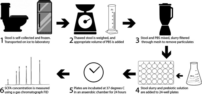
To measure SCFA production by gut microbiota, we adapted the in vitro approach of Edwards et al. (41). This method was specifically designed to study fermentation of starch in the human lower GI tract and has since been used to measure metabolite production from human stool samples when exposed to prebiotic fiber (42–44). In brief, we homogenized previously frozen feces in reduced phosphate-buffered saline (PBS; pH 7.0 ± 0.1) to create a fecal slurry with a final concentration of 100 g/liter (Fig. 1). These fecal slurries were then supplied with each of five prebiotic carbon sources, as well as a carbon-free control, and allowed to ferment at 37°C in anaerobic conditions for 24 h to approximate colonic transit time (45). After the incubation period, the concentrations of SCFAs in the samples were measured by gas chromatography. To control for differences in overall cell viability or stool slurry nutrient content between donors, we corrected measurements of SCFA concentration by dividing the treatment SCFA concentration by the control SCFA concentration.
FIG 1.
Overview of in vitro fermentation methods.
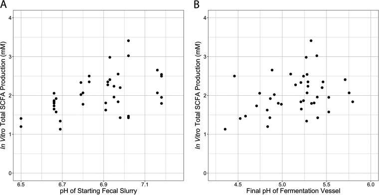
To validate our assay, we ran a series of experiments using feces from validation sample sets. We verified that our control-corrected SCFA production data were not influenced by bacterial abundance (P = 0.38, ρ = 0.14, Spearman correlation; see Fig. S1 in the supplemental material). Absolute (not relativized to control) SCFA concentrations are supplied in the supplement (see Fig. S2 and S3). Since our fermentation experiments used previously frozen fecal samples, we verified that total SCFA production was strongly correlated between fresh samples and twice freeze-thawed samples (P < 0.0001, ρ = 0.75, Spearman correlation; see Fig. S4A). Since we elected to not provide our fermentation reactions with nutrients in excess of what was contained in the fecal slurries, we verified that there existed strong correlation in total SCFA production between PBS-grown and colonic medium-grown cultures (46), both when supplied with dextrin and inulin (dextrin: P = 0.001, ρ = 0.68; inulin: P = 0.02, ρ = 0.51, Spearman correlations; see Fig. S5). We found that total SCFA production over control was positively correlated with the pH of starting fecal slurries (P = 0.003, ρ = 0.46, Spearman correlation; Fig. 2A). A weaker correlation may exist between SCFA production and the final pH of the fermentation vessels (P = 0.067, ρ = 0.29, Spearman correlation; Fig. 2B).
FIG 2.
Relationship between in vitro SCFA production and pH. (A) In vitro total SCFA production over control is positively correlated with the pH of starting fecal slurries (P = 0.003, ρ = 0.46; Spearman correlation). (B) Relationship between SCFA production and the final pH of fermentation vessels (P = 0.067, ρ = 0.29; Spearman correlation).
Bacterial abundance in stool, as measured by DNA concentration, does not correlate with control-corrected total SCFA production in in vitro cultures (P = 0.38, ρ = 0.14; Spearman correlation) Download FIG S1, PDF file, 0.4 MB (416.5KB, pdf) .
Copyright © 2020 Holmes et al.
This content is distributed under the terms of the Creative Commons Attribution 4.0 International license.
To control for differences in overall cell viability or stool slurry nutrient content between donors, we corrected measurements of SCFA concentration by dividing the treatment SCFA concentration by the control SCFA concentration. The resulting fold-change data do not contain information about absolute SCFA production. We examined the potential for this artifact to influence our interpretation and found that fold changes of SCFA concentrations after prebiotic treatment relative to the control were correlated with absolute control treatment levels (P < 0.0001, ρ = −0.77, Spearman correlation); absolute (not corrected to control) SCFA concentrations are presented in Fig. S3. Download FIG S2, PDF file, 0.4 MB (427.6KB, pdf) .
Copyright © 2020 Holmes et al.
This content is distributed under the terms of the Creative Commons Attribution 4.0 International license.
Total SCFA concentration of in vitro fermentation vessels after 24-h fermentation, plotted for each donor across five prebiotic treatments and the unsupplemented control vessel. The vertical red line indicates the total SCFA concentration of the starting fecal slurry prior to fermentation. Instances where SCFA concentration decreases during fermentation may be explained by net SCFA consumption by the community when no fermentable carbon is supplied or by a lack of change in concentration coupled with technical variation in our measurements. Instances where SCFA concentration is increased in the control treatment suggest that some unmetabolized carbohydrate may have remained in the stool to be metabolized during in vitro fermentation. Download FIG S3, PDF file, 1.4 MB (1.7MB, pdf) .
Copyright © 2020 Holmes et al.
This content is distributed under the terms of the Creative Commons Attribution 4.0 International license.
(A) In vitro total SCFA production from unfrozen stool samples and from twice frozen stool samples is highly correlated (P < 0.0001, ρ = 0.75, Spearman correlation). (B) In contrast, in vitro butyrate production is not correlated between unfrozen and twice frozen stool samples (P = 0.18, ρ = 0.19, Spearman correlation). Download FIG S4, PDF file, 1.2 MB (1.2MB, pdf) .
Copyright © 2020 Holmes et al.
This content is distributed under the terms of the Creative Commons Attribution 4.0 International license.
In vitro total SCFA production from inulin (A) and dextrin (B) is well correlated between cultures grown in PBS and cultures grown in a medium designed to mimic the colonic environment (P = 0.001, ρ = 0.68 [A]; P = 0.02, ρ = 0.51 [B], Spearman correlations). Download FIG S5, PDF file, 2.8 MB (3.1MB, pdf) .
Copyright © 2020 Holmes et al.
This content is distributed under the terms of the Creative Commons Attribution 4.0 International license.
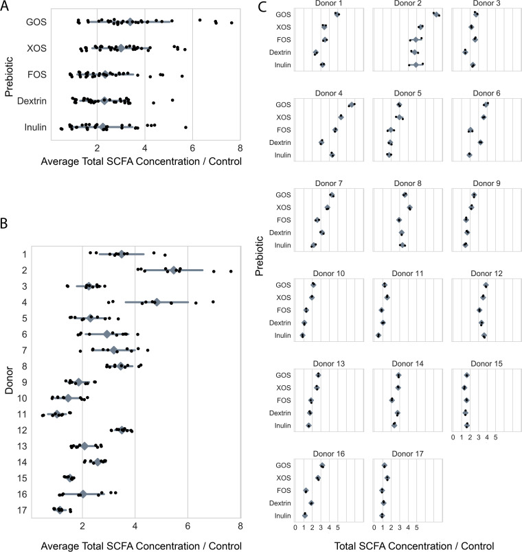
We subsequently applied our assay to fecal microbiota from a cohort of 17 children (6 male, 10 female, one unknown) ranging in age from 10 to 18 years old (average age, 15.7 years), Tanner stages 2 to 5, and a body mass index (BMI) of 25.9 to 75.3 (average BMI, 34.9) (see Table S1 in the supplemental material). One patient provided samples used in all analyses but was lost to follow-up before providing clinical metadata. This cohort was a subset of a cohort of patients enrolled in the Pediatric Obesity Microbiome and Metabolism study (47). We found all 17 individuals demonstrated a net gain of SCFAs relative to the control in at least one prebiotic treatment, which led us to conclude that all tested cultures were viable and metabolically active (Fig. 3).
FIG 3.
In vitro SCFA production by prebiotic (A), donor (B), and individually (C). In a two-way ANOVA of the effects of “donor” and “prebiotic” on “SCFA concentration/control,” “donor,” “prebiotic,” and their interaction were all statistically significant (P < 0.0001, P < 0.0001, and P < 0.0001, respectively). Shown is the total SCFA concentration of an in vitro culture after 24 h of anaerobic incubation, divided by the SCFA concentration of the corresponding prebiotic-free control culture, for each of five prebiotic growth conditions across 17 donors (black dots). Gray diamonds are means, and gray bars are standard deviations. (Absolute SCFA concentrations are depicted in Fig. S3.)
Demographic characteristics of participants in this study. One patient provided samples used in all analyses but was lost to follow-up before providing clinical metadata. That patient is only counted in the total column. Download Table S1, DOCX file, 0.01 MB (13.4KB, docx) .
Copyright © 2020 Holmes et al.
This content is distributed under the terms of the Creative Commons Attribution 4.0 International license.
Donor and prebiotic both impact SCFA production in vitro.
We next tested the hypothesis that different prebiotics equally promote the production of SCFAs by performing statistical analysis of SCFA production as a function of the prebiotic type and individual identity. Our analysis revealed heterogeneity in the efficacy of prebiotic supplements (two-way analysis of variance [ANOVA], P < 0.001; see Table S2; Fig. 3A), ranging from inulin, which resulted in a 2.35 mean fold change in total SCFAs, to galactooligosaccharides (GOS), which resulted in 3.55 mean fold change in total SCFAs. Frequently, only two or three of the five tested prebiotics resulted in increased total SCFA production within an individual. Our statistical testing also revealed consistent patterns between individuals’ gut microbiota in terms of SCFA production (two-way ANOVA, P < 0.001; see Table S2; Fig. 3B), with mean fold changes in SCFAs over control ranging from 2.37 to 6.12. Within individuals, the average fold change in SCFA concentration in the prebiotic treatments often appeared to be driven by a few strongly acidogenic prebiotics. Last, our analysis indicated a significant interaction between prebiotic type and individual identity (two-way ANOVA, P < 0.001; see Table S2; Fig. 3C). Because our statistical analysis considered technical replicates as separate experimental conditions, this result suggests the presence of consistent prebiotic/individual responses across in vitro assay replicate runs—not whether such interactions are consistent within an individual over time.
Two-way ANOVA of in vitro SCFA production across donors and prebiotics. Download Table S2, DOCX file, 0.01 MB (13KB, docx) .
Copyright © 2020 Holmes et al.
This content is distributed under the terms of the Creative Commons Attribution 4.0 International license.
SCFA production in vitro predicts the abundance of bacteria in the starting culture.
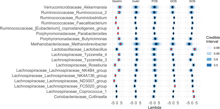
If interindividual differences in gut microbiota mediated responses to prebiotic treatment, we would expect that specific bacterial taxa, which varied between individuals, could also be associated with SCFA production. To evaluate this hypothesis, we used the R package stray (48) to create a Bayesian multinomial logistic normal linear regression (pibble) model that tested for correlations between in vitro SCFA production in response to each prebiotic and 16S rRNA community composition of patient stool used in the fermentations at the genus level. This analysis revealed that SCFA production from prebiotics was correlated with the relative abundances of 18 different bacterial genera (95% credible interval not covering 0; Fig. 4). Of the 13 genera positively associated with SCFA production, 9 are known or likely fiber degraders (49–53), one, Akkermansia, is often observed to increase in abundance after prebiotic treatment (54), and one, Methanobrevibacter, an archaeon hydrogenotrophic methanogen, is known to increase the efficiency of carbohydrate metabolism by the microbiota (55) (Table 1). Most genera identified by stray were associated with SCFA production in a limited set of prebiotic treatments. One genus, Lactobacillus, is positively associated with SCFA production on xylooligosaccharides (XOS) but was negatively associated with SCFA production on GOS. Overall, the presence of specific associations between bacterial taxa and different prebiotics supports a model where different individuals vary in their levels of prebiotic degrading gut bacteria.
FIG 4.
Eighteen genera were found to be credibly associated with SCFA production in at least one of our five prebiotic growth conditions. Shown are the mean lambda values and 99%, 95%, 80%, and 50% credible intervals for all 18 genera credibly associated with at least one prebiotic growth condition, plotted on centered log-ratio (CLR) coordinates. Red centers denote associations with 95% credible intervals that do not cover 0. Lambda represents the strength of the effect of each covariate on each taxa. A lambda value of 1 reflects a unit fold change in SCFA concentration over control as being associated with a unit fold change in the CLR-transformed relative abundance of the genus.
TABLE 1.
Associations between microbial genera and SCFA production on five different prebiotic substrates
| Genus | Association with SCFA production |
Fiber degrader status | Reference | ||||
|---|---|---|---|---|---|---|---|
| Dextrin | XOS | GOS | FOS | Inulin | |||
| Akkermansia | + | Supporter | 87 | ||||
| Ruminococcus_2 | + | Degrader | 49 | ||||
| Coprostanoligenes_group | + | No evidence | 88 | ||||
| Parabacteroides | + | Degrader | 50 | ||||
| Butyricimonas | + | Associated | 51 | ||||
| Methanobrevibacter | + | Supporter | 89 | ||||
| Tyzzerella_4 | + | Degrader | 52 | ||||
| Tyzzerella_3 | + | Degrader | 52 | ||||
| Lachnospiraceae_NK4B4 | + | Degrader | 52 | ||||
| Lactobacillus | + | – | Degrader | 50 | |||
| Coprococcus_1 | + | Degrader | 53 | ||||
| Collinsella | + | No evidence | 90 | ||||
| Lachnospiraceae_FCS020 | + | Degrader | 52 | ||||
Metrics of obesity do not appear to correlate with SCFA production capacity of stool.
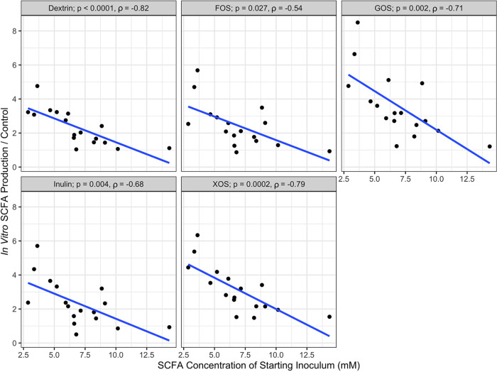
Finally, we tested the hypothesis that in vitro SCFA production would be associated with obesity-related phenotypes. We compared clinical metadata from individuals, which included BMI, insulin, and HbA1c, with average total SCFA production across prebiotics and found no significant correlations in our population (Spearman correlation; Table 2). Fecal microbial SCFA production capacity may not be directly associated with obesity though because rates of host SCFA uptake likely vary, and this variance may influence host intestinal physiology (56–58). Indeed, in support of the idea that SCFA absorption rate (which was not measured in this study) shape metabolic homeostasis and host health, we observed a negative association between fecal SCFA concentrations and in vitro SCFA production across the range of tested prebiotics (Fig. 5). Furthermore, if SCFA absorption efficiencies varied by individual, residual fecal SCFA concentrations may not directly reflect the complete effect of bacterial metabolism on obesity. Consistent with this notion, no significant relationships were apparent between concentrations of SCFA in patient stool and clinical markers of obesity measured at enrollment, including BMI, insulin levels, and HbA1c (Table 2), although this may also be explained by uncontrolled patient parameters.
TABLE 2.
Neither average SCFA production in vitro nor fecal SCFA concentration correlated with metrics of obesity measured in individuals at the time of enrollmenta
| Parameter | BMI |
Insulin |
HbA1c |
|||
|---|---|---|---|---|---|---|
| P | ρ | P | ρ | P | ρ | |
| Avg net SCFA production | 0.98 | –0.007 | 0.63 | 0.13 | 0.75 | 0.083 |
| Fecal SCFA concentration | 0.65 | –0.12 | 0.61 | 0.13 | 0.72 | –0.09 |
P and ρ values were determined from Spearman correlations.
FIG 5.
Spearman correlations between in vitro SCFA production and SCFA concentration of the starting fecal inoculum. SCFA production is the average of technical replicates, with the linear regression line plotted.
DISCUSSION
In this study, we found that the microbiota of all tested adolescents with obesity increased total SCFA production when exposed in vitro to at least one prebiotic. Both donor and prebiotic were significant factors in determining SCFA production in vitro, as was their interaction. Our modeling revealed distinct associations between specific microbial taxa and SCFA production on different prebiotics. We interpret this result as suggesting that the associated bacteria play a role in the fiber fermenting capacity of the community. We observed no correlations between either stool SCFA concentrations or in vitro acidogenic capacity of communities and any metrics of obesity (Table 2).
We have recapitulated previous findings that both donor and prebiotic are important in determining the SCFA production from in vitro prebiotic supplementation (31, 51, 59), and we found that not all prebiotics appear equally acidogenic (51). Since our in vitro system removes the host as a potential source of variation, our data support a gut microbial role for interdonor variation in fecal SCFA production. In addition, the strength of the interaction between donor and prebiotic strongly suggests that prebiotics are not “one size fits all”; rather, inconsistent results from prior studies of prebiotics in pediatric obesity (32, 34, 60) may be due to variation in the SCFA production capacity of individuals’ gut microbiota across the tested prebiotics. Future therapeutic efforts involving prebiotics in patients with obesity may benefit from stratified or personalized treatments. Nutritional therapies that are personalized to individuals’ microbiota are already in development (61).
Murine and in vitro studies show that increased signaling through GPCRs, mediated by acetate, propionate, and butyrate, increases satiety and insulin sensitivity, while decreasing adipogenesis (12, 23, 62); yet, we did not observe associations between fecal SCFA levels and metrics of obesity. The effects of SCFA on obesity may be masked by uncontrolled patient factors, such as differences in caloric intake and variation in individual nutrient harvest and utilization. In order to observe the effects of SCFA on obesity, it would be necessary to control for these variable physiological and lifestyle parameters, which we did not attempt. These patient factors may also have influenced our inability to observe an association between acidogenic capacity of microbiota and fecal SCFA concentrations. However, this may also be explained by the potential uncoupling of fecal SCFA production and fecal SCFA concentration. In vitro, increased luminal concentrations of butyrate have been shown to upregulate the sodium-coupled monocarboxylate transporter SLC5A8 (56), and the addition of physiological mixtures of SCFA has been shown to upregulate the monocarboxylate transporter SLC16A1 (63), both of which uptake acetate, propionate, and butyrate from the lumen. Since gut epithelia have the capacity to absorb up to 95% of SCFA before excretion (64), increased host SCFA uptake (triggered by increased gut bacterial production) could therefore lead to constant or even decreased fecal SCFA concentrations. This complex relationship could explain the absence of positive correlations we observed between stool SCFA levels and the acidogenic capacity of gut microbiota. It may be necessary to delve further upstream of fecal SCFA concentration by measuring proxies for host SCFA uptakes, such as the expression of SCFA transporters (SLC5A8 and SLC16A1) and SCFA receptors (GPR43, GPR41, and GPR109A) (56).
The primary limitations of this study involve constraints common to in vitro culture studies. First, many factors affecting bacterial SCFA production in vivo are difficult to replicate in vitro, including the availability of nutrients such as nitrogen, the starting concentration of SCFAs, the redox state of the environment, and the efficiency of cross-feeding interactions (65, 66). Different metabolic results between prebiotics may have occurred if we provided alternative cometabolites or nutrients, in addition to the tested prebiotics. We chose our culture conditions, namely, a medium-free approach that does not add any nutrients beyond what is present in the stool, in an effort to avoid inducing artificial selective conditions within our cultures. Prior experimental digestion studies have shown that prebiotic response patterns can be recapitulated across various culture conditions (42, 44). Indeed, we found strong correlation in SCFA production between cultures grown with our medium-free approach and those grown in a more conventional medium containing added nitrogen, vitamins, minerals, and acetate. Further, this approach allowed us to minimize the influence of the host on measurements of microbiota production of SCFA. We did observe shifts in community composition during the 24 h fermentations (Fig. S6); however, we remained able to find statistical associations between SCFA production capacity and prefermentation community composition. A second set of limitations in this study involves our reliance on patient collection of stool. Interdonor variation in prebiotic response could have originated in technical variation between how patients exposed stool to aerobic conditions (67) or how they froze their samples (68), which in turn could have affected the fraction of viable microbial cells in stool samples. Still, we found a significant correlation between in vitro total SCFA production from fresh stool and stool that had been frozen and thawed twice. Variation in donor prebiotic response could also have biological origins due to physiological differences between people (e.g., efficiency of food digestion, consistency of stool [69]) or differences in diet, which can lead to variation in stool microbial load and nutrient content (70). Rather than control for a myriad of different sources of variation whose origins we did not measure, we chose the straightforward approach of standardizing donor samples by employing a consistent concentration of stool slurry (5% [wt/vol] stool in PBS) in our experiments.
Microbial community composition changes during the course of a 24-h fermentation. We performed 16S rRNA sequencing on pre- and postfermentation samples from 20 donors across three treatments (inulin, dextrin, and control). We then measured the Shannon diversity of the fecal slurries both before and after fermentation (all treatments averaged) and found a significant decrease in Shannon diversity over the course of fermentation (P < 0.0001 [paired t test]). To characterize the changes in community composition associated with this decrease in diversity, we tested the pre- and postfermentation samples for differential abundance of taxa at the species level. We found 10 taxa with significantly different abundances between the two sample sets (P < 0.05 [Benjamini-Hochberg corrected Wilcoxon rank sum tests]). Of these 10 taxa, only Escherichia and Shigella spp. increased in relative abundance after fermentation, whereas nine other taxa each decreased in relative abundance (Anaerostipes hadrus, Bacteroides acidifaciens, Blautia faecis, Blautia wexlerae and two other Blautia of undetermined species, two undetermined species in the Eubacterium hallii group, and Ruminococcaceae_UCG-004 spp.). We attribute these changes to differences in growth rates among bacteria in our in vitro system and the inability of our fermentation medium to support the growth of some community members. Download FIG S6, PDF file, 1.3 MB (1.6MB, pdf) .
Copyright © 2020 Holmes et al.
This content is distributed under the terms of the Creative Commons Attribution 4.0 International license.
Future work to address these limitations could test multiple stool samples per subject to confirm whether the observed variation in prebiotic response is durable between individuals over time. Future studies could also examine the correlation between the metabolic effects of prebiotic supplementation in vitro and in vivo using randomized human trials that couple human prebiotic supplementation, in vivo measurement of SCFA production, and in vitro tests of microbiota metabolic activity. It would also be useful for such studies to explore the impact of prebiotic supplementation on host physiology, both in vitro and in vivo. Specifically, the effects of prebiotic supplementation on colonic epithelial barrier integrity, SCFA receptor (GPR41, GPR43, and GPR109A) expression, and SCFA transporter (MCT1 and SMCT1) expression could provide greater insight into the health impacts of prebiotic supplementation, as well as explain why fecal SCFA concentrations may not mirror the metabolic capacity of gut microbiota.
MATERIALS AND METHODS
Cohort.
Stool was collected from human donors under a protocol approved by the Duke Health Institutional Review Board (Duke Health IRB Pro00074547) for a prospective longitudinal cohort study and biorepository. Participants whose samples were used in this study were treatment-seeking adolescents with obesity who were newly enrolled in a multidisciplinary weight management program. All subjects received family-based intensive lifestyle modification. Based on clinical necessity, some participants also were placed on a low-carbohydrate diet, medications to facilitate weight loss, or underwent weight loss surgery (see Table S3). Due to the low number of patients assigned to each treatment arm, we did not attempt to base any analyses on patient treatment plan. Patients were 10 to 18 years old, with a BMI ≥ 95th percentile. None had antibiotic use in the 1 month prior to enrollment, used medications known to interfere with the intestinal microbiome, or had other significant medical problems. Stool samples used in this study were from enrollment, 3-month, 4.5-month, and 6-month follow-up visits (see Table S3). The clinical metadata used for correlations was collected at enrollment, 3 months, and 6 months. The metadata collected nearest to the stool sample collection date was used in our analyses.
Treatment arms and sample time points of patients in this study. Download Table S3, DOCX file, 0.01 MB (13.9KB, docx) .
Copyright © 2020 Holmes et al.
This content is distributed under the terms of the Creative Commons Attribution 4.0 International license.
Stool collection.
Patients collected intact stool samples in the clinic or at home using a plastic stool collection container (Fisher Scientific, 02-544-208) and were asked to immediately store this container in their home freezer. Patients then returned the sample by either bringing it to the study team or scheduling a home pickup within 18 h of stooling. Stool was transported frozen in an insulated container with an ice pack. Upon receipt in the lab, samples were placed on dry ice until transferred to a –80°C freezer for long-term storage. All patient samples were frozen at –80°C within 19 h of stooling (range, 0.08 h to 18.83 h; median, 11.42 h), except for one which was stored 44.03 h after stooling. The time between stooling and freezing at –80°C did not have a significant effect on average SCFA production (P = 0.58, ρ = −0.15, Pearson correlation). Stool samples for analysis were processed by removing containers from –80°C storage and thawing on ice in a biological safety cabinet until soft enough to aliquot. Thawed containers of stool were opened to atmosphere for a maximum of 10 min while samples were aliquoted. After primary aliquoting, the remaining stool was transferred to an anaerobic chamber (COY Laboratory Products, 5% hydrogen, 5% CO2, 90% nitrogen) and further portioned into ∼2-g aliquots for this study. These aliquots were then stored as solid stool pellets at –80°C until used for this study.
In vitro fermentation.
See Fig. 1 for an overview of in vitro fermentation methods. Aliquoted stool was thawed at room temperature in an anaerobic chamber. Once thawed, stool was weighed and placed into a polyethylene filter bag with 0.33-mm pore size (Whirl-Pak B01385), and 10 ml of anaerobic 1× PBS was added for each gram of stool, resulting in a 10% (wt/vol) fecal slurry, similar to previous studies (41, 42, 71, 72). During our validation experiments, a medium designed to simulate colonic contents was used in place of 1× PBS to create stool slurries (46). The filter bag was then closed and placed into a stomacher (Seward Stomacher 80) where the contents were homogenized on the medium speed setting for 60 s. The liquid fraction was removed from the downstream side of the filter membrane, and the solid fraction was discarded. A 1-ml aliquot of this liquid fraction was removed for analysis of the SCFA concentration to determine the SCFA concentration of the starting stool sample. During our validation experiments, two separate 1-ml aliquots of this liquid fraction were removed: one was used to estimate relative bacteria abundance of starting fecal slurries using total extracted DNA concentration, as has been previously published (73), and the remaining aliquot was used to determine the pH of the starting fecal slurry using a handheld pH meter (Elite pH Spear; Thermo Fisher Scientific). The remaining liquid fraction was incubated in duplicate across six different treatments, either supplemented with inulin (Now Foods Inulin Powder, part 2944), fructooligosaccharides (FOS; Cargill, part 100047199), galactooligosaccharides (GOS; Bimuno Powder), xylooligosaccharides (XOS; BioNutrition prebiotic with Llife-Oligo, part 359), wheat dextrin (Benefiber Original), or unsupplemented. For each reaction, 1 ml of 10% fecal slurry was placed in one well of a 24-well cell culture plate. Each well was then delivered 1 ml of 1% (wt/vol) prebiotic solution in 1× PBS or 1 ml of 1× PBS without prebiotic. During our validation experiments, prebiotics were dissolved in colonic medium instead of 1× PBS. The resulting fermentation conditions were therefore 5% fecal slurry with 0.5% prebiotic (wt/vol). A 5% fecal slurry was selected because its fermentative capacity has been previously demonstrated to be insensitive to small variations in concentration and is feasible to work with using this method (42). A 0.5% final concentration of prebiotic in the context of a 5% fecal slurry is analogous to an average adult consuming 20 g of dietary fiber per day, assuming an average daily stool mass of 200 g (74). Fermentation reactions were carried out in an anaerobic chamber at 37°C for 24 h. After fermentation, 1 ml of medium was taken from each reaction vessel for SCFA quantification. During our validation experiments, a separate 1-ml aliquot was taken for pH measurement.
Simulation of freeze-thaws experienced by study samples.
To test the effects of freeze-thaw cycles on in vitro SCFA production, we collected fresh, whole fecal samples from four healthy adults who were not patients in the study cohort. Informed consent was obtained from volunteers and the protocol was approved by the Duke Health Institutional Review Board. Samples were brought into an anaerobic chamber after voiding. Once in anaerobic conditions, these samples were divided into three aliquots. One aliquot was processed immediately following the same in vitro fermentation protocol used in our study. The other two aliquots were transferred to –80°C storage. After a minimum of 24 h, one of these two aliquots was removed from the freezer and thawed at room temperature for 2 h, before being returned to –80°C for an additional minimum of 24 h. Each of these frozen aliquots was thawed and processed following the same in vitro fermentation protocol. This allowed direct comparison of samples that had been used in fermentations immediately after voiding to those that had been frozen and thawed one and two times.
Medium preparation.
To validate our methods, namely, our use of a 5% fecal slurry in PBS, without supplementation of other nutrient components, we compared SCFA production with our methods to SCFA production when stool was instead resuspended in a medium designed to simulate the large intestine. We used a slightly modified medium derived from Gamage et al. (46). The media contained the following (per liter): peptone, 0.5 g; yeast extract, 0.5 g; NaHCO3, 6 g; hemin solution (0.5% [wt/vol] hemin and 0.2% [wt/vol] NaOH), 100 μl; l-cysteine HCl monohydrate, 0.53 g; bile salts (glycocholic acid and taurocholic acid), 0.5 g (Ward’s Science, 470300-380); vitamin supplement (ATCC MD-VS), 1 ml; K2HPO4, 0.228 g; KH2PO4, 0.228 g; (NH4)2SO4, 0.228 g; NaCl, 0.456 g; MgSO4, 0.0456 g; CaCl2, 0.0460 g; trace mineral supplement (ATCC MD-TMS), 1 ml; and glacial acetic acid, 287 μl. The pH of the medium was adjusted to 7.0 ± 0.1.
Quantification of SCFA.
The SCFA concentration of fecal slurries and fermentation vessels was determined following a protocol adapted from Zhao et al. (75). First, a 1-ml aliquot of either 10% fecal slurry in PBS or the fermentation vessel contents was obtained. To this, 50 μl of 6 N HCl was added to acidify the solution to a pH below 3. The mixture was vortexed and then centrifuged at 14,000 relative centrifugal force for 5 min at 4°C to remove particles. Avoiding the pellet, 750 μl of this supernatant was passed through a 0.22-μm spin column filter. The resulting filtrate was then transferred to a glass autosampler vial (VWR, part 66009-882).
Filtrates were analyzed on an Agilent 7890b gas chromatograph equipped with a flame ionization detector and an Agilent HP-FFAP free fatty-acid column (25 m × 0.2 mm [inner diameter] × 0.3 μm film). A volume of 0.5 μl of the filtrate was injected into a sampling port heated to 220°C and equipped with a split injection liner. The column temperature was maintained at 120°C for 1 min, ramped to 170°C at a rate of 10°C/min, and then maintained at 170°C for 1 min. The helium carrier gas was run at a constant flow rate of 1 ml/min, giving an average velocity of 35 cm/s. After each sample, we ran a 1-min postrun at 220°C and a carrier gas flow rate of 1 ml/min to clear any residual sample. All C2:C5 short-chain fatty acids were identified and quantified in each sample by comparing to an 8-point standard curve that encompassed the sample concentration range. Standards contained 0.1, 0.2, 0.5, 1, 2, 4, 8, and 16 mM concentrations of each SCFA.
DNA extraction, PCR amplification, and sequencing.
We performed 16S rRNA gene amplicon sequencing on human stool samples to determine microbiota community composition. DNA was extracted from frozen fecal samples with a Qiagen DNeasy PowerSoil DNA extraction kit (ID 12888-100). Amplicon sequencing was performed using custom barcoded primers targeting the V4 region of the 16S gene (76), using published protocols (76–78). The sequencing library was diluted to a 10 nM concentration and sequenced using an Illumina MiniSeq and a MiniSeq Mid Output kit (FC420-1004) with paired-end 150-bp reads.
Identifying sequence variants and taxonomy assignment.
We used an analysis pipeline with DADA2 (79) to identify and quantify sequence variants, as previously published by Silverman et al. (80). To prepare data for denoising with DADA2, 16S rRNA primer sequences were trimmed from paired sequencing reads using Trimmomatic v0.36 without quality filtering (81). Barcodes corresponding to reads that were dropped during trimming were removed using a custom python script. Reads were demultiplexed without quality filtering using Python scripts provided with Qiime v1.9 (82). Bases between positions 10 and 150 were retained for the forward reads and between positions 0 and 140 were retained for the reverse reads. This trimming, as well as minimal quality filtering of the demultiplexed reads, was performed using the function fastqPairedFilter provided with the DADA2 R package (v1.8.0). Sequence variants were inferred by DADA2 independently for the forward and reverse reads of each of the two sequencing runs using error profiles learned from all 20 samples. Forward and reverse reads were merged. Bimeras were removed using the function removeBimeraDenovo with default settings. Taxonomy was assigned using the function assignTaxonomy from DADA2, trained using version 123 of the Silva database.
Modeling microbial composition data.
To associate microbial genera to SCFA production on different prebiotics, the sequence variant table was amalgamated to the genus level using the R package phyloseq (82). Genera that were observed with at least three counts in at least three samples were retained. This filtering step retained 99.3% of the sequence variant counts and a total of 97 genera.
To associate microbial composition to SCFA production on different prebiotics, we used Bayesian multinomial logistic-normal linear regression, implemented in the R package stray as the function pibble (83). We chose this method to account for uncertainty due to counting and compositional constraints as expressed in Silverman et al. (80) and Grantham et al. (84). Our regression model was defined for the jth sample by the covariate vector:
where xj(Inulin) is the amount of total SCFA produced by the community in sample j as assessed by our in vitro assay and the preceding 1 represents a constant intercept. The regression model priors required that four hyperparameters—gamma, theta, xi, and upsilon—be specified. We set the hyperparameter theta to a D × Q matrix of zeros (where D = 97 is the number of sequence variants and Q = 5 is the number of covariates) representing our prior assumption that, on average, the association between each prebiotic and each taxon is zero.
We set the hyperparameter gamma to be the matrix:
which was chosen to reflect the following prior information: (i) the relative scale of gamma11 to gammakk (for) implies that we have little knowledge regarding the mean composition between individuals, but that we conservatively expect that the association between SCFA production and microbial composition is small comparatively; (ii) the value of 0.6 states that, on average across genera, we assume that the effects of each prebiotic are correlated with an average correlation of 0.3; (iii) in concert with our prior choices for xi and upsilon (below), the scale of gamma represents our assumption that the technical noise in our community measurements is smaller (by a factor of) ≈e2 than the magnitude of the biological variation between samples. This later prior regarding technical versus biological variation was informed by Silverman et al. (80). All prior choices were further investigated using prior predictive checks (85). To reflect a weak prior assumption that the absolute abundance of each taxon is uncorrelated we choose upsilon = D + 3 and xi to be the (D – 1) × (D – 1) matrix with elements xiii = (upsilon – D) and xiij = (upsilon – D)/2 for j ≠ i (86). While the model fit by stray and our corresponding priors were specified with respect to additive log-ratio coordinates, we utilized theory from compositional data analysis to transform these results into centered log-ratio coordinates for interpretation (80). Credible intervals and figures reflect 2,000 samples from the posterior distribution of the corresponding multivariate regression model.
ACKNOWLEDGMENTS
This study was supported in part by National Institutes of Health grants R24-DK110492 and 1R01DK116187, Translational Research Institute through Cooperative Agreement NNX16AO69A, the Damon Runyon Cancer Research Foundation, and the UNC CGIBD (NIDDK P30DK034987). This study used a high-performance computing facility partially supported by grant 2016-IDG-1013 (HARDAC+: Reproducible HPC for Next-Generation Genomics) from the North Carolina Biotechnology Center.
We thank Jessica McCann, Cameron Catherine, Charles Sarria, Alexandra Zizzi, and Janet Wooton for their contributions to this work.
L.A.D. was a member of the Kaleido Biosciences Strategic Advisory Board and retains equity in the company.
Footnotes
Citation Holmes ZC, Silverman JD, Dressman HK, Wei Z, Dallow EP, Armstrong SC, Seed PC, Rawls JF, David LA. 2020. Short-chain fatty acid production by gut microbiota from children with obesity differs according to prebiotic choice and bacterial community composition. mBio 11:e00914-20. https://doi.org/10.1128/mBio.00914-20.
Contributor Information
Thomas Mitchell Schmidt, University of Michigan—Ann Arbor.
Jose C. Clemente, Icahn School of Medicine at Mount Sinai.
REFERENCES
- 1.Skinner AC, Skelton JA. 2014. Prevalence and trends in obesity and severe obesity among children in the United States, 1999-2012. JAMA Pediatr 168:561–566. doi: 10.1001/jamapediatrics.2014.21. [DOI] [PubMed] [Google Scholar]
- 2.Freedman DS, Fulton JE, Dietz WH, Pan L, Nihiser AJ, Srinivasan SR, Berenson GS. 2010. The identification of children with adverse risk factor levels by body mass index cutoffs from 2 classification systems: the Bogalusa Heart Study. Am J Clin Nutr 92:1298–1305. doi: 10.3945/ajcn.2010.29758. [DOI] [PMC free article] [PubMed] [Google Scholar]
- 3.Freedman DS, Goodman A, Contreras OA, DasMahapatra P, Srinivasan SR, Berenson GS. 2012. Secular trends in BMI and blood pressure among children and adolescents: the Bogalusa Heart Study. Pediatrics 130:e159–e166. doi: 10.1542/peds.2011-3302. [DOI] [PMC free article] [PubMed] [Google Scholar]
- 4.Finkelstein EA, Graham WC, Malhotra R. 2014. Lifetime direct medical costs of childhood obesity. Pediatrics 133:854–862. doi: 10.1542/peds.2014-0063. [DOI] [PubMed] [Google Scholar]
- 5.Whitlock EP, O’Connor EA, Williams SB, Beil TL, Lutz KW. 2010. Effectiveness of weight management interventions in children: a targeted systematic review for the USPSTF. Pediatrics 125:e396–e418. doi: 10.1542/peds.2009-1955. [DOI] [PubMed] [Google Scholar]
- 6.Greenblum S, Turnbaugh PJ, Borenstein E. 2012. Metagenomic systems biology of the human gut microbiome reveals topological shifts associated with obesity and inflammatory bowel disease. Proc Natl Acad Sci U S A 109:594–599. doi: 10.1073/pnas.1116053109. [DOI] [PMC free article] [PubMed] [Google Scholar]
- 7.Jumpertz R, Le DS, Turnbaugh PJ, Trinidad C, Bogardus C, Gordon JI, Krakoff J. 2011. Energy-balance studies reveal associations between gut microbes, caloric load, and nutrient absorption in humans. Am J Clin Nutr 94:58–65. doi: 10.3945/ajcn.110.010132. [DOI] [PMC free article] [PubMed] [Google Scholar]
- 8.Ley RE, Turnbaugh PJ, Klein S, Gordon JI. 2006. Microbial ecology: human gut microbes associated with obesity. Nature 444:1022–1023. doi: 10.1038/4441022a. [DOI] [PubMed] [Google Scholar]
- 9.Turnbaugh PJ, Backhed F, Fulton L, Gordon JI. 2008. Diet-induced obesity is linked to marked but reversible alterations in the mouse distal gut microbiome. Cell Host Microbe 3:213–223. doi: 10.1016/j.chom.2008.02.015. [DOI] [PMC free article] [PubMed] [Google Scholar]
- 10.Turnbaugh PJ, Ley RE, Mahowald MA, Magrini V, Mardis ER, Gordon JI. 2006. An obesity-associated gut microbiome with increased capacity for energy harvest. Nature 444:1027–1031. doi: 10.1038/nature05414. [DOI] [PubMed] [Google Scholar]
- 11.Thangaraju M, Cresci GA, Liu K, Ananth S, Gnanaprakasam JP, Browning DD, Mellinger JD, Smith SB, Digby GJ, Lambert NA, Prasad PD, Ganapathy V. 2009. GPR109A is a G-protein-coupled receptor for the bacterial fermentation product butyrate and functions as a tumor suppressor in colon. Cancer Res 69:2826–2832. doi: 10.1158/0008-5472.CAN-08-4466. [DOI] [PMC free article] [PubMed] [Google Scholar]
- 12.Samuel BS, Shaito A, Motoike T, Rey FE, Backhed F, Manchester JK, Hammer RE, Williams SC, Crowley J, Yanagisawa M, Gordon JI. 2008. Effects of the gut microbiota on host adiposity are modulated by the short-chain fatty-acid binding G protein-coupled receptor, Gpr41. Proc Natl Acad Sci U S A 105:16767–16772. doi: 10.1073/pnas.0808567105. [DOI] [PMC free article] [PubMed] [Google Scholar]
- 13.Brown AJ, Goldsworthy SM, Barnes AA, Eilert MM, Tcheang L, Daniels D, Muir AI, Wigglesworth MJ, Kinghorn I, Fraser NJ, Pike NB, Strum JC, Steplewski KM, Murdock PR, Holder JC, Marshall FH, Szekeres PG, Wilson S, Ignar DM, Foord SM, Wise A, Dowell SJ. 2003. The Orphan G protein-coupled receptors GPR41 and GPR43 are activated by propionate and other short chain carboxylic acids. J Biol Chem 278:11312–11319. doi: 10.1074/jbc.M211609200. [DOI] [PubMed] [Google Scholar]
- 14.Thomas C, Pellicciari R, Pruzanski M, Auwerx J, Schoonjans K. 2008. Targeting bile-acid signalling for metabolic diseases. Nat Rev Drug Discov 7:678–693. doi: 10.1038/nrd2619. [DOI] [PubMed] [Google Scholar]
- 15.Sonnenburg JL, Bäckhed F. 2016. Diet-microbiota interactions as moderators of human metabolism. Nature 535:56–64. doi: 10.1038/nature18846. [DOI] [PMC free article] [PubMed] [Google Scholar]
- 16.Roediger WE. 1982. Utilization of nutrients by isolated epithelial cells of the rat colon. Gastroenterology 83:424–429. doi: 10.1016/S0016-5085(82)80339-9. [DOI] [PubMed] [Google Scholar]
- 17.Davie JR. 2003. Inhibition of histone deacetylase activity by butyrate. J Nutr 133:2485S–2493S. doi: 10.1093/jn/133.7.2485S. [DOI] [PubMed] [Google Scholar]
- 18.Candido EP, Reeves R, Davie JR. 1978. Sodium butyrate inhibits histone deacetylation in cultured cells. Cell 14:105–113. doi: 10.1016/0092-8674(78)90305-7. [DOI] [PubMed] [Google Scholar]
- 19.Smith PM, Howitt MR, Panikov N, Michaud M, Gallini CA, Bohlooly YM, Glickman JN, Garrett WS. 2013. The microbial metabolites, short-chain fatty acids, regulate colonic Treg cell homeostasis. Science 341:569–573. doi: 10.1126/science.1241165. [DOI] [PMC free article] [PubMed] [Google Scholar]
- 20.Lee C, Kim BG, Kim JH, Chun J, Im JP, Kim JS. 2017. Sodium butyrate inhibits the NF-kappa B signaling pathway and histone deacetylation, and attenuates experimental colitis in an IL-10 independent manner. Int Immunopharmacol 51:47–56. doi: 10.1016/j.intimp.2017.07.023. [DOI] [PubMed] [Google Scholar]
- 21.Yan H, Ajuwon KM. 2017. Butyrate modifies intestinal barrier function in IPEC-J2 cells through a selective upregulation of tight junction proteins and activation of the Akt signaling pathway. PLoS One 12:e0179586. doi: 10.1371/journal.pone.0179586. [DOI] [PMC free article] [PubMed] [Google Scholar]
- 22.Hatayama H, Iwashita J, Kuwajima A, Abe T. 2007. The short chain fatty acid, butyrate, stimulates MUC2 mucin production in the human colon cancer cell line, LS174T. Biochem Biophys Res Commun 356:599–603. doi: 10.1016/j.bbrc.2007.03.025. [DOI] [PubMed] [Google Scholar]
- 23.Tolhurst G, Heffron H, Lam YS, Parker HE, Habib AM, Diakogiannaki E, Cameron J, Grosse J, Reimann F, Gribble FM. 2012. Short-chain fatty acids stimulate glucagon-like peptide-1 secretion via the G-protein-coupled receptor FFAR2. Diabetes 61:364–371. doi: 10.2337/db11-1019. [DOI] [PMC free article] [PubMed] [Google Scholar]
- 24.Vinolo MAR, Rodrigues HG, Festuccia WT, Crisma AR, Alves VS, Martins AR, Amaral CL, Fiamoncini J, Hirabara SM, Sato FT, Fock RA, Malheiros G, dos Santos MF, Curi R. 2012. Tributyrin attenuates obesity-associated inflammation and insulin resistance in high-fat-fed mice. Am J Physiol Endocrinol Metab 303:E272–E282. doi: 10.1152/ajpendo.00053.2012. [DOI] [PubMed] [Google Scholar]
- 25.Gao Z, Yin J, Zhang J, Ward RE, Martin RJ, Lefevre M, Cefalu WT, Ye J. 2009. Butyrate improves insulin sensitivity and increases energy expenditure in mice. Diabetes 58:1509–1517. doi: 10.2337/db08-1637. [DOI] [PMC free article] [PubMed] [Google Scholar]
- 26.Henagan TM, Stefanska B, Fang Z, Navard AM, Ye J, Lenard NR, Devarshi PP. 2015. Sodium butyrate epigenetically modulates high-fat diet-induced skeletal muscle mitochondrial adaptation, obesity and insulin resistance through nucleosome positioning. Br J Pharmacol 172:2782–2798. doi: 10.1111/bph.13058. [DOI] [PMC free article] [PubMed] [Google Scholar]
- 27.Lu Y, Fan C, Li P, Lu Y, Chang X, Qi K. 2016. Short chain fatty acids prevent high-fat-diet-induced obesity in mice by regulating G protein-coupled receptors and gut microbiota. Sci Rep 6:37589. doi: 10.1038/srep37589. [DOI] [PMC free article] [PubMed] [Google Scholar]
- 28.Frost G, Sleeth ML, Sahuri-Arisoylu M, Lizarbe B, Cerdan S, Brody L, Anastasovska J, Ghourab S, Hankir M, Zhang S, Carling D, Swann JR, Gibson G, Viardot A, Morrison D, Louise Thomas E, Bell JD. 2014. The short-chain fatty acid acetate reduces appetite via a central homeostatic mechanism. Nat Commun 5:3611. doi: 10.1038/ncomms4611. [DOI] [PMC free article] [PubMed] [Google Scholar]
- 29.Lin HV, Frassetto A, Kowalik EJ Jr, Nawrocki AR, Lu MM, Kosinski JR, Hubert JA, Szeto D, Yao X, Forrest G, Marsh DJ. 2012. Butyrate and propionate protect against diet-induced obesity and regulate gut hormones via free fatty acid receptor 3-independent mechanisms. PLoS One 7:e35240. doi: 10.1371/journal.pone.0035240. [DOI] [PMC free article] [PubMed] [Google Scholar]
- 30.McOrist AL, Miller RB, Bird AR, Keogh JB, Noakes M, Topping DL, Conlon MA. 2011. Fecal butyrate levels vary widely among individuals but are usually increased by a diet high in resistant starch. J Nutr 141:883–889. doi: 10.3945/jn.110.128504. [DOI] [PubMed] [Google Scholar]
- 31.Venkataraman A, Sieber JR, Schmidt AW, Waldron C, Theis KR, Schmidt TM. 2016. Variable responses of human microbiomes to dietary supplementation with resistant starch. Microbiome 4:9. doi: 10.1186/s40168-016-0178-x. [DOI] [PMC free article] [PubMed] [Google Scholar]
- 32.Abrams SA, Griffin IJ, Hawthorne KM, Ellis KJ. 2007. Effect of prebiotic supplementation and calcium intake on body mass index. J Pediatr 151:293–298. doi: 10.1016/j.jpeds.2007.03.043. [DOI] [PubMed] [Google Scholar]
- 33.Nicolucci AC, Hume MP, Martínez I, Mayengbam S, Walter J, Reimer RA. 2017. Prebiotics reduce body fat and alter intestinal microbiota in children who are overweight or with obesity. Gastroenterology 153:711–722. doi: 10.1053/j.gastro.2017.05.055. [DOI] [PubMed] [Google Scholar]
- 34.Liber A, Szajewska H. 2014. Effect of oligofructose supplementation on body weight in overweight and obese children: a randomised, double-blind, placebo-controlled trial. Br J Nutr 112:2068–2074. doi: 10.1017/S0007114514003110. [DOI] [PubMed] [Google Scholar]
- 35.Baxter NT, Schmidt AW, Venkataraman A, Kim KS, Waldron C, Schmidt TM. 2019. Dynamics of human gut microbiota and short-chain fatty acids in response to dietary interventions with three fermentable fibers. mBio 10:e02566-18. doi: 10.1128/mBio.02566-18. [DOI] [PMC free article] [PubMed] [Google Scholar]
- 36.Murugesan S, Ulloa-Martínez M, Martínez-Rojano H, Galván-Rodríguez FM, Miranda-Brito C, Romano MC, Piña-Escobedo A, Pizano-Zárate ML, Hoyo-Vadillo C, García-Mena J. 2015. Study of the diversity and short-chain fatty acids production by the bacterial community in overweight and obese Mexican children. Eur J Clin Microbiol Infect Dis 34:1337–1346. doi: 10.1007/s10096-015-2355-4. [DOI] [PubMed] [Google Scholar]
- 37.Nicolucci AC, Reimer RA. 2017. Prebiotics as a modulator of gut microbiota in paediatric obesity. Pediatr Obes 12:265–273. doi: 10.1111/ijpo.12140. [DOI] [PubMed] [Google Scholar]
- 38.Faith JJ, Guruge JL, Charbonneau M, Subramanian S, Seedorf H, Goodman AL, Clemente JC, Knight R, Heath AC, Leibel RL, Rosenbaum M, Gordon JI. 2013. The long-term stability of the human gut microbiota. Science 341:1237439. doi: 10.1126/science.1237439. [DOI] [PMC free article] [PubMed] [Google Scholar]
- 39.Ruppin H, Bar-Meir S, Soergel KH, Wood CM, Schmitt MG Jr.. 1980. Absorption of short-chain fatty acids by the colon. Gastroenterology 78:1500–1507. doi: 10.1016/S0016-5085(19)30508-6. [DOI] [PubMed] [Google Scholar]
- 40.Rechkemmer G, Rönnau K, Engelhardt WV. 1988. Fermentation of polysaccharides and absorption of short chain fatty acids in the mammalian hindgut. Comp Biochem Physiol 90:563–568. doi: 10.1016/0300-9629(88)90668-8. [DOI] [PubMed] [Google Scholar]
- 41.Edwards CA, Gibson G, Champ M, Jensen BB, Mathers JC, Nagengast F, Rumney C, Quehl A. 1996. In vitro method for quantification of the fermentation of starch by human faecal bacteria. J Sci Food Agric 71:209–217. doi:. [DOI] [Google Scholar]
- 42.Coles LT, Moughan PJ, Awati A, Darragh AJ. 2013. Optimization of inoculum concentration and incubation duration for an in vitro hindgut dry matter digestibility assay. Food Chem 136:624–631. doi: 10.1016/j.foodchem.2012.08.045. [DOI] [PubMed] [Google Scholar]
- 43.Rotbart A, Yao CK, Ha N, Chrisp MD, Muir JG, Gibson PR, Kalantar-Zadeh K, Ou JZ. 2017. Designing an in-vitro gas profiling system for human faecal samples. Sensors Actuators B Chem 238:754–764. doi: 10.1016/j.snb.2016.07.120. [DOI] [Google Scholar]
- 44.Coles LT, Moughan PJ, Awati A, Darragh AJ. 2011. Influence of assay conditions on the in vitro hindgut digestibility of dry matter. Food Chem 125:1351–1358. doi: 10.1016/j.foodchem.2010.10.004. [DOI] [PubMed] [Google Scholar]
- 45.Proano M, Camilleri M, Phillips SF, Brown ML, Thomforde GM. 1990. Transit of solids through the human colon: regional quantification in the unprepared bowel. Am J Physiol 258:G856–G862. doi: 10.1152/ajpgi.1990.258.6.G856. [DOI] [PubMed] [Google Scholar]
- 46.Gamage H, Tetu SG, Chong RWW, Ashton J, Packer NH, Paulsen IT. 2017. Cereal products derived from wheat, sorghum, rice and oats alter the infant gut microbiota in vitro. Sci Rep 7:14312. doi: 10.1038/s41598-017-14707-z. [DOI] [PMC free article] [PubMed] [Google Scholar]
- 47.McCann JR, Bihlmeyer NA, Roche K, Catherine C, Jawahar J, Kwee LC, Younge NE, Silverman J, Ilkayeva O, Sarria CJ, Zizzi A, Wootton J, Poppe L, Anderson PR, Arlotto M, Wei Z, Granek JA, Valdivia RH, David LA, Dressman HK, Newgard CB, Shah SH, Seed PC, Rawls JF, Armstrong SC. 2020. The Pediatric Obesity Microbiome and Metabolism Study (POMMS): methods, baseline data, and early insights. medRxiv doi: 10.1101/2020.06.09.20126763. [DOI] [PMC free article] [PubMed] [Google Scholar]
- 48.Silverman JD, Roche K, Holmes ZC, David LA, Mukherjee S. 2019. Bayesian multinomial logistic normal models through marginally latent matrix-T processes. arXiv arXiv:1903.11695 https://arxiv.org/abs/1903.11695.
- 49.Flint HJ, Scott KP, Duncan SH, Louis P, Forano E. 2012. Microbial degradation of complex carbohydrates in the gut. Gut Microbes 3:289–306. doi: 10.4161/gmic.19897. [DOI] [PMC free article] [PubMed] [Google Scholar]
- 50.Holscher HD. 2017. Dietary fiber and prebiotics and the gastrointestinal microbiota. Gut Microbes 8:172–184. doi: 10.1080/19490976.2017.1290756. [DOI] [PMC free article] [PubMed] [Google Scholar]
- 51.Gamage H, Tetu SG, Chong RWW, Bucio-Noble D, Rosewarne CP, Kautto L, Ball MS, Molloy MP, Packer NH, Paulsen IT. 2018. Fiber supplements derived from sugarcane stem, wheat dextrin and psyllium husk have different in vitro effects on the human gut microbiota. Front Microbiol 9:1618. doi: 10.3389/fmicb.2018.01618. [DOI] [PMC free article] [PubMed] [Google Scholar]
- 52.Biddle A, Stewart L, Blanchard J, Leschine S. 2013. Untangling the genetic basis of fibrolytic specialization by Lachnospiraceae and Ruminococcaceae in diverse gut communities. Diversity 5:627–640. doi: 10.3390/d5030627. [DOI] [Google Scholar]
- 53.Holdeman LV, Moore W. 1974. New genus, Coprococcus, 12 new species, and emended descriptions of 4 previously described species of bacteria from human feces. Int J Syst Bacteriol 24:260–277. doi: 10.1099/00207713-24-2-260. [DOI] [Google Scholar]
- 54.Belzer C, Chia LW, Aalvink S, Chamlagain B, Piironen V, Knol J, de Vos WM. 2017. Microbial metabolic networks at the mucus layer lead to diet-independent butyrate and vitamin B12 production by intestinal symbionts. mBio 8:e00770-17. doi: 10.1128/mBio.00770-17. [DOI] [PMC free article] [PubMed] [Google Scholar]
- 55.Samuel BS, Gordon JI. 2006. A humanized gnotobiotic mouse model of host-archaeal-bacterial mutualism. Proc Natl Acad Sci U S A 103:10011–10016. doi: 10.1073/pnas.0602187103. [DOI] [PMC free article] [PubMed] [Google Scholar]
- 56.Mathewson ND, Jenq R, Mathew AV, Koenigsknecht M, Hanash A, Toubai T, Oravecz-Wilson K, Wu S-R, Sun Y, Rossi C, Fujiwara H, Byun J, Shono Y, Lindemans C, Calafiore M, Schmidt TM, Honda K, Young VB, Pennathur S, van den Brink M, Reddy P. 2016. Gut microbiome–derived metabolites modulate intestinal epithelial cell damage and mitigate graft-versus-host disease. Nat Immunol 17:505–513. doi: 10.1038/ni.3400. [DOI] [PMC free article] [PubMed] [Google Scholar]
- 57.Senga T, Iwamoto S, Yoshida T, Yokota T, Adachi K, Azuma E, Hamaguchi M, Iwamoto T. 2003. LSSIG is a novel murine leukocyte-specific GPCR that is induced by the activation of STAT3. Blood 101:1185–1187. doi: 10.1182/blood-2002-06-1881. [DOI] [PubMed] [Google Scholar]
- 58.Ang Z, Ding JL. 2016. GPR41 and GPR43 in obesity and inflammation: protective or causative? Front Immunol 7:28. doi: 10.3389/fimmu.2016.00028. [DOI] [PMC free article] [PubMed] [Google Scholar]
- 59.Poeker SA, Geirnaert A, Berchtold L, Greppi A, Krych L, Steinert RE, de Wouters T, Lacroix C. 2018. Understanding the prebiotic potential of different dietary fibers using an in vitro continuous adult fermentation model (PolyFermS). Sci Rep 8:4318. doi: 10.1038/s41598-018-22438-y. [DOI] [PMC free article] [PubMed] [Google Scholar]
- 60.Hume MP, Nicolucci AC, Reimer RA. 2017. Prebiotic supplementation improves appetite control in children with overweight and obesity: a randomized controlled trial. Am J Clin Nutr 105:790–799. doi: 10.3945/ajcn.116.140947. [DOI] [PubMed] [Google Scholar]
- 61.Zeevi D, Korem T, Zmora N, Israeli D, Rothschild D, Weinberger A, Ben-Yacov O, Lador D, Avnit-Sagi T, Lotan-Pompan M, Suez J, Mahdi JA, Matot E, Malka G, Kosower N, Rein M, Zilberman-Schapira G, Dohnalová L, Pevsner-Fischer M, Bikovsky R, Halpern Z, Elinav E, Segal E. 2015. Personalized nutrition by prediction of glycemic responses. Cell 163:1079–1094. doi: 10.1016/j.cell.2015.11.001. [DOI] [PubMed] [Google Scholar]
- 62.Kimura I, Ozawa K, Inoue D, Imamura T, Kimura K, Maeda T, Terasawa K, Kashihara D, Hirano K, Tani T, Takahashi T, Miyauchi S, Shioi G, Inoue H, Tsujimoto G. 2013. The gut microbiota suppresses insulin-mediated fat accumulation via the short-chain fatty acid receptor GPR43. Nat Commun 4:1829. doi: 10.1038/ncomms2852. [DOI] [PMC free article] [PubMed] [Google Scholar]
- 63.Zhan K, Jiang M, Gong X, Zhao G. 2018. Effect of short-chain fatty acids on the expression of genes involved in short-chain fatty acid transporters and inflammatory response in goat jejunum epithelial cells. In Vitro Cell Dev Biol Anim 54:311–320. doi: 10.1007/s11626-017-0226-2. [DOI] [PubMed] [Google Scholar]
- 64.den Besten G, van Eunen K, Groen AK, Venema K, Reijngoud DJ, Bakker BM. 2013. The role of short-chain fatty acids in the interplay between diet, gut microbiota, and host energy metabolism. J Lipid Res 54:2325–2340. doi: 10.1194/jlr.R036012. [DOI] [PMC free article] [PubMed] [Google Scholar]
- 65.Louis P, Flint HJ. 2009. Diversity, metabolism and microbial ecology of butyrate-producing bacteria from the human large intestine. FEMS Microbiol Lett 294:1–8. doi: 10.1111/j.1574-6968.2009.01514.x. [DOI] [PubMed] [Google Scholar]
- 66.Pryde SE, Duncan SH, Hold GL, Stewart CS, Flint HJ. 2002. The microbiology of butyrate formation in the human colon. FEMS Microbiol Lett 217:133–139. doi: 10.1111/j.1574-6968.2002.tb11467.x. [DOI] [PubMed] [Google Scholar]
- 67.Chu ND, Smith MB, Perrotta AR, Kassam Z, Alm EJ. 2017. Profiling living bacteria informs preparation of fecal microbiota transplantations. PLoS One 12:e0170922. doi: 10.1371/journal.pone.0170922. [DOI] [PMC free article] [PubMed] [Google Scholar]
- 68.Song SJ, Amir A, Metcalf JL, Amato KR, Xu ZZ, Humphrey G, Knight R. 2016. Preservation methods differ in fecal microbiome stability, affecting suitability for field studies. mSystems 1:e00021-16. doi: 10.1128/mSystems.00021-16. [DOI] [PMC free article] [PubMed] [Google Scholar]
- 69.Vandeputte D, Falony G, Vieira-Silva S, Tito RY, Joossens M, Raes J. 2016. Stool consistency is strongly associated with gut microbiota richness and composition, enterotypes and bacterial growth rates. Gut 65:57–62. doi: 10.1136/gutjnl-2015-309618. [DOI] [PMC free article] [PubMed] [Google Scholar]
- 70.Stephen AM, Cummings JH. 1980. Mechanism of action of dietary fibre in the human colon. Nature 284:283–284. doi: 10.1038/284283a0. [DOI] [PubMed] [Google Scholar]
- 71.O’Donnell MM, Rea MC, O’Sullivan Ó, Flynn C, Jones B, McQuaid A, Shanahan F, Ross RP. 2016. Preparation of a standardised faecal slurry for ex-vivo microbiota studies which reduces inter-individual donor bias. J Microbiol Methods 129:109–116. doi: 10.1016/j.mimet.2016.08.002. [DOI] [PubMed] [Google Scholar]
- 72.Barry JL, Hoebler C, Macfarlane GT, Macfarlane S, Mathers JC, Reed KA, Mortensen PB, Nordgaard I, Rowland IR, Rumney CJ. 1995. Estimation of the fermentability of dietary fibre in vitro: a European interlaboratory study. Br J Nutr 74:303–322. doi: 10.1079/bjn19950137. [DOI] [PubMed] [Google Scholar]
- 73.Contijoch EJ, Britton GJ, Yang C, Mogno I, Li Z, Ng R, Llewellyn SR, Hira S, Johnson C, Rabinowitz KM, Barkan R, Dotan I, Hirten RP, Fu S-C, Luo Y, Yang N, Luong T, Labrias PR, Lira S, Peter I, Grinspan A, Clemente JC, Kosoy R, Kim-Schulze S, Qin X, Castillo A, Hurley A, Atreja A, Rogers J, Fasihuddin F, Saliaj M, Nolan A, Reyes-Mercedes P, Rodriguez C, Aly S, Santa-Cruz K, Peters L, Suárez-Fariñas M, Huang R, Hao K, Zhu J, Zhang B, Losic B, Irizar H, Song W-M, Di Narzo A, Wang W, Cohen BL, DiMaio C, Greenwald D, Itzkowitz S, Lucas A, Marion J, Maser E, Ungaro R, Naymagon S, Novak J, Shah B, Ullman T, Rubin P, George J, Legnani P, Telesco SE, Friedman JR, Brodmerkel C, Plevy S, Cho JH, Colombel J-F, Schadt EE, Argmann C, Dubinsky M, Kasarskis A, Sands B, Faith JJ. 2019. Gut microbiota density influences host physiology and is shaped by host and microbial factors. Elife 8:e40553. doi: 10.7554/eLife.40553. [DOI] [PMC free article] [PubMed] [Google Scholar]
- 74.Davies GJ, Crowder M, Reid B, Dickerson JW. 1986. Bowel function measurements of individuals with different eating patterns. Gut 27:164–169. doi: 10.1136/gut.27.2.164. [DOI] [PMC free article] [PubMed] [Google Scholar]
- 75.Zhao G, Nyman M, Jonsson JA. 2006. Rapid determination of short-chain fatty acids in colonic contents and faeces of humans and rats by acidified water-extraction and direct-injection gas chromatography. Biomed Chromatogr 20:674–682. doi: 10.1002/bmc.580. [DOI] [PubMed] [Google Scholar]
- 76.Caporaso JG, Lauber CL, Walters WA, Berg-Lyons D, Lozupone CA, Turnbaugh PJ, Fierer N, Knight R. 2011. Global patterns of 16S rRNA diversity at a depth of millions of sequences per sample. Proc Natl Acad Sci U S A 108:4516–4522. doi: 10.1073/pnas.1000080107. [DOI] [PMC free article] [PubMed] [Google Scholar]
- 77.Caporaso JG, Lauber CL, Walters WA, Berg-Lyons D, Huntley J, Fierer N, Owens SM, Betley J, Fraser L, Bauer M, Gormley N, Gilbert JA, Smith G, Knight R. 2012. Ultra-high-throughput microbial community analysis on the Illumina HiSeq and MiSeq platforms. ISME J 6:1621–1624. doi: 10.1038/ismej.2012.8. [DOI] [PMC free article] [PubMed] [Google Scholar]
- 78.Maurice Corinne F, Haiser Henry J, Turnbaugh Peter J. 2013. Xenobiotics shape the physiology and gene expression of the active human gut microbiome. Cell 152:39–50. doi: 10.1016/j.cell.2012.10.052. [DOI] [PMC free article] [PubMed] [Google Scholar]
- 79.Callahan BJ, McMurdie PJ, Rosen MJ, Han AW, Johnson AJ, Holmes SP. 2016. DADA2: high-resolution sample inference from Illumina amplicon data. Nat Methods 13:581–583. doi: 10.1038/nmeth.3869. [DOI] [PMC free article] [PubMed] [Google Scholar]
- 80.Silverman JD, Durand HK, Bloom RJ, Mukherjee S, David LA. 2018. Dynamic linear models guide design and analysis of microbiota studies within artificial human guts. Microbiome 6:202. doi: 10.1186/s40168-018-0584-3. [DOI] [PMC free article] [PubMed] [Google Scholar]
- 81.Caporaso JG, Kuczynski J, Stombaugh J, Bittinger K, Bushman FD, Costello EK, Fierer N, Pena AG, Goodrich JK, Gordon JI, Huttley GA, Kelley ST, Knights D, Koenig JE, Ley RE, Lozupone CA, McDonald D, Muegge BD, Pirrung M, Reeder J, Sevinsky JR, Turnbaugh PJ, Walters WA, Widmann J, Yatsunenko T, Zaneveld J, Knight R. 2010. QIIME allows analysis of high-throughput community sequencing data. Nat Methods 7:335–336. doi: 10.1038/nmeth.f.303. [DOI] [PMC free article] [PubMed] [Google Scholar]
- 82.McMurdie PJ, Holmes S. 2013. phyloseq: an R package for reproducible interactive analysis and graphics of microbiome census data. PLoS One 8:e61217. doi: 10.1371/journal.pone.0061217. [DOI] [PMC free article] [PubMed] [Google Scholar]
- 83.Silverman JD. 2018. stray: multinomial logistic-normal linear regression models. https://githubcom/jsilve24/stray.
- 84.Grantham NS, Reich BJ, Borer ET, Gross K. 2017. MIMIX: a Bayesian mixed-effects model for microbiome data from designed experiments. arXiv arXiv:1703.07747 https://arxiv.org/abs/1703.07747.
- 85.Gelman A, Carlin J, Stern H, Dunson D, Vehtari A, Rubin D. 2013. Bayesian data analysis. Chapman and Hall/CRC, New York, NY. [Google Scholar]
- 86.Aitchison J. 1982. The statistical analysis of compositional data. J R Stat Soc Series B Stat Methodol 44:139–177. doi: 10.1111/j.2517-6161.1982.tb01195.x. [DOI] [Google Scholar]
- 87.Kageyama A, Benno Y. 2000. Catenibacterium mitsuokai gen. nov., sp. nov., a Gram-positive anaerobic bacterium isolated from human faeces. Int J Syst Evol Microbiol 50:1595–1599. doi: 10.1099/00207713-50-4-1595. [DOI] [PubMed] [Google Scholar]
- 88.Freier TA, Beitz DC, Li L, Hartman PA. 1994. Characterization of Eubacterium coprostanoligenes sp. nov., a cholesterol-reducing anaerobe. Int J Syst Bacteriol 44:137–142. doi: 10.1099/00207713-44-1-137. [DOI] [PubMed] [Google Scholar]
- 89.Sakamoto M, Takagaki A, Matsumoto K, Kato Y, Goto K, Benno Y. 2009. Butyricimonas synergistica gen. nov., sp. nov. and Butyricimonas virosa sp. nov., butyric acid-producing bacteria in the family ‘Porphyromonadaceae’ isolated from rat faeces. Int J Syst Evol Microbiol 59:1748–1753. doi: 10.1099/ijs.0.007674-0. [DOI] [PubMed] [Google Scholar]
- 90.Candela M, Biagi E, Soverini M, Consolandi C, Quercia S, Severgnini M, Peano C, Turroni S, Rampelli S, Pozzilli P, Pianesi M, Fallucca F, Brigidi P. 2016. Modulation of gut microbiota dysbioses in type 2 diabetic patients by macrobiotic Ma-Pi 2 diet. Br J Nutr 116:80–93. doi: 10.1017/S0007114516001045. [DOI] [PMC free article] [PubMed] [Google Scholar]
Associated Data
This section collects any data citations, data availability statements, or supplementary materials included in this article.
Supplementary Materials
Bacterial abundance in stool, as measured by DNA concentration, does not correlate with control-corrected total SCFA production in in vitro cultures (P = 0.38, ρ = 0.14; Spearman correlation) Download FIG S1, PDF file, 0.4 MB (416.5KB, pdf) .
Copyright © 2020 Holmes et al.
This content is distributed under the terms of the Creative Commons Attribution 4.0 International license.
To control for differences in overall cell viability or stool slurry nutrient content between donors, we corrected measurements of SCFA concentration by dividing the treatment SCFA concentration by the control SCFA concentration. The resulting fold-change data do not contain information about absolute SCFA production. We examined the potential for this artifact to influence our interpretation and found that fold changes of SCFA concentrations after prebiotic treatment relative to the control were correlated with absolute control treatment levels (P < 0.0001, ρ = −0.77, Spearman correlation); absolute (not corrected to control) SCFA concentrations are presented in Fig. S3. Download FIG S2, PDF file, 0.4 MB (427.6KB, pdf) .
Copyright © 2020 Holmes et al.
This content is distributed under the terms of the Creative Commons Attribution 4.0 International license.
Total SCFA concentration of in vitro fermentation vessels after 24-h fermentation, plotted for each donor across five prebiotic treatments and the unsupplemented control vessel. The vertical red line indicates the total SCFA concentration of the starting fecal slurry prior to fermentation. Instances where SCFA concentration decreases during fermentation may be explained by net SCFA consumption by the community when no fermentable carbon is supplied or by a lack of change in concentration coupled with technical variation in our measurements. Instances where SCFA concentration is increased in the control treatment suggest that some unmetabolized carbohydrate may have remained in the stool to be metabolized during in vitro fermentation. Download FIG S3, PDF file, 1.4 MB (1.7MB, pdf) .
Copyright © 2020 Holmes et al.
This content is distributed under the terms of the Creative Commons Attribution 4.0 International license.
(A) In vitro total SCFA production from unfrozen stool samples and from twice frozen stool samples is highly correlated (P < 0.0001, ρ = 0.75, Spearman correlation). (B) In contrast, in vitro butyrate production is not correlated between unfrozen and twice frozen stool samples (P = 0.18, ρ = 0.19, Spearman correlation). Download FIG S4, PDF file, 1.2 MB (1.2MB, pdf) .
Copyright © 2020 Holmes et al.
This content is distributed under the terms of the Creative Commons Attribution 4.0 International license.
In vitro total SCFA production from inulin (A) and dextrin (B) is well correlated between cultures grown in PBS and cultures grown in a medium designed to mimic the colonic environment (P = 0.001, ρ = 0.68 [A]; P = 0.02, ρ = 0.51 [B], Spearman correlations). Download FIG S5, PDF file, 2.8 MB (3.1MB, pdf) .
Copyright © 2020 Holmes et al.
This content is distributed under the terms of the Creative Commons Attribution 4.0 International license.
Demographic characteristics of participants in this study. One patient provided samples used in all analyses but was lost to follow-up before providing clinical metadata. That patient is only counted in the total column. Download Table S1, DOCX file, 0.01 MB (13.4KB, docx) .
Copyright © 2020 Holmes et al.
This content is distributed under the terms of the Creative Commons Attribution 4.0 International license.
Two-way ANOVA of in vitro SCFA production across donors and prebiotics. Download Table S2, DOCX file, 0.01 MB (13KB, docx) .
Copyright © 2020 Holmes et al.
This content is distributed under the terms of the Creative Commons Attribution 4.0 International license.
Microbial community composition changes during the course of a 24-h fermentation. We performed 16S rRNA sequencing on pre- and postfermentation samples from 20 donors across three treatments (inulin, dextrin, and control). We then measured the Shannon diversity of the fecal slurries both before and after fermentation (all treatments averaged) and found a significant decrease in Shannon diversity over the course of fermentation (P < 0.0001 [paired t test]). To characterize the changes in community composition associated with this decrease in diversity, we tested the pre- and postfermentation samples for differential abundance of taxa at the species level. We found 10 taxa with significantly different abundances between the two sample sets (P < 0.05 [Benjamini-Hochberg corrected Wilcoxon rank sum tests]). Of these 10 taxa, only Escherichia and Shigella spp. increased in relative abundance after fermentation, whereas nine other taxa each decreased in relative abundance (Anaerostipes hadrus, Bacteroides acidifaciens, Blautia faecis, Blautia wexlerae and two other Blautia of undetermined species, two undetermined species in the Eubacterium hallii group, and Ruminococcaceae_UCG-004 spp.). We attribute these changes to differences in growth rates among bacteria in our in vitro system and the inability of our fermentation medium to support the growth of some community members. Download FIG S6, PDF file, 1.3 MB (1.6MB, pdf) .
Copyright © 2020 Holmes et al.
This content is distributed under the terms of the Creative Commons Attribution 4.0 International license.
Treatment arms and sample time points of patients in this study. Download Table S3, DOCX file, 0.01 MB (13.9KB, docx) .
Copyright © 2020 Holmes et al.
This content is distributed under the terms of the Creative Commons Attribution 4.0 International license.


