Abstract
Calcific tendinopathy is a condition that is related to the deposition of calcium, mostly hydroxyapatite crystals, within the tendons. The shoulder and the hip are commonly affected joints, but calcific tendinopathy may occur in any tendon of the body. While there is an extensive literature on the ultrasound diagnosis of calcific tendinopathy of the shoulder, there are only sporadic reports on other sites. This review combines the experience of our centers and a thorough analysis of the literature from the last 45 years (1972–2017) in order to highlight the localizations beyond the rotator cuff, their ultrasound characteristics and therapeutic possibilities.
Keywords: Ultrasound, Tendon, Calcific tendinopathy
Introduction
Calcific tendinopathy is a common condition related to deposition of calcium, mostly hydroxyapatite crystals, within the tendons. More rarely the pathology can affect also other anatomical structures as the ligaments. The condition is unique and distinct from degenerative tendons disease, and indeed calcium deposition in degenerative tendinopathy has a different chemical composition than in calcific tendinitis. The shoulder and the hip are the most commonly affected joints [1], but calcific tendinitis may occur in any tendons of the body [2, 3]. In many cases asymptomatic, it can sometimes be a cause of severe pain. The pathogenesis is not completely understood, but it seems related to areas of hypoxia in tendons, which lead to fibrocartilaginous metaplasia, followed by the formation of a calcium deposit, typically in healthy tendons with no pathologic findings.
Calcific tendinopathy is a dynamic process that evolves through successive stages, characterized by distinct imaging, pathologic and clinical features. Four stages of disease are described in the Uhthoff Cycle [4]: pre-calcific, in which fibrocartilaginous transformation occurs within tendon fibers, usually asymptomatic (Stage 1); formative, in which calcifications are formed, usually poorly symptomatic and including sub-acute low-grade pain, increasing at night (Stage 2); resorptive, in which the tendon develops increased vasculature and calcium deposits are usually removed by phagocytes, but calcifications may migrate into the adjacent structures (Stage 3); and post-calcific, in which there is self-healing and repair of the tendon fibers over several months, which may be associated with pain and restricted function (Stage 4).
While there is an extensive literature on the ultrasound diagnosis of the calcific tendinopathy of the shoulder and its therapies [5–10], there are only sporadic reports on the other sites. This article aims to provide a systematic review of the literature to highlight the localizations of calcific tendinopathy beyond the rotator cuff, its ultrasound characteristics and the therapeutic possibilities.
This review is generated from the combined experience of our centers, as indicated by the references in the text, and a thorough analysis of the literature from the last 45 years (1972–2017). A systematic search of the literature was performed in PubMed and included original studies and review articles. Case reports and case series were selected according to clinical relevance. Of the 192 selected articles on PubMed animal and cadaver studies were excluded (8 articles), so 184 articles were evaluated (Tables 1, 2, 3).
Table 1.
Articles reporting neck calcific tendinopathy
| Article type | Author | Site | Muscle | N° | Age | Sex | Year |
|---|---|---|---|---|---|---|---|
| Case report | Abdelbaki A. et al. [11] | Neck | Longus colli (C1–C2) | 2 | 38 | M | 2017 |
| 53 | F | ||||||
| Case report | Ahmed O. H. et al. [12] | Neck | Longus colli | 2 | 2012 | ||
| Case report | Alamoudi U. et al. [13] | Neck | Longus colli (C1–C2) | 1 | 53 | M | 2017 |
| Case report | Andrade C. S. et al. [14] | Neck | Longus colli (C1–C2) | 1 | 54 | M | 2015 |
| Case report | Bailey C. W. et al. [15] | Neck | Longus colli | 1 | 2015 | ||
| Case report | Benanti J. C. et al. [16] | Neck | Longus colli (C1–C2) | 5 | 27–32–33–54 | M | 1986 |
| 41 | F | ||||||
| Case report | Bladt O. et al. [17] | Neck | Longus colli (C1–C2) | 1 | 2008 | ||
| Case report | Blome S. A. et al. [18] | Neck | Longus colli (C1–C2) | 1 | 57 | F | 1987 |
| Case report | Boikov A. S. et al. [19] | Neck | Longus colli (C4–C5) | 1 | 68 | M | 2012 |
| Case report | Borrmann A. et al. [20] | Neck | Longus colli (C1–C2) | 2 | 41–43 | F | 2008 |
| Case report | Chen C-H. et al. [21] | Neck | Longus colli (C1–C2) | 1 | 47 | M | 2015 |
| Case report | Chung T. et al. [22] | Neck | Longus colli (C1–C2) | 1 | 36 | F | 2005 |
| Case report | Colella D. M. et al. [23] | Neck | Longus colli (C1–C2) | 1 | 44 | F | 2016 |
| Case report | Coulier B. et al. [24] | Neck | Longus colli (C1–C2) | 1 | 63 | F | 2011 |
| Case report | De Maeseneer M. et al. [25] | Neck | Longus colli | 1 | 1997 | ||
| Case report | De Temmerman G. et al. [26] | Neck | Longus colli | 1 | 2007 | ||
| Case report | Desmots F. et al. [27] | Neck | Longus colli (C1–C2) | 1 | 43 | M | 2013 |
| Case report | Eastwood J. D. et al. [28] | Neck | Longus colli (C1–C2) | 3 | 40 | M | 1998 |
| 31–50 | F | ||||||
| Case report | Ellika S. K. et al. [29] | Neck | Longus colli (C1–C2) | 2 | 35 | M | 2008 |
| 41 | F | ||||||
| Case report | Estimable K. et al. [30] | Neck | Longus colli (C1–C2) | 1 | 45 | M | 2015 |
| Retrospective study | Fahlgren H. [31] | Neck | Longus colli (C1–C2) | 28 | Mean 51.5 (range 26–81) | 14 M | 1986 |
| 14 F | |||||||
| Figler T. [32] | Neck | Longus colli | 1 | 1993 | |||
| Case report | Gabra N. et al. [33] | Neck | Longus colli (C1–C2) | 4 | 52–63 | M | 2013 |
| 36–40 | F | ||||||
| Case report | Hall F. M. et al. [34] | Neck | Longus colli (C1–C2) | 1 | 50 | F | 1986 |
| Case report | Haun C. L. et al. [35] | Neck | Longus colli (C1–C2) | 4 | 57 | M | 1978 |
| 32–37–53 | F | ||||||
| Case report | Horowitz G. et al. [36] | Neck | Longus colli (C1–C2) | 8 | 36.6 + − 5.2 (range 26–44) | 3 M | 2013 |
| 5 F | |||||||
| Case report | Jimenez S. et al. [37] | Neck | Longus colli (C1–C2) | 1 | 58 | M | 2007 |
| Case report | Joshi G. S. et al. [38] | Neck | Longus colli (C1–C2) | 1 | 46 | M | 2016 |
| Case report | Kanzaria H. et al. [39] | Neck | Longus colli (C1–C2) | 1 | 48 | F | 2011 |
| Case report | Kaplan M. J. et al. [40] | Neck | Longus colli (C1–C2) | 5 | 38 | M | 1984 |
| 22–28–32–46 | F | ||||||
| Case report | Karasick D. et al. [41] | Neck | Longus colli (C1–C2) | 1 | 49 | M | 1981 |
| Case report | Kenzaka T. et al. [42] | Neck | Longus colli (C1–C2) | 1 | 47 | M | 2017 |
| Case report | Khurana B. et al. [43] | Neck | Longus colli (C1–C2) | 1 | 49 | F | 2012 |
| Case report | Kim Y-J. et al. [44] | Neck | Longus colli (C1–C2) | 8 | 42–43–46–48–49 | M | 2017 |
| 41–42–45 | F | ||||||
| Case report | Kupferman T. A. et al. [45] | Neck | Longus colli (C1–C2) | 1 | 34 | M | 2007 |
| Case report | Kusunoki T. et al. [46] | Neck | Longus colli (C1–C2) | 1 | 34 | F | 2006 |
| Case report | Lee S. et al. [47] | Neck | Longus colli (C4–C5) | 1 | 30 | F | 2011 |
| Case report | Leep Hunderfund A. N. et al. [48] | Neck | Longus colli (C1–C2) | 1 | 36 | F | 2008 |
| Case report | Mannoji C. et al. [49] | Neck | Longus colli (C1–C2) | 1 | 45 | F | 2015 |
| Case report | Martindale J. L. et al. [50] | Neck | Longus colli (C1–C2) | 1 | 58 | M | 2012 |
| Case report | Mihmanli I. et al. [51] | Neck | Longus colli | 1 | 2001 | ||
| Case report | Naqshabandi A. M. et al. [52] | Neck | Longus colli (C1–C2) | 1 | 45 | M | 2011 |
| Case report | Newmark H. et al. [53] | Neck | Longus colli (C1–C2) | 4 | 21–39–44–49 | M | 1978 |
| Case report | Newmark H. et al. [54] | Neck | Longus colli (C1–C2) | 1 | 62–66 | M | 1981 |
| 50 | F | ||||||
| Case report | Newmark H. et al. [55] | Neck | Longus colli (C1–C2) | 1 | 32 | F | 1986 |
| Case report | Nozu T. et al. [56] | Neck | Longus colli (C1–C2) | 1 | 42 | F | 2015 |
| Case report | Nunes C. et al. [57] | Neck | Longus colli (C1–C2) | 1 | 48 | F | 2012 |
| Case report | Offiah C. E. et al. [58] | Neck | Longus colli (C1–C2) | 3 | 51 | M | 2009 |
| 37–66 | F | ||||||
| Case report | Oh J. Y. et al. [59] | Neck | Longus colli (C1–C2) | 1 | 25 | M | 2016 |
| Case report | Omezzine S. J. et al. [60] | Neck | Longus colli (C1–C2) | 1 | 60 | M | 2008 |
| Case report | Park R. et al. [61] | Neck | Longus colli (C1–C2) | 1 | 30 | F | 2010 |
| Case report | Park S. Y. et al. [62] | Neck | Longus colli (C5–C6) | 1 | 41 | F | 2010 |
| Case report | Pellicer Garcia V. et al. [63] | Neck | Longus colli (C1–C2) | 1 | 48 | F | 2012 |
| Case report | Queinnec S. et al. [64] | Neck | Longus colli (C1–C2) | 1 | 56 | M | 2011 |
| Case report | Razon R. V. B. et al. [65] | Neck | Longus colli (C1–C2) | 1 | 43 | M | 2009 |
| 30 | F | ||||||
| Case report | Sanghvi D. A. et al. [66] | Neck | Longus colli | 1 | 2006 | ||
| Case report | Sarkozi J. et al. [67] | Neck | Longus colli (C1–C2) | 1 | 42 | M | 1984 |
| Case report | Shibuki T. et al. [68] | Neck | Longus colli (C1–C2) | 1 | 74 | F | 2017 |
| Case report | Shin D-E. et al. [69] | Neck | Longus colli (C1–C2) | 2 | 51 | M | 2010 |
| 22 | F | ||||||
| Retrospective study | Silva C. F. et al. [70] | Neck | Longus colli (C1–C2) | 9 | Mean age 44 + − 6.9 | 5 M | 2014 |
| 4 F | |||||||
| Case report | Siwiec R. M. et al. [71] | Neck | Longus colli (C1–C2) | 1 | 37 | F | 2009 |
| Case report | Sokolov M. et al. [72] | Neck | Longus colli (C1–C2) | 1 | 28 | F | 2009 |
| Case report | Sierra Solis A. et al. [73] | Neck | Longus colli (C1–C2) | 1 | 49 | F | 2017 |
| Case report | Southwell K. et al. [74] | Neck | Longus colli (C1–C2) | 1 | 56 | M | 2008 |
| Case report | Suyama Y. et al. [75] | Neck | Longus colli (C1–C2) | 1 | 32 | M | 2015 |
| Case report | Szelei N. et al. [76] | Neck | Longus colli | 2 | 2001 | ||
| Case report | Tagashira Y. et al. [77] | Neck | Longus colli (C1–C2) | 1 | 40 | M | 2015 |
| Case report | Tamm A. et al. [78] | Neck | Longus colli (C1–C2) | 1 | 41 | F | 2015 |
| Case report | Tezuka F. et al. [79] | Neck | Longus colli (C1–C2) | 1 | 59 | F | 2014 |
| Case report | Torbati S. S. et al. [80] | Neck | Longus colli | 1 | 2014 | ||
| Case report | Uchiyama D. et al. [81] | Neck | Longus colli (C1–C2) | 1 | 47 | F | 2016 |
| Case report | Ulusoy O. L. et al. [82] | Neck | Longus colli (C1–C2) | 1 | 35 | F | 2016 |
| Case report | Van Kerkhove F. et al. [83] | Neck | Longus colli (C1–C2) | 1 | 65 | F | 2007 |
| Case report | Wakabayashi Y. et al. [84] | Neck | Longus colli (C1–C2) | 1 | 74 | F | 2012 |
| Case report | Widius D. M. [85] | Neck | Longus colli (C1–C2) | 2 | 40 | M | 1985 |
| 21 | F | ||||||
| Case report | Wolzak H. et al. [86] | Neck | Longus colli (C1–C2) | 1 | 66 | F | 2010 |
| Case report | Yaylaci S. et al. [87] | Neck | Longus colli (C1–C2) | 2 | 42 | M | 2015 |
| 47 | F | ||||||
| Case report | Zapolsky N. et al. [88] | Neck | Longus colli (C1–C2) | 1 | 52 | M | 2017 |
| Case report | Zibis A. H. et al. [89] | Neck | Longus colli (C1–C2) | 1 | 36 | F | 2013 |
Table 2.
Articles reporting upper extremities calcific tendinopathy
| Article type | Author | Site | Muscle | N° | Age | Sex | Year |
|---|---|---|---|---|---|---|---|
| Case report | Abate et al [90] | Elbow | Common extensor | 34 | F | 2016 | |
| Case report | Ali S. N. et al. [91] | Hand | Flexor digitorum superficialis of III finger | 66 | M | 2004 | |
| Case report | Cahir J. et al. [92] | Arm | Pectoralis major | 40 | M | 2005 | |
| Case report | Dilley D. F. et al. [93] | Hand | Abductor pollicis longus | 3 | 28–32–62 | F | 1991 |
| Flexor carpi radialis | |||||||
| Flexor carpi ulnaris | |||||||
| Abductor pollicis brevis | |||||||
| Case report | Durr H. R. et al. [94] | Arm | Pectoralis major | 31 | F | 1997 | |
| Case report | El-Essawy M. T. et al. [95] | Arm | Pectoralis major | 64 | M | 2012 | |
| Case report | Galliani I. et al. [96] | Elbow | Common extensor | 25 | F | 1998 | |
| Case report | Garayoa S. A. et al. [97] | Elbow | Biceps (distal) | 61 | F | 2010 | |
| Review | Goldman A. B. [98] | Shoulder | Biceps (long head) | 19 | 1989 | ||
| Case report | Gossner J. [99] | Elbow | Biceps (distal) | 89 | F | 2018 | |
| Case report | Greene T. L. et al. [100] | Hand | Intrinsic (II–III metacarpal head) | 2 | 24 | F | 1980 |
| Intrinsic (III–IV metacarpal head) | 36 | M | |||||
| Case report | Hakozaki M. et al. [101] | Hand | Extensor pollicis longus | 10 | M | 2007 | |
| Case report | Hansen U. et al. [111] | Hand | Flexor digitorum superficialis | 8 | M | 2007 | |
| Case report | Harris A. R. et all [102] | Wrist | Flexor within carpal tunnel | 45 | F | 2009 | |
| Case report | Hayes C. W. et al. [1] | Hip | Gluteus maximus | 2 | 2 M | 1990 | |
| Adductor magnus | 1 | Mean 51 (range 39–62) | |||||
| Chest | Pectoralis major | 2 | 3F | ||||
| Case report | Huntley J. S. et al. [103] | Hand | Flexor index | 42 | F | 2003 | |
| Case report | Ikegawa S. [104] | Arm | Pectoralis major | 2 | 61 | M | 1996 |
| 65 | F | ||||||
| Case report | Kheterpal A. et al. [105] | Wrist | Flexor pollicis longus | 8 | M | 2014 | |
| Case report | Kim J. H. et al. [106] | Hand | Distal interphalangeal joint (IV finger) | 72 | F | 2016 | |
| Case report | Kim K. C. et al. [107] | Shoulder | Biceps (long head) | 41 | M | 2007 | |
| Case report | Lee H. O. et al. [108] | Foot | Flexor hallucis brevis | 4 | 32–42 | F | 2012 |
| Hand | Abductor digiti minimi | 34–61 | M | ||||
| Abductor pollicis brevis | |||||||
| Case report | Munjal A. et al. [109] | Hand | Flexor digitorum profundus | 51 | F | 2013 | |
| Case report | Murase T. et al. [110] | Elbow | Biceps (distal) | 67 | F | 1994 | |
| Case report | Nofsinger C. C. et al. [111] | Shoulder | Trapezius | 43 | F | 1999 | |
| Case report | Park J-Y. et al. [112] | Elbow | Biceps (distal) | 52 | F | 2008 | |
| Case report | Ryan W. G. [113] | Wrist | Flexor carpi ulnaris | 47 | F | 1993 | |
| Case report | Sakamoto K. et al. [114] | Elbow | Biceps (distal) | 3 | M | 2002 | |
| Case report | Saleh W. R. et al. [115] | Wrist | Flexor within carpal tunnel | 94 | F | 2008 | |
| Case report | Schneider D. et al. [116] | Hand | First and second dorsal interosseous of the hand | 68 | F | 2017 | |
| Case report | Seiler J. G. et al. [117] | Wrist | Flexor digitorum profundus | 11 | M | 1995 | |
| Case report | Selby C. [118] | Hand | First interphalangeal joint (pollicis) | 57 | F | 1984 | |
| Case report | Shields J. S. et al. [119] | Hand | Abductor pollicis brevis | 2 | 20 | M | 2007 |
| 20 | F | ||||||
| Case report | Torbati S. S. et al. [120] | Wrist | Flexor carpi ulnaris | 27 | M | 2013 | |
| Case report | Walocko F. M. et al. [121] | Hand | Flexor index | 9 | M | 2017 | |
| Case report | Yasen S. [122] | Wrist | Flexor carpi ulnaris | 64 | F | 2012 |
Table 3.
Articles reporting lower extremities calcific tendinopathy
| Article type | Author | Site | Muscle | N° | Age | Sex | Year |
|---|---|---|---|---|---|---|---|
| Case report | Abram S. G. F. et al. [123] | Knee | Quadriceps | 43 | M | 2012 | |
| Case report | Almedghio S. et al. [124] | Hip | Gluteus medius | 2 | 37 | M | 2014 |
| 51 | F | ||||||
| Cross-sectional study | Beebe J. A. et al. [125] | Knee | Patellar | 2013 | |||
| Case report | Berney J. W. [126] | Hip | Gluteus maximus | 62 | M | 1972 | |
| Case report | Braun-Moscovici Y. et al. [127] | Hip | Rectus femoris (proximal) | 3 | 51–60 | M | 2006 |
| 45 | F | ||||||
| Case report | Choudur H. N. et al. [128] | Hip | Gluteus maximus | 4 | 46–60–68 | F | 2006 |
| 46 | M | ||||||
| Case report | Cox D. et al. [129] | Foot | Peroneus longus | 50 | F | 1991 | |
| Longitudinal study | Craig T. Gillis et al. [130] | Foot | Achilles | 14 | 2016 | ||
| Case report | Doucet C. et al. [131] | Leg | Popliteus | 48 | F | 2017 | |
| Case report | Duncan Tennent T. et al. [132] | Leg | Popliteus | 47 | M | 2003 | |
| Case report | Durst H. B. et al. [133] | Hip | Gluteus maximus | 50 | M | 2006 | |
| Case report | Ferraro A. et al. [134] | Hip | Gluteus maximus | 4 | 1995 | ||
| Case report | Garner H. W. et al. [135] | Foot | Flexor hallucis brevis | 40 | F | 2013 | |
| Case report | Harries L. et al. [136] | Foot | Tibialis posterior | 42 | F | 2011 | |
| Case report | Hayes C. W. et al. [1] | Hip | Gluteus maximus | 2 | Mean 51 (range 39–62) | 2 M | 1990 |
| Adductor magnus | 1 | ||||||
| Chest | Pectoralis major | 2 | 3 F | ||||
| Case report | Hottat N. et al. [137] | Hip | Gluteus maximus | 2 | 48–50 | F | 1999 |
| Longitudinal study | Howell M. A. et al. [138] | Foot | Achilles | 40 | 2016 | ||
| Case report | Huang K. et al. [139] | Hip | Gluteus maximus | 53 | F | 2017 | |
| Case report | Jo H. et al. [140] | Hip | Gluteus medius | 56 | F | 2016 | |
| Longitudinal study | Johnson K.W. et all [141] | Foot | Achilles | 25 | Mean 48 (range 17–75) | 10 M + 15 F | 2006 |
| Case report | Kandemir U. et al. [142] | Hip | Gluteus medius and minimus | 63 | F | 2003 | |
| Case report | Karakida O. et al. [143] | Hip | Gluteus maximus | 4 | 1995 | ||
| Case report | Kim Y. S. et al. [144] | Hip | Rectus femoris (proximal) | 37 | M | 2013 | |
| Case report | Klammer G. et al. [145] | Foot | Peroneus longus | 22 | F | 2011 | |
| Case report | Kobayashi H. et al. [146] | Hip | Rectus femoris (proximal) | 2 | 38 and 40 | F | 2015 |
| Longitudinal study | Kristian Jarl Johan Johansson et al. [147] | Foot | Achilles | 34 | Mean 42 (range 23–68) | 26 M + 8 F | 2013 |
| Case report | Kurtoğlu S. et al. [148] | Foot | Achilles | 16 | M | 2015 | |
| Case report | Lee H. O. et al. [108] | Foot | Flexor hallucis brevis | 4 | 32–42 | F | 2012 |
| Hand | Abductor digiti minimi | 34–61 | M | ||||
| Abductor pollicis brevis | |||||||
| Case report | Lesavre A. et al. [149] | Hip | Gluteus maximus | 46 | M | 2006 | |
| Case report | Lim C. H. et al. [150] | Hip | Gluteus maximus | 48 | F | 2017 | |
| Case report | Lin T. C. et al. [151] | Foot | Achilles | 49 | F | 2012 | |
| Longitudinal study | Maffulli N.et al. [152] | Foot | Achilles | 21 | Mean 46.9 ± 6.4 | 15 M + 6 F | 2004 |
| Longitudinal study | Miao X.D. et al. [153] | Foot | Achilles | 34 | Mean 25.2 ± 10.9 (range 24–62) | 24 M + 10 F | 2016 |
| Case report | Mizutani H. et al. [154] | Hip | Gluteus maximus | 1 | 1994 | ||
| Case report | Moon S. G. et al. [155] | Pelvis | Ischiococcygeus | 35 | M | 2012 | |
| Case report | Mouzopoulos G. et al. [156] | Foot | Peroneus longus | 32 | M | 2009 | |
| Retrospective study | Paik N. C. et al. [157] | Hip | Gluteus medius | 6 | 54–62 | 2 M | 2014 |
| 35–33−54–62 | 4F | ||||||
| Cases series | Park S-M [158] | Hip | Gluteus medius | 15 | Mean 51.5 (range 28–78) | 7 M | 2014 |
| Rectus femoris | 10 | ||||||
| Iliopsoas | 1 | 22 F | |||||
| Piriformis | 1 | ||||||
| Capsule | 3 | ||||||
| Case report | Peng X. et al. [159] | Hip | Rectus femoris (proximal) | 3 | 45–38–55 | F | 2013 |
| Case report | Pierannunzi L. et al. [160] | Hip | Rectus femoris (proximal) | 43 | F | 2010 | |
| Case report | Pope T.L. Jr. et al. [161] | Hip | Rectus femoris (proximal) | 2 | 37 | F | 1992 |
| 38 | N.A. | ||||||
| Case report | Ramon F.A. et al. [162] | Thigh | Vastus lateralis | 3 | 66 | M | 1991 |
| 45 | M | ||||||
| 45 | M | ||||||
| Case report | Rhodes R. A. et al. [163] | Foot | Flexors of the forefoot | 33 | F | 1986 | |
| Case report | Rozenbaum M. et al. [164] | Hip | Rectus femoris (proximal) | 3 | 30–46–31 | F | 2008 |
| Case report | Sakai T. et al. [165] | Hip | Gluteus medius | 69 | M | 2004 | |
| Case report | Sarkar J.S. et al [166] | Hip | Rectus femoris (proximal) | 6 | 43—30–36–45 | F | 1996 |
| 49–41 | |||||||
| Case report | Shenoy P.M. et al [167] | Leg | Popliteus | 45 | M | 2009 | |
| Case report | Singh J.R. et al [168] | Hip | Gluteus Maximus | 47 | M | 2015 | |
| Case report | Stark P. et al [169] | Hip | Piriform | 1983 | |||
| Case report | Tamangani J. et al [170] | Hip | Adductor brevis | 52 | F | 2009 | |
| Case report | Thomason H.C. et al [171] | Hip | Gluteus Maximus | 2001 | |||
| Case report | Thornton M. J. et al [172] | Hip | Gluteus Maximus | 3 | 40 | M | 1998 |
| 47–63 | F | ||||||
| Case report | Tibrewal S.B. et al [173] | Leg | Popliteus | 3 | Mean 35,2 (range 27–49) | 1M+2F | 2002 |
| Case report | Tomlinson M. P. et al [174] | Foot | Exstensor Hallucis Longus | 47 | F | 2006 | |
| Case report | Trujeque L. et al [175] | Knee | Quadriceps | 59 | M | 1977 | |
| Case report | Van Damme K. et al [176] | Hip | Gluteus Maximus | 2 | 52–73 | M | 2017 |
| 75–75–75 | F | ||||||
| Case report | Varghese B. et al [177] | Knee | Quadriceps | 46 | M | 2006 | |
| Longitudinal study | Watanabe H. et al [178] | Hip | Rectus Femoris (Proximal) | 6 | N.A. | N.A. | 1998 |
| Case report | Wepfer J. F. et al [179] | Hip | Gluteus Maximus | 7 | 1983 | ||
| Case report | Williams A. A. et al [180] | Hip | Gluteus Maximus | 32 | M | 2016 | |
| Case report | Yang I. et al [181] | Hip | Gluteus Medius | 56 | F | 2002 | |
| Case report | Yang J-H. et al [182] | Hip | Rectus Femoris (Proximal) | 50 | F | 2013 | |
| Longitudinal study | Yi S. R. et al [183] | Hip | Gluteus Medius | 15 | Mean age 51 (range 32–74) | 21 M | 2015 |
| Rectus Femoris | 6 | 7 F | |||||
| Adductor | 4 | ||||||
| Vastus Lateralis | 2 | ||||||
| Sartorius | 1 | ||||||
| Case report | Yun H.H. et al [184] | Hip | Rectus Femoris (Proximal) | 6 | Mean 41 (range 33–49) | 5F + 1M | 2009 |
| Case report | Zajonz D. et al [185] | Hip | Ileopsoas | 41 | 41 | F | 2013 |
| Case report | Zini R. et al [186] | Hip | Rectus Femoris (Proximal) | 6 | Mean 32,6 | F | 2014 |
Neck
The neck, despite being a rare site of calcific tendinopathy, is the most frequently reported site in the literature, with 157 patients in 79 different articles. Calcific deposits tend to occur anteriorly to C1 and C2, near the insertion of the longus colli muscle, in two cases anteriorly to C4–C5 and, only in one, to C5–C6. Calcific tendinopathy may manifest with cervical and shoulder pain [75], neck stiffness, dysphagia or odynophagia, sore throat, fever and mild leukocytosis. Clinical findings of calcific retropharyngeal tendonitis are similar to the retropharyngeal abscess that represents a medical emergency [46]. Other conditions, like meningitis, pharyngitis, epiglottitis, infectious spondylitis, traumatic injury, cervical disk herniation, muscle spasm, foreign body aspiration and neoplasm, must be excluded [23, 39].
Standard latero-lateral radiographs of the cervical spine, showing calcification at the insertion of the longus colli muscle, are usually adequate for a diagnosis. Computed tomography may be useful for a differential diagnosis with retropharyngeal abscess. Magnetic resonance imaging can identify prevertebral edema [77]. There are no descriptions of ultrasound evaluations of calcific tendinopathy of the neck muscles, most likely because they are rarely examined using ultrasound.
Shoulder and arm
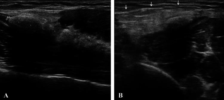
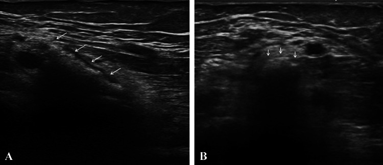
The shoulder is the joint most commonly affected by calcific tendinopathy, mostly the rotator cuff tendons [3, 10, 187], rarely the other tendons (pectoralis major, trapezius, biceps brachii) (Fig. 1). Pectoralis major calcific tendinopathy was described in four articles. Radiographs all showed a lithic area of the lateral humeral cortex with periosteal reaction. Second-level examinations (computed tomography and magnetic resonance imaging) and, in one case biopsy, were necessary for the aspecific findings of the radiographs. A single case of calcific tendinopathy is reported in the trapezius tendon. Biceps brachii calcific tendinopathy has been reported in both the proximal and the distal insertions (Fig. 2), except of the brief head. Calcific tendinopathy was also reported concomitant in the biceps tendon and in the rotator cuff tendon [98]. One of the five cases of calcific distal biceps tendinitis affected a 3-year-old boy [114]. Physical examination shows pain, tenderness, swelling and functional limitation, without a history of traumatic events. The differential diagnoses of extra-articular calcific tendinopathy are with calcific bursitis and loose bodies in the biceps tendon recess and synovial osteochondromatosis [98]. Radiography and ultrasound are sufficient for diagnosis.
Fig. 1.
Long head of the biceps tendon calcific tendinopathy at mid-arm level. a Long axis; b, c short axis. Ultrasonography shows calcifications (a calipers, b arrows) with absence of posterior acoustic shadowing (resorptive phase)
Fig. 2.
Distal biceps tendon calcific tendinopathy at the level of the elbow, proximal to the insertion on the radial tubercle. a Long axis, b short axis. Ultrasonography shows calcifications (arrows) with absence of posterior acoustic shadowing (resorptive phase)
Elbow and forearm
Two cases of calcific tendinopathy of the common extensor tendon of the elbow were described in two young women exhibiting pain, swelling and functional limitation. Radiograph and ultrasound showed soft-fluid calcification near the muscle insertion [90]. To our knowledge, the common flexor tendon has not been reported in the literature as affected.
Hand and wrist
Tendons of the hand and wrist [188, 189] are rarely reported as affected by calcific tendinopathy (incidence of 2%) [116], but more often the flexor tendons than the extensors (flexor carpi ulnaris four cases, flexor digitorum profundus two cases, flexor digitorum superficialis two cases, abductor pollicis brevis 2). Authors have described calcific tendinopathy in the flexor carpi radialis, abductor digiti minimi, extensor pollicis longus, abductor pollicis longus, flexor pollicis longus and the tendons of the intrinsic muscles. Two cases of calcific tendinopathy of the carpal tunnel tendons have been reported in literature, both with carpal tunnel syndrome.
The differential diagnosis includes soft-tissue infection, bone fracture, metabolic disorder (hyperparathyroidism, gout, pseudogout, hypervitaminosis D, hypercalcemia), degenerative or inflammatory or autoimmune conditions. Clinical presentation, ultrasound and radiographs can differentiate calcific tendinopathy from other etiologies [121].
Hip
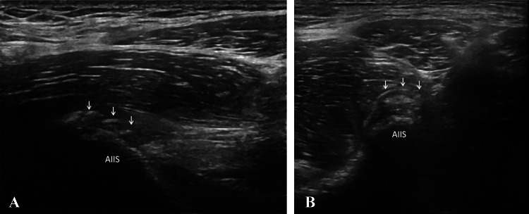
The hip [190] is the second most common site of calcific tendinopathy, after the shoulder [158]. The tendons of the rectus femoris are the most commonly involved (Fig. 3) described in 56 patients in 14 different articles. Both direct and indirect tendon components may be affected by this pathology, with prevalence, in our experience, of the direct tendon. But it is also necessary to distinguish the tendinous calcifications from calcifications of their insertional bursae, just beneath the direct and indirect tendon, that may occur quite frequently. The second most common group are the gluteal tendons (42 cases of the gluteus medius and 36 cases of the gluteus maximus reported). Adductor magnus, adductor longus and adductor brevis calcific tendinopathy have been described in six cases (1 magnus, 1 brevis and 4 unknown). Other rare sites are the piriformis (2 cases), the iliopsoas (2 cases), the ischiococcygeus (1 case) and the sartorius (1 case).
Fig. 3.
Direct tendon of rectus femoris calcific tendinopathy at the level of the anteroinferior iliac spine. a Long axis, b short axis. Ultrasonography shows calcifications (arrows) with absence of posterior acoustic shadowing (resorptive phase). AIIS anteroinferior iliac spine
Usually occurring in middle-age, patients have functional limitation, tenderness, pain and a positive Patrick’s test [183]. Depending on the affected tendon, the differential diagnosis includes infection [124, 180], arthritis, lumbar radiculopathy [126, 140, 168], os acetabuli, avulsion fracture, insertional calcified bursitis, sesamoid bones, myositis ossificans and chondrosarcoma [146]. Sonography and standard radiographs can be used for diagnosis, showing the calcification. Sometimes computed tomography is useful for bone evaluation, and MRIs to show soft-tissue edema and bone marrow edema.
Thigh, knee and leg
Thigh, knee and leg are rarely affected by calcific tendinopathy, and only a few cases involving the quadriceps tendon, patellar tendon (Fig. 4) and iliotibial band (Fig. 5) [191] are described. In many articles [125, 162] there is no distinction between calcific tendinopathy and calcifications of tendinous tendons, but the majority are calcifications in tendinous tendons. As usual, patients have functional limitations, tenderness and pain. The articles confirm the main role of ultrasound in the diagnosis and in the management of calcific tendinopathy, even of the less common ones.
Fig. 4.
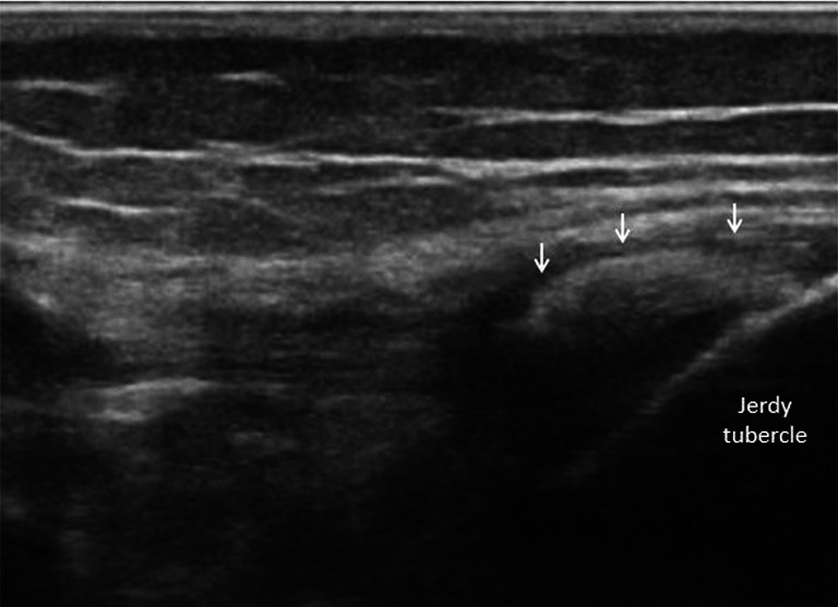
Iliotibial band calcific tendinopathy at the level of the Gerdy’s tubercle. Ultrasonography shows calcifications (arrows) with absence of posterior acoustic shadowing (resorptive phase)
Fig. 5.
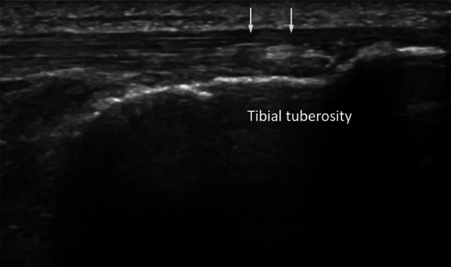
Patellar tendon calcific tendinopathy at the level of the tibial tuberosity. Ultrasonography shows calcifications (arrows) with absence of posterior acoustic shadowing (resorptive phase)
Foot and ankle
Calcific tendinopathy of the ankle [192] and foot is frequently misdiagnosed because of its rare occurrence and a clinical presentation that is similar to other entities. Achilles tendon calcific tendinopathy was described in eight articles, in both sexes, but more commonly in males. The second most commonly involved tendons are the peroneus longus (3 cases) and the flexor hallucis brevis (2 cases). Single cases have been described in other tendons (extensor hallucis longus, tibialis posterior and flexor of the forefoot).
Motion restriction secondary to pain, erythema, swelling and tenderness are the most frequent symptoms, in the absence of acute trauma [108]. The differential diagnosis is broad and includes gout or pseudogout, avulsion fractures, sesamoid bones, myositis ossificans and infection [156]. Ultrasonography and radiography can be used to make a diagnosis of calcific tendinopathy of the ankle and the foot, while computed tomography and MRI have few indications [136].
Ligaments
Calcifications of the ligaments, that can produce an important pain symptomatology like the calcific tendinopathy of the rotator cuff, are more frequent in the medial collateral ligament (proximal insertion) of the knee, where they can also become of considerable size [193, 194]. Other ligaments less frequently affected by the pathology are the lateral collateral ligament, the anterior or posterior cruciate ligament of the knee and sometimes Wrisberg ligament [195, 196].
Differential diagnosis
Depending on the affected tendon the differential diagnosis includes many diseases. Among the idiopathic ones the most known is the diffuse idiopathic skeletal hyperostosis (DISH) which predominantly affects the spine while ligaments and tendons of the appendicular skeleton are rarely involved [197]. In these cases, the US differential diagnosis is not possible and is generally related to the distribution and site of the calcifications.
The differential diagnosis with the calcification in a degenerative tendinopathy is more easy even with the ultrasound because the affected tendon appear as normal in calcific tendinopathy while shows diffuse signs of degeneration (e.g., hypoechogenicity, loss of the fibrillar aspect) around the calcification in a degenerative tendinopathy. In the most challenging cases, CT and MRI may be necessary.
Non-surgical treatment (or conservative, or minimally invasive treatments)
Calcific tendinopathy is usually a self-limited condition, so the initial management of pain is conservative, with physical therapy and oral administration of NSAIDs. If these treatments fail, other non-surgical therapeutic options may be considered: extra-corporeal shock wave therapy (ESWT), steroid injection (ultrasound-guided or unguided) and US-guided percutaneous aspiration of calcific tendinopathy (US-PICT). ESWT is based on the application of repetitive pulses over the affected site. The results are variable and the exact underlying mechanism of the therapeutic effect on calcific tendinopathy is still debated. It seems to be related to the phagocytosis of calcium deposition induced by the neovascularization response and leukocyte chemotaxis [198]; ESWT therapy is painful, expensive and not widely available. The use of conservative treatment or ESWT in patients with acute pain from calcific tendinopathy in resorption seems to be suboptimal, and often fails. The symptoms in this phase significantly impact quality of life [9, 199]. Minimally invasive interventional techniques (steroid injection of calcific tendinitis) may be used in these cases (US-guided or unguided) and/or US-guided percutaneous aspiration of calcific tendinopathy (US-PICT) [200]. A study by de Witta et al. reports that US-PICT is a superior method compared to steroid injection in the calcific tendinitis of the rotator cuff [201]. In cases of hard calcifications in mildly symptomatic patients, elective treatments should be considered [202]. Percutaneous treatment is not indicated when patients are asymptomatic, and calcification is very small (≤ 5 mm) [203]. Different approaches have been reported in recent studies and all include the use of a fluid: local anesthetic or saline solution to dissolve calcium deposits; one needle or two needles are used to inject and retrieve the fluid to dissolve calcium deposits. Recent evidence has suggested that a double-needle approach might be more appropriate for treating harder deposits, while one needle may be more useful in treating fluid calcifications. Some advantages of US-PICT are that the procedure does not require any hospitalization, is performed under local anesthesia, the patient can return home about 30 min after the procedure is complete, there is no need for post-procedural immobilization, and the patient can return to work sooner [10].
Surgical treatment
Arthroscopic treatment of calcific tendinitis involves selected cases in which conservative or less invasive approaches have failed. Calcification removal techniques vary according to the type of tendon incision and the instrumentation used to remove the calcium deposit. The surgery allows the removal of calcification and a thorough cleaning of the joint of interest. Surgery requires hospitalization, general anesthesia or sedation, however, and a relatively long rehabilitation period after treatment.
Conclusion
Calcific tendinopathy is commonly found in non-rotator cuff tendons. It is easily diagnosed using ultrasound, although ultrasound is rarely used in some anatomic sites, such as neck muscles. Depending on the affected tendon the differential diagnosis includes many diseases, and CT and MRI may be necessary. Usually occurring in middle-age, it can however affect patients of all ages, and a case was reported in a 3-year-old boy. Patients with calcific tendinopathy have functional limitation, tenderness and pain.
Resorption of deposits generally occurs spontaneously, although some patients show persistent clinical symptoms and require therapy. Various therapies can be used, although ultrasonographic guided therapeutic procedures (steroid injection or percutaneous aspiration) seem to be the most effective, particularly for calcific tendinopathy in resorption. Surgery remains an option in cases where other approaches have failed.
Compliance with ethical standards
Conflict of interest
C.B. is a consultant for Bracco Imaging and Doc. Congress; the other authors have nothing to disclose.
Ethical standards
Exams have been performed in accordance with the ethical standards laid down in the Helsinki Declaration of 1975 and its late amendments. Additional informed consent was obtained from all patients for whom identifying information was not included in this article.
Footnotes
Publisher's Note
Springer Nature remains neutral with regard to jurisdictional claims in published maps and institutional affiliations.
References
- 1.Hayes CW, Conway WF. Calcium hydroxyapatite deposition disease. RadioGraphics. 1990;10(6):1031–1048. doi: 10.1148/radiographics.10.6.2175444. [DOI] [PubMed] [Google Scholar]
- 2.Faure G, Daculsi G. Calcified tendinitis: a review. Ann Rheum Dis. 1983;42(Suppl 1):49–53. doi: 10.1136/ard.42.suppl_1.49. [DOI] [PMC free article] [PubMed] [Google Scholar]
- 3.Siegal DS, Wu JS, Newman JS, et al. Calcific tendinitis: a pictorial review. Can Assoc Radiol J J Assoc Can Radiol. 2009;60(5):263–272. doi: 10.1016/j.carj.2009.06.008. [DOI] [PubMed] [Google Scholar]
- 4.Uhthoff HK, Sarkar K. Calcifying tendinitis. Baillieres Clin Rheumatol. 1989;3(3):567–581. doi: 10.1016/s0950-3579(89)80009-3. [DOI] [PubMed] [Google Scholar]
- 5.Messina C, Banfi G, Orlandi D, et al. Ultrasound-guided interventional procedures around the shoulder. Br J Radiol. 2016;89(1057):20150372. doi: 10.1259/bjr.20150372. [DOI] [PMC free article] [PubMed] [Google Scholar]
- 6.Lanza E, Banfi G, Serafini G, et al. Ultrasound-guided percutaneous irrigation in rotator cuff calcific tendinopathy: what is the evidence? A systematic review with proposals for future reporting. Eur Radiol. 2015;25(7):2176–2183. doi: 10.1007/s00330-014-3567-1. [DOI] [PubMed] [Google Scholar]
- 7.Tagliafico A, Russo G, Boccalini S, et al. Ultrasound-guided interventional procedures around the shoulder. Radiol Med (Torino) 2014;119(5):318–326. doi: 10.1007/s11547-013-0351-2. [DOI] [PubMed] [Google Scholar]
- 8.Draghi F, Scudeller L, Draghi AG, et al. Prevalence of subacromial-subdeltoid bursitis in shoulder pain: an ultrasonographic study. J Ultrasound. 2015;18(2):151–158. doi: 10.1007/s40477-015-0167-0. [DOI] [PMC free article] [PubMed] [Google Scholar]
- 9.Robotti G, Canepa MG, Bortolotto C, et al. Interventional musculoskeletal US: an update on materials and methods. J Ultrasound. 2013;16(2):45–55. doi: 10.1007/s40477-013-0018-9. [DOI] [PMC free article] [PubMed] [Google Scholar]
- 10.Draghi F, Robotti G, Jacob D, et al. Interventional musculoskeletal ultrasonography: precautions and contraindications. J Ultrasound. 2010;13(3):126–133. doi: 10.1016/j.jus.2010.09.004. [DOI] [PMC free article] [PubMed] [Google Scholar]
- 11.Abdelbaki A, Abdelbaki S, Bhatt N, et al. Acute calcific tendinitis of the longus colli muscle: report of two cases and review of the literature. Cureus. 2017;9(8):e1597. doi: 10.7759/cureus.1597. [DOI] [PMC free article] [PubMed] [Google Scholar]
- 12.Ahmed OH, German MA, Handwerker J, et al. Radiology quiz case 2. Acute calcific tendinitis of the longus colli (also known as calcific retropharyngeal/prevertebral tendinitis) Arch Otolaryngol Head Neck Surg. 2012;138(6):599–600. [PubMed] [Google Scholar]
- 13.Alamoudi U, Al-Sayed AA, AlSallumi Y, et al. Acute calcific tendinitis of the longus colli muscle masquerading as a retropharyngeal abscess: a case report and review of the literature. Int J Surg Case Rep. 2017;41:343–346. doi: 10.1016/j.ijscr.2017.10.063. [DOI] [PMC free article] [PubMed] [Google Scholar]
- 14.Andrade CS, Peixoto CAR, Cevasco FKI, et al. Longus colli calcific acute tendinitis: typical features with distinct imaging modalities. Arq Neuropsiquiatr. 2015;73(1):69–70. doi: 10.1590/0004-282X20140186. [DOI] [PubMed] [Google Scholar]
- 15.Bailey CW, Connolly BD, Sterner K, et al. Acute calcific longus colli tendonitis. W V Med J. 2015;111(2):10–12. [PubMed] [Google Scholar]
- 16.Benanti JC, Gramling P, Bulat PI, et al. Retropharyngeal calcific tendinitis: report of five cases and review of the literature. J Emerg Med. 1986;4(1):15–24. doi: 10.1016/0736-4679(86)90108-3. [DOI] [PubMed] [Google Scholar]
- 17.Bladt O, Vanhoenacker R, Bevernage C, et al. Acute calcific prevertebral tendinitis. JBR-BTR Organe Soc R Belge Radiol SRBR Orgaan Van K Belg Ver Voor Radiol KBVR. 2008;91(4):158–159. [PubMed] [Google Scholar]
- 18.Blome SA. Retropharyngeal calcific tendinitis. Australas Radiol. 1987;31(2):142–143. doi: 10.1111/j.1440-1673.1987.tb01800.x. [DOI] [PubMed] [Google Scholar]
- 19.Boikov AS, Griffith B, Stemer M, et al. Acute calcific longus colli tendinitis: an unusual location and presentation. Arch Otolaryngol Head Neck Surg. 2012;138(7):676–679. doi: 10.1001/archoto.2012.910. [DOI] [PubMed] [Google Scholar]
- 20.Borrmann A, Niedermeyer HP, Arnold W. Retropharyngeal tendinitis–a rare differential diagnosis of retropharyngeal abscess. Laryngorhinootologie. 2008;87(3):186–189. doi: 10.1055/s-2007-966887. [DOI] [PubMed] [Google Scholar]
- 21.Chen C-H, Lu Y-C, Wong T-Y. Acute calcific prevertebral tendinitis: rare cause of neck pain. Acute Med Surg. 2015;2(3):199–201. doi: 10.1002/ams2.92. [DOI] [PMC free article] [PubMed] [Google Scholar]
- 22.Chung T, Rebello R, Gooden EA. Retropharyngeal calcific tendinitis: case report and review of literature. Emerg Radiol. 2005;11(6):375–380. doi: 10.1007/s10140-005-0427-y. [DOI] [PubMed] [Google Scholar]
- 23.Colella DM, Calderón Sandoval F, et al. A rare cause of dysphagia to remember: calcific tendinitis of the longus colli muscle. Case Rep Gastroenterol. 2016;10(3):755–759. doi: 10.1159/000452199. [DOI] [PMC free article] [PubMed] [Google Scholar]
- 24.Coulier B, Macsim M, Desgain O. Retropharyngeal calcific tendinitis–longus colli tendinitis–an unusual cause of acute dysphagia. Emerg Radiol. 2011;18(5):449–451. doi: 10.1007/s10140-011-0959-2. [DOI] [PubMed] [Google Scholar]
- 25.De Maeseneer M, Vreugde S, Laureys S, et al. Calcific tendinitis of the longus colli muscle. Head Neck. 1997;19(6):545–548. doi: 10.1002/(sici)1097-0347(199709)19:6<545::aid-hed13>3.0.co;2-4. [DOI] [PubMed] [Google Scholar]
- 26.De Temmerman G, Marcelis S, Beeckman P. Acute calcific retropharyngeal tendonitis. JBR-BTR Organe Soc R Belge Radiol SRBR Orgaan Van K Belg Ver Voor Radiol KBVR. 2007;90(3):184–185. [PubMed] [Google Scholar]
- 27.Desmots F, Derkenne R, Couppey N. Calcific retropharyngeal tendinitis of the longus colli muscle Answer to the e-quid. ‘Acute cervical pain and dysphagia in a 43 year-old man’. Diagn Interv Imaging. 2013;94(4):470. doi: 10.1016/j.diii.2012.09.006. [DOI] [PubMed] [Google Scholar]
- 28.Eastwood JD, Hudgins PA, Malone D. Retropharyngeal effusion in acute calcific prevertebral tendinitis: diagnosis with CT and MR imaging. AJNR Am J Neuroradiol. 1998;19(9):1789–1792. [PMC free article] [PubMed] [Google Scholar]
- 29.Ellika SK, Payne SC, Patel SC, et al. Acute calcific tendinitis of the longus colli: an imaging diagnosis. Dento Maxillo Facial Radiol. 2008;37(2):121–124. doi: 10.1259/dmfr/23211511. [DOI] [PubMed] [Google Scholar]
- 30.Estimable K, Rizk C, Pujalte GGA. A rare case of neck pain: acute longus colli calcific tendinitis in a possibly immunocompromised individual. J Am Board Fam Med JABFM. 2015;28(1):146–150. doi: 10.3122/jabfm.2015.01.140124. [DOI] [PubMed] [Google Scholar]
- 31.Fahlgren H. Retropharyngeal tendinitis. Cephalalgia Int J Headache. 1986;6(3):169–174. doi: 10.1046/j.1468-2982.1986.0603169.x. [DOI] [PubMed] [Google Scholar]
- 32.Figler T. Radiologic case study Calcific tendonitis of the longissimus colli muscle. Orthopedics. 1993;16(12):1365. doi: 10.3928/0147-7447-19931201-15. [DOI] [PubMed] [Google Scholar]
- 33.Gabra N, Belair M, Ayad T. Retropharyngeal calcific tendinitis mimicking a retropharyngeal phlegmon. Case Rep Otolaryngol. 2013;2013:912628. doi: 10.1155/2013/912628. [DOI] [PMC free article] [PubMed] [Google Scholar]
- 34.Hall FM, Docken WP, Curtis HW. Calcific tendinitis of the longus coli: diagnosis by CT. AJR Am J Roentgenol. 1986;147(4):742–743. doi: 10.2214/ajr.147.4.742. [DOI] [PubMed] [Google Scholar]
- 35.Haun CL. Retropharyngeal tendinitis. AJR Am J Roentgenol. 1978;130(6):1137–1140. doi: 10.2214/ajr.130.6.1137. [DOI] [PubMed] [Google Scholar]
- 36.Horowitz G, Ben-Ari O, Brenner A, et al. Incidence of retropharyngeal calcific tendinitis (longus colli tendinitis) in the general population. Otolaryngol-Head Neck Surg Off J Am Acad Otolaryngol-Head Neck Surg. 2013;148(6):955–958. doi: 10.1177/0194599813482289. [DOI] [PubMed] [Google Scholar]
- 37.Jiménez S, Millán JM. Calcific retropharyngeal tendinitis: a frequently missed diagnosis. Case report. J Neurosurg Spine. 2007;6(1):77–80. doi: 10.3171/spi.2007.6.1.77. [DOI] [PubMed] [Google Scholar]
- 38.Joshi GS, Fomin DA, Joshi GS, et al. Unusual case of acute neck pain: acute calcific longus colli tendinitis. BMJ Case Rep. 2016;2:2016. doi: 10.1136/bcr-2016-216041. [DOI] [PMC free article] [PubMed] [Google Scholar]
- 39.Kanzaria H, Stein JC. A severe sore throat in a middle-aged man: calcific tendonitis of the longus colli tendon. J Emerg Med. 2011;41(2):151–153. doi: 10.1016/j.jemermed.2008.01.016. [DOI] [PubMed] [Google Scholar]
- 40.Kaplan MJ, Eavey RD. Calcific tendinitis of the longus colli muscle. Ann Otol Rhinol Laryngol. 1984;93(3 Pt 1):215–219. doi: 10.1177/000348948409300305. [DOI] [PubMed] [Google Scholar]
- 41.Karasick D, Karasick S. Calcific retropharyngeal tendinitis. Skeletal Radiol. 1981;7(3):203–205. doi: 10.1007/BF00361865. [DOI] [PubMed] [Google Scholar]
- 42.Kenzaka T, Kumabe A. Acute calcific prevertebral tendinitis. Intern Med Tokyo Jpn. 2017;56(12):1611. doi: 10.2169/internalmedicine.56.8266. [DOI] [PMC free article] [PubMed] [Google Scholar]
- 43.Khurana B. Calcific tendinitis mimicking acute prevertebral abscess. J Emerg Med. 2012;42(1):e15–e16. doi: 10.1016/j.jemermed.2009.08.046. [DOI] [PubMed] [Google Scholar]
- 44.Kim Y-J, Park J-Y, Choi K-Y, et al. Case reports about an overlooked cause of neck pain: calcific tendinitis of the longus colli: case reports. Medicine (Baltimore) 2017;96(46):e8343. doi: 10.1097/MD.0000000000008343. [DOI] [PMC free article] [PubMed] [Google Scholar]
- 45.Kupferman TA, Rice CH, Gage-White L. Acute prevertebral calcific tendinitis: a nonsurgical cause of prevertebral fluid collection. Ear Nose Throat J. 2007;86(3):164–166. [PubMed] [Google Scholar]
- 46.Kusunoki T, Muramoto D, Murata K. A case of calcific retropharyngeal tendinitis suspected to be a retropharyngeal abscess upon the first medical examination. Auris Nasus Larynx. 2006;33(3):329–331. doi: 10.1016/j.anl.2005.11.014. [DOI] [PubMed] [Google Scholar]
- 47.Lee S, Joo KB, Lee KH, et al. Acute retropharyngeal calcific tendinitis in an unusual location: a case report in a patient with rheumatoid arthritis and atlantoaxial subluxation. Korean J Radiol. 2011;12(4):504–509. doi: 10.3348/kjr.2011.12.4.504. [DOI] [PMC free article] [PubMed] [Google Scholar]
- 48.Leep Hunderfund AN, Robertson CE, Bell ML, et al. Calcific retropharyngeal tendinitis: unusual cause of acute neck pain with nuchal rigidity. Neurology. 2008;71(10):778. doi: 10.1212/01.wnl.0000324917.44950.8b. [DOI] [PubMed] [Google Scholar]
- 49.Mannoji C, Koda M, Furuya T, et al. Calcific tendinitis of the longus colli. Intern Med. 2015;54(12):1573. doi: 10.2169/internalmedicine.54.4342. [DOI] [PubMed] [Google Scholar]
- 50.Martindale JL, Senecal EL. Atraumatic neck pain and rigidity: a case of calcific retropharyngeal tendonitis. Am J Emerg Med. 2012;30(4):636.e1–636.e2. doi: 10.1016/j.ajem.2011.02.004. [DOI] [PubMed] [Google Scholar]
- 51.Mihmanli I, Karaarslan E, Kanberoglu K. Inflammation of vertebral bone associated with acute calcific tendinitis of the longus colli muscle. Neuroradiology. 2001;43(12):1098–1101. doi: 10.1007/s002340100644. [DOI] [PubMed] [Google Scholar]
- 52.Naqshabandi AM, Srinivasan J. Teaching neuroimages: acute calcific tendinitis of longus colli mimicking meningismus. Neurology. 2011;76(17):e81. doi: 10.1212/WNL.0b013e318217e793. [DOI] [PubMed] [Google Scholar]
- 53.Newmark H, Forrester DM, Brown JC, et al. Calcific tendinitis of the neck. Radiology. 1978;128(2):355–358. doi: 10.1148/128.2.355. [DOI] [PubMed] [Google Scholar]
- 54.Newmark H, Zee CS, Frankel P, et al. Chronic calcific tendinitis of the neck. Skeletal Radiol. 1981;7(3):207–208. doi: 10.1007/BF00361866. [DOI] [PubMed] [Google Scholar]
- 55.Newmark H, Blackford D, Roberts D, et al. Computed tomography of acute cervical spine tendinitis. J Comput Tomogr. 1986;10(4):373–375. doi: 10.1016/0149-936x(86)90035-4. [DOI] [PubMed] [Google Scholar]
- 56.Nozu T, Kumei S, Ohhira M, et al. Retropharyngeal calcific tendinitis. Intern Med Tokyo Jpn. 2015;54(17):2277. doi: 10.2169/internalmedicine.54.4682. [DOI] [PubMed] [Google Scholar]
- 57.Nunes C, Casimiro C, Carreiro I. Imagiology of the acute calcific tendinitis of the longus colli. Acta Med Port. 2012;25(Suppl 1):51–55. [PubMed] [Google Scholar]
- 58.Offiah CE, Hall E. Acute calcific tendinitis of the longus colli muscle: spectrum of CT appearances and anatomical correlation. Br J Radiol. 2009;82(978):e117–e121. doi: 10.1259/bjr/19797697. [DOI] [PubMed] [Google Scholar]
- 59.Oh JY, Lim JH, Kim YS, et al. Misconceived retropharyngeal calcific tendinitis during management of myofascial neck pain syndrome. Korean J Pain. 2016;29(1):48–52. doi: 10.3344/kjp.2016.29.1.48. [DOI] [PMC free article] [PubMed] [Google Scholar]
- 60.Omezzine SJ, Hafsa C, Lahmar I, et al. Calcific tendinitis of the longus colli: diagnosis by CT. Jt Bone Spine Rev Rhum. 2008;75(1):90–91. doi: 10.1016/j.jbspin.2007.03.002. [DOI] [PubMed] [Google Scholar]
- 61.Park R, Halpert DE, Baer A, et al. Retropharyngeal calcific tendinitis: case report and review of the literature. Semin Arthritis Rheum. 2010;39(6):504–509. doi: 10.1016/j.semarthrit.2009.04.002. [DOI] [PubMed] [Google Scholar]
- 62.Park SY, Jin W, Lee SH, et al. Acute retropharyngeal calcific tendinitis: a case report with unusual location of calcification. Skeletal Radiol. 2010;39(8):817–820. doi: 10.1007/s00256-010-0879-3. [DOI] [PubMed] [Google Scholar]
- 63.Pellicer García V, Pérez Moya C, Magán Martín A. Acute calcific prevertebral tendinitis: case report and literature review. Rev Espanola Cirugia Ortop Traumatol. 2012;56(5):389–392. doi: 10.1016/j.recot.2012.05.009. [DOI] [PubMed] [Google Scholar]
- 64.Queinnec S, Petrover D, Guigui P, et al. Benign febrile cervicalgia due to calcific retropharyngeal tendinitis: case study. Orthop Traumatol Surg Res OTSR. 2011;97(3):341–344. doi: 10.1016/j.otsr.2010.09.021. [DOI] [PubMed] [Google Scholar]
- 65.Razon RVB, Nasir A, Wu GS, et al. Retropharyngeal calcific tendonitis: report of two cases. J Am Board Fam Med JABFM. 2009;22(1):84–88. doi: 10.3122/jabfm.2009.01.080034. [DOI] [PubMed] [Google Scholar]
- 66.Sanghvi DA, Jankharia BG, Purandare NC, et al. Radiologic case study Acute calcific retropharyngeal tendinitis. Orthopedics. 2006;29(7):561. doi: 10.3928/01477447-20060701-17. [DOI] [PubMed] [Google Scholar]
- 67.Sarkozi J, Fam AG. Acute calcific retropharyngeal tendinitis: an unusual cause of neck pain. Arthritis Rheum. 1984;27(6):708–710. doi: 10.1002/art.1780270618. [DOI] [PubMed] [Google Scholar]
- 68.Shibuki T, Goto H, Fukushima K, et al. Acute prevertebral calcific tendinitis. Intern Med Tokyo Jpn. 2017;56(10):1275. doi: 10.2169/internalmedicine.56.8197. [DOI] [PMC free article] [PubMed] [Google Scholar]
- 69.Shin D-E, Ahn C-S, Choi J-P. The acute calcific prevertebral tendinitis: report of two cases. Asian Spine J. 2010;4(2):123–127. doi: 10.4184/asj.2010.4.2.123. [DOI] [PMC free article] [PubMed] [Google Scholar]
- 70.Silva CF, Soffia PS, Pruzzo E. Acute prevertebral calcific tendinitis: a source of non-surgical acute cervical pain. Acta Radiol Stockh Swed. 2014;55(1):91. doi: 10.1177/0284185113492151. [DOI] [PubMed] [Google Scholar]
- 71.Siwiec RM, Kushner DJ, Morrison JL. Retropharyngeal calcific tendinitis. J Rheumatol. 2009;36(7):1546–1547. doi: 10.3899/jrheum.081161. [DOI] [PubMed] [Google Scholar]
- 72.Sokolov M, Yaffe D, Ophir D. Retropharyngeal calcific tendinitis. Isr Med Assoc J IMAJ. 2009;11(11):701–702. [PubMed] [Google Scholar]
- 73.Sierra Solís A, Fernández Prudencio L. Acute calcific tendinitis of the longus colli muscle. Acta Otorrinolaringol Esp. 2017;68(4):244–245. doi: 10.1016/j.otorri.2016.04.001. [DOI] [PubMed] [Google Scholar]
- 74.Southwell K, Hornibrook J, O’Neill-Kerr D. Acute longus colli calcific tendonitis causing neck pain and dysphagia. Otolaryngol-Head Neck Surg Off J Am Acad Otolaryngol-Head Neck Surg. 2008;138(3):405–406. doi: 10.1016/j.otohns.2007.12.004. [DOI] [PubMed] [Google Scholar]
- 75.Suyama Y, Kishimoto M, Nozaki T, et al. Acute calcific tendinitis of the longus colli muscle. Arthritis Rheumatol Hoboken NJ. 2015;67(9):2446. doi: 10.1002/art.39184. [DOI] [PubMed] [Google Scholar]
- 76.Szelei N, Tassart M, Le Breton C, et al. Calcific retropharyngeal tendinitis: unusual diagnosis. J Radiol. 2001;82(9 Pt 1):1001–1004. [PubMed] [Google Scholar]
- 77.Tagashira Y, Watanuki S. Acute calcific retropharyngeal tendonitis. CMAJ Can Med Assoc J J Assoc Medicale Can. 2015;187(13):995. doi: 10.1503/cmaj.140214. [DOI] [PMC free article] [PubMed] [Google Scholar]
- 78.Tamm A, Jeffery CC, Ansari K, et al. Acute prevertebral calcific tendinitis. J Radiol Case Rep. 2015;9(11):1–5. doi: 10.3941/jrcr.v9i11.2494. [DOI] [PMC free article] [PubMed] [Google Scholar]
- 79.Tezuka F, Sakai T, Miyagi R, et al. Complete resolution of a case of calcific tendinitis of the longus colli with conservative treatment. Asian Spine J. 2014;8(5):675–679. doi: 10.4184/asj.2014.8.5.675. [DOI] [PMC free article] [PubMed] [Google Scholar]
- 80.Torbati SS, Vos EM, Bral D, et al. Calcific tendinitis of the longus colli muscle. Ear Nose Throat J. 2014;93(12):492–493. [PubMed] [Google Scholar]
- 81.Uchiyama D, Nakamura K, Yoshida S, et al. Calcific tendinitis of the longus colli. Intern Med Tokyo Jpn. 2016;55(5):553. doi: 10.2169/internalmedicine.55.5846. [DOI] [PubMed] [Google Scholar]
- 82.Ulusoy OL, Tutar S, Ozturk E, et al. Calcific tendinitis of the longus colli muscle: a rare cause of neck pain. Neurol India. 2016;64(5):1085–1086. doi: 10.4103/0028-3886.190267. [DOI] [PubMed] [Google Scholar]
- 83.Van Kerkhove F, Geusens E, Knockaert D. Retropharyngeal calcific tendonitis. Eur J Emerg Med Off J Eur Soc Emerg Med. 2007;14(5):269–271. doi: 10.1097/MEJ.0b013e3280c60cac. [DOI] [PubMed] [Google Scholar]
- 84.Wakabayashi Y, Hori Y, Kondoh Y, et al. Acute calcific prevertebral tendonitis mimicking tension-type headache. Neurol Med Chir (Tokyo) 2012;52(9):631–633. doi: 10.2176/nmc.52.631. [DOI] [PubMed] [Google Scholar]
- 85.Widlus DM. Calcific tendonitis of the longus colli muscle: a cause of atraumatic neck pain. Ann Emerg Med. 1985;14(10):1014–1017. doi: 10.1016/s0196-0644(85)80253-5. [DOI] [PubMed] [Google Scholar]
- 86.Wolzak H, van de Rest M, Geurts M, et al. Acute calcific tendinitis of the longus colli muscle: an often unrecognized cause of severe neck pain. J Clin Rheumatol Pract Rep Rheum Musculoskelet Dis. 2010;16(5):240–241. doi: 10.1097/RHU.0b013e3181e968b6. [DOI] [PubMed] [Google Scholar]
- 87.Yaylacı S, Öztürk TC, Aksoy E, et al. Retropharyngeal calcific tendinitis: report of two cases. J Emerg Trauma Shock. 2015;8(2):119–120. doi: 10.4103/0974-2700.145408. [DOI] [PMC free article] [PubMed] [Google Scholar]
- 88.Zapolsky N, Heller M, Felberbaum M, et al. Calcific tendonitis of the longus colli: an uncommon but benign cause of throat pain that closely mimics retropharyngeal abscess. J Emerg Med. 2017;52(3):358–360. doi: 10.1016/j.jemermed.2016.08.016. [DOI] [PubMed] [Google Scholar]
- 89.Zibis AH, Giannis D, Malizos KN, et al. Acute calcific tendinitis of the longus colli muscle: case report and review of the literature. Eur Spine J Off Publ Eur Spine Soc Eur Spinal Deform Soc Eur Sect Cerv Spine Res Soc. 2013;22(Suppl 3):S434–S438. doi: 10.1007/s00586-012-2584-5. [DOI] [PMC free article] [PubMed] [Google Scholar]
- 90.Abate M, Salini V, Schiavone C. Ultrasound-guided percutaneous lavage in the treatment of calcific tendinopathy of elbow extensor tendons: a case report. Malays Orthop J. 2016;10(2):53–55. doi: 10.5704/MOJ.1607.011. [DOI] [PMC free article] [PubMed] [Google Scholar]
- 91.Ali SN, Kelly JL. Acute calcific tendinitis of the finger–a case report. Hand Surg Int J Devoted Hand Up Limb Surg Relat Res J Asia-Pac Fed Soc Surg Hand. 2004;9(1):105–107. doi: 10.1142/s0218810404001954. [DOI] [PubMed] [Google Scholar]
- 92.Cahir J, Saifuddin A. Calcific tendonitis of pectoralis major: CT and MRI findings. Skeletal Radiol. 2005;34(4):234–238. doi: 10.1007/s00256-004-0842-2. [DOI] [PubMed] [Google Scholar]
- 93.Dilley DF, Tonkin MA. Acute calcific tendinitis in the hand and wrist. J Hand Surg Edinb Scotl. 1991;16(2):215–216. doi: 10.1016/0266-7681(91)90181-m. [DOI] [PubMed] [Google Scholar]
- 94.Dürr HR, Lienemann A, Silbernagl H, et al. Acute calcific tendinitis of the pectoralis major insertion associated with cortical bone erosion. Eur Radiol. 1997;7(8):1215–1217. doi: 10.1007/s003300050277. [DOI] [PubMed] [Google Scholar]
- 95.El-Essawy MT, Vanhoenacker FM. Calcific tendinopathy of the pectoralis major insertion with intracortical protrusion of calcification. JBR-BTR Organe Soc R Belge Radiol SRBR Orgaan Van K Belg Ver Voor Radiol KBVR. 2012;95(6):374. doi: 10.5334/jbr-btr.733. [DOI] [PubMed] [Google Scholar]
- 96.Galliani I, Columbaro M, Ferri S, et al. A case of calcific lateral epicondylitis: a histological and ultrastructural study. Br J Rheumatol. 1998;37(2):235–236. doi: 10.1093/rheumatology/37.2.235. [DOI] [PubMed] [Google Scholar]
- 97.Garayoa SA, Romero-Muñoz LM, Pons-Villanueva J. Acute compartment syndrome of the forearm caused by calcific tendinitis of the distal biceps. Musculoskelet Surg. 2010;94(3):137–139. doi: 10.1007/s12306-010-0079-2. [DOI] [PubMed] [Google Scholar]
- 98.Goldman AB. Calcific tendinitis of the long head of the biceps brachii distal to the glenohumeral joint: plain film radiographic findings. AJR Am J Roentgenol. 1989;153(5):1011–1016. doi: 10.2214/ajr.153.5.1011. [DOI] [PubMed] [Google Scholar]
- 99.Gossner J. Large bone erosion due to calcific tendinitis of the distal biceps tendon. Jt Bone Spine Rev Rhum. 2018;85(2):247. doi: 10.1016/j.jbspin.2017.03.008. [DOI] [PubMed] [Google Scholar]
- 100.Greene TL, Louis DS. Calcifying tendinitis in the hand. Ann Emerg Med. 1980;9(8):438–440. doi: 10.1016/s0196-0644(80)80160-0. [DOI] [PubMed] [Google Scholar]
- 101.Hakozaki M, Iwabuchi M, Konno S, et al. Acute calcific tendinitis of the thumb in a child: a case report. Clin Rheumatol. 2007;26(5):841–844. doi: 10.1007/s10067-006-0384-1. [DOI] [PubMed] [Google Scholar]
- 102.Harris AR, McNamara TR, Brault JS, et al. An unusual presentation of acute calcific tendinitis in the hand. Hand N Y N. 2009;4(1):81–83. doi: 10.1007/s11552-008-9121-3. [DOI] [PMC free article] [PubMed] [Google Scholar]
- 103.Huntley JS. Dougall TW Mistaken identity: calcific tendinitis in the finger. Hosp Med Lond Engl. 2003;64(12):747. doi: 10.12968/hosp.2003.64.12.2370. [DOI] [PubMed] [Google Scholar]
- 104.Ikegawa S. Calcific tendinitis of the pectoralis major insertion. A report of two cases. Arch Orthop Trauma Surg. 1996;115(2):118. doi: 10.1007/BF00573455. [DOI] [PubMed] [Google Scholar]
- 105.Kheterpal A, Zoga A, McClure K. Acute calcific tendinitis of the flexor pollicis longus in an 8-year-old boy. Skeletal Radiol. 2014;43(10):1471–1475. doi: 10.1007/s00256-014-1908-4. [DOI] [PubMed] [Google Scholar]
- 106.Kim JH, Lee JH, Park JU, et al. Acute calcific tendinitis in the distal interphalangeal joint. Arch Plast Surg. 2016;43(3):301–303. doi: 10.5999/aps.2016.43.3.301. [DOI] [PMC free article] [PubMed] [Google Scholar]
- 107.Kim KC, Rhee KJ, Shin HD, et al. A SLAP lesion associated with calcific tendinitis of the long head of the biceps brachii at its origin. Knee Surg Sports Traumatol Arthrosc Off J ESSKA. 2007;15(12):1478–1481. doi: 10.1007/s00167-007-0323-y. [DOI] [PubMed] [Google Scholar]
- 108.Lee HO, Lee YH, Mun SH, et al. Calcific tendinitis of the hand and foot: a report of four cases. J Korean Soc Magn Reson Med. 2012;16(2):177. [Google Scholar]
- 109.Munjal A, Munjal P, Mahajan A. Diagnostic dilemma: acute calcific tendinitis of flexor digitorum profundus. Hand N Y N. 2013;8(3):352–353. doi: 10.1007/s11552-013-9514-9. [DOI] [PMC free article] [PubMed] [Google Scholar]
- 110.Murase T, Tsuyuguchi Y, Hidaka N, et al. Calcific tendinitis at the biceps insertion causing rotatory limitation of the forearm: a case report. J Hand Surg. 1994;19(2):266–268. doi: 10.1016/0363-5023(94)90017-5. [DOI] [PubMed] [Google Scholar]
- 111.Nofsinger CC, Williams GR, Iannotti JP. Calcific tendinitis of the trapezius insertion. J Shoulder Elbow Surg. 1999;8(2):162–164. doi: 10.1016/s1058-2746(99)90011-3. [DOI] [PubMed] [Google Scholar]
- 112.Park J-Y, Gupta A, Park H-K. Calcific tendinitis at the radial insertion of the biceps brachii: a case report. J Shoulder Elbow Surg. 2008;17(6):e19–e21. doi: 10.1016/j.jse.2008.02.023. [DOI] [PubMed] [Google Scholar]
- 113.Ryan WG. Calcific tendinitis of flexor carpi ulnaris: an easy misdiagnosis. Arch Emerg Med. 1993;10(4):321–323. doi: 10.1136/emj.10.4.321. [DOI] [PMC free article] [PubMed] [Google Scholar]
- 114.Sakamoto K, Kozuki K. Calcific tendinitis at the biceps brachii insertion of a child: a case report. J Shoulder Elbow Surg. 2002;11(1):88–91. doi: 10.1067/mse.2002.119854. [DOI] [PubMed] [Google Scholar]
- 115.Saleh WR, Yajima H, Nakanishi A. Acute carpal tunnel syndrome secondary to calcific tendinitis: case report. Hand Surg Int J Devoted Hand Up Limb Surg Relat Res J Asia-Pac Fed Soc Surg Hand. 2008;13(3):197–200. doi: 10.1142/S0218810408004080. [DOI] [PubMed] [Google Scholar]
- 116.Schneider D, Hirsch M. Acute calcific tendonitis of dorsal interosseous muscles of the hand: uncommon site of a frequent disease. Reumatismo. 2017;69(1):43–46. doi: 10.4081/reumatismo.2017.950. [DOI] [PubMed] [Google Scholar]
- 117.Seiler JG, Kerwin GA. Adolescent trigger finger secondary to post-traumatic chronic calcific tendinitis. J Hand Surg. 1995;20(3):425–427. doi: 10.1016/S0363-5023(05)80100-5. [DOI] [PubMed] [Google Scholar]
- 118.Selby CL. Acute calcific tendinitis of the hand: an infrequently recognized and frequently misdiagnosed form of periarthritis. Arthritis Rheum. 1984;27(3):337–340. doi: 10.1002/art.1780270314. [DOI] [PubMed] [Google Scholar]
- 119.Shields JS, Chhabra AB, Pannunzio ME. Acute calcific tendinitis of the hand: 2 case reports involving the abductor pollicis brevis. Am J Orthop Belle Mead NJ. 2007;36(11):605–607. [PubMed] [Google Scholar]
- 120.Torbati SS, Bral D, Geiderman JM. Acute calcific tendinitis of the wrist. J Emerg Med. 2013;44(2):352–354. doi: 10.1016/j.jemermed.2012.08.028. [DOI] [PubMed] [Google Scholar]
- 121.Walocko FM, Sando IC, Haase SC, et al. Acute calcific tendinitis of the index finger in a child. Hand N Y N. 2017;12(5):NP84. doi: 10.1177/1558944716675146. [DOI] [PMC free article] [PubMed] [Google Scholar]
- 122.Yasen S. Acute calcific tendinitis of the flexor carpi ulnaris causing acute compressive neuropathy of the ulnar nerve: a case report. J Orthop Surg Hong Kong. 2012;20(3):414–416. doi: 10.1177/230949901202000333. [DOI] [PubMed] [Google Scholar]
- 123.Abram SGF, Sharma AD, Arvind C. Atraumatic quadriceps tendon tear associated with calcific tendonitis. BMJ Case Rep. 2012;27:2012. doi: 10.1136/bcr-2012-007031. [DOI] [PMC free article] [PubMed] [Google Scholar]
- 124.Almedghio S, Garneti N. The acute and chronic presentation of gluteus medius calcific tendinitis—a case report of two. J Orthop Case Rep. 2014;4(4):48–50. doi: 10.13107/jocr.2250-0685.225. [DOI] [PMC free article] [PubMed] [Google Scholar]
- 125.Beebe JA, Cross PS. Patellar tendinopathy: preliminary surgical results. Sports Health. 2013;5(3):220–224. doi: 10.1177/1941738113478768. [DOI] [PMC free article] [PubMed] [Google Scholar]
- 126.Berney JW. Calcifying peritendinitis of the gluteus maximus tendon. Radiology. 1972;102(3):517–518. doi: 10.1148/102.3.517. [DOI] [PubMed] [Google Scholar]
- 127.Braun-Moscovici Y, Schapira D, Nahir AM. Calcific tendinitis of the rectus femoris. J Clin Rheumatol Pract Rep Rheum Musculoskelet Dis. 2006;12(6):298–300. doi: 10.1097/01.rhu.0000249896.43792.62. [DOI] [PubMed] [Google Scholar]
- 128.Choudur HN, Munk PL. Image-guided corticosteroid injection of calcific tendonitis of gluteus maximus. J Clin Rheumatol Pract Rep Rheum Musculoskelet Dis. 2006;12(4):176–178. doi: 10.1097/01.rhu.0000230457.51606.62. [DOI] [PubMed] [Google Scholar]
- 129.Cox D, Paterson FW. Acute calcific tendinitis of peroneus longus. J Bone Jt Surg Br. 1991;73(2):342. doi: 10.1302/0301-620X.73B2.2005172. [DOI] [PubMed] [Google Scholar]
- 130.Gillis CT, Lin JS. Use of a central splitting approach and near complete detachment for insertional calcific achilles tendinopathy repaired with an achilles bridging suture. J Foot Ankle Surg Off Publ Am Coll Foot Ankle Surg. 2016;55(2):235–239. doi: 10.1053/j.jfas.2015.10.002. [DOI] [PubMed] [Google Scholar]
- 131.Doucet C, Gotra A, Reddy SMV, et al. Acute calcific tendinopathy of the popliteus tendon: a rare case diagnosed using a multimodality imaging approach and treated conservatively. Skeletal Radiol. 2017;46(7):1003–1006. doi: 10.1007/s00256-017-2623-8. [DOI] [PubMed] [Google Scholar]
- 132.Tennent TD, Goradia VK. Arthroscopic management of calcific tendinitis of the popliteus tendon. Arthrosc J Arthrosc Relat Surg Off Publ Arthrosc Assoc N Am Int Arthrosc Assoc. 2003;19(4):E35. doi: 10.1053/jars.2003.50124. [DOI] [PubMed] [Google Scholar]
- 133.Durst HB, Kuster MS. Extracorporeal shock-wave lithotripsy for the treatment of calcific tendonitis of the glutaeus maximus tendon. Z Orthop Ihre Grenzgeb. 2006;144(5):516–518. doi: 10.1055/s-2006-942238. [DOI] [PubMed] [Google Scholar]
- 134.Ferraro A, Mercuri M, Ruggieri P, et al. Calcific tendinitis of the gluteus maximus. Chir Organi Mov. 1995;80(3):335–340. [PubMed] [Google Scholar]
- 135.Garner HW, Whalen JL. Acute calcific tendinosis of the flexor hallucis brevis: case report. Foot Ankle Int. 2013;34(10):1451–1455. doi: 10.1177/1071100713491562. [DOI] [PubMed] [Google Scholar]
- 136.Harries L, Kempson S, Watura R. Calcific tendonitis of the tibialis posterior tendon at the navicular attachment. J Radiol Case Rep. 2011;5(6):25–30. doi: 10.3941/jrcr.v5i6.764. [DOI] [PMC free article] [PubMed] [Google Scholar]
- 137.Hottat N, Fumière E, Delcour C. Calcific tendinitis of the gluteus maximus tendon: CT findings. Eur Radiol. 1999;9(6):1104–1106. doi: 10.1007/s003300050799. [DOI] [PubMed] [Google Scholar]
- 138.Howell MA, Catanzariti AR. Flexor hallucis longus tendon transfer for calcific insertional achilles tendinopathy. Clin Podiatr Med Surg. 2016;33(1):113–123. doi: 10.1016/j.cpm.2015.07.002. [DOI] [PubMed] [Google Scholar]
- 139.Huang K, Murphy D, Dehghan N. Calcific tendinitis of the gluteus maximus of a 53-year-old woman. CMAJ Can Med Assoc J J Assoc Medicale Can. 2017;189(50):E1561. doi: 10.1503/cmaj.171308. [DOI] [PMC free article] [PubMed] [Google Scholar]
- 140.Jo H, Kim G, Baek S, et al. Calcific tendinopathy of the gluteus medius mimicking lumbar radicular pain successfully treated with barbotage: a case report. Ann Rehabil Med. 2016;40(2):368–372. doi: 10.5535/arm.2016.40.2.368. [DOI] [PMC free article] [PubMed] [Google Scholar]
- 141.Johnson KW, Zalavras C, Thordarson DB. Surgical management of insertional calcific achilles tendinosis with a central tendon splitting approach. Foot Ankle Int. 2006;27(4):245–250. doi: 10.1177/107110070602700404. [DOI] [PubMed] [Google Scholar]
- 142.Kandemir U, Bharam S, Philippon MJ, et al. Endoscopic treatment of calcific tendinitis of gluteus medius and minimus. Arthrosc J Arthrosc Relat Surg Off Publ Arthrosc Assoc N Am Int Arthrosc Assoc. 2003;19(1):E4. doi: 10.1053/jars.2003.50021. [DOI] [PubMed] [Google Scholar]
- 143.Karakida O, Aoki J, Fujioka F, et al. Radiological and anatomical investigation of calcific tendinitis of the gluteus maximus tendon. Nihon Igaku Hoshasen Gakkai Zasshi Nippon Acta Radiol. 1995;55(7):483–487. [PubMed] [Google Scholar]
- 144.Kim YS, Lee HM, Kim JP. Acute calcific tendinitis of the rectus femoris associated with intraosseous involvement: a case report with serial CT and MRI findings. Eur J Orthop Surg Traumatol Orthop Traumatol. 2013;23(Suppl 2):S233–S239. doi: 10.1007/s00590-012-1156-z. [DOI] [PubMed] [Google Scholar]
- 145.Klammer G, Iselin LD, Bonel HM, et al. Calcific tendinitis of the peroneus longus: case report. Foot Ankle Int. 2011;32(6):638–640. doi: 10.3113/FAI.2011.0638. [DOI] [PubMed] [Google Scholar]
- 146.Kobayashi H, Kaneko H, Homma Y, et al. Acute calcific tendinitis of the rectus femoris: a case series. J Orthop Case Rep. 2015;5(3):32–34. doi: 10.13107/jocr.2250-0685.301. [DOI] [PMC free article] [PubMed] [Google Scholar]
- 147.Johansson KJJ, Sarimo JJ, Lempainen LL, et al. Calcific spurs at the insertion of the Achilles tendon: a clinical and histological study. Muscles Ligaments Tendons J. 2013;2(4):273–277. [PMC free article] [PubMed] [Google Scholar]
- 148.Kurtoğlu S, Akın L, Kendirci M, et al. An unusual presentation of parathyroid adenoma in an adolescent: calcific achilles tendinitis. J Clin Res Pediatr Endocrinol. 2015;7(4):333–335. doi: 10.4274/jcrpe.2193. [DOI] [PMC free article] [PubMed] [Google Scholar]
- 149.Lesavre A, Miquel A, Menu Y. Misleading paradiaphyseal calcifications: calcific tendonitis. Presse Medicale Paris Fr. 2006;35(9 Pt 1):1273. doi: 10.1016/s0755-4982(06)74803-5. [DOI] [PubMed] [Google Scholar]
- 150.Lim CH, Lin C-T, Chen Y-H. Acute calcific tendinitis of gluteus maximus tendon due to tumoral calcium pyrophosphate dihydrate deposition disease. Int J Rheum Dis. 2017;20(12):2249–2252. doi: 10.1111/1756-185X.12969. [DOI] [PubMed] [Google Scholar]
- 151.Lin T-C, Lin C-Y, Chou C-L, et al. Achilles tendon tear following shock wave therapy for calcific tendinopathy of the Achilles tendon: a case report. Phys Ther Sport Off J Assoc Chart Physiother Sports Med. 2012;13(3):189–192. doi: 10.1016/j.ptsp.2011.08.002. [DOI] [PubMed] [Google Scholar]
- 152.Maffulli N, Testa V, Capasso G, et al. Calcific insertional Achilles tendinopathy: reattachment with bone anchors. Am J Sports Med. 2004;32(1):174–182. doi: 10.1177/0363546503258923. [DOI] [PubMed] [Google Scholar]
- 153.Miao X-D, Jiang H, Wu Y-P, et al. Treatment of calcified insertional achilles tendinopathy by the posterior midline approach. J Foot Ankle Surg Off Publ Am Coll Foot Ankle Surg. 2016;55(3):529–534. doi: 10.1053/j.jfas.2016.01.016. [DOI] [PubMed] [Google Scholar]
- 154.Mizutani H, Ohba S, Mizutani M, et al. Calcific tendinitis of the gluteus maximus tendon with cortical bone erosion: CT findings. J Comput Assist Tomogr. 1994;18(2):310–312. doi: 10.1097/00004728-199403000-00030. [DOI] [PubMed] [Google Scholar]
- 155.Moon SG, Kim NR, Choi JW, et al. Acute coccydynia related to precoccygeal calcific tendinitis. Skeletal Radiol. 2012;41(4):473–476. doi: 10.1007/s00256-011-1326-9. [DOI] [PubMed] [Google Scholar]
- 156.Mouzopoulos G, Lasanianos N, Nikolaras G, et al. Peroneus longus acute calcific tendinitis: a case report. Cases J. 2009;1(2):7453. doi: 10.1186/1757-1626-2-7453. [DOI] [PMC free article] [PubMed] [Google Scholar]
- 157.Paik NC. Acute calcific tendinitis of the gluteus medius: an uncommon source for back, buttock, and thigh pain. Semin Arthritis Rheum. 2014;43(6):824–829. doi: 10.1016/j.semarthrit.2013.12.003. [DOI] [PubMed] [Google Scholar]
- 158.Park S-M, Baek J-H, Ko Y-B, et al. Management of acute calcific tendinitis around the hip joint. Am J Sports Med. 2014;42(11):2659–2665. doi: 10.1177/0363546514545857. [DOI] [PubMed] [Google Scholar]
- 159.Peng X, Feng Y, Chen G, et al. Arthroscopic treatment of chronically painful calcific tendinitis of the rectus femoris. Eur J Med Res. 2013;23(18):49. doi: 10.1186/2047-783X-18-49. [DOI] [PMC free article] [PubMed] [Google Scholar]
- 160.Pierannunzii L, Tramontana F, Gallazzi M. Case report: calcific tendinitis of the rectus femoris: a rare cause of snapping hip. Clin Orthop. 2010;468(10):2814–2818. doi: 10.1007/s11999-009-1208-9. [DOI] [PMC free article] [PubMed] [Google Scholar]
- 161.Pope TL, Keats TE. Case report 733. Calcific tendinitis of the origin of the medial and lateral heads of the rectus femoris muscle and the anterior iliac spin (AIIS) Skeletal Radiol. 1992;21(4):271. doi: 10.1007/BF00243072. [DOI] [PubMed] [Google Scholar]
- 162.Ramon FA, Degryse HR, De Schepper AM, et al. Calcific tendinitis of the vastus lateralis muscle. A report of three cases. Skeletal Radiol. 1991;20(1):21. doi: 10.1007/BF00243716. [DOI] [PubMed] [Google Scholar]
- 163.Rhodes RA, Stelling CB. Calcific tendinitis of the flexors of the forefoot. Ann Emerg Med. 1986;15(6):751–753. doi: 10.1016/s0196-0644(86)80444-9. [DOI] [PubMed] [Google Scholar]
- 164.Rozenbaum M, Slobodin G, Boulman N, et al. Calcific tendonitis of the rectus femoris. J Clin Rheumatol Pract Rep Rheum Musculoskelet Dis. 2008;14(1):57. doi: 10.1097/RHU.0b013e318163bdad. [DOI] [PubMed] [Google Scholar]
- 165.Sakai T, Shimaoka Y, Sugimoto M, et al. Acute calcific tendinitis of the gluteus medius: a case report with serial magnetic resonance imaging findings. J Orthop Sci Off J Jpn Orthop Assoc. 2004;9(4):404–407. doi: 10.1007/s00776-004-0799-y. [DOI] [PubMed] [Google Scholar]
- 166.Sarkar JS, Haddad FS, Crean SV, et al. Acute calcific tendinitis of the rectus femoris. J Bone Jt Surg Br. 1996;78(5):814–816. [PubMed] [Google Scholar]
- 167.Shenoy PM, Kim DH, Wang KH, et al. Calcific tendinitis of popliteus tendon: arthroscopic excision and biopsy. Orthopedics. 2009;32(2):127. [PubMed] [Google Scholar]
- 168.Singh JR, Yip K. Gluteus maximus calcific tendonosis: a rare cause of sciatic pain. Am J Phys Med Rehabil. 2015;94(2):165–167. doi: 10.1097/PHM.0000000000000190. [DOI] [PubMed] [Google Scholar]
- 169.Stark P, Hildebrandt-Stark HE. Calcific tendinitis of the piriform muscle. ROFO Fortschr Geb Rontgenstr Nuklearmed. 1983;138(1):111–112. doi: 10.1055/s-2008-1055696. [DOI] [PubMed] [Google Scholar]
- 170.Tamangani J, Davies AM, James SLJ, et al. Calcific tendonitis of the adductor brevis insertion. Clin Radiol. 2009;64(9):940–943. doi: 10.1016/j.crad.2009.01.014. [DOI] [PubMed] [Google Scholar]
- 171.Thomason HC, Bos GD, Renner JB. Calcifying tendinitis of the gluteus maximus. Am J Orthop Belle Mead NJ. 2001;30(10):757–758. [PubMed] [Google Scholar]
- 172.Thornton MJ, Harries SR, Hughes PM, et al. Calcific tendinitis of the gluteus maximus tendon with abnormalities of cortical bone. Clin Radiol. 1998;53(4):296–301. doi: 10.1016/s0009-9260(98)80131-1. [DOI] [PubMed] [Google Scholar]
- 173.Tibrewal SB. Acute calcific tendinitis of the popliteus tendon–an unusual site and clinical syndrome. Ann R Coll Surg Engl. 2002;84(5):338–341. doi: 10.1308/003588402760452475. [DOI] [PMC free article] [PubMed] [Google Scholar]
- 174.Tomlinson MP, Williams LA. Extensor hallucis longus calcific tendonitis: a case report. Foot Ankle Int. 2006;27(2):144–145. doi: 10.1177/107110070602700214. [DOI] [PubMed] [Google Scholar]
- 175.Trujeque L, Spohn P, Bankhurst A, et al. Patellar whiskers and acute calcific quadriceps tendinitis in a general hospital population. Arthritis Rheum. 1977;20(7):1409–1412. doi: 10.1002/art.1780200716. [DOI] [PubMed] [Google Scholar]
- 176.Van Damme K, De Coster L, Mermuys K, et al. Bone scan findings in calcific tendinitis at the gluteus maximus insertion: some illustrative cases. Radiol Case Rep. 2017;12(1):168–174. doi: 10.1016/j.radcr.2016.11.009. [DOI] [PMC free article] [PubMed] [Google Scholar]
- 177.Varghese B, Radcliffe GS. Groves C Calcific tendonitis of the quadriceps. Br J Sports Med. 2006;40(7):652. doi: 10.1136/bjsm.2005.020917. [DOI] [PMC free article] [PubMed] [Google Scholar]
- 178.Watanabe H, Sano K, Shinozaki T, et al. Calcific tendinitis in the posterior proximal thigh as a self-limited condition: pathogenic role of inflammatory responses. J Rheumatol. 1998;25(5):970–974. [PubMed] [Google Scholar]
- 179.Wepfer JF, Reed JG, Cullen GM, et al. Calcific tendinitis of the gluteus maximus tendon (gluteus maximus tendinitis) Skeletal Radiol. 1983;9(3):198–200. doi: 10.1007/BF00352555. [DOI] [PubMed] [Google Scholar]
- 180.Williams AA, Stang TS, Fritz J, et al. Calcific tendinitis of the gluteus maximus in a golfer. Orthopedics. 2016;39(5):e997–e1000. doi: 10.3928/01477447-20160616-08. [DOI] [PubMed] [Google Scholar]
- 181.Yang I, Hayes CW, Biermann JS. Calcific tendinitis of the gluteus medius tendon with bone marrow edema mimicking metastatic disease. Skeletal Radiol. 2002;31(6):359–361. doi: 10.1007/s00256-002-0516-x. [DOI] [PubMed] [Google Scholar]
- 182.Yang J-H, Oh K-J. Endoscopic treatment of calcific tendinitis of the rectus femoris in a patient with intractable pain. J Orthop Sci Off J Jpn Orthop Assoc. 2013;18(6):1046–1049. doi: 10.1007/s00776-012-0250-8. [DOI] [PubMed] [Google Scholar]
- 183.Yi SR, Lee MH, Yang BK, et al. Characterizing the progression of varying types of calcific tendinitis around hip. Hip Pelvis. 2015;27(4):265–272. doi: 10.5371/hp.2015.27.4.265. [DOI] [PMC free article] [PubMed] [Google Scholar]
- 184.Yun HH, Park JH, Park JW, et al. Calcific tendinitis of the rectus femoris. Orthopedics. 2009;32(7):490. doi: 10.3928/01477447-20090527-13. [DOI] [PubMed] [Google Scholar]
- 185.Zajonz D, Moche M, Tiepold S, et al. Acute hip pseudoparalysis with calcific tendinitis at the insertion of the psoas muscle. Case report and first description of an atypical location. Z Rheumatol. 2013;72(2):178–183. doi: 10.1007/s00393-012-1046-0. [DOI] [PubMed] [Google Scholar]
- 186.Zini R, Panascì M, Papalia R, et al. Rectus femoris tendon calcification: arthroscopic excision in 6 top amateur athletes. Orthop J Sports Med. 2014;2(12):2325967114561585. doi: 10.1177/2325967114561585. [DOI] [PMC free article] [PubMed] [Google Scholar]
- 187.Precerutti M, Garioni E, Madonia L, et al. US anatomy of the shoulder: pictorial essay. J Ultrasound. 2010;13(4):179–187. doi: 10.1016/j.jus.2010.10.005. [DOI] [PMC free article] [PubMed] [Google Scholar]
- 188.Normal Sonographic Anatomy of the Wrist With Emphasis on Assessment of Tendons, Nerves, and Ligaments—Gitto—2016–Journal of Ultrasound in Medicine–Wiley Online Library [Internet]. [cited 2018 Sep 5]. Available from: https://onlinelibrary.wiley.com/doi/full/10.7863/ultra.15.06105. Accessed 5 June 2019 [DOI] [PubMed]
- 189.Presazzi A, Bortolotto C, Zacchino M, et al. Carpal tunnel: normal anatomy, anatomical variants and ultrasound technique. J Ultrasound. 2011;14(1):40–46. doi: 10.1016/j.jus.2011.01.006. [DOI] [PMC free article] [PubMed] [Google Scholar]
- 190.Molini L, Precerutti M, Gervasio A, et al. Hip: anatomy and US technique. J Ultrasound. 2011;14(2):99–108. doi: 10.1016/j.jus.2011.03.004. [DOI] [PMC free article] [PubMed] [Google Scholar]
- 191.Cocco G, Draghi F, Schiavone C Ultrasonographic diagnosis and percutaneous treatment of insertional calcific tendinopathy of iliotibial band: case report [Internet]. 10.1594/EURORAD/CASE.15881
- 192.Precerutti M, Bonardi M, Ferrozzi G, et al. Sonographic anatomy of the ankle. J Ultrasound. 2014;17(2):79–87. doi: 10.1007/s40477-013-0025-x. [DOI] [PMC free article] [PubMed] [Google Scholar]
- 193.Vampertzis T, Agathangelidis F, Gkouliopoulou E, Papastergiou S. Massive non-traumatic calcification of the medial collateral ligament of the knee. BMJ Case Rep. 2016 doi: 10.1136/bcr-2016-217743. [DOI] [PMC free article] [PubMed] [Google Scholar]
- 194.Jang EC, Lee HJ, Kim SH, Kwak YH. Arthroscopic treatment of symptomatic calcific periarthritis on the medial side of the knee after posterior cruciate ligament reconstruction: case report and literature review. Acta Orthop Traumatol Turc. 2017;51(6):495–498. doi: 10.1016/j.aott.2017.03.017. [DOI] [PMC free article] [PubMed] [Google Scholar]
- 195.Hayashi H, Fischer H. Incidental anterior cruciate ligament calcification: case report. J Radiol Case Rep. 2016;10(3):20–27. doi: 10.3941/jrcr.v10i3.2319. [DOI] [PMC free article] [PubMed] [Google Scholar]
- 196.Koukoulias NE, Papastergiou SE. Isolated posterior cruciate ligament calcification. BMJ Case Rep. 2011 doi: 10.1136/bcr.10.2011.4916. [DOI] [PMC free article] [PubMed] [Google Scholar]
- 197.Mader R, Novofastovski I, Iervolino S, et al. Ultrasonography of peripheral entheses in the diagnosis and understanding of diffuse idiopathic skeletal hyperostosis (DISH) Rheumatol Int. 2015;35:493–497. doi: 10.1007/s00296-014-3190-0. [DOI] [PubMed] [Google Scholar]
- 198.Kim Y-S, Lee H-J, Kim Y, et al. Which method is more effective in treatment of calcific tendinitis in the shoulder? Prospective randomized comparison between ultrasound-guided needling and extracorporeal shock wave therapy. J Shoulder Elbow Surg. 2014;23(11):1640–1646. doi: 10.1016/j.jse.2014.06.036. [DOI] [PubMed] [Google Scholar]
- 199.del Cura JL, Torre I, Zabala R, et al. Sonographically guided percutaneous needle lavage in calcific tendinitis of the shoulder: short- and long-term results. AJR Am J Roentgenol. 2007;189(3):W128–W134. doi: 10.2214/AJR.07.2254. [DOI] [PubMed] [Google Scholar]
- 200.De Zordo T, Ahmad N, Ødegaard F, et al. US-guided therapy of calcific tendinopathy: clinical and radiological outcome assessment in shoulder and non-shoulder tendons. Ultraschall Med Stuttg Ger. 1980;2011(32 Suppl 1):S117–S123. doi: 10.1055/s-0029-1245333. [DOI] [PubMed] [Google Scholar]
- 201.de Witte PB, Selten JW, Navas A, et al. Calcific tendinitis of the rotator cuff: a randomized controlled trial of ultrasound-guided needling and lavage versus subacromial corticosteroids. Am J Sports Med. 2013;41(7):1665–1673. doi: 10.1177/0363546513487066. [DOI] [PubMed] [Google Scholar]
- 202.Serafini G, Sconfienza LM, Lacelli F, et al. Rotator cuff calcific tendonitis: short-term and 10-year outcomes after two-needle us-guided percutaneous treatment–nonrandomized controlled trial. Radiology. 2009;252(1):157–164. doi: 10.1148/radiol.2521081816. [DOI] [PubMed] [Google Scholar]
- 203.Sconfienza LM, Albano D, Messina C, et al. How, when, why in magnetic resonance arthrography: an international survey by the european society of musculoskeletal radiology (ESSR) Eur Radiol. 2018;28(6):2356–2368. doi: 10.1007/s00330-017-5208-y. [DOI] [PubMed] [Google Scholar]


