Abstract
ABA is involved in plant responses to a broad range of pathogens and exhibits complex antagonistic and synergistic relationships with salicylic acid (SA) and ethylene (ET) signaling pathways, respectively. However, the specific receptor of ABA that triggers the positive and negative responses of ABA during immune responses remains unknown. Through a reverse genetic analysis, we identified that PYR1, a member of the family of PYR/PYL/RCAR ABA receptors, is transcriptionally upregulated and specifically perceives ABA during biotic stress, initiating downstream signaling mediated by ABA-activated SnRK2 protein kinases. This exerts a damping effect on SA-mediated signaling, required for resistance to biotrophic pathogens, and simultaneously a positive control over the resistance to necrotrophic pathogens controlled by ET. We demonstrated that PYR1-mediated signaling exerted control on a priori established hormonal cross-talk between SA and ET, thereby redirecting defense outputs. Defects in ABA/PYR1 signaling activated SA biosynthesis and sensitized plants for immune priming by poising SA-responsive genes for enhanced expression. As a trade-off effect, pyr1-mediated activation of the SA pathway blunted ET perception, which is pivotal for the activation of resistance towards fungal necrotrophs. The specific perception of ABA by PYR1 represented a regulatory node, modulating different outcomes in disease resistance.
Keywords: ABA, ethylene, pathogens, plant immunity, PYR1, salicylic acid
1. Introduction
Pathogen recognition triggers the altered accumulation of three major defense hormones: salicylic acid (SA), jasmonic acid (JA), and ethylene (ET). SA is essential for establishing resistance to many virulent biotrophic pathogens, especially as a component of systemic acquired resistance (SAR) [1,2], while JA and ET tend to be associated with resistance to fungal necrotrophic pathogens [3,4]. While JA and ET interact synergistically to activate certain disease responses, the JA and ET pathways act at least independently or even antagonistically with respect to the SA-dependent pathway [4,5]. Antagonistic interactions between SA and JA hormone signaling networks have been characterized [6,7,8]. JA levels decline soon after SA begins to accumulate [9]; this, therefore, suggests that, in response to a pathogen that can induce synthesis of both SA and JA, cross-talk is used by the plant to adjust the response in favor of the more effective pathway (i.e., the SA-mediated pathway). Similarly, SA acts antagonistically with ET [10,11,12,13], and their biosynthesis pathways can be mutually repressed [14,15]. More recently, Huang et al. [16] revealed a mechanism by which SA antagonizes ET signaling: the direct interaction of NPR1 (the core component of SA signaling) with EIN3 (the transcription factor mediating ET-responses) blocks transcription of EIN3-induced genes, and this interaction is further enhanced by SA. Therefore, tradeoffs between plant defenses against pathogens with different lifestyles must be strictly regulated [4,17], implying the fine-tuned deployment of conserved defense signals in different plant-pathogen interactions.
ABA is another major phytohormone involved in the regulation of a great variety of abiotic stress responses in plants. In addition, ABA assists in controlling many developmental and growth characteristics of plants, including seed germination and dormancy, leaf abscission, closure of stomata, or inhibition of fruit ripening [18]. ABA also controls the responses of plants to biotic stresses caused by a broad range of plant pathogens [19,20,21,22]. However, the ABA effect varies in different pathosystems, being the outcome influenced by the infection biology. ABA biosynthesis is required for effective disease resistance against necrotrophic fungal pathogens [23,24,25], whereas ABA has been shown to be involved in conferring susceptibility against bacterial diseases, with ABA-deficient mutants showing resistance enhancement [21,26,27]. In fact, some bacteria have acquired new virulence strategies for exploiting their host through the secretion of type III virulence effectors that promote enhancement of ABA levels in the infected plant [28,29,30]. Therefore, endogenous ABA synergizes with JA and exhibits a complex antagonistic relationship with SA during disease development [6,7,29]. Likewise, antagonistic interactions between components of the ABA and ET signaling pathways seem to modulate gene expression in response to biotic and abiotic stress (Fujimoto et al., 2000; Chen et al., 2002; Anderson et al., 2004; Yang et al., 2005; Broekaert et al., 2006) [5,31,32,33,34], but it remains unknown whether a convergent point exists between these two signaling pathways or whether they operate in parallel. Despite all these evidences, the specific components of the ABA signaling apparatus, which exploit the positive and negative responses of ABA during immune responses, remain unknown. Therefore, understanding the regulatory system of ABA-mediated responses to pathogens is critical for improving agricultural issues related to disease resistance. In contrast, specific components of ABA perception have been recently identified for stomatal closure signal integration [35]. Thus, PYL2 is sufficient for guard-cell ABA-induced response, and PYL4/5 are essential receptors for a guard-cell response to CO2 [35].
Three major protein families form the core ABA signaling pathway; (i) the soluble ABA receptors, which are 14 members of pyrabactin resistance 1 (PYR1) and PYR1-like (PYL) proteins, also known as regulatory component of ABA receptors (RCAR) family and collectively referred to as PYR/PYL/RCAR, (ii) group A of type 2C protein phosphatases (PP2Cs), and (iii) SNF1-related protein kinases (SnRKs) subfamily 2 (SnRK2s), namely SnRK2.2, 2.3 and 2.6 (Cutler et al., 2010; Hubbard et al., 2010; Klingler et al., 2010; Raghavendra et al., 2010) [18,36,37,38]. In the absence of ABA, PP2Cs dephosphorylate and inactivate SnRK2s, repressing ABA-dependent responses [39,40]. When ABA concentration increases in response to stress conditions or developmental cues, ABA binds to receptors of the PYR/PYL/RCAR family, which leads to the formation of ternary complexes with PP2Cs, thereby inactivating them [41,42,43]. This results in the activation of SnRK2s, which subsequently phosphorylate a myriad of substrate proteins [44].
The PYR/PYL/RCAR ABA receptor family is unusually large, comprising 14 members in Arabidopsis and even more in crops, such as tomato, maize, or soybean (Gonzalez-Guzman et al., 2012; Gonzalez-Guzman et al., 2014; Helander et al., 2016) [45,46,47]. However, the biological roles of the individual PYR/PYL/RCAR members are still being established, which is complicated by functional redundancy. At least 13 PYR/PYL/RCAR members are able to perceive ABA, and the generation of quadruple, pentuple, and sextuple mutants is required to obtain robust ABA-insensitive phenotypes [41,43,45]. Moreover, the analysis of combined pyr/pyl mutants shows quantitative regulation of both stomatal aperture and transcriptional response to ABA [45]. Inactivation of six highly transcribed members, PYR1, PYL1, PYL2, PYL4, PYL5, and PYL8, generates a mutant that is practically blind to ABA in the classical assays that measure ABA sensitivity [45]. However, in spite of the receptor gene expression patterns and biochemical analyses of different receptor-phosphatase complexes suggesting that the function of ABA receptors is not completely redundant [45,48,49], only the single pyl8 mutant has been reported to show a non-redundant role in root sensitivity to ABA [50]. In contrast, pyr1 shows wild-type sensitivity to ABA and only shows a conditional phenotype -pyrabactin resistance in germination assays in medium supplemented with the ABA agonist pyrabactin [43]. In eukaryotes, functional diversification follows the evolutionary expansion of a gene family. Identification of specific roles for members of a multigene family is usually limited by laboratory conditions, whereas the plethora of conditions found in complex biological contexts offers chances to identify specific roles. Here, we were able to unveil a non-redundant role in plant immunity for PYR1, one of the 13 members of the multigene ABA receptor family, and revealed that the PYR1 receptor is pivotal in modulating the cross-talk between the SA and ET signaling pathways during the defense.
2. Results
2.1. The SnRK2s Protein Kinases are Engaged in Disease Resistance to Fungal Infection
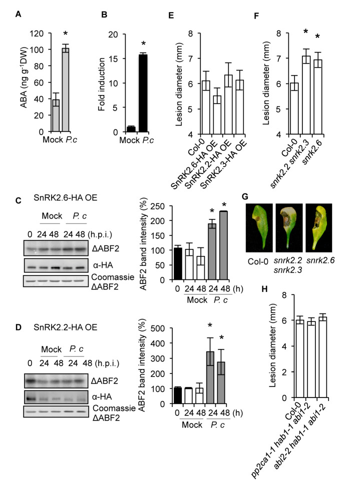
Liquid chromatography-mass spectrometry (LC-MS) showed marked accumulation of ABA in full expanded leaves of Arabidopsis plants at 72 h after drop inoculation with a spore suspension of the fungal necrotroph Plectosphaerella cucumerina (Figure 1A). ABA enhancement supported the upregulation of ABI4 gene expression, an ABA-responsive gene encoding a transcription factor [23] (Figure 1B). Therefore, ABA biosynthesis and signaling were triggered by P. cucumerina infection. The ABA-mediated activation of three monomeric SnRK2s (i.e., SnRK2.2, −2.3, and −2.6) is central to ABA signaling [51], so we investigated whether SnRK2s were engaged in the defense responses to this pathogen. Transgenic lines overexpressing HA-tagged SnRK2.6 (SnRK2.6-HA/OE) and SnRK2.2 (SnRK2.2-HA/OE) were inoculated with P. cucumerina or mock-treated, and leaf samples were collected at 0, 24, and 48 h post-inoculation (h.p.i.). Immunoprecipitation of SnRK2.2-HA and SnRK2.6-HA and the subsequent kinase assay of the immunoprecipitate were performed by determining the incorporation of 32P to purified ABF2 protein fragment substrate (amino acids Gly-73 to Gln-119) [52] in gel-kinase assays. Results revealed two- and three-fold enhancement for SnRK2.6 and SnRK2.2 kinase activity, respectively, following fungal inoculation (Figure 1C,D). For both kinases, enhanced activity occurred at 24 h.p.i., and the activation was sustained at 48 h.p.i. Therefore, ABA-activated SnRK2s were actively engaged in response to this fungal pathogen.
Figure 1.
Participation of SnRK2s kinases in the response of Arabidopsis plants to infection by the fungal pathogen P. cucumerina. (A) ABA accumulation determined in mock and P. cucumerina-infected Col-0 plants. (B) RT-qPCR of ABI4 in mock and in P. cucumerina-infected Col-0. (C,D) P. cucumerina-mediated activation of SnRK2.6 (C) and SnRK2.2 (D). Transgenic Arabidopsis plants expressing HA-tagged versions of the kinases were inoculated with P. cucumerina, or were mocked, and leaf samples were taken at 0, 24, and 48 h.p.i., and the protein extracts were immunoprecipitated with anti-HA antibodies. The immunoprecipitates were incubated with a His-ABF2 fragment (Gly73 to Gln 119; ΔABF2) in the presence of [γ-32P]ATP, and the proteins were resolved by SDS-PAGE. Bands corresponding to ΔABF2 fragments and to SnRK2.6 and SnRK2.2 kinases are indicated. Radioactivities of ΔABF2 fragment bands were measured with a phosphoimager, and the values were plotted on the graphs shown at the right of the figures. Error bars indicate S.E.M.; n = 3. (E) Disease resistance towards P. cucumerina of transgenic plants overexpressing SnRK2.6, SnRK2.2, and SnRK2.3 in comparison to Col-0. (F) Disease resistance towards P. cucumerina in the double snrk2.2 snrk2.3 mutant and in snrk2.6 mutant plants. (G) Representative leaves from each genotype at 12 days following inoculation with P. cucumerina. (H) Disease resistance towards P. cucumerina in the triple PP2C mutants pp2ca1 1hab1 1abi1-2 and abi2-2 hab1 abi1-2. For the bioassays with P. cucumerina, lesion diameter of 25 plants per genotype and four leaves per plant were determined 12 d following inoculation with P. cucumerina. Data points represent the average lesion size ± SE of measurements. An ANOVA was conducted to assess significant differences in the activation of SnRKs, ABA accumulation, ABI4 transcript accumulation, and disease symptoms, with a priori p < 0.05 level of significance; the asterisks * above the bars indicate statistically significant differences regarding mock treatments or Col-0 plants. Asterisks above the bars indicate different homogeneous groups with statistically significant differences.
We then investigated whether gain-of-function or loss-of-function in SnRK2s altered disease resistance to P. cucumerina. Symptoms of the fungal disease appear in the form of necrotic lesions, which are measured to quantify the degree of plant susceptibility [25,53,54]. Inoculation of transgenic plants individually overexpressing (OE) SnRK2.2, −2.3, and −2.6 revealed no significant variation in disease susceptibility towards P. cucumerina when compared to Col-0 plants (Figure 1E); thus, either endogenous SnRK2s levels are sufficient to achieve pathogen-triggered ABA signaling or overexpression of SnRK2s additionally requires increased ABA levels to enhance their activity. Although functional redundancy between SnRK2.2 and SnRK2.3 exists, functional segregation between SnRK2.6 and SnRK2.2/2.3 has been described [52]. Therefore, we inoculated an snrk2.2/2.3 double mutant and the single snrk2.6 mutant with P. cucumerina and recorded disease resistance. The triple snrk2.2/2.3/2.6 mutant, which is drastically affected in plant growth [51], was not compatible with the pathogenic assay and was, therefore, not used in the present study. Figure 1F–G show that snrk2.2/2.3 and snrk2.6 plant resistance to P. cucumerina was severely compromised. Moreover, an ABA deficient mutant (i.e., aba2) was similarly affected in disease resistance to this pathogen (Figure 2A). In summary, our results indicated that pathogen-induced ABA accumulation and concurrent activation of SnRK2s positively regulated disease resistance to P. cucumerina.
Figure 2.
PYR1 is required for disease resistance towards P. cucumerina. (A). Disease resistance towards P. cucumerina in Col-0, the resistant ocp3-1 mutant, the susceptible aba2-1, and the triple and quadruple multi-locus mutants pyl4 pyl5 pyl8, pyr1 pyl4 pyl8, pyr1 pyl4 pyl5, and pyr1 pyl1 pyl2 pyl4. (B) Disease resistance in single pyl1, pyr1, and pyl4 mutants, in a transgenic line overexpressing PYR1 (PYR1-OE), and in Col-0. Below the graph, the representative leaves from each genotype are shown at 12 days following inoculation with P. cucumerina. (C) Comparative disease resistance towards P. cucumerina among the allelic pyr1-1, pyr1-2, and pyr1-8 mutants. Data points represent the average lesion size ± SE of measurements. An ANOVA was conducted to assess significant differences in disease symptoms (p < 0.05); the letters above the bars indicate different homogeneous groups with statistically significant differences.
ABA signaling through SnRK2s is negatively regulated by clade A protein phosphatase type 2C (PP2C), particularly by ABI1, ABI2, PP2CA/AHG3, AHG1, HAB1, and HAB2 (see [55] and references therein). Therefore, clade A PP2Cs might negatively regulate ABA-mediated disease resistance to P. cucumerina. Because of the demonstrated redundancy existing for these PP2Cs, combined inactivation of selected groups of these phosphatases is required to determine functionality. We combined loss-of-function mutations in ABI1, ABI2, HAB1, and PP2CA genes to determine their contribution to ABA-mediated disease resistance. Different combinations of mutations were used with two triple mutants, pp2ca1-1;hab1-1;abi1-2 and abi2-2;hab1-1;abi1-2, which represent four of the nine closely related group A PP2Cs. Both multi-locus mutants showed an extreme response to exogenous ABA, partial constitutive response to endogenous ABA, and partial constitutive activation of SnRK2s in pp2ca1-1;hab1-1;abi1-2 [51,55]. Inoculation of both triple mutants with P. cucumerina showed no defective disease resistance (Figure 1H). This result suggests that the demonstrated redundancy of PP2Cs masks the manifestation of a clear phenotype upon pathogen inoculation. Additionally, ABA response in triple pp2c mutants was partially equivalent to that of lines OE SnRK2s, which did not show altered disease resistance to the pathogen (Figure 1E). It is also possible that other members of the large PP2C family, represented by 76 homologous genes [56], are key for resistance to P. cucumerina. This interpretation is supported by previous studies showing that a distinct PP2C member (i.e., AtDBP1) is required for other aspects of plant immunity [57,58].
2.2. The Requirement of the PYR1 Receptor for Antifungal Resistance
We next investigated which one of 14 soluble PYR/PYL/RCAR receptors perceived the ABA produced during P. cucumerina infection. Partial functional redundancy of ABA receptors has been demonstrated by genetic analysis; however, PYL8 plays a non-redundant role to regulate root sensitivity to ABA [45,46]. Additionally, both transcriptional and physiological ABA responses and signaling of environmental cues in guard cells mediated by individual receptors are starting to be elucidated [35]. We characterized disease resistance to P. cucumerina in a series of multi-locus mutants from different PYR/PYL receptors. The triple pyl4;pyl5;pyl8, pyr1;pyl4;pyl8, and pyr1;pyl4;pyl5 mutants, and the quadruple pyr1;pyl1;pyl2;pyl4 mutant, representing the highest genetic impairment in PYR/PYL function without affecting plant growth [45], were inoculated with P. cucumerina, and their impact on disease resistance was compared to aba2-1 (which enhances susceptibility [25]), to ocp3-1 (which enhances resistance [53]), and Col-0 plants. The two triple mutants incorporating the pyr1 mutation (i.e., pyr1;pyl4;pyl8 and pyr1;pyl4;pyl5) exhibited noticeably enhanced disease susceptibility (Figure 2A), which was of a magnitude similar to that observed in aba2-1 plants. Conversely, the disease resistance of the triple pyl4;pyl5;pyl8 mutant was unaltered compared to Col-0 plants. The quadruple mutant (also containing the pyr1 mutation) enhanced disease susceptibility to P. cucumerina. The results showed that the PYR1 receptor was pivotal for eliciting ABA-mediated defense responses towards P. cucumerina.
The specificity of PYR1 at eliciting plant immune responses was further tested by assaying the single pyr1-1 mutant. The individual pyr1-1 mutant had a compromised disease resistance phenotype (Figure 2B), contrasting to other single pyl mutants (e.g., pyl1, pyl4) for which resistance to the fungus remained intact. Moreover, the overexpression of the PYR1 receptor (PYR1-OE line) conferred significant enhancement of resistance to the fungus (Figure 2B). Other mutant alleles of the PYR1 receptor, predicted to produce a variety of defects in PYR1 (i.e., pyr1-2 and pyr1-8 [43]), consistently compromised disease resistance to P. cucumerina, showing pyr1-2 mutant allele as the strongest phenotype (Figure 2C). These results supported that the PYR1 receptor positively promoted ABA-dependent plant immunity against P. cucumerina. Interestingly, these results also indicated that other major receptors for ABA response, i.e., PYL1, PYL4, PYL5, PYL8, were not recruited in plant response against P. cucumerina. Furthermore, PYR1 appeared similarly to be required for the immune activation to Alternaria brassicicola, another fungal necrotroph and the causal agent of black spot disease in Brassica species. Results shown in Supplemental Figure S1 indicate that upon inoculation with A. brassicicola, both aba2 and pyr1 plants, compared to Col-0, pyl1, and pyl4 plants, showed remarkable enhancement in disease susceptibility to this pathogen. The enhancement of necrosis in A. brassicicola-inoculated leaves of pyr1 plants gave further support to the importance of PYR1-mediated perception of ABA for mounting effective defense responses towards necrotrophs.
2.3. Local Induction of PYR1 Gene Expression by P. cucumerina
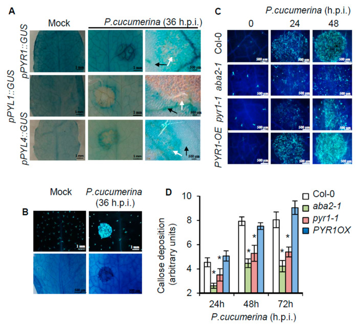
A reasonable explanation for the specific role of PYR1 in plant immunity might be the specific upregulation of PYR1 expression in response to the pathogen. Therefore, we next investigated whether transcriptional reprogramming occurred to enhance PYR1 expression upon pathogen inoculation. Transgenic plants expressing the promoter of the PYR1 gene fused to the β-glucuronidase GUS reporter gene (pPYR1::GUS) [45] were used to detect potential P. cucumerina-mediated activation of PYR1. Transgenic lines carrying the pPYL1::GUS and pPYL4::GUS gene constructs were also assayed to determine specificity. Local infection, i.e., by drop inoculation on the upper leaf surface with a P. cucumerina spore suspension, of transgenic pPYR1::GUS plants revealed early transcriptional activation of PYR1 triggered by the pathogen (Figure 3A). PYR1 induction mostly occurred within the vascular bundles of the primary and secondary veins of the P. cucumerina-inoculated leaf sectors. This highly localized induced expression pattern was specific to PYR1 because neither PYL1 nor PYL4 genes were transcriptionally activated under similar circumstances (Figure 3A). The local induction of pPYR1::GUS concurred with local synthesis and deposition of callose (Figure 3B) and later on with cell death (Figure 3C). These microscopy markers demarcated inoculated tissue sectors in advance to the appearance of visible necrosis and served to delimit local transcriptional responses. Moreover, callose deposition was compromised in pyr1-1 and aba2-1 mutants following fungal infection (Figure 3D), thus supporting the participation of ABA and PYR1 in this local process.
Figure 3.
Local activation of PYR1 gene expression at pathogen inoculation sites, and the requirement of PYR1 for pathogen-induced callose deposition. (A) Comparative histochemical analysis of GUS activity in rosette leaves from transgenic plants carrying pPYR1::GUS, pPYL1::GUS, and pPYL4::GUS gene constructs and those were either mocked or inoculated P. cucumerina. Leaves were stained for GUS activity at 36 h.p.i. The left panel corresponds to mocked plants. The central and right panels correspond to enlargements of the inoculated leaf sectors. Black arrow points towards leaf tissues proximal to the inoculation point, and white arrows denote tissues that directly received the spore inoculum. Note that pPYR1::GUS is heavily induced in leaf veins within the inoculated sector. (B) Characteristic spore-inoculated leaf sector, similar to those shown in A, stained with aniline blue to detect pathogen-induced callose deposition (top panel), or with trypan blue (lower panel) to identify incipient cell deterioration due to fungal infection at 36 h.p.i. (C) Aniline blue staining and epifluorescence microscopy were applied to visualize callose accumulation. Micrographs indicate P. cucumerina inoculation and infection site in the different Arabidopsis genotypes at 0 h.p.i (right panel), at 24 h.p.i. (central panel), and at 48 h.p.i. (right panel). (D) The number of yellows pixels (corresponding to pathogen-induced callose) per million on digital photographs of infected leaves were used as a means to express arbitrary units (i.e., to quantify the image) at the indicated times. Bars represent mean ± SD, n = 15 independent replicates. An ANOVA was conducted to assess significant differences in callose deposition (p < 0.05); the asterisks * above the bars indicate statistically significant differences regarding Col-0 plants.
2.4. Resistance Enhancement of pyr1 Plants to Pseudomonas syringae DC3000
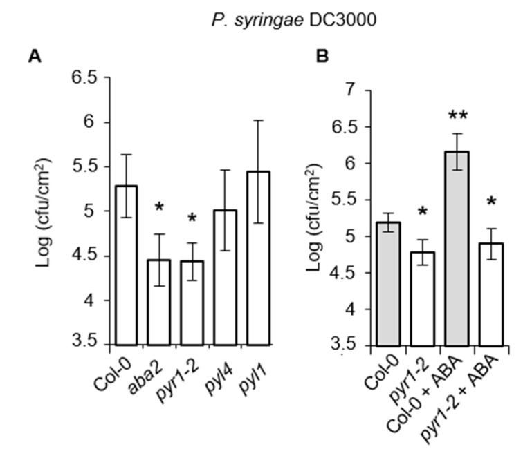
In marked contrast to the results shown above, the role of ABA in repressing plant immunity against the (hemi) biotrophic pathogens P. syringae DC3000 has been previously documented [6,19,20,21]. Therefore, we asked whether the negative role of ABA in plant immunity against P. syringae DC3000 could similarly be funneled through PYR1. If so, we would expect resistance enhancement in pyr1 plants. pyr1 plants were inoculated by leaf infiltration with P. syringae DC3000, and the rate of bacterial growth in the inoculated leaves was determined at 3 days post-inoculation in comparison to aba2, pyl1, pyl4, and Col-0 plants. Figure 4A shows that bacterial growth was reduced 10-fold in both aba2 and pyr1 mutants compared to Col-0, pyl1, and pyl4. This result confirmed the negative role of ABA in resistance towards P. syringae DC3000 and demonstrated the specific requirement of PYR1 for the negative role of ABA during this plant-pathogen interaction. Moreover, pre-treatment of Col-0 with 150 μM ABA, applied by drenching, predictably provoked disease susceptibility enhancement to P. syringae DC3000 (Figure 4B), denoting a damping effect of ABA on SA signaling. This ABA-mediated enhancement in susceptibility to P. syringae DC3000 did not occur in pyr1-2 plants whose enhanced resistance was not altered by the hormone (Figure 4B).
Figure 4.
Response of pyr1 plants to infection by P. syringae DC3000. (A) Col-0, aba2-1, pyr1-2, pyl1, and pyl4 mutants were inoculated with P. syringae DC3000, and their disease responses were recorded. (B) Col-0 and pyr1 plants were pre-treated with 150 μM ABA, applied by drenching, before inoculation with P. syringae DC3000, and the growth of the bacteria was recorded in comparison to mocked plants. Growth of P. syringae DC3000 was measured at 3 d.p.i. Error bars represent standard deviation (n = 12). An ANOVA was conducted to assess significant differences in disease symptoms, with a priori p < 0.05 level of significance; the asterisks *, ** above the bars indicate different homogeneous groups with statistically significant differences.
Therefore, our results indicated that the dual antagonistic role of ABA in plant immunity was mediated through the PYR1 receptor, which reciprocally activates and represses immune responses towards necrotrophic and biotrophic pathogens, respectively.
2.5. SA–Responsive Defense Genes are Activated in PYR1 Defective Mutants
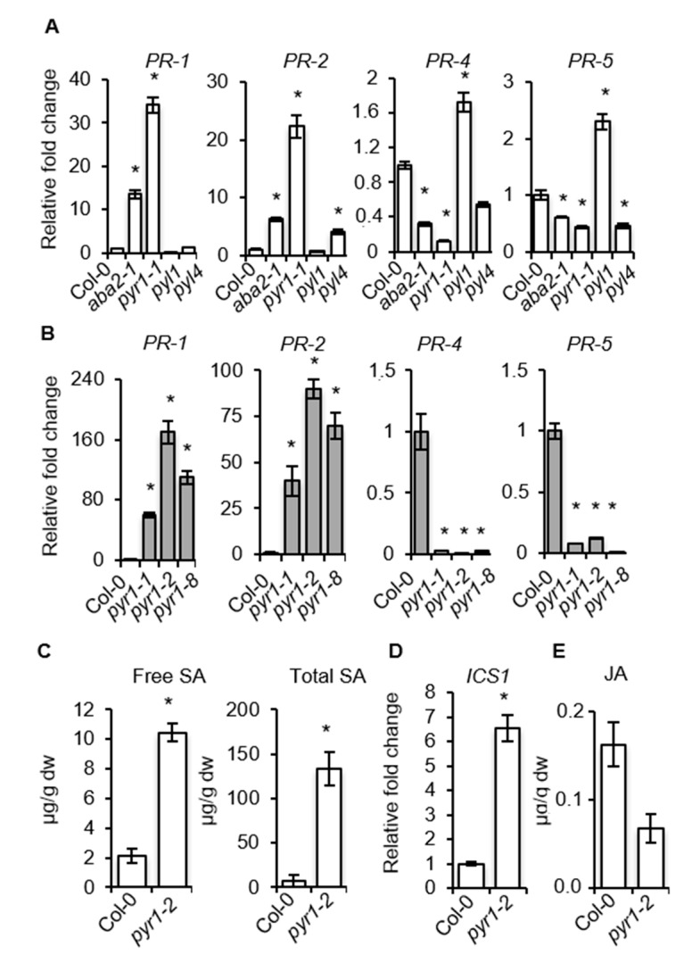
We investigated whether pyr1 and aba2 plants carried constitutive elevated expression of SA-responsive genes, which might explain the observed enhanced resistance to P. syringae DC3000 (Figure 4). The accumulation of PR-1 and PR-2 transcript, which are SA- and pathogen-responsive genes, was examined by RT-qPCR. In addition, we examined PR-4 and PR-5, which are also pathogen-responsive genes but are simultaneously influenced by SA and ET [59]. Transcript accumulation was also evaluated in pyl1 and pyl4 mutants, which served as additional controls. Figure 5A shows that pyr1 and aba2 plants carried constitutive elevated levels of SA-dependent PR-1 and PR-2 transcripts compared to Col-0, pyl1, or pyl4 plants. Conversely, the constitutive levels of transcript accumulation for PR-4 and PR-5 occurring in Col-0 were repressed in pyr1 and aba2 plants, and only partially enhanced in pyl1 plants (Figure 5A). The enhanced expression of PR-1 and PR-2 and the concerted repression of PR-4 and PR-5 were corroborated by the pyr1 allelic series, with the pyr1-2 allele showing the strongest differences (Figure 5B). Thus, the ABA/PYR1 module might function as an integration node regulating distinct branches of defenses. The constitutive activation of PR1- and -2 in pyr1 plants supported the enhanced accumulation of both free and conjugated SA observed in the mutant, which concurred also with elevated expression of ICS1, encoding isochorismate synthase, a pivotal enzyme controlling SA biosynthesis [60,61] (Figure 5C,D). On the other hand, pyr1 plants only showed a moderate reduction, less than two-fold, of JA content in comparison to Col-0 (Figure 5E). The conspicuous enhancement in SA content in healthy pyr1 plants, therefore, explained the resistance phenotypes of the mutant when confronted with P. syringae DC3000. However, the notorious enhanced susceptibility of pyr1 plants to the fungal necrotrophs P. cucumerina and A. brassicicola remained unsolved, as it could not simply be explained by the moderate reduction of JA levels as attained in the mutant.
Figure 5.
Expression of SA-responsive and ET-responsive genes in pyr1 and aba2 mutants. (A,B) RT-qPCR analysis showing constitutive expression levels of PR-1, PR-2, PR-4, and PR-5 genes in (A) Col-0, aba2-1, pyr1-1, pyl1, and pyl4 plants, and (B) their comparative expression levels in the allelic pyr1-1, pyr1-2, and pyr1-8 mutants. Data represent mean ± SD; n = 3 replicates. The expression was normalized to the constitutive ACT2 and ACT8 genes and then to the expression in Col-0 plants. (C–E) Accumulation of free SA, total SA, and total JA in Col-0 and pyr1-2 plants. Data represent the average of three biological replicates. An ANOVA was conducted to assess significant differences in RT-qPCR and hormone analysis, with a priori p < 0.05 level of significance; the asterisks * above the bars indicate statistically significant differences regarding Col-0 plants.
2.6. Enhanced Activation of MAPK Kinases in pyr1 Plants
We next investigated whether enhanced resistance to P. syringae DC3000 in pyr1 plants was associated with elevated MAPKs activation, which is linked to the activation of immune responses following pathogen perception. We employed an antibody recognizing the phosphorylated residues within the MAPK activation loop (i.e., the pTEpY motif). Western blot analysis of protein extracts derived from healthy Col-0 and pyr1 plants showed positive immunoreactive signals in two polypeptides corresponding to MPK6 and MPK3 (Beckers et al., 2009) (Figure 6), and the densitometric scanning of blots indicated that the MPK3 immunoreactive band was more intense in pyr1 plants. Inoculation with P. syringae DC3000 promoted further activation-associated dual TEY phosphorylation of MPKs, which was noticeably higher for MPK3 in pyr1 compared to Col-0 plants at 24 h.p.i. (Figure 6). At the latter stages of infection (i.e., 48 h.p.i.), the MPK activation was similar in Col-0 and pyr1 plants. Therefore, MPK activation may be prone to activation in plants defective of ABA perception through the PYR1 receptor. Indeed, partial pre-activation of MPK was reflected in detectable PR-1 protein accumulating in pyr1 plants at time zero (Figure 6). This result was in agreement with the higher expression level of the PR-1 gene determined by RT-qPCR (Figure 5A,B). Interestingly, inoculation with P. syringae DC3000 promoted the further accumulation of the PR-1 protein, which progressively increased over time to a much higher level in the pyr1 mutant compared to Col-0 plants (Figure 6).
Figure 6.
Loss of PYR1 function confers enhanced mitogen-activated kinase activation and PR-1 protein accumulation following P. syringae DC3000 infection. Western blot with anti-pTEpY and anti-PR-1 antibodies of crude protein extracts derived from Col-0, pyr1-2 plants at 0, 24, and 48 h.p.i with P. syringae DC3000. Equal protein loading was check by Ponceau-S staining of the nitrocellulose filter. MPK6 and MPK3 migrating bands are indicated on the right. The experiments were repeated three times with similar results. Scan quantification of protein bands corresponding to MPK3 and PR-1 is shown below the Western blot. Data represent the mean ± SD; n = 3 replicates. An ANOVA was conducted to assess significant differences in RT-qPCR analysis, with a priori p < 0.05 level of significance; the asterisks * above the bars indicate statistically significant differences regarding Col-0 plants.
Thus, we hypothesize that the lack of ABA perception through the PYR1 receptor de-represses a pathway that allows cell sensitization through MPKs activation and downstream defense gene reprogramming, even in the absence of pathogen infection. Sensitized cells may be ready for the enhanced induction of this defense pathway following pathogen infection, which, in turn, may explain why aba and pyr1 plants exhibit enhanced disease resistance to P. syringae DC3000. These observations support that ABA and PYR1 function as a repressor module of SA-mediated onset of resistance.
2.7. SA-Mediated Defense Genes are Poised for Enhanced Activation through Chromatin Remodeling in pyr1 Plants
We then asked whether other markers diagnostic of an immune status were also activated in pyr1 plants. The expression of the extracellular subtilase SBT3.3 gene has been recently described to be a switch for poising SA-related gene expression and immune priming [62]. Moreover, constitutive SBT3.3 expression, MPK activation, and readied SA-related genes convey in plants defective in the RNA-directed DNA methylation (RdDM) pathway, which negatively regulates immune priming [54]. Consequently, the expression level of genes encoding SBT3.3 and either of the two subunits of RNA Pol V (i.e., NRPD2 and NRPE1) controlling RdDM were determined by RT-qPCR. Figure 7A shows the constitutive upregulation of SBT3.3 and concurrent downregulation of NRPD2 in pyr1 plants compared to Col-0, congruent with the activation of immune priming in the mutant. The downregulation was specific for NRPD2, encoding the second large subunit of Pol V, because the expression of the gene encoding the large NRPE1 subunit exhibited a minimal variation in pyr1 plants (Figure 7A).
Figure 7.
Loss of PYR1 function provokes the setting of hallmarks characteristic of primed immunity. (A) Comparative RT-qPCR of SBT3.3, NRPD2, and NRPE1 transcript levels between healthy Col-0 and pyr1-2 plants. The expression was normalized to the constitutive ACT2/8 gene and then to the expression in Col-0 plants. (B) Chromatin immunoprecipitation (ChIP) and comparison between Col-0 and pyr1-2 plants of the level of histone H3 Lys4 trimethylation (H3K4me3) and histone H3 Lys9 acetylation (H3K9ac) on the SBT3.3, PR-1, WRKY6, and WRKY53 gene promoters as present in leaf samples. The setting of histone marks in ACTIN2 was used as an internal control. Data are standardized for Col-0 histone modification levels. Data represent the mean ± SD; n = 3 biological replicates. An ANOVA was conducted to assess significant differences between MPKs activation and PR1 accumulation (p < 0.05); the asterisks * above the bars indicate statistically significant differences regarding Col-0 plants.
In plants defective in RdDM-mediated epigenetic control, immune priming is activated concurrently with chromatin histone activation marks being enriched in SA-related genes, including the SBT3.3 gene itself [54,62]. Thus, we hypothesized that SA-related defense genes and SBT3.3 in pyr1-2 plants are poised for enhanced expression by differential histone modification. We used chromatin immunoprecipitation (ChIP) to analyze H3K4me3 and H3K9ac activation marks on the SBT3.3 and PR-1 gene promoter regions in pyr1 and Col-0 plants. We also examined the genes encoding WRKY6 and WRKY53, transcriptional regulators of SA-defense genes. Figure 7B shows that H3K4me3 marks in the SBT3.3 promoter region notably increased in pyr1 plants compared to Col-0 plants, while H3K9ac marks remained invariant (Figure 7B), supporting previous descriptions of plants constitutively expressing primed immunity [54,62,63]. On the PR1 promoter, both H3K4me3 and H3K9ac activation marks increased three- and two-fold, respectively, in pyr1 plants compared to Col-0 (Figure 7B). Likewise, histone activation marks also moderately increased in the WRKY6 and WRKY53 promoters of pyr1 plants compared to Col-0 plants (Figure 7B). The setting of histone marks in pyr1 plants remained unchanged in the ACTIN2 gene promoter, which was used as the control (Figure 7B). Therefore, chromatin activation marks proliferated in the promoter regions of the priming regulatory gene SBT3.3 and the SA-responsive genes in pyr1 plants and would explain why the PR-1 protein showed accelerated and enhanced accumulation in pyr1 plants following pathogen inoculation (Figure 6). Our results indicated that ABA and its PYR1-mediated perception represented novel integral components of a signaling process, repressing SA-mediated immunity.
2.8. NahG Plants Abrogate the Altered Disease Resistance Response of pyr1 Plants
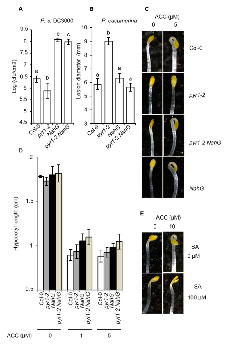
To evaluate the role of SA for pyr1-altered resistance, we generated a pyr1;NahG double mutant. In plants carrying the NahG transgene, salicylate hydroxylase depletes the plant of this defense hormone [64]. Compared to Col-0, NahG plants showed an anticipated increase in susceptibility to P. syringae DC3000 due to SA depletion (Figure 8A). Interestingly, in pyr1;NahG plants, the pyr1-mediated enhanced resistance was abrogated, and instead enhanced susceptibility to P. syringae DC3000 emerged (Figure 8A). Moreover, when assayed against the fungal pathogen P. cucumerina, NahG plants behaved like Col-0, both showing the same degree of susceptibility (Figure 8B), suggesting normal metabolic levels of SA played no major role in the resistance towards this pathogen. Surprisingly, in pyr1;NahG plants, the pyr1-mediated-enhanced susceptibility to P. cucumerina was abrogated (Figure 8B), with pyr1;NahG plants to be behaving as Col-0 or NahG plants. This suggested that the PYR1-mediated perception of ABA negatively regulated the SA pathway. When this negative regulation failed, such as in pyr1 plants, the SA levels increased, and the resistance to P. syringae DC3000 was activated. As a trade-off effect, the elevated SA levels presumably interfered with JA or ET signaling pathways required for mounting a resistance response to fungal pathogens.
Figure 8.
Effect of NahG on disease resistance and insensitivity to ACC of pyr1 plants and seedlings. (A,B) Comparative disease resistance towards P.s. DC3000 and P. cucumerina among Col-0, pyr1-2, NahG, and pyr1-2NahG plants. Growth of P. syringae DC3000 was measured at 3 d.p.i. Error bars represent standard deviation (n = 12). For P. cucumerina, data points represent the average lesion size ± SE of measurements. An ANOVA was conducted to assess significant differences in disease symptoms (p < 0.05); the letters above the bars indicate different homogeneous groups with statistically significant differences. (C) Apical hook region of the indicated seedlings germinated and grown on MS/2 in the dark for 4 d in the presence of the indicated concentration of ACC. (D) Hypocotyl length of seedlings germinated and grown in the dark for 4 d on MS/2 medium supplemented with the denoted concentrations of ACC. Error bars represent standard deviation (n = 50). An ANOVA was conducted, and no significant differences were observed in hypocotyl length (p < 0.05). (E) Apical hook region of Col-0 seedlings germinated and grown on MS/2 in the dark for 4 d in the presence of the indicated concentration of ACC and SA.
2.9. pyr1-Mediated Enhanced SA Content Blocks ET Perception
The SA and JA signal pathways are under an antagonistic equilibrium. Therefore, we wondered if the enhanced SA levels of pyr1 plants could be affecting JA signaling in this mutant. We studied pyr1 plants for altered responses to JA using the widely applied root growth inhibition assay. In the absence of JA, primary root length of pyr1 seedlings was comparable to that of Col-0 plants (Supplemental Figure S2), and in the presence of JA, root growth reduction in the mutant was also similar to that observed in Col-0 plants (Supplemental Figure S2), providing evidence that JA perception was not impaired in the mutant. In addition, comparison of the expression level of different JA-responsive genes at different times following P. cucumerina inoculation in Col-0 and pyr1 plants revealed that JA signaling appeared to be not affected in the mutant (Supplemental Figure S3). Instead, for some of the genes analyzed, a higher induction was recorded in pyr1 plants. Therefore, JA signaling was not compromised in pyr1 plants.
We next asked whether ET signaling, which is also pivotal for resistance to fungal pathogens [10,12,13], could be the one impaired in pyr1 plants due to the elevated levels of SA. This hypothesis gained even more relevance in view of the recently described mechanism explaining the antagonism between SA and ET in the suppression of apical hook formation and early seedling establishment via NPR1-mediated repression of EIN3 and EIL1 [16]. We, therefore, assayed Col-0 and pyr1 seedlings, grown in the dark in the presence or absence of a low concentration of the ethylene precursor ACC (5 μM), for the induction of the ET-mediated triple response. The triple response in Arabidopsis consists of shortening and thickening of hypocotyls and roots and exaggeration of the curvature of apical hooks. Compared to Col-0, the assay revealed that pyr1 seedlings showed no curvature of the apical hook (Figure 8C) and also showed less shortened hypocotyls (Figure 8D) when grown in the presence of ACC. Thus, the pyr1 mutant was impaired in ET perception. The enhanced SA content in pyr1 seedlings was the causal link mediating insensitivity to ET since in pyr1;NahG double mutant, normal sensitivity to ET was re-established (Figure 8C,D). Moreover, when Col-0 seedlings were assayed in the presence of high amounts of SA (100 μM), the ACC-induced triple response was abrogated (Figure 8E), further sustaining that the elevated levels of SA in pyr1 plants blunted ET perception. Thus, our results suggested that perception of ABA through PYR1 acted primarily as a module negatively controlling the SA pathway. When ABA/PYR1 failed, the SA pathway was released, and the resistance to P. syringae DC3000 was activated. As a trade-off effect, the enhanced accumulation of the SA pathway blocked the ET pathway, the later required for resistance to fungal necrotrophic pathogens.
3. Discussion
Despite the demonstrated role of ABA on the final outcome of immune responses, the specific components of the ABA signaling apparatus and the specific mechanisms that exploit ABA to positively and negatively influence immune responses to specific plant-pathogen interactions have remained largely unknown. Here, we showed that SnRK2s kinases were actively engaged in activating resistance towards P. cucumerina, whereas the loss-of-function of any of the three individual SnRK2s compromised this resistance. Furthermore, we demonstrated that PYR1 was pivotal and played a positive role in disease resistance to P. cucumerina since overexpression of PYR1 (i.e., PYR1-OE transgenic line) conferred significantly enhanced resistance. Conversely, in PYR1 loss-of-function mutants, the resistance was compromised. Therefore, the PYR1 receptor had functional specificity in perceiving ABA produced in response to fungal infection to activate plant immunity. This study provided novel information about a specific ABA receptor-mediating specific plant immune responses and pinpointed ABA-activated SnRK2s as cardinal components for plant resistance. This information helps construct a functional classification scheme of the different members of the PYR/PYL receptor family with respect to their downstream signaling pathways in a true biological context. Thus, specific non-redundant roles for PYR1 and PYL8 have been reported in plant immunity (this work) and root ABA sensitivity [50], respectively. An explanation for the specific role of PYR1 in pathogen response could be the selective and highly localized pathogen-induced expression of PYR1 in vascular bundles (Figure 3A). This expression pattern mirrors the expression of genes encoding ABA-biosynthetic enzymes [65,66,67,68]. Therefore, the synthesis of ABA and the pathogen-induced expression of PYR1 spatially concur in the vasculature, supporting the hypothesis that vascular tissues function as an integrating node, triggering stress signaling that sets in motion the local and systemic immune responses in the plant [67,69,70].
This study showed that resistance to A. brassicicola was also dependent on ABA and PYR1, reinforcing the importance of this signal pathway for activating immunity against necrotrophs. This further reconciled with results shown above and also with previous studies showing that ABA promotes enhanced resistance to the necrotroph P. cucumerina [23,25,71]. Moreover, when a fungal necrotroph is a shift to a biotrophic lifestyle by changing the inoculation method and also the developmental stage of the plant [72], as reported for P. cucumerina [22], then ABA exerts an opposite effect, and the resistance to this same pathogen is suppressed. This contradictory role of ABA at controlling the disease resistance has also been observed for biotrophic pathogens (e.g., P. syringae DC3000), with resistance appearing negatively regulated by ABA, whereas resistance is enhanced in ABA-deficient mutants (Mohr & Cahill, 2007; Jensen et al., 2008; Fan et al., 2009; Verhage et al., 2010) [4,21,26,27]. In fact, we showed that the growth of P. syringae DC3000 was severely restricted in pyr1 plants, as documented for aba2 aao3 plants or the ABA-insensitive abi1-1 and abi2-1 mutants [8,28]. These results demonstrated the Janus functions of PYR1 in disease resistance, mediating repression of immunity against biotrophic pathogens, whereas activation against necrotrophs. Consequently, PYR1 may regulate which of these two plant immune programs prevails. This hypothesis supports previous observations of ABA as a hormone that interacts antagonistically or synergistically with the SA-JA-ET backbone of the plant immune signaling network, redirecting defense outputs [4,28,73,74,75]. Yet, how does the ABA/PYR1 module interfere with immunity to drive simultaneously the repression and activation of the SA and JA/ET defense pathways, respectively? Hormone cross-talk allows different hormone signaling pathways to act antagonistically or synergistically, providing the powerful regulatory potential to flexibly tailor the plant’s adaptive response to a range of environmental cues [4]. Our results showed basal activation of the SA-dependent pathway in pyr1 mutants, and that pyr1 was insensitive to the damping effect of ABA on SA signaling. This finding supported previous work demonstrating the negative role of ABA on disease resistance to biotrophs, and that P. syringae-induced ABA levels in Col-0 suppress SA biosynthesis and action, enhancing susceptibility to this pathogen [21,28,73,75,76]. Interestingly, our finding that JA perception remained intact in pyr1 plant but ET perception became compromised added a degree of specificity for the understanding of the disease resistance phenotype of the mutant. The observation that in pyr1;NahG plants, the pyr1-mediated ET-insensitivity was reversed, and that SA per se could block ET perception in Col-0 plants (Figure 8 and Huang et al., 2020), pointed towards SA-mediated repression of ET signaling modulated by ABA and PYR1 during pathogenesis. The positive effect of ABA at promoting ET-dependent resistance to fungal pathogens may be indirect: perception of pathogenic ABA by PYR1 dampens SA signaling, which, in turn, stops ET pathway repression by SA. This ABA and PYR1-modulated cross-talk regulation of SA and ET pathways may provide the plant with a powerful regulatory potential to boost its defenses according to the lifestyle of the attacker. This phenomenon may also explain why disease-promoting biotrophic pathogens (e.g., P. syringae DC3000) have developed strategies to alter the host ABA physiology as part of the infection strategy [28,76].
How does then ABA/PYR1-mediated signaling control SA-mediated defenses? pyr1 plants bear constitutive activation of ICS expression and moderate enhanced level of SA. Besides, pyr1 plants carry the hallmarks of immune priming, including (1) basal activation of MPKs; (2) repression of NRPD2 and, therefore, the RdDM mechanisms that negatively control the onset of defense; (3) activation of the SBT3.3 subtilase; and (4) readying of SA-related genes for enhanced expression by pertinent chromatin modifications. Therefore, pyr1 plants mirror the phenotypes of RdDM defective mutants, which exhibit simultaneous enhanced susceptibility and resistance to necrotrophs and biotrophs, respectively [54], supporting SA signaling activation in pyr1 plants. The fact that immune priming and SA-mediated resistance are negatively regulated by the RdDM, and that in pyr1 plants, the ABA repression of SA pathway is relieved, both observations unveil the importance of ABA/PYR1 as new element participating in an epigenetic mechanism of control of gene expression in plant immunity.
4. Materials and Methods
4.1. Plants Growth Conditions
Arabidopsis thaliana plants were grown in a growth chamber (19–23 °C, 85% relative humidity, 100 mEm−2 s−1 fluorescent illumination) on a 10-h-light and 14-hr-dark cycle. All mutants and transgenic plants are in Col-0 background; SnRK2.6-HA/OE and SnRK2.2-HA/OE were previously described [77,78]; snrk2.2 snrk2.3 and snrk2.6 were described in [44,45,51]; the triple mutants pp2ca1-1 hab1-1 abi2-2 and abi2-2 hab1-1 abi1-2 were described in [22]; ocp3-1 and aba2-1 mutants described in [25,53], and the triple pyl4 pyl5 pyl8, pyr1 pyl4 pyl8, and pyr1 pyl4 pyl5 mutants, along with the quadruple pyr1 pyl1 pyl2 pyl4 and single pyl1, pyl4, pyr1-1, pyr1-2 and pyr1-8 mutants were described in [41,45]. Transgenic lines carrying pPYR1::GUS, pPYL1::GUS, pPYL4::GUS were described previously [45]. NahG plants were described previously [64].
4.2. Gene Expression Analysis
Total RNA was extracted from plant tissues using TRIzol (Invitrogen, Waltham, MA, USA) and purified by lithium chloride precipitation. Reverse transcription was done using the RevertAid H Minus First Strand cDNA Synthesis Kit (Fermentas Life Sciences, Waltham, MA, USA). Quantitative PCR (qPCR) was performed using an ABI PRISM 7000 sequence detection system and SYBR-Green (Perkin-Elmer Applied Biosystems, Foster, CA, USA). ACTIN2 and ACTIN8 were the reference genes. The primers used for RT-qPCR experiments are provided in Table S1. RT-qPCR analyses were performed at least three times using sets of cDNA samples from independent experiments.
4.3. Immunoprecipitation of HA–SnRKs and In Vitro Phosphorylation
HA-tagged SnRK2.2 and SnRK2.6 were immunoprecipitated and used for in vitro kinase assay, as described previously [78].
4.4. Chromatin Immunoprecipitation
Chromatin isolation and immunoprecipitation were performed, as described [54,62]. Chip samples, derived from three biological replicates, were amplified in triplicate and measured by quantitative PCR using primers for SBT3.3, PR-1, WRKY6, WRKY53, and Actin2, as reported [54,62]. All ChIP experiments were performed in three independent biological replicates. The antibodies used for the immunoprecipitation of modified histones from 2 g of leaf material were antiH3K4m3 (#07-473 Millipore) and antiH3K9ac (#07-352 Millipore).
4.5. Western Blot
Protein crude extracts were prepared by homogenizing ground frozen leaf material with Tris-buffered saline (TBS) supplemented with 5 mM DTT, protease inhibitor cocktail (Sigma-Aldrich), and protein phosphatase inhibitors (PhosStop, Roche). Protein concentration was measured using Bradford reagent; 25 μg of total protein was separated by SDS-PAGE (12% acrylamide w/v) and transferred to nitrocellulose filters. The filter was stained with Ponceau-S after transfer and used as a loading control.
4.6. Pathogen Assays
Pseudomonas syringae DC3000 was grown for two days, and a culture with O.D. 2 × 10⁻⁴ was used to infect 5-week-old Arabidopsis leaves by infiltration, and the bacterial growth was determined following [54,62]. Twelve samples were used for each data point and represented as the mean ± SD of log c.f.u./cm2. For Plectosphaerella cucumerina and Alternaria brassicicola bioassays, 5-week-old plants were inoculated, as described [23,24], with a suspension of fungal spores of 2.5 × 10⁴, 5 × 10⁶, and 5 × 10⁶ spores/mL, respectively. The challenged plants were maintained at 100% relative humidity. Disease symptoms were evaluated by determining the lesion diameter of at least 100 lesions (25 plants per genotype and four leaves per plant) at 3, 12, and 8 days after inoculation with P. cucumerina and A. brassicicola, respectively. For pathogen-induced callose deposition analyses, infected leaves were stained at 24, 48, and 72 h.p.i. with aniline blue, and callose deposition quantifications were performed, as described by [53].
4.7. Determination of Plant Hormones and Metabolites
ABA, JA, SA levels were determined, as described previously [25,71].
Acknowledgments
We acknowledge V. Flors for the technical assistance in the analysis of plant metabolites and hormones.
Supplementary Materials
The following are available online at https://www.mdpi.com/1422-0067/21/16/5852/s1, Figure S1: Disease resistance towards Alternaria brassicicola in Col-0, aba2-1, pyr1, pyl2 and pyl4 plants and comparison of Col-0 and pyr1-2 plants upon inoculation with Altenaria brassicicola. Figure S2: Response of Col-0 and pyr1-2 seedlings to JA. Figure S3: Comparative RT-qPCR analysis for the expression of different JA-responsive and biosynthesis genes in either mocked or P. cucumerina-inoculated Col-0 and pyr1 plants. Table S1: primer sequences.
Author Contributions
M.G.-G. and P.L.R. performed SnRKs kinase assays and provided genetic resources. J.G.-A. and B.G. performed the rest of the experiment shown in the manuscript. P.V. wrote the manuscript. All authors have read and agreed to the published version of the manuscript.
Funding
This research was founded by the Spanish AEI agency by grant BIO2017-82503-R to P.L.R. and by grant RTI2018-098501-B-I00 to P.V.
Conflicts of Interest
Authors declare no competing financial interest.
References
- 1.Malamy J., Carr J.P., Klessig D.F., Raskin I. Salicylic acid: A likely endogenous signal in the resistance response of tobacco to viral infection. Science. 1990;250:1002–1004. doi: 10.1126/science.250.4983.1002. [DOI] [PubMed] [Google Scholar]
- 2.Métraux J.P., Signer H., Ryals J., Ward E., WyssBenz M., Gaudin J., Raschdorf K., Schmid E., Blum W., Inverardi B. Increase in salicylic acid at the onset of systemic acquired resistance in cucumber. Science. 1990;250:1004–1006. doi: 10.1126/science.250.4983.1004. [DOI] [PubMed] [Google Scholar]
- 3.Glazebrook J. Contrasting mechanisms of defense against biotrophic and necrotrophic pathogens. Annu. Rev. Phytopathol. 2005;43:205–227. doi: 10.1146/annurev.phyto.43.040204.135923. [DOI] [PubMed] [Google Scholar]
- 4.Verhage A., van Wees S.C.M., Pieterse C.M.J. Plant immunity: It’s the hormones talking, but what do they say? Plant Physiol. 2010;154:536–540. doi: 10.1104/pp.110.161570. [DOI] [PMC free article] [PubMed] [Google Scholar]
- 5.Broekaert W.F., Delaure S.L., De Bolle M.F., Cammue B.P. The role of ethylene in host-pathogen interactions. Annu. Rev. Phytopathol. 2006;44:393–416. doi: 10.1146/annurev.phyto.44.070505.143440. [DOI] [PubMed] [Google Scholar]
- 6.Grant M.R., Jones J.D. Hormone (dis)harmony moulds plant health and disease. Science. 2009;324:750–752. doi: 10.1126/science.1173771. [DOI] [PubMed] [Google Scholar]
- 7.Pieterse C.M.J., Van der Does D., Zamioudis C., Leon-Reyes A., Van Wees S.C. Hormonal modulation of plant immunity. Annu. Rev. Cell Dev. Biol. 2012;28:489–521. doi: 10.1146/annurev-cellbio-092910-154055. [DOI] [PubMed] [Google Scholar]
- 8.Thaler J.S., Bostock R.M. Interactions between abscisic-acid-mediated responses and plant resistance to pathogens and insects. Ecology. 2004;85:48–58. doi: 10.1890/02-0710. [DOI] [Google Scholar]
- 9.Spoel S.H., Koornneef A., Claessens S.M., Korzelius J.P., Van Pelt J.A., Mueller M.J., Buchala A.J., Metraux J.P., Brown R., Kazan K., et al. NPR1 modulates cross-talk between salicylate- and jasmonate-dependent defense pathways through a novel function in the cytosol. Plant Cell. 2003;15:760–770. doi: 10.1105/tpc.009159. [DOI] [PMC free article] [PubMed] [Google Scholar]
- 10.Thomma B.P.H.J., Eggermont K., Tierens K.F.M., Broekaert W.F. Requirement of functional ethylene-insensitive 2 gene for efficient resistance of Arabidopsis to infection by Botrytis cinerea. Plant Physiol. 1999;121:1093–1102. doi: 10.1104/pp.121.4.1093. [DOI] [PMC free article] [PubMed] [Google Scholar]
- 11.Gu Y.Q., Yang C., Thara V.K., Zhou J., Martin G.B. Pti4 is induced by ethylene and salicylic acid, and its product is phosphorylated by the Pto kinase. Plant Cell. 2000;12:771–786. doi: 10.1105/tpc.12.5.771. [DOI] [PMC free article] [PubMed] [Google Scholar]
- 12.Berrocal-Lobo M., Molina A., Solano R. Constitutive expression of ETHYLENE-RESPONSE-FACTOR1 in Arabidopsis confers resistance to several necrotrophic fungi. Plant J. 2002;29:23–32. doi: 10.1046/j.1365-313x.2002.01191.x. [DOI] [PubMed] [Google Scholar]
- 13.Díaz J., ten Have A., van Kan J.A.L. The role of ethylene and wound signaling in resistance of tomato to Botrytis cinerea. Plant Physiol. 2002;129:1341–1351. doi: 10.1104/pp.001453. [DOI] [PMC free article] [PubMed] [Google Scholar]
- 14.Leslie C.A., Romani R.J. Inhibition of ethylene biosynthesis by salicylic acid. Plant Physiol. 1988;88:833–837. doi: 10.1104/pp.88.3.833. [DOI] [PMC free article] [PubMed] [Google Scholar]
- 15.Chen H., Xue L., Chintamanani S., Germain H., Lin H., Cui H., Cai R., Zuo J., Tang X., Li X., et al. ETHYLENE INSENSITIVE3 and ETHYLENE INSENSITIVE3-LIKE1 repress SALICYLIC ACID INDUCTION DEFICIENT2 expression to negatively regulate plant innate immunity in Arabidopsis. Plant Cell. 2009;21:2527–2540. doi: 10.1105/tpc.108.065193. [DOI] [PMC free article] [PubMed] [Google Scholar]
- 16.Huang P., Dong Z., Guo P., Zhang X., Qiu Y., Li B., Wang Y., Guo H. Salicylic Acid Suppresses Apical Hook Formation via NPR1-Mediated Repression of EIN3 and EIL1 in Arabidopsis. Plant Cell. 2020;32:612–629. doi: 10.1105/tpc.19.00658. [DOI] [PMC free article] [PubMed] [Google Scholar]
- 17.Spoel S.H., Johnson J.S., Dong X. Regulation of tradeoffs between plant defenses against pathogens with different lifestyles. Proc. Natl. Acad. Sci. USA. 2007;104:18842–18847. doi: 10.1073/pnas.0708139104. [DOI] [PMC free article] [PubMed] [Google Scholar]
- 18.Cutler S.R., Rodriguez P.L., Finkelstein R.R., Abrams S.R. Abscisic acid: Emergence of a core signaling network. Annu. Rev. Plant Biol. 2010;61:651–679. doi: 10.1146/annurev-arplant-042809-112122. [DOI] [PubMed] [Google Scholar]
- 19.Mauch-Mani B., Mauch F. The role of abscisic acid in plant-pathogen interactions. Curr. Opin. Plant Biol. 2005;8:409–414. doi: 10.1016/j.pbi.2005.05.015. [DOI] [PubMed] [Google Scholar]
- 20.Asselbergh B., De Vleesschauwer D., Hofte M. Global switches and fine-tuning-aba modulates plant pathogen defense. Mol. Plant Microbe Interact. 2008;21:709–719. doi: 10.1094/MPMI-21-6-0709. [DOI] [PubMed] [Google Scholar]
- 21.Fan J., Hill L., Crooks C., Doerner P., Lamb C. Abscisic acid has a key role in modulating diverse plant-pathogen interactions. Plant Physiol. 2009;150:1750–1761. doi: 10.1104/pp.109.137943. [DOI] [PMC free article] [PubMed] [Google Scholar]
- 22.Sanchez-Vallet A., Lopez G., Ramos B., Delgado-Cerezo M., Riviere M.P., Llorente F., Fernandez P.V., Miedes E., Estevez J.M., Grant M., et al. Disruption of abscisic acid signaling constitutively activates arabidopsis resistance to the necrotrophic fungus plectosphaerella cucumerina. Plant Physiol. 2012;160:2109–2124. doi: 10.1104/pp.112.200154. [DOI] [PMC free article] [PubMed] [Google Scholar]
- 23.Ton J., Mauch-Mani B. Beta-amino-butyric acid-induced resistance against necrotrophic pathogens is based on aba-dependent priming for callose. Plant J. 2004;38:119–130. doi: 10.1111/j.1365-313X.2004.02028.x. [DOI] [PubMed] [Google Scholar]
- 24.Adie B.A., Perez-Perez J., Perez-Perez M.M., Godoy M., Sanchez-Serrano J.J., Schmelz E.A., Solano R. ABA is an essential signal for plant resistance to pathogens affecting JA biosynthesis and the activation of defenses in Arabidopsis. Plant Cell. 2007;19:1665–1681. doi: 10.1105/tpc.106.048041. [DOI] [PMC free article] [PubMed] [Google Scholar]
- 25.García-Andrade J., Ramírez V., Flors V., Vera P. Arabidopsis ocp3 mutant reveals a mechanism linking aba and ja to pathogen-induced callose deposition. Plant J. 2011;67:783–794. doi: 10.1111/j.1365-313X.2011.04633.x. [DOI] [PubMed] [Google Scholar]
- 26.Mohr P.G., Cahill D.M. Suppression by ABA of salicylic acid and lignin accumulation and the expression of multiple genes, in arabidopsis infected with Pseudomonas syringae pv. tomato. Funct. Integr. Genomics. 2007;7:181–191. doi: 10.1007/s10142-006-0041-4. [DOI] [PubMed] [Google Scholar]
- 27.Jensen M.K., Hagedorn P.H., de Torres-Zabala M., Grant M.R., Rung J.H., Collinge D.B., Lyngkjaer M.F. Transcriptional regulation by an nac (nam-ataf1,2-cuc2) transcription factor attenuates aba signalling for efficient basal defence towards Blumeria graminis f. sp. Hordei in Arabidopsis. Plant J. 2008;56:867–880. doi: 10.1111/j.1365-313X.2008.03646.x. [DOI] [PubMed] [Google Scholar]
- 28.Torres-Zabala M., Truman W., Bennett M.H., Lafforgue G., Mansfield J.W., Rodriguez Egea P., Bögre L., Grant M. Pseudomonas syringae pv. tomato hijacks the Arabidopsis abscisic acid signalling pathway to cause disease. EMBO J. 2007;26:1434–1443. doi: 10.1038/sj.emboj.7601575. [DOI] [PMC free article] [PubMed] [Google Scholar]
- 29.Mine A., Berens M.L., Nobori T., Anver S., Fukumoto K., Winkelmüller T.M., Takeda A., Becker D., Tsuda K. Pathogen exploitation of an abscisic acid- and jasmonate-inducible MAPK phosphatase and its interception by Arabidopsis immunity. Proc. Natl. Acad. Sci. USA. 2017;114:7456–7461. doi: 10.1073/pnas.1702613114. [DOI] [PMC free article] [PubMed] [Google Scholar]
- 30.Peng Z., Hu Y., Zhang J., Huguet-Tapia J., Block A.K., Park S., Sapkota S., Liu Z., Liu S., White F.F. Xanthomonas translucens commandeers the host rate-limiting step in ABA biosynthesis for disease susceptibility. Proc. Natl Acad. Sci. USA. 2019;116:20938–20946. doi: 10.1073/pnas.1911660116. [DOI] [PMC free article] [PubMed] [Google Scholar]
- 31.Fujimoto S.Y., Ohta M., Usui A., Shinshi H., Ohme-Takagi M. Arabidopsis ethylene-responsive element binding factors act as transcriptional activators or repressors of gcc box-mediated gene expression. Plant Cell. 2000;12:393–404. doi: 10.1105/tpc.12.3.393. [DOI] [PMC free article] [PubMed] [Google Scholar]
- 32.Chen W., Provart N.J., Glazebrook J., Katagiri F., Chang H.S., Eulgem T., Mauch F., Luan S., Zou G., Whitham S.A., et al. Expression profile matrix of arabidopsis transcription factor genes suggests their putative functions in response to environmental stresses. Plant Cell. 2002;14:559–574. doi: 10.1105/tpc.010410. [DOI] [PMC free article] [PubMed] [Google Scholar]
- 33.Anderson J.P., Badruzsaufari E., Schenk P.M., Manners J.M., Desmond O.J., Ehlert C., Maclean D.J., Ebert P.R., Kazan K. Antagonistic interaction between abscisic acid and jasmonate-ethylene signaling pathways modulates defense gene expression and disease resistance in arabidopsis. Plant Cell. 2004;16:3460–3479. doi: 10.1105/tpc.104.025833. [DOI] [PMC free article] [PubMed] [Google Scholar]
- 34.Yang Z., Tian L., Latoszek-Green M., Brown D., Wu K. Arabidopsis erf4 is a transcriptional repressor capable of modulating ethylene and abscisic acid responses. Plant Mol. Biol. 2005;58:585–596. doi: 10.1007/s11103-005-7294-5. [DOI] [PubMed] [Google Scholar]
- 35.Dittrich M., Mueller H.M., Bauer H., Peirats-Llobet M., Rodriguez P.L., Geilfus C.M., Carpentier S.C., Al Rasheid K.A.S., Kollist H., Merilo E., et al. The role of Arabidopsis ABA receptors from the PYR/PYL/RCAR family in stomatal acclimation and closure signal integration. Nat. Plants. 2019;5:1002–1011. doi: 10.1038/s41477-019-0490-0. [DOI] [PubMed] [Google Scholar]
- 36.Hubbard K.E., Nishimura N., Hitomi K., Getzoff E.D., Schroeder J.I. Early abscisic acid signal transduction mechanisms: Newly discovered components and newly emerging questions. Genes Dev. 2010;24:1695–1708. doi: 10.1101/gad.1953910. [DOI] [PMC free article] [PubMed] [Google Scholar]
- 37.Klingler J.P., Batelli G., Zhu J.K. Aba receptors: The start of a new paradigm in phytohormone signalling. J. Exp. Bot. 2010;61:3199–3210. doi: 10.1093/jxb/erq151. [DOI] [PMC free article] [PubMed] [Google Scholar]
- 38.Raghavendra A.S., Gonugunta V.K., Christmann A., Grill E. ABA perception and signalling. Trends Plant Sci. 2010;15:395–401. doi: 10.1016/j.tplants.2010.04.006. [DOI] [PubMed] [Google Scholar]
- 39.Umezawa T., Sugiyama N., Mizoguchi M., Hayashi S., Myouga F., Yamaguchi-Shinozaki K., Ishihama Y., Hirayama T., Shinozaki K. Type 2c protein phosphatases directly regulate abscisic acid-activated protein kinases in arabidopsis. Proc. Natl. Acad. Sci. USA. 2009;106:17588–17593. doi: 10.1073/pnas.0907095106. [DOI] [PMC free article] [PubMed] [Google Scholar]
- 40.Vlad F., Rubio S., Rodrigues A., Sirichandra C., Belin C., Robert N., Leung J., Rodriguez P.L., Lauriere C., Merlot S. Protein phosphatases 2c regulate the activation of the snf1-related kinase ost1 by abscisic acid in arabidopsis. Plant Cell. 2009;21:3170–3184. doi: 10.1105/tpc.109.069179. [DOI] [PMC free article] [PubMed] [Google Scholar]
- 41.Fujii H., Chinnusamy V., Rodrigues A., Rubio S., Antoni R., Park S.-Y., Cutler S.R., Sheen J., Rodriguez P.L., Zhu J.-K. In vitro reconstitution of an aba signaling pathway. Nature. 2009;462:660–664. doi: 10.1038/nature08599. [DOI] [PMC free article] [PubMed] [Google Scholar]
- 42.Ma Y., Szostkiewicz I., Korte A., Moes D., Yang Y., Christmann A., Grill E. Regulators of pp2c phosphatase activity function as abscisic acid sensors. Science. 2009;324:1064–1068. doi: 10.1126/science.1172408. [DOI] [PubMed] [Google Scholar]
- 43.Park S.Y., Fung P., Nishimura N., Jensen D.R., Fujii H., Zhao Y., Lumba S., Santiago J., Rodrigues A., Chow T.F., et al. Abscisic acid inhibits type 2c protein phosphatases via the pyr/pyl family of start proteins. Science. 2009;324:1068–1071. doi: 10.1126/science.1173041. [DOI] [PMC free article] [PubMed] [Google Scholar]
- 44.Umezawa T., Sugiyama N., Takahashi F., Anderson J.C., Ishihama Y., Peck S.C., Shinozaki K. Genetics and phosphoproteomics reveal a protein phosphorylation network in the abscisic acid signaling pathway in arabidopsis thaliana. Sci. Signal. 2013;6:rs8. doi: 10.1126/scisignal.2003509. [DOI] [PubMed] [Google Scholar]
- 45.Gonzalez-Guzman M., Pizzio G.A., Antoni R., Vera-Sirera F., Merilo E., Bassel G.W., Fernandez M.A., Holdsworth M.J., Perez-Amador M.A., Kollist H., et al. Arabidopsis pyr/pyl/rcar receptors play a major role in quantitative regulation of stomatal aperture and transcriptional response to abscisic acid. Plant Cell. 2012;24:2483–2496. doi: 10.1105/tpc.112.098574. [DOI] [PMC free article] [PubMed] [Google Scholar]
- 46.Gonzalez-Guzman M., Rodriguez L., Lorenzo-Orts L., Pons C., Sarrion-Perdigones A., Fernandez M.A., Peirats-Llobet M., Forment J., Moreno-Alvero M., Cutler S.R., et al. Tomato pyr/pyl/rcar abscisic acid receptors show high expression in root, differential sensitivity to the abscisic acid agonist quinabactin, and the capability to enhance plant drought resistance. J. Exp. Bot. 2014;65:4451–4464. doi: 10.1093/jxb/eru219. [DOI] [PMC free article] [PubMed] [Google Scholar]
- 47.Helander J.D., Vaidya A.S., Cutler S.R. Chemical manipulation of plant water use. Bioorg. Med. Chem. 2016;24:493–500. doi: 10.1016/j.bmc.2015.11.010. [DOI] [PubMed] [Google Scholar]
- 48.Antoni R., Gonzalez-Guzman M., Rodriguez L., Rodrigues A., Pizzio G.A., Rodriguez P.L. Selective inhibition of clade a phosphatases type 2c by pyr/pyl/rcar abscisic acid receptors. Plant Physiol. 2012;158:970–980. doi: 10.1104/pp.111.188623. [DOI] [PMC free article] [PubMed] [Google Scholar]
- 49.Tischer S.V., Wunschel C., Papacek M., Kleigrewe K., Hofmann T., Christmann A., Grill E. Combinatorial interaction network of abscisic acid receptors and coreceptors from arabidopsis thaliana. Proc. Natl. Acad. Sci. USA. 2017;114:10280–10285. doi: 10.1073/pnas.1706593114. [DOI] [PMC free article] [PubMed] [Google Scholar]
- 50.Antoni R., Gonzalez-Guzman M., Rodriguez L., Peirats-Llobet M., Pizzio G.A., Fernandez M.A., De Winne N., De Jaeger G., Dietrich D., Bennett M.J., et al. Pyrabactin resistance1-like8 plays an important role for the regulation of abscisic acid signaling in root. Plant Physiol. 2013;161:931–941. doi: 10.1104/pp.112.208678. [DOI] [PMC free article] [PubMed] [Google Scholar]
- 51.Fujii H., Zhu J.K. Arabidopsis mutant deficient in 3 abscisic acid-activated protein kinases reveals critical roles in growth, reproduction, and stress. Proc. Natl. Acad. Sci. USA. 2009;106:8380–8385. doi: 10.1073/pnas.0903144106. [DOI] [PMC free article] [PubMed] [Google Scholar]
- 52.Fujii H., Verslues P.E., Zhu J.K. Identification of two protein kinases required for abscisic acid regulation of seed germination, root growth, and gene expression in arabidopsis. Plant Cell. 2007;19:485–494. doi: 10.1105/tpc.106.048538. [DOI] [PMC free article] [PubMed] [Google Scholar]
- 53.García-Andrade J., Ramirez V., Lopez A., Vera P. Mediated plastid rna editing in plant immunity. PLoS Pathog. 2013;9:e1003713. doi: 10.1371/journal.ppat.1003713. [DOI] [PMC free article] [PubMed] [Google Scholar]
- 54.López A., Ramírez V., García-Andrade J., Flors V., Vera P. The RNA silencing enzyme RNA polymerase V is required for plant immunity. PLoS Genet. 2011;7:e1002434. doi: 10.1371/journal.pgen.1002434. [DOI] [PMC free article] [PubMed] [Google Scholar]
- 55.Rubio S., Rodrigues A., Saez A., Dizon M.B., Galle A., Kim T.H., Santiago J., Flexas J., Schroeder J.I., Rodriguez P.L. Triple loss of function of protein phosphatases type 2c leads to partial constitutive response to endogenous abscisic acid. Plant Physiol. 2009;150:1345–1355. doi: 10.1104/pp.109.137174. [DOI] [PMC free article] [PubMed] [Google Scholar]
- 56.Schweighofer A., Hirt H., Meskiene I. Plant PP2C phosphatases: Emerging functions in stress signaling. Trends Plant Sci. 2004;9:236–243. doi: 10.1016/j.tplants.2004.03.007. [DOI] [PubMed] [Google Scholar]
- 57.Carrasco J.L., Ancillo G., Mayda E., Vera P. A novel transcription factor involved in plant defense endowed with protein phosphatase activity. EMBO J. 2003;22:3376–3384. doi: 10.1093/emboj/cdg323. [DOI] [PMC free article] [PubMed] [Google Scholar]
- 58.Carrasco J.L., Castello M.J., Naumann K., Lassowskat I., Navarrete-Gomez M., Scheel D., Vera P. Arabidopsis protein phosphatase dbp1 nucleates a protein network with a role in regulating plant defense. PLoS ONE. 2014;9:e90734. doi: 10.1371/journal.pone.0090734. [DOI] [PMC free article] [PubMed] [Google Scholar]
- 59.Clarke J.D., Volko S.M., Ledford H., Ausubel F.M., Dong X. Roles of salicylic acid, jasmonic acid, and ethylene in cpr-induced resistance in Arabidopsis. Plant Cell. 2000;12:2175–2190. doi: 10.1105/tpc.12.11.2175. [DOI] [PMC free article] [PubMed] [Google Scholar]
- 60.Wildermuth M.C., Dewdney J., Wu G., Ausubel F.M. Isochorismate synthase is required to synthesize salicylic acid for plant defence. Nature. 2001;414:562–565. doi: 10.1038/35107108. [DOI] [PubMed] [Google Scholar]
- 61.Garcion C., Lohmann A., Lamodière E., Catinot J., Buchala A., Doermann P., Métraux J.-P. Characterization and biological function of the isochorismate synthase2 gene of Arabidopsis. Plant Physiol. 2008;147:1279–1287. doi: 10.1104/pp.108.119420. [DOI] [PMC free article] [PubMed] [Google Scholar]
- 62.Ramírez V., López A., Mauch-Mani B., Gil M.J., Vera P. An extracellular subtilase switch for immune priming in arabidopsis. PLoS Pathog. 2013;9:e1003445. doi: 10.1371/journal.ppat.1003445. [DOI] [PMC free article] [PubMed] [Google Scholar]
- 63.Mauch-Mani B., Baccelli I., Luna E., Flors V. Defense priming: An adaptive part of induced resistance. Annu. Rev. Plant Biol. 2017;68:485–512. doi: 10.1146/annurev-arplant-042916-041132. [DOI] [PubMed] [Google Scholar]
- 64.Gaffney T., Friedrich L., Vernooij B., Negrotto D., Nye G., Uknes S., Ward E., Kessmann H., Ryals J. Requirement of salicylic acid for the induction of systemic acquired resistance. Science. 1993;261:754–756. doi: 10.1126/science.261.5122.754. [DOI] [PubMed] [Google Scholar]
- 65.Cheng W.H., Endo A., Zhou L., Penney J., Chen H.C., Arroyo A., Leon P., Nambara E., Asami T., Seo M., et al. A unique short-chain dehydrogenase/reductase in arabidopsis glucose signaling and abscisic acid biosynthesis and functions. Plant Cell. 2002;14:2723–2743. doi: 10.1105/tpc.006494. [DOI] [PMC free article] [PubMed] [Google Scholar]
- 66.Koiwai H., Nakaminami K., Seo M., Mitsuhashi W., Toyomasu T., Koshiba T. Tissue-specific localization of an abscisic acid biosynthetic enzyme, AAO3, in Arabidopsis. Plant Physiol. 2004;134:1697–1707. doi: 10.1104/pp.103.036970. [DOI] [PMC free article] [PubMed] [Google Scholar]
- 67.Endo A., Koshiba T., Kamiya Y., Nambara E. Vascular system is a node of systemic stress responses: Competence of the cell to synthesize abscisic acid and its responsiveness to external cues. Plant Signal Behav. 2008;3:1138–1140. doi: 10.4161/psb.3.12.7145. [DOI] [PMC free article] [PubMed] [Google Scholar]
- 68.Endo A., Sawada Y., Takahashi H., Okamoto M., Ikegami K., Koiwai H., Seo M., Toyomasu T., Mitsuhashi W., Shinozaki K., et al. Drought induction of arabidopsis 9-cis-epoxycarotenoid dioxygenase occurs in vascular parenchyma cells. Plant Physiol. 2008;147:1984–1993. doi: 10.1104/pp.108.116632. [DOI] [PMC free article] [PubMed] [Google Scholar]
- 69.Alvarez M.E., Pennell R.I., Meijer P.J., Ishikawa A., Dixon R.A., Lamb C. Reactive oxygen intermediates mediate a systemic signal network in the establishment of plant immunity. Cell. 1998;92:773–784. doi: 10.1016/S0092-8674(00)81405-1. [DOI] [PubMed] [Google Scholar]
- 70.Yu A., Lepere G., Jay F., Wang J., Bapaume L., Wang Y., Abraham A.L., Penterman J., Fischer R.L., Voinnet O., et al. Dynamics and biological relevance of DNA demethylation in Arabidopsis antibacterial defense. Proc. Natl. Acad. Sci. USA. 2013;110:2389–2394. doi: 10.1073/pnas.1211757110. [DOI] [PMC free article] [PubMed] [Google Scholar]
- 71.Flors V., Ton J., van Doorn R., Jakab G., Garcia-Agustin P., Mauch-Mani B. Interplay between JA, SA and ABA signalling during basal and induced resistance against pseudomonas syringae and alternaria brassicicola. Plant J. 2008;54:81–92. doi: 10.1111/j.1365-313X.2007.03397.x. [DOI] [PubMed] [Google Scholar]
- 72.Petriacq P., Stassen J.H., Ton J. Spore density determines infection strategy by the plant pathogenic fungus plectosphaerella cucumerina. Plant Physiol. 2016;170:2325–2339. doi: 10.1104/pp.15.00551. [DOI] [PMC free article] [PubMed] [Google Scholar]
- 73.Koornneef A., Pieterse C.M. Cross talk in defense signaling. Plant Physiol. 2008;146:839–844. doi: 10.1104/pp.107.112029. [DOI] [PMC free article] [PubMed] [Google Scholar]
- 74.Ton J., Flors V., Mauch-Mani B. The multifaceted role of aba in disease resistance. Trends Plant Sci. 2009;14:310–317. doi: 10.1016/j.tplants.2009.03.006. [DOI] [PubMed] [Google Scholar]
- 75.De Vleesschauwer D., Yang Y., Cruz C.V., Hofte M. Abscisic acid-induced resistance against the brown spot pathogen cochliobolus miyabeanus in rice involves map kinase-mediated repression of ethylene signaling. Plant Physiol. 2010;152:2036–2052. doi: 10.1104/pp.109.152702. [DOI] [PMC free article] [PubMed] [Google Scholar]
- 76.de Torres Zabala M., Bennett M.H., Truman W.H., Grant M.R. Antagonism between salicylic and abscisic acid reflects early host-pathogen conflict and moulds plant defence responses. Plant J. 2009;59:375–386. doi: 10.1111/j.1365-313X.2009.03875.x. [DOI] [PubMed] [Google Scholar]
- 77.Belin C., de Franco P.O., Bourbousse C., Chaignepain S., Schmitter J.M., Vavasseur A., Giraudat J., Barbier-Brygoo H., Thomine S. Identification of features regulating ost1 kinase activity and ost1 function in guard cells. Plant Physiol. 2006;141:1316–1327. doi: 10.1104/pp.106.079327. [DOI] [PMC free article] [PubMed] [Google Scholar]
- 78.Planes M.D., Ninoles R., Rubio L., Bissoli G., Bueso E., Garcia-Sanchez M.J., Alejandro S., Gonzalez-Guzman M., Hedrich R., Rodriguez P.L., et al. A mechanism of growth inhibition by abscisic acid in germinating seeds of arabidopsis thaliana based on inhibition of plasma membrane H+-atpase and decreased cytosolic pH, K+, and anions. J. Exp. Bot. 2015;66:813–825. doi: 10.1093/jxb/eru442. [DOI] [PMC free article] [PubMed] [Google Scholar]
Associated Data
This section collects any data citations, data availability statements, or supplementary materials included in this article.


