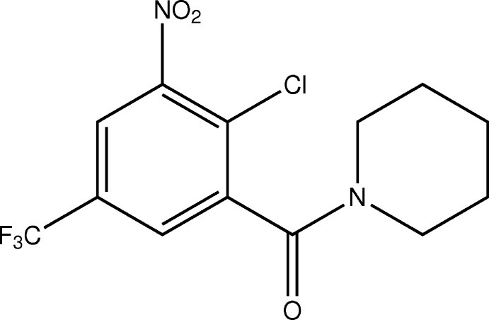
The crystal and molecular structure of [2-chloro-3-nitro-5-(trifluoromethyl)phenyl](piperidin-1-yl)methanone, a side product in the synthesis of an 8-nitro-1,3-benzothiazin-4-one, which belongs to a class of new anti-tuberculosis drug candidates, is reported.
Keywords: benzothiazinones, nitrobenzamides, anti-tuberculosis drugs, reaction mechanism, crystal structure
Abstract
1,3-Benzothiazin-4-ones (BTZs) are a promising new class of anti-tuberculosis drug candidates, some of which have reached clinical trials. The title compound, the benzamide derivative [2-chloro-3-nitro-5-(trifluoromethyl)phenyl](piperidin-1-yl)methanone, C13H12ClF3N2O3, occurs as a side product as a result of competitive reaction pathways in the nucleophilic attack during the synthesis of the BTZ 8-nitro-2-(piperidin-1-yl)-6-(trifluoromethyl)-1,3-benzothiazin-4-one, following the original synthetic route, whereby the corresponding benzoyl isothiocyanate is reacted with piperidine as secondary amine. In the title compound, the nitro group and the nearly planar amide group are significantly twisted out of the plane of the benzene ring. The piperidine ring adopts a chair conformation. The trifluoromethyl group exhibits slight rotational disorder with a refined ratio of occupancies of 0.972 (2):0.028 (2). There is structural evidence for intermolecular weak C—H⋯O hydrogen bonds.
Chemical context
1,3-Benzothiazin-4-ones (BTZs) are promising anti-tuberculosis drug candidates, some of which have already reached clinical trials (Mikušová et al., 2014 ▸; Makarov & Mikušová, 2020 ▸). Various methods for the synthesis of BTZs have been reported (Makarov et al., 2007 ▸; Moellmann et al., 2009 ▸; Makarov, 2011 ▸; Rudolph, 2014 ▸; Rudolph et al., 2016 ▸; Zhang & Aldrich, 2019 ▸). In the original synthesis, 2-chlorobenzoyl chloride derivatives are reacted with ammonium or alkali metal thiocyanates to form the corresponding 2-chlorobenzoyl isothiocyanates (Makarov et al., 2007 ▸; Moellmann et al., 2009 ▸). These are reactive species and are treated in situ with secondary amines to afford the corresponding thiourea derivatives, which undergo ring closure to give 1,3-thiazin-4-ones via an intramolecular nucleophilic substitution reaction. The latter step is favoured when electron-withdrawing substituents are present on the benzene ring.
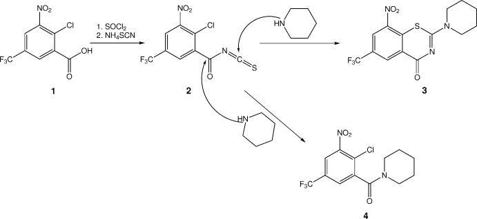
Fig. 1 ▸ depicts the synthesis following the original procedure for a BTZ previously reported by us (Rudolph, 2014 ▸; Rudolph et al., 2016 ▸; Richter, Rudolph et al., 2018 ▸). After treatment of 2-chloro-3-nitro-5-(trifluoromethyl)benzoic acid (1) with thionyl chloride and subsequently ammonium thiocyanate, the corresponding 2-chloro-3-nitro-5-(trifluoromethyl)benzoyl isothiocyanate (2) was reacted with piperidine. As illustrated, nucleophilic attack of the piperidine nitrogen atom at the isothiocyanate carbon atom leads to the anticipated 8-nitro-2-(piperidin-1-yl)-6-(trifluoromethyl)-1,3-benzothiazin-4-one (3). The alternative nucleophilic attack at the carbonyl carbon atom affords the side product (2-chloro-3-nitro-5-(trifluoromethyl)phenyl)(piperidin-1-yl)methanone (4), which was structurally characterized by X-ray crystallography in the present work. The ratio of 3 to 4 was found to vary depending on the reaction conditions. Temperatures at or below 283 K favour the formation of the anticipated 3, whereas substantial amounts of 4 form at elevated temperatures (Rudolph, 2014 ▸). Since BTZs are in clinical development [see, for example, Makarov & Mikušová (2020 ▸) or Mariandyshev et al. (2020 ▸)], this observation is not only important for the improvement of synthetic yields but also for the compilation of known synthetic side products for drug quality control.
Figure 1.
Synthetic pathway from 2-chloro-3-nitro-5-(trifluoromethyl)benzoic acid (1) to BTZ 3 and side product 4, illustrating the two different points of nucleophilic attack of piperidine at the intermediate 2-chloro-3-nitro-5-(trifluoromethyl)benzoyl isothiocyanate (2), resulting in 3 and 4 (Rudolph, 2014 ▸).
It is interesting to note that dinitrobenzamide derivatives related to 4 have been found to have some anti-mycobacterial activity (Christophe et al., 2009 ▸; Trefzer et al., 2010 ▸; Tiwari et al., 2013 ▸), and the non-chlorinated analogue of 4 was reported to have anticoccidial activity (Welch et al., 1969 ▸).
Structural commentary
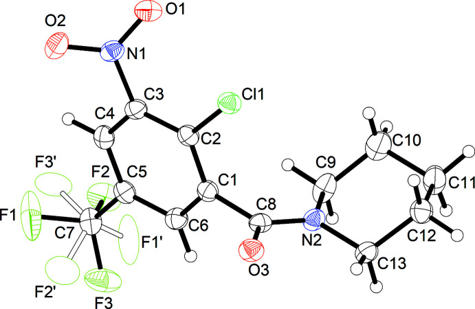
Fig. 2 ▸ shows the molecular structure of 4 in the solid state. Selected geometric parameters are listed in Table 1 ▸. The dihedral angle between the plane of the nitro group and the mean plane of the benzene ring is 38.1 (2)°, which can be attributed to the steric demand of the neighbouring chloro substituent at the benzene ring. The trifluoromethyl group exhibits rotational disorder over two sites with 97.2 (2)% occupancy for the major site. The plane of the amide group, as defined by C8, O3 and N2, is tilted out of the mean plane of the benzene ring by 79.6 (1)°. The Winkler–Dunitz parameters for the amide linkage τ (twist angle) = 1.2° and χN (pyramidalization at nitrogen) = 4.0° indicate an almost planar amide group (Winkler & Dunitz, 1971 ▸). In the IR spectrum (see supporting information), the band at 1639 cm−1 can be assigned to the C=O stretching vibration of the amide group. The molecule is axially chiral, although the centrosymmetric crystal structure contains both enantiomers. The 13C NMR spectrum of 4 in methanol-d 4 as well as chloroform-d at room temperature (see supporting information) displays five distinct signals in the aliphatic region, which are assigned to the piperidine carbon atoms, indicating that the rotation about the amide C—N bond is slow in solution under these conditions. The 13C NMR chemical shift of the α-carbon atom C13 syn to the carbonyl oxygen atom of the amide group is shielded compared with that of the anti α-carbon atom C9. In chloroform-d, the observed shielding magnitude of ΔδC = 5.0 ppm is within the range expected for a benzoylpiperidine (Rubiralta et al., 1991 ▸). In the corresponding 1H NMR spectrum, the syn protons with respect to the amide carbonyl oxygen atom are deshielded compared with those in the anti position (ΔδH = 0.58 ppm). Complete assignments of 1H and 13C NMR data in chloroform-d by 13C,1H-HSQC and -HMBC NMR spectra can be found in the supporting information. Notably, the two separated methylene 1H NMR signals assigned to C10 in chloroform-d appear as one signal in methanol-d 4.
Figure 2.
Molecular structure of 4. Displacement ellipsoids are drawn at the 50% probability level. H atoms are represented by small spheres of arbitrary radii. The minor occupancy component of the disordered trifluoromethyl group is depicted by empty ellipsoids.
Table 1. Selected geometric parameters (Å, °).
| C1—C8 | 1.510 (3) | C7—F3 | 1.328 (3) |
| C2—Cl1 | 1.725 (2) | C7—F2 | 1.336 (3) |
| C3—N1 | 1.468 (3) | C8—O3 | 1.234 (2) |
| C5—C7 | 1.497 (3) | C8—N2 | 1.342 (3) |
| C7—F1 | 1.325 (3) | ||
| C4—C3—N1 | 116.41 (17) | N2—C9—C10 | 110.59 (18) |
| C2—C3—N1 | 122.35 (18) | C9—C10—C11 | 110.61 (19) |
| F1—C7—F3 | 107.69 (19) | C12—C11—C10 | 109.74 (18) |
| F1—C7—F2 | 105.98 (19) | C13—C12—C11 | 111.01 (18) |
| F3—C7—F2 | 105.59 (17) | N2—C13—C12 | 111.35 (17) |
| F1—C7—C5 | 112.43 (17) | O2—N1—O1 | 124.48 (17) |
| F3—C7—C5 | 112.80 (18) | O2—N1—C3 | 117.04 (17) |
| F2—C7—C5 | 111.86 (17) | O1—N1—C3 | 118.44 (16) |
| O3—C8—N2 | 124.72 (18) | C8—N2—C13 | 120.26 (16) |
| O3—C8—C1 | 118.43 (18) | C8—N2—C9 | 124.74 (16) |
| N2—C8—C1 | 116.85 (17) | C13—N2—C9 | 114.89 (16) |
| C4—C3—N1—O2 | 36.6 (2) | O3—C8—N2—C13 | 3.0 (3) |
| C2—C3—N1—O2 | −143.29 (19) | C1—C8—N2—C13 | −176.62 (17) |
| C4—C3—N1—O1 | −141.34 (18) | O3—C8—N2—C9 | 179.0 (2) |
| C2—C3—N1—O1 | 38.8 (3) | C1—C8—N2—C9 | −0.6 (3) |
In the solid state, the piperidine ring in 4 adopts a low-energy chair conformation with some minor angular deviations from ideal tetrahedral values, resulting from planarity at N2 due to involvement in the amide linkage. The puckering parameters of the piperidine six-membered ring, as calculated with PLATON (Spek, 2020 ▸), are Q = 0.555 (2) Å, θ = 4.1 (2)° and φ = 161 (3)°. By way of comparison, the total puckering amplitude Q is 0.63 Å and the magnitude of distortion θ is 0° for an ideal cyclohexane chair (Cremer & Pople, 1975 ▸).
Supramolecular features
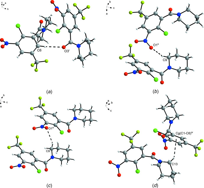
In general, the crystal structure of 4 appears to be dominated by close packing. According to Kitaigorodskii (1973 ▸), the space group Pbca is among those available for the densest packing of molecules of arbitrary shape. Nevertheless, the solid-state supramolecular structure features C—H⋯O contacts between an aromatic CH moiety and the amide oxygen atom of an adjacent molecule (Fig. 3 ▸ a). The corresponding geometric parameters (Table 2 ▸) support the interpretation as a weak hydrogen bond (Thakuria et al., 2017 ▸). These interactions link the molecules into strands extending by 21 screw symmetry in the [010] direction. The α-methylene groups of the piperidine ring, on which the amide group should exert an electron-withdrawing effect, also form intermolecular C—H⋯O and C—H⋯π contacts, respectively, to the nitro group and the benzene ring of adjacent molecules (Fig. 3 ▸ b–d). The corresponding geometric parameters (Table 2 ▸), however, reveal that these contacts may not have the same significance here as the aforementioned Caromatic—H⋯Oamide short contact (Wood et al., 2009 ▸). It is also worth noting that π–π stacking of the aromatic rings is not observed.
Figure 3.
Short contacts (dashed lines) between adjacent molecules in the crystal structure of 4. The minor component of the disordered trifluoromethyl group is omitted for clarity. Symmetry codes: (i) −x + 1, y +  , −z +
  ; (ii) −x +
  , y +
, y +  , z; (iii) x, y + 1, z; (iv) x +
  , −y +
  , −z.
, −z.
Table 2. Hydrogen-bond geometry (Å, °).
| D—H⋯A | D—H | H⋯A | D⋯A | D—H⋯A |
|---|---|---|---|---|
| C6—H6⋯O3i | 0.95 | 2.59 | 3.526 (3) | 169 |
| C9—H9A⋯O1ii | 0.99 | 2.45 | 3.361 (3) | 154 |
| C9—H9B⋯O1iii | 0.99 | 2.58 | 3.369 (3) | 137 |
| C13—H13A⋯Cg(C1–C6)iv | 0.99 | 2.92 | 3.447 (2) | 114 |
Symmetry codes: (i)  ; (ii)
  ; (iii)
  ; (iv)
  .
.
Database survey
A search of the Cambridge Structural Database (CSD; version 5.41 with March 2020 updates; Groom et al., 2016 ▸) for related substituted N-benzoyl-piperidine compounds revealed about 30 structures, of which (2-chloro-3,5-dinitrophenyl)(piperidin-1-yl)methanone (CSD refcode: URALIJ; Luo et al., 2011 ▸) is structurally most related to 4. Similar to 4, the 3-nitro group with the neighbouring chloro substituent is tilted out of the mean plane of the benzene ring by 36.2°. At 75.8°, the dihedral angle between the amide plane and the mean plane of the benzene ring is comparable with that in 4. Likewise, the piperidine ring exhibits a chair conformation with a planar structure at the nitrogen atom. In contrast to 4, the solid-state supramolecular structure of URALIJ exhibits π–π stacking of the aromatic rings. Interestingly, a CSD search for the 2-chloro-3-nitro-5-(trifluoromethyl)phenyl moiety present in 4 led to only one structure, viz. 2-chloro-1,3-dinitro-5-(trifluoromethyl)benzene (JIHNUM; del Casino et al., 2018 ▸), also known as chloralin, which is active against Plasmodium, but which also shows toxicity in mice.
Anti-mycobacterial evaluation
The anti-mycobacterial activity of 4 was evaluated against Mycobacterium smegmatis mc2 155 and Mycobacterium abscessus ATCC19977, using broth microdilution assays [for the assay protocols, see the supporting information and Richter, Strauch et al. (2018 ▸)]. For both mycobacterial species, no growth inhibition was detectable up to a concentration of 100 µM. For M. smegmatis, the findings are consistent with the activity data for a related nitrobenzamide derivative reported by Tiwari et al. (2013 ▸). CT319, a 3-nitro-5-(trifluoromethyl)benzamide derivative, however, showed activity against M. smegmatis mc2 155 and other mycobacterial strains (Trefzer et al., 2010 ▸).
Synthesis and crystallization
Chemicals were purchased and used as received. The synthesis of 1 is described elsewhere (Welch et al., 1969 ▸). Solvents were of reagent grade and were distilled before use. The IR spectrum was measured on a Bruker TENSOR II FT–IR spectrometer at a resolution of 4 cm−1. NMR spectra were recorded at room temperature on an Agilent Technologies VNMRS 400 MHz NMR spectrometer (abbreviations: d = doublet, q = quartet, m = multiplet). Chemical shifts are referenced to the residual signals of methanol-d 4 (δH = 3.35 ppm, δC = 49.3 ppm) or chloroform-d (δH = 7.26 ppm, δC = 77.2 ppm).
2.7 mL (37.0 mmol) of SOCl2 were added to a stirred solution of 1 (5.00 g,18.5 mmol) in toluene, and the mixture was heated to reflux for two h. The solvent was subsequently removed under reduced pressure, and the acid chloride thus obtained was used without purification. The residue was taken up in 6.5 mL of acetonitrile and a solution of 1.41 g (18.5 mmol) NH4SCN in 55 mL of acetonitrile was added dropwise with stirring to obtain 2 in situ. After stirring for 5 min at 313 K, the resulting NH4Cl precipitate was filtered off, and 3.7 mL (37.0 mmol) of piperidine were added. The mixture was refluxed overnight, and then the solvent was removed under reduced pressure. Water was added to the residue and, after extraction with dichloromethane, the organic phase was washed with 10% aqueous NaHCO3 and dried over MgSO4. After removal of the solvent, the crude product was subjected to flash chromatography on silica gel, eluting with ethyl acetate/n-heptane (gradient 10–50% v/v), to isolate 1.09 g (3.0 mmol, 16%) of 3 and a minor amount of the side product 4. 1H and 13C NMR spectroscopic and mass spectrometric data of 3 were in agreement with those in the literature (Rudolph, 2014 ▸; Rudolph et al., 2016 ▸). Crystals of 4 suitable for X-ray crystallography were obtained from a solution in ethyl acetate/heptane (1:1) by slow evaporation of the solvents at room temperature. NMR spectroscopic data for 4:
1H NMR (400 MHz, CD3OD) δ 8.42 (d, 4 J meta = 2.2 Hz, 1H, Ar—H), 8.09 (d, 4 J meta = 2.2 Hz, 1H, Ar—H), 3.88–3.71 (m, 2H, N—CH 2), 3.33–3.21 (m, 2H, N—CH 2), 1.76 (m, 4H, CH2), 1.64 (m, 2H, CH2) ppm; 13C NMR (101 MHz, CD3OD) δ 165.5, 150.7, 141.8, 132.3 (q, 2 J C,F = 35 Hz), 129.2 (q, 3 J C,F = 4 Hz), 128.1, 124.4 (q, 3 J C,F = 4 Hz), 124.1 (q, 1 J C,F = 273 Hz), 49.5, 44.3, 27.6, 26.7, 25.5 ppm.
1H NMR (400 MHz, CDCl3 δ) 8.07 (d, 4 J meta = 2.0 Hz, 1H, C4—H), 7.73 (d, 4 J meta = 2.0 Hz, 1H, C6—H), 3.83–3.68 (m, 2H, C13—CH 2), 3.22 (ddd, 2 J gem = 13.2 Hz, 3 J vic = 7.1, 4.0 Hz, 1H, C9—CH 2), 3.15 (ddd, 2 J gem = 13.2 Hz, 3 J vic = 7.1, 4.0 Hz, 1H, C9—CH 2), 1.70 (m, 4H, C11, C12—CH 2), 1.65–1.57 (m, 1H, C10—CH 2), 1.56–1.47 (m, 1H, C10—CH 2) ppm; 13C NMR (101 MHz, CDCl3 δ) 163.5 (C8, C=O), 148.9 (C3), 141.1 (C1), 131.2 (q, 2 J C,F = 35 Hz, C5), 127.8 (q, 3 J C,F = 4 Hz, C6), 127.6 (C2), 122.7 (q, 3 J C,F = 4 Hz, C4), 122.4 (q, 1 J C,F = 273 Hz, C7), 48.3 (C9), 43.3 (C13), 26.7 (C10), 25.7 (C12), 24.6 (C11) ppm.
Refinement
Crystal data, data collection and structure refinement details are summarized in Table 3 ▸. The rotational disorder of the trifluoromethyl group was refined using a split model with similar distance restraints on the 1,2- and 1,3-distances and equal atomic displacement parameters for opposite fluorine atoms belonging to different disorder sites. Refinement of the ratio of occupancies by means of a free variable resulted in 0.972 (2):0.028 (2). Hydrogen-atom positions were calculated geometrically with Ca—H = 0.95 Å and Cm—H = 0.99 Å (a = aromatic and m = methylene), and refined with the appropriate riding model and U iso(H) = 1.2 U eq(C).
Table 3. Experimental details.
| Crystal data | |
| Chemical formula | C13H12ClF3N2O3 |
| M r | 336.70 |
| Crystal system, space group | Orthorhombic, P b c a |
| Temperature (K) | 100 |
| a, b, c (Å) | 18.0904 (7), 7.8971 (3), 19.8043 (8) |
| V (Å3) | 2829.28 (19) |
| Z | 8 |
| Radiation type | Cu Kα |
| μ (mm−1) | 2.88 |
| Crystal size (mm) | 0.59 × 0.50 × 0.44 |
| Data collection | |
| Diffractometer | Bruker Kappa Mach3 APEXII |
| Absorption correction | Gaussian (SADABS; Krause et al., 2015 ▸) |
| T min, T max | 0.297, 0.586 |
| No. of measured, independent and observed [I > 2σ(I)] reflections | 49954, 2784, 2699 |
| R int | 0.041 |
| (sin θ/λ)max (Å−1) | 0.617 |
| Refinement | |
| R[F 2 > 2σ(F 2)], wR(F 2), S | 0.043, 0.115, 1.15 |
| No. of reflections | 2784 |
| No. of parameters | 209 |
| No. of restraints | 45 |
| H-atom treatment | H-atom parameters constrained |
| Δρmax, Δρmin (e Å−3) | 0.32, −0.32 |
Supplementary Material
Crystal structure: contains datablock(s) global, I. DOI: 10.1107/S2056989020010658/vm2238sup1.cif
Structure factors: contains datablock(s) I. DOI: 10.1107/S2056989020010658/vm2238Isup2.hkl
Supporting information file. DOI: 10.1107/S2056989020010658/vm2238Isup3.cdx
IR and NMR spectra, assay protocol for anti-mycobacterial activity. DOI: 10.1107/S2056989020010658/vm2238sup4.pdf
Supporting information file. DOI: 10.1107/S2056989020010658/vm2238Isup5.cml
CCDC reference: 2021003
Additional supporting information: crystallographic information; 3D view; checkCIF report
Acknowledgments
We thank Professor Christian W. Lehmann for providing access to the X-ray diffraction facility and Heike Schucht for technical assistance.
supplementary crystallographic information
Crystal data
| C13H12ClF3N2O3 | Dx = 1.581 Mg m−3 |
| Mr = 336.70 | Cu Kα radiation, λ = 1.54178 Å |
| Orthorhombic, Pbca | Cell parameters from 9968 reflections |
| a = 18.0904 (7) Å | θ = 4.9–71.6° |
| b = 7.8971 (3) Å | µ = 2.88 mm−1 |
| c = 19.8043 (8) Å | T = 100 K |
| V = 2829.28 (19) Å3 | Block, colourless |
| Z = 8 | 0.59 × 0.50 × 0.44 mm |
| F(000) = 1376 |
Data collection
| Bruker Kappa Mach3 APEXII diffractometer | 2784 independent reflections |
| Radiation source: 0.2 × 2 mm2 focus rotating anode | 2699 reflections with I > 2σ(I) |
| MONTEL graded multilayer optics monochromator | Rint = 0.041 |
| Detector resolution: 66.67 pixels mm-1 | θmax = 72.2°, θmin = 4.9° |
| φ– and ω–scans | h = −21→22 |
| Absorption correction: gaussian (SADABS; Krause et al., 2015) | k = −9→9 |
| Tmin = 0.297, Tmax = 0.586 | l = −24→24 |
| 49954 measured reflections |
Refinement
| Refinement on F2 | Primary atom site location: dual |
| Least-squares matrix: full | Secondary atom site location: difference Fourier map |
| R[F2 > 2σ(F2)] = 0.043 | Hydrogen site location: inferred from neighbouring sites |
| wR(F2) = 0.115 | H-atom parameters constrained |
| S = 1.15 | w = 1/[σ2(Fo2) + (0.0508P)2 + 2.7994P] where P = (Fo2 + 2Fc2)/3 |
| 2784 reflections | (Δ/σ)max < 0.001 |
| 209 parameters | Δρmax = 0.32 e Å−3 |
| 45 restraints | Δρmin = −0.32 e Å−3 |
Special details
| Geometry. All esds (except the esd in the dihedral angle between two l.s. planes) are estimated using the full covariance matrix. The cell esds are taken into account individually in the estimation of esds in distances, angles and torsion angles; correlations between esds in cell parameters are only used when they are defined by crystal symmetry. An approximate (isotropic) treatment of cell esds is used for estimating esds involving l.s. planes. |
Fractional atomic coordinates and isotropic or equivalent isotropic displacement parameters (Å2)
| x | y | z | Uiso*/Ueq | Occ. (<1) | |
| C1 | 0.41556 (10) | 0.3250 (3) | 0.23581 (10) | 0.0230 (4) | |
| C2 | 0.38370 (10) | 0.1651 (3) | 0.23833 (10) | 0.0224 (4) | |
| C3 | 0.35007 (10) | 0.1004 (3) | 0.18023 (10) | 0.0227 (4) | |
| C4 | 0.34763 (10) | 0.1927 (3) | 0.12111 (10) | 0.0243 (4) | |
| H4 | 0.325215 | 0.146458 | 0.081847 | 0.029* | |
| C5 | 0.37831 (10) | 0.3541 (3) | 0.11961 (10) | 0.0242 (4) | |
| C6 | 0.41190 (11) | 0.4206 (3) | 0.17666 (10) | 0.0250 (4) | |
| H6 | 0.432417 | 0.531321 | 0.175487 | 0.030* | |
| C7 | 0.37425 (12) | 0.4549 (3) | 0.05572 (11) | 0.0298 (5) | |
| C8 | 0.45848 (10) | 0.3906 (2) | 0.29575 (10) | 0.0232 (4) | |
| C9 | 0.34929 (13) | 0.5683 (3) | 0.32422 (11) | 0.0327 (5) | |
| H9A | 0.325497 | 0.503425 | 0.287381 | 0.039* | |
| H9B | 0.351745 | 0.688568 | 0.310277 | 0.039* | |
| C10 | 0.30335 (12) | 0.5528 (3) | 0.38813 (13) | 0.0404 (6) | |
| H10A | 0.296350 | 0.431647 | 0.399304 | 0.048* | |
| H10B | 0.254021 | 0.603627 | 0.380559 | 0.048* | |
| C11 | 0.34134 (13) | 0.6421 (3) | 0.44689 (11) | 0.0373 (5) | |
| H11A | 0.344826 | 0.764952 | 0.437412 | 0.045* | |
| H11B | 0.311898 | 0.626777 | 0.488581 | 0.045* | |
| C12 | 0.41843 (12) | 0.5691 (3) | 0.45695 (10) | 0.0294 (5) | |
| H12A | 0.443955 | 0.632047 | 0.493365 | 0.035* | |
| H12B | 0.414464 | 0.449164 | 0.470978 | 0.035* | |
| C13 | 0.46343 (11) | 0.5807 (3) | 0.39245 (10) | 0.0277 (4) | |
| H13A | 0.473754 | 0.701122 | 0.382205 | 0.033* | |
| H13B | 0.511333 | 0.522352 | 0.398982 | 0.033* | |
| N1 | 0.31581 (9) | −0.0681 (2) | 0.17845 (9) | 0.0253 (4) | |
| N2 | 0.42423 (9) | 0.5038 (2) | 0.33544 (8) | 0.0249 (4) | |
| O1 | 0.28104 (8) | −0.1161 (2) | 0.22819 (8) | 0.0324 (4) | |
| O2 | 0.32229 (8) | −0.1493 (2) | 0.12611 (8) | 0.0341 (4) | |
| O3 | 0.52198 (7) | 0.33806 (19) | 0.30471 (7) | 0.0288 (3) | |
| F1 | 0.38438 (14) | 0.3603 (2) | 0.00122 (7) | 0.0711 (7) | 0.972 (2) |
| F2 | 0.30831 (8) | 0.52847 (19) | 0.04797 (8) | 0.0418 (4) | 0.972 (2) |
| F3 | 0.42321 (8) | 0.5801 (2) | 0.05379 (8) | 0.0470 (4) | 0.972 (2) |
| F1' | 0.352 (3) | 0.612 (3) | 0.0688 (18) | 0.0711 (7) | 0.028 (2) |
| F2' | 0.4387 (11) | 0.472 (6) | 0.0269 (18) | 0.0418 (4) | 0.028 (2) |
| F3' | 0.325 (2) | 0.397 (5) | 0.0129 (15) | 0.0470 (4) | 0.028 (2) |
| Cl1 | 0.38924 (3) | 0.05417 (6) | 0.31320 (2) | 0.02635 (16) |
Atomic displacement parameters (Å2)
| U11 | U22 | U33 | U12 | U13 | U23 | |
| C1 | 0.0194 (9) | 0.0250 (9) | 0.0246 (9) | 0.0021 (7) | 0.0012 (7) | −0.0019 (8) |
| C2 | 0.0186 (8) | 0.0246 (9) | 0.0239 (9) | 0.0021 (7) | 0.0011 (7) | 0.0002 (8) |
| C3 | 0.0166 (8) | 0.0219 (9) | 0.0296 (10) | 0.0011 (7) | 0.0009 (7) | −0.0021 (8) |
| C4 | 0.0202 (9) | 0.0288 (10) | 0.0239 (9) | 0.0050 (8) | −0.0004 (7) | −0.0031 (8) |
| C5 | 0.0219 (9) | 0.0261 (10) | 0.0247 (10) | 0.0056 (8) | 0.0021 (7) | 0.0011 (8) |
| C6 | 0.0248 (10) | 0.0226 (9) | 0.0277 (10) | 0.0006 (8) | 0.0004 (8) | 0.0002 (8) |
| C7 | 0.0350 (11) | 0.0292 (11) | 0.0251 (10) | 0.0056 (9) | 0.0005 (8) | −0.0002 (8) |
| C8 | 0.0225 (9) | 0.0226 (9) | 0.0245 (9) | −0.0035 (8) | −0.0003 (7) | 0.0033 (8) |
| C9 | 0.0335 (11) | 0.0298 (11) | 0.0347 (11) | 0.0094 (9) | −0.0117 (9) | −0.0088 (9) |
| C10 | 0.0200 (10) | 0.0517 (15) | 0.0495 (14) | 0.0058 (9) | −0.0038 (9) | −0.0188 (11) |
| C11 | 0.0324 (11) | 0.0474 (14) | 0.0322 (11) | 0.0039 (10) | −0.0011 (9) | −0.0114 (10) |
| C12 | 0.0308 (11) | 0.0327 (11) | 0.0247 (10) | −0.0010 (9) | −0.0013 (8) | 0.0002 (8) |
| C13 | 0.0267 (10) | 0.0306 (10) | 0.0257 (10) | −0.0070 (8) | −0.0028 (8) | −0.0018 (8) |
| N1 | 0.0190 (8) | 0.0260 (9) | 0.0310 (9) | −0.0009 (6) | −0.0037 (7) | −0.0016 (7) |
| N2 | 0.0213 (8) | 0.0277 (9) | 0.0257 (8) | −0.0010 (7) | −0.0042 (7) | −0.0030 (7) |
| O1 | 0.0267 (7) | 0.0334 (8) | 0.0371 (8) | −0.0062 (6) | 0.0010 (6) | 0.0028 (7) |
| O2 | 0.0348 (8) | 0.0320 (8) | 0.0356 (8) | −0.0010 (6) | −0.0045 (6) | −0.0098 (7) |
| O3 | 0.0204 (7) | 0.0324 (8) | 0.0337 (8) | 0.0005 (6) | −0.0017 (6) | −0.0003 (6) |
| F1 | 0.151 (2) | 0.0382 (9) | 0.0239 (7) | 0.0278 (10) | 0.0154 (9) | −0.0002 (6) |
| F2 | 0.0336 (7) | 0.0457 (8) | 0.0460 (8) | 0.0053 (6) | −0.0056 (6) | 0.0175 (7) |
| F3 | 0.0389 (8) | 0.0567 (10) | 0.0453 (8) | −0.0142 (7) | −0.0052 (6) | 0.0251 (7) |
| F1' | 0.151 (2) | 0.0382 (9) | 0.0239 (7) | 0.0278 (10) | 0.0154 (9) | −0.0002 (6) |
| F2' | 0.0336 (7) | 0.0457 (8) | 0.0460 (8) | 0.0053 (6) | −0.0056 (6) | 0.0175 (7) |
| F3' | 0.0389 (8) | 0.0567 (10) | 0.0453 (8) | −0.0142 (7) | −0.0052 (6) | 0.0251 (7) |
| Cl1 | 0.0267 (3) | 0.0273 (3) | 0.0250 (3) | −0.00271 (18) | 0.00002 (17) | 0.00364 (17) |
Geometric parameters (Å, º)
| C1—C2 | 1.389 (3) | C8—N2 | 1.342 (3) |
| C1—C6 | 1.395 (3) | C9—N2 | 1.465 (3) |
| C1—C8 | 1.510 (3) | C9—C10 | 1.519 (3) |
| C2—C3 | 1.398 (3) | C9—H9A | 0.9900 |
| C2—Cl1 | 1.725 (2) | C9—H9B | 0.9900 |
| C3—C4 | 1.380 (3) | C10—C11 | 1.525 (3) |
| C3—N1 | 1.468 (3) | C10—H10A | 0.9900 |
| C4—C5 | 1.390 (3) | C10—H10B | 0.9900 |
| C4—H4 | 0.9500 | C11—C12 | 1.522 (3) |
| C5—C6 | 1.386 (3) | C11—H11A | 0.9900 |
| C5—C7 | 1.497 (3) | C11—H11B | 0.9900 |
| C6—H6 | 0.9500 | C12—C13 | 1.518 (3) |
| C7—F2' | 1.304 (14) | C12—H12A | 0.9900 |
| C7—F3' | 1.314 (14) | C12—H12B | 0.9900 |
| C7—F1 | 1.325 (3) | C13—N2 | 1.465 (2) |
| C7—F3 | 1.328 (3) | C13—H13A | 0.9900 |
| C7—F1' | 1.332 (14) | C13—H13B | 0.9900 |
| C7—F2 | 1.336 (3) | N1—O2 | 1.225 (2) |
| C8—O3 | 1.234 (2) | N1—O1 | 1.229 (2) |
| C2—C1—C6 | 120.16 (18) | C10—C9—H9A | 109.5 |
| C2—C1—C8 | 119.82 (17) | N2—C9—H9B | 109.5 |
| C6—C1—C8 | 119.90 (17) | C10—C9—H9B | 109.5 |
| C1—C2—C3 | 118.92 (18) | H9A—C9—H9B | 108.1 |
| C1—C2—Cl1 | 117.93 (15) | C9—C10—C11 | 110.61 (19) |
| C3—C2—Cl1 | 123.13 (16) | C9—C10—H10A | 109.5 |
| C4—C3—C2 | 121.24 (18) | C11—C10—H10A | 109.5 |
| C4—C3—N1 | 116.41 (17) | C9—C10—H10B | 109.5 |
| C2—C3—N1 | 122.35 (18) | C11—C10—H10B | 109.5 |
| C3—C4—C5 | 119.32 (18) | H10A—C10—H10B | 108.1 |
| C3—C4—H4 | 120.3 | C12—C11—C10 | 109.74 (18) |
| C5—C4—H4 | 120.3 | C12—C11—H11A | 109.7 |
| C6—C5—C4 | 120.33 (18) | C10—C11—H11A | 109.7 |
| C6—C5—C7 | 120.58 (19) | C12—C11—H11B | 109.7 |
| C4—C5—C7 | 119.09 (18) | C10—C11—H11B | 109.7 |
| C5—C6—C1 | 119.98 (19) | H11A—C11—H11B | 108.2 |
| C5—C6—H6 | 120.0 | C13—C12—C11 | 111.01 (18) |
| C1—C6—H6 | 120.0 | C13—C12—H12A | 109.4 |
| F2'—C7—F3' | 111.1 (19) | C11—C12—H12A | 109.4 |
| F1—C7—F3 | 107.69 (19) | C13—C12—H12B | 109.4 |
| F2'—C7—F1' | 105.2 (19) | C11—C12—H12B | 109.4 |
| F3'—C7—F1' | 104.0 (19) | H12A—C12—H12B | 108.0 |
| F1—C7—F2 | 105.98 (19) | N2—C13—C12 | 111.35 (17) |
| F3—C7—F2 | 105.59 (17) | N2—C13—H13A | 109.4 |
| F2'—C7—C5 | 112.3 (15) | C12—C13—H13A | 109.4 |
| F3'—C7—C5 | 113.2 (15) | N2—C13—H13B | 109.4 |
| F1—C7—C5 | 112.43 (17) | C12—C13—H13B | 109.4 |
| F3—C7—C5 | 112.80 (18) | H13A—C13—H13B | 108.0 |
| F1'—C7—C5 | 110.3 (15) | O2—N1—O1 | 124.48 (17) |
| F2—C7—C5 | 111.86 (17) | O2—N1—C3 | 117.04 (17) |
| O3—C8—N2 | 124.72 (18) | O1—N1—C3 | 118.44 (16) |
| O3—C8—C1 | 118.43 (18) | C8—N2—C13 | 120.26 (16) |
| N2—C8—C1 | 116.85 (17) | C8—N2—C9 | 124.74 (16) |
| N2—C9—C10 | 110.59 (18) | C13—N2—C9 | 114.89 (16) |
| N2—C9—H9A | 109.5 | ||
| C6—C1—C2—C3 | 1.9 (3) | C6—C5—C7—F1' | −47 (3) |
| C8—C1—C2—C3 | −174.16 (17) | C4—C5—C7—F1' | 133 (3) |
| C6—C1—C2—Cl1 | −179.32 (15) | C6—C5—C7—F2 | −99.4 (2) |
| C8—C1—C2—Cl1 | 4.6 (2) | C4—C5—C7—F2 | 80.3 (2) |
| C1—C2—C3—C4 | −0.5 (3) | C2—C1—C8—O3 | 77.8 (2) |
| Cl1—C2—C3—C4 | −179.22 (14) | C6—C1—C8—O3 | −98.3 (2) |
| C1—C2—C3—N1 | 179.37 (16) | C2—C1—C8—N2 | −102.6 (2) |
| Cl1—C2—C3—N1 | 0.6 (3) | C6—C1—C8—N2 | 81.4 (2) |
| C2—C3—C4—C5 | −0.9 (3) | N2—C9—C10—C11 | 55.5 (3) |
| N1—C3—C4—C5 | 179.25 (16) | C9—C10—C11—C12 | −56.9 (3) |
| C3—C4—C5—C6 | 0.9 (3) | C10—C11—C12—C13 | 55.8 (3) |
| C3—C4—C5—C7 | −178.87 (18) | C11—C12—C13—N2 | −53.4 (2) |
| C4—C5—C6—C1 | 0.5 (3) | C4—C3—N1—O2 | 36.6 (2) |
| C7—C5—C6—C1 | −179.75 (18) | C2—C3—N1—O2 | −143.29 (19) |
| C2—C1—C6—C5 | −1.9 (3) | C4—C3—N1—O1 | −141.34 (18) |
| C8—C1—C6—C5 | 174.13 (18) | C2—C3—N1—O1 | 38.8 (3) |
| C6—C5—C7—F2' | 70 (2) | O3—C8—N2—C13 | 3.0 (3) |
| C4—C5—C7—F2' | −110 (2) | C1—C8—N2—C13 | −176.62 (17) |
| C6—C5—C7—F3' | −163 (2) | O3—C8—N2—C9 | 179.0 (2) |
| C4—C5—C7—F3' | 17 (2) | C1—C8—N2—C9 | −0.6 (3) |
| C6—C5—C7—F1 | 141.5 (2) | C12—C13—N2—C8 | −130.0 (2) |
| C4—C5—C7—F1 | −38.8 (3) | C12—C13—N2—C9 | 53.6 (2) |
| C6—C5—C7—F3 | 19.5 (3) | C10—C9—N2—C8 | 129.2 (2) |
| C4—C5—C7—F3 | −160.82 (18) | C10—C9—N2—C13 | −54.6 (2) |
Hydrogen-bond geometry (Å, º)
| D—H···A | D—H | H···A | D···A | D—H···A |
| C6—H6···O3i | 0.95 | 2.59 | 3.526 (3) | 169 |
| C9—H9A···O1ii | 0.99 | 2.45 | 3.361 (3) | 154 |
| C9—H9B···O1iii | 0.99 | 2.58 | 3.369 (3) | 137 |
| C13—H13A···Cg(C1–C6)iv | 0.99 | 2.92 | 3.447 (2) | 114 |
Symmetry codes: (i) −x+1, y+1/2, −z+1/2; (ii) −x+1/2, y+1/2, z; (iii) x, y+1, z; (iv) x+3/2, −y+1/2, −z.
Funding Statement
This work was funded by Deutsche Forschungsgemeinschaft grant .
References
- Allen, F. H., Johnson, O., Shields, G. P., Smith, B. R. & Towler, M. (2004). J. Appl. Cryst. 37, 335–338.
- Brandenburg, K. (2018). DIAMOND. Crystal Impact GbR, Bonn, Germany.
- Bruker (2004). SAINT. Bruker AXS Inc., Madison, Wisconsin, USA.
- Bruker (2017). APEX3. Bruker AXS Inc., Madison, Wisconsin, USA.
- Casino, A. del, Lukinović, V., Bhatt, R., Randle, L. E., Dascombe, M. J., Fennell, B. J., Drew, M. G. B., Bell, A., Fielding, A. J. & Ismail, F. M. D. (2018). ChemistrySelect 3, 7572–7580.
- Christophe, T., Jackson, M., Jeon, H. K., Fenistein, D., Contreras-Dominguez, M., Kim, J., Genovesio, A., Carralot, J.-P., Ewann, F., Kim, E. H., Lee, S. Y., Kang, S., Seo, M. J., Park, E. J., Škovierová, H., Pham, H., Riccardi, G., Nam, J. Y., Marsollier, L., Kempf, M., Joly-Guillou, M.-L., Oh, T., Shin, W. K., No, Z., Nehrbass, U., Brosch, R., Cole, S. T. & Brodin, P. (2009). PLoS Pathog. 5, e1000645. [DOI] [PMC free article] [PubMed]
- Cremer, D. & Pople, J. A. (1975). J. Am. Chem. Soc. 97, 1354–1358.
- Groom, C. R., Bruno, I. J., Lightfoot, M. P. & Ward, S. C. (2016). Acta Cryst. B72, 171–179. [DOI] [PMC free article] [PubMed]
- Kitaigorodskii, A. I. (1973). Molecular Crystals and Molecules. London: Academic Press.
- Krause, L., Herbst-Irmer, R., Sheldrick, G. M. & Stalke, D. (2015). J. Appl. Cryst. 48, 3–10. [DOI] [PMC free article] [PubMed]
- Luo, X., Huang, Y.-C., Gao, C. & Yu, L.-T. (2011). Acta Cryst. E67, o1066. [DOI] [PMC free article] [PubMed]
- Makarov, V. (2011). PCT Int. Appl. WO 2011132070 A1.
- Makarov, V., Cole, S. T. & Moellmann, U. (2007). PCT Int. Appl. WO 2007134625 A1.
- Makarov, V. & Mikušová, K. (2020). Appl. Sci. 10, 2269.
- Mariandyshev, A. O., Khokhlov, A. L., Smerdin, S. V., Shcherbakova, V. S., Igumnova, O. V., Ozerova, I. V., Bolgarina, A. A. & Nikitina, N. A. (2020). Ter. Arkh. 92, 61–72. [DOI] [PubMed]
- Mikušová, K., Makarov, V. & Neres, J. (2014). Curr. Pharm. Des. 20, 4379–4403. [DOI] [PubMed]
- Moellmann, U., Makarov, V. & Cole, S. T. (2009). PCT Int. Appl. WO 2009010163 A1.
- Richter, A., Rudolph, I., Möllmann, U., Voigt, K., Chung, C., Singh, O. M. P., Rees, M., Mendoza-Losana, A., Bates, R., Ballell, L., Batt, S., Veerapen, N., Fütterer, K., Besra, G., Imming, P. & Argyrou, A. (2018). Sci. Rep. 8, 13473. [DOI] [PMC free article] [PubMed]
- Richter, A., Strauch, A., Chao, J., Ko, M. & Av-Gay, Y. (2018). Antimicrob. Agents Chemother. 62, e00828–18. [DOI] [PMC free article] [PubMed]
- Rubiralta, M., Giralt, E. & Diez, A. (1991). 7 - N-Acylpiperidines. A Useful Tool for Stereocontrol in Organic Synthesis. In: Studies in Organic Chemistry, vol. 43, pp. 193–224. Amsterdam: Elsevier.
- Rudolph, A. I. (2014). PhD thesis, Martin-Luther-Universität Halle-Wittenberg, Halle (Saale), Germany.
- Rudolph, I., Imming, P. & Richter, A. (2016). Ger. Offen. DE 102014012546 A1 20160331.
- Sheldrick, G. M. (2015a). Acta Cryst. A71, 3–8.
- Sheldrick, G. M. (2015b). Acta Cryst. C71, 3–8.
- Spek, A. L. (2020). Acta Cryst. E76, 1–11. [DOI] [PMC free article] [PubMed]
- Thakuria, R., Sarma, B. & Nangia, A. (2017). Hydrogen Bonding in Molecular Crystals. In: Comprehensive Supramolecular Chemistry II, vol. 7, edited by J. L. Atwood, pp. 25–48. Oxford: Elsevier.
- Tiwari, R., Moraski, G. C., Krchňák, V., Miller, P. A., Colon-Martinez, M., Herrero, E., Oliver, A. G. & Miller, M. J. (2013). J. Am. Chem. Soc. 135, 3539–3549. [DOI] [PMC free article] [PubMed]
- Trefzer, C., Rengifo-Gonzalez, M., Hinner, M. J., Schneider, P., Makarov, V., Cole, S. T. & Johnsson, K. (2010). J. Am. Chem. Soc. 132, 13663–13665. [DOI] [PubMed]
- Welch, D. E., Baron, R. R. & Burton, B. A. (1969). J. Med. Chem. 12, 299–303. [DOI] [PubMed]
- Westrip, S. P. (2010). J. Appl. Cryst. 43, 920–925.
- Winkler, F. K. & Dunitz, J. D. (1971). J. Mol. Biol. 59, 169–182. [DOI] [PubMed]
- Wood, P. A., Allen, F. H. & Pidcock, E. (2009). CrystEngComm, 11, 1563–1571.
- Zhang, G. & Aldrich, C. C. (2019). Acta Cryst. C75, 1031–1035. [DOI] [PubMed]
Associated Data
This section collects any data citations, data availability statements, or supplementary materials included in this article.
Supplementary Materials
Crystal structure: contains datablock(s) global, I. DOI: 10.1107/S2056989020010658/vm2238sup1.cif
Structure factors: contains datablock(s) I. DOI: 10.1107/S2056989020010658/vm2238Isup2.hkl
Supporting information file. DOI: 10.1107/S2056989020010658/vm2238Isup3.cdx
IR and NMR spectra, assay protocol for anti-mycobacterial activity. DOI: 10.1107/S2056989020010658/vm2238sup4.pdf
Supporting information file. DOI: 10.1107/S2056989020010658/vm2238Isup5.cml
CCDC reference: 2021003
Additional supporting information: crystallographic information; 3D view; checkCIF report


